Embed Size (px)
Citation preview
Acute Kidney Injury
David E. Leaf, MD, MMSc, FASN
Assistant Professor of Medicine, Harvard Medical School
Director of Clinical and Translational Research in Acute Kidney Injury
Brigham and Women’s Hospital
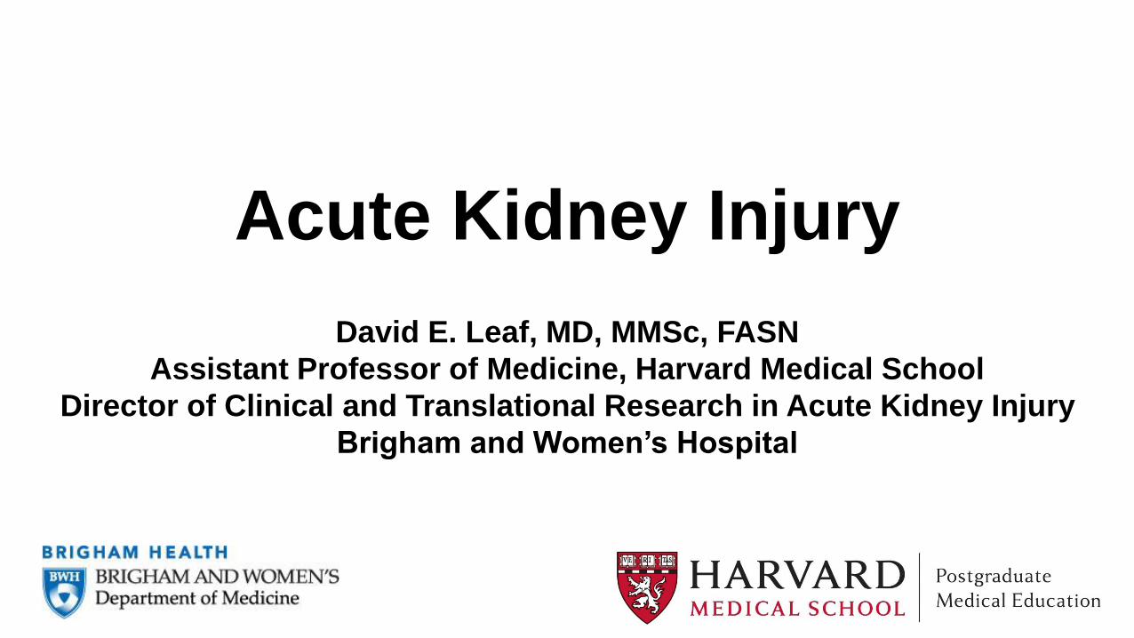
David Leaf, MD, MMSc
▪ Undergrad @UPenn
▪ Medical School @NYU
▪ Medicine Residency @Columbia
▪ Nephrology Fellowship @BWH/MGH
▪ MMSc (Clinical Investigation) @HMS
▪ Assistant Professor of Medicine @HMS
▪ Clinical and Research Focus: AKI

▪ None
Disclosures
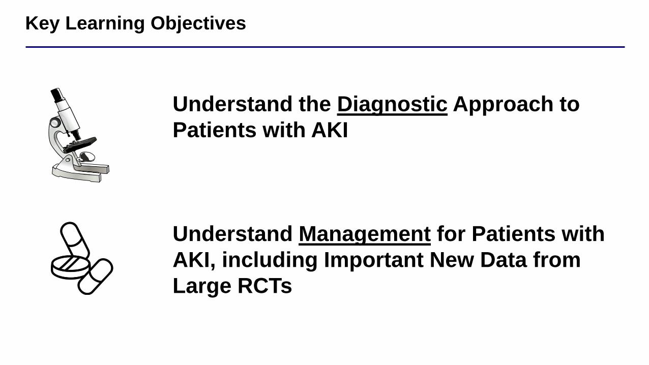
Key Learning Objectives
Understand the Diagnostic Approach to
Patients with AKI
Understand Management for Patients with
AKI, including Important New Data from
Large RCTs
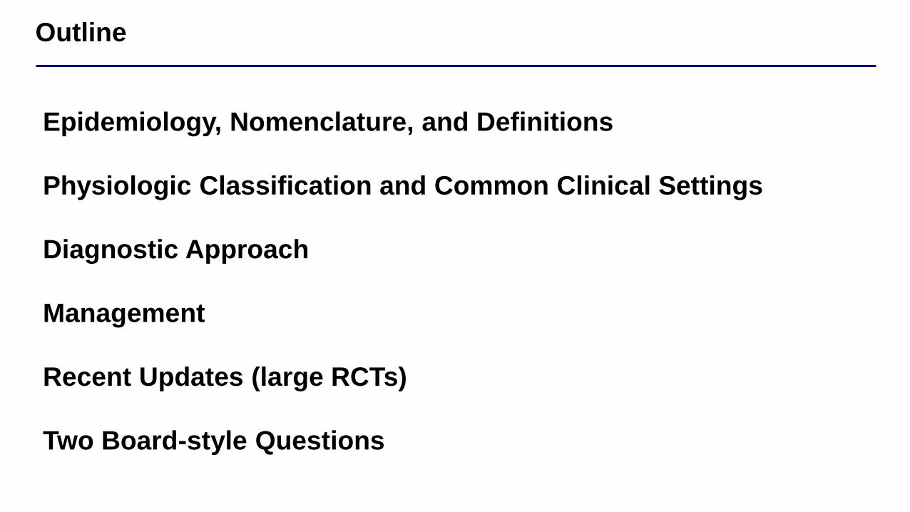
Outline
Epidemiology, Nomenclature, and Definitions
Physiologic Classification and Common Clinical Settings
Diagnostic Approach
Management
Recent Updates (large RCTs)
Two Board-style Questions
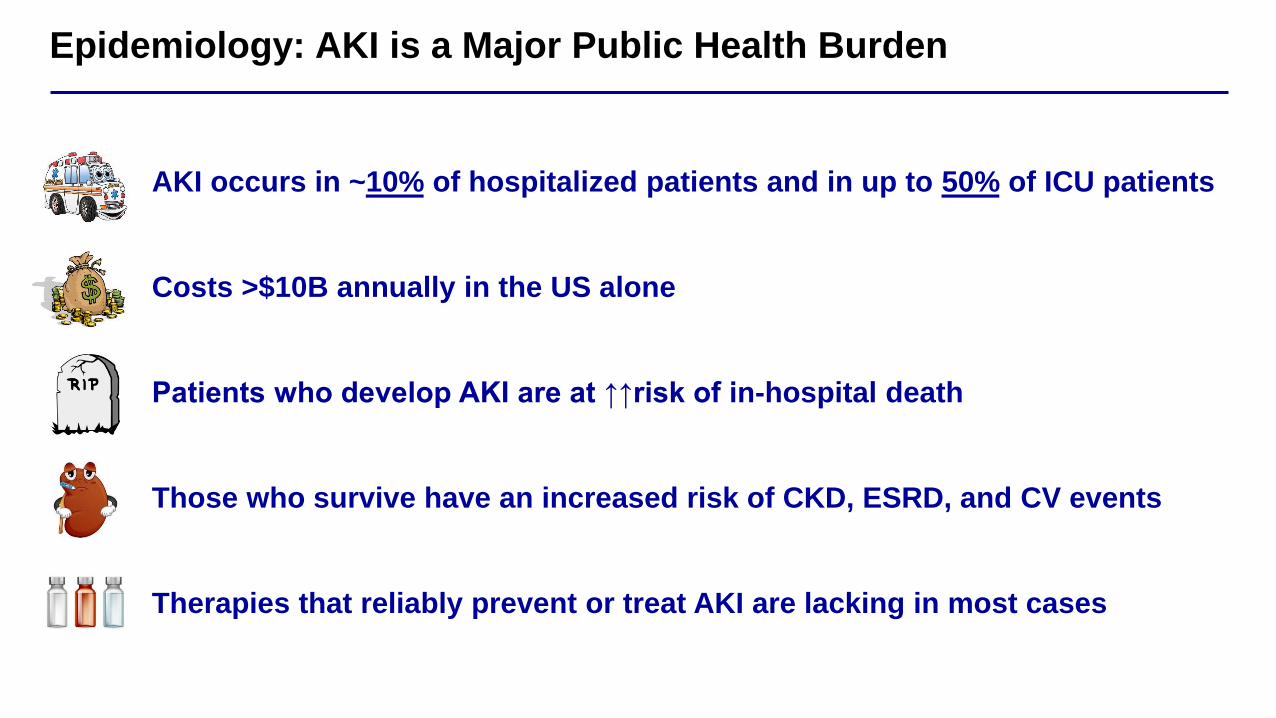
Epidemiology: AKI is a Major Public Health Burden
AKI occurs in ~10% of hospitalized patients and in up to 50% of ICU patients
Costs >$10B annually in the US alone
Patients who develop AKI are at ↑↑risk of in-hospital death
Those who survive have an increased risk of CKD, ESRD, and CV events
Therapies that reliably prevent or treat AKI are lacking in most cases
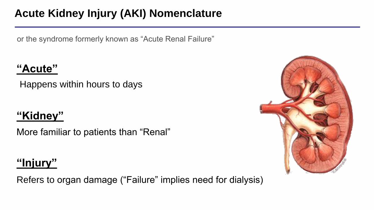
Acute Kidney Injury (AKI) Nomenclature
or the syndrome formerly known as “Acute Renal Failure”
“Acute”
“Kidney”
“Injury”
Happens within hours to days
More familiar to patients than “Renal”
Refers to organ damage (“Failure” implies need for dialysis)
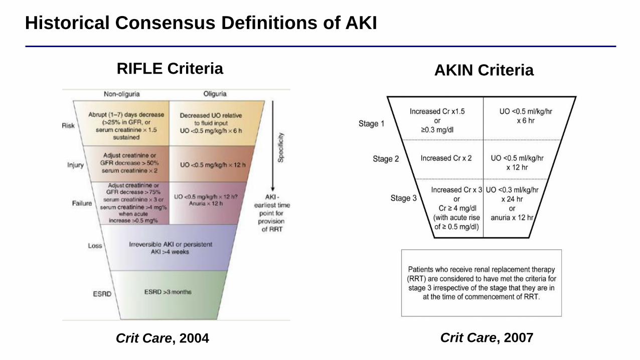
Historical Consensus Definitions of AKI
Crit Care, 2004
RIFLE Criteria
Crit Care, 2007
AKIN Criteria
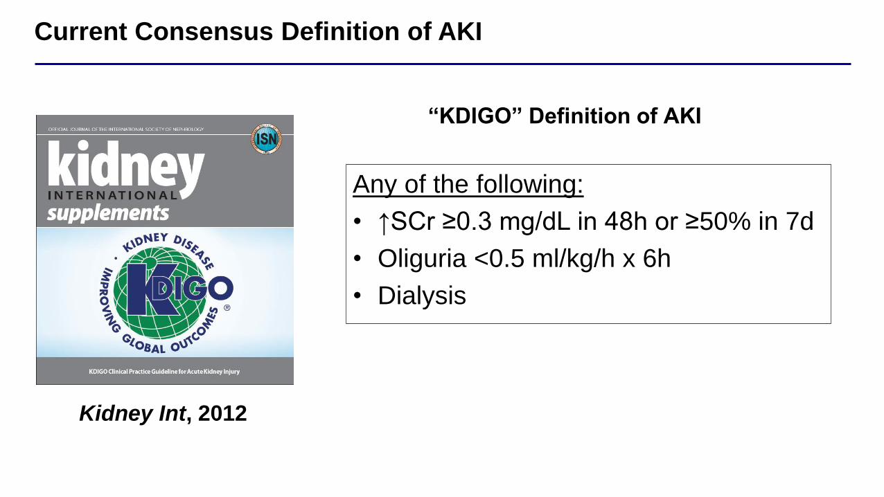
Current Consensus Definition of AKI
Any of the following:
• ↑SCr ≥0.3 mg/dL in 48h or ≥50% in 7d
• Oliguria <0.5 ml/kg/h x 6h
• Dialysis
Kidney Int, 2012
“KDIGO” Definition of AKI
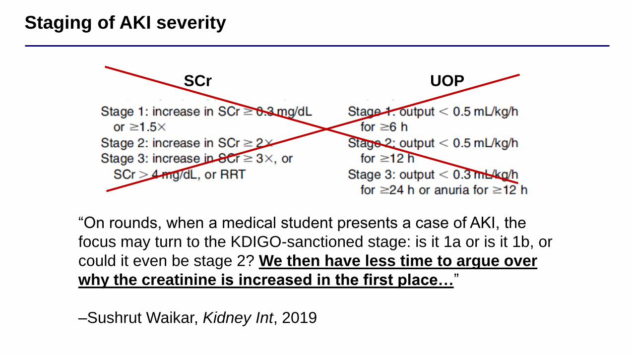
Staging of AKI severity
SCr UOP
“On rounds, when a medical student presents a case of AKI, the
focus may turn to the KDIGO-sanctioned stage: is it 1a or is it 1b, or
could it even be stage 2? We then have less time to argue over
why the creatinine is increased in the first place…”
–Sushrut Waikar, Kidney Int, 2019
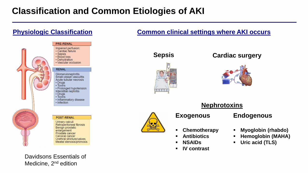
Classification and Common Etiologies of AKI
Physiologic Classification
Davidsons Essentials of
Medicine, 2nd edition
Common clinical settings where AKI occurs
Exogenous
▪ Chemotherapy
▪ Antibiotics
▪ NSAIDs
▪ IV contrast
Cardiac surgerySepsis
Endogenous
▪ Myoglobin (rhabdo)
▪ Hemoglobin (MAHA)
▪ Uric acid (TLS)
Nephrotoxins
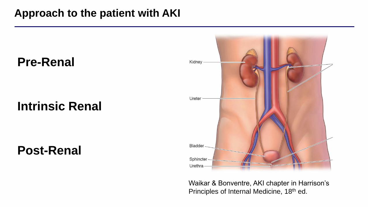
Approach to the patient with AKI
Pre-Renal
Intrinsic Renal
Post-Renal
Waikar & Bonventre, AKI chapter in Harrison’s
Principles of Internal Medicine, 18th ed.

Pre-renal AKI azotemia
Waikar & Bonventre, AKI chapter in Harrison’s
Principles of Internal Medicine, 18th ed.
Overview
• No structural injury to kidney
• SCr increases due to renal hypoperfusion
• Restoration of hemodynamics -> rapid recovery
Causes
• “True” Volume depletion
– GI losses, hemorrhage
• ↓Effective arterial blood volume
– CHF, HRS
• Impaired renal hemodynamics
– NSAIDs (afferent vasoconstriction)
– ACE-I/ARBs (efferent vasodilation)
Intrinsic renal disease
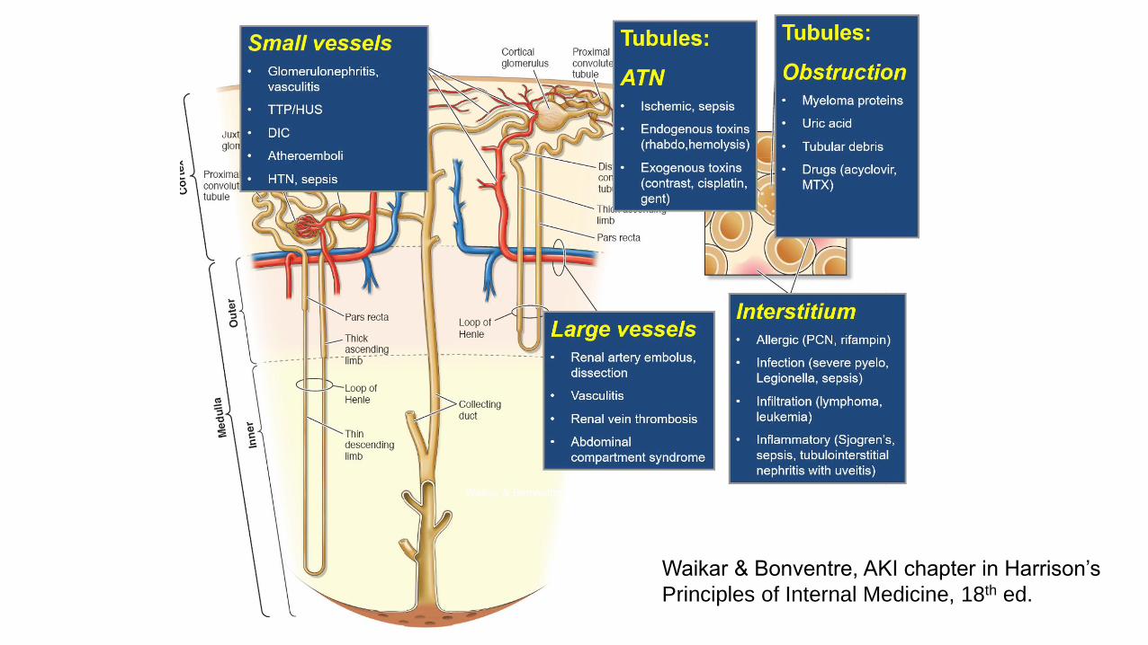
Waikar & Bonventre, AKI chapter in Harrison’s
Principles of Internal Medicine, 18th ed.
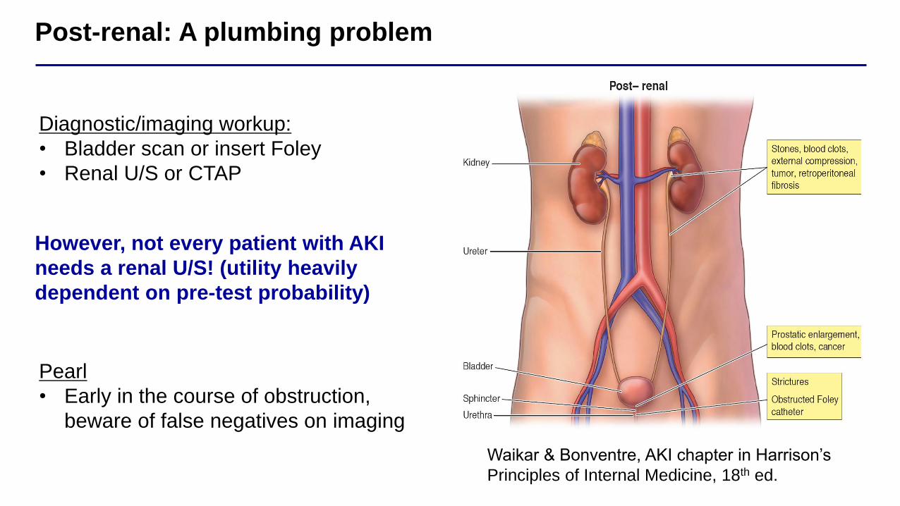
Post-renal: A plumbing problem
Waikar & Bonventre, AKI chapter in Harrison’s
Principles of Internal Medicine, 18th ed.
Diagnostic/imaging workup:
• Bladder scan or insert Foley
• Renal U/S or CTAP
Pearl
• Early in the course of obstruction,
beware of false negatives on imaging
However, not every patient with AKI
needs a renal U/S! (utility heavily
dependent on pre-test probability)
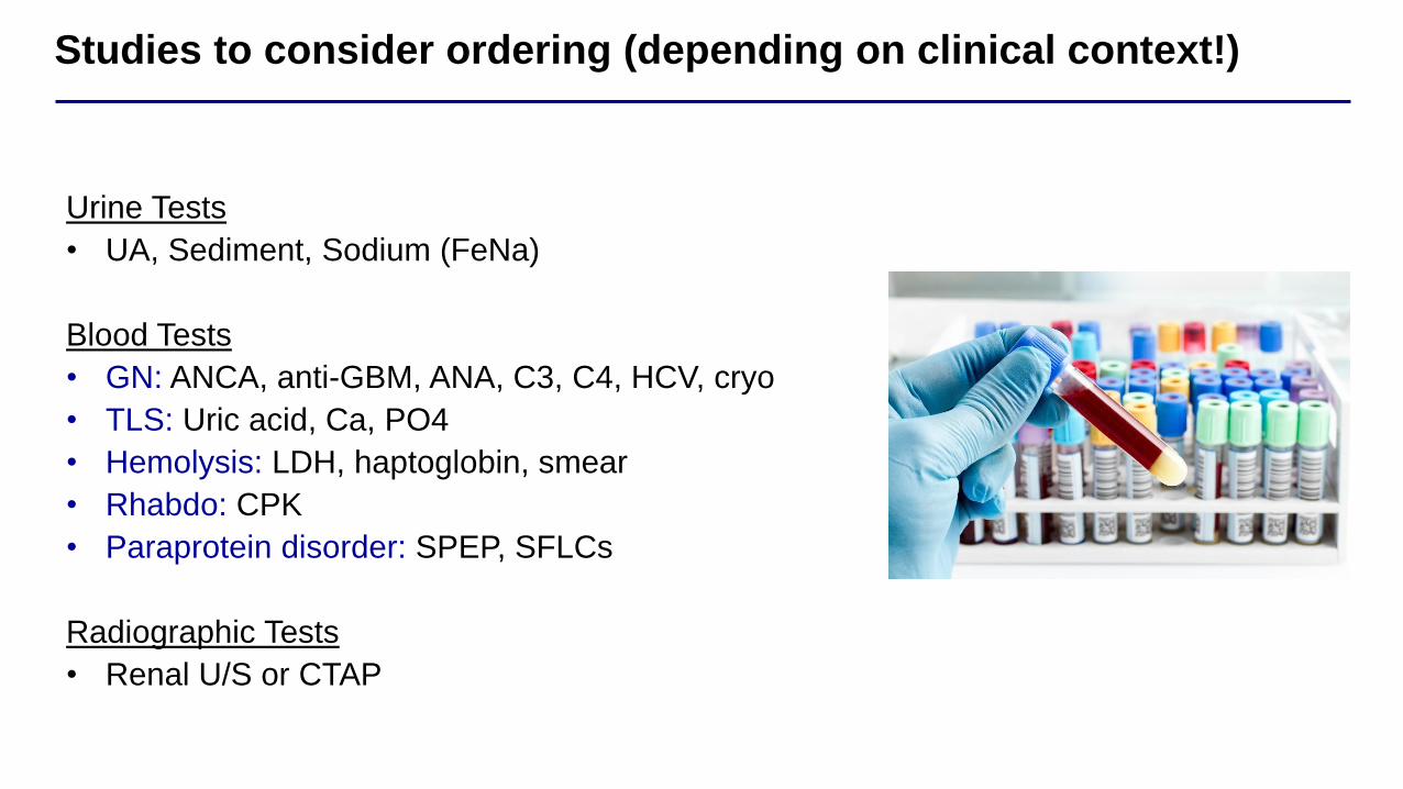
Studies to consider ordering (depending on clinical context!)
Urine Tests
• UA, Sediment, Sodium (FeNa)
Blood Tests
• GN: ANCA, anti-GBM, ANA, C3, C4, HCV, cryo
• TLS: Uric acid, Ca, PO4
• Hemolysis: LDH, haptoglobin, smear
• Rhabdo: CPK
• Paraprotein disorder: SPEP, SFLCs
Radiographic Tests
• Renal U/S or CTAP
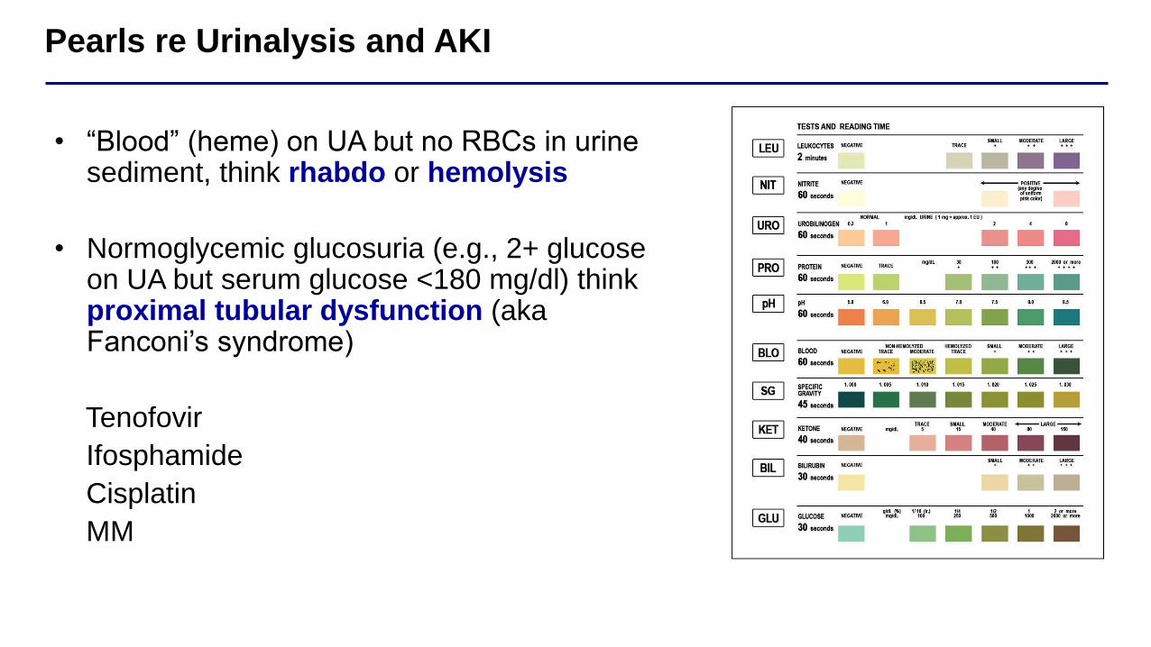
Pearls re Urinalysis and AKI
• “Blood” (heme) on UA but no RBCs in urine sediment, think rhabdo or hemolysis
• Normoglycemic glucosuria (e.g., 2+ glucose on UA but serum glucose <180 mg/dl) think proximal tubular dysfunction (aka Fanconi’s syndrome)
Tenofovir
Ifosphamide
Cisplatin
MM
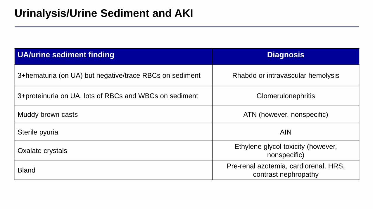
Urinalysis/Urine Sediment and AKI
UA/urine sediment finding Diagnosis
3+hematuria (on UA) but negative/trace RBCs on sediment Rhabdo or intravascular hemolysis
3+proteinuria on UA, lots of RBCs and WBCs on sediment Glomerulonephritis
Muddy brown casts ATN (however, nonspecific)
Sterile pyuria AIN
Oxalate crystalsEthylene glycol toxicity (however,
nonspecific)
BlandPre-renal azotemia, cardiorenal, HRS,
contrast nephropathy
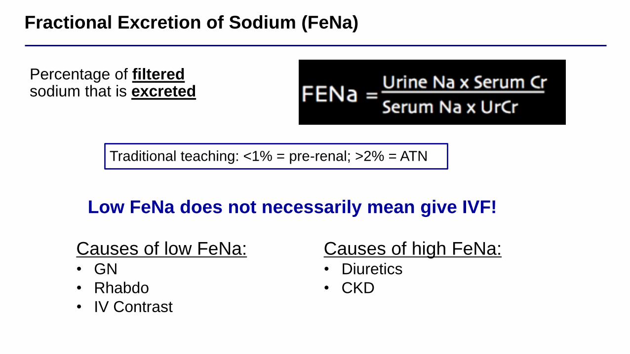
Fractional Excretion of Sodium (FeNa)
Traditional teaching: <1% = pre-renal; >2% = ATN
Percentage of filteredsodium that is excreted
Causes of low FeNa:• GN
• Rhabdo
• IV Contrast
Low FeNa does not necessarily mean give IVF!
Causes of high FeNa:• Diuretics
• CKD
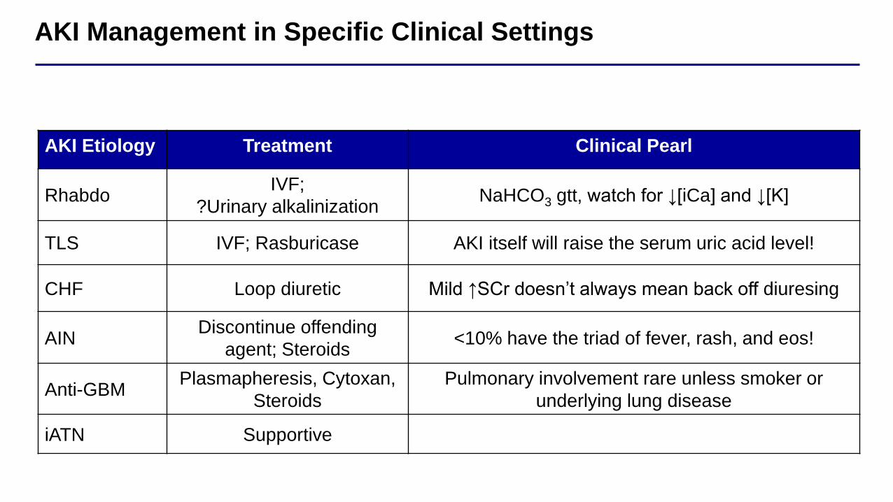
AKI Management in Specific Clinical Settings
AKI Etiology Treatment Clinical Pearl
RhabdoIVF;
?Urinary alkalinizationNaHCO3 gtt, watch for ↓[iCa] and ↓[K]
TLS IVF; Rasburicase AKI itself will raise the serum uric acid level!
CHF Loop diuretic Mild ↑SCr doesn’t always mean back off diuresing
AINDiscontinue offending
agent; Steroids<10% have the triad of fever, rash, and eos!
Anti-GBMPlasmapheresis, Cytoxan,
Steroids
Pulmonary involvement rare unless smoker or
underlying lung disease
iATN Supportive
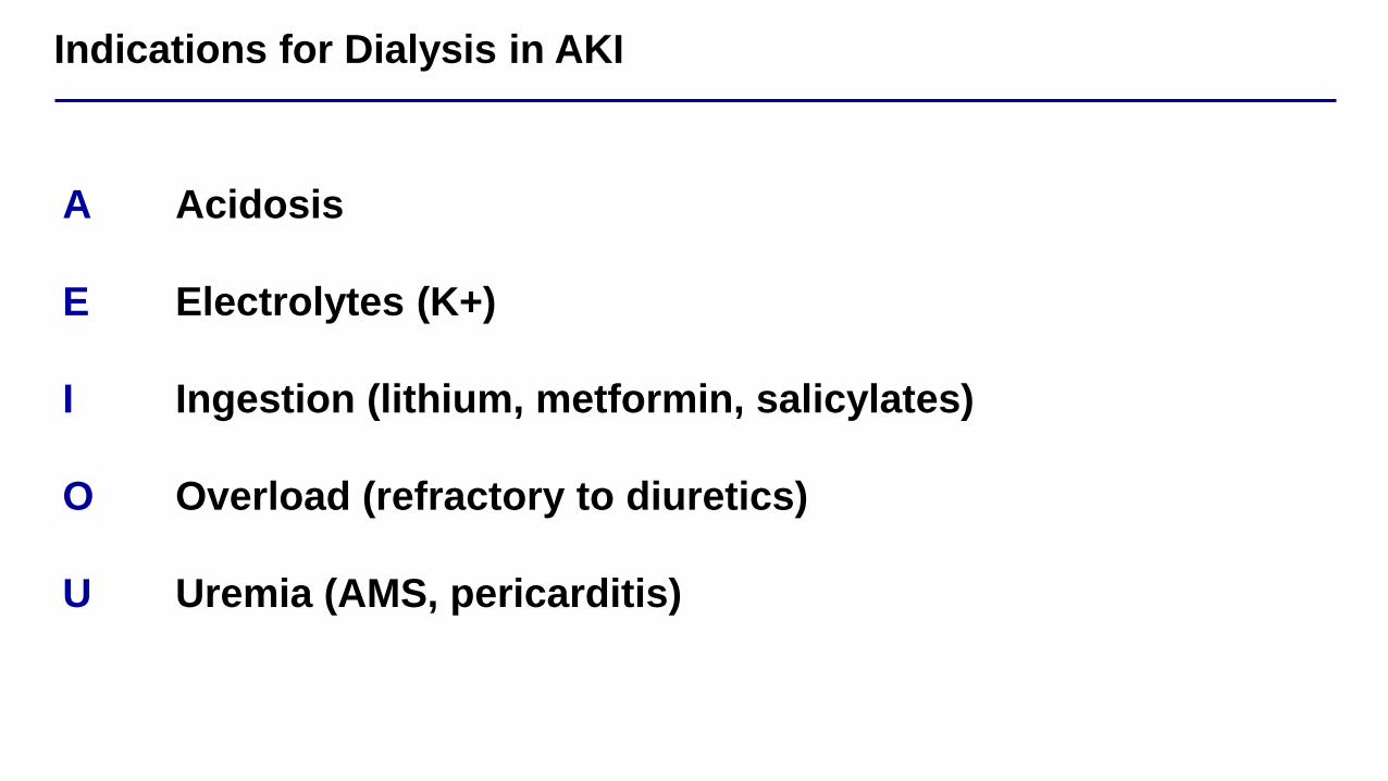
Indications for Dialysis in AKI
A
E
I
O
U
Acidosis
Electrolytes (K+)
Ingestion (lithium, metformin, salicylates)
Overload (refractory to diuretics)
Uremia (AMS, pericarditis)
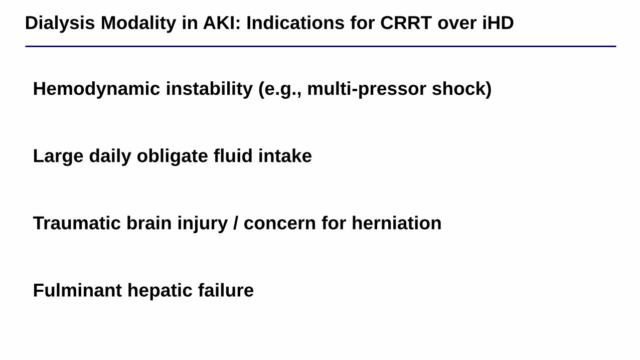
Dialysis Modality in AKI: Indications for CRRT over iHD
Hemodynamic instability (e.g., multi-pressor shock)
Large daily obligate fluid intake
Traumatic brain injury / concern for herniation
Fulminant hepatic failure
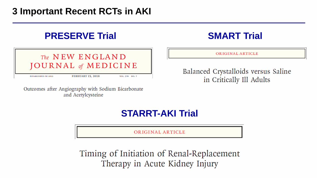
3 Important Recent RCTs in AKI
PRESERVE Trial SMART Trial
STARRT-AKI Trial
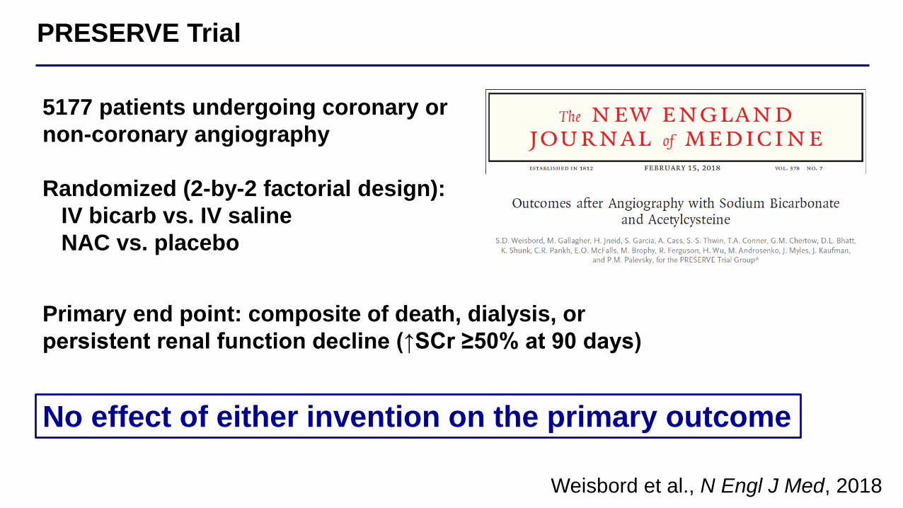
PRESERVE Trial
Weisbord et al., N Engl J Med, 2018
No effect of either invention on the primary outcome
5177 patients undergoing coronary or
non-coronary angiography
Randomized (2-by-2 factorial design):
IV bicarb vs. IV saline
NAC vs. placebo
Primary end point: composite of death, dialysis, or
persistent renal function decline (↑SCr ≥50% at 90 days)
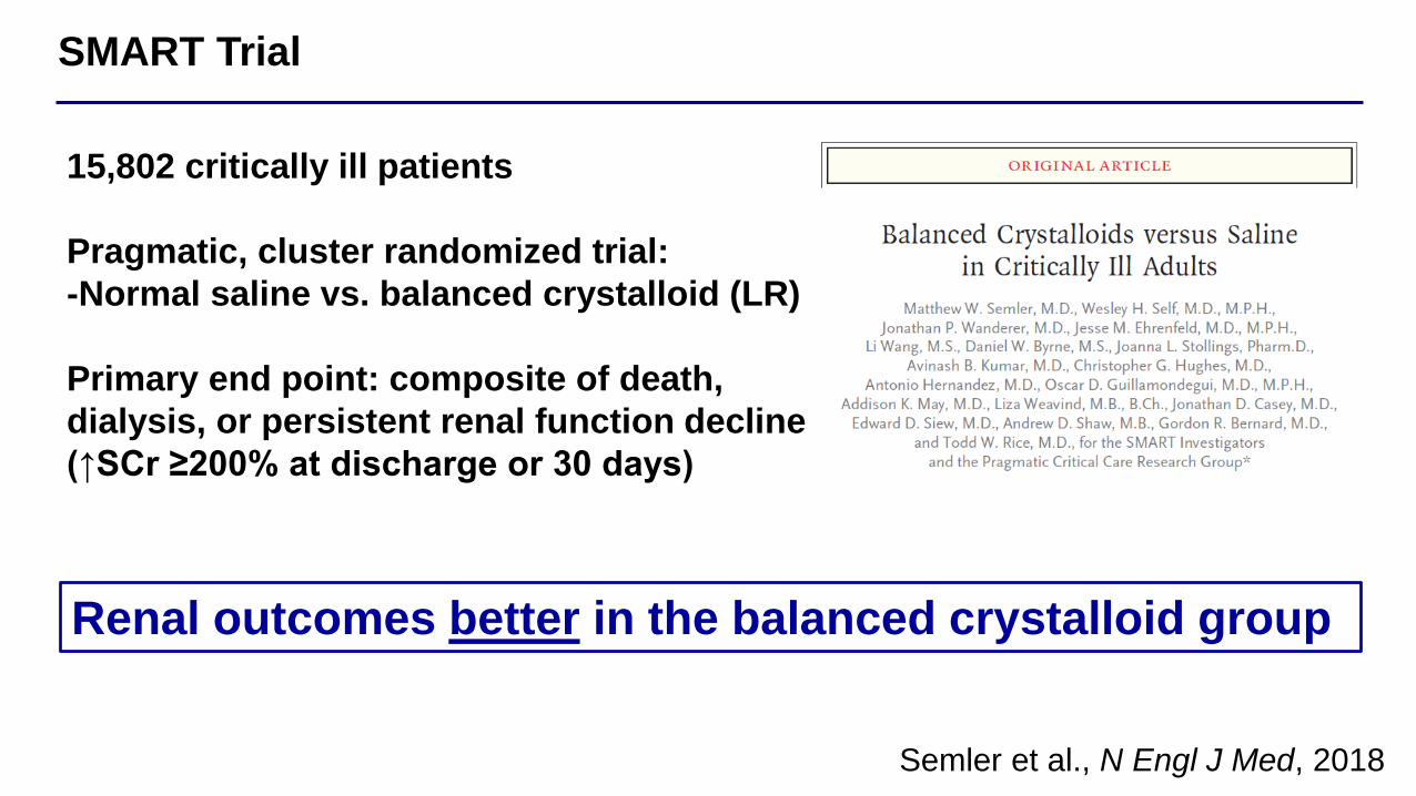
SMART Trial
Semler et al., N Engl J Med, 2018
15,802 critically ill patients
Pragmatic, cluster randomized trial:
-Normal saline vs. balanced crystalloid (LR)
Primary end point: composite of death,
dialysis, or persistent renal function decline
(↑SCr ≥200% at discharge or 30 days)
Renal outcomes better in the balanced crystalloid group
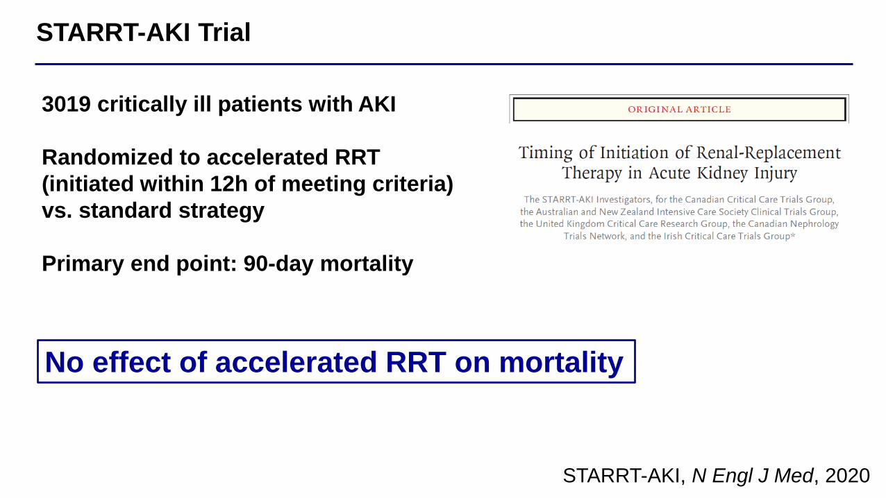
STARRT-AKI Trial
STARRT-AKI, N Engl J Med, 2020
3019 critically ill patients with AKI
Randomized to accelerated RRT
(initiated within 12h of meeting criteria)
vs. standard strategy
Primary end point: 90-day mortality
No effect of accelerated RRT on mortality
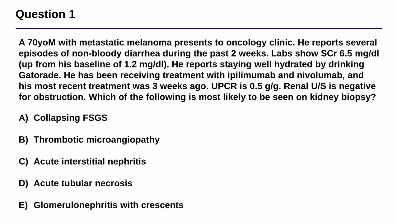
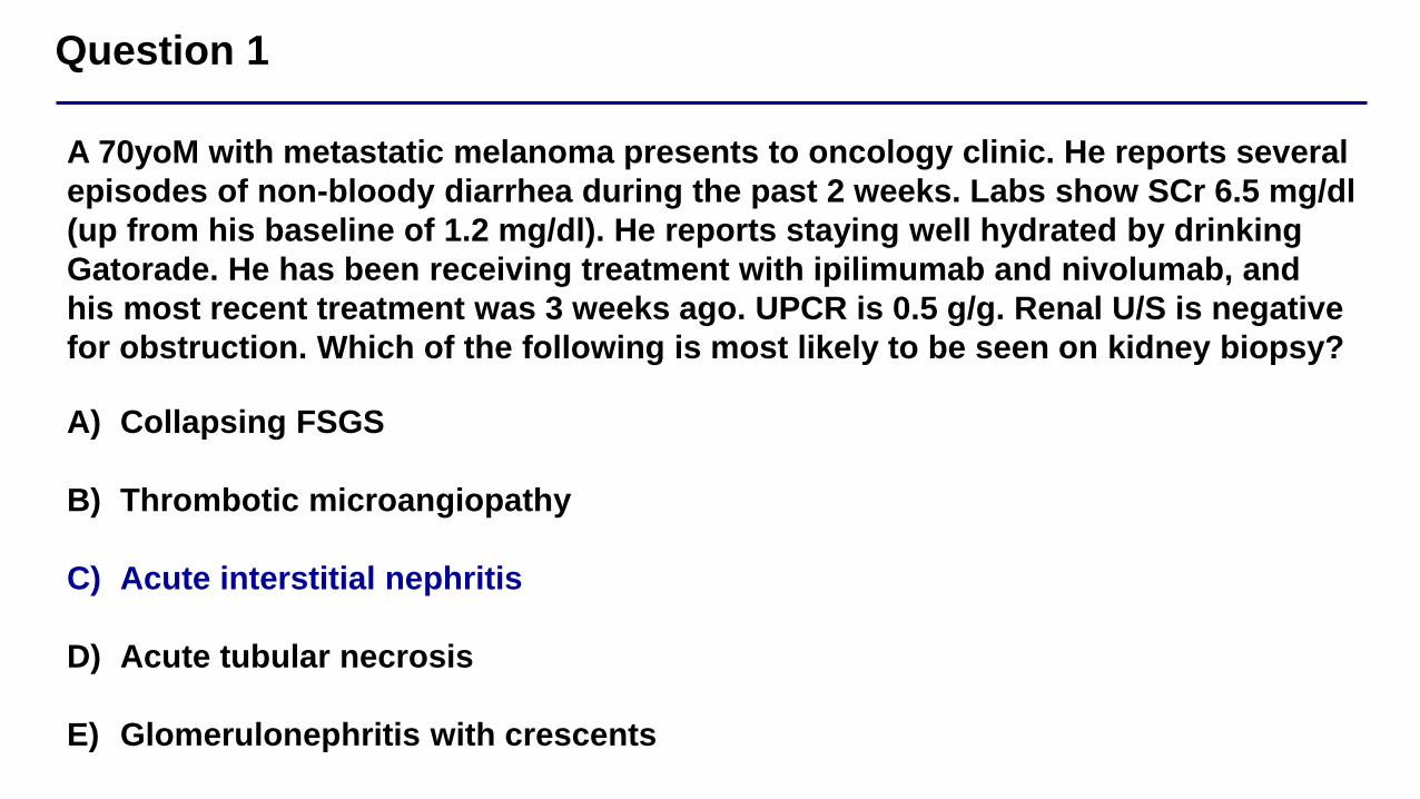
Question 1
A 70yoM with metastatic melanoma presents to oncology clinic. He reports several
episodes of non-bloody diarrhea during the past 2 weeks. Labs show SCr 6.5 mg/dl
(up from his baseline of 1.2 mg/dl). He reports staying well hydrated by drinking
Gatorade. He has been receiving treatment with ipilimumab and nivolumab, and
his most recent treatment was 3 weeks ago. UPCR is 0.5 g/g. Renal U/S is negative
for obstruction. Which of the following is most likely to be seen on kidney biopsy?
A) Collapsing FSGS
B) Thrombotic microangiopathy
C) Acute interstitial nephritis
D) Acute tubular necrosis
E) Glomerulonephritis with crescents
Question 1
Explanation
The answer is C. This patient’s presentation is consistent with acute interstitial
nephritis from immune checkpoint inhibitors.
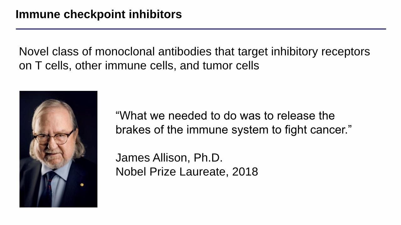
Immune checkpoint inhibitors
Novel class of monoclonal antibodies that target inhibitory receptors
on T cells, other immune cells, and tumor cells
“What we needed to do was to release the
brakes of the immune system to fight cancer.”
James Allison, Ph.D.
Nobel Prize Laureate, 2018
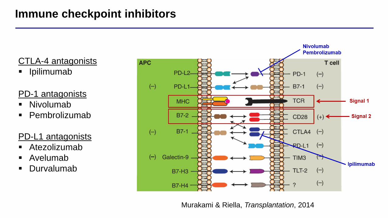
Immune checkpoint inhibitors
CTLA-4 antagonists
▪ Ipilimumab
PD-1 antagonists
▪ Nivolumab
▪ Pembrolizumab
PD-L1 antagonists
▪ Atezolizumab
▪ Avelumab
▪ Durvalumab
Murakami & Riella, Transplantation, 2014
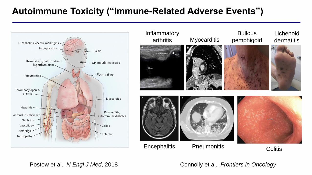
Autoimmune Toxicity (“Immune-Related Adverse Events”)
Postow et al., N Engl J Med, 2018 Connolly et al., Frontiers in Oncology
Inflammatory
arthritis MyocarditisBullous
pemphigoidLichenoid
dermatitis
Encephalitis Pneumonitis Colitis
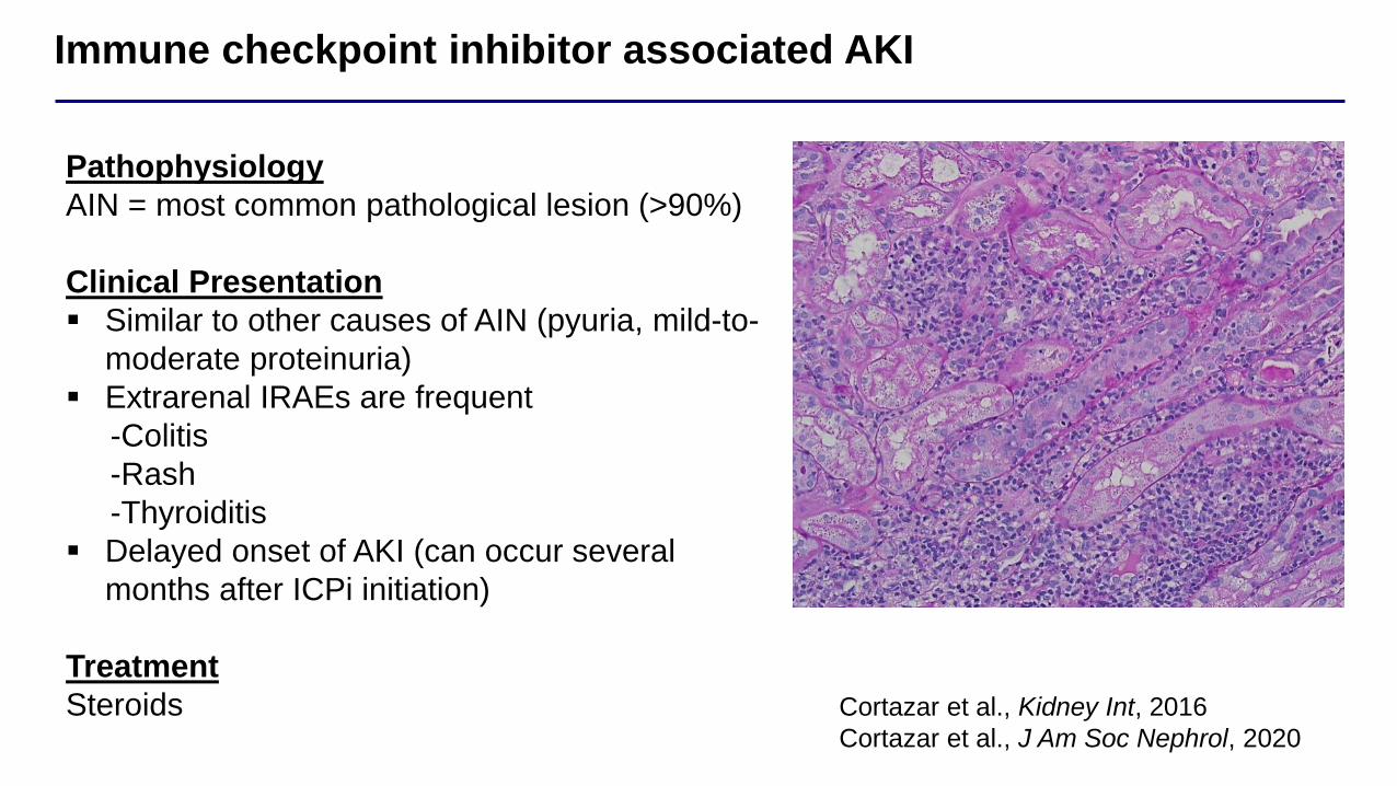
Immune checkpoint inhibitor associated AKI
Pathophysiology
AIN = most common pathological lesion (>90%)
Clinical Presentation
▪ Similar to other causes of AIN (pyuria, mild-to-
moderate proteinuria)
▪ Extrarenal IRAEs are frequent
-Colitis
-Rash
-Thyroiditis
▪ Delayed onset of AKI (can occur several
months after ICPi initiation)
Treatment
Steroids Cortazar et al., Kidney Int, 2016
Cortazar et al., J Am Soc Nephrol, 2020
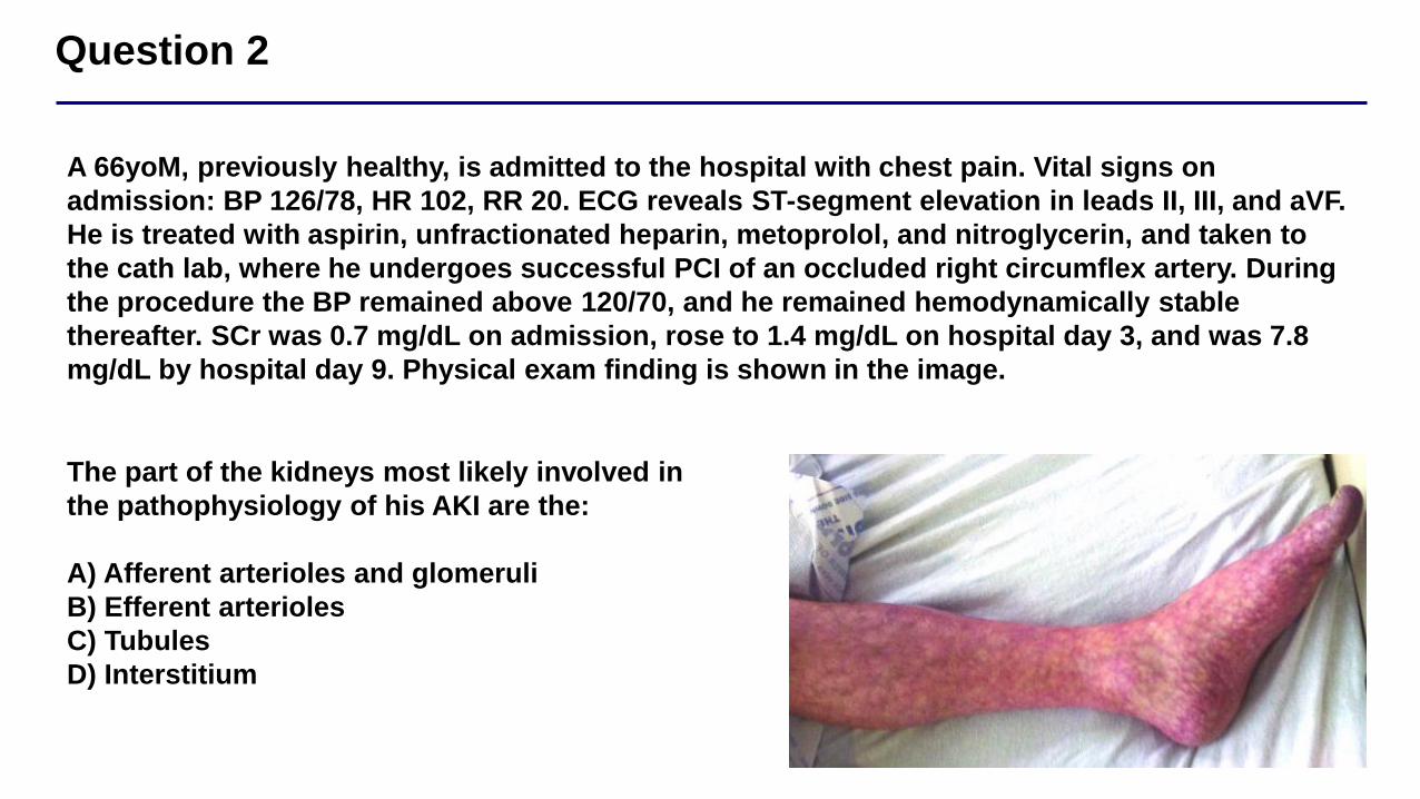
Question 2
A 66yoM, previously healthy, is admitted to the hospital with chest pain. Vital signs on
admission: BP 126/78, HR 102, RR 20. ECG reveals ST-segment elevation in leads II, III, and aVF.
He is treated with aspirin, unfractionated heparin, metoprolol, and nitroglycerin, and taken to
the cath lab, where he undergoes successful PCI of an occluded right circumflex artery. During
the procedure the BP remained above 120/70, and he remained hemodynamically stable
thereafter. SCr was 0.7 mg/dL on admission, rose to 1.4 mg/dL on hospital day 3, and was 7.8
mg/dL by hospital day 9. Physical exam finding is shown in the image.
The part of the kidneys most likely involved in
the pathophysiology of his AKI are the:
A) Afferent arterioles and glomeruli
B) Efferent arterioles
C) Tubules
D) Interstitium
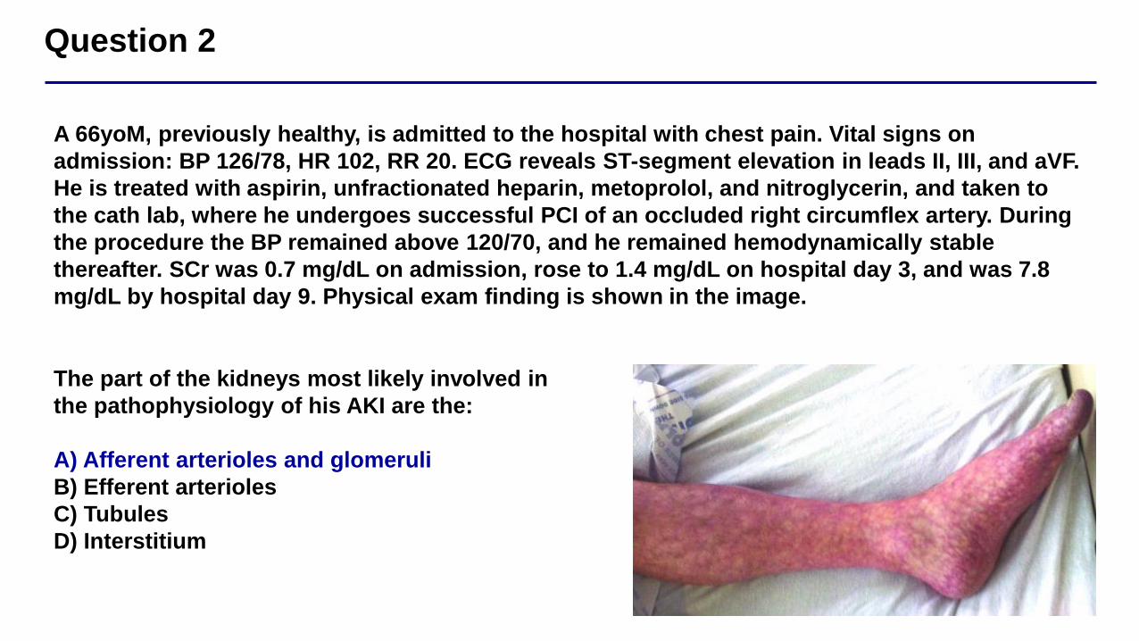
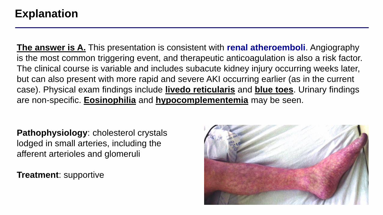
Explanation
The answer is A. This presentation is consistent with renal atheroemboli. Angiography
is the most common triggering event, and therapeutic anticoagulation is also a risk factor.
The clinical course is variable and includes subacute kidney injury occurring weeks later,
but can also present with more rapid and severe AKI occurring earlier (as in the current
case). Physical exam findings include livedo reticularis and blue toes. Urinary findings
are non-specific. Eosinophilia and hypocomplementemia may be seen.
Pathophysiology: cholesterol crystals
lodged in small arteries, including the
afferent arterioles and glomeruli
Treatment: supportive
Take Home Points
▪ In approaching the DDx for AKI, consider pre-, intrinsic-, and post-renal
causes, and order diagnostic tests based on clinical suspicion
▪ Know the diagnoses associated with common UA/sediment findings
▪ Know the treatment for AKI in specific clinical scenarios
▪ Recent Data
▪ PRESERVE Trial found no benefit with IV NaHCO3 (vs. IV NS) or NAC
(vs. placebo) in preventing contrast nephropathy
▪ SMART Trial found benefit with balanced crystalloid (vs. NS) in
critically ill patients
References
▪ Bellomo et al., Acute renal failure – definition, outcome measures, animal models, fluid therapy and information
technology needs: the Second International Consensus Conference of the Acute Dialysis Quality Initiative (ADQI)
Group. Crit Care, 2004
▪ Mehta et al., Acute Kidney Injury Network: report of an initiative to improve outcomes in acute kidney injury. Crit
Care, 2007
▪ Kidney Disease: Improving Global Outcomes (KDIGO) Acute Kidney Injury Work Group. KDIGO clinical practice
guideline for acute kidney injury. Kidney Int Suppl, 2012
▪ Davidson’s Essentials of Medicine, 2nd Edition, 2015
▪ Harrison’s Principles of Internal Medicine (AKI Chapter), 18th Edition, 2011
▪ Weisbord et al., Outcomes after Angiography with Sodium Bicarbonate and Acetylcystein. N Engl J Med, 2018
▪ Semler et al., Balanced Crystalloids versus Saline in Critically Ill Adults. N Engl J Med, 2018
▪ STARRT-AKI Investigators. Timing of Initiation of Renal-Replacement Therapy in Acute Kidney Injury. N Engl J
Med, 2020
▪ Murakami & Riella, Co-inhibitory pathways and their importance in immune regulation. Transplantation, 2014
▪ Postow et al., Immune-Related Adverse Events Associated with Immune Checkpoint Blockade. N Engl J Med,
2018
▪ Cortazar et al., Clinical Features and Outcomes of Immune Checkpoint Inhibitor-Associated AKI: A Multicenter
Study. J Am Soc Nephrol, 2020
Thank [email protected]