Embed Size (px)
Citation preview
242 © Copyright The Korean Academy of Asthma, Allergy and Clinical Immunology • The Korean Academy of Pediatric Allergy and Respiratory Diseasehttp://e-aair.org
INTRODUCTION
Acute eosinophilic pneumonia (AEP) is characterized by acute development of respiratory symptoms, such as cough, short-ness of breath, and fever with eosinophilic lung infiltration.1 In most cases, the cause of AEP is unknown. Some investigators have suggested that it is an acute hypersensitivity reaction to in-haled antigens.2 Typical cases of AEP were reported among young males with recent onset of smoking.3 Many patients with AEP show progressive respiratory failure without correct diag-nosis and proper treatment. Fortunately, patients with AEP are uniformly responsive to intravenous or oral corticosteroid ther-apy.4 The response is usually so dramatic that if a patient fails to respond to corticosteroids, an alternative diagnosis should be considered. Therefore, it is very unusual to suspect AEP when acute respiratory disease develops in an older, non-smoking fe-male currently taking systemic corticosteroids. Here, we pres-ent a case of AEP that resulted in acute respiratory failure in a 69-year-old female patient with rheumatoid arthritis who had been taking prednisolone (5 mg once a day) regularly for 2 years.
CASE REPORT
A 69-year-old female patient visited the emergency room with fever and dyspnea. She had been taking prednisolone (5 mg once per day) and methotrexate (2.5 mg once per week) for
Acute Eosinophilic Pneumonia Leading to Acute Respiratory Failure in a Current Systemic Corticosteroid UserHwa Yong Shin,1 Ju Won Choe,2 Minsuk Kwon,3 Ju Young Jang,3 Jae Woo Jung,3 Jae Chol Choi,3 Jong Wook Shin,3 In Won Park,3 Byoung Whui Choi,3 Jae Yeol Kim3*
Departments of Anesthesiology & Pain Medicine,1 Chest Surgery,2 and Internal Medicine,3 Chung-Ang University College of Medicine, Seoul, Korea
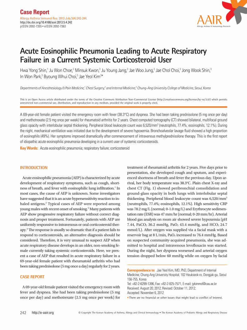
treatment of rheumatoid arthritis for 2 years. Five days prior to presentation, she developed cough and sputum, and experi-enced shortness of breath and fever the previous day. Upon ar-rival, her body temperature was 38.3°C. Plain chest X-ray and chest CT (Fig. 1) showed peribronchial consolidation and ground-glass opacity in both lungs with interlobular septal thickening. Peripheral blood leukocyte count was 6,520/mm3 (neutrophils, 77.4%; eosinophils, 12.1%). High sensitivity CRP was 78.2 mg/L (normal; 0-1.0 mg/L) and Erythrocyte sedimen-tation rate (ESR) was 47 mm/hr (normal; 0-20 mm/hr). Arterial blood gas analysis on room air showed severe hypoxemia (pH 7.44, PaCO2 36.2 mmHg, PaO2 43.4 mmHg, and HCO3 24.7 mmol/L). After oxygen was supplied via a facial mask with a reservoir bag at 8 L/min, PaO2 increased to 76.4 mmHg. Based on suspected community-acquired pneumonia, she was ad-mitted to hospital and intravenous levofloxacin was started. During the night, her dyspnea worsened and arterial oxygen tension dropped below 60 mmHg while on oxygen by facial
Case ReportAllergy Asthma Immunol Res. 2013 July;5(4):242-244.http://dx.doi.org/10.4168/aair.2013.5.4.242pISSN 2092-7355 • eISSN 2092-7363
A 69-year-old female patient visited the emergency room with fever (38.3°C) and dyspnea. She had been taking prednisolone (5 mg once per day) and methotrexate (2.5 mg once per week) for rheumatoid arthritis for 2 years. Chest computed tomography (CT) showed bilateral, multifocal ground glass opacity with interlobular septal thickening. Peripheral blood leukocyte count was 6,520/mm3 (neutrophils, 77.4%; eosinophils, 12.1%). During the night, mechanical ventilation was initiated due to the development of severe hypoxemia. Bronchoalveolar lavage fluid showed a high proportion of eosinophils (49%). Her symptoms improved dramatically after commencement of intravenous methylprednisolone therapy. This is the first report of idiopathic acute eosinophilic pneumonia developing in a current user of systemic corticosteroids.
Key Words: Acute eosinophilic pneumonia; respiratory failure; corticosteroid
This is an Open Access article distributed under the terms of the Creative Commons Attribution Non-Commercial License (http://creativecommons.org/licenses/by-nc/3.0/) which permits unrestricted non-commercial use, distribution, and reproduction in any medium, provided the original work is properly cited.
Correspondence to: Jae Yeol Kim, MD, PhD, Department of Internal Medicine, Chung-Ang University Hospital, 102 Heukseok-ro, Dongjak-gu, Seoul 156-755, KoreaTel: +82-2-6299-1396, Fax: +82-2-825-7571, E-mail: [email protected]: August 20, 2012; Revised: October 11, 2012; Accepted: November 6, 2012•There are no financial or other issues that might lead to conflict of interest.
Acute Eosinophilic Pneumonia in a Steroid User
Allergy Asthma Immunol Res. 2013 July;5(4):242-244. http://dx.doi.org/10.4168/aair.2013.5.4.242
AAIR
243http://e-aair.org
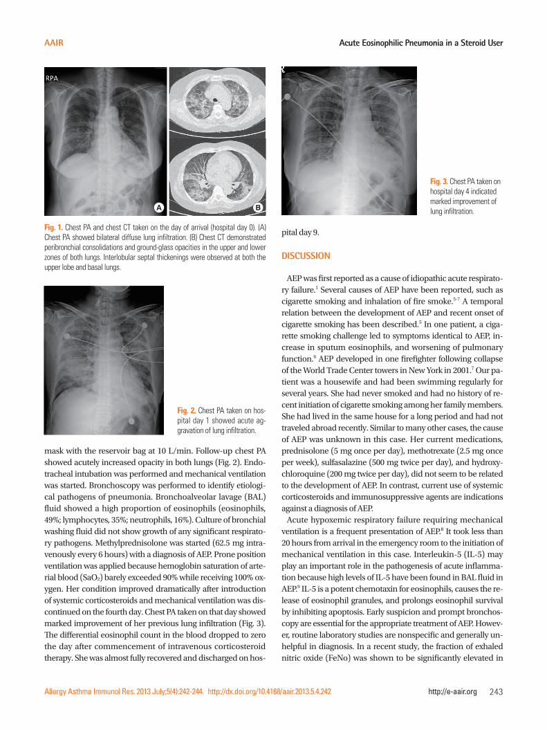
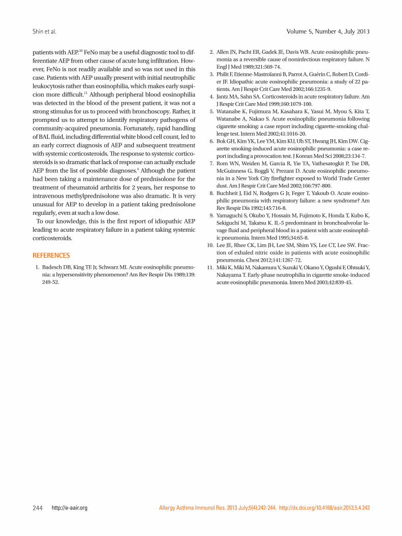
mask with the reservoir bag at 10 L/min. Follow-up chest PA showed acutely increased opacity in both lungs (Fig. 2). Endo-tracheal intubation was performed and mechanical ventilation was started. Bronchoscopy was performed to identify etiologi-cal pathogens of pneumonia. Bronchoalveolar lavage (BAL) fluid showed a high proportion of eosinophils (eosinophils, 49%; lymphocytes, 35%; neutrophils, 16%). Culture of bronchial washing fluid did not show growth of any significant respirato-ry pathogens. Methylprednisolone was started (62.5 mg intra-venously every 6 hours) with a diagnosis of AEP. Prone position ventilation was applied because hemoglobin saturation of arte-rial blood (SaO2) barely exceeded 90% while receiving 100% ox-ygen. Her condition improved dramatically after introduction of systemic corticosteroids and mechanical ventilation was dis-continued on the fourth day. Chest PA taken on that day showed marked improvement of her previous lung infiltration (Fig. 3). The differential eosinophil count in the blood dropped to zero the day after commencement of intravenous corticosteroid therapy. She was almost fully recovered and discharged on hos-
pital day 9.
DISCUSSION
AEP was first reported as a cause of idiopathic acute respirato-ry failure.1 Several causes of AEP have been reported, such as cigarette smoking and inhalation of fire smoke.5-7 A temporal relation between the development of AEP and recent onset of cigarette smoking has been described.5 In one patient, a ciga-rette smoking challenge led to symptoms identical to AEP, in-crease in sputum eosinophils, and worsening of pulmonary function.6 AEP developed in one firefighter following collapse of the World Trade Center towers in New York in 2001.7 Our pa-tient was a housewife and had been swimming regularly for several years. She had never smoked and had no history of re-cent initiation of cigarette smoking among her family members. She had lived in the same house for a long period and had not traveled abroad recently. Similar to many other cases, the cause of AEP was unknown in this case. Her current medications, prednisolone (5 mg once per day), methotrexate (2.5 mg once per week), sulfasalazine (500 mg twice per day), and hydroxy-chloroquine (200 mg twice per day), did not seem to be related to the development of AEP. In contrast, current use of systemic corticosteroids and immunosuppressive agents are indications against a diagnosis of AEP.
Acute hypoxemic respiratory failure requiring mechanical ventilation is a frequent presentation of AEP.8 It took less than 20 hours from arrival in the emergency room to the initiation of mechanical ventilation in this case. Interleukin-5 (IL-5) may play an important role in the pathogenesis of acute inflamma-tion because high levels of IL-5 have been found in BAL fluid in AEP.9 IL-5 is a potent chemotaxin for eosinophils, causes the re-lease of eosinophil granules, and prolongs eosinophil survival by inhibiting apoptosis. Early suspicion and prompt bronchos-copy are essential for the appropriate treatment of AEP. Howev-er, routine laboratory studies are nonspecific and generally un-helpful in diagnosis. In a recent study, the fraction of exhaled nitric oxide (FeNo) was shown to be significantly elevated in
Fig. 1. Chest PA and chest CT taken on the day of arrival (hospital day 0). (A) Chest PA showed bilateral diffuse lung infiltration. (B) Chest CT demonstrated peribronchial consolidations and ground-glass opacities in the upper and lower zones of both lungs. Interlobular septal thickenings were observed at both the upper lobe and basal lungs.
A B
Fig. 2. Chest PA taken on hos-pital day 1 showed acute ag-gravation of lung infiltration.
Fig. 3. Chest PA taken on hospital day 4 indicated marked improvement of lung infiltration.
Shin et al.
Allergy Asthma Immunol Res. 2013 July;5(4):242-244. http://dx.doi.org/10.4168/aair.2013.5.4.242
Volume 5, Number 4, July 2013
244 http://e-aair.org
patients with AEP.10 FeNo may be a useful diagnostic tool to dif-ferentiate AEP from other cause of acute lung infiltration. How-ever, FeNo is not readily available and so was not used in this case. Patients with AEP usually present with initial neutrophilic leukocytosis rather than eosinophilia, which makes early suspi-cion more difficult.11 Although peripheral blood eosinophilia was detected in the blood of the present patient, it was not a strong stimulus for us to proceed with bronchoscopy. Rather, it prompted us to attempt to identify respiratory pathogens of community-acquired pneumonia. Fortunately, rapid handling of BAL fluid, including differential white blood cell count, led to an early correct diagnosis of AEP and subsequent treatment with systemic corticosteroids. The response to systemic cortico-steroids is so dramatic that lack of response can actually exclude AEP from the list of possible diagnoses.4 Although the patient had been taking a maintenance dose of prednisolone for the treatment of rheumatoid arthritis for 2 years, her response to intravenous methylprednisolone was also dramatic. It is very unusual for AEP to develop in a patient taking prednisolone regularly, even at such a low dose.
To our knowledge, this is the first report of idiopathic AEP leading to acute respiratory failure in a patient taking systemic corticosteroids.
REFERENCES
1. Badesch DB, King TE Jr, Schwarz MI. Acute eosinophilic pneumo-nia: a hypersensitivity phenomenon? Am Rev Respir Dis 1989;139: 249-52.
2. Allen JN, Pacht ER, Gadek JE, Davis WB. Acute eosinophilic pneu-monia as a reversible cause of noninfectious respiratory failure. N Engl J Med 1989;321:569-74.
3. Philit F, Etienne-Mastroïanni B, Parrot A, Guérin C, Robert D, Cordi-er JF. Idiopathic acute eosinophilic pneumonia: a study of 22 pa-tients. Am J Respir Crit Care Med 2002;166:1235-9.
4. Jantz MA, Sahn SA. Corticosteroids in acute respiratory failure. Am J Respir Crit Care Med 1999;160:1079-100.
5. Watanabe K, Fujimura M, Kasahara K, Yasui M, Myou S, Kita T, Watanabe A, Nakao S. Acute eosinophilic pneumonia following cigarette smoking: a case report including cigarette-smoking chal-lenge test. Intern Med 2002;41:1016-20.
6. Bok GH, Kim YK, Lee YM, Kim KU, Uh ST, Hwang JH, Kim DW. Cig-arette smoking-induced acute eosinophilic pneumonia: a case re-port including a provocation test. J Korean Med Sci 2008;23:134-7.
7. Rom WN, Weiden M, Garcia R, Yie TA, Vathesatogkit P, Tse DB, McGuinness G, Roggli V, Prezant D. Acute eosinophilic pneumo-nia in a New York City firefighter exposed to World Trade Center dust. Am J Respir Crit Care Med 2002;166:797-800.
8. Buchheit J, Eid N, Rodgers G Jr, Feger T, Yakoub O. Acute eosino-philic pneumonia with respiratory failure: a new syndrome? Am Rev Respir Dis 1992;145:716-8.
9. Yamaguchi S, Okubo Y, Hossain M, Fujimoto K, Honda T, Kubo K, Sekiguchi M, Takatsu K. IL-5 predominant in bronchoalveolar la-vage fluid and peripheral blood in a patient with acute eosinophil-ic pneumonia. Intern Med 1995;34:65-8.
10. Lee JE, Rhee CK, Lim JH, Lee SM, Shim YS, Lee CT, Lee SW. Frac-tion of exhaled nitric oxide in patients with acute eosinophilic pneumonia. Chest 2012;141:1267-72.
11. Miki K, Miki M, Nakamura Y, Suzuki Y, Okano Y, Ogushi F, Ohtsuki Y, Nakayama T. Early-phase neutrophilia in cigarette smoke-induced acute eosinophilic pneumonia. Intern Med 2003;42:839-45.