Embed Size (px)
Citation preview
1
1
Cynthia Webner DNP, RN, CCNS, CCRN-CMC, CHFN
Cardiovascular Nursing Education Associates / Key Choice
2015 Regional Health Heart and Vascular Symposium
2015
2
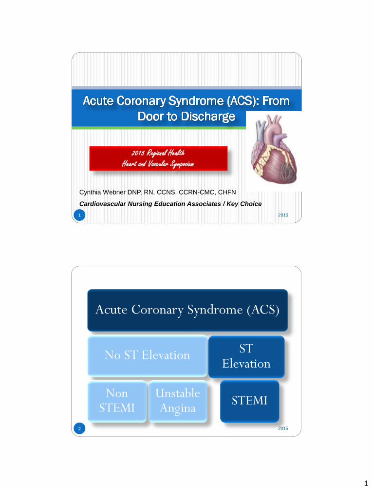
Acute Coronary Syndrome (ACS)
No ST Elevation
Non STEMI
Unstable Angina
ST Elevation
STEMI
2015
2
3
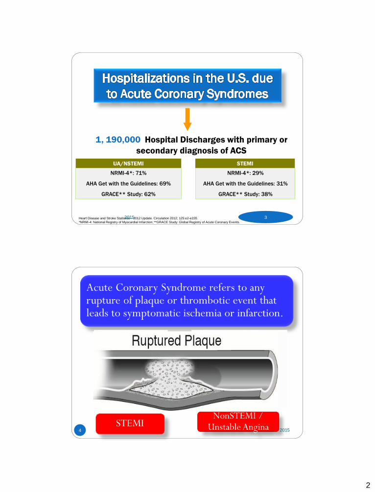
1, 190,000 Hospital Discharges with primary or
secondary diagnosis of ACS
UA/NSTEMI STEMI
NRMI-4*: 71%
AHA Get with the Guidelines: 69%
GRACE** Study: 62%
NRMI-4*: 29%
AHA Get with the Guidelines: 31%
GRACE** Study: 38%
Heart Disease and Stroke Statistics – 2012 Update. Circulation 2012; 125:e2-e220.
*NRMI-4: National Registry of Myocardial Infarction; **GRACE Study: Global Registry of Acute Coronary Events.
2015
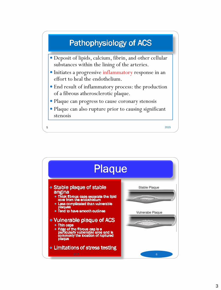
Acute Coronary Syndrome refers to any rupture of plaque or thrombotic event that leads to symptomatic ischemia or infarction.
STEMI NonSTEMI /
Unstable Angina 2015 4
3
5
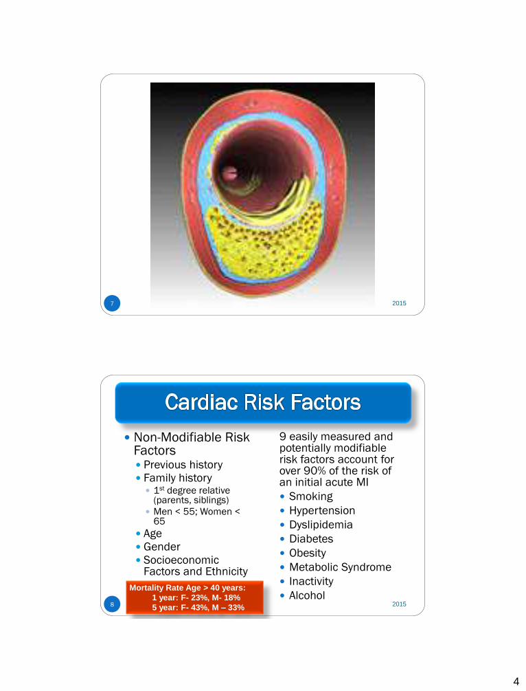
Deposit of lipids, calcium, fibrin, and other cellular substances within the lining of the arteries.
Initiates a progressive inflammatory response in an effort to heal the endothelium.
End result of inflammatory process: the production of a fibrous atherosclerotic plaque.
Plaque can progress to cause coronary stenosis
Plaque can also rupture prior to causing significant stenosis
2015
2015 6
4
7 2015
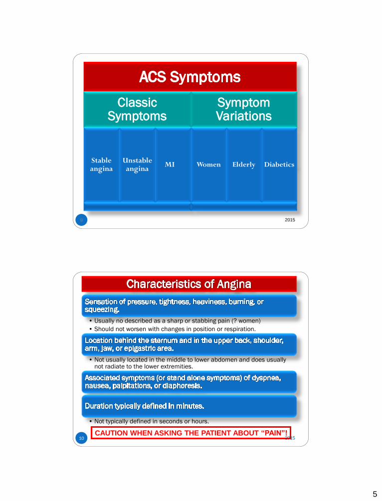
Non-Modifiable Risk Factors Previous history Family history
1st degree relative (parents, siblings)
Men < 55; Women < 65
Age
Gender
Socioeconomic Factors and Ethnicity
9 easily measured and potentially modifiable risk factors account for over 90% of the risk of an initial acute MI
Smoking
Hypertension
Dyslipidemia
Diabetes
Obesity
Metabolic Syndrome
Inactivity
Alcohol
Mortality Rate Age > 40 years:
1 year: F- 23%, M- 18%
5 year: F- 43%, M – 33% 2015 8
5
Classic Symptoms
Stable angina
Unstable angina
MI
Symptom Variations
Women Elderly Diabetics
9 2015
10
• Usually no described as a sharp or stabbing pain (? women)
• Should not worsen with changes in position or respiration.
• Not usually located in the middle to lower abdomen and does usually not radiate to the lower extremities.
• Not typically defined in seconds or hours.
CAUTION WHEN ASKING THE PATIENT ABOUT “PAIN”! 2015
6
11 2015
29-38% of ACS patients
Complete occlusion of a vessel by a thrombus
Fibrin stable clot (red clot)
Classified more specifically by the portion of the left ventricle suffering injury.
Mortality is greatest within the first 24 to 48 hours of symptom onset
TREATMENT FOCUS = REPERFUSION 12 2015
7
Nationally under treated according to evidence based practice guidelines (Crusade Registry)
Pathophysiology often involves a platelet plug or white clot
Less stable clot
Opportunity for spontaneous reperfusion
Differentiated from unstable angina by troponin levels
TREATMENT FOCUS = ANTIPLATELET THERAPY 13 2015
14
Increase myocardial oxygen demand: Hyperthermia
Hypertension
Tachycardia
Conditions producing over stimulation of the sympathetic nervous system (cocaine use, hyperthyroidism)
Decrease myocardial oxygen delivery: Anemia
Pulmonary disease.
Increase myocardial oxygen demand and decrease myocardial oxygen supply: Aortic stenosis
Hypertrophic cardiomyopathy
Type 2 MI
2015
8
15 2015
16 2015
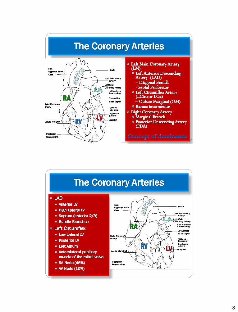
9
17 2015
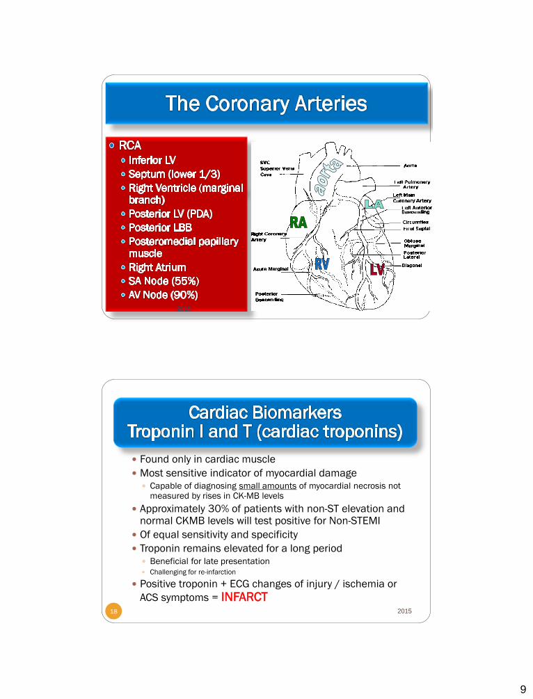
Found only in cardiac muscle
Most sensitive indicator of myocardial damage Capable of diagnosing small amounts of myocardial necrosis not
measured by rises in CK-MB levels
Approximately 30% of patients with non-ST elevation and normal CKMB levels will test positive for Non-STEMI
Of equal sensitivity and specificity
Troponin remains elevated for a long period Beneficial for late presentation Challenging for re-infarction
Positive troponin + ECG changes of injury / ischemia or
ACS symptoms = INFARCT 2015 18
10
Non infarct cardiac causes of elevated troponin: heart failure, left ventricular hypertrophy, tachyarrhythmias, pericarditis, cardiac trauma
Non CAD causes of troponin elevation (sepsis, pulmonary emboli, chronic kidney disease, chemotherapy, respiratory failure, burns, neurological disease )
Troponin I more specific in renal dysfunction Patients with ESRD commonly have elevated troponin T
Not a false positive - relates to overall dysfunction of the cardiorenal system
< 10% of patients with ESRD have elevated troponin I in absence of ACS
Elevated troponin levels are marker of risk and associated with an increased mortality – even when diagnosis is not myocardial infarction
Degree of troponin elevation correlates with risk of death
New high sensitivity troponin T
2015 19
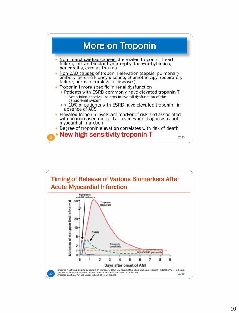
Timing of Release of Various Biomarkers After
Acute Myocardial Infarction
20
Shapiro BP, Jaffe AS. Cardiac biomarkers. In: Murphy JG, Lloyd MA, editors. Mayo Clinic Cardiology: Concise Textbook. 3rd ed. Rochester,
MN: Mayo Clinic Scientific Press and New York: Informa Healthcare USA, 2007:773–80.
Anderson JL, et al. J Am Coll Cardiol 2007;50:e1–e157, Figure 5. 2015
11
21
ASA: 325 mg (non enteric coated) If fibrinolytic therapy – 162-325 mg
P2Y12 inhibitor (loading dose before or at time of PCI) If fibrinolytic therapy - clopidogrel only
Anticoagulants (related to reperfusion strategy) If fibrinolytic – weight based heparin x 48 hours
Oral beta blockers ASAP IV if hypertensive or tachycardic
NTG – Sublingual vs IV Morphine Sulfate (Class I) Oxygen if hypoxemic (arterial oxygen saturation < 90%) High intensity statin therapy D/C NSAIDS ACE Inhibitors (within 24 hours)
Greatest benefit in anterior wall MI, LVEF < 40%, HTN, diabetes or chronic kidney disease
Aldosterone Antagonists Initiate within 7 days in those with LVEF <40% , HF , or diabetes
2015
22
Reperfusion is number one treatment strategy
Primary Coronary Intervention (PCI) preferred treatment strategy if within 90 minutes Goal: 90 minutes from 1st medical contact
Fibrinolytics within 30 minutes of hospital presentation (or 30 minutes from EMS to
fibrinolytics)
2015
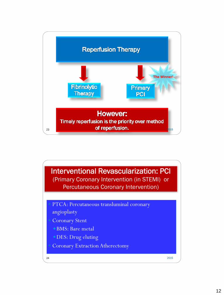
12
23
The Winner!
2015
Interventional Revascularization: PCI (Primary Coronary Intervention (in STEMI) or
Percutaneous Coronary Intervention)
PTCA: Percutaneous transluminal coronary
angioplasty
Coronary Stent
BMS: Bare metal
DES: Drug eluting
Coronary Extraction Atherectomy
2015 24
13
2015 25
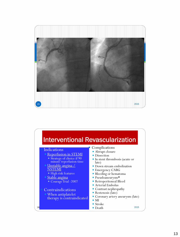
Interventional Revascularization
Indications Reperfusion in STEMI
Strategy of choice if 90 minute reperfusion time
Unstable angina / NSTEMI High risk features
Stable angina Courage Trial -2007
Contraindications When antiplatelet
therapy is contraindicated
Complications Abrupt closure Dissection In stent thrombosis (acute or
late) Down stream embolization Emergency CABG Bleeding or hematoma Pseudoaneurysm* Retroperitoneal Bleed Arterial Embolus Contrast nephropathy Restenosis (late) Coronary artery aneurysm (late) MI Stroke Death
2015 26
14
27
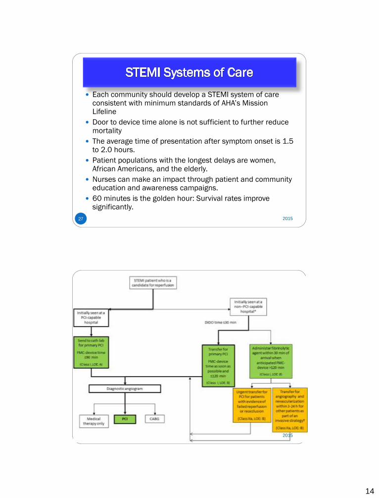
Each community should develop a STEMI system of care consistent with minimum standards of AHA’s Mission Lifeline
Door to device time alone is not sufficient to further reduce mortality
The average time of presentation after symptom onset is 1.5 to 2.0 hours.
Patient populations with the longest delays are women, African Americans, and the elderly.
Nurses can make an impact through patient and community education and awareness campaigns.
60 minutes is the golden hour: Survival rates improve significantly.
2015
28 2015
15
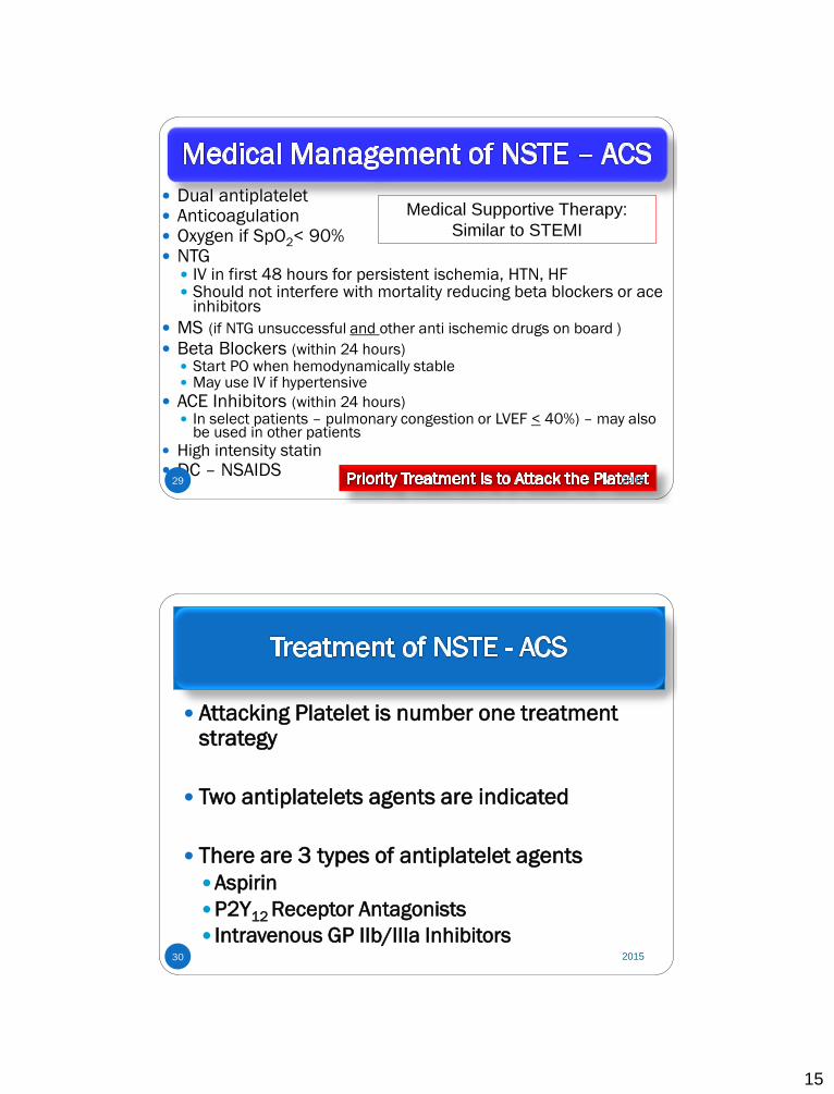
Dual antiplatelet Anticoagulation Oxygen if SpO2< 90% NTG
IV in first 48 hours for persistent ischemia, HTN, HF Should not interfere with mortality reducing beta blockers or ace
inhibitors
MS (if NTG unsuccessful and other anti ischemic drugs on board )
Beta Blockers (within 24 hours) Start PO when hemodynamically stable May use IV if hypertensive
ACE Inhibitors (within 24 hours) In select patients – pulmonary congestion or LVEF < 40%) – may also
be used in other patients
High intensity statin
DC – NSAIDS
Medical Supportive Therapy:
Similar to STEMI
2015 29
30
Attacking Platelet is number one treatment strategy
Two antiplatelets agents are indicated
There are 3 types of antiplatelet agents
Aspirin
P2Y12 Receptor Antagonists
Intravenous GP IIb/IIIa Inhibitors
2015
16
31
Dual antiplatelet therapy for invasive strategies in medium to high risk patients ASA (and one of
following) P2Y12 / ADP Receptor
blockers Clopidogrel Prasugral Ticagrelor
GP II b / III a Inhibitors
(*eptifibatide, * tirofiban, abciximab) * preferred agents Used only in special
circumstances
Antiplatelet therapy also in conservative treatment Prasugrel not unless
PCI is planned Abciximab not unless
PCI is planned
Dual antiplatelet
therapy is also
used after STEMI
and after any
coronary
intervention. 2015
2015 32
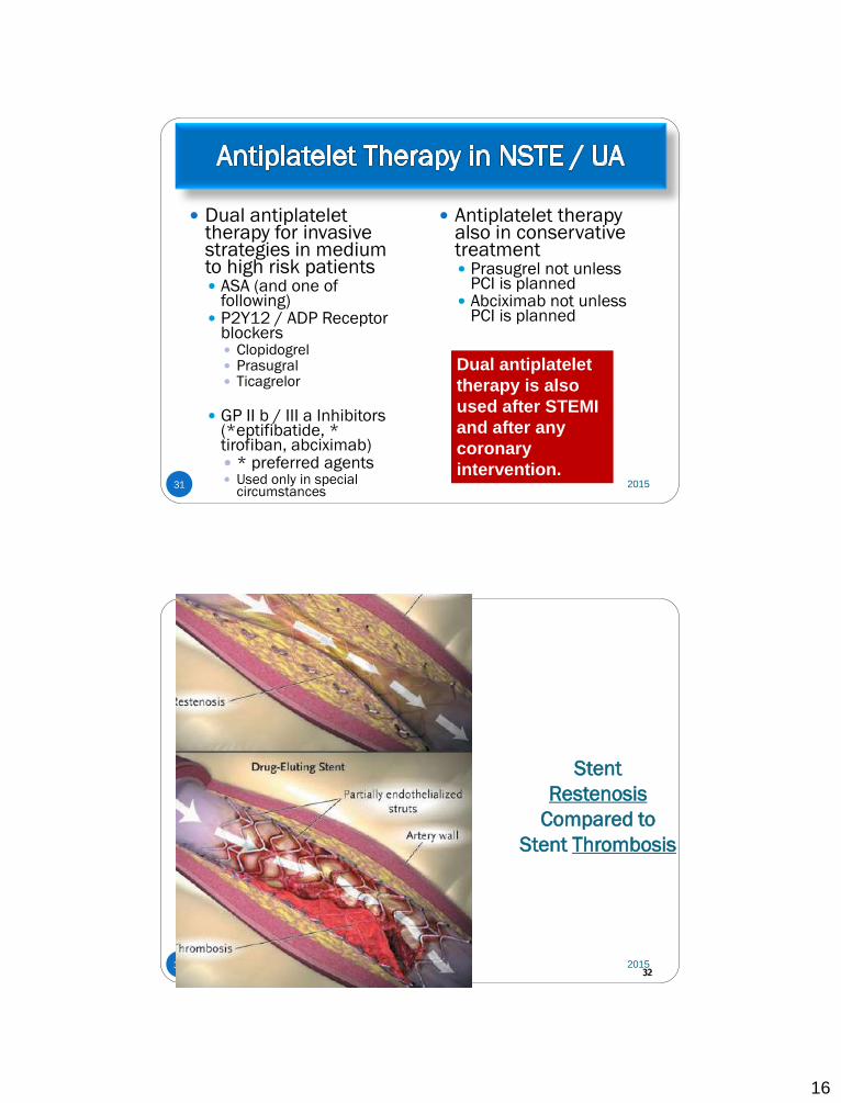
Stent
Restenosis
Compared to
Stent Thrombosis
32
17
33
What is it? Not waiting for failed medical treatment Not waiting for + noninvasive test Angiography with intent of revascularization Done within 12 to 24 hours
2015
34
When to do it? Refractory angina Hemodynamic
instability Electrical instability Initially stable
patients with a high risk for clinical events
Excluded: very frail elderly, severe hepatic, renal or pulmonary disease / active or inoperable cancer
Early invasive therapy is not recommended in patients with acute chest pain with a low likelihood of ACS
Early invasive therapy
is not recommended in patients who do not want to consent to revascularization.
2015
18
35
Recurrent angina / ischemia Rest or low level activity with medical treatment
Troponin +
New or presumed new ST depression
S&S HF or worsening mitral regurgitation
High risk findings on noninvasive testing EF < 35%, large anterior perfusion defect, multiple perfusion defects)
Hemodynamic instability
Sustained VT
PCI within 6 months
Prior CABG
Reduced LV Function
High risk TIMI or GRACE Score
2015
36
Medications to improve prognosis Aspirin
ASA benefits > in those > 65 years Long term benefit with 81 mg
Clopidogrel / Prasugrel / Ticagrelor Dual antiplatelet therapy in conservative management
for 12 months Higher risk of bleeding with dual antiplatelet therapy
No elderly sub group data for clopidogrel
Statins Have greater benefit in elderly for reduction of future MI
and death than in younger patient populations
2015
19
37
Medications to improve prognosis Beta-blockers ACE inhibitors Definite in select patients / reasonable in
all ARBs if ACE-I intolerant
Aldosterone antagonists EF < 40 with HF or diabetes
2015
SL NTG Instruction
38
No more than 1 dose of SL NTG If chest discomfort is unimproved or is
worsening 5 min after 1 NTG call 9-1-1
immediately before taking additional NTG.
May take additional NTG while waiting EMS.
Chew ASA while waiting EMS.
In chronic stable angina if symptoms are significantly
improved by 1 dose of NTG may repeat NTG every 5
min for a maximum of 3 doses and call 9-1-1 if
symptoms have not resolved completely.
2015
20
39
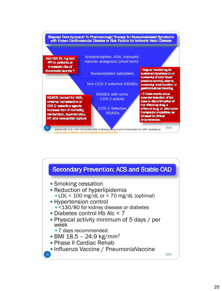
• Acetaminophen, ASA, tramadol,
narcotic analgesics (short term)
• COX-2 Selective
NSAIDs
• Nonacetylated salicylates
• Non COX-2 selective NSAIDs
• NSAIDs with some
COX-2 activity
Select patients at low risk
of thrombotic events
Prescribe lowest dose
required to control symptoms
* Addition of ASA may not be sufficient protection
Antman EM, et al. J Am Coll Cardiol 2008. Published ahead of print on December 10, 2007. Available at
http://content.onlinejacc.org/cgi/content/full/j.jacc.2007.10.001.
2015
40
Smoking cessation Reduction of hyperlipidemia
LDL < 100 mg/dL or < 70 mg/dL (optimal)
Hypertension control <130/80 for kidney disease or diabetes
Diabetes control Hb AIc < 7 Physical activity minimum of 5 days / per
week 7 days recommended
BMI 18.5 – 24.9 kg/mm2
Phase II Cardiac Rehab Influenza Vaccine / PneumoniaVaccine
2015
21
41
Use oxygen for hypoxemia
Assess response to beta-blocker therapy. HR / BP
Arrhythmia control
Assess for complications related to specific type of MI Assess heart sounds for new holosystolic murmurs
Risk for myocardial rupture
Observe for signs of left ventricular dysfunction, including hypotension or clinical signs of heart failure.
Monitor ECG for conduction disturbances and arrhythmias
Assess for presence of RV infarct 2015
42
Management of arterial access site
Assessment for contrast nephropathy
Restrict activity for the first 12 hours, and then begin Phase I Cardiac Rehabilitation (progressive mobility)
Referral to Phase II Cardiac Rehabilitation
Utilize cardiac monitoring ST-segment monitoring
Uninterrupted monitoring for first 24-48 hours
Address addiction to nicotine Consideration for nicotine withdrawal
Focus on holistic approach to anxiety reduction Include the family. Family visits do not have a negative
impact on vital signs or cardiac rhythm 2015
22
43 2015
44
Cellular edema produces an inflammatory response.
Recruitment of some stem cells leads to some tissue
regeneration.
Damaged tissue is bruised and cyanotic.
Catecholamines are released from myocardial cells, thus
increasing the risk of arrhythmias.
Cardiac biomarkers are released.
White blood cells invade the necrotic tissue within 2 to 3 days.
Scavenger cells release enzymes to break down necrotic tissue.
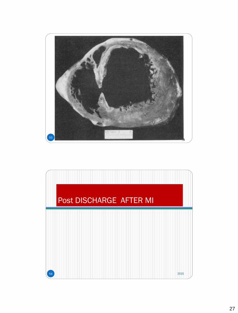
The necrotic wall can become very thin during this phase, and
the patient is at risk for cardiac rupture.
2015
23
45
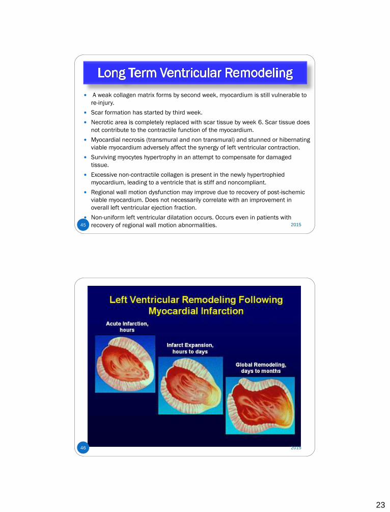
A weak collagen matrix forms by second week, myocardium is still vulnerable to
re-injury.
Scar formation has started by third week.
Necrotic area is completely replaced with scar tissue by week 6. Scar tissue does
not contribute to the contractile function of the myocardium.
Myocardial necrosis (transmural and non transmural) and stunned or hibernating
viable myocardium adversely affect the synergy of left ventricular contraction.
Surviving myocytes hypertrophy in an attempt to compensate for damaged
tissue.
Excessive non-contractile collagen is present in the newly hypertrophied
myocardium, leading to a ventricle that is stiff and noncompliant.
Regional wall motion dysfunction may improve due to recovery of post-ischemic
viable myocardium. Does not necessarily correlate with an improvement in
overall left ventricular ejection fraction.
Non-uniform left ventricular dilatation occurs. Occurs even in patients with
recovery of regional wall motion abnormalities. 2015
46 2015
24
47
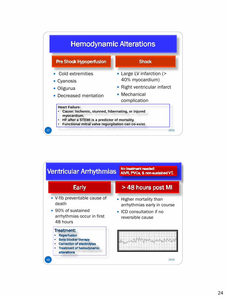
Cold extremities
Cyanosis
Oligurua
Decreased mentation
Large LV infarction (>
40% myocardium)
Right ventricular infarct
Mechanical
complication
Heart Failure:
• Cause: Ischemic, stunned, hibernating, or injured
myocardium.
• HF after a STEMI is a predictor of mortality.
• Functional mitral valve regurgitation can co-exist.
2015
48
V-fib preventable cause of
death
90% of sustained
arrhythmias occur in first
48 hours
Higher mortality than
arrhythmias early in course
ICD consultation if no
reversible cause
2015
25
49 2015
50
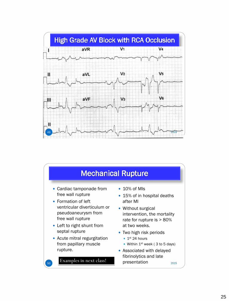
Cardiac tamponade from
free wall rupture
Formation of left
ventricular diverticulum or
pseudoaneurysm from
free wall rupture
Left to right shunt from
septal rupture
Acute mitral regurgitation
from papillary muscle
rupture.
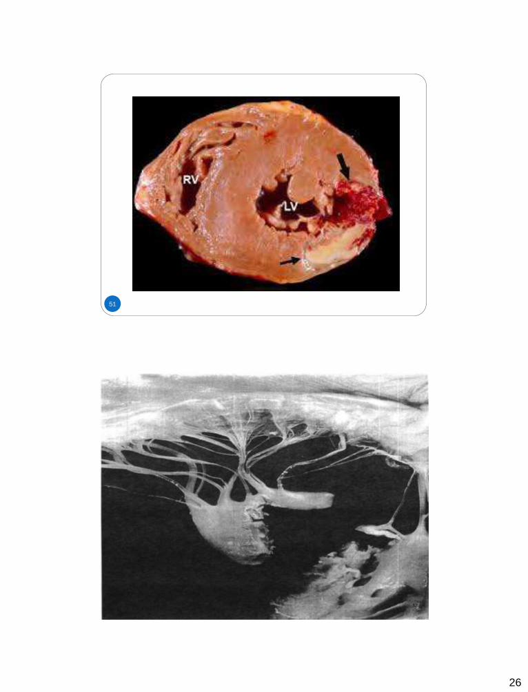
10% of MIs
15% of in hospital deaths
after MI
Without surgical
intervention, the mortality
rate for rupture is > 80%
at two weeks.
Two high risk periods
1st 24 hours
Within 1st week ( 3 to 5 days)
Associated with delayed
fibrinolytics and late
presentation Examples in next class! 2015
27
53 53
Post DISCHARGE AFTER MI
2015 54
28
After the patient achieves a rehabilitation level equivalent with
activities of daily living, he/she can begin a walking program
3 to 4 METS
Should be by time of discharge
Begin walking 5 to 10 minutes at a time
Patients should rate activity as moderate
Shortness of breath means overexertion. Other signs of
activity intolerance include: angina, dizziness, diaphoresis,
prolonged fatigue, and nausea.
The use of force to open windows or tight jar lids should be
avoided in patients with lifting restrictions.
2015 55
2015 56
29
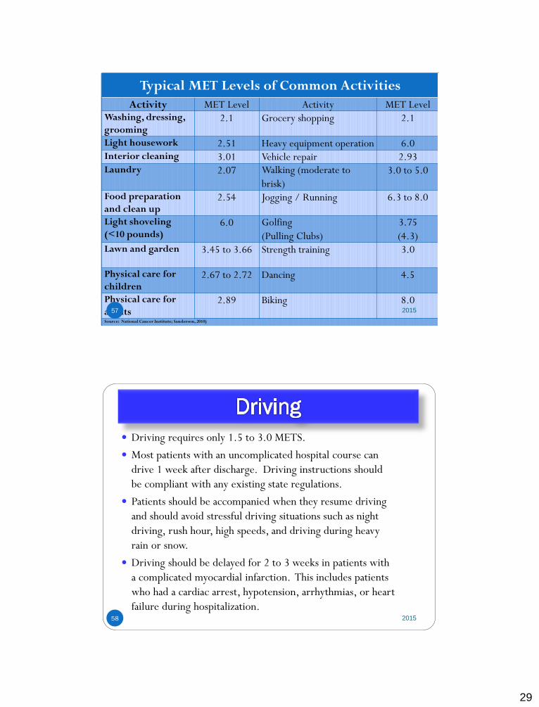
Typical MET Levels of Common Activities
Activity MET Level Activity MET Level Washing, dressing,
grooming 2.1 Grocery shopping 2.1
Light housework 2.51 Heavy equipment operation 6.0 Interior cleaning 3.01 Vehicle repair 2.93 Laundry 2.07 Walking (moderate to
brisk)
3.0 to 5.0
Food preparation
and clean up 2.54 Jogging / Running 6.3 to 8.0
Light shoveling
(<10 pounds) 6.0 Golfing
(Pulling Clubs)
3.75
(4.3) Lawn and garden 3.45 to 3.66 Strength training 3.0
Physical care for
children 2.67 to 2.72 Dancing 4.5
Physical care for
adults 2.89 Biking 8.0
Source: National Cancer Institute; Sanderson, 2010)
2015 57
Driving requires only 1.5 to 3.0 METS.
Most patients with an uncomplicated hospital course can
drive 1 week after discharge. Driving instructions should
be compliant with any existing state regulations.
Patients should be accompanied when they resume driving
and should avoid stressful driving situations such as night
driving, rush hour, high speeds, and driving during heavy
rain or snow.
Driving should be delayed for 2 to 3 weeks in patients with
a complicated myocardial infarction. This includes patients
who had a cardiac arrest, hypotension, arrhythmias, or heart
failure during hospitalization.
2015 58
30
Travel Patients can usually travel by air within 2 weeks if accompanied
by a travel companion, and if the patient has sublingual
nitroglycerin
If free of all angina symptoms and complications of their myocardial
infarction
Patients should also have airport transportation assistance to avoid
excessive stress and rushing in the airport
Patients should also take precautions when traveling to avoid the
development of deep vein thrombosis
2015 59
Sex After an acute coronary syndrome, stable patients can resume sexual
activity with their usual partner in one week to 10 days (Anderson et al., 2011).
Patients are uncomfortable asking about resuming sexual relationships, so instructions regarding sexual activity should be included as a routine part of all discharge instructions.
Patients with a history of angina during sexual relationships may be instructed to take nitroglycerin prior to engaging in sexual activities.
The average intimate session ranges from 2.5-4 METS for most people.
Walking at 2 mph on level ground is 2.5 METS. Mowing the lawn with a power mower or walking at 3.5 mph is 4 METS. Climbing up a flight of stairs is 8 METS.
The biggest risk with sex in the cardiac patient is the possibility of arrhythmias, which is associated with sympathetic activity increased during arousal. Patients with uncontrolled or untreated hypertension need to discuss specific guidelines with their physician (Sotile & Cantor-Cooke, 2003).
2015 60
31
Return to Work Low risk myocardial infarction (LVEF > 45%, successful
revascularization with PCI, age < 70 years) can generally return to work after 2 weeks.
Most myocardial infarction adverse events reach a low steady state at 10 weeks. This may guide decision making in some types of employment.
Patients who need to return to physically demanding activities can have an exercise stress test that compares their performance on the stress test to the METs required for the activity. This will provide information about the ability and safety of engaging in activities based on the MET level achieved during exercise stress test.
(Anderson et al., 2011).
2015 61
Cardiac Rehabilitation Goals:
Increase functional capacity
Reduce disability
Improve quality of life
Modify cardiac risk factors
Reduce morbidity and mortality.
Pooled data from a meta-analysis of studies involving the
exercise portion of cardiac rehabilitation show a benefit of
reduced all-cause mortality of approximately 25% when
compared to usual care.
In one study of over 600,000 Medicare patients, mortality
rates were 21% to 34% lower in patients who participated in
cardiac rehabilitation (Suaya, Stason, Ades, Normand, & Shepard, 2009).
2015 62
32
Cardiac Rehabilitation
Low-risk patients can implement an exercise prescription at
home or in a community setting. Low-risk patients include
those with absence of ischemia or arrhythmias on a stress test.
High-risk patients should be in medically supervised exercise
programs. They are defined as patients with ischemia or
serious arrhythmias on a stress test.
Under utilization of cardiac rehabilitation.
2015 63
Treating the Whole Patient
Depression
Approximately 1 in 5 patients hospitalized with MI have major depression. There is also evidence that depression continues for several months after discharge (Fihn et al., 2012; Bush et al., 2005).
There is strong evidence that patients who are depressed post MI have a higher rate of mortality from both cardiac and non-cardiac causes (Bush et al., 2005).
Anxiety and Stress
In post MI patients, interventions to reduce stress can reduce recurrent cardiac events by as much as 35-75% (Gibbons et al., 2002).
Social Support
Role Identity 2015 64
33
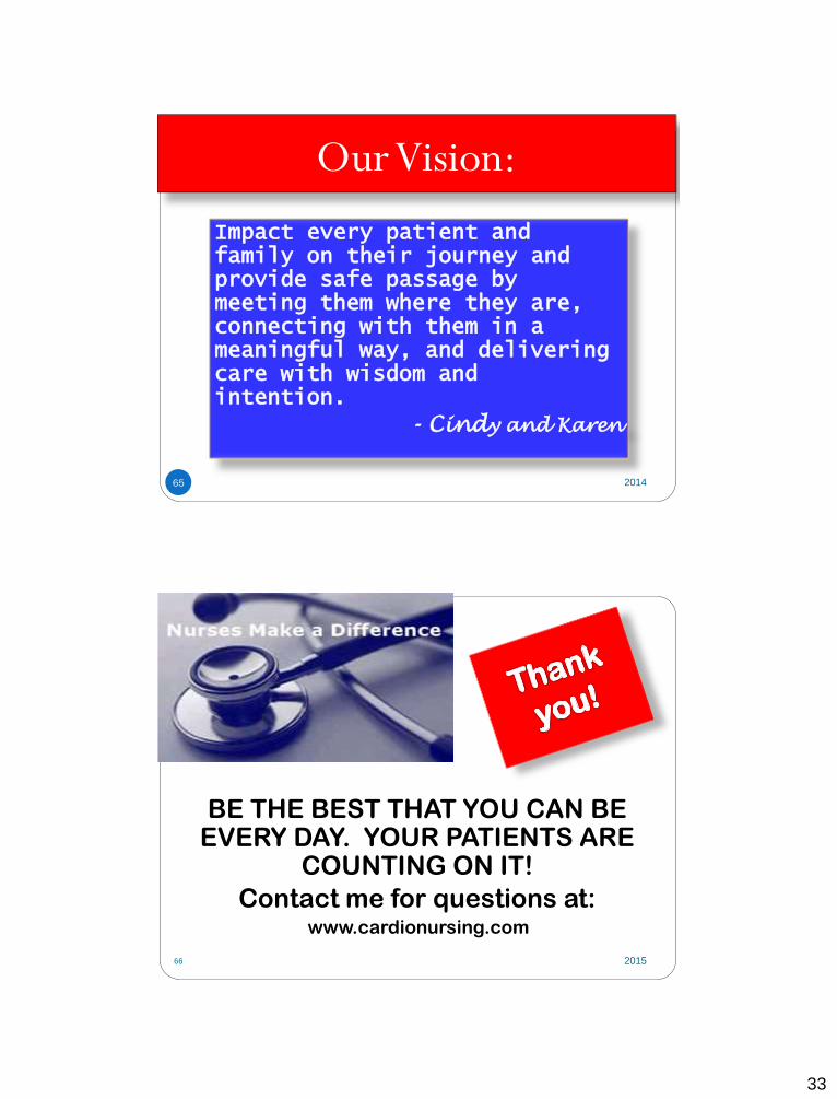
Our Vision:
Impact every patient and family on their journey and provide safe passage by meeting them where they are, connecting with them in a meaningful way, and delivering care with wisdom and intention.
- Cindy and Karen
65 2014
66
BE THE BEST THAT YOU CAN BE EVERY DAY. YOUR PATIENTS ARE
COUNTING ON IT!
Contact me for questions at: www.cardionursing.com
2015