Embed Size (px)
Citation preview
British Journal of Ophthalmology, 1978, 62, 843-849
Acute follicular conjunctivitis and keratoconjunctivitisdue to herpes simplex virus in LondonS. DAROUGAR, P. A. HUNTER, M. VISWALINGAM, J. A. GIBSON, ANDB. R. JONESFrom the Department of Clinical Ophthalmology, Institute of Ophthalmology, andExternal Eye Disease Clinic, Moorfields Eye Hospital, London
SUMMARY During the 18 months January 1975 to June 1976, 25 cases of acute herpetic follicularconjunctivitis and keratoconjunctivitis resembling adenovirus ocular infection presented in theExternal Eye Disease Clinic, Moorfields Eye Hospital, City Road, London. Herpes simplex viruswas isolated in HEp2 cells in 22 patients, and the remaining 3 patients were identified by aminimum 4-fold rise in the level of antiherpes simplex virus antibody in their blood. No adenoviruswas isolated from these patients, but complement fixation test for adenovirus was positive in 1
patient with cultural test positive for herpes simplex virus. Most patients were between 20 and 35years old and the ratio of males to females was 12 to 13. At the initial visit the clinical featuresof disease were moderate to severe conjunctival papillary and follicular reactions with epithelialand subepithelial punctate keratitis but little systemic disease. In the absence of typical herpeticlesions of face, lids, or cornea the disease resembled adenovirus types 8 or 19 keratoconjunctivitis.Of these 25 patients 5 subsequently developed typical herpetic lesions of lids or cornea. In theremaining 20 cases the correct diagnosis could be made only by cultural or serological tests.Virological diagnosis provides a rational basis for antiherpetic chemotherapy, which appears toshorten the course of infection.
Acute primary herpetic blepharokeratoconjuncti-vitis and chronic recurrent keratitis are commonmanifestations of herpes simplex virus (HSV)infection. Less well recognised is acute follicularconjunctivitis without characteristic lid or corneallesions.
This report presents the findings in 25 cases ofacute herpetic follicular conjunctivitis which pre-sented without characteristic lid, face, or cornealsigns of herpetic infections. In most of them, thecorrect diagnosis could be made only by culturalor serological tests for HSV infection.
Methods and patients
Patients with acute follicular conjunctivitis orkeratoconjunctivitis without facial, lid, or corneallesions typical of HSV infection at the first visit,but with positive cultural or serological tests forHSV, were included.The patients were examined with a Haag-Streit
Address for reprints: Dr S. Darougar, Institute of Ophthal-mology, Judd Street, London WC1H 9QS
slit lamp and symptoms were graded on a 0 to 3scale (mild, moderate, severe) as described previously(Darougar et al., 1977b). Conjunctival swabbingswere placed in plastic capsules containing 2 SPtransport medium (Gordon et al., 1969) with 3%fetal bovine serum and stored in a refrigerator at-700C. Each clinical specimen was inoculated into2 tubes containing HEp2 cells. Cultures weremaintained for 21 days and examined frequently forthe presence of cytopathic effect (CPE) (McSwigganet al., 1975). Isolates were identified by the flu-orescent antibody test.
Sera collected by venepuncture at intervals of2 to 3 weeks were tested by a complement fixationtest (CFT) for herpes simplex virus and adenovirusgroup antibodies. Conjunctival impressions takenfrom some patients by specially-designed plasticspatulae were fixed, stained, and examined by themethod described by Thatcher et al. (1977).
Results
During the 18 months January 1975 to June 197625 patients with acute follicular conjunctivitis and
843
copyright. on M
arch 2, 2020 by guest. Protected by
http://bjo.bmj.com
/B
r J Ophthalm
ol: first published as 10.1136/bjo.62.12.843 on 1 Decem
ber 1978. Dow
nloaded from

S. Darougar, P. A. Hunter, M. Viswalingam, J. A. Gibson, and B. R. Jones
no herpetic lesions of the lids, face, or cornea atthe time of their first visit to the clinic, but withpositive cultural or serological tests for herpessimplex virus, were identified. Of these 25 patients5 subsequently developed palpebral vesicles ordendritic ulcers.The patients' ages ranged from 8 to 50 years, the
majority being between 20 and 35 years. The ratioof males to females was 12 to 13. Of these 25 cases18 were diagnosed in 1975 and 7 in the first 6months of 1976. No seasonal pattern of incidencewas observed.
PREVIOUS HISTORYFive patients gave a history of previous infectionsuggestive of herpes simplex: 3 had recurrent coldsores on nose or lips, 1 had lid disease (possiblyherpetic) 5 years previously, and 1 had a history ofa possible corneal ulcer 5 years previously. Inno case was there a clear history of previous ocularherpetic infection.Of the 25 patients 2 had been in contact with
patients with cold sores, 3 in contact with otherpersons suffering from conjunctivitis, and a further3 had been in swimming pools 9 to 21 days beforethe onset of their ocular infection. In 1 of thislatter group some fellow swimmers had also de-veloped conjunctivitis at the same time. In theother 17 cases there was no history of contact withother patients nor had they attended hospitals,clinics, or a swimming pool.
In 5 cases there was a history of pharyngitis andrhinitis during the week preceding the conjuncti-vitis. Another patient had a cold 3 weeks beforedeveloping eye disease. One patient had Hodgkin'sdisease and was on systemic treatment with pred-nisolone 5 mg daily.
SYMPTOMS AND SIGNSFifteen of the 25 patients had enlarged preauricularlymph nodes, which in 12 cases were tender. In 8cases in which the disease affected both eyes thelymphadenopathy was bilateral.The commonest symptoms were moderate hyper-
aemia associated with lacrimation, discharge, grit-tiness, and swelling of the lids (Table 1 and Fig. 1).
In 8 cases the second eye became inflamed inless than 1 week from the onset of the infection.In the other 17 the infection remained unilateral.The average duration of conjunctival inflammationwas 4 weeks (range 2 to 12 weeks). In 17 cases lidsshowed diffuse oedema and mild to moderateerythema. No vesicle or ulceration was observedin these cases at the initial examination. However,in 4 cases vesicles or ulcers developed on the lid 2to 3 days after the first examination.
The bulbar conjunctiva showed mild to moderateh'yperaemia in 23 cases. Limbal follicles were notedin 2 and a moderate ecchymosis in 1 (Fig. 2). Thepalpebral conjunctiva showed moderate to severehyperaemia and papillary hypertrophy in 21 cases
Table 1 Prevalence and severity ofsymptoms at firstvisit in 25 cases ofherpetic follicular conjunctivitis
Severity
Symptoms Mild Moderate Severe
Swelling of lids 7 5 1
Hyperaemia I 1 13 0
Lacrimation 7 9 2
Discharge 15 6 0
Grittiness 11 5 0
Itching 6 2 0
Photophobia 4 2 1
Pain 4 1 0
Blurred vision 6 1 0
Duration and severity of symptoms and signs
6 3 7 10 14 17 21 28 2tl2 3112 4/12Days
Fig. 1 Duration and severity ofsymptoms and signs ofdisease in 25 cases ofacute herpeticfollicular conjunctivitisand keratoconjunctivitis
844
copyright. on M
arch 2, 2020 by guest. Protected by
http://bjo.bmj.com
/B
r J Ophthalm
ol: first published as 10.1136/bjo.62.12.843 on 1 Decem
ber 1978. Dow
nloaded from

Acute follicular conjunctivitis and keratoconjunctivitis due to herpes simplex virus in London
Fig. 2 Moderate ecchymosis in a case ofacute herpeticfollicular conjunctivitis
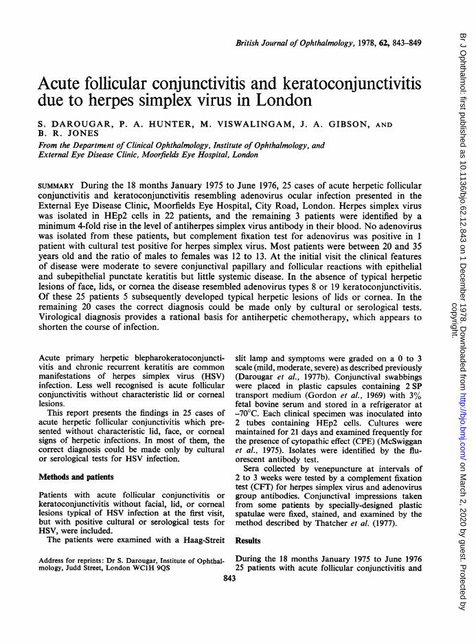
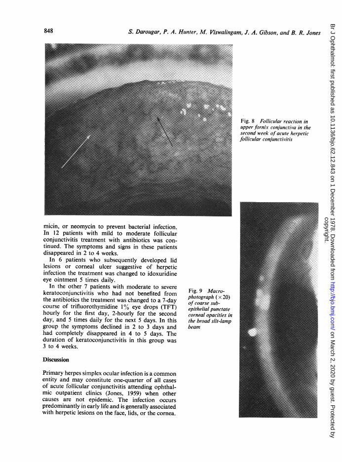
and moderate to severe follicular reaction in 19(Table 2). Papillary hypertrophy was most severein the upper tarsus as well as the lower lid (Figs.3, 4, 5) and lasted for 2 to 12 weeks (Fig. 1). Follicu-lar reaction was present in all cases. Moderate tosevere follicular hypertrophy was present mainlyin the lower and upper fornices (Figs. 6, 7, 8). Thefollicles were small, discrete, and lasted 2 to 8weeks (Fig. 1).Nine of the 25 cases developed moderate coarse
epithelial punctate keratitis (Jones, 1962). Thiswas generally preceded by fine punctate keratitis(Jones, 1962). In 5 cases mild to moderate sub-
Fig. 3 Severe papillary reaction
in upper tarsal conjunctiva in the
first week of acute herpetic
follicular conjunctivits
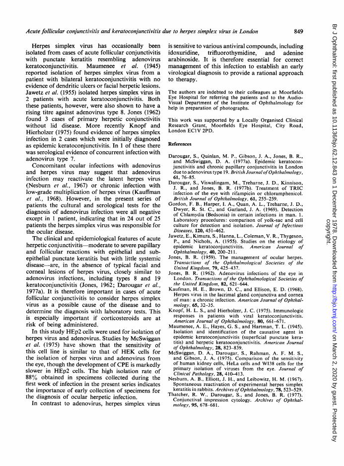
epithelial punctate keratitis (Fig. 9) resemblingthat of epidemic keratoconjunctivitis (Jones, 1962)developed. The opacities were coarse, fewer than15 in number, and mainly located in the inter-palpebral fissure. The average time from the onsetof symptoms to the occurrence of subepitheliallesions was 17 days (Fig. 1). The subepithelialpunctate keratitis persisted between 1 and 4 months.
In 2 cases small dendritic ulcers developed ondays 4 and 9 respectively. They both respondedwell to therapy with idoxuridine 05% eye ointment.Of the cases with keratitis 2 developed a milduveitis which responded quickly to therapy withtrifluorothymidine 1% eye drops.
PATHOLOGYIn all, 38 conjunctival specimens were collectedfrom 25 patients. In 22 of 25 cases herpes simplexvirus was isolated in HEp2 cells.Of the 24 specimens collected during the first
week of infection 21 (88%) were positive for herpes
Table 2 Prevalence and severity of signs in thepalpebral conjunctiva at first visit in 25 cases of acuteherpetic jollicular conjunctivitis
Severity
Sign Mild Moderate Severe
Hyperaemia 8 13 4
Papillary hypertrophy 4 12 9
Follicular hypertrophy 6 15 4
845
copyright. on M
arch 2, 2020 by guest. Protected by
http://bjo.bmj.com
/B
r J Ophthalm
ol: first published as 10.1136/bjo.62.12.843 on 1 Decem
ber 1978. Dow
nloaded from
S. Darougar, P. A. Hunter, M. Viswalingam, J. A. Gibson, and B. R. Jones
Fig. 4 Severe papillary reactionin lower lid conjunctiva in thefirst week of acute herpeticfollicular conjunctivitis
Fig. 5 Severe papillary reactionin upper fornix conjunctiva in thefirst week of acute herpeticfollicular conjunctivitis
virus but only 4 out of 10 specimens collected duringthe second week gave positive results. No virus wasisolated after the 11th day of infection. Adenoviruswas not isolated from these patients.
Paired or triplicate sera were tested in 14 cases.A 4-fold or greater rise in the level of herpes simplexvirus antibody was observed in 4 cases. In 3 ofthese no herpes virus was isolated. In 1 case inwhich the conjunctival swab was positive for herpesvirus a 4-fold rise in adenovirus antibody titre wasobtained.
Conjunctival impressions taken from 7 patientsduring the course of their disease showed a mixedpopulation of inflammatory cells in which poly-morphonuclear cells and monocytes were evenlydistributed. No multinucleated epithelial cells wereobserved in these impressions.
TREATMENTWith a preliminary diagnosis of an adenovirusinfection all patients were initially treated with eyedrops or eye ointment of chloramphenicol, genta-
846
copyright. on M
arch 2, 2020 by guest. Protected by
http://bjo.bmj.com
/B
r J Ophthalm
ol: first published as 10.1136/bjo.62.12.843 on 1 Decem
ber 1978. Dow
nloaded from

Acute follicular conjunctivitis and keratoconjunctivitis due to herpes simplex virus in London
Fig. 6 Follicular reaction inupper tarsal conjunctiva in thesecond week of acute herpeticfollicular conjunctivitis
Fig. 7 Follicular reaction in (a)tarsal and (b) fornix area of thelower lid conjunctiva in thesecond week of acute herpeticfollicular conjunctivitis
847
copyright. on M
arch 2, 2020 by guest. Protected by
http://bjo.bmj.com
/B
r J Ophthalm
ol: first published as 10.1136/bjo.62.12.843 on 1 Decem
ber 1978. Dow
nloaded from
S. Darougar, P. A. Hunter, M. Viswalingam, J. A. Gibson, and B. R. Jones
Fig. 8 Follicular reaction inupper fornix conjunctiva in thesecond week of acute herpeticfollicular conjunctivitis
micin, or neomycin to prevent bacterial infection.In 12 patients with mild to moderate follicularconjunctivitis treatment with antibiotics was con-tinued. The symptoms and signs in these patientsdisappeared in 2 to 4 weeks.
In 6 patients who subsequently developed lidlesions or corneal ulcer suggestive of herpeticinfection the treatment was changed to idoxuridineeye ointment 5 times daily.
In the other 7 patients with moderate to severekeratoconjunctivitis who had not benefited fromthe antibiotics the treatment was changed to a 7-daycourse of trifluorothymidine 1% eye drops (TFT)hourly for the first day, 2-hourly for the secondday, and 5 times daily for the next 5 days. In thisgroup the symptoms declined in 2 to 3 days andhad completely disappeared in 4 to 5 days. Theduration of keratoconjunctivitis in this group was3 to 4 weeks.
Fig. 9 Macro-photograph ( x 20)of coarse sub-epithelial punctatecorneal opacities inthe broad slit-lampbeam
Discussion
Primary herpes simplex ocular infection is a commonentity and may constitute one-quarter of all casesof acute follicular conjunctivitis attending ophthal-mic outpatient clinics (Jones, 1959) when othercauses are not epidemic. The infection occurspredominantly in early life and is generally associatedwith herpetic lesions on the face, lids, or the cornea.
848
copyright. on M
arch 2, 2020 by guest. Protected by
http://bjo.bmj.com
/B
r J Ophthalm
ol: first published as 10.1136/bjo.62.12.843 on 1 Decem
ber 1978. Dow
nloaded from
Acute follicular conjunctivitis and keratoconjunctivitis due to herpes simplex virus in London
Herpes simplex virus has occasionally beenisolated from cases of acute follicular conjunctivitiswith punctate keratitis resembling adenoviruskeratoconjunctivitis. Maumenee et al. (1945)reported isolation of herpes simplex virus from apatient with bilateral keratoconjunctivitis with noevidence of dendritic ulcers or facial herpetic lesions.Jawetz et al. (1955) isolated herpes simplex virus in2 patients with acute keratoconjunctivitis. Boththese patients, however, were also shown to have arising titre against adenovirus type 8. Jones (1962)found 3 cases of primary herpetic conjunctivitiswithout lid disease. More recently Knopf andHierholzer (1975) found evidence of herpes simplexinfection in 2 cases which were initially diagnosedas epidemic keratoconjunctivitis. In 1 of these therewas serological evidence of concurrent infection withadenovirus type 7.
Concomitant ocular infections with adenovirusand herpes virus may suggest that adenovirusinfection may reactivate the latent herpes virus(Nesburn et al., 1967) or chronic infection withlow-grade multiplication of herpes virus (Kauffmanet al., 1968). However, in the present series ofpatients the cultural and serological tests for thediagnosis of adenovirus infection were all negativeexcept in 1 patient, indicating that in 24 out of 25patients the herpes simplex virus was responsible forthe ocular disease.The clinical and epidemiological features of acute
herpetic conjunctivitis-moderate to severe papillaryand follicular reactions with epithelial and sub-epithelial punctate keratitis but with little systemicdisease-are, in the absence of typical facial andcorneal lesions of herpes virus, closely similar toadenovirus infections, including types 8 and 19keratoconjunctivitis (Jones, 1962; Darougar et al.,1977a). It is therefore important in cases of acutefollicular conjunctivitis to consider herpes simplexvirus as a possible cause of the disease and todetermine the diagnosis with laboratory tests. Thisis especially important if corticosteroids are atrisk of being administered.
In this study HEp2 cells were used for isolation ofherpes virus and adenovirus. Studies by McSwigganet al. (1975) have shown that the sensitivity ofthis cell line is similar to that of HEK cells forthe isolation of herpes virus and adenovirus fromthe eye, though the development of CPE is markedlyslower in HEp2 cells. The high isolation rate of88% obtained in specimens collected during thefirst week of infection in the present series indicatesthe importance of early collection of specimens forthe diagnosis of ocular herpetic infection.
In contrast to adenovirus, herpes simplex virus
is sensitive to various antiviral compounds, includingidoxuridine, trifluorothymidine, and adeninearabinoside. It is therefore essential for correctmanagement of this infection to establish an earlyvirological diagnosis to provide a rational approachto therapy.
The authors are indebted to their colleagues at MoorfieldsEye Hospital for referring the patients and to the Audio-Visual Department of the Institute of Ophthalmology forhelp in preparation of photographs.
This work was supported by a Locally Organised ClinicalResearch Grant, Moorfields Eye Hospital, City Road,London ECIV 2PD.
References
Darougar, S., Quinlan, M. P., Gibson, J. A., Jones, B. R.,and McSwiggan, D. A. (1977a). Epidemic keratocon-junctivitis and chronic papillary conjunctivitis in Londondue to adenovirus type 19. British Journal ofOphthalmology,61, 76-85.
Darougar, S., Viswalingam, M., Treharne, J. D., Kinnison,J. R., and Jones, B. R. (1977b). Treatment of TRICinfection of the eye with rifampicin or chloramphenicol.British Journal of Ophthalmology, 61, 255-259.
Gordon, F. B., Harper, I. A., Quan, A. L., Treharne, J. D.,Dwyer, R. St. C., and Garland, J. A. (1969). Detectionof Chlamydia (Bedsonia) in certain infections in man. 1.Laboratory procedures: comparison of yolk-sac and cellculture for detection and isolation. Journal of InfectiousDiseases, 120, 451-462.
Jawetz, E., Kimura, S., Hanna, L., Coleman, V. R., Thygeson,P., and Nichols, A. (1955). Studies on the etiology ofepidemic keratoconjunctivitis. American Journal ofOphthalmology, 40, 200-211.
Jones, B. R. (1959). The management of ocular herpes.Transactions of the Ophthalmological Societies of theUnited Kingdom, 79, 425-437.
Jones, B. R. (1962). Adenovirus infections of the eye inLondon. Transactions of the Ophthalmological Societies ofthe United Kingdom, 82, 621-644.
Kaufman, H. E., Brown, D. C., and Ellison, E. D. (1968).Herpes virus in the lacrimal gland conjunctiva and corneaof man: a chronic infection. American Journal of Ophthal-mology, 65, 32-35.
Knopf, H. L. S., and Hierholzer, J. C. (1975). Immunologicresponses in patients with viral keratoconjunctivitis.American Journal of Ophthalmology, 80, 661-671.
Maumenee, A. E., Hayes, G. S., and Hartman, T. L. (1945).Isolation and identification of the causative agent inepidemic keratoconjunctivitis (superficial punctate kera-titis) and herpetic keratoconjunctivitis. American Journalof Ophthalmology, 28, 823-839.
McSwiggan, D. A., Darougar, S., Rahman, A. F. M. S.,and Gibson, J. A. (1975). Comparison of the sensitivityof human kidney cells, HeLa cells and W138 cells for theprimary isolation of viruses from the eye. Journal ofClinical Pathology, 28, 410-413.
Nesburn, A. B., Elliott, J. H., and Leibowitz, H. M. (1967).Spontaneous reactivation of experimental herpes simplexkeratitis in rabbits. Archives ofOphthalmology, 78, 523-529.
Thatcher, R. W., Darougar, S., and Jones, B. R. (1977).Conjunctival impression cytology. Archives of Ophthal-mology, 95, 678-681.
849
copyright. on M
arch 2, 2020 by guest. Protected by
http://bjo.bmj.com
/B
r J Ophthalm
ol: first published as 10.1136/bjo.62.12.843 on 1 Decem
ber 1978. Dow
nloaded from