Embed Size (px)
Citation preview
REVIEWS
Acupuncture’s Cardiovascular Actions:A Mechanistic Perspective
John Longhurst, MD, PhD
ABSTRACT
Over the last several decades, there has been an explosion of articles on acupuncture, including studies that
have begun to explore mechanisms underlying its analgesic and cardiovascular actions. Modulation of
cardiovascular function is most effective during manual and low-frequency, low-intensity electroacupuncture
(EA) at a select set of acupoints situated along meridians located over deep somatic nerves on the upper and
lower extremities. Stimulation at these acupoints activates underlying sensory neural pathways that project to
a number of regions in the central nervous system (CNS) that ultimately regulate autonomic outflow and
hence cardiovascular function. A long-loop pathway involving the hypothalamus, midbrain, and medulla
underlies EA modulation of reflex increases in blood pressure (BP). Actions of excitatory and inhibitory
neurotransmitters in the supraspinal CNS underlie processing of the somatic input and adjustment of auto-
nomic outflow during EA. Acupuncture also decreases elevated blood pressure through actions in the thoracic
spinal cord. Reflexes that lower BP likewise are modulated by EA through its actions on sympathetic and
parasympathetic nuclei in the medulla. The autonomic influence of acupuncture is slow in onset but pro-
longed in duration, typically lasting beyond the period of stimulation. Clinical studies suggest that acu-
puncture can be used to treat cardiac diseases, such as myocardial ischemia and hypertension, associated with
overactivity of the sympathetic nervous system.
Key Words: Autonomic Nervous System, Integrative Physiology, Somatic Afferents, Blood Pressure, Sympathetic
Nervous System, Parasympathetic Nervous System
INTRODUCTION
The practice of acupuncture began 2000–3000 years
ago. Until the last 50 years, acupuncture developed
empirically and its art was passed on from teacher to student
through practical application. More recently, practitioners
began to find that acupuncture had a rightful place in
mainstream medicine and could be used to treat a number of
conditions and symptoms. The public outside the Orient has
accepted acupuncture because of a perception that it reduces
pain effectively and successfully reverses a number of other
medical problems. Western medical and scientific commu-
nities have been more reluctant to accept this practice be-
cause of the absence of controlled clinical trials and scant
scientific evidence for its mechanisms of action. However,
there may be reason for this skepticism to change. The
number of articles published on acupuncture research (451
articles worldwide in 2009) has been increasing almost ex-
ponentially over the last several decades, with the United
States and China both taking lead roles in advancing
Samueli Center for Integrative Medicine, University of California, Irvine, Irvine, CA.
Research cited in this review was funded by two grants from the National Institutes of Health, (R01-HL63313 and R01-HL72125), theAdolph Coors Foundation, the Peterson Family Foundation, the Larry K. Dodge Chair of Integrative Biology, and the Susan SamueliChair of Integrative Medicine.
MEDICAL ACUPUNCTUREVolume 25, Number 2, 2013# Mary Ann Liebert, Inc.DOI: 10.1089/acu.2013.0960
101
understanding of this ancient therapy.1 With mounting evi-
dence that acupuncture can be used to treat a number of
clinical conditions, including cardiovascular dysfunctions,
such as hypertension and hypotension, the rationale for
achieving a better understanding of the actions of acu-
puncture at organ system, cellular, and subcellular levels has
become more compelling.2 This article is a review of recent
experimental studies exploring mechanisms underlying the
cardiovascular actions of acupuncture, a focus of the
laboratory of the Samueli Center for Integrative Medicine
and Department of Medicine, University of California, Ir-
vine, over the last decade and a half.
EARLY STUDIES: BEGINNINGS OF A NEWERA IN ACUPUNCTURE RESEARCH
Two pioneers who used accepted Western approaches to
study the mechanisms of actions of acupuncture were Drs.
Ji-Sheng Han (PhD) at Beijing University and Peng Li (MD)
at Shanghai Medical University. Dr. Han focused on pain
while Dr. Li, together with Dr. Tai Yao, (MD) studied
acupuncture’s action on cardiovascular function. Both re-
searchers described the importance of the central nervous
system (CNS) in mediating acupuncture’s physiological
actions. These researchers found that the endogenous opioid
system in the CNS was responsible for much of acupunc-
ture’s modulation of pain and hypertension.3,4 Small studies
of patients with coronary disease in the late 1980s and early
1990s suggested that acupuncture reduces electrocardio-
graphic evidence of myocardial ischemia and increases
angina threshold.4–6 In the mid 1990s, I began a long-term
collaboration with Dr. Li, exploring peripheral and central
neural mechanisms underlying the actions of electro-
acupuncture (EA) on cardiovascular function. The collabo-
ration involved studying experimental models of myocardial
ischemia, reflex-induced hypertension, and, more recently,
reflex hypotension. Each model was based on the observation
that acupuncture’s cardiovascular actions are most prominent
when autonomic outflow is stimulated, for example, during
visceral sympathoexcitatory reflexes. These investigations
over the last 16 years have been reported in a series of more
than 30 studies using combined whole animal or human
physiological, electrophysiological, anatomical, pharmaco-
logical, and, more recently, molecular approaches.
ACUPUNCTURE IN DEMAND-INDUCEDMYOCARDIAL ISCHEMIA
Early studies originating from Europe have suggested
that acupuncture reduces ischemia in patients with symp-
tomatic coronary artery disease.5–9 To explore the mecha-
nisms underlying this clinical effect, Dr. Li and I developed
a feline model of demand-induced myocardial ischemia, in
which the left anterior descending (LAD) coronary artery
was ligated, partially allowing normal coronary blood flow
at rest but causing an insufficient flow, and hence transient
ischemia, identified by the regional myocardial dysfunction,
during reflex stimulation evoked by applying bradykinin to
the gallbladder.9,10 This model simulates the most common
form of cardiac ischemia experienced during stress by pa-
tients with symptomatic coronary atherosclerosis. Thirty
minutes of median nerve stimulation with low-frequency,
low-current (5 Hz, 4 mA) or, alternatively, percutaneous EA
at the PC 5 and PC 6 acupoints (Jianshi and Neiguan along
the Pericardium meridian on the volar surface of the wrist
(Fig. 1), using similar stimulus parameters (2 Hz, 4 mA),
abolished the stress-induced ischemia.11 EA reversed the
ischemia by blunting the reflex-related increase in blood
pressure (BP) significantly but not by increasing LAD blood
flow, as measured with a pulsed Doppler flowmeter. A
follow-up study showing that acupuncture’s action on is-
chemia could be reversed with intravenous (I.V.) naloxone
implicated the endogenous opioid system as an underlying
neurotransmitter mechanism (Fig. 2).12 The site of opioid
action during EA was not determined. Given that acu-
puncture’s anti-ischemic effect was noted to be predomi-
nately through its BP-lowering effect, subsequent studies
have focused on this hemodynamic action of acupuncture.
Because percutaneous EA mimicked direct nerve stimula-
tion, it was concluded that the peripheral nervous system
and CNS were involved in the EA-cardiovascular response.
OPIOIDS IN THE MEDULLA PROCESSSOMATIC SENSORY INPUT DURING EA
The rostral ventrolateral medulla (rVLM) is a significant
source of premotor sympathetic neurons and hence consti-
tutes an important brainstem region that processes somatic
and visceral sensory nerve input capable of adjusting sym-
pathetic outflow and, ultimately, cardiovascular function.13–16
Early investigations from several laboratories suggested
that opioids, c-aminobutyric acid (GABA) and serotonin (5-
hydroxytryptamine, 5-HT), might participate in acupuncture
regulation of BP.4,17,18 Recent immunohistochemical stud-
ies revealed EA-induced c-Fos nuclear expression (a marker
of neuronal activation) in the rVLM. These EA-activated
neurons contain enkephalin, while b-endorphin is present in
closely located axons.19,20 Both neurotransmitters thus have
the potential to participate in processing sympathetic out-
flow during EA. Pharmacological studies investigating the
roles of l-, d-, and j-opioid receptors indicate that b-en-
dorphin (and possibly endomorphin) and enkephalins, but
not dynorphin, are involved in rVLM modulation of hy-
pertensive responses during EA.21 The rVLM and possibly
the nucleus raphe palladus (NRP) are the source of en-
kephalins for the rVLM, while b-endorphin originates in
the arcuate nucleus of the hypothalamus.19,20,22 Visceral
102 LONGHURST
reflexes initiated by gallbladder stimulation and gastric dis-
tension stimulate rVLM neurons by releasing the excitatory
neurotransmitter glutamate to increase sympathetic outflow
and raise BP.23 Through these opioid mechanisms, acupunc-
ture reduces glutamate release in the rVLM and hence mod-
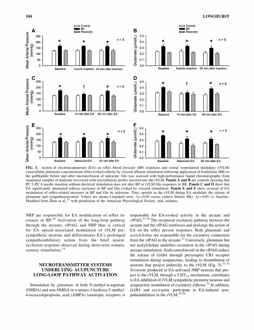
ulates reflex sympathoactivation and elevated BP (Fig. 3).23
LONG-LOOP PATHWAY IN ACUPUNCTUREMODULATION OF EXCITATORYCARDIOVASCULAR REFLEXES
EA stimulation of somatic nerves applied for at least
10–15 minutes activates a ‘‘long-loop’’ pathway in the
hypothalamus, midbrain, and medulla that leads to opioid-
mediated regulation of rVLM neurons.20 As part of this
pathway, the arcuate nucleus in the ventral hypothalamus
participates in modulation of BP elevations evoked by the
defense reaction when EA is applied at the ST 36 and ST
37 acupoints located over the deep peroneal nerve.4,24,25
Direct axonal projections from the arcuate to the rVLM
are a source of b-endorphin for the rVLM.20 Electro-
physiological and anatomical studies recently have docu-
mented direct reciprocal projections between the arcuate
and the midbrain ventrolateral periaqueductal gray
(vlPAG), another important depressor region that processes
somatic sensory input during EA (Fig. 4).11,19,22,26,27 Both
the arcuate and vlPAG receive input during EA at the PC
5, PC 6, ST 36, and ST 37 acupoints on the fore- and
hindlimbs of cats, which are analogous anatomically to
acupoints along the Pericardium and Stomach meridians
on the wrists and lateral legs of humans.20,28 The NRP in
the midline medulla also forms part of the long-loop
pathway activated during EA stimulation.29 Indirect
projections from the vlPAG to the rVLM though the
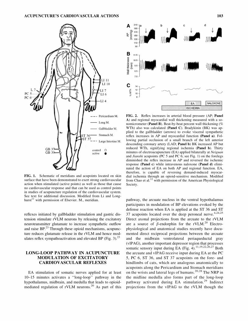
FIG. 2. Reflex increases in arterial blood pressure (AP; PanelA) and regional myocardial wall thickening measured with a so-nomicrometer (Panel B). Beat-by-beat percent wall thickening (%WTh) also was calculated (Panel C). Bradykinin (BK) was ap-plied to the gallbladder (arrows) to evoke visceral sympatheticreflex increases in AP and myocardial function (Panel a). Fol-lowing partial occlusion of a small branch of the left anteriordescending coronary artery (LAD; Panel b) BK increased AP butreduced WTh, signifying regional ischemia (Panel b). Thirtyminutes of electroacupuncture (EA) applied bilaterally at Neiguanand Jianshi acupoints (PC 5 and PC 6; see Fig. 1) on the forelegsdiminished the reflex increase in AP and reversed the ischemicresponse (Panel c) while intravenous naloxone (Panel d) elimi-nated the action of EA on both AP and regional function. EA,therefore, is capable of reversing demand-induced myocar-dial ischemia through an opioid-sensitive mechanism. Modifiedfrom Chao et al.12 with permission of the American PhysiologicalSociety.
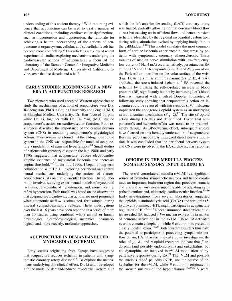
FIG. 1. Schematic of meridians and acupoints located on skinsurface that have been demonstrated to exert strong cardiovascularaction when stimulated (active points) as well as those that causeno cardiovascular response and that can be used as control pointsin studies of acupuncture regulation of the cardiovascular system.See text for additional discussion. Modified from Li and Long-hurst11 with permission of Elsevier. M., meridian.
ACUPUNCTURE’S CARDIOVASCULAR ACTIONS 103
NRP are responsible for EA modification of reflex in-
creases in BP.30 Activation of the long-loop pathway
through the arcuate, vlPAG, and NRP thus is critical
for EA opioid–associated modulation of rVLM pre-
sympathetic neurons and differentiates EA’s prolonged
sympathoinhibitory action from the brief neural-
occlusion response observed during short-term somato-
sensory stimulation.14
NEUROTRANSMITTER SYSTEMSUNDERLYING ACUPUNCTURE
LONG-LOOP PATHWAY ACTIVATION
Stimulation by glutamate of both N-methyl-d-aspartate
(NMDA) and non-NMDA or a-amino-3-hydroxy-5-methyl-
4-isoxazolepropionic acid (AMPA) ionotropic receptors is
responsible for EA-evoked activity in the arcuate and
vlPAG.27,30 The reciprocal excitatory pathway between the
arcuate and the vlPAG reinforces and prolongs the action of
EA on the reflex pressor responses. Both glutamate and
acetylcholine are responsible for the excitatory connection
from the vlPAG to the arcuate.27 Conversely, glutamate but
not acetylcholine underlies excitation in the vlPAG during
arcuate stimulation. Endocannabinoids in the vlPAG reduce
the release of GABA through presynaptic CB1 receptor
stimulation during acupuncture, leading to disinhibition of
neurons that project indirectly to the rVLM (Fig. 5).31,32
Serotonin produced in EA-activated NRP neurons that pro-
ject to the rVLM, through a 5-HT1A mechanism, contributes
to EA inhibition of rVLM sympathetic premotor neurons and
acupuncture modulation of excitatory reflexes.33 In addition,
GABA and nociceptin, participate in EA-induced sym-
pathoinhibition in the rVLM.16,34
FIG. 3. Action of electroacupuncture (EA) on reflex blood pressure (BP) responses and rostral ventrolateral medullary (rVLM)extracellular glutamate concentrations (Glu) evoked reflexly by visceral afferent stimulation following application of bradykinin (BK) tothe gallbladder before and after microperfusion of naloxone. Glu was assessed with high-performance liquid chromatography fromsequential samples of dialysate recovered with microdialysis probes inserted into the rVLM. Panels A and B are controls showing thatPC 5–PC 6 needle insertion without electrical stimulation does not alter BP or rVLM Glu responses to BK. Panels C and D show thatEA significantly attenuated reflexes increases in BP and Glu evoked by visceral stimulation. Panels E and F show reversal of EAmodulation of reflex-related increases in BP and Glu by naloxone. Thus, opioids in the rVLM during EA modulate the release ofglutamate and sympathoactivation. Values are means – standard error. *p < 0.05 versus control (before BK). {p < 0.05 vs. baseline.Modified from Zhou et al.,23 with permission of the American Physiological Society. min, minutes.
104 LONGHURST
SPINAL MECHANISMS IN ACUPUNCTURE–CARDIOVASCULAR MODULATION
Transcutaneous low-frequency pulsed electromagnetic
stimulation, like acupuncture, inhibits reflex increases in BP
through a naloxone-sensitive mechanism at the spinal
level.35 Enkephalins and dynorphin appear to predominate
in spinal processing of the cardiovascular responses because
the influence of magnetic stimulation is blocked by d-
and j-, but not l-opioid antagonists administered intra-
thecally.35 Conventional EA likewise reduces visceral
sympathoexcitation through both opioid and nonopioid
(nociceptin) mechanisms in the spinal cord dorsal horn
and intermediolateral column (IML).36 EA’s dorsal horn
action implies inhibition of sensory inflow during reflex
stimulation, while EA’s action in the IML suggests that
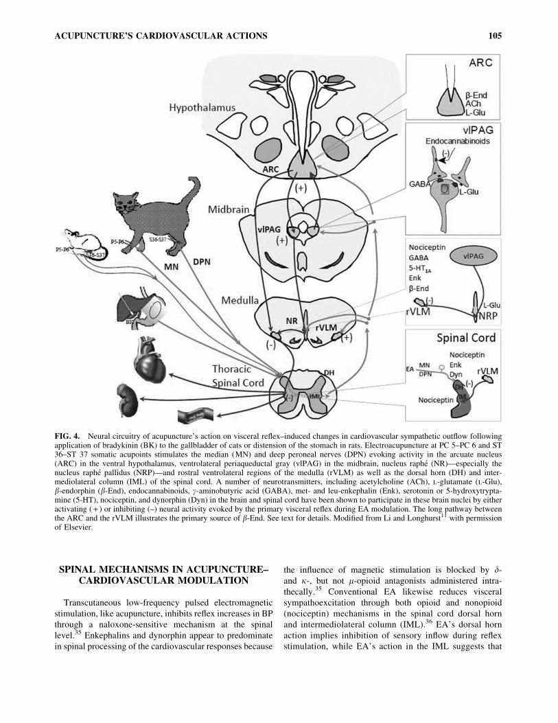
FIG. 4. Neural circuitry of acupuncture’s action on visceral reflex–induced changes in cardiovascular sympathetic outflow followingapplication of bradykinin (BK) to the gallbladder of cats or distension of the stomach in rats. Electroacupuncture at PC 5–PC 6 and ST36–ST 37 somatic acupoints stimulates the median (MN) and deep peroneal nerves (DPN) evoking activity in the arcuate nucleus(ARC) in the ventral hypothalamus, ventrolateral periaqueductal gray (vlPAG) in the midbrain, nucleus raphe (NR)—especially thenucleus raphe pallidus (NRP)—and rostral ventrolateral regions of the medulla (rVLM) as well as the dorsal horn (DH) and inter-mediolateral column (IML) of the spinal cord. A number of neurotransmitters, including acetylcholine (ACh), l-glutamate (l-Glu),b-endorphin (b-End), endocannabinoids, c-aminobutyric acid (GABA), met- and leu-enkephalin (Enk), serotonin or 5-hydroxytrypta-mine (5-HT), nociceptin, and dynorphin (Dyn) in the brain and spinal cord have been shown to participate in these brain nuclei by eitheractivating ( + ) or inhibiting (–) neural activity evoked by the primary visceral reflex during EA modulation. The long pathway betweenthe ARC and the rVLM illustrates the primary source of b-End. See text for details. Modified from Li and Longhurst11 with permissionof Elsevier.
ACUPUNCTURE’S CARDIOVASCULAR ACTIONS 105
acupuncture also modulates sympathetic outflow in the
spinal cord.
MODULATION OF LOW BPBY ACUPUNCTURE
Several studies have explored the effect of acupuncture in
various experimental models of hypotension. For example,
acupuncture partially reverses hypotension associated with
nitroprusside infusion or hemorrhage.37,38 We have used two
models to investigate the central regions and neurotransmitter
systems involved in acupuncture’s BP-raising capability.
First, we used an I.V. infusion of the 5-HT3 receptor agonist
phenylbiguanide (PBG) to stimulate cardiopulmonary vagal
afferent endings and reflexly evoke bradycardia and hypo-
tension.39–41 This model mimics vasovagal syncope, which is
thought to be caused by mechanical stimulation of cardio-
pulmonary sensory nerve endings by a hypercontractile
myocardium.42,43 We found that preganglionic cholinergic
(i.e., parasympathetic) neurons in the nucleus ambiguus in
close proximity to axons containing enkephalin are activated
by 30 minutes of EA.44 In fact, during EA, both enkephalin
and GABA in the nucleus ambiguus modulated PBG-evoked
reflex vagal bradycardia (Fig. 6).45 A second model of reflex
hypotension involved gastric distension in hypercapnia-
induced acidosis. Under these conditions, both spinal and
vagal afferent pathways are stimulated by gastric distension
to lower BP through a combination of sympathetic with-
drawal and increased parasympathetic outflow.46 Recent
observations indicate that, through GABAergic mechanisms
in the rVLM and caudal ventrolateral medulla (cVLM), EA
limits the distension-related sympathetic withdrawal, while,
in the nucleus ambiguus, EA inhibits the distension-induced
increase in parasympathetic outflow and hence reduces the
reflex hypotension and bradycardia.47 No clinical studies on
acupuncture’s BP-raising actions are available, but it seems
conceivable that acupuncture may be a therapeutic option for
patients with recurrent vasovagal syncope or perhaps other
forms of symptomatic hypotension.
Overall, consistent with Traditional Chinese Medicine
(TCM) philosophy of achieving homeostasis, acupuncture
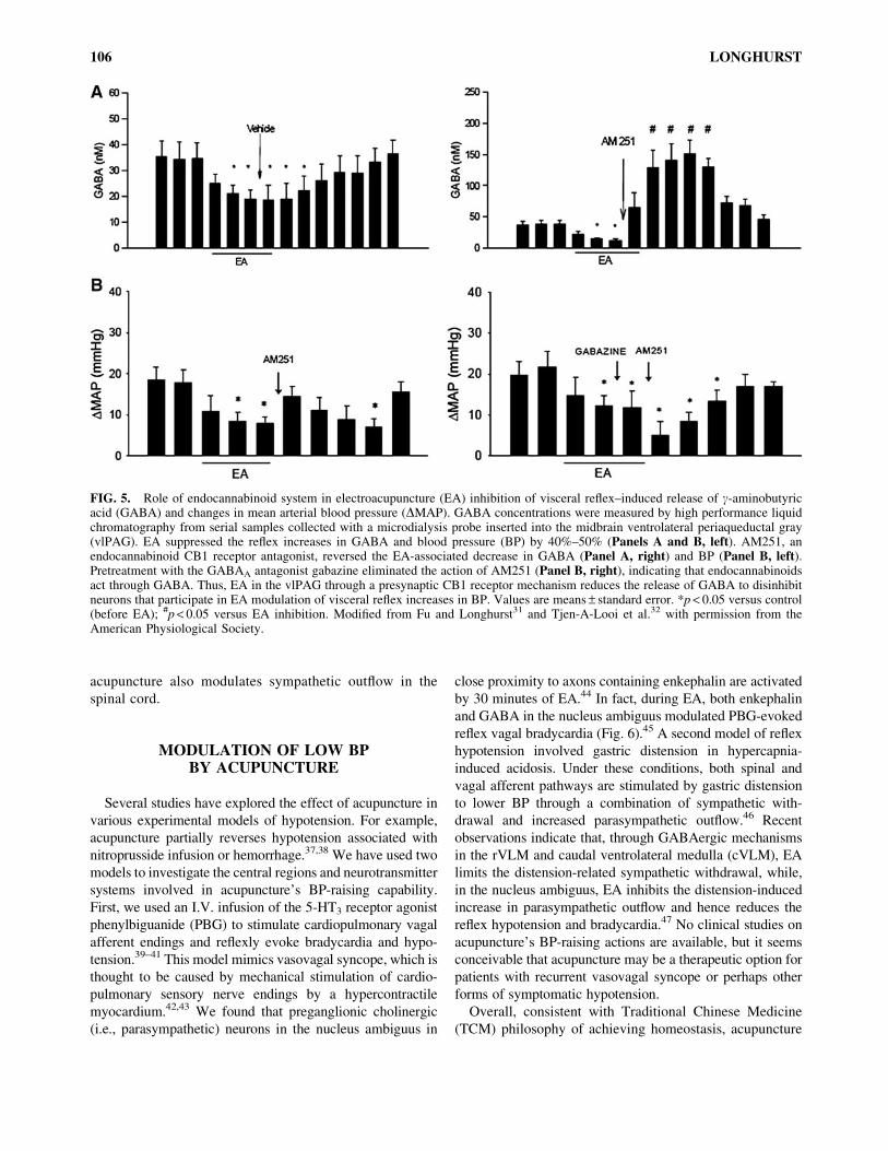
FIG. 5. Role of endocannabinoid system in electroacupuncture (EA) inhibition of visceral reflex–induced release of c-aminobutyricacid (GABA) and changes in mean arterial blood pressure (DMAP). GABA concentrations were measured by high performance liquidchromatography from serial samples collected with a microdialysis probe inserted into the midbrain ventrolateral periaqueductal gray(vlPAG). EA suppressed the reflex increases in GABA and blood pressure (BP) by 40%–50% (Panels A and B, left). AM251, anendocannabinoid CB1 receptor antagonist, reversed the EA-associated decrease in GABA (Panel A, right) and BP (Panel B, left).Pretreatment with the GABAA antagonist gabazine eliminated the action of AM251 (Panel B, right), indicating that endocannabinoidsact through GABA. Thus, EA in the vlPAG through a presynaptic CB1 receptor mechanism reduces the release of GABA to disinhibitneurons that participate in EA modulation of visceral reflex increases in BP. Values are means – standard error. *p < 0.05 versus control(before EA); #p < 0.05 versus EA inhibition. Modified from Fu and Longhurst31 and Tjen-A-Looi et al.32 with permission from theAmerican Physiological Society.
106 LONGHURST
appears to be capable of normalizing BP by lowering ele-
vated BP and elevating depressed BP. Somatic sensory
nerve evoked input during acupuncture, acting through a
number of neurotransmitter systems in several cardiovas-
cular regions of the brainstem, essentially restores altered
neuronal activity back toward a stable baseline. If, for ex-
ample, the increase in activity is predominately sym-
pathoexcitation, then acupuncture decreases the extent of
excitation associated with increased sympathetic outflow
and lowers elevated BP. However, if acupuncture is applied
in the presence of reflex sympathetic withdrawal and/or
increased parasympathetic outflow, the somatic sensory
input activates modulatory neurotransmitter systems to re-
duce the extent of hypotension and bradycardia.
PERIPHERAL SENSORY NERVOUS SYSTEMIN ACUPUNCTURE
Acupuncture needles are typically inserted at acupuncture
points (acupoints) located along meridians. Although many
studies have attempted to locate meridians anatomically
using a variety of anatomical and physiological methods, to
date, the only reproducible and scientifically valid studies
suggest that meridians are not physical entities but simply
comprise a road map that guide acupuncturists regarding
where to stimulate along the body’s surface to evoke clini-
cally meaningful responses.25,48 Nerve bundles located be-
neath meridians are responsible for the action of acupuncture,
and the only constant anatomical structures located in the
vicinity of acupoints are nerves and nerve endings.49 Thus,
local anesthetic infusion into the region of an acupoint but not
inflation of a BP cuff to suprasystolic pressures interrupts
acupuncture analgesia.50,51 Likewise, nerve transection
abolishes acupuncture modulation of excitatory cardiovas-
cular responses.52 Interruption of sensory rather than motor
nerve fibers is responsible for the influence of chemical or
surgical denervation on the acupuncture–cardiovascular re-
sponse, because motor paralysis does not influence acu-
puncture’s action.45 These studies prove that the nervous
system, particularly somatic sensory nerve fiber stimulation
underlies acupuncture’s analgesic and cardiovascular actions.
This conclusion is consistent with the clinical observation of
many TCM practitioners who ask patients if they feel the
sensation of De Qi, described by patients as a burning sen-
sation, a fullness or heaviness in the extremity or trunk where
acupuncture is applied. The therapist knows that an optimal
clinical response will not be achieved without this neural
sensation or paresthesia, thus confirming an important role
for the sensory nervous system in acupuncture treatment.
Anatomical observations using light microscopy and
multiunit recording studies suggest that myelinated sensory
nerves conduct information centrally during acupuncture
stimulation.53–55 Neither procedure is capable of evaluating
the role of small diameter Group IV afferents because they
are not easily visualized or recorded. Group IV afferents are
thought to convey nociceptive (painful) information to the
CNS and, because acupuncture is not perceived to be
painful, this sensory fiber type has not been thought to play
an important role in signaling the brain during needling.
However, single-unit afferent recording studies have dem-
onstrated that acupuncture stimulates both finely myelinated
Group III and unmyelinated Group IV afferent fibers in a
ratio of 70:30.9 Furthermore, when Group IV fibers are
destroyed by capsaicin, acupuncture’s hypotensive effect is
nearly eliminated.56 Thus, although low intensity EA
stimulates many more Group III than Group IV sensory
fibers and despite the fact that acupuncture typically is not a
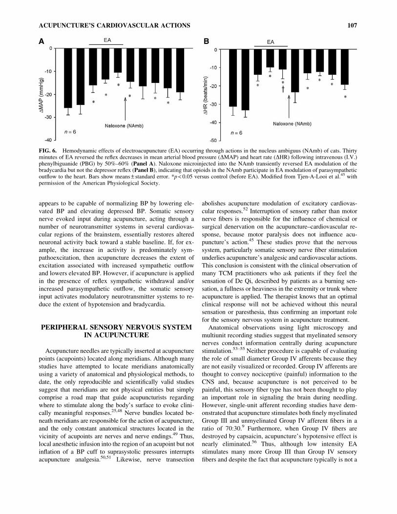
FIG. 6. Hemodynamic effects of electroacupuncture (EA) occurring through actions in the nucleus ambiguus (NAmb) of cats. Thirtyminutes of EA reversed the reflex decreases in mean arterial blood pressure (DMAP) and heart rate (DHR) following intravenous (I.V.)phenylbiguanide (PBG) by 50%–60% (Panel A). Naloxone microinjected into the NAmb transiently reversed EA modulation of thebradycardia but not the depressor reflex (Panel B), indicating that opioids in the NAmb participate in EA modulation of parasympatheticoutflow to the heart. Bars show means – standard error. *p < 0.05 versus control (before EA). Modified from Tjen-A-Looi et al.45 withpermission of the American Physiological Society.
ACUPUNCTURE’S CARDIOVASCULAR ACTIONS 107
painful stimulus, small-diameter, slowly conducting, un-
myelinated, somatic sensory fibers constitute a necessary
part of the afferent pathway for the cardiovascular actions of
somatic acupuncture.
EXPLORATION OF UNIQUE FEATURESOF ACUPUNCTURE
Manual Acupuncture Versus EA
Our University’s work has emphasized cardiovascular
responses to EA because this form of acupuncture is easy to
standardize. However, most acupuncturists use manual
acupuncture, during which needles are inserted and then
intermittently manipulated to strengthen the acupuncture
response. To compare EA with the more commonly used
manual acupuncture the magnitude and duration of acu-
puncture’s hypotensive action was evaluated during and
after 30 minutes of stimulation.57 When the two modalities
were matched for stimulation frequency (2 Hz) their impact
on elevated BP was virtually identical, probably because the
two forms of stimulation caused very similar activation of
somatic sensory nerves, which link somatic needling with
central neural modulation of sympathetic outflow.
Low-Frequency Versus High-Frequency EA
Studies of acupuncture analgesia suggest that both low-
and high-frequency EA (2 and 100 Hz, respectively) raise
the pain threshold, although to some extent in different lo-
cations in the brain and through different neurotransmitter
mechanisms. Low-frequency EA used to treat pain ap-
pears to be linked to the action of enkephalins, while high-
frequency EA is associated with dynorphin acting in dif-
ferent regions of the brain.3,58 However, a study of EA’s
action on cardiovascular function led to a different con-
clusion.57 Thus, while low-frequency (2 Hz) EA at PC 5 and
PC 6 reduced sympathoexcitatory BP responses by *40%,
neither middle- (20–40 Hz) nor high-frequency (100 Hz) EA
influenced these reflex responses. Low-frequency EA led to
much greater activation of afferent fibers than higher fre-
quencies of stimulation, indicating that, with the latter form
of stimulation, there is simply less information traveling
centrally to inhibit presympathetic activity in regions such
as the rVLM. Differences between studies from China
showing that high-frequency EA modulates pain effectively
and our studies demonstrating that there is no discernible
influence of high-frequency acupuncture on elevated BP
presently cannot be reconciled. Further research is war-
ranted.
Acupoint Combinations
The first treatise on acupuncture, the Inner Classic of
the Yellow Emperor, published between 100 and 200 bc
described 160 acupoints.59 This number has been gradually
expanded first to 349 in the A–Z Classic of Acupuncture and
Moxibustion published in 300 ad, and, more recently, to 361
acupoints in modern textbooks.60,61 Clinical acupuncture
typically involves stimulation of several acupoints in com-
bination, presumably to reinforce and increase acupunc-
ture’s action.57,62 Early studies on acupuncture’s role in pain
suggested that using a combination of two points, for ex-
ample Hoku or Hegu (LI 4) located along the Large Intestine
meridian between the thumb and the first finger (over
branches of the radial and median nerves) and Zusanli (ST
36) along the Stomach meridian on the lateral leg just below
the knee over the deep peroneal nerve, produced a greater
effect using either one acupoint alone.63 However, using EA
to control the magnitude and frequency of stimulation pre-
cisely—and hence input to the CNS—we found that bilat-
eral stimulation of two combinations of acupoints (PC 5–PC
6 and ST 36–ST 37)—which evoke strong cardiovascular
responses independently—does not evoke larger decreases
in elevated BP than stimulation of each individual set of
acupoints.15,57 More studies on the potential additive or
synergistic effects of stimulating combinations of acu-
points—including, for example, combinations of somatic
and auricular acupoints—to maximize clinical responses are
needed.
Point Specificity
An important concept in TCM is point specificity, which
implies that stimulation of some acupoints are important for
addressing certain clinical conditions, whereas other acu-
points are less effective or are ineffective. A systematic
review of 12 studies was designed to answer the question:
‘‘Are acupoints specific for diseases?’’ The reviewers con-
cluded that approximately half of the trials produced evi-
dence for point specificity and half did not.64 However, a
number of trials included in this review were biased. Five
with a low risk of bias showed no difference between sham
and true acupuncture. Thus, support for the concept of point
specificity has been weak and several questions have
emerged. First, if point specificity does not exist, can ap-
propriate controls that incorporate acupoints, which are in-
active in certain conditions, be developed for acupuncture?
Use of such controls to assess sham actions of acupuncture
allows rigorous investigation of its point-specific clinical
actions, an underlying tenet of acupuncture philosophy.
However, there are many studies in the acupuncture litera-
ture that either do not incorporate control stimulation or that
use weak controls.65 Second, are rigorous studies available
that show clear point-specific responses and, if such studies
have been conducted, what was the underlying mechanism
of point specificity? Dr. Han argues from a neurobiological
perspective that there is an uneven distribution of nerves
along the body, so it is irrational to assume that needling
different places would elicit the same response.3 Furthermore,
108 LONGHURST
it is unlikely that all sensory neurons project identically to
centers in the brain and, as such, stimulation of different
acupoints along separate neural pathways should evoke
quite different acupuncture responses. In a study examin-
ing potential answers these questions from a cardiovas-
cular perspective, we showed that point-specific responses
to EA at different acupoints exist.15 Stimulation of some
points results in significant reductions in elevated BP,
while others cause more-modest changes or no change at
all. In general, stimulation of acupoints (PC 5, PC 6, ST 36,
ST 37, LI 4, LI 10, and LI 11; Fig. 1) located over deep
somatic nerves, such as the median or deep peroneal
nerves, reduced elevated BP, whereas EA at acupoints (LI
6, LI 7, KI 1, BL 67; Fig. 1) located over superficial
(cutaneous) nerves, such as the superficial radial and tib-
ial nerves, have produced little cardiovascular effect.11
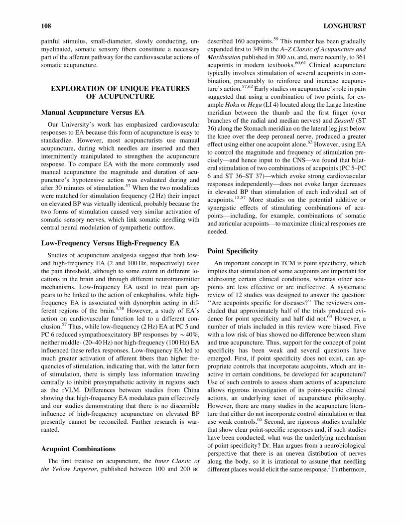
Stimulation of nerves underlying acupoints that reduced
elevated BP the most during EA evoked the greatest rVLM
discharge activity (Fig. 7) suggesting that ‘‘hard wiring’’
of somatic nerves that project indirectly to regions of the
brain concerned with regulation of sympathetic outflow
underlies the capability of certain acupoints to lower BP
effectively.15 As noted above in the first question posed,
our study also has implications for selection of effective
controls for future studies of acupuncture in both experi-
mental and clinical situations. We have found that it is
possible to use either (1) inactive acupoints or (2) active
acupoints in which a needle is placed but not stimulated as
two strong controls, which can be compared with re-
sponses to stimulation of active acupoints.57 In the former
paradigm, there is little input to cardiovascular centers of
the brain, while, in the latter, there is brief, transient but not
sustained sensory stimulation and hence a negligible in-
fluence of sham acupuncture over and above placebo. Yet,
in both cases, acupuncture needles are inserted and un-
derlying neural pathways are stimulated (at least briefly) to
evoke De Qi, which cannot be differentiated by patients.
Certainly, the debate over the existence of point specificity
is likely to continue. It is difficult to resolve this question in
humans in whom placebo may play a role during acu-
puncture, because stimulation of any point is perceived
consciously and may be believed by patients to potentially
offer relief. However, it does seem clear, from experi-
mental and even clinical studies, such as in our laboratory,
that point-specific responses can occur when the acupunc-
ture stimulus and experimental paradigm are controlled
carefully.66
DISCUSSION
Despite tremendous advances in understanding of how
acupuncture works, a number of unanswered or partially
answered questions remain in acupuncture research.
Are Experimental Studies Applicable to ClinicalAcupuncture?
The answer is probably ‘‘yes.’’ As shown by our exper-
imental findings, application of EA does not alter BP when
it is not elevated but does reduce exercise-associated pressor
responses in human subjects.67 Currently, preliminary on-
going studies of acupuncture in patients with mild-to-
moderate hypertension suggest that using acupoints dem-
onstrated experimentally to have the greatest influence on
reflex elevations in BP—and using a stimulus paradigm that
is most effective (low-frequency, low-intensity EA bilater-
ally at PC 5, PC 6, ST 36 and ST 37 applied once weekly for
30 minutes)—reduces systolic and, to a lesser extent, dia-
stolic arterial BP by 8–12 mmHg in *70% of patients
studied.66 The reduction in BP is slow in onset, beginning
2–4 weeks after initiating EA, and is prolonged in duration,
extending for several weeks after termination of an 8-week
trial of acupuncture. More patients need to be evaluated and
compared to control acupuncture involving treatment at
inactive cardiovascular acupoints, to verify that the ob-
served responses are not a placebo effect. In addition, on-
going experimental studies of nonanesthetized hypertensive
conditions (conscious rats in a cold environment) subjected
to repeated acupuncture may provide additional clues about
how to best apply acupuncture clinically.
FIG. 7. Relationship between changes in mean arterial bloodpressure (MAP) and evoked activity displayed as impulses (imp)in the rostral ventrolateral medulla (rVLM) during electro-acupuncture (EA) modulation of visceral sympathoexcitation.EAwas maintained at low frequency (2 Hz) for 30 minutes, whileevoked activity in the rVLM consisted of needle stimulation at thesame acupoints and frequency for 15 seconds. A strong correlationwas observed between acupoints that evoked large increases inrVLM activity and decreases in the reflex increases in MAP (e.g.,PC 5–PC 6) as well as those that evoked little activity in thismedullary region and did not influence the reflex sympathoexci-tation, indicating that acupoints overlying nerves that project to aregion of the brain that is known to regulate sympathetic activityare most capable of influencing cardiovascular function throughtheir actions on autonomic outflow. See Fig. 1 and text for ex-planation of acupoint nomenclature. Stim, stimulation. Modifiedfrom Tjen-A-Looi et al.15 with permission of the American Phy-siological Society.
ACUPUNCTURE’S CARDIOVASCULAR ACTIONS 109
Why Does Acupuncture Have a Prolonged Action?
Experimental studies involving anesthetized prepara-
tions demonstrate that cardiovascular hypotensive re-
sponses to acupuncture typically last for 1–1.5 hours
beyond the period of stimulation.16 As discussed above,
this prolonged action in response to 30 minutes of stimu-
lation, in part, is related to activation of a long-loop
hypothalamic–midbrain–medullary pathway, which leads
to the release of a number of inhibitory neurotransmitters
that ultimately modify sympathetic outflow.25 Within the
long-loop pathway reciprocal excitatory projections be-
tween the arcuate nucleus in the hypothalamus and the
vlPAG in the midbrain reinforce and prolong the
acupuncture-evoked somatic input and hence EA’s action
on elevated BP.19,22,26,27 Also, both opioids and GABA in
the rVLM participate in the prolonged response to a single
application of acupuncture in anesthetized animals.16
However, additional mechanisms underlying EA’s pro-
longed action appear to be operative when acupuncture is
applied repeatedly over a period of days, weeks, or months.
In this regard, a single application of acupuncture over 30
minutes stimulates preproenkephalin mRNA expression,
the precursor of enkephalin, for a period lasting 90 min-
utes.68 Repeated acupuncture over several days causes
longer elevations of the message and protein (enkephalin)
expression, lasting hours or days after acupuncture is
terminated.44 So, through transcriptional and possibly
translational regulation of the precursors of modulatory
neurotransmitters, such as enkephalins, repeated acupuncture
exerts a very prolonged action on BP. More experimental and
clinical studies are needed to determine how often acu-
puncture treatment must be reinforced to continue to suppress
elevated BP after an initial period of application that lowers it
effectively.66 Our current hypothesis is that continued treat-
ment can maintain low BP effectively, when it treatment is
given once or twice each month.
How Can Acupuncture EffectivenessBe Improved?
Studies from a number of groups studying acupuncture
analgesia indicate that *70% of patients respond to
acupuncture.62,69–72 We have made similar observations in
studies evaluating cardiovascular responses to acupunc-
ture.66,67 But why are some individuals unresponsive to
acupuncture treatment? Recent preliminary investigations
in the University’s laboratory suggest that nonresponders
can be converted to responders by administering an antag-
onist to the octapeptide of cholecystokinin.73 Cholecystoki-
nin (CCK) is produced in the intestine, where CCK delays
gastric emptying, contracts the gallbladder, releases bile and
causes secretion of pancreatic enzymes. The octapeptide of
CCK or CCK-8 is distributed widely throughout the
brain.74,75–77 CCK in the brain antagonizes the action of
morphine, leading to tolerance, a form of nonresponsive-
ness.78–80 CCK-8 exerts an antiopioid effect in the brain.78,81
Molecular studies suggest that CCK, through a CCK-A re-
ceptor mechanism in the hypothalamus, may reduce
responsiveness to EA.82–84 Recent preliminary studies sug-
gest that CCK-8 in the rVLM, through a CCK-A receptor
mechanism, contributes to the absence of EA’s antihyper-
tensive action in rats.73 These studies may provide clues for
conversion of some EA nonresponders into responders.
CONCLUSIONS
Laboratories in China and the United States have begun to
elucidate the mechanisms through which acupuncture re-
duces pain and cardiovascular dysfunction, including myo-
cardial ischemia, hypertension, and hypotension. Over the
last two decades, substantial information has been developed
to show that acupuncture acts mainly through the peripheral
nervous system and the CNS. It is now known that acu-
puncture leads to the release of a number of excitatory and
inhibitory neurotransmitters in the CNS to alter processing of
sensory information and, ultimately, autonomic outflow, and
hence cardiovascular function. It has been exciting to play a
role in developing new knowledge of the mechanisms of
acupuncture’s cardiovascular actions. However, while the
information base has been considerably expanded, much
work remains to be accomplished to understand fully the
clinical actions of this ancient therapy.
ACKNOWLEDGMENTS
The authors thank the National Institutes of Health (for
grants R01-HL63313 and R01-HL72125), the Adolph Coors
Foundation, the Peterson Family Foundation, the Larry K.
Dodge Chair of Integrative Biology, and the Samueli
Chair of Integrative Medicine for their support of the re-
search described in this review. The assistance of Stephanie
Tjen-A-Looi, PhD, in preparing some of the figures and
Melissa Chiang, BS, for her help with the manuscript is
appreciated.
DISCLOSURE STATEMENT
No competing financial interests exist.
REFERENCES
1. Han J-S, Ho YS. Global trends and performances of acupunc-
ture research. Neurosci Biobehav Rev. 2011;35(3):680–687.
2. World Health Organization. Acupuncture: Review and Ana-
lysis of Reports on Controlled Clinical Trials [serial online].
110 LONGHURST
2003. Online document at: http://apps.who.int/medicinedocs/
pdf/s4926e/s4926e.pdf Accessed February 12, 2013.
3. Han J-S. Acupuncture analgesia: Areas of consensus and
controversy. Pain. 2011;152(3):S41–S81.
4. Li P, Yao T. Pressor effect of electroacupuncture or somatic
nerve stimulation on experimental hypotension. In: Mechan-
ism of the Modulatory Effect of Acupuncture on Abnormal
Cardiovascular Functions. Shanghai, China: Shanghai Med-
ical University Press; 1992:32–40.
5. Ballegaard S, Meyer CN, Trojaborg W. Acupuncture in an-
gina pectoris: Does acupuncture have a specific effect? J In-
tern Med. 1991;229(4):357–362.
6. Ballegaard S, Pedersen F, Pietersen A, et al. Effects of acu-
puncture in moderate stable angina pectoris: A controlled
study. J Intern Med. 1990;227(1):25–30.
7. Ballegaard S, Jensen G, Pedersen F, et al. Acupuncture in
severe, stable angina pectoris: A randomized trial. Acta Med
Scand. 1986;220(4):307–313.
8. Richter A, Herlitz J, Hjalmarson A. Effect of acupuncture in
patients with angina pectoris. Eur Heart J. 1991;12(2):
175–178.
9. Li P, Pitsillides KF, Rendig SV, et al. Reversal of reflex-
induced myocardial ischemia by median nerve stimulation: A
feline model of electroacupuncture. Circulation. 1998;97(12):
1186–1194.
10. Pitsillides KF, Longhurst JC. An ultrasonic system for mea-
surement of absolute myocardial thickness using a single
transducer. Am J Physiol. 1995;37(3):H1358–H1367.
11. Li P, Longhurst JC. Neural mechanism of electroacupuncture’s
hypotensive effects. Auton Neurosci. 2010;157:24–30.
12. Chao DM, Shen LL, Tjen-A-Looi SC, et al. Naloxone re-
verses inhibitory effect of electroacupuncture on sympathetic
cardiovascular reflex responses. Am J Physiol. 1999;276(45):
H2127–H2134.
13. Tjen-A-Looi SC, Li P, Longhurst JC. Midbrain vIPAG inhibits
rVLM cardiovascular sympathoexcitatory responses during
acupuncture. Am J Physiol. 2006;290(6):H2543–H2553.
14. Tjen-A-Looi SC, Li P, Longhurst JC. Prolonged inhibition of
rostral ventral lateral medullary premotor sympathetic neuron
by electroacupuncture in cats. Auton Neurosci. 2003;106(2):
119–131.
15. Tjen-A-Looi SC, Li P, Longhurst JC. Medullary substrate and
differential cardiovascular response during stimulation of
specific acupoints. Am J Physiol. 2004;287(4):R852–R862.
16. Tjen-A-Looi SC, Li P, Longhurst JC. Role of medullary
GABA, opioids, and nociceptin in prolonged inhibition of
cardiovascular sympathoexcitatory reflexes during electro-
acupuncture in cats. Am J Physiol. 2007;293(6):H3627–H3635.
17. Huangfu D, Li P. Role of nucleus raphe obscurus in the in-
hibition of defence reaction by deep peroneal nerve stimula-
tion. Chin J Physiol Sci. 1988;4(1):77–83.
18. Zhang Z, Lin RJ, Fan W, et al. Involvement of 5-HT and
GABA in the inhibition induced by NRO stimulation of
rVLM-defense-reaction-related neurons. Chin J Physiol.
1992;8(3):208–215.
19. Guo Z-L, Moazzami AR, Longhurst JC. Electroacupuncture
induces c-Fos expression in the rostral ventrolateral medulla
and periaqueductal gray in cats: Relation to opioid containing
neurons. Brain Res. 2004;1030(1):103–115.
20. Li P, Tjen-A-Looi SC, Guo ZL, et al. Long-loop pathways in
cardiovascular electroacupuncture responses. J Appl Physiol.
2009;106(2):620–630.
21. Li P, Tjen-A-Looi SC, Longhurst JC. Rostral ventrolateral
medullary opioid receptor subtypes in the inhibitory effect of
electroacupuncture on reflex autonomic response in cats.
Autonomic Neuroscience. 2001;89(1–2):38–47.
22. Guo Z, Longhurst J. Expression of c-Fos in arcuate nucleus
induced by electroacupuncture: Relations to neurons con-
taining opioids and glutamate. Brain Res. 2007;1166:65–76.
23. Zhou W, Fu L-W, Guo ZL, et al. Role of glutamate in rostral
ventrolateral medulla in acupuncture-related modulation of
visceral reflex sympathoexcitation. Am J Physiol.
2007;292(4):H1868–H1875.
24. Cheung L, Li P, Wong C. The Mechanism of Acupuncture
Therapy and Clinical Case Studies. New York: Taylor and
Francis; 2001.
25. Li P, Tjen-A-Looi SC, Longhurst JC. Acupuncture’s role in
cardiovascular homeostasis. In: Xia Y, Dong G, Wu G-C, eds.
Current Research in Acupuncture. New York: Springer
Science + Business Media; 2012:457–486.
26. Guo Z-L, Longhurst JC. Activation of reciprocal pathways
between arcuate nucleus and ventrolateral periaqueductal
gray during electroacupuncture: Involvement of VGLUT3.
Brain Res. 2010;1360:77–88.
27. Li P, Tjen-A-Looi SC, Guo ZL, et al. An arcuate-ventrolateral
periaqueductal gray reciprocal circuit participates in electro-
acupuncture cardiovascular inhibition. Auton Neurosci.
2010;158(1–2):13–23.
28. Li P, Tjen-A-Looi SC, Longhurst JC. Excitatory projections
from arcuate nucleus to ventrolateral periaqueductal gray in
electroacupuncture inhibition of cardiovascular reflexes. Am J
Physiol. 2006;209(6):H2535–H2542.
29. Guo Z-L, Moazzami A, Tjen-A-Looi S, et al. Responses of
opioid and serotonin containing medullary raphe neurons to
electroacupuncture. Brain Res. 2008;1229:125–136.
30. Li P, Tjen-A-Looi SC, Longhurst JC. Nucleus raphe pallidus
participates in midbrain–medullary cardiovascular sym-
pathoinhibition during electroacupuncture. Am J Physiol
Regul Integr Comp Physiol. 2010;299(5):R1369–R1376.
31. Fu L-W, Longhurst C. Electroacupuncture modulates vlPAG
release of GABA through presynaptic cannabinoid CB1 re-
ceptor. J Appl Physiol. 2009;106(6):1800–1809.
32. Tjen-A-Looi SC, Li P, Longhurst JC. Processing cardiovas-
cular information in the vlPAG during electroacupuncture in
rats: Roles of endocannabinoids and GABA. J Appl Physiol.
2009;106(6):1793–1799.
33. Moazzami A, Tjen-A-Looi SC, Guo Z-L, et al. Serotonergic
projection from nucleus raphe pallidus to rostral ventrolateral
medulla modulates cardiovascular reflex responses during
acupuncture. J Appl Physiol. 2010;108(5):1336–1346.
34. Crisostomo M, Li P, Tjen-A-Looi SC, et al. Nociceptin in
rVLM mediates electroacupuncture inhibition of cardiovas-
cular reflex excitatory response in rats. J Appl Physiol.
2005;98(6):2056–2063.
35. Zhou W, Hsiao I, Lin V, et al. Modulation of cardiovascular
excitatory responses in rats by transcutaneous magnetic
stimulation: Role of the spinal cord. J Appl Physiol.
2006;100(3):926–932.
ACUPUNCTURE’S CARDIOVASCULAR ACTIONS 111
36. Zhou W, Mahajan A, Longhurst JC. Spinal nociceptin me-
diates electroacupuncture-related modulation of visceral
sympathoexcitatory reflex responses in rats. Am J Physiol.
2009;297(2):H859–H865.
37. Xiao YF, Lin SX, Deng ZF, et al. Mechanism of pressor
effect of electroacupuncture on nitroprusside hypotension in
dogs [in Chinese]. Acta Physio Sin. 1983;35:257–263.
38. Syuu Y, Matsubara H, Hosogi S, et al. Pressor effect of
electroacupuncture on hemorrhagic hypotension. Am J Phy-
siol Heart Circ Physiol. 2003;285(6):R1446–R1452.
39. Coleridge H, Coleridge J. Cardiovascular afferents involved in
regulation of peripheral vessels. Annu Rev Physiol. 1980;42:
413–427.
40. Fu L-W, Longhurst JC. Reflex pressor response to arterial
phenylbiguanide: Role of abdominal sympathetic visceral
afferents. Am J Physiol. 1998;275(6):H2025–H2035.
41. Jeggo R, Kellett D, Wang Y, et al. The role of central 5-HT3
receptors in vagal reflex inputs to neurones in the nucleus
tractus solitarius of anaesthetized rats. J Physiol. 2005;566(3):
939–953.
42. Calkins H, Zipes DP. Hypotension and syncope. In: Libby P,
Bonow RO, Mann D, et al., eds. Braunwald’s Heart Disease:
A Textbook of Cardiovascular Medicine. Philadelphia:
Saunders Elsevier; 2008:975–984.
43. Kapa S, Somers VK. Cardiovascular Manifestations of Auto-
nomic Disorders. In: Libby P, Bonow RO, Mann D, et al., eds.
Braunwald’s Heart Disease: A Textbook of Cardiovascular
Medicine. Philadelphia: Saunders Elsevier; 2008:2171–2183.
44. Guo Z-L, Li M, Longhurst J. Nucleus ambiguus cholinergic
neurons activated by acupuncture: Relation to enkephalin.
Brain Res. 2012;1442:25–35.
45. Tjen-A-Looi SC, Li P, Li M, et al. Modulation of cardio-
pulmonary depressor reflex in nucleus ambiguus by electro-
acupuncture: Roles of opioids and gamma aminobutyric acid.
Am J Physiol. 2012;302(7):R833–R844.
46. Tjen-A-Looi SC, Hsiao AF, Longhurst JC. Central and pe-
ripheral mechanisms underlying gastric distention inhibitory
reflex responses in hypercapnic-acidotic rats. Am J Physiol.
2011;300(3):H1003–H1012.
47. Tjen-A-Looi SC, Li P, Hsiao A-F, et al. Central processing by
electroacupuncture of cardiovascular reflex vasodepression
[abstr]. FASEB J. 2012;26(702):2.
48. Longhurst J.C. Defining meridians: A modern basis of un-
derstanding. J Acupunct Meridian Stud. 2010;3(2):67–74.
49. Yu AS, Ahao YX, Li XL, et al. Morphological research in
Neiguan (Pe 6)’s three dimensional structure. Shanghai J
Acup Moxibustion. 1996;15:30–31.
50. Chiang CY, Chang HT, Cicero TJ, et al. Peripheral afferent
pathway for acupuncture analgesia. Scientia Sinica. 1973;16:
210–217.
51. Han J-S. The Neurochemical Basis of Pain Relief by Acu-
puncture. Beijing: China Medical and Pharmaceutical Tech-
nology; 1987.
52. Kline RL, Yeung KY, Calaresu FR. Role of somatic nerves in
the cardiovascular responses to stimulation of an acupuncture
point in anesthetized rabbits. Exp Neurol. 1978;61(3):561–570.
53. Toda I, Ichioka M. Electroacupuncture: Relations between
forelimb afferent impulses and suppression of jaw-opening
reflex in the rat. Exp Neurol. 1978;61(2):465–470.
54. Pomeranz B, Paley D. Electroacupuncture hyperalgesia is
mediated by afferent nerve impulses: An electrophysiological
study in mice. Exp Neurol. 1979;66(2):398–402.
55. Lu G-W. Neurobiologic research on acupuncture in China as
exemplified by acupuncture analgesia. Anesth Analg.
1983;62(3):335–340.
56. Tjen-A-Looi SC, Fu L-W, Zhou W, et al. Role of unmy-
elinated fibers in electroacupuncture cardiovascular re-
sponses. Auton Neurosci. 2005;118(1–2):43–50.
57. Zhou W, Fu L-W, Tjen-A-Looi SC, et al. Afferent mecha-
nisms underlying stimulation modality-related modulation of
acupuncture-related cardiovascular responses. J Appl Physiol.
2005;98(3):872–880.
58. Wang JQ, Mao L, Han JS. Comparison of the antinociceptive
effects induced by electroacupuncture and transcutaneous
electrical nerve stimulation in the rat. Int J Neurosci.
1992;65(1–4):117–129.
59. Huangdi Neijing: The Yellow Emperor’s Inner Canon: Basic
Questions. Beijing: Renmin Weisheng Press; 1983.
60. Jia Zhenjiu Yi Jing: A–Z Classic of Acupuncture and Mox-
ibustion. Beijing: Renmin Weisheng Press; 1979.
61. The Academy of Traditional Chinese Medicine. An Outline of
Chinese Acupuncture. Beijing: Foreign Languages Press; 1975.
62. Ulett G, Han S, Han J-S. Electroacupuncture: Mechanisms and
clinical application. Biol Psychiatry. 1998;44(2):129–138.
63. Research Group of Acupuncture Analgesia BMC. The effect
of acupuncture on the human skin pain threshold. Chin Med J.
1973;3:151–157.
64. Zhang H, Bian ZX, Lin. Are acupoints specific for diseases?
A systematic review of the randomized controlled trials with
sham acupuncture controls. Chin Med. 2010;5(1):1–7.
65. Mayer DJ. Acupuncture: An evidence-based review of the
clinical literature. Annu Rev Med. 2000;51:49–63.
66. Li P, Longhurst JC. Long-lasting inhibitory effect of EA on
blood pressure in patients with mild to moderate hypertension
[abstr]. Soc Neurosci. 2007;37.
67. Li P, Ayannusi O, Reed C, et al. Inhibitory effect of electro-
acupuncture (EA) on the pressor response induced by exercise
stress. Clinical Autonomic Research. 2004;14(3):182–188.
68. Li M, Tjen ALS, Guo ZL, et al. Repetitive electroacupuncture
causes prolonged increased met-enkephalin expression in the
rVLM of conscious rats. Auton Neurosci. 2012;170(1–2):30–
35.
69. Andersson SA, Hansson G, Homgren E, et al. Evaluation of
the pain suppressive effect of different frequencies of pe-
ripheral electrical stimulation in chronic pain conditions. Acta
Orthop Scand. 1976;47(2):149–157.
70. Ng L, Katims J, Lee M. Acupuncture. In: Aronoff G, ed.
Evaluation and Treatment of Chronic Pain. Baltimore, MD:
Williams & Wilkins; 1992:291–298.
71. Thomas M, Lundeberg T. Importance of modes of acupunc-
ture in the treatment of chronic nociceptive low back pain.
Acta Anaesthesiol Scand. 1994;38(1):63–69.
72. Ulett G. Acupuncture. In: Tollison C, Kriegel M, eds. Inter-
disciplinary Rehabilitation of Low Back Pain. Baltimore,
MD: Williams & Wilkins; 1989:85–100.
73. Li M, Tjen-A-Looi S, Choi E, et al. Cholecystokinin antago-
nizes opioid function during electroacupuncture modulation of
reflex hypertension in rats [abstr]. FASEB J. 2012;26(1091):74.
112 LONGHURST
74. Larsson L, Rehfeld J. Localization and molecular heteroge-
neity of cholecystokinin in the central and peripheral nervous
system. Brain Res. 1979;165(2):201–218.
75. Ingram S, Krause II R, Baldino Jr F, et al. Neuronal locali-
zation of cholecystokinin mRNA in the rat brain by using in
situ hybridization histochemistry. J Comp Neurol. 1989;287(2):
260–272.
76. Mantyh PW, Hunt SP. Evidence for cholecystokinin-like
immunoreactive neurons in the rat medulla oblongata
which project to the spinal cord. Brain Res. 1983;291(1):
49–54.
77. Mercer LD, Le VQ, Nunan J, et al. Direct visualization of
cholecystokinin subtype2 receptors in rat central nervous sys-
tem using anti-peptide antibodies. Neurosci Lett. 2004;293(3):
167–170.
78. Noble F, Derrien M, Roques BP. Modulation of opioid anti-
nociception by CCK at the supraspinal level: Evidence of
regulatory mechanisms between CCK and enkephalin systems
in the control of pain. Br J Pharmacol. 1993;109(4):1064–
1070.
79. Tang NM, Dong HW, Wang XM, et al. Cholecystokinin
antisense RNA increases the analgesic effect induced by
electroacupuncture or low dose morphine: Conversion of
low responder rats to high responders. Brain Res Mol Brain
Res. 1997;71(1):71–80.
80. Zhao ZQ. Neural mechanism underlying acupuncture anal-
gesia. Prog Neurobiol. 2008;85(4):355–375.
81. Heinricher MM, McGaraughty S, Tortorici V. Circuitry un-
derlying antiopioid actions of cholecystokinin within the
rostral ventromedial medulla. J Neurophysiol. 2001;85(1):
280–286.
82. Kim S, Moon H, Park J, et al. The maintenance of individual
differences in the sensitivity of acute and neuropathic pain
behaviors to electroacupuncture in rats. Brain Res Bull.
2007;74(5):357–360.
83. Ko ES, Kim SK, Kim JT, et al. The difference in mRNA
expressions of hypothalamic CCK and CCK-A and -B re-
ceptors between responder and non-responder rats to high
frequency electroacupuncture analgesia. Peptides. 2006;
27(7):1841–1845.
84. Lee G, Rho S, Shin M, et al. The association of cholecysto-
kinin-A receptor expression with the responsiveness of elec-
troacupuncture analgesic effects in rat. Neurosci Lett.
2002;325(1):17–20.
Address correspondence to:
John Longhurst, MD, PhD
Samueli Center for Integrative Medicine
University of California, Irvine
Medical Sciences C, Room C240
821 Health Sciences Road
Irvine, CA 92697
E-mail: [email protected]
ACUPUNCTURE’S CARDIOVASCULAR ACTIONS 113