Embed Size (px)
Citation preview
The hornworts belong to a group of ancient land
plants that emerged about 450 million years ago and that
includes, according to different sources, from 200 to 300
species [1]. Until recently, the hornworts were considered
as a class of mosses [2], but now they are considered as a
separate division in plant classification [3, 4]. Due to the
cytological and morphological similarity of hornworts to
both algae and higher plants, they have become the sub-
ject of particular interest for molecular biologists and
geneticists [3, 5]. For example, hornworts have chloro-
plasts characteristic of algae with central pyrenoid con-
taining ribulose biphosphate carboxylase and, hence,
have a carbon assimilation mechanism unique for land
plants [1]. Unlike the liverworts, which have symbiosis
with blue-green algae only in four orders out of more than
340, in hornworts symbiosis was found among representa-
tives of all 14 orders [1, 6]. From a practical point of view,
the hornworts are investigated as sources of rosmarinic
acid and other biologically active compounds [2].
Phylogenetic and structural analyses suggest that evolu-
tionarily hornworts might be a sister group to modern vas-
cular plants [3, 5]. It is likely that the ancient hornwort-
like plants were intermediate upon transition from organ-
isms with life cycle in which the generation with haploid
gametophyte dominates to the organisms with diploid
sporophyte [5].
The majority of hornworts inhabit disrupted and
grass-free locations, being the first to occupy an open
moist soil. At the same time, it is known that the horn-
worts are extremely drought resistant. Unlike the vegeta-
tive organs of higher vascular plants that are extremely
sensitive to dehydration, the hornwort thallus exhibits
phenomenal ability to retain viability after the loss of 95%
of water [7, 8]. Due to their high tolerance to the action
of adverse environmental factors, the hornworts represent
a unique model for investigation of mechanisms for the
formation of resistance in higher plants. Despite the obvi-
ISSN 0006-2979, Biochemistry (Moscow), 2015, Vol. 80, No. 9, pp. 1157-1168. © Pleiades Publishing, Ltd., 2015.
Original Russian Text © A. V. Chasov, R. P. Beckett, F. V. Minibayeva, 2015, published in Biokhimiya, 2015, Vol. 80, No. 9, pp. 1391-1404.
Originally published in Biochemistry (Moscow) On-Line Papers in Press, as Manuscript BM15-017, June 21, 2015.
1157
Abbreviations: CW, cell wall; DOPA, 3,4-dihydroxyphenylala-
nine; ECS, extracellular solution; О2�, superoxide anion-radi-
cal; PIB, post-infiltration buffer; ROS, reactive oxygen species;
SOD, superoxide dismutase; XTT, 2,3-bis-(2-methoxy-4-
nitro-5-sulfophenyl)-2H-tetrazolium-5-carboxanilide.
* To whom correspondence should be addressed.
Activity of Redox Enzymes in the Thallus of Anthoceros natalensis
A. V. Chasov1*, R. P. Beckett2, and F. V. Minibayeva1
1Kazan Institute of Biochemistry and Biophysics, Kazan Scientific Center, Russian Academy of Sciences,
420111 Kazan, Russia; fax: (843) 292-7347; E-mail: [email protected] of Life Sciences, University of KwaZulu-Natal, Private Bag X01,
Pietermaritzburg, Scottsville 3209, South Africa; fax: +27-33-260-5105
Received January 16, 2015
Revision received April 15, 2015
Abstract—Anthocerotophyta (hornworts) belong to a group of ancient nonvascular plants and originate from a common
ancestor with contemporary vascular plants. Hornworts represent a unique model for investigating mechanisms of forma-
tion of stress resistance in higher plants due to their high tolerance to the action of adverse environmental factors. In this
work, we demonstrate that the thallus of Anthoceros natalensis exhibits high redox activity changing under stress.
Dehydration of the thallus is accompanied by the decrease in activities of intracellular peroxidases, DOPA-peroxidases, and
tyrosinases, while catalase activity increases. Subsequent rehydration results in the increase in peroxidase and catalase activ-
ities. Kinetic features of peroxidases and tyrosinases were characterized as well as the peroxidase isoenzyme composition of
different fractions of the hornwort cell wall proteins. It was shown that the hornwort peroxidases are functionally similar to
peroxidases of higher vascular plants including their ability to form superoxide anion-radical. The biochemical mechanism
was elucidated, supporting the possible participation of peroxidases in the formation of reactive oxygen species (ROS) via
substrate–substrate interactions in the hornwort thallus. It has been suggested that the ROS formation by peroxidases is an
evolutionarily ancient process that emerged as a protective mechanism for enhancing adaptive responses of higher land
plants and their adaptation to changing environmental conditions and successful colonization of various ecological niches.
DOI: 10.1134/S0006297915090060
Key words: hornwort, peroxidase, superoxide, catalase, tyrosinases, dehydration, rehydration
1158 CHASOV et al.
BIOCHEMISTRY (Moscow) Vol. 80 No. 9 2015
ous importance, the biochemical mechanisms of resist-
ance including kinetic characteristics of redox reactions
in plants remain topics of discussions. In particular, elu-
cidation of the dual role of peroxidases and switching
from the antioxidant functioning mode to the prooxidant
one is still a hot topic. In the cells of vascular plants such
as, for example, wheat roots, the apoplastic peroxidases
capable of both decomposition of H2O2 and formation of
reactive oxygen species (ROS) are among the key
enzymes of redox metabolism under stress [9, 10]. This
multifunctionality of peroxidases as well as a multitude
of isoforms and genes encoding them defines the impor-
tant role of peroxidases in plant resistance. Despite
intensive investigation of the reaction chemistry and evo-
lution of the peroxidase structures [11-13], the possible
mechanisms and factors facilitating ROS-forming activ-
ity of the peroxidases are still unsolved. Investigation of
previously unknown features of peroxidases from horn-
worts that are on a lower level of evolution in comparison
with vascular plants is a promising approach for solving
this problem.
An increase in ROS level can occur under stress in
the cells of bryophytes similarly to that in the majority of
plants. Previously we showed for the first time that a
number of lichens, mosses, as well as liverworts and horn-
worts demonstrate high redox activity such as formation
of superoxide anion-radicals (О2�) [8]. It is remarkable
that the highest activity was observed in the thallus of
Anthoceros natalensis Steph. Despite obvious importance,
the biochemical mechanisms of resistance mediated by
redox enzymes including peroxidases in hornwort cells
are practically unstudied. In this context, the aim of this
study was to analyze the kinetic characteristics of horn-
wort redox enzymes. Particular emphasis was placed on
the investigation of the changes in peroxidases activities
under the stress induced by dehydration and the following
rehydration of the hornwort thallus, as well as on the pos-
sible participation of peroxidases in ROS formation.
MATERIALS AND METHODS
Object of study and preparation of samples for deter-
mination of enzyme activity. Experiments were conducted
with thallus of A. natalensis Steph. collected in the
Botanical Garden of the University of KwaZulu Natal,
South Africa. The hornwort thallus was dehydrated fol-
lowed by rehydration in experiments on investigation of
the activity of redox enzymes under stress conditions [8,
14]. Aliquots (0.5 g) of thallus samples (water content –
30 g H2O per 1 g dry mass) were placed into weighing
beakers. The thallus was dehydrated in a desiccator by
keeping samples over saturated CaCl2 solution for 0 to
68 h. For calculation of the relative water content, the
material was weighed at the following time intervals: 2.5,
20, 23.5, 24, 44, 48, 67, and 68 h. The samples were taken
for determination of enzyme activity after 24, 48, and
68 h. The thallus after dehydration was subsequently
rehydrated in distilled H2O for 1 and 3 h. The activities of
redox enzymes were analyzed in the intracellular fraction
and in the extracellular solution (ECS). Following the
rehydration of the hornwort thallus in distilled H2O, the
thallus was homogenized in 5 ml of 0.05 M Sorensen’s
phosphate buffer (Na2HPO4/KH2PO4, pH 7.0), cen-
trifuged at 4300g for 15 min at 5°C, and intracellular
enzyme activity was determined in the supernatant. The
solution in which the thallus was rehydrated (ECS) was
used for determination of extracellular enzyme activity.
To investigate the dependence of redox activity in the
cells and apoplast of the hornwort thallus on the degree of
the enzyme binding to the structural elements of cell wall
(CW), the activity of the redox enzymes was analyzed in
the soluble intracellular fraction and in the fraction of the
CW proteins. For this purpose, the proteins were extract-
ed from the hornwort thallus using sequentially: Tris-HCl
buffer, Sorensen’s phosphate buffer, digitonin, and NaCl
[15]. The following fractions were obtained: intracellular
soluble fraction (C) and fractions of proteins bound to
CW by hydrogen bonds (B1), van der Waals bonds and
hydrophobic interactions (B2), and ionic bonds (B3).
A post-infiltration buffer (PIB) of wheat roots con-
taining apoplastic peroxidases was used for comparison of
the spectral characteristics of oxidation of substrates of
the hornwort and the wheat root peroxidases. For this
purpose, excised roots of 5-day-old seedlings of spring
wheat (Triticum aestivum L.) cultivar Kazanskaya
Yubileinaya were vacuum infiltrated in 0.1 M Na-citrate
buffer, pH 7.0 [16].
Determination of activity of redox enzymes. The activ-
ity of enzymes cited below was determined spectrophoto-
metrically from accumulation of the respective product or
consumption of the respective substrate using a Lambda
25 spectrophotometer (Perkin Elmer, USA) and presented
in nmol/(s·g dry mass). In all cases, the reaction was initi-
ated by the addition of substrate into the reaction mixture
(total volume 0.5 ml). The activity of tyrosinase (3,4-dihydr-
oxyphenylalanine (DOPA) oxidase) (EC 1.14.18.1) was
assessed based on formation of DOPAchrome (ε475 =
3.6 mM–1⋅cm–1). The reaction mixture contained 0.08 M
Na-citrate buffer, pH 7.0, 1 mM DOPA, and 0.05 ml of
enzyme extract. The activities of ascorbate peroxidase (EC
1.11.1.11) and ascorbate oxidase (EC 1.10.3.3) were deter-
mined from the decline of ascorbic acid in the reaction
mixture according to methods described previously [9,
17], and the activity of catalase (EC 1.11.1.6) from the
decline of H2O2 in the reaction mixture [17]. A solution of
1 mM o-dianisidine (ε460 = 30.0 mM–1⋅cm–1) or 1 mM
DOPA in 0.07 M Na-citrate buffer, pH 5.5, with addition
of 1 mM H2O2 were used as substrates for recording per-
oxidase activity (EC 1.11.1.7) [9].
Solutions of 0.08 M Na-citrate buffer with pH from
2.5 to 8.0 were used for determination of the tyrosinase
REDOX ENZYMES OF Anthoceros natalensis 1159
BIOCHEMISTRY (Moscow) Vol. 80 No. 9 2015
and peroxidase pH optima with the rest of the reaction
mixture composition being the same as described above
for those enzymes.
For determination of substrate specificity of the per-
oxidase in fraction B1, the reaction mixture contained
50 µl of the substrate at the respective concentration, 5 µl
of fraction B1, 0.1 mM H2O2, and 0.07 M Na-citrate
buffer, pH 5.5. The following substrates were used: o-dian-
isidine and ascorbic, sinapic (ε305 = 10.61 mM–1⋅cm–1),
chlorogenic (ε323 = 16.90 mM–1⋅cm–1), p-coumaric (ε285 =
17.21 mM–1⋅cm–1), caffeic (ε311 = 9.91 mM–1
⋅cm–1), and
ferulic (ε310 = 13.74 mM–1⋅cm–1) acids.
Spectral analysis. Absorption spectra of different
compounds were recorded in the range from 1100 to
190 nm. Various concentrations of NADH and ferulic
acid were used. Changes of the absorption spectrum of
mixture containing 50 µl of the enzyme extract corre-
sponding to the respective fraction of the hornwort thal-
lus, 0.11 mM NADH, and 0.1 mM of ferulic acid in
0.73 µM Na-citrate buffer, pH 5.5 or 7.0, were registered
for 20 min in the presence or absence of 1 mM KCN with
scanning rate of 1920 nm/min and period of 1 min. The
total volume of the reaction mixture was 0.5 ml; a cuvette
with distilled H2O was used as the reference.
Intensity of superoxide anion-radical formation. The
optical density of the mixture of compounds containing
100 µl of the respective fraction of hornwort thallus,
0.1 mM NADH, and 0.1 mM 2,3-bis-(2-methoxy-4-
nitro-5-sulfophenyl)-2H-tetrazolium-5-carboxanilide
(XTT) in 0.08 M Na-citrate buffer, pH 5.5 or 7.0, in the
presence or absence of 250 units/ml of superoxide dismu-
tase (SOD) was scanned at the rate of 1920 nm/min for
50 min. The XTT in the presence of О2� was converted
into XTT formazan, formation of which was detected by
the difference in optical density (ε470 = 21.6 mM–1⋅cm–1)
[18].
Electrophoretic protein separation. Electrophoresis
was conducted in a 10% polyacrylamide gel in the modi-
fied Laemmli system [19] using a Mini-PROTEAN Tetra
Cell (Bio-Rad, USA) under native conditions without
addition of SDS, mercaptoethanol, and the sample heat-
ing. Peroxidase activity was visualized using 5 mM o-
dianisidine prepared in the Na-citrate buffer, pH 5.5,
with addition of 1 mM H2O2.
Isoelectrofocusing was conducted using an instru-
ment (Hiiu Kalur, Estonia) in 5% polyacrylamide gel
with addition of Ampholine of pH 3.5-10.0 (LKB,
Sweden). A set of IEF-M1A standards (3.6-9.3) (Sigma,
USA) was used for determination of isoelectric points of
the proteins. Following reverse dialysis, a 25-µl aliquot
of the sample was loaded into a well. The peroxidase
activity in the loaded samples was 1.0, 0.3, 0.3, and
1.2 mmol of oxidized o-dianisidine/(min⋅liter) in the
fractions C, B1, B2, and B3, respectively. The activity of
peroxidase isoenzymes in the gel was visualized as
described above.
The experiments were conducted in at least three bio-
logical replicates. Representative results for each experi-
mental series ae given. All experimental data demonstrat-
ed normal distribution. Table 1 and Figs. 1 and 2 present
arithmetic mean values and their standard deviations,
where n is the number of data points used for calculation
of the mean. The data in Fig. 2 were statistically processed
using Microsoft Excel software (Student’s t-test).
The following reagents were used: XTT (Alfa Aesar,
UK); NADH (Reanal, Hungary); SOD (Serva,
Germany); ferulic acid (Fluka, Switzerland); DOPA, o-
dianisidine, and N,N′-methylene-bis-acrylamide (Acros
Organics, Belgium); caffeic and p-coumaric acids (Ferak,
Germany); acrylamide (Sigma, USA). All other reagents
were of the chemically pure or pure for analysis grades
(Reakhim, Russia).
RESULTS
Enzyme activity. Catalase demonstrated the lowest
activity among the redox enzymes of the intracellular
fraction of the hornwort thallus (Table 1). The activities of
the ascorbate oxidase and ascorbate peroxidase were sev-
eral-fold higher, and the activities of the DOPA-oxidase
and DOPA-peroxidase exceeded the catalase activity by
an order of magnitude. The o-dianisidine activity of per-
oxidase was in turn 25-fold higher than the DOPA perox-
idase activity (Table 1). The maximum activity of the
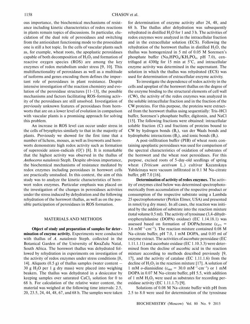
intracellular peroxidase was observed at pH 5.5 (Fig. 1a),
and the optimum of the intracellular tyrosinase activity –
in the range from pH 6.0 to 7.0 (Fig. 1b). Kinetic analysis
of the peroxidase activity in different fractions of the thal-
lus proteins showed that the intracellular peroxidases and
tyrosinases had the maximum reaction rates (Table 2).
The apoplastic peroxidases from fractions B1-B3 exhibit-
ed the highest substrate affinity because they had the low-
est Michaelis constants (Table 2). The Michaelis con-
stants for tyrosinases increased in the series: B1 < B2 <
C < B3 (Table 2).
S, nmol/(s·g of dry mass)
10.6 ± 0.3 (3)44.0 ± 4.9 (3)50.5 ± 4.1 (3)
P, nmol/(s·g of dry mass)
114.6 ± 9.9 (3)129.0 ± 8.8 (3)
3314.8 ± 366.5 (3)
Enzyme
CatalaseAscorbate oxidase Ascorbate peroxidase
DOPA-oxidase DOPA-peroxidasePeroxidase
Table 1. Enzyme activities in the intracellular fraction of
A. natalensis thallus expressed as a rate of substrate (S)
decomposition or product (P) formation
1160 CHASOV et al.
BIOCHEMISTRY (Moscow) Vol. 80 No. 9 2015
Ascorbic acid demonstrated the highest affinity to
the apoplastic peroxidase (B1). The Michaelis constant
increased in the series: ascorbic acid < o-dianisidine <
sinapic acid < caffeic acid < ferulic acid < chlorogenic
acid < p-coumaric acid (Table 3). The maximum velocity
of the reaction increased in the series: sinapic acid <
ascorbic acid < o-dianisidine < p-coumaric acid < caffeic
acid < ferulic acid < chlorogenic acid (Table 3). Among
the natural phenolic substrates, the peroxidases demon-
strated the highest sensitivity to sinapic acid (Table 3).
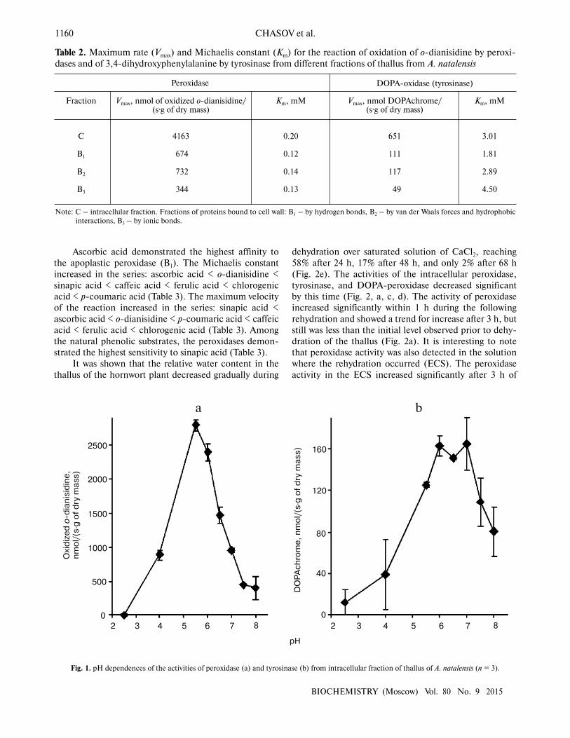
It was shown that the relative water content in the
thallus of the hornwort plant decreased gradually during
dehydration over saturated solution of CaCl2, reaching
58% after 24 h, 17% after 48 h, and only 2% after 68 h
(Fig. 2e). The activities of the intracellular peroxidase,
tyrosinase, and DOPA-peroxidase decreased significant
by this time (Fig. 2, a, c, d). The activity of peroxidase
increased significantly within 1 h during the following
rehydration and showed a trend for increase after 3 h, but
still was less than the initial level observed prior to dehy-
dration of the thallus (Fig. 2a). It is interesting to note
that peroxidase activity was also detected in the solution
where the rehydration occurred (ECS). The peroxidase
activity in the ECS increased significantly after 3 h of
Fig. 1. pH dependences of the activities of peroxidase (a) and tyrosinase (b) from intracellular fraction of thallus of A. natalensis (n = 3).
500
0
2 3 4 5 6 7
рН
Ox
idiz
ed
o-d
ian
isid
ine
,n
mo
l/(s
·g o
f d
ry m
as
s)
2500
2000
1500
1000
8 2 3 4 5 6 7 8
0
DO
PA
ch
rom
e,
nm
ol/
(s·g
of
dry
ma
ss
) 160
120
80
40
a b
Fraction
С
B1
B2
B3
Km, mM
3.01
1.81
2.89
4.50
Table 2. Maximum rate (Vmax) and Michaelis constant (Km) for the reaction of oxidation of o-dianisidine by peroxi-
dases and of 3,4-dihydroxyphenylalanine by tyrosinase from different fractions of thallus from A. natalensis
Vmax, nmol DOPAchrome/(s·g of dry mass)
651
111
117
49
Km, mM
0.20
0.12
0.14
0.13
Vmax, nmol of oxidized o-dianisidine/(s·g of dry mass)
4163
674
732
344
Peroxidase DOPA-oxidase (tyrosinase)
Note: C – intracellular fraction. Fractions of proteins bound to cell wall: B1 – by hydrogen bonds, B2 – by van der Waals forces and hydrophobic
interactions, B3 – by ionic bonds.
REDOX ENZYMES OF Anthoceros natalensis 1161
BIOCHEMISTRY (Moscow) Vol. 80 No. 9 2015
rehydration in comparison with the ECS level after 1 h of
rehydration (Fig. 2a). The activity of intracellular tyrosi-
nase increased slightly but reliably following the rehydra-
tion (Fig. 2d). Only trace amounts of the DOPA-peroxi-
dase, tyrosinase, and catalase were found in the ECS
(Fig. 2, b-d). The catalase activity increased significantly
following 24 h of rehydration, decreased after 48 h, and
remained on a level close to that observed prior to the
dehydration even after 68 h of incubation during rehydra-
tion (Fig. 2b).
Electrophoretic separation and isoelectrofocusing of
proteins. Analysis of results of electrophoretic separation
of proteins under native conditions revealed the presence
of five peroxidase isoenzymes in the intracellular fraction
of the hornwort with Rf 0.99, 0.82, 0.54, 0.53, and 0.52
(data not shown). Isoelectrofocusing revealed 14 different
Substrate
Ascorbic acid
o-Dianisidine
Caffeic acid
p-Coumaric acid
Sinapic acid
Ferulic acid
Chlorogenic acid
Vmax, µM/min
2.4
4.9
15.8
15.3
2.0
23.4
32.2
Km, µM
2
17
33
114
28
43
111
Table 3. Maximum rate (Vmax) and Michaelis constant (Km)
for the reaction of oxidation of different substrates by apo-
plastic peroxidases of fraction B1 from thallus of A. natalensis
Note: Standard 0.5-ml reaction mixture contained 50 µl of substrate at
respective concentration, 5 µl of fraction B1, 0.1 mM H2O2, and
0.07 M Na-citrate buffer, pH 5.5.
Fig. 2. Activity of redox enzymes – (a) peroxidase, (b) catalase, (c) DOPA-peroxidase, and (d) tyrosinase in intracellular fraction of thallus of
A. natalensis; e) relative water content (RWC) in the thallus of the plant: 1) prior to dehydration (control); 2-4) after dehydration for 24, 48, and
68 h, respectively; 5, 6) after rehydration for 1 and 3 h, respectively; 7, 8) extracellular solution obtained 1 and 3 h after incubation and removal
of the thallus from the rehydration solution, respectively. Difference is significant at P � 0.05 (*), P � 0.01 (**), P � 0.001 (***), n = 6.
1000
01 2 3 4 5 6 7 8 1 2 3 4 5 6 7 8
Ox
idiz
ed
o-d
ian
isid
ine
,n
mo
l/(s
·g o
f d
ry m
as
s)
5000
4000
3000
2000
0
25
20
15
10
a b
20
60
40
*****
***
***
***
2
1H2O
2,
nm
ol/
(s·g
of
dry
ma
ss
)
DO
PA
ch
rom
e,
nm
ol/
(s·g
of
dry
ma
ss
)
******
*
1 2 3 4 5 6 7 8 1 2 3 4 5 6 7 8
12 24 36 48 60
Time, h
c d e
250
200
150
100
30
25
20
15
10
5
0
140
120
100
80
60
15
10
5
0
100
75
50
25
0
RW
C,
%
1162 CHASOV et al.
BIOCHEMISTRY (Moscow) Vol. 80 No. 9 2015
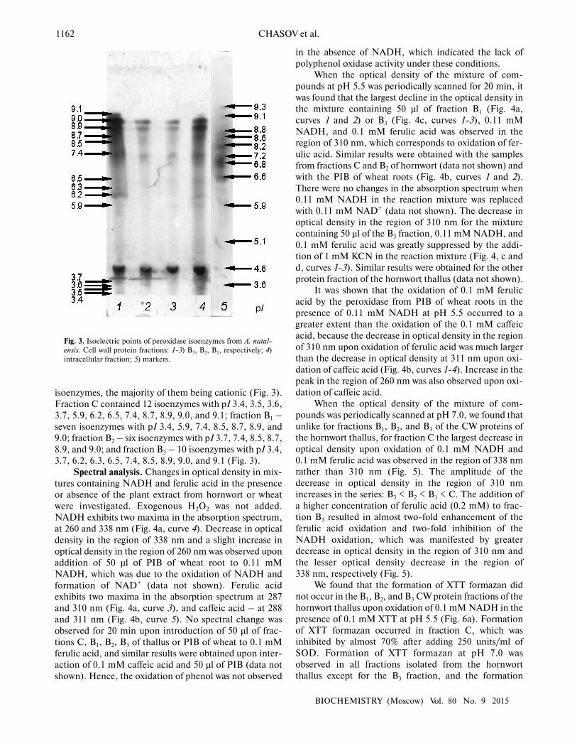
isoenzymes, the majority of them being cationic (Fig. 3).
Fraction C contained 12 isoenzymes with pI 3.4, 3.5, 3.6,
3.7, 5.9, 6.2, 6.5, 7.4, 8.7, 8.9, 9.0, and 9.1; fraction B1 –
seven isoenzymes with pI 3.4, 5.9, 7.4, 8.5, 8.7, 8.9, and
9.0; fraction B2 – six isoenzymes with pI 3.7, 7.4, 8.5, 8.7,
8.9, and 9.0; and fraction B3 – 10 isoenzymes with pI 3.4,
3.7, 6.2, 6.3, 6.5, 7.4, 8.5, 8.9, 9.0, and 9.1 (Fig. 3).
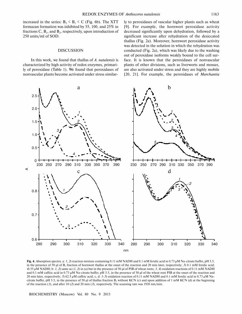
Spectral analysis. Changes in optical density in mix-
tures containing NADH and ferulic acid in the presence
or absence of the plant extract from hornwort or wheat
were investigated. Exogenous H2O2 was not added.
NADH exhibits two maxima in the absorption spectrum,
at 260 and 338 nm (Fig. 4a, curve 4). Decrease in optical
density in the region of 338 nm and a slight increase in
optical density in the region of 260 nm was observed upon
addition of 50 µl of PIB of wheat root to 0.11 mM
NADH, which was due to the oxidation of NADH and
formation of NAD+ (data not shown). Ferulic acid
exhibits two maxima in the absorption spectrum at 287
and 310 nm (Fig. 4a, curve 3), and caffeic acid – at 288
and 311 nm (Fig. 4b, curve 5). No spectral change was
observed for 20 min upon introduction of 50 µl of frac-
tions C, B1, B2, B3 of thallus or PIB of wheat to 0.1 mM
ferulic acid, and similar results were obtained upon inter-
action of 0.1 mM caffeic acid and 50 µl of PIB (data not
shown). Hence, the oxidation of phenol was not observed
in the absence of NADH, which indicated the lack of
polyphenol oxidase activity under these conditions.
When the optical density of the mixture of com-
pounds at pH 5.5 was periodically scanned for 20 min, it
was found that the largest decline in the optical density in
the mixture containing 50 µl of fraction B1 (Fig. 4a,
curves 1 and 2) or B3 (Fig. 4c, curves 1-3), 0.11 mM
NADH, and 0.1 mM ferulic acid was observed in the
region of 310 nm, which corresponds to oxidation of fer-
ulic acid. Similar results were obtained with the samples
from fractions C and B2 of hornwort (data not shown) and
with the PIB of wheat roots (Fig. 4b, curves 1 and 2).
There were no changes in the absorption spectrum when
0.11 mM NADH in the reaction mixture was replaced
with 0.11 mM NAD+ (data not shown). The decrease in
optical density in the region of 310 nm for the mixture
containing 50 µl of the B3 fraction, 0.11 mM NADH, and
0.1 mM ferulic acid was greatly suppressed by the addi-
tion of 1 mM KCN in the reaction mixture (Fig. 4, c and
d, curves 1-3). Similar results were obtained for the other
protein fraction of the hornwort thallus (data not shown).
It was shown that the oxidation of 0.1 mM ferulic
acid by the peroxidase from PIB of wheat roots in the
presence of 0.11 mM NADH at pH 5.5 occurred to a
greater extent than the oxidation of the 0.1 mM caffeic
acid, because the decrease in optical density in the region
of 310 nm upon oxidation of ferulic acid was much larger
than the decrease in optical density at 311 nm upon oxi-
dation of caffeic acid (Fig. 4b, curves 1-4). Increase in the
peak in the region of 260 nm was also observed upon oxi-
dation of caffeic acid.
When the optical density of the mixture of com-
pounds was periodically scanned at pH 7.0, we found that
unlike for fractions B1, B2, and B3 of the CW proteins of
the hornwort thallus, for fraction C the largest decrease in
optical density upon oxidation of 0.1 mM NADH and
0.1 mM ferulic acid was observed in the region of 338 nm
rather than 310 nm (Fig. 5). The amplitude of the
decrease in optical density in the region of 310 nm
increases in the series: B3 < B2 < B1 < C. The addition of
a higher concentration of ferulic acid (0.2 mM) to frac-
tion B3 resulted in almost two-fold enhancement of the
ferulic acid oxidation and two-fold inhibition of the
NADH oxidation, which was manifested by greater
decrease in optical density in the region of 310 nm and
the lesser optical density decrease in the region of
338 nm, respectively (Fig. 5).
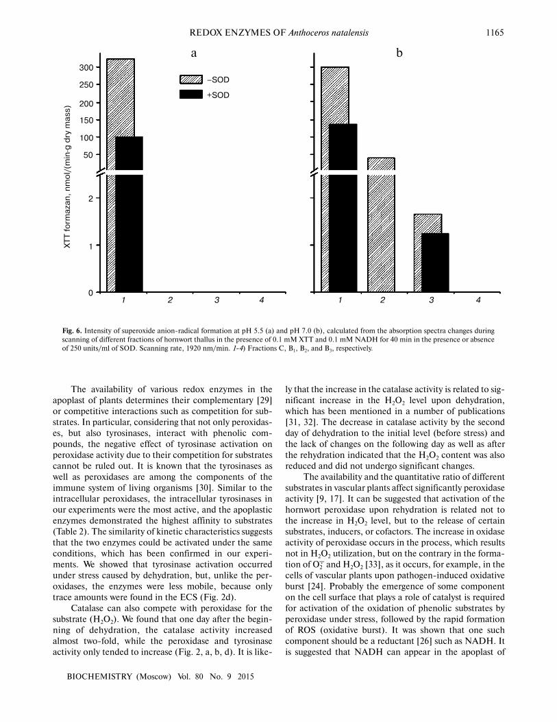
We found that the formation of XTT formazan did
not occur in the B1, B2, and B3 CW protein fractions of the
hornwort thallus upon oxidation of 0.1 mM NADH in the
presence of 0.1 mM XTT at pH 5.5 (Fig. 6a). Formation
of XTT formazan occurred in fraction C, which was
inhibited by almost 70% after adding 250 units/ml of
SOD. Formation of XTT formazan at pH 7.0 was
observed in all fractions isolated from the hornwort
thallus except for the B3 fraction, and the formation
Fig. 3. Isoelectric points of peroxidase isoenzymes from A. natal-
ensis. Cell wall protein fractions: 1-3) B3, B2, B1, respectively; 4)
intracellular fraction; 5) markers.
pI
REDOX ENZYMES OF Anthoceros natalensis 1163
BIOCHEMISTRY (Moscow) Vol. 80 No. 9 2015
increased in the series: B2 < B1 < C (Fig. 6b). The XTT
formazan formation was inhibited by 55, 100, and 25% in
fractions C, B1, and B2, respectively, upon introduction of
250 units/ml of SOD.
DISCUSSION
In this work, we found that thallus of A. natalensis is
characterized by high activity of redox enzymes, primari-
ly of peroxidase (Table 1). We found that peroxidases of
nonvascular plants become activated under stress similar-
ly to peroxidases of vascular higher plants such as wheat
[9]. For example, the hornwort peroxidase activity
decreased significantly upon dehydration, followed by a
significant increase after rehydration of the desiccated
thallus (Fig. 2a). Moreover, hornwort peroxidase activity
was detected in the solution in which the rehydration was
conducted (Fig. 2a), which was likely due to the washing
out of peroxidase isoforms weakly bound to the cell sur-
face. It is known that the peroxidases of nonvascular
plants of other divisions, such as liverworts and mosses,
are also activated under stress and they are highly mobile
[20, 21]. For example, the peroxidases of Marchantia
Fig. 4. Absorption spectra. a: 1, 2) reaction mixture containing 0.11 mM NADH and 0.1 mM ferulic acid in 0.73 µM Na-citrate buffer, pH 5.5,
in the presence of 50 µl of B1 fraction of hornwort thallus at the onset of the reaction and 20 min later, respectively; 3) 0.1 mM ferulic acid;
4) 55 µM NADH; b: 1, 2) same as (1, 2) in (a) but in the presence of 50 µl of PIB of wheat roots; 3, 4) oxidation reactions of 0.11 mM NADH
and 0.1 mM caffeic acid in 0.73 µM Na-citrate buffer, pH 5.5, in the presence of 50 µl of the wheat root PIB at the onset of the reaction and
20 min later, respectively; 5) 62.5 µM caffeic acid; c, d: 1-3) oxidation reaction of 0.11 mM NADH and 0.1 mM ferulic acid in 0.73 µM Na-
citrate buffer, pH 5.5, in the presence of 50 µl of thallus fraction B3 without KCN (c) and upon addition of 1 mM KCN (d) at the beginning
of the reaction (1), and after 10 (2) and 20 min (3), respectively. The scanning rate was 1920 nm/min.
1
0.5
0
2
0.8
0.7
230
3
4
250 270 290 310 330
nm
a b
2.5
2.0
1.5
1.0
1
2
3
1
2
3
5
4
1
2
3
0.6
350 370 390 230 250 270 290 310 330 350 370 390
280 290 300 310 320 330 340 280 290 300 310 320 330 340
А
c d
1164 CHASOV et al.
BIOCHEMISTRY (Moscow) Vol. 80 No. 9 2015
polymorpha L. suspension culture were secreted into the
culture medium in response to the chemical
stressor, bornyl acetate [20], and the peroxidases of sus-
pension culture of Physcomitrella patens (Hedw.) B.S.G.
and Racomitrium japonicum Dozy & Molk. mosses were
also activated in the culture medium in response to the
fungal elicitor chitosan [21]. The mobility of apoplastic
peroxidases is explained by the fact that these enzymes
are present on the cell surface [11], from which they can
be easily released under stress conditions even before their
stress-induced synthesis begins [12, 21, 22].
The peroxidases in vascular plants are among the
main components of the adaptive response involved in
formation of resistance to various stresses [12, 23, 24]. It
is likely that because peroxidases are so important for
plants, evolution has resulted in formation of a large
number of genes encoding these enzymes. For example,
138 genes located in 12 chromosomes were identified in
rice (Oryza sativa L.) [11], 73 in thale cress (Arabidopsis
thaliana (L.) Heynh.) [25], and 48 in P. patens moss [21].
Perhaps the availability of such a large number of genes
results in the presence of several isoforms of the enzyme
in the plants [10, 15]. Peroxidase activity is increased in
plants that are more resistant to stressors [23]. At least two
strategies are likely to exist in plants for using the protec-
tive properties of peroxidase: to keep a high level of activ-
ity of this enzyme permanently, or to increase the peroxi-
dase activity from low to high level quickly at the location
required for the plant in response to the changes in envi-
ronmental conditions.
Kinetic analysis of peroxidase activity in the intra-
cellular fraction and in the different CW protein fractions
of the hornwort thallus showed that the intracellular per-
oxidases have the maximum reaction velocity, while the
apoplastic peroxidases exhibit the highest affinity to the
substrates as they had the lowest Michaelis constants
(Table 2). Hence, a minimal amount of a substrate is suf-
ficient for activation of the apoplastic peroxidase. It is
known that the fast activation of apoplastic peroxidase
under stress conditions could be a key factor of oxidative
burst in plant cells [26, 27], and this could play an impor-
tant role in initiation of signaling cascades and subse-
quent formation of immunity in plants [28]. Substrate-
dependent peroxidase activation is also supported by the
fact that peroxidase secretion under stress conditions
could accompany substrate secretion as was shown, for
example, for lunularin in suspension culture of M. poly-
morpha [20].
Fig. 5. Difference calculated from the absorption spectra during the reaction of peroxidase oxidation of substrates: 0.1 mM NADH and fer-
ulic acid in 0.73 µM Na-citrate buffer, pH 7.0, for 20 min in the presence of different hornwort fractions. Scanning rate, 1920 nm/min. 1-4)
Fractions C, B1, B2, and B3, respectively, with 0.1 mM ferulic acid; 5) fraction B3 with 0.2 mM ferulic acid; 260 and 338 nm – maxima of
absorption spectrum of NADH; 287 and 310 nm – maxima of absorption spectrum of ferulic acid.
–4
–5
–6
–7
1 2 3 4 5
∆А
, µ
l·c
m/(
min
·g w
et
ma
ss
)
0
–1
–2
–3
–8
–9
260 nm
338 nm
287 nm
310 nm
REDOX ENZYMES OF Anthoceros natalensis 1165
BIOCHEMISTRY (Moscow) Vol. 80 No. 9 2015
The availability of various redox enzymes in the
apoplast of plants determines their complementary [29]
or competitive interactions such as competition for sub-
strates. In particular, considering that not only peroxidas-
es, but also tyrosinases, interact with phenolic com-
pounds, the negative effect of tyrosinase activation on
peroxidase activity due to their competition for substrates
cannot be ruled out. It is known that the tyrosinases as
well as peroxidases are among the components of the
immune system of living organisms [30]. Similar to the
intracellular peroxidases, the intracellular tyrosinases in
our experiments were the most active, and the apoplastic
enzymes demonstrated the highest affinity to substrates
(Table 2). The similarity of kinetic characteristics suggests
that the two enzymes could be activated under the same
conditions, which has been confirmed in our experi-
ments. We showed that tyrosinase activation occurred
under stress caused by dehydration, but, unlike the per-
oxidases, the enzymes were less mobile, because only
trace amounts were found in the ECS (Fig. 2d).
Catalase can also compete with peroxidase for the
substrate (H2O2). We found that one day after the begin-
ning of dehydration, the catalase activity increased
almost two-fold, while the peroxidase and tyrosinase
activity only tended to increase (Fig. 2, a, b, d). It is like-
ly that the increase in the catalase activity is related to sig-
nificant increase in the H2O2 level upon dehydration,
which has been mentioned in a number of publications
[31, 32]. The decrease in catalase activity by the second
day of dehydration to the initial level (before stress) and
the lack of changes on the following day as well as after
the rehydration indicated that the H2O2 content was also
reduced and did not undergo significant changes.
The availability and the quantitative ratio of different
substrates in vascular plants affect significantly peroxidase
activity [9, 17]. It can be suggested that activation of the
hornwort peroxidase upon rehydration is related not to
the increase in H2O2 level, but to the release of certain
substrates, inducers, or cofactors. The increase in oxidase
activity of peroxidase occurs in the process, which results
not in H2O2 utilization, but on the contrary in the forma-
tion of О2� and H2O2 [33], as it occurs, for example, in the
cells of vascular plants upon pathogen-induced oxidative
burst [24]. Probably the emergence of some component
on the cell surface that plays a role of catalyst is required
for activation of the oxidation of phenolic substrates by
peroxidase under stress, followed by the rapid formation
of ROS (oxidative burst). It was shown that one such
component should be a reductant [26] such as NADH. It
is suggested that NADH can appear in the apoplast of
Fig. 6. Intensity of superoxide anion-radical formation at pH 5.5 (a) and pH 7.0 (b), calculated from the absorption spectra changes during
scanning of different fractions of hornwort thallus in the presence of 0.1 mM XTT and 0.1 mM NADH for 40 min in the presence or absence
of 250 units/ml of SOD. Scanning rate, 1920 nm/min. 1-4) Fractions C, B1, B2, and B3, respectively.
150
100
50
2
a bX
TT
fo
rma
za
n,
nm
ol/
(min
·g d
ry m
as
s)
300
250
200
1
0
–SOD
+SOD
1 2 3 4 1 2 3 4
1166 CHASOV et al.
BIOCHEMISTRY (Moscow) Vol. 80 No. 9 2015
vascular plants because of malate- or lactate-dehydroge-
nase activity of the plasmalemma or CW, and its interac-
tion with the peroxidase could result in formation of ROS
[10, 24, 33]. As is known, peroxidase exhibits NADH-
oxidase activity, and the radical form of NAD• and H2O2
are formed in the process of interaction of this enzyme
with oxygen [24, 33, 34]. A chain of interconversions can
occur thereafter. It is known that during the combined
oxidation by peroxidase of substrates with very different
reactivity, effects of their activation or inhibition are
observed [35]. It is the authors’ opinion that the activa-
tion of the slowly oxidized substrate occurs together with
partial or complete inhibition of conversion of the rapid-
ly oxidized substrate (activator). Hence, the phenol radi-
cals and/or radical from of the reductant can interact with
each other and with oxygen, forming О2� [33-35].
To evaluate the possibility of this redox mechanism
functioning in the cells of nonvascular higher plants, we
analyzed features of the combined oxidation of NADH
and ferulic acid, which is a natural phenolic substrate of
the peroxidase, by the peroxidase of the hornwort thallus.
Various derivatives of hydroxycinnamic acids have been
found in hornworts [2]. It was found that at pH 5.5 when
NADH and ferulic acid are present and exogenous H2O2
is absent, the largest decrease in optical density was
observed in the region of 310 nm, which was likely due to
the oxidation of ferulic acid rather than NADH (Fig. 4a).
The significant inhibition of this reaction by the addition
of cyanide, such as for example in fraction B3 (Fig. 4, c
and d), indirectly confirms participation of peroxidases in
this process. Despite the fact that NADH can be a slowly
oxidizing substrate of peroxidase [35], it is possible that in
our experiments NADH plays the role of activator of oxi-
dation of phenolic acids. To test this possibility, the
enzyme of vascular plants (extracellular wheat peroxidase
from PIB) was used. It was found that similarly to the
hornwort peroxidases, the wheat peroxidase at pH 5.5
oxidized ferulic acid in the presence of NADH and ferulic
acid, and in the absence of exogenous H2O2 even to a
greater degree than caffeic acid (Fig. 4b). It is known that
for peroxidase-mediated oxidative burst to occur, the
change in pH toward alkaline values is required addition-
ally to the presence of reductant [26]. Therefore, it was
important to test if the oxidation of ferulic acid in the
presence of NADH by the hornwort peroxidases would
depend on pH of the reaction mixture. It was found that
at pH 7.0 as well as at pH 5.5 the oxidation of ferulic acid
occurred to a greater degree than the oxidation of NADH
in all fractions of the hornwort except for the fraction C,
which was manifested by the larger decrease in optical
density at 310 nm than at 338 nm (Fig. 5). All soluble
compounds are present in the cytoplasmic fraction
including endogenous phenols and other peroxidase sub-
strates that may be why NADH is oxidized to a larger
degree in this case than exogenous ferulic acid. The per-
oxidases from hornwort fraction B3 oxidized ferulic acid
the least; however, its oxidation increased almost two-fold
with two-fold increase in the ferulic acid concentration
(0.2 mM), and the NADH oxidation decreased two-fold
in the process (Fig. 5). This is a possible indication of the
competition between these substrates during the oxida-
tion catalyzed by peroxidase.
We showed that the hornwort peroxidases are capable
of О2� formation (Fig. 6), and that this process is pH-
dependent. The О2� acceptor XTT [18] was converted into
XTT formazan in the presence of 0.1 mM NADH at
pH 5.5 only in the intracellular hornwort fraction, and
this reaction was sensitive to SOD (Fig. 6a). The SOD-
sensitive formation of XTT formazan at pH 7.0 indicates
that the О2� was formed in all fractions except for fraction
B3 (Fig. 6b). Such significant difference in this reaction at
acidic and neutral pH is probably because more alkaline
medium is required for both О2� formation by peroxidase
and for the reaction of О2� with XTT [18, 26]. The differ-
ences in the inhibition of XTT formazan formation by
fractions C, B1, and B2 following SOD addition and the
lack of the XTT oxidation by the peroxidases from frac-
tion B3 (Fig. 6b) are likely due to the different ability of
the peroxidase isoforms from different fractions for О2�
formation. We found that the composition of peroxidase
isoforms was different in the fractions (Fig. 3). It is possi-
ble that the peroxidases capable of О2� formation are lack-
ing in fraction B3, in particular, the isoform with pI 8.7 is
absent in the B3 fraction, while it is present in the other
fractions. Two anionic isoforms with pI 3.5 and 3.6 are
present in fraction C that are absent in other fractions.
The peroxidase isoform with pI 5.9 is present only the
fractions C and B1 (Fig. 3), and it is exactly in these frac-
tions the formation of XTT formazan exceeds its produc-
tion in fraction B2 by one order of magnitude (Fig. 3).
Hence, we suggest that the isoforms of the hornwort per-
oxidases have different ability for production of О2�. In
future, detailed analysis of the specificity of the О2�-form-
ing activity of the separate peroxidase isoforms would
allow elucidating their contribution to oxidative burst
under stress.
Hence, assuming that the release of NADH or any
other reductant into the apoplast happens under stress
conditions, the amount of it required for the induction of
oxidative burst would be very small. Because of the differ-
ential oxidation of substrates by peroxidase, such a reduc-
tant can play the role of an inducer stimulating conver-
sion of other substrates such as phenols released under
stress into the apoplast. Considering that a pool of soluble
peroxidases is always present in apoplast, possibly togeth-
er with slowly oxidizing substrates, it can be suggested
that the release of a small amount of the readily oxidized
substrate or any cofactor would be sufficient for the
immediate response. The following increase in the
response could be through either the release of additional
enzyme or the emergence of substrates. Thus, the key role
of the extracellular peroxidase comprises the regulation of

REDOX ENZYMES OF Anthoceros natalensis 1167
BIOCHEMISTRY (Moscow) Vol. 80 No. 9 2015
the ROS balance in the apoplast of plant cells, and this
regulation is carried out due to the competitive and com-
plementary interactions of different peroxidase sub-
strates.
It was shown for the first time in this work that, along
with known cytological and morphological similarity with
the vascular plants, the hornworts demonstrate certain
similarity in functioning features of their redox enzymes.
The kinetic characteristics of peroxidases and tyrosinases
of hornworts were analyzed for the first time, and the pos-
sibility of participation of the peroxidase of the hornwort
thallus in anti- and prooxidant processes was demonstrat-
ed. We suggest that the revealed biochemical mechanism
of the possible participation of peroxidases in ROS for-
mation through substrate–substrate interaction is of
importance for adaptation and survival of the hornwort
under stress conditions such as dehydration/rehydration.
It seems important to decipher in future the elements of
the signaling pathways mediated by the changes in the
redox state and facilitating formation of the protective
responses in hornworts. We suggest that the formation of
ROS by peroxidases is an evolutionarily ancient process
that emerged as a protective mechanism of the land high-
er plants that enhances the adaptive mechanisms to
ensure their adaptation to the changing environmental
conditions and successful colonization of various ecolog-
ical niches.
This work was financially supported by the Russian
Foundation for Basic Research (grant 14-04-93962), by
the Program of the President of the Russian Federation
for supporting of leading scientific schools (grant NSh-
825.2012.4), and by the Program of Fundamental
Research of the Presidium of the Russian Academy of
Sciences “Molecular and Cell Biology” (leader A. N.
Grechkin).
REFERENCES
1. Villarreal, J. C., Cargill, D. C., Söderström, L., Hagborg,
A., and Renzaglia, K. S. (2010) A synthesis of hornwort
diversity: patterns, causes and future work, Phytotaxa, 9,
150-166.
2. Asakawa, Y. (1995) in Progress in the Chemistry of Organic
Natural Products (Herz, W., Kirby, G. W., Moore, R. E.,
Steglich, W., and Tamm, Ch., eds.) Vol. 65, Springer,
Vienna, pp. 1-562.
3. Troitsky, A. V., Ignatov, M. S., Bobrova, V. K., and
Milyutina, I. A. (2007) Contribution of genosystematics to
current concepts of phylogeny and classification of
bryophytes, Biochemistry (Moscow), 72, 1368-1376.
4. Chang, Y., and Graham, S. W. (2011) Inferring the higher-
order phylogeny of mosses (Bryophyta) and relatives using
a large, multigene plastid data set, Am. J. Bot., 98, 839-849.
5. Qiu, Y.-L., Li, L., Wang, B., Chen, Z., Knoop, V., Groth-
Malonek, M., Dombrovska, O., Lee, J., Kent, L., Rest, J.,
Estabrook, G. F., Hendry, T. A., Taylor, D. W., Testa, C.
M., Ambros, M., Crandall-Stotler, B., Duff, R. J.,
Stech, M., Frey, W., Quandt, D., and Davis, C. C. (2006)
The deepest divergences in land plants inferred from phy-
logenomic evidence, PNAS, 103, 15511-15516.
6. Adams, D. G., and Duggan, P. S. (2008) Cyanobacteria–
bryophyte symbioses, J. Exp. Bot., 59, 1047-1058.
7. Wood, A. J. (2007) The nature and distribution of vegetative
desiccation-tolerance in hornworts, liverworts and mosses,
Bryologist, 110, 163-177.
8. Minibayeva, F., and Beckett, R. P. (2001) High rates of
extracellular superoxide production in bryophytes and
lichens, and an oxidative burst in response to rehydration
following desiccation, New Phytologist, 152, 333-341.
9. Chasov, A. V., and Minibayeva, F. V. (2009) Effect of exoge-
nous phenols on superoxide production by extracellular
peroxidase from wheat seedling roots, Biochemistry
(Moscow), 74, 766-774.
10. Minibayeva, F., Kolesnikov, O., Chasov, A., Beckett, R. P.,
Lüthje, S., Vylegzhanina, N., Buck, F., and Böttger, M.
(2009) Wound-induced apoplastic peroxidase activities:
their roles in the production and detoxification of reactive
oxygen species, Plant Cell Environ., 32, 497-508.
11. Passardi, F., Longet, D., Penel, C., and Dunand, C. (2004)
The class III peroxidase multigenic family in rice and its
evolution in land plants, Phytochemistry, 65, 1879-1893.
12. Almagro, L., Gómez Ros, L. V., Belchi-Navarro, S., Bru,
R., Ros Barceló, A., and Pedreño, M. A. (2009) Class III
peroxidases in plant defense reactions, J. Exp. Bot., 60,
377-390.
13. Mathé, C., Barre, A., Jourda, C., and Dunand, C. (2010)
Evolution and expression of class III peroxidases, Arch.
Biochem. Biophys., 500, 58-65.
14. Mayaba, N., and Beckett, R. P. (2003) Increased activities
of superoxide dismutase and catalase are not the mecha-
nism of desiccation tolerance induced by hardening in the
moss Atrichum androgynum, J. Bryol., 25, 281-286.
15. Li, J. L., Sulaiman, M., Beckett, R. P., and Minibayeva, F.
V. (2010) Cell wall peroxidases in the liverwort Dumortiera
hirsuta are responsible for extracellular superoxide produc-
tion, and can display tyrosinase activity, Physiol. Plant.,
138, 474-484.
16. Chasov, A. V., and Minibayeva, F. V. (2014) Methodological
approaches for studying apoplastic redox activity: 1.
Mechanisms of peroxidase release, Russ. J. Plant Physiol.,
61, 556-563.
17. Chasov, A. V., and Minibayeva, F. V. (2014) Methodological
approaches for studying apoplastic redox activity: 2.
Regulation of peroxidase activity, Russ. J. Plant Physiol.,
61, 626-633.
18. Sutherland, M. W., and Learmonth, B. A. (1997) The tetra-
zolium dyes MTS and XTT provide new quantitative assays
for superoxide and superoxide dismutase, Free Rad. RPS,
27, 283-289.
19. Laemmli, U. K. (1970) Cleavage of structural proteins dur-
ing the assembly of the head of bacteriophage T4, Nature,
227, 680-685.
20. Hirata, T., Ashida, Y., Mori, H., Yoshinaga, D., and Goad,
L. J. (2000) A 37-kDa peroxidase secreted from liverworts
in response to chemical stress, Phytochemistry, 55, 197-
202.
21. Lehtonen, M. T., Akita, M., Kalkkinen, N., Ahola-
Iivarinen, E., Rönnholm, G., Somervuo, P., Thelander,
1168 CHASOV et al.
BIOCHEMISTRY (Moscow) Vol. 80 No. 9 2015
M., and Valkonen, J. P. (2009) Quickly-released peroxidase
of moss in defense against fungal invaders, New Phytol.,
183, 432-443.
22. Van Loon, L. C., Rep, M., and Pieterse, C. M. J. (2006)
Significance of inducible defence-related proteins in
infected plants, Ann. Rev. Phytopathol., 44, 135-162.
23. Pshenichnov, E., Khashimova, N., Akhunov, A.,
Golubenko, Z., and Stipanovic, R. D. (2011) Participation
of chitin-binding peroxidase isoforms in the wilt pathogen-
esis of cotton, AJPS, 2, 43-49.
24. O’Brien, J. A., Daudi, A., Butt, V. S., and Bolwell, G. P.
(2012) Reactive oxygen species and their role in plant
defense and cell wall metabolism, Planta, 236, 765-779.
25. Tognolli, M., Penel, C., Greppin, H., and Simon, P. (2002)
Analysis and expression of the class III peroxidase large
gene family in Arabidopsis thaliana, Gene, 288, 129-138.
26. Bolwell, G. P., Bindschedler, L. V., Blee, K. A., Butt, V. S.,
Davies, D. R., Gardner, S. L., Gerrish, C., and
Minibayeva, F. (2002) The apoplastic oxidative burst in
response to biotic stress in plants: a three-component sys-
tem, J. Exp. Bot., 53, 1367-1376.
27. Lehtonen, M. T., Akita, M., Frank, W., Reski, R., and
Valkonen, J. P. T. (2012) Involvement of a class III peroxi-
dase and the mitochondrial protein TSPO in oxidative burst
upon treatment of moss plants with a fungal elicitor,
MPMI, 25, 363-371.
28. Tarchevskii, I. A. (2001) Metabolism of Plants under Stress
[in Russian], Fen, Kazan.
29. Roach, T., Colville, L., Beckett, R. P., Minibayeva, F. V.,
Havaux, M., and Kranner, I. (2015) A proposed interplay
between peroxidase, amine oxidase and lipoxygenase in the
wounding-induced oxidative burst in Pisum sativum
seedlings, Phytochemistry, 112, 130-138.
30. Mayer, A. M. (2006) Polyphenol oxidases in plants and
fungi: going places? A review, Phytochemistry, 67, 2318-
2331.
31. Lee, B. R., Kim, K. Y., Jung, W. J., Avice, J. C., Ourry, A.,
and Kim, T. H. (2007) Peroxidases and lignification in rela-
tion to the intensity of water-deficit stress in white clover
(Trifolium repens L.), J. Exp. Bot., 58, 1271-1279.
32. Chen, Q., Yang, L., Ahmad, P., Wan, X., and Hu, X. (2011)
Proteomic profiling and redox status alteration of recalci-
trant tea (Camellia sinensis) seed in response to desiccation,
Planta, 233, 583-592.
33. Halliwell, B. (1978) Lignin synthesis: the generation of
hydrogen peroxide and superoxide by horseradish peroxi-
dase and its stimulation by manganese (II) and phenols,
Planta, 140, 81-88.
34. Lebedeva, O. V., and Ugarova, N. N. (1997) Steady-state
kinetics of NADH oxidation by hydrogen peroxide in the
presence of horseradish peroxidase, Biochemistry (Moscow),
62, 212-216.
35. Lebedeva, O. V., and Ugarova, N. N. (1996) Mechanism of
peroxidase-catalyzed oxidation. Substrate–substrate acti-
vation in horseradish peroxidase-catalyzed reactions, Russ.
Chem. Bull., 45, 18-25.