Embed Size (px)
Citation preview
JOURNAL OF VIROLOGY, Nov. 1995, p. 7264–7268 Vol. 69, No. 110022-538X/95/$04.0010Copyright q 1995, American Society for Microbiology
Active Foamy Virus Proteinase Is Essential for Virus Infectivitybut Not for Formation of a Pol PolyproteinJAN KONVALINKA,† MARTIN LOCHELT, HANSWALTER ZENTGRAF,
ROLF M. FLUGEL, AND HANS-GEORG KRAUSSLICH*
Angewandte Tumorvirologie, Deutsches Krebsforschungszentrum,D-69009 Heidelberg, Germany
Received 15 May 1995/Accepted 31 July 1995
To analyze proteolytic processing of foamy (spuma) retroviruses, two mutations were generated in thepresumed active-site triplet Asp-Ser-Gly in the predicted proteinase (PR) region of the human foamy virus(HSRV). The mutations changed either the presumed catalytic aspartic acid residue to a catalytically incom-petent alanine or the adjacent serine to a threonine found in most cellular and retroviral proteases at thisposition. Both mutations were cloned into the full-length infectious HSRV DNA clone. Wild-type and S/Tmutant genomes directed the synthesis of particles with similar infectious titers, while the HSRV D/A PRmutant was noninfectious. Immunoblot analysis of transfected cells revealed identical patterns for the wild-type and for the S/T PR mutant. HSRV D/A mutant-transfected cells expressed only a single Gag polyproteinof 78 kDa instead of the 78-kDa–74-kDa doublet found in HSRV-infected or wild-type-transfected cells.Analysis with pol-specific antisera yielded a protein of approximately 120 kDa reactive with antisera againstpol- but not gag-specific domains. No Gag-Pol polyprotein was detected in this study. Electron microscopyanalysis of transfected cells showed heterogeneous particle morphology in the case of the D/A mutant, withparticles of normal appearance and particles of aberrant size and shape. These results indicate that foamy viruseshave an aspartic PR that is essential for infectivity but not for formation of the 120-kDa Pol polyprotein.
Proteolytic processing of the structural Gag and Gag-Polpolyproteins by the virus-encoded virion-associated proteinase(PR) is essential for the production of infectious retroviralparticles. Inactivation of PR, either by mutation of its active-site residues or by use of specific inhibitors, abolished retrovi-ral infectivity and led to the release of immature-appearingnoninfectious particles consisting of uncleaved viral polypro-teins (12, 28). In the case of human immunodeficiency virus(HIV) and other retroviruses, the viral PR has been shown tocleave the Gag and Gag-Pol polyproteins at defined sites toproduce all mature viral proteins (reviewed in references 13and 15). Cellular proteases are believed not to be involved inretrovirus Gag and Gag-Pol polyprotein processing. RetroviralPRs belong to the family of aspartic proteases and are active ashomodimers, with the active-site triplets (Asp-Thr/Ser-Gly; D-T/S-G) from both chains contributing to the symmetric activesite of the enzyme (26). The catalytic apparatus of asparticproteases consists primarily of the two aspartic acid residueswithin the conserved D-T/S-G triplets which are located in thecenter of the substrate-binding cleft and are essentially in thesame spatial relationship in the three-dimensional structuresdetermined to date (33; reviewed in reference 32). A particularfeature of the active site is the side-chain-to-backbone inter-action between the hydroxyl group of the Thr residue in theDTG triplet and the main chain of the other PR molecule inthe dimer. These interactions form a network of two symmet-rical pairs of hydrogen bonds, stabilizing the structure of theactive site which has been described as ‘‘fireman’s grip’’ (6).The only other small amino acid capable of forming such an
interaction is serine, and a DSG triplet has been found in thePR region of avian retroviruses, yeast retrotransposons, andfoamy viruses (30).Foamy viruses (spumaviruses) are a group of complex ret-
roviruses that have been isolated from a number of animalspecies and are believed to be apathogenic in their animal host(19). The human foamy virus (HSRV) was originally isolatedfrom a patient with nasopharyngeal carcinoma (1). The HSRVproviral genome has been cloned and sequenced, and the ge-netic organization and some unusual aspects of its replicationcycle have been studied (reviewed in references 16 and 20).Foamy virus morphogenesis involves the assembly of intracy-toplasmic core particles, similar to B- and D-type retroviruses,which bud at the plasma membrane but preferentially at intra-cellular membranes (7). A particular feature of foamy virusreplication is the fact that viral structural antigens remainmainly cell associated and few extracellular virions are found.Most intracellular particles appear to be immature, and anal-ysis of foamy virus Gag proteins has revealed mostly precursorpolyproteins, with the main Gag-reactive proteins being a dou-blet of 78 kDa-74 kDa and very limited amounts of processedproducts (3, 23). Furthermore, the sizes of pol-derived prod-ucts corresponding to the viral PR, reverse transcriptase (RT)-RNase H, and integrase (IN) have been reported to be 10, 80,and 39 kDa, respectively (11, 24, 25). However, foamy viruspolyprotein processing has not been studied in detail, and noanalysis of the viral PR activity has been reported.The amino acid sequence derived from the 59 part of the
foamy virus pol gene, predicted to be expressed by a11 frame-shift event unusual in retroviruses, contains a homologousDSG sequence that determines the location of the putative PRregion (30). Outside the conserved active-site region (with aSer residue instead of the more frequent Thr residue), there isa second sequence motif, GlyArgLys, corresponding to thepattern GlyArg(Asp/Asn) found in most other viral asparticPRs (26). Apart from these characteristic features, foamy virus
* Corresponding author. Present address: Abteilung Zellbiologieund Virologie, Heinrich-Pette-Institut, Martinistr. 52, D-20251 Ham-burg, Germany. Phone: (40) 48051-241. Fax: (40) 48051-184.† Present address: Department of Biochemistry, Institute of Organic
Chemistry and Biochemistry, Czech Academy of Science, 166 10 Prague6, Czech Republic.
7264
PR exhibits very little similarity to other retroviral and cellularproteases. In particular, a conserved structurally flexible region(‘‘flap’’) that is normally found between the described con-served elements and that is involved in substrate binding can-not be easily identified in the foamy virus PR region. Consid-ering the limited sequence similarity and the observation thatfoamy virus Gag proteins are mostly detectable as precursorproteins, we hypothesized that there may be special features infoamy virus polyprotein processing. In order to prove thatfoamy viruses contain an aspartic PR essential for virus repli-cation and to analyze polyprotein processing, we generated amutation which changed the presumed active-site Asp residueto an Ala residue and analyzed the phenotype of the resultingproviral clone pHSRV-D/A regarding virus infectivity, poly-protein processing, and particle morphology. A second muta-tion was based on the hypothesis that a PR containing a DTGinstead of the DSG triplet may be more active and changed theSer residue to the more commonly found Thr residue(pHSRV-S/T). The corresponding reverse mutation in theHIV type 1 (HIV-1) PR had been shown to reduce PR activityby a factor of 5 to 10 (12a).The two mutations were generated by oligonucleotide-di-
rected mutagenesis on a single-stranded DNA template ac-cording to the method of Kunkel (14). As the template, weused single-stranded DNA derived from plasmid pBS-H(N/P)that contains a NarI-PstI fragment of the infectious proviralplasmid pHSRV13 (nucleotides [nt] 1126 to 5063 of the HSRVgenome) in pBluescript (Stratagene). The sequences of the twooligonucleotides used were as follows (altered nucleotides areunderlined): 59-CC CAC TGG GCT TCA GGG GC-39 (D/A;nt 3178 to 3196 of the HSRV genome) and 59-CAC TGGGATACA GGG GCA AC-39 (S/T; nt 3180 to 3199 of the HSRVgenome). Following mutagenesis, the DraIII-to-Bsu36I frag-ments (nt 3098 to 3631 of the HSRV genome spanning theputative PR region) of the D/A and S/T mutant plasmids werecloned into pHSRV13 (18) to yield plasmids pHSRV-D/A andpHSRV-S/T, respectively. The presence of the mutations wasconfirmed by sequence analysis of the region between theDraIII and Bsu36I sites.
To analyze the influence of the mutations in the putative PRactive site on viral infectivity, we titrated the infectivity of virusparticles harvested after transient transfection of BHK21 orBHK FAB cells (34) with plasmids pHSRV13, pHSRV-D/A,and pHSRV-S/T, respectively (Table 1). Cleared culture mediacollected 2 to 3 days after transfection were normalized fortransfection efficiency, and serial dilutions were used to infectfresh HSRV-permissive BHK FAB cells. No infectious virusparticles were recovered in four independent transfectionswith pHSRV-D/A proviral DNA, although similar transfectionefficiencies were achieved, and synthesis of foamy virus pro-teins and particle assembly was easily detected in mutant-transfected cells (see below). Culture media from pHSRV-S/T-transfected cells, on the other hand, yielded titers similar tothose obtained for the media from the wild-type (wt)-trans-fected cells (Table 1). On the basis of these results, we con-clude that HSRV contains an aspartic PR essential for virusinfectivity as observed in a number of other retroviral systems.Changing the active-site triplet to the more commonly foundDTG triplet did not alter infectious titers, suggesting that theunusual Ser residue in the foamy virus PR active site does notsignificantly impair virus infectivity. Similarly, mutation of thePR active-site DSG of the yeast transposon Ty3 to the com-monly found DTG had little if any effect on polyprotein pro-cessing and viral replication (10). Furthermore, a reverse mu-tation changing the HIV PR active site from DTG to DSG hasbeen shown to reduce PR activity in in vitro assay systems by afactor of 5 to 10, with little effect on processing of particle-associated polyproteins and no change in virus infectivity(12a). Since this mutation primarily affected the Km values, itwas concluded that the observed reduced activity of the mu-tated PR may not be phenotypically relevant for proteolysisinside the virion, where substrate concentrations should besaturating.Figure 1 shows an immunoblot analysis of extracts from
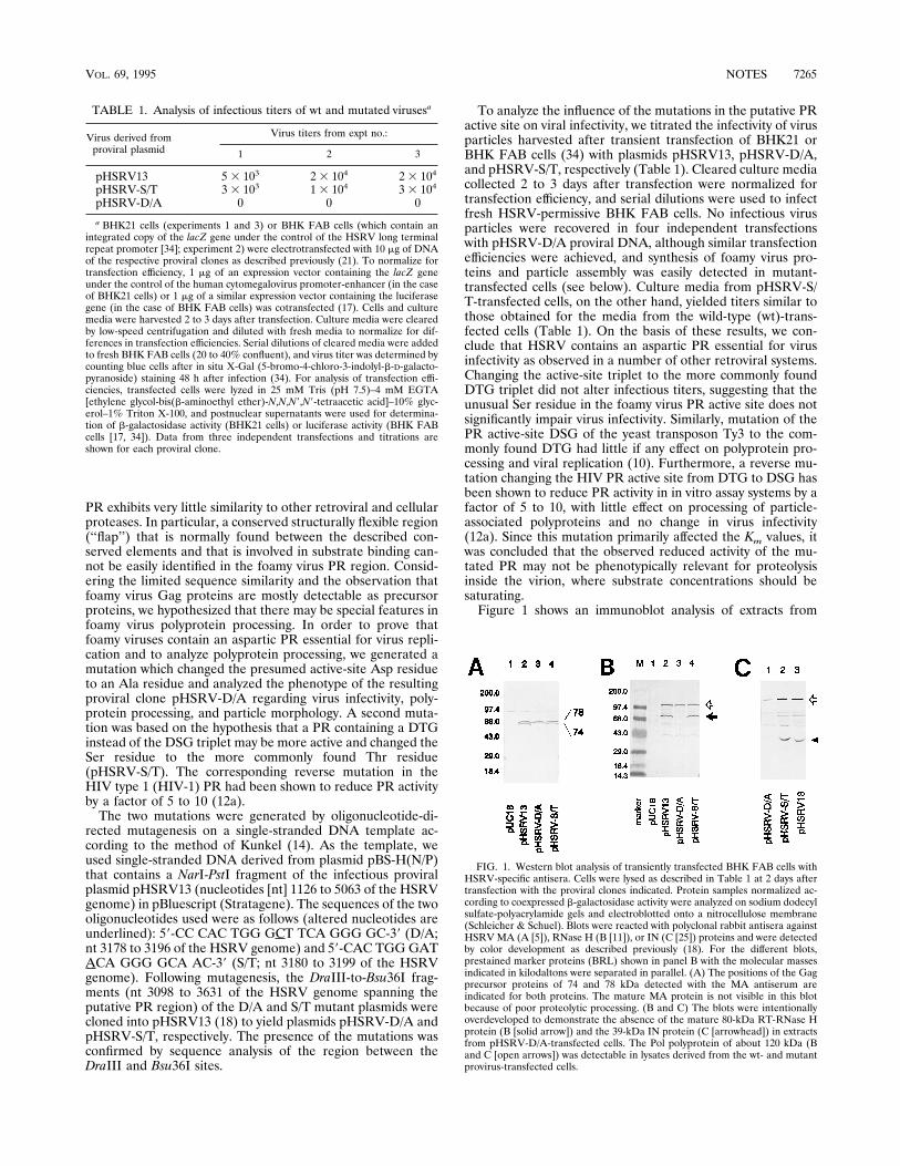
FIG. 1. Western blot analysis of transiently transfected BHK FAB cells withHSRV-specific antisera. Cells were lysed as described in Table 1 at 2 days aftertransfection with the proviral clones indicated. Protein samples normalized ac-cording to coexpressed b-galactosidase activity were analyzed on sodium dodecylsulfate-polyacrylamide gels and electroblotted onto a nitrocellulose membrane(Schleicher & Schuel). Blots were reacted with polyclonal rabbit antisera againstHSRVMA (A [5]), RNase H (B [11]), or IN (C [25]) proteins and were detectedby color development as described previously (18). For the different blots,prestained marker proteins (BRL) shown in panel B with the molecular massesindicated in kilodaltons were separated in parallel. (A) The positions of the Gagprecursor proteins of 74 and 78 kDa detected with the MA antiserum areindicated for both proteins. The mature MA protein is not visible in this blotbecause of poor proteolytic processing. (B and C) The blots were intentionallyoverdeveloped to demonstrate the absence of the mature 80-kDa RT-RNase Hprotein (B [solid arrow]) and the 39-kDa IN protein (C [arrowhead]) in extractsfrom pHSRV-D/A-transfected cells. The Pol polyprotein of about 120 kDa (Band C [open arrows]) was detectable in lysates derived from the wt- and mutantprovirus-transfected cells.
TABLE 1. Analysis of infectious titers of wt and mutated virusesa
Virus derived fromproviral plasmid
Virus titers from expt no.:
1 2 3
pHSRV13 5 3 103 2 3 104 2 3 104
pHSRV-S/T 3 3 103 1 3 104 3 3 104
pHSRV-D/A 0 0 0
a BHK21 cells (experiments 1 and 3) or BHK FAB cells (which contain anintegrated copy of the lacZ gene under the control of the HSRV long terminalrepeat promoter [34]; experiment 2) were electrotransfected with 10 mg of DNAof the respective proviral clones as described previously (21). To normalize fortransfection efficiency, 1 mg of an expression vector containing the lacZ geneunder the control of the human cytomegalovirus promoter-enhancer (in the caseof BHK21 cells) or 1 mg of a similar expression vector containing the luciferasegene (in the case of BHK FAB cells) was cotransfected (17). Cells and culturemedia were harvested 2 to 3 days after transfection. Culture media were clearedby low-speed centrifugation and diluted with fresh media to normalize for dif-ferences in transfection efficiencies. Serial dilutions of cleared media were addedto fresh BHK FAB cells (20 to 40% confluent), and virus titer was determined bycounting blue cells after in situ X-Gal (5-bromo-4-chloro-3-indolyl-b-D-galacto-pyranoside) staining 48 h after infection (34). For analysis of transfection effi-ciencies, transfected cells were lyzed in 25 mM Tris (pH 7.5)–4 mM EGTA[ethylene glycol-bis(b-aminoethyl ether)-N,N,N9,N9-tetraacetic acid]–10% glyc-erol–1% Triton X-100, and postnuclear supernatants were used for determina-tion of b-galactosidase activity (BHK21 cells) or luciferase activity (BHK FABcells [17, 34]). Data from three independent transfections and titrations areshown for each proviral clone.
VOL. 69, 1995 NOTES 7265
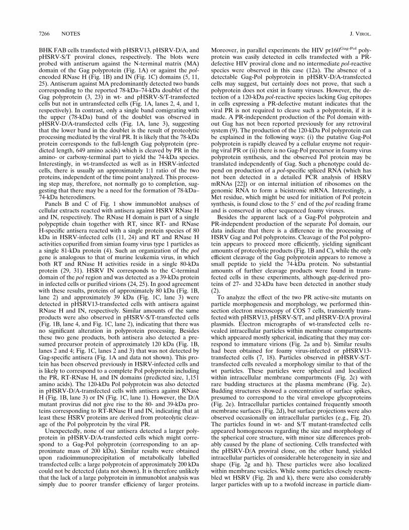
BHK FAB cells transfected with pHSRV13, pHSRV-D/A, andpHSRV-S/T proviral clones, respectively. The blots wereprobed with antiserum against the N-terminal matrix (MA)domain of the Gag polyprotein (Fig. 1A) or against the pol-encoded RNase H (Fig. 1B) and IN (Fig. 1C) domains (5, 11,25). Antiserum against MA predominantly detected two bandscorresponding to the reported 78-kDa–74-kDa doublet of theGag polyprotein (3, 23) in wt- and pHSRV-S/T-transfectedcells but not in untransfected cells (Fig. 1A, lanes 2, 4, and 1,respectively). In contrast, only a single band comigrating withthe upper (78-kDa) band of the doublet was observed inpHSRV-D/A-transfected cells (Fig. 1A, lane 3), suggestingthat the lower band in the doublet is the result of proteolyticprocessing mediated by the viral PR. It is likely that the 78-kDaprotein corresponds to the full-length Gag polyprotein (pre-dicted length, 649 amino acids) which is cleaved by PR in theamino- or carboxy-terminal part to yield the 74-kDa species.Interestingly, in wt-transfected as well as in HSRV-infectedcells, there is usually an approximately 1:1 ratio of the twoproteins, independent of the time point analyzed. This process-ing step may, therefore, not normally go to completion, sug-gesting that there may be a need for the formation of 78-kDa–74-kDa heterodimers.Panels B and C of Fig. 1 show immunoblot analyses of
cellular extracts reacted with antisera against HSRV RNase Hand IN, respectively. The RNase H domain is part of a singlepolypeptide chain together with RT, since RT- and RNaseH-specific antisera reacted with a single protein species of 80kDa in HSRV-infected cells (11, 24) and RT and RNase Hactivities copurified from simian foamy virus type 1 particles asa single 81-kDa protein (4). Such an organization of the polgene is analogous to that of murine leukemia virus, in whichboth RT and RNase H activities reside in a single 80-kDaprotein (29, 31). HSRV IN corresponds to the C-terminaldomain of the pol region and was detected as a 39-kDa proteinin infected cells or purified virions (24, 25). In good agreementwith these results, proteins of approximately 80 kDa (Fig. 1B,lane 2) and approximately 39 kDa (Fig. 1C, lane 3) weredetected in pHSRV13-transfected cells with antisera againstRNase H and IN, respectively. Similar amounts of the sameproducts were also observed in pHSRV-S/T-transfected cells(Fig. 1B, lane 4, and Fig. 1C, lane 2), indicating that there wasno significant alteration in polyprotein processing. Besidesthese two gene products, both antisera also detected a pre-sumed precursor protein of approximately 120 kDa (Fig. 1B,lanes 2 and 4; Fig. 1C, lanes 2 and 3) that was not detected byGag-specific antisera (Fig. 1A and data not shown). This pro-tein has been observed previously in HSRV-infected cells andis likely to correspond to a complete Pol polyprotein includingthe PR, RT-RNase H, and IN domains (predicted size, 1,151amino acids). The 120-kDa Pol polyprotein was also detectedin pHSRV-D/A-transfected cells with antisera against RNaseH (Fig. 1B, lane 3) or IN (Fig. 1C, lane 1). However, the D/Amutant provirus did not give rise to the 80- and 39-kDa pro-teins corresponding to RT-RNase H and IN, indicating that atleast these HSRV proteins are derived from proteolytic cleav-age of the Pol polyprotein by the viral PR.Unexpectedly, none of our antisera detected a larger poly-
protein in pHSRV-D/A-transfected cells which might corre-spond to a Gag-Pol polyprotein (corresponding to an ap-proximate mass of 200 kDa). Similar results were obtainedupon radioimmunoprecipitation of metabolically labelledtransfected cells: a large polyprotein of approximately 200 kDacould not be detected (data not shown). It is therefore unlikelythat the lack of a large polyprotein in immunoblot analysis wassimply due to poorer transfer efficiency of larger proteins.
Moreover, in parallel experiments the HIV pr160Gag-Pol poly-protein was easily detected in cells transfected with a PR-defective HIV proviral clone and no intermediate pol-reactivespecies were observed in this case (12a). The absence of adetectable Gag-Pol polyprotein in pHSRV-D/A-transfectedcells may suggest, but certainly does not prove, that such apolyprotein does not exist in foamy viruses. However, the de-tection of a 120-kDa pol-reactive species lacking Gag epitopesin cells expressing a PR-defective mutant indicates that theviral PR is not required to cleave such a polyprotein, if it ismade. A PR-independent production of the Pol domain with-out Gag has not been reported previously for any retroviralsystem (9). The production of the 120-kDa Pol polyprotein canbe explained in the following ways: (i) the putative Gag-Polpolyprotein is rapidly cleaved by a cellular enzyme not requir-ing viral PR or (ii) there is no Gag-Pol precursor in foamy viruspolyprotein synthesis, and the observed Pol protein may betranslated independently of Gag. Such a phenotype could de-pend on production of a pol-specific spliced RNA (which hasnot been detected in a detailed PCR analysis of HSRVmRNAs [22]) or on internal initiation of ribosomes on thegenomic RNA to form a bicistronic mRNA. Interestingly, aMet residue, which might be used for initiation of Pol proteinsynthesis, is found close to the 59 end of the pol reading frameand is conserved in other sequenced foamy viruses.Besides the apparent lack of a Gag-Pol polyprotein and
PR-independent production of the separate Pol domain, ourdata indicate that there is a difference in the processing ofHSRV Gag and Pol polyproteins. Cleavage of the Pol polypro-tein appears to proceed more efficiently, yielding significantamounts of proteolytic products (Fig. 1B and C), while the onlyefficient cleavage of the Gag polyprotein appears to remove asmall peptide to yield the 74-kDa protein. No substantialamounts of further cleavage products were found in trans-fected cells in these experiments, although gag-derived pro-teins of 27- and 32-kDa have been detected in another study(2).To analyze the effect of the two PR active-site mutants on
particle morphogenesis and morphology, we performed thin-section electron microscopy of COS 7 cells, transiently trans-fected with pHSRV13, pHSRV-S/T, and pHSRV-D/A proviralplasmids. Electron micrographs of wt-transfected cells re-vealed intracellular particles within membrane compartmentswhich appeared mostly spherical, indicating that they may cor-respond to immature virions (Fig. 2a and b). Similar resultshad been obtained for foamy virus-infected or pHSRV13-transfected cells (7, 18). Particles observed in pHSRV-S/T-transfected cells revealed a morphology similar to that of thewt particles. These particles were spherical and localizedwithin intracellular membrane compartments (Fig. 2c) withrare budding structures at the plasma membrane (Fig. 2e).Budding structures showed a concentration of surface spikes,presumed to correspond to the viral envelope glycoproteins(Fig. 2e). Intracellular particles contained frequently smoothmembrane surfaces (Fig. 2d), but surface projections were alsoobserved occasionally on intracellular particles (e.g., Fig. 2f).The particles found in wt- and S/T mutant-transfected cellsappeared homogeneous regarding the size and morphology ofthe spherical core structure, with minor size differences prob-ably caused by the plane of sectioning. Cells transfected withthe pHSRV-D/A proviral clone, on the other hand, yieldedintracellular particles of considerable heterogeneity in size andshape (Fig. 2g and h). These particles were also localizedwithin membrane vesicles. While some particles closely resem-bled wt HSRV (Fig. 2h and k), there were also considerablylarger particles with up to a twofold increase in particle diam-
7266 NOTES J. VIROL.

FIG. 2. Electron micrographs showing thin sections of COS 7 cells transfected with pHSRV13 (a and b), pHSRV-S/T (c to f), or pHSRV-D/A (g to k). At 48 h aftertransfection, the cells were fixed in situ with 2.5% glutaraldehyde in 50 mM sodium cacodylate (pH 7.2) for 30 min at 48C. Fixed cells were scraped from the plate,collected by low-speed centrifugation, successively stained with 2% osmium tetroxide–0.5% uranyl acetate, and processed for ultrathin sectioning. Micrographs were taken witha Zeiss EM-10 electron microscope at 80 kV. Magnification,354,000 (panel c); 390,000 (all other panels). Bar, 200 nm (panel c) and 100 nm (all other panels).
7267
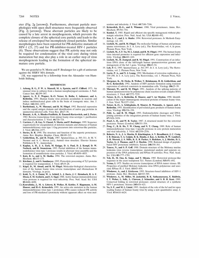
eter (Fig. 2g [arrows]). Furthermore, aberrant particle mor-phologies with open shell structures were frequently observed(Fig. 2j [arrows]). These aberrant particles are likely to becaused by a late arrest in morphogenesis, which prevents thecomplete closure of the spherical core particle and leads to therelease of enveloped but incompletely closed particles. Similarmorphologies have been reported for PR-defective mutants ofHIV-1 (21, 27) and for PR-inhibitor-treated HIV-1 particles(8). These observations suggest that PR activity may not onlybe required for condensation of the viral core during virionmaturation but may also play a role in an earlier step of virusmorphogenesis leading to the formation of the spherical im-mature core particle.
We are grateful to N. Morin and P. Boulanger for a gift of antiserumagainst the HSRV MA domain.J.K. was supported by a fellowship from the Alexander von Hum-
boldt Stiftung.
REFERENCES
1. Achong, B. G., P. W. A. Mansell, M. A. Epstein, and P. Clifford. 1971. Anunusual virus in cultures from a human nasopharyngeal carcinoma. J. Natl.Cancer Inst. 42:299–307.
2. Aguzzi, A., E. F. Wagner, K.-O. Netzer, K. Bothe, I. Anhauser, and A.Rethwilm. 1993. Human foamy virus proteins accumulate in neurons andinduce multinucleated giant cells in the brain of transgenic mice. Am. J.Pathol. 142:1061–1072.
3. Bartholoma, A., W. Muranyi, and R. M. Flugel. 1992. Bacterial expressionand the capsid antigen domain and identification of native gag proteins inspumavirus-infected cells. Virus Res. 23:27–38.
4. Benzair, A.-B., A. Rhodes-Feuillette, R. Emanoıl-Ravicovitch, and J. Peries.1982. Reverse transcriptase from simian foamy virus serotype 1: purificationand characterization. J. Virol. 44:720–724.
5. Carriere, C., B. Gay, N. Chazal, N. Morin, and P. Boulanger. 1995. Sequencerequirements for encapsidation of deletion mutants and chimeras of humanimmunodeficiency virus type 1 Gag precursors into retrovirus-like particles.J. Virol. 69:2366–2377.
6. Davies, D. R. 1990. The structure and function of the aspartic proteinases.Annu. Rev. Biophys. Biophys. Chem. 19:189–215.
7. Gelderblom, H., and H. Frank. 1987. Spumavirinae, p. 305–311. In M. V.Nermut and A. C. Steven (ed.), Animal virus structure. Elsevier SciencePublishers B. V., Amsterdam.
8. Kaplan, A. H., J. A. Zack, M. Knigge, D. A. Paul, D. J. Kempf, D. W.Norbeck, and R. Swanstrom. 1993. Partial inhibition of the human immu-nodeficiency virus type 1 protease results in aberrant virus assembly and theformation of noninfectious virus particles. J. Virol. 67:4050–4055.
9. Katz, R. A., and A. M. Skalka. 1994. The retroviral enzymes. Annu. Rev.Biochem. 63:133–173.
10. Kirchner, J., and S. Sandmeyer. 1993. Proteolytic processing of Ty3 proteinsis required for transposition. J. Virol. 67:19–28.
11. Kogel, D., M. Aboud, and R. M. Flugel. Molecular-biological characteriza-tion of the human foamy virus reverse transcriptase and ribonuclease Hdomains. Virology, in press.
12. Kohl, N., E. A. Emini, W. A. Schleif, L. J. Davis, J. C. Heimbach, R. A. F.Dixon, E. M. Scolnick, and I. S. Sigal. 1988. Active human immunodeficiencyvirus protease is required for viral infectivity. Proc. Natl. Acad. Sci. USA85:4686–4690.
12a.Konvalinka, J., M. A. Litterst, R. Welker, H. Kottler, F. Rippmann, A.-M.Hauser, and H.-G. Krausslich. 1995. An active-site mutation in the humanimmunodeficiency virus type 1 proteinase (PR) causes reduced PR activityand loss of PR-mediated cytotoxicity without apparent effect on virus mat-
uration and infectivity. J. Virol. 69:7180–7186.13. Krausslich, H.-G., and E. Wimmer. 1988. Viral proteinases. Annu. Rev.
Biochem. 57:701–754.14. Kunkel, T. 1985. Rapid and efficient site-specific mutagenesis without phe-
notypic selection. Proc. Natl. Acad. Sci. USA 82:488–492.15. Kuo, L. C., and J. A. Shafer. 1994. Retroviral proteases. In Methods Enzy-
mology, vol. 241.16. Lochelt, M., and R. M. Flugel. The molecular biology of human and primate
spuma retroviruses. In J. A. Levy (ed.), The Retroviridae, vol. 4, in press.Plenum Press, New York.
17. Lochelt, M., S. F. Yu, M. L. Linial, and R. M. Flugel. 1995. The human foamyvirus internal promoter is required for efficient gene expression and infec-tivity. Virology 206:601–610.
18. Lochelt, M., H. Zentgraf, and R. M. Flugel. 1991. Construction of an infec-tious DNA clone of the full-length human spumaretrovirus genome andmutagenesis of the bel 1 gene. Virology 184:43–54.
19. Loh, P. C. 1993. Spumaviruses, p. 361–397. In J. A. Levy (ed.), The Retro-viridae, vol. 2. Plenum Press, New York.
20. Luciw, P. A., and N. J. Leung. 1992. Mechanism of retrovirus replication, p.159–298. In J. A. Levy (ed.), The Retroviridae, vol. 1. Plenum Press, NewYork.
21. Mergener, K., M. Facke, R. Welker, V. Brinkmann, H. R. Gelderblom, andH.-G. Krausslich. 1992. Analysis of HIV particle formation using transientexpression of subviral constructs in mammalian cells. Virology 186:25–39.
22. Muranyi, W., and R. M. Flugel. 1991. Analysis of the splicing patterns ofhuman spumaretrovirus by polymerase chain reaction reveals complex RNAstructures. J. Virol. 65:7272–7235.
23. Netzer, K.-O., A. Rethwilm, B. Maurer, and V. ter Meulen. 1990. Identifi-cation of the major immunogenic structural proteins of human foamy virus.J. Gen. Virol. 71:1237–1241.
24. Netzer, K.-O., A. Schliephake, B. Maurer, R. Watanabe, A. Aguzzi, and A.Rethwilm. 1993. Identification of pol-related gene products of human foamyvirus. Virology 192:336–338.
25. Pahl, A., and R. M. Flugel. 1993. Endonucleolytic cleavages and DNA-joining activities of the integration protein of human foamy virus. J. Virol.67:5426–5434.
26. Pearl, L. H., and W. R. Taylor. 1987. A structural model for the retroviralproteases. Nature (London) 329:351–354.
27. Peng, C., B. K. Ho, T. W. Chang, and N. T. Chang. 1989. Role of humanimmunodeficiency virus type 1-specific protease in core protein maturationand viral infectivity. J. Virol. 63:2550–2556.
28. Roberts, N. A., J. A. Martin, D. Kinchington, A. V. Broadhurst, J. C. Craig,I. B. Duncan, S. A. Galpin, B. K. Handa, J. Kay, A. Krohn, R. W. Lambert,J. H. Merrett, J. S. Mills, K. E. B. Parkes, S. Redshaw, A. J. Ritchie, D. L.Taylor, G. J. Thomas, and P. J. Machin. 1990. Rational design of peptide-based HIV proteinase inhibitors. Science 248:358–361.
29. Tanese, N., and S. P. Goff. 1988. Domain structure of the Moloney murineleukemia virus reverse transcriptase: mutational analysis and separate ex-pression of the DNA polymerase and RNase H activities. Proc. Natl. Acad.Sci. USA 85:1777–1781.
30. Toh, H., M. Ono, K. Saigo, and T. Miyata. 1985. Retroviral protease-likesequence in the yeast transposon Ty1. Nature (London) 315:691–692.
31. Verma, I. 1975. Studies on reverse transcriptase of RNA tumor viruses. III.Properties of purified Moloney leukemia virus DNA polymerase and asso-ciate RNase H. J. Virol. 15:843–854.
32. Wlodawer, A., and J. Ericksson. 1993. Structure-based inhibitors of HIV-1protease. Annu. Rev. Biochem. 62:543–585.
33. Wlodawer, A., M. Miller, M. Jaskolski, B. K. Sathyanarayna, E. Baldwin,I. T. Weber, L. Selk., L. Clawson, J. Schneider, and S. B. H. Kent. 1989.Conserved folding in retroviral proteases: crystal structure of a syntheticHIV-1 proteinase. Science 245:616–621.
34. Yu, S. F., and M. L. Linial. 1993. Analysis of the role of the bel and bet openreading frames of human foamy virus by using a new quantitative assay. J.Virol. 67:6618–6624.
7268 NOTES J. VIROL.