Embed Size (px)
Citation preview
1
ACTIVATION OF THE PI3K/Akt PATHWAY EARLY DURING VACCINIA AND
COWPOX VIRUS INFECTION IS REQUIRED FOR BOTH HOST SURVIVAL
AND VIRAL REPLICATION
JAMÁRIA A. P. SOARES1,2§••••, FLÁVIA G. G. LEITE1,2§, LUCIANA G.
ANDRADE1,2, ALICE A. TORRES1,2, LIRLÂNDIA P. DE SOUSA3,4, LUCÍOLA S.
BARCELOS4,5, MAURO M. TEIXEIRA4, PAULO C. P. FERREIRA2, ERNA G.
KROON2, THAÍS SOUTO-PADRÓN6 AND CLÁUDIO A. BONJARDIM1,2*
(1) Grupo de Transdução de Sinal, (2) Laboratório de Vírus, (3) Departamento de
Microbiologia, Setor de Patologia – Coltec/UFMG, (4) Laboratório de
Imunofarmacologia, Departamento de Bioquímica e Imunologia, (5) Departamento de
Fisiologia e Biofísica, Instituto de Ciências Biológicas, Universidade Federal de Minas
Gerais, 31270-901 - Belo Horizonte, Minas Gerais, (6) Laboratório de Biologia Celular e
Ultraestrutura, Universidade Federal do Rio de Janeiro, 21941-590 - Rio de Janeiro, Brazil
*Address correspondence to: C. A. BONJARDIM, Ph.D., Grupo de Transdução de Sinal -
Laboratório de Vírus, Departamento de Microbiologia, Instituto de Ciências Biológicas,
Universidade Federal de Minas Gerais, Av. Antônio Carlos, 6627 - Campus Pampulha,
31270-901 - Belo Horizonte, MG – Brazil. Fax: 55-31 3443-6482; E-mail:
(§) - These authors contributed equally to this work.
Copyright © 2009, American Society for Microbiology and/or the Listed Authors/Institutions. All Rights Reserved.J. Virol. doi:10.1128/JVI.00245-09 JVI Accepts, published online ahead of print on 22 April 2009
on February 2, 2019 by guest
http://jvi.asm.org/
Dow
nloaded from
2
Running title: Role played by PI3K/Akt in Orthopoxvirus biology
(●) - Current address: Department of Microbiology and Molecular Genetics, Medical
College of Wisconsin, Milwaukee, USA.
on February 2, 2019 by guest
http://jvi.asm.org/
Dow
nloaded from
3
ABSTRACT
Viral manipulation of the transduction pathways associated with key cellular functions
such as actin remodeling, microtubule stabilization and survival may favor a productive
viral infection. Here we show that consistent with the Vaccinia virus (VACV) and Cowpox
virus (CPXV) requirement for cytoskeleton alterations early during the infection cycle,
PBK/Akt was phosphorylated at S473 (Akt-S473-P), a modification associated with the
mammalian target of rapamycin complex 2 (mTORC2), which was paralleled by
phosphorylation at T308 (Akt-T308-P) by PI3K/PDK1, that is required for host survival.
Notably, while VACV stimulated Akt (S473-P/T308-P) at early (1 hpi) and late times (24
hpi) during the infective cycle, CPXV stimulated Akt at early times only. Pharmacological
and genetic inhibition of PI3K (LY294002) or Akt (Akt-X and dominant negative form of
Akt - K179M) resulted in a significant decline in virus yield (from 80 to ≥ 90%). This
decline was secondary to the inhibition of late viral gene expression, which, in turn, led to
an arrest of virion morphogenesis at the immature virion stage of the viral growth cycle.
Furthermore, cleavage of both caspase-3 and poly ADP-ribose polymerase and TUNEL
assays confirmed that the permissive spontaneously immortalized cells such as A31 and
MEFs underwent apoptosis upon Orthopoxvirus infection plus LY294002. Thus, in A31
and MEFs, early viral-receptor mediated signals transmitted via the PI3K/Akt pathway are
required and precede the expression of viral anti-apoptotic genes. Additionally, the
inhibition of these signals resulted in apoptosis of the infected cells and a significant
decline in viral titers.
on February 2, 2019 by guest
http://jvi.asm.org/
Dow
nloaded from
4
INTRODUCTION
Poxviridae is a family of large linear double-stranded DNA viruses that carries out its
entire life cycle within the cytoplasmic compartment of infected cells. Vaccinia virus
(VACV) is a prototypical member of the Orthopoxvirus genus, which also includes the
closely related Cowpox virus (CPXV) (12, 52). The genomes of these viruses are
approximately 200 kbp in length with a coding capacity of approximately 200 genes. The
genes involved in the virus-host interaction are situated at both ends of the genome and are
associated with the evasion of host immune defenses (1). These evasion mechanisms
mainly operate extracellularly. For example, the secretion of soluble cytokine- and
chemokine-receptor homologues blocks the receptor recognition by intercepting the
cognate cytokine/chemokine in the extracellular environment. This mechanism facilitates
viral attachment and entry into cells (1, 70). Therefore, decoy receptors for interferon
(IFN) α, β and γ and tumor necrosis factor-α (TNF-α) play an important
immunomodulatory role by affecting both the host antiviral and apoptotic responses.
To counteract the host pro-apoptotic response, poxviruses have developed a number of
anti-apoptotic strategies, including the inhibition of apoptotic signals triggered by the
extrinsic pathway (those mediated by death receptors such as TNF and Fas ligand) or the
intrinsic pathway (mediated by the mitochondria and triggered upon viral infection) (1, 25,
70, 74). Many studies have previously identified viral inhibitors that block specific steps of
the intrinsic pathway. These include the VAVC-encoded E3L, F1L and N1L genes and the
Myxoma virus (MYXV)-encoded M11L gene that block cytochrome c release (14, 20, 34,
on February 2, 2019 by guest
http://jvi.asm.org/
Dow
nloaded from
5
39, 45, 75, 90) and the CPXV-encoded cytokine response modifier gene (CrmA) as well as
the VACV-encoded SPI-2 gene that inhibits both caspase 1 and 8 (25, 58, 61, 74).
An emerging body of evidence has also highlighted the pivotal role played by intracellular
signaling pathways in Orthopoxvirus biology (18, 48, 92). We and others have shown that
poxvirus manipulation of signaling pathways can be virus specific. For example, while
both VACV and CPXV stimulate the MEK/ERK/EGR-1 pathway during a substantial
length of time of their infective cycle, the pathway is required only for VACV replication,
whereas its role in CPXV biology has yet to be identified (71). MYXV, a rabbit-specific
poxvirus, also activates the MEK/ERK pathway in a mouse model of poxvirus-host
interaction. However, this stimulation led to the expression of IFN-β that consequently
blocked virus replication and possibly explains why MYXV has such a restricted host-
range (87).
Another signaling molecule associated with viral replication is Akt kinase (also known as
protein kinase B/PKB). The MYXV host-range factor M-T5 is able to reprogram the
intracellular environment thereby increasing human tumor cell permissiveness to viral
replication, which is directly associated with the levels of phosphorylated Akt (88). In
addition, M-T5 is functionally replaced by the host PIKE-A (phosphatidylinositol 3-kinase
enhancer A) protein (92).
The transmission of intracellular signals mediated by the serine/threonine kinase Akt to
downstream molecules in response to diverse stimuli such as growth factors, insulin, and
hormones, is dependent upon the phosphorylation of serine 473 (S473-P) and threonine
on February 2, 2019 by guest
http://jvi.asm.org/
Dow
nloaded from
6
308 (T308-P). This phosphorylation is mediated by the mammalian target of rapamycin
complex 2 (mTORC2) and phosphoinositide-dependent protein kinase 1 (PDK1), which
act as downstream effector of the phosphatidylinositol 3-kinase (PI3K)/Akt/mTORC1
pathway (2, 66). PI3Ks are a family of enzymes (Class I-III) that generate lipid second
messengers by the phosphorylation of plasma membrane phosphoinositides. Class IA
PI3Ks consist of a catalytic subunit (p110, comprising the three isoforms α, β and δ) and
an adaptor/regulatory subunit (p85), comprising the two isoforms α and β (for a detailed
review see 80).
The Akt family of proteins is comprised of the three isoforms α, β and γ, which are
composed of an N-terminal pleckstrin (PH) homology domain, a central catalytic domain
and a C-terminal hydrophobic domain. Akt is recruited to the plasma membrane through
the binding of its PH domain to the phosphatidylinositol 3, 4, 5-triphosphate (PI (3, 4, 5) P3
- PIP3), which is a product of PI3K that is anchored to the plasma membrane. PDK1 is also
recruited to the plasma membrane through interactions with PIP3. As both PDK1 and Akt
interact with PIP3, PDK1 co-localizes with Akt and activates it by phosphorylating the
threonine 308 (T308-P) (2, 66). Following its activation, Akt phosphorylates a number of
downstream substrates such as caspase-9, BAD, glycogen synthase kinase-3β (GSK-3β)
and FKHR. This leads to the suppression of apoptosis, cell growth, survival and
proliferation (11, 16, 56).
Another downstream target of PI3K/Akt is mTOR, a serine/threonine kinase that plays a
central role in the regulation of cell growth, proliferation, survival and protein synthesis
on February 2, 2019 by guest
http://jvi.asm.org/
Dow
nloaded from
7
(26). The mTOR kinase has recently been found to be associated with two functionally
distinct complexes in mammalian cells and known as mTORC1 and mTORC2 (63, 66).
Although these multi-protein complexes share molecules in common, distinct adaptor
proteins are recruited into each complex: raptor (regulatory-associated protein of TOR) is
recruited into mTORC1, while rictor (rapamycin-insensitive companion of TOR) is
recruited into mTORC2 (33, 64). While mTORC1 controls cell growth and protein
translation and has proven to be rapamycin-sensitive, mTORC2 regulates the actin
cytoskeleton and is assumed to be rapamycin-insensitive, even though under prolonged
exposure to the drug appears to inhibit mTORC2 assembly (29, 64, 65). Additionally, it
has been demonstrated that mTORC2 regulates the activity of Akt through the
phosphorylation of S473 (S473-P). S473-P appears to be required for the full activation of
Akt, since S473-P has been shown to enhance the subsequent phosphorylation of T308 by
PDK1 (66, 67, 94). Moreover, the phosphorylation of both S473 and T308 results in a
four- to five-fold increase in Akt activity when compared to T308-P by PDK1 alone (66).
The PI3K/PDK1/Akt(T308)/mTORC1 pathway regulates vital cellular processes that are
important in viral replication and propagation, including cell growth, proliferation and
protein translation. This pathway is particularly important in the replication of DNA
viruses, as their replication is cap-dependent. However, the Akt signaling pathway can also
negatively affect viral replication. The stress response downstream of Akt signaling,
including hypoxia and energy and amino acid depletion, inhibits mTORC1 (5, 9, 69).
Therefore, DNA viruses must overcome these constraints to translate their mRNAs.
on February 2, 2019 by guest
http://jvi.asm.org/
Dow
nloaded from
8
Pharmacological disruption of the PI3K/Akt pathway with the specific PI3K inhibitor,
LY294002 (82), has been reported to not only increase the cleavage of downstream
molecules associated with pro-apoptotic activity (e.g. PARP - (poly ADP-ribose
polymerase and the executioner caspase-3) (38, 41), but also to promote microtubule
stabilization, actin filament remodeling/cell migration and bleb formation/viral infectivity
(10, 35, 49, 54, 59).
Because the PI3K/Akt and PI3K/Akt/mTOR pathways influence diverse cellular functions
and possibly a healthy anti-viral response, usurping these pathways could support an
increase in viral replication. In support of this, a number of reports have demonstrated that
either the PI3K/Akt or the PI3K/Akt/mTOR pathway plays a role in the replication of
many viruses, including flavivirus (38), hepatitis C (27), HIV-1 (93), HPV (44, 96), RSV
(77), Coxsakievirus B3 (19), Epstein-Barr (17, 50, 73), HCMV (36, 37, 72), HSV-1 (7,
83), Varicella-zoster (60), KSHV (89), Adenovirus (55) and SV40 (95). With this in mind,
we also investigated whether the PI3K/Akt pathway played a pivotal role in Orthopoxvirus
biology. In this study, we show that the VACV- and CPXV-stimulated PI3K/Akt pathway,
not only contributes to the prevention of host-cell death, but also plays a beneficial role in
the viral replication cycle.
on February 2, 2019 by guest
http://jvi.asm.org/
Dow
nloaded from
9
MATERIALS AND METHODS
Cell culture, antibodies and chemicals - A spontaneously immortalized cell line (A31),
which is derived from mouse Balb/c 3T3, wild-type mouse embryonic fibroblasts (MEFs)
(81), SV40-LT-immortalized MEFs (MEFs-LT), kindly provided by Dr. C. Ronald Kahn -
Joslin Diabetes Center, USA, and BSC-40 cells was cultured in Dulbecco’s modified
Eagle’s medium (DMEM) supplemented with 7.5% (v/v) heat-inactivated fetal bovine
serum (FBS) (Cultilab, Campinas, SP, Brazil) and antibiotics in 5% CO2 at 37º C. After
reaching 80-90% confluence, the medium was then changed to 1% FBS and the cells were
incubated for 12 h. Antibodies against phospho-Akt (Ser473 and Thr308); ERK1/2;
PARP, which recognizes full-length PARP (116KDa) and the large (89KDa) and small
fragments (24KDa) of PARP resulting from caspase cleavage; caspase-3, which detects
full-length caspase-3 (35kDa) and the large fragment of caspase-3 resulting from cleavage
(17kDa), and cleaved caspase-3 (Asp175), which detects the large fragment of cleaved
caspase-3 (17-19kDa), were purchased from Cell Signaling Technology (Beverly, MA,
USA). LY294002 (2-morpholino-8-phenyl-4H-1-benzopyran-4-one), a pharmacological
inhibitor of PI3K; rapamycin, a pharmacological inhibitor of mTORC1; the pan-caspase
inhibitor benzyloxycarbonyl-Val-Ala-Asp-fluoromethylketone (zVAD.fmk); the viral
DNA synthesis inhibitor cytosine arabinoside (Ara C) and the anti-β-actin antibody were
purchased from Sigma-Aldrich (São Paulo, Brazil). The inhibitor of Akt, AktX (10-(4′-(N-
diethylamino) butyl)-2-chlorophenoxazine) and geneticin (G418) were purchased from
Calbiochem (São Paulo, Brazil). The specific antibodies for the viral H3L, D8L, A14L and
F18R proteins, were a generous gift from Dr B. Moss (NIAID, Bethesda, USA). The
on February 2, 2019 by guest
http://jvi.asm.org/
Dow
nloaded from
10
antibody that detects CrmA (SPI-2) was from Dr D. Pickup (Department of Molecular
Genetics and Microbiology, Duke University Medical Center, Durham, USA).
Viruses and viral infection – Wild-type VACV, strain WR, and CPXV, strain BR, were
propagated in BSC-40 cells and highly purified by sucrose gradient sedimentation as
previously described (31). The infective form of VACV and CPXV intracellular mature
virus (IMV), which represents the majority of the infectious viral progeny, was used to
carry out the experiments presented in this study. For experiments involving UV-irradiated
viruses, viral stocks were exposed for 5-10 min. to an UV lamp producing irradiation
predominantly at 365 nm. UV-irradiated viruses were then tested for virus infectivity.
Viruses unable to form plaques or in which late viral gene expression could not be detected
by western blots were considered UV-inactivated. Viral infections were carried out when
cell cultures reached 80-90% confluence. Cells were infected in the absence of FBS at the
indicated multiplicity of infection (MOI) for the times shown. Cells were treated with the
indicated drugs for 30 min. prior to viral infection and then incubated in its continued
presence for the indicated times.
Virus infectivity assays - A31, MEFs and MEFs-LT cells were cultured and starved as
above at a density of 5 x 10⁵ cells per well, in a 6-well culture dish and then exposed to
virus. Infections of A31 cells were carried out at an MOI of 10 for 3, 6, 12, 24, 36 and 48
h, either in the absence or in the presence of LY294002 (20 µM), or at the same MOI for
12 h or 24 h either in the absence or in the presence of rapamycin (50 nM), or Akt-X (15
µM), respectively. MEFs and MEFs-LT were infected for 24 h either in the absence or
presence of LY294002 as indicated above. Cultures were then washed with cold PBS and
on February 2, 2019 by guest
http://jvi.asm.org/
Dow
nloaded from
11
cells were disrupted by freezing and thawing. Virus was collected from the supernatant of
centrifuged cells and assayed for infectivity as described (15). Each experiment was run in
duplicate and the results are reported as the average values. The data were confirmed by at
least three independent experiments with identical results.
AKT dominant-negative cell lines - Cell lines stably expressing dominant-negative Akt,
were generated by transfecting A31 cells with 10 µg of plasmid DNA encoding a N-
terminal HA-tagged kinase-defective Akt mutant (Akt K179M; DN-Akt-HA), an Akt
mutant in which the lysine residue at position 179 (the ATP binding site) was changed to a
methionine (22) or the empty vector (pCDNA3) using a standard calcium phosphate
transfection protocol. Transfected cells were then ring-cloned after selection with 800
µg/ml geneticin for at least 21 days. The expression of the mutant Akt protein was
evaluated by western blot analysis. Cell extracts were blotted and then probed with anti-
HA or anti-Akt (S473-P) antibodies as described below.
Electron microscopy – A31 cells were infected with VACV or CPXV at an MOI of 2,
either in the presence or absence of LY294002 (20 µM), and incubated at 37º C for 18 or
22 h, respectively. Cells were fixed with 2.5% glutaraldehyde in 0.1M phosphate buffer
(pH 7.4) for one hour at room temperature, scraped gently and collected by centrifugation.
The cells were then washed with cacodylate buffer, fixed with 1% osmium tetroxide,
dehydrated in acetone and then processed for conventional transmission electron
microscopy. Thin sections were examined using a Morgagni transmission electron
microscope operating at 80 kV.
on February 2, 2019 by guest
http://jvi.asm.org/
Dow
nloaded from
12
Western blotting - a) Lysate preparation – Cells were grown, starved as above, and
infected with VACV or CPXV at an MOI of 10 for the times shown. Cells were left
untreated or pre-incubated with the indicated inhibitor for 30 min. and then exposed to
virus in the continued presence of the drugs as shown. Cells were then washed twice with
cold PBS and lysed on ice with lysis buffer (20 mM Tris acetate, pH 7.0; 1 mM EDTA; 1%
Triton X-100; 10 mM β-glycerophosphate; 50 mM NaF; 5 mM sodium pyrophosphate; 4
µg/ml leupeptin; 1 mM sodium orthovanadate). Lysates were scraped, collected into
Eppendorf tubes and centrifuged at 13.500 x g for 15 min. at 4º C. Protein concentrations
were determined using the Bio-Rad assay.
b) Electrophoresis and immunoblotting - Forty micrograms of the cell lysate per sample
were separated by electrophoresis on a 10 or 15% SDS-polyacrylamide gel and then
transferred onto nitrocellulose membranes as previously described (16). Membranes were
blocked at room temperature for 1 h with PBS containing 5% (w/v) non-fat milk and 0.1%
Tween-20. The membranes were washed three times with PBS containing 0.1% Tween-20
and then incubated with the specific rabbit or mouse polyclonal or monoclonal antibody
(1:1000 - 1:3000) in PBS containing 5% (w/v) BSA and 0.1% Tween-20. After washing,
the membranes were incubated with horseradish peroxidase-conjugated secondary anti-
rabbit (1:3000) or anti-mouse Ab (1:5000). Immunoreactive bands were visualized using
an ECL detection system as recommended by the manufacturer (GE Healthcare, Brazil).
TUNEL apoptosis assay - A31 cells were grown and starved as described above and then
infected with VACV or CPXV at an MOI of 10 for 4 h. Cells were left untreated or pre-
incubated with LY294002 (20 µM) for 30 min. and then incubated with virus in the
continued presence of the drug. Cells were then fixed with 4% paraformaldehyde. The
on February 2, 2019 by guest
http://jvi.asm.org/
Dow
nloaded from
13
terminal deoxynucleotidyl transferase-mediated deoxyuridine nick end-labeling (TUNEL)
assay was performed according to the manufacturer’s instructions (TdT-FragEL DNA
Fragmentation Kit, Calbiochem). The 3'-OH ends of the fragmented nucleosomal DNA in
apoptotic cells were specifically labeled using exogenous terminal deoxynucleotidyl
transferase (TdT) and biotin-labeled dNTP. Labeled ends were then detected using a
streptavidin-horseradish peroxidase (HRP) conjugate and diaminobenzidine (DAB). Nuclei
were counterstained with methyl green. At least 300 cells (in ten randomly captured
microscopic fields at a 600x magnification) were scored to calculate the percentage of
TUNEL-positive nuclei. The data are expressed as the apoptosis index which is the average
percentage of apoptotic nuclei. The experiment was performed in triplicate and repeated
two times.
Densitometric analysis - Phosphorylated-Akt and cleaved caspase-3 were quantified using
densitometric analysis software (LabImage) and the levels were normalized to the levels of
β-actin in the same sample. The changes in protein phosphorylation and cleavage with
respect to control values were estimated. The results were expressed as the Akt-P, cleaved
caspase-3 or H3L-A14L to β-actin ratio measured in arbitrary units.
on February 2, 2019 by guest
http://jvi.asm.org/
Dow
nloaded from
14
RESULTS
1 - VACV AND CPXV STIMULATE AKT (S473 AND T308)
PHOSPHORYLATION
In order to generate new progeny, poxviruses manipulate essential host signaling
pathways, such as the MEK/ERK pathway (4) and the PBK/Akt pathway (88) and,
consequently, the intracellular environment, to allow increased viral replication.
Because early events in the virus-host interaction involve alterations in the cytoskeleton,
from which Orthopoxvirus may benefit either at virus attachment/penetration,
morphogenesis or release (43, 49, 53, 68), we initially investigated whether Akt was
phosphorylated upon infection with VACV or CPXV at S473, a phosphorylation that is
mediated by mTORC2. Since survival signals are also important and may benefit viral
replication, we also analyzed the phosphorylation of Akt at T308, a role played by PDK1
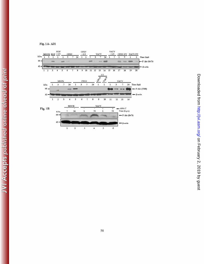
(9, 63, 66). A31 cells were infected at an MOI of 10 and cell lysates were collected from
the infected cells at 1 to 24 hpi and subjected to western blotting using an anti-phospho Akt
antibody to evaluate the phosphorylation status of Akt. As shown in Figure 1A top – upper
panel (S473) and top - lower panel (T308), both VACV and CPXV were able to induce
Akt phosphorylation. The amount of Akt-P was verified throughout the course of VACV
infection and the highest levels of Akt-P were observed at early (1 hpi) and at late time
points (24 hpi) (upper/lower panels - lanes 11 and 14) and intermediate levels were
detected at 3 and 7 hpi (lanes 12-13). In contrast, infection with CPXV was found to
on February 2, 2019 by guest
http://jvi.asm.org/
Dow
nloaded from
15
stimulate Akt-P (S473/T308) only at early time points (1 – 3 hpi) of the infection (lanes 5-
8).
We then asked whether the virus-stimulated signal leading to Akt-P (308) was mediated by
the upstream kinase PI3K. LY294002, a pharmacological inhibitor of PI3K, inhibits PI3K
activity through competitive inhibition of the ATP binding site located on the regulatory
subunit of PI3K (24, 79, 82) and inhibition of Akt-P (308) may result, though indirectly, in
reduced phosphorylation of Akt-P (473). Cells were left untreated or pre-incubated with
LY294002 (20 µM) for 30 min and then infected with CPXV or VACV, respectively, at an
MOI of 10 for the times shown in (Fig. 1A, top - upper panel – lanes 9, 10 and 15, 16 and
lower panel, lanes 9 and 10). Pre-incubation of the cells with LY294002 abrogated Akt-P,
suggesting the requirement for PI3K in both VACV- and CPXV-induced Akt-P (T308)
and, indirectly, to the Akt-P (S473) as well. As a control for Akt activation, we exposed the
serum-starved cells to a well-known Akt stimulator, epidermal growth factor (EGF) at 100
nM. As shown in Fig. 1A, pre-incubation with LY294002 abrogated the EGF-stimulated
Akt-P (S473) (lanes 3, 4).
Next, we raised the question as to whether viral-stimulation of Akt-P (S473) was
dependent on viral replication. To inhibit replication, viruses were inactivated with UV-
radiation. Although viral irradiation prevents virus replication, it does not block virus
penetration and expression of some early genes (6, 51). Cells were exposed to the
inactivated viruses for one and three hours (Fig. 1A, top - upper panel, lanes 17-20). Cell
lysates were then collected and subjected to western blotting with the anti-phospho-Akt
(S473) antibody. As seen in Figure 1A – top - upper panel, replication-competent virions,
on February 2, 2019 by guest
http://jvi.asm.org/
Dow
nloaded from
16
either CPXV (lanes 17-18) or VACV (upper panel, lanes 19-20), were not required for the
induction of Akt phosphorylation, suggesting that receptor-mediated events are sufficient
to trigger the phosphorylation of Akt. Taken together, these data implied that viral
induction of Akt-P takes place before viral replication and that viral attachment and entry
are sufficient for Akt-P. This is consistent with the viral requirement for actin remodeling
early during the infective cycle (43, 49, 68) and viral activation of Akt-S473-P by
mTORC2, as well as stimulation of early survival signals, a role played by the
PI3K/PDK/Akt (T308-P) pathway (reviewed in 9).
Because the kinetics of Akt phosphorylation upon VACV or CPXV infection were quite
unique, e.g., Akt-P (S473/T308) was detected at late times (24 hpi) only upon infection
with VACV, this suggested that VACV may require post-replicative gene expression in
order to phosphorylate Akt. To test this hypothesis, A31 cells were incubated for 30 min
with cytosine arabinoside (Ara C) (40 µg/ml), prior to VACV infection at an MOI of 10 for
the times shown. Experiments were carried out either in the absence or in the continued
presence of Ara C and then cell lysates were collected and subjected to western blotting
using the anti-phospho Akt antibody. As shown in Fig. 1B, while in the presence of Ara C,
Akt (S473-P) was detected only at early times, but not at 24 hpi (compares lanes 5 and 6).
As the incubation with Ara C compromises intermediate/late gene expression, this suggests
that the phosphorylation of Akt late during the VACV infective cycle is dependent on post-
DNA replication events. Based on this observation we hypothesized that Akt (T308-P) at
late times during the VACV infective cycle is also dependent on the same events.
on February 2, 2019 by guest
http://jvi.asm.org/
Dow
nloaded from
17
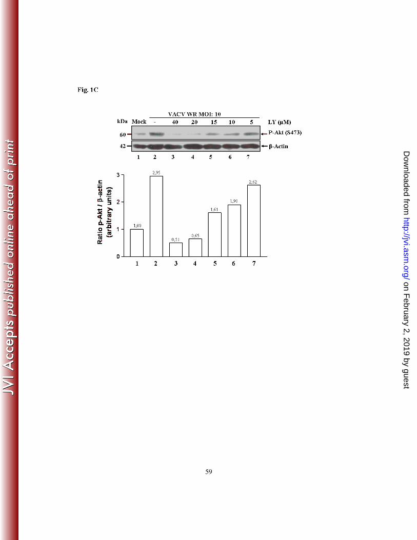
In order to examine whether the pharmacological inhibition, though indirect, of Akt (S473-
P) was effective in a dose-dependent manner, A31 cells were incubated with increasing
concentrations (5, 10, 20 and 40 µM) of LY294002 for 30 min prior to VACV infection at
an MOI of 10 for one hour. Cell lysates were then collected and subjected to western
blotting using the anti-P-Akt antibody. Pharmacological inhibition of Akt-P in response to
viral infection was found to be dose-dependent, as seen in Fig. 1C. Similarly, LY294002
inhibited the induction of Akt-P in a dose-dependent manner during CPXV infection (data
not shown).
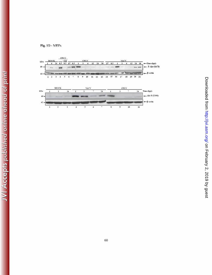
To further investigate whether the induction of Akt-P following infection with VACV or
CPXV, and its blockade by pre-incubation with LY294002, occurred in cells other than
A31 cells, another spontaneously immortalized cell line, mouse embryonic fibroblasts
(MEFs) (81) were grown, starved as indicated for A31 cells and then infected with VACV
or CPXV at an MOI of 10. Cell lysates were then collected at the indicated times post-
infection and analyzed by western blotting using the anti-phospho-Akt (S473 and T308)
antibodies to evaluate Akt-P. As demonstrated in Fig. 1D – top - upper and lower panels,
the kinetics of the viral induction of Akt-P (S473/T308), and its dependence either directly,
or indirectly of PI3K (only the inhibition associated with CPXV infection is shown), was
also confirmed with MEFs. Of note, the kinetics of Akt-P were consistent with its late
stimulation occurring only upon VACV infection (top - upper panel, lanes 20-21). Early
induction of Akt-P is also consonant with the cytoskeletal alterations required during early
infection. Taken together, these results were very similar to those seen with the A31 cells,
which strongly suggest that the findings are not cell-type specific.
on February 2, 2019 by guest
http://jvi.asm.org/
Dow
nloaded from
18
2 - THE PI3K/AKT SIGNALING PATHWAY IS REQUIRED FOR VIRAL
GROWTH
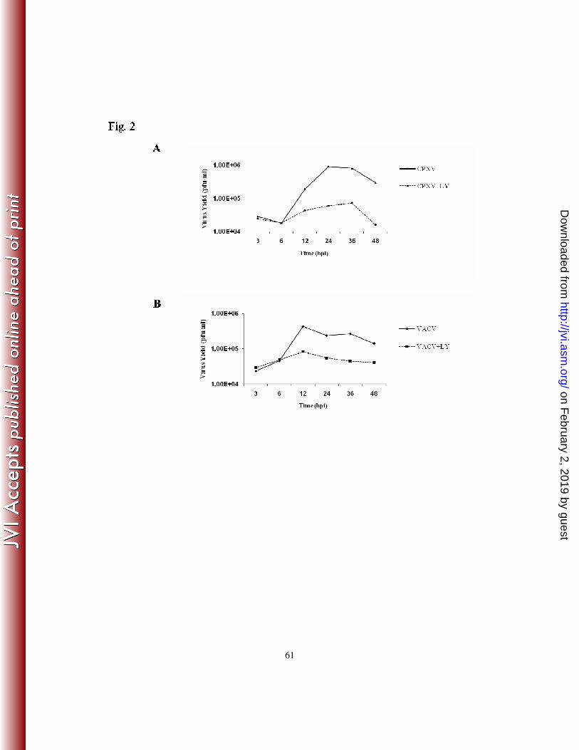
In order to investigate whether the Orthopoxvirus-stimulated PI3K/Akt pathway was of
biological relevance to the virus, we performed a one-step viral growth curve experiment,
either in the presence or absence of LY294002. A31 cells were left untreated or treated
with LY294002 (20 µM) for 30 min. prior to viral infection at an MOI of 10. Viruses were
then collected at 3, 6, 12, 24, 36 and 48 hpi and assayed for infectivity. The data indicate
that the PI3K/Akt pathway did play a relevant role in both VACV and CPXV biology. A
significant reduction in viral titers (≥ 90%) was observed when either the CPXV (Fig. 2A)
or VACV (Fig. 2B) infection was carried out in the continued presence of the PI3K
inhibitor.
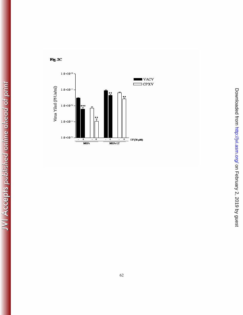
In order to verify that the inhibitory effect associated with LY294002 was not restricted to
the A31 cells, MEFs were also infected with VACV or CPXV as described above. As
shown in Fig. 2C, LY294002 caused a significant decline in viral titers (85-90%), thereby
demonstrating that viral inhibition is not cell-type specific. Next, we investigated the viral
replication in the context of a non-spontaneously immortalized cell line such as SV40-LT-
immortalized MEFs (MEFs-LT). Cells were infected with VACV or CPXV under the same
experimental conditions used for MEFs and then the titers were measured. Our findings
revealed that virus titers were decreased by 50 (2.0-fold) and 58% (2.4-fold), respectively.
Next, to rule out the possibility of the non-specific pharmacological inhibition of
PI3K/Akt, we generated cell lines stably expressing dominant-negative form of Akt (DN-
on February 2, 2019 by guest
http://jvi.asm.org/
Dow
nloaded from
19
Akt – K179M). Clones were then monitored for the expression of HA-Akt by western blot
using an anti-HA antibody and the VACV stimulation of Akt (S473-P). Two representative
clones expressing dominant-negative Akt (DN-Akt) are shown in Fig. 2D (top - left panel,
Lanes 2 and 3), as well as the control cells transfected with the empty vector only (A31*,
Lane 1), and in Fig. 2D – top - right panel, where the levels of Akt-P was significantly
reduced upon viral infection (lanes 4 and 6). The importance of Akt in viral replication was
determined by measuring the viral titers following the infection of cells expressing DN-
Akt. The representative clones were infected with VACV or CPXV at an MOI of 10 and
24 hpi viruses were collected and the titers determined. As shown in Fig. 2D (bottom
panel), viral titers were significantly decreased (~70-80%) in cells expressing DN-Akt,
suggesting that the PI3K/Akt pathway does play an important role in the VACV and
CPXV life cycle.
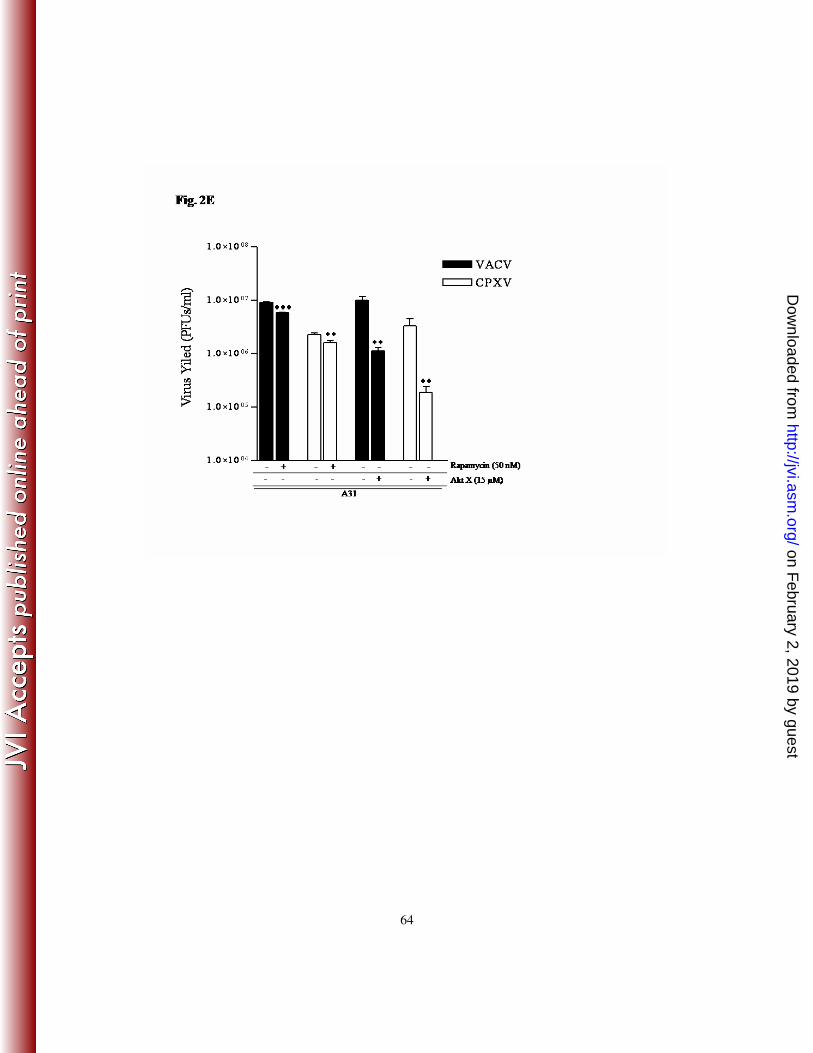
To further strengthen the involvement of Akt in mediating upstream signals after viral-
stimulation of PI3K, we pretreated the cell with the pharmacological inhibitor of Akt, Akt-
X (15 µM), infected the cells at an MOI of 10 with VACV or CPXV in the continued
presence of the inhibitor and 24 hpi virus was collected and assayed for infectivity. Our
data showed that the viral titers were significantly reduced by ≥ 90% (Fig. 2E).
Collectively these data strongly suggest that the PI3k/Akt pathway is beneficial for VACV
and CPXV replication.
Since mTORC1, the downstream target of PI3K/PDK1/Akt (T308-P) regulates key cellular
events such as survival, proliferation and translation, and because maintenance of
translation is important for viral cap-dependent mRNA translation, we investigated
on February 2, 2019 by guest
http://jvi.asm.org/
Dow
nloaded from
20
whether mTORC1 could be required for VACV and CPXV replication. A31 cells were left
untreated or incubated for 30 min. prior to virus infection with rapamycin (50 nM), a
specific pharmacological inhibitor of mTORC1. Cells were infected in the continued
presence of rapamycin at an MOI of 10 and 12 hpi viruses were collected and assayed for
infectivity. This period of time was chosen not only because it is the earliest time where a
significant decline in virus yield was verified (Fig. 2A-B), but also to rule out the
possibility of the non-specific inhibition of mTORC2 observed after prolonged exposure of
the cells to rapamycin (65). Our findings demonstrate that VACV and CPXV replication
was only partially affected (~35% - 1.5-fold decrease) following mTORC1 inhibition (Fig.
2E). Similar levels of inhibition were also observed following incubation with different
concentrations of rapamycin (30 and 70 nM), while higher drug concentration (100 nM)
appeared to increase the cytotoxic effect (data not shown).
3 - DISRUPTION OF THE PI3K/AKT PATHWAY IS FOLLOWED BY ALTERED
EXPRESSION OF EARLY/LATE VIRAL GENES
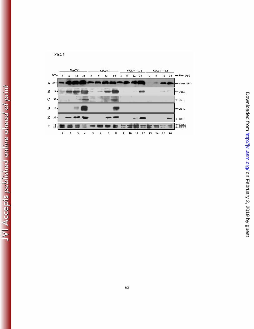
To gain insight into the mechanism(s) underlying the decreased viral yield upon treatment
with LY294002, experiments were designed to investigate whether early (CrmA/SPI-2)
and/or late (F18R, H3L, A14L and D8L) viral gene expression was affected following the
pre-incubation of the cells with the inhibitor. Cells were cultured either in the presence or
absence of LY294002 and infected with VACV or CPXV at an MOI of 10 for the indicated
times. Cellular lysates were then collected and subjected to western blotting using
antibodies raised against the viral proteins CrmA/SPI-2 (A), F18R (B), H3L (C), A14L (D)
and D8L (E). As shown in Fig. 3, viral gene expression was remarkably affected upon
on February 2, 2019 by guest
http://jvi.asm.org/
Dow
nloaded from
21
treatment with LY294002 and protein expression was either abrogated (panels B-D) or
delayed (panels A, E). This emphasizes the critical role played by the PI3K/Akt pathway in
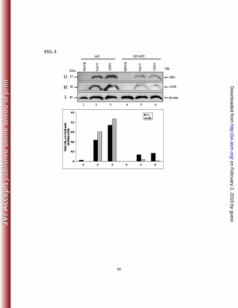
the regulation of Orthopoxvirus gene expression. It was also provided genetic evidence
that activation of the PI3K/Akt pathway is required for viral late gene expression.
Similarly, the infection of cells expressing DN-Akt demonstrated that both A14L and H3L
expression were significantly reduced (Fig. 3G-H).
4 - ALTERED VIRAL EARLY/LATE GENE EXPRESSION IS ACCOMPANIED
BY AN ARREST IN ORTHOPOXVIRUS MORPHOGENESIS
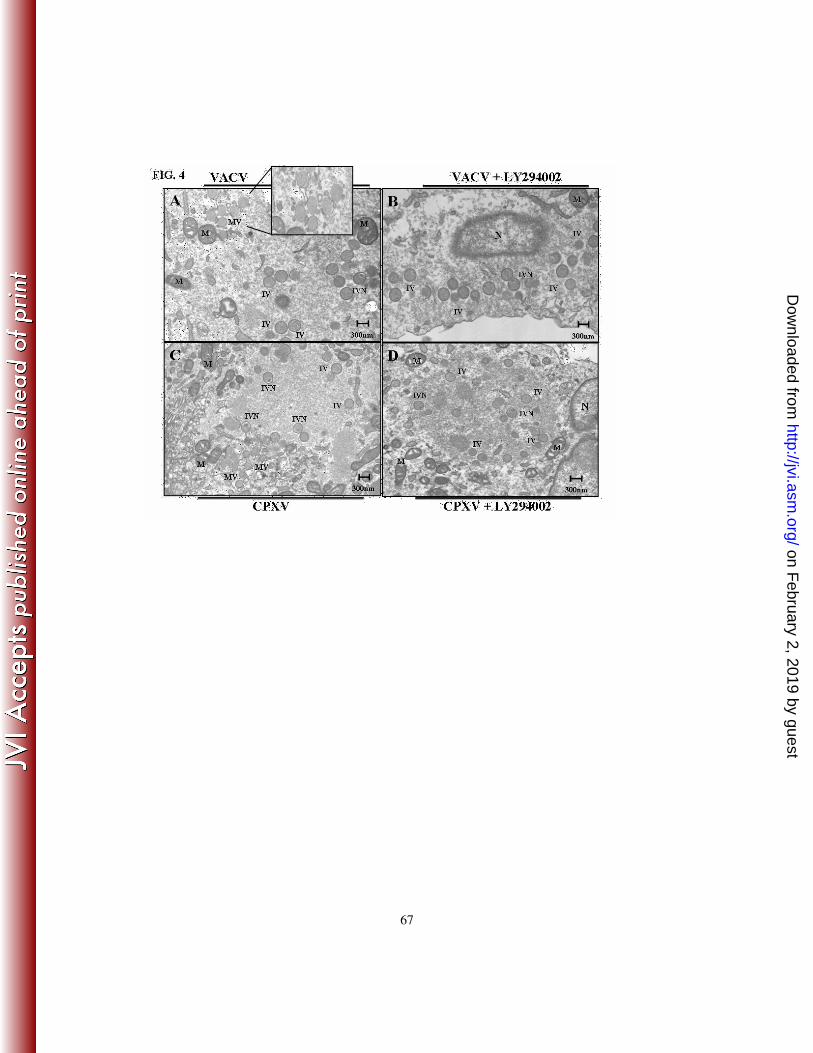
In order to investigate whether the altered expression of the viral early/late genes
demonstrated in Fig. 3, was accompanied by an arrest in virion morphogenesis, cell
cultures were left untreated (panels A and C) or were pretreated with LY294002 (20 µM)
(panels B and D) and then infected with VACV (A, B) or CPXV (C, D) at an MOI of 2 for
18 and 22 h, respectively. As shown in Fig. 4, while cultures of infected cells alone (panels
A and C) contained the full spectrum of normal intermediates and mature virions typically
seen in virion morphogenesis, cells pre-incubated with LY294002 (panels B and D)
resulted in an infectious cycle arrested at the immature virion (IV) or IV with nucleoids
(IVN) stage of the virion morphogenic cycle. Therefore, the inhibition of the PI3K/Akt
pathway resulted in an arrest that equally affected the same stages of the morphogenic
cycle of both orthopoxviruses. Furthermore, the arrest in virion morphogenesis is
consistent with the altered viral gene expression induced by both the PI3K inhibitor and
DN-Akt.
on February 2, 2019 by guest
http://jvi.asm.org/
Dow
nloaded from
22
5 - VIRAL STIMULATION OF THE PI3K/AKT PATHWAY INDUCES THE
CLEAVAGE OF PARP AND CASPASE-3
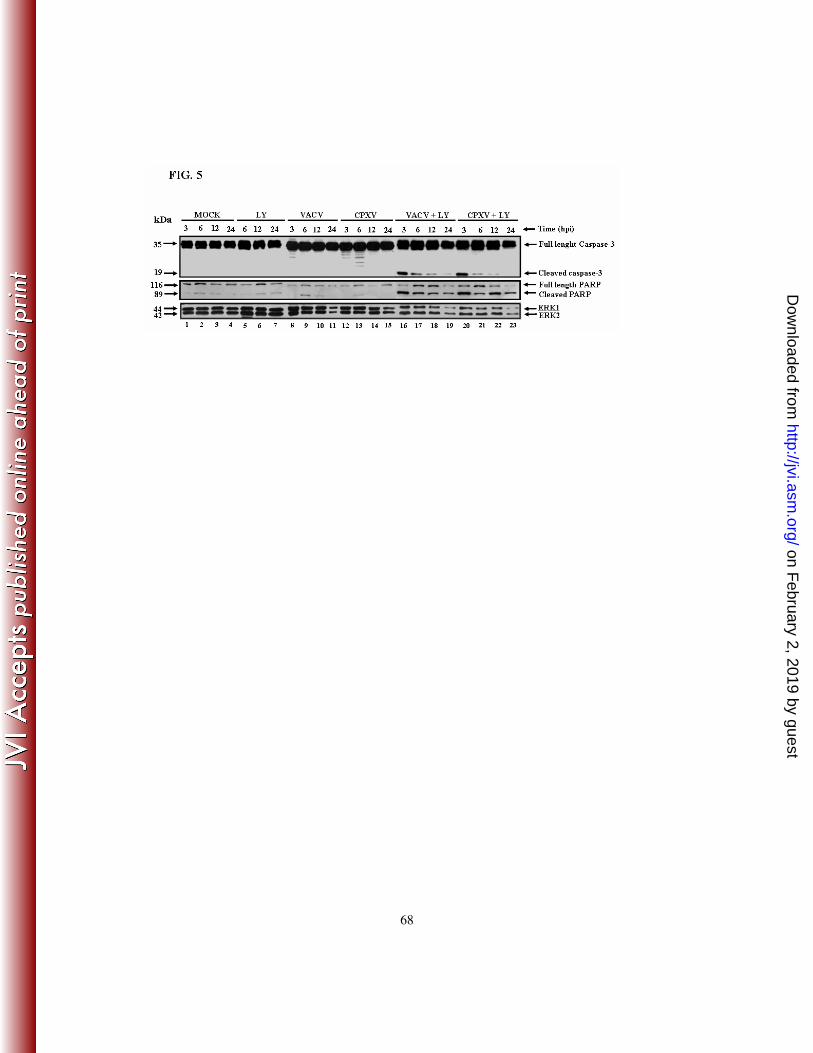
To further elucidate the role of the PI3K/Akt pathway in regulating the survival/apoptosis
of Orthopoxvirus-infected host cells, we blocked the pathway by pre-incubating the cells
with LY294002, infected the cells with virus, and then investigated whether the cleavage
of host proteins associated with characteristic hallmark features of apoptosis, such as
caspase-3 and PARP, was affected. Cells were mock-infected or infected with VACV or
CPXV at an MOI of 10 for 3, 6, 12 or 24 h, in either the presence or the absence of
LY294002 (20 µM). Cell lysates were then harvested and subjected to western blotting
with anti-caspase-3 (Fig. 5 - top panel) and anti-PARP (Fig. 5 – middle panel) antibodies.
Pharmacological inhibition of the PI3K/Akt pathway resulted in the cleavage of caspase-3
and PARP in Orthopoxvirus-infected cells (Fig. 5– top/middle panels, lanes 16-23). These
findings strongly suggest that the viral stimulation of the PI3K/Akt pathway has an anti-
apoptotic/pro-survival effect.
6 - INHIBITION OF APOPTOSIS IS FOLLOWED BY AN ENHANCEMENT OF
THE VIRUS-STIMULATED SURVIVAL PATHWAY
We have demonstrated that the pharmacological blockade of the PI3K/Akt pathway is
followed by a decline in Orthopoxvirus replication (Fig. 2). Combined with the pro-
apoptotic data presented in Fig. 5, these findings strongly suggest that, at least in part, the
increased cytopathic effect observed in the infected cells (see Fig. 6C and 6D – compare
on February 2, 2019 by guest
http://jvi.asm.org/
Dow
nloaded from
23
panels d and h with b and f) were due to the pharmacological inhibition of the anti-
apoptotic activity mediated by the PI3K/Akt pathway.
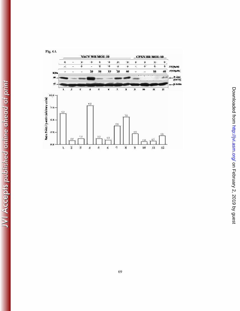
In order to further investigate this, A31 cells were pre-incubated for 30 min. with
increasing concentrations (10, 15, 20 and 40 µM) of the pan-caspase inhibitor zVAD.fmk,
either in the absence or presence of LY294002 (20 µM). Cells were then infected with
virus at an MOI of 10 and 3 hpi, cell lysates were collected and subjected to western
blotting with the anti-Akt-S473-P antibody. As shown in Fig. 6A, incubation with
zVAD.fmk (20 µM) prior to VACV infection increased the levels of Akt-P (lane 4), which
were even more pronounced (1.25-fold) than the level seen in VACV infection alone (lane
1). In contrast, pharmacological inhibition of the PI3K/Akt pathway resulted in decreased
levels of Akt-P upon infection (lane 3). Remarkably, incubation with zVAD.fmk at 10, 15,
20 or 40 µM reversed the levels of VACV-mediated Akt phosphorylation in a dose-
dependent manner, even in the presence of LY294002 (lanes 5-8). Although not as
pronounced as those verified with VACV infection, similar results were also observed
when the infections were performed with CPXV (lanes 9-12).
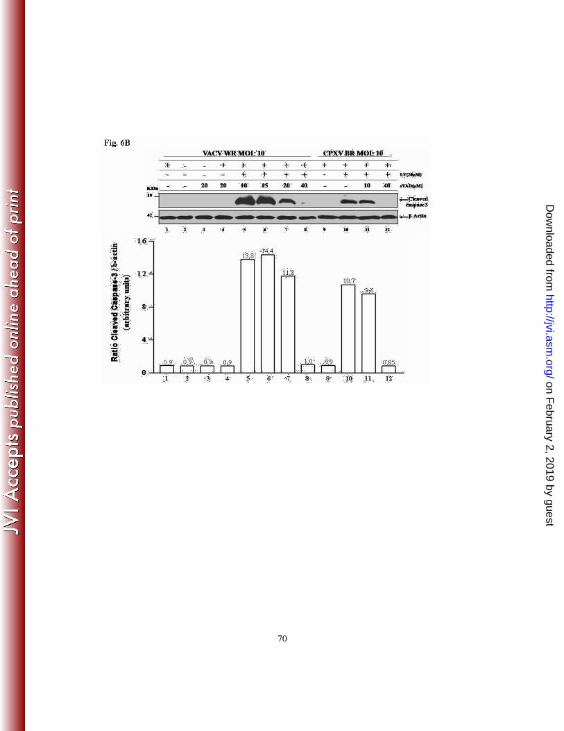
Given that the inhibition of pro-apoptotic signals (by zVAD.fmk) is accompanied by
enhanced host survival signals (e.g., increased Akt-P) upon VACV and CPXV infection,
one would expect that under this condition, the cleavage of caspase-3 after exposure to
zVAD.fmk would also be inhibited in a dose-dependent manner. To investigate this
hypothesis, A31 cells were incubated with 10, 15, 20 or 40 µM of zVAD.fmk for 30 min.,
either in the absence or presence of LY294002 (20 µM), prior to viral infection at an MOI
of 10 for 3 h. Cell lysates were collected and subjected to western blotting with an anti-
on February 2, 2019 by guest
http://jvi.asm.org/
Dow
nloaded from

24
caspase-3 antibody that specifically detects the cleaved form of caspase-3 (17-19 kDa). As
demonstrated in Fig. 6B, the cleavage of caspase-3, as a consequence of inhibition of the
survival pathway by LY294002 (lanes 5-8), was inhibited in a dose-dependent manner by
the anti-apoptotic compound zVAD.fmk. This would increase cell viability and,
subsequently, favor viral replication. The same set of experiments was performed after
infection with CPXV and the results were similar to those found upon VACV infection
(lanes 9-12).
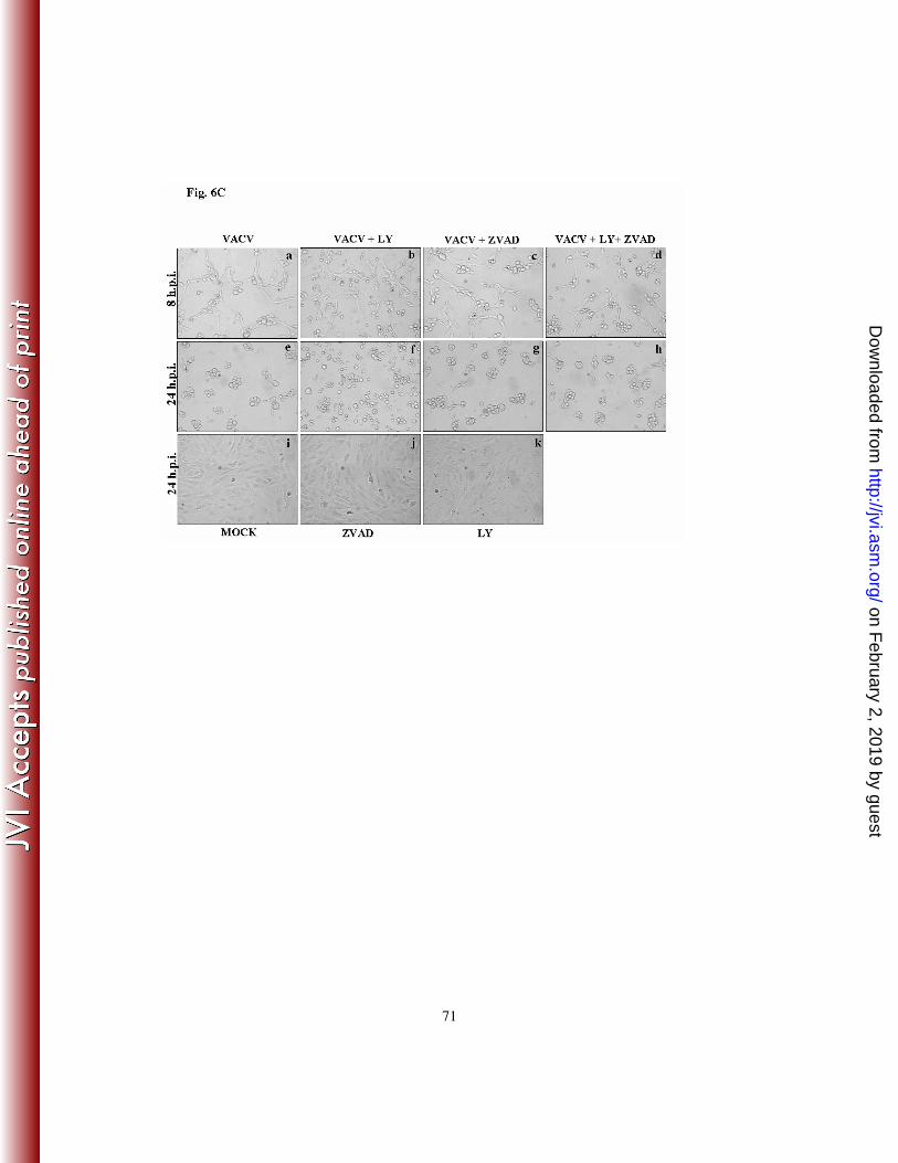
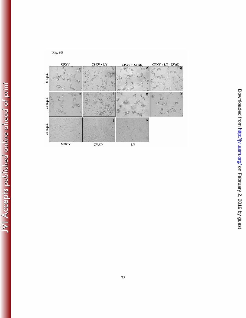
With this in mind, we then investigated whether pre-incubation with zVAD.fmk was
sufficient, at least in part, to reverse the increased cytopathic effect observed with the
infections carried out in the presence of LY294002. Cells were pre-incubated with
LY294002 (20 µM) for 30 min. and then treated with zVAD.fmk (40 µM) for an additional
30 min. before infection at an MOI of 10 for 8 or 24 h with VACV (Fig. 6C) or CPXV
(Fig. 6D). The infected cells were then examined by phase-contrast microscopy. Our
findings suggest that the anti-apoptotic signals induced upon the exposure to zVAD.fmk
not only decreased the cytopathic effect, but also increased the cell viability in both
infection models (Fig. 6C and 6D - compare panels d and h with b and f). Furthermore,
while the infections carried out for 24 h in the presence of LY294002 alone led to an
abundant number of detached cells that were recovered from the supernatant, cells infected
in the simultaneous presence of LY294002 and zVAD.fmk not only remained significantly
more attached to the substrate but were recovered in the supernatant to a lesser extent (data
not shown). Altogether, these data indicate that the inhibition of pro-apoptotic signals,
accompanied by enhanced host survival signals and increased cell viability upon VACV or
CPXV infection, play an important role during the viral infective cycle.
on February 2, 2019 by guest
http://jvi.asm.org/
Dow
nloaded from
25
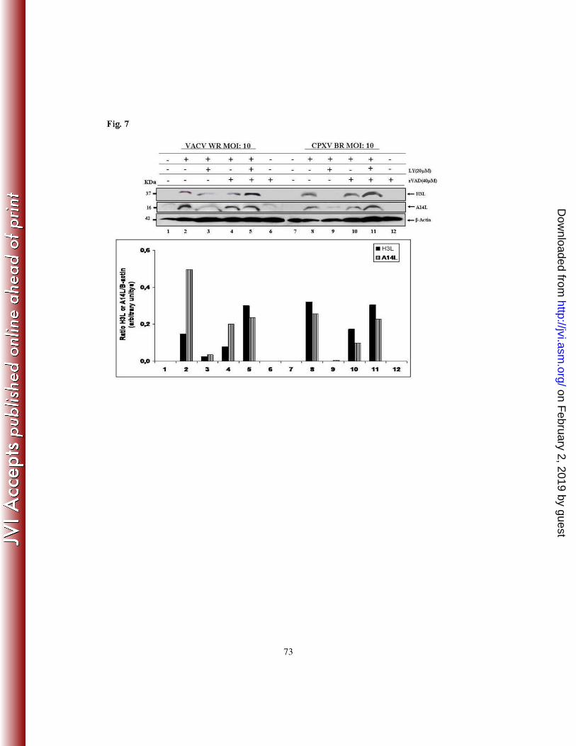
7 - INHIBITION OF VIRAL LATE PROTEIN EXPRESSION BY LY294002 IS
CASPASE-DEPENDENT
In order to investigate whether the LY294002-mediated inhibition of viral late protein
expression is due to an acceleration of apoptosis in infected cells, we compared the
expression of the viral proteins A14L and H3L in the absence or presence of zVAD.fmk.
A31 cells were incubated with LY294002 (20 µM) for 30 min. and then treated with
zVAD.fmk (40 µM) for an additional 30 min. prior to VACV or CPXV infection at an
MOI of 10 for 24 h. Cell lysates were collected and subjected to western blotting with anti-
A14L or anti-H3L antibodies. Remarkably, our results indicate that the inhibition of
caspase-3 cleavage (Fig. 6B) and apoptosis by the general pan-caspase inhibitor
zVAD.fmk reverses the inhibitory effect of LY294002 on viral A14L and H3L expression
(Fig. 7). The observation that zVAD.fmk is capable of blocking caspase-3 cleavage in
infected cells (Fig. 6B) in association with the reversion of A14L and H3L expression,
even in the presence of LY294002 (Fig. 7 – lanes 5 and 11), indicates that the regulatory
effect of the PI3k/Akt pathway exerted during Orthopoxvirus replication, is a caspase-
dependent event.
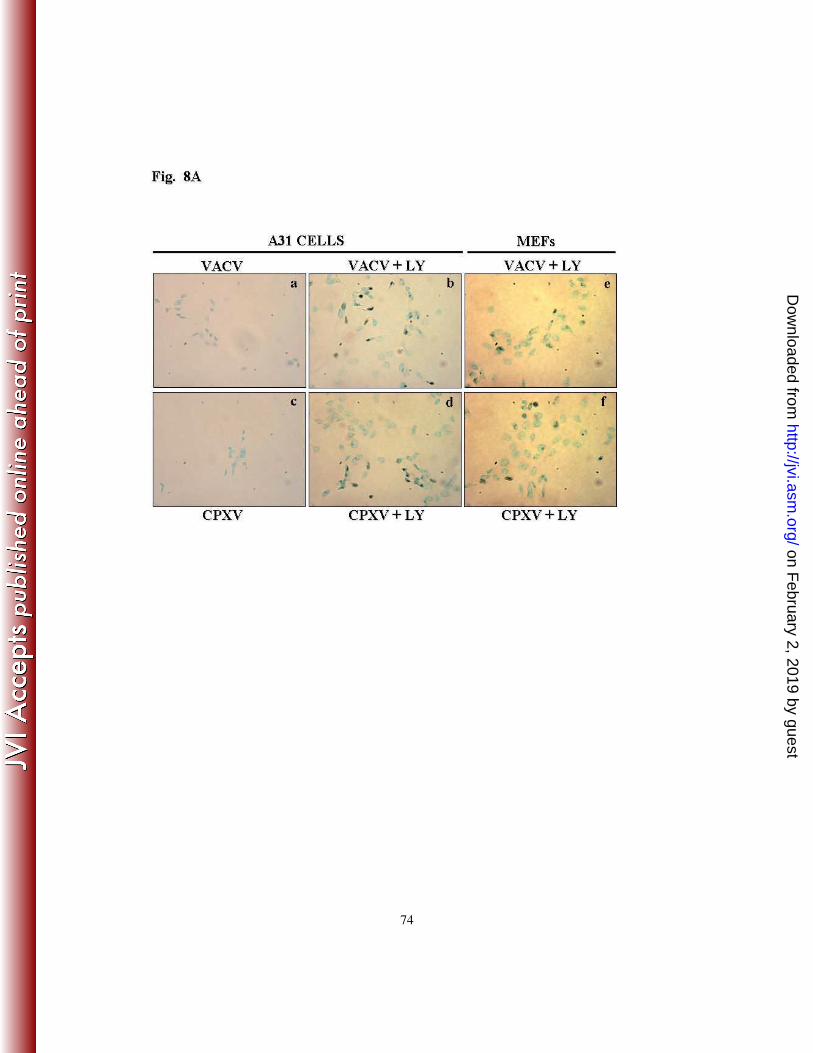
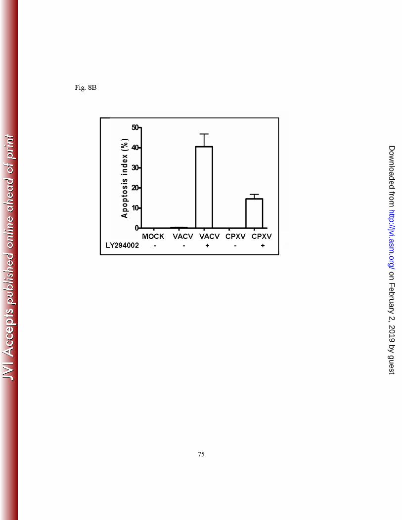
8 - INHIBITION OF THE PI3K/AKT PATHWAY BY LY294002 RESULTS IN
APOPTOSIS OF INFECTED CELLS.
In order to firmly establish that the inhibition of the PI3k/Akt pathway by LY294002
results in the apoptosis of the infected cells, a TUNEL assay was used to monitor apoptosis
on February 2, 2019 by guest
http://jvi.asm.org/
Dow
nloaded from
26
in individual cells. A31 cells were incubated with LY294002 (20 µM) for 30 min. prior to
viral infection with VACV or CPXV at an MOI of 10 for 4 and 6 h, respectively. A brown
precipitate confined to the nucleus was considered a TUNEL-positive cell. As shown in
Fig. 8A, TUNEL-positive nuclei were barely observed in the absence of LY294002 in the
mock-infected cells (not shown) and in the VACV-infected (panel a) or the CPXV-infected
cells (panel c). However, the number of TUNEL-positive cells increased significantly only
in cultures pre-incubated with LY294002 and infected with VACV or CPXV (panel b and
d). The apoptosis index was calculated and the pre-treatment with LY294002 resulted in a
41% ± SD and a 16% ± SD increase in the number of apoptotic cells following infection
with VACV or CPXV, respectively (Fig. 8B). To certify that the inhibition of the PI3k/Akt
pathway that results in apoptosis of A31-infected cells was not a cell-type specific event,
the same approach was used with MEFs. As demonstrated in Fig. 8A (panels e and f),
VACV- or CPXV-infected MEFs also underwent apoptosis upon treatment with
LY294002, while apoptosis was not observed in viral-infected cells when in the absence of
LY294002 (data not shown). The apoptosis index in these cells was also determined and it
was found similar to that shown in Fig. 8B (data not shown). Thus, these findings not only
confirm the hypothesis that the pharmacological inhibition of the PI3k/Akt pathway
induced the infected spontaneously immortalized cells to undergo apoptosis, but also
suggest that, under these conditions, CPXV appears to be more effective than VACV in
inhibiting apoptosis.
on February 2, 2019 by guest
http://jvi.asm.org/
Dow
nloaded from
27
DISCUSSION
Virus replication and assembly relies on a delicate balance between host-defense
mechanisms and the viral manipulation of signaling pathways that govern host responses.
Furthermore, viral activation of intracellular pathways associated with the transmission of
mitogenic and host survival signals can improve viral utilization of the host resources and
metabolism required to efficiently produce viral progeny (13, 18).
We have previously demonstrated that both VACV and CPXV stimulate the mitogen-
activated protein kinase/extracellular-signal regulated kinases 1/2 (MAPK/ERK1/2)
throughout the viral life cycle (4, 71). However, while stimulation of the pathway is
beneficial for VACV multiplication, the biological significance of CPXV-mediated
MAPK/ERK1/2 activation requires further clarification.
In the present report, we demonstrate that both VACV and CPXV stimulate a signaling
pathway that results in the phosphorylation of protein kinase B/Akt on both S473 and
T308, modifications that are mediated by mTORC2 and PI3K/PDK1, respectively (9, 63,
66). We also demonstrate that incubation of cells with a pharmacological inhibitor of
PI3K, LY294002, prior to viral infection impairs the signal transduction required for the
induction of Akt-P in a dose-dependent manner (Fig. 1A, C). Our findings are also
consistent with the hypothesis that the phosphorylation of Akt (S473/T308) is an early
event that takes place soon after viral attachment/entry (e.g., 1-3 hpi; Fig. 1D). Moreover,
the induction of Akt-P does not require replication-competent virions, since infection with
UV-inactivated virus also induced Akt-P (Fig. 1A). Importantly, this is consistent with the
on February 2, 2019 by guest
http://jvi.asm.org/
Dow
nloaded from
28
previously described viral requirement for cytoskeletal alterations early during the
infection (43, 49) and activation of mTORC2, as well as viral-stimulated early survival
signals. Notably, the kinetics of Akt-P induction were distinct for these orthopoxviruses.
Thus, while Akt-P (S473/T308) is induced by CPXV only at early time points following
infection (1-3 hpi), a time-frame in which the UV-irradiated virus is also able to induce
Akt-P (Fig 1A, lanes 17- 20), late stimulation of Akt-P upon VACV infection requires
post-DNA replication events (Fig. 1B). Thus, the induction of Akt-P may be a consequence
of the interaction between a viral gene product and the cellular PI3K. Such an event may
result in the transmission of signals to the downstream effector, Akt. A similar signaling
pathway has been described for the host range factor MT-5 from MYXV: activation of Akt
by MYXV is dependent on the association between MT-5 and Akt (88). However, the
precise mechanisms underlying VACV- and CPXV-mediated stimulation of the PI3K/Akt
pathway remain to be elucidated.
Our data also provide evidence of the relevant role played by the PI3K/Akt pathway in the
life cycle of these orthopoxviruses. Both VACV and CPXV growth are significantly
reduced by ≥ 90% (one log unit – 10-fold) in the presence of LY294002 (Fig. 2A-B). This
observation was further supported by the treatment of the infected cells with the specific
Akt inhibitor, Akt-X. Akt-X treatment resulted in a reduction (≥ 90%) in the titers of both
VACV and CPXV (Fig. 2E) and therefore of the same magnitude verified with LY294002.
Furthermore, the infection of cell lines expressing DN-Akt (Fig. 2D) confirms the
involvement of the PI3K/Akt pathway in viral replication (about 70-80% decrease in viral
titers) and rules out the possibility of a non-specific pharmacological inhibition of
LY294002. Considering that: 1) PI3K is targeted by LY294002 and that PDK1 is the
on February 2, 2019 by guest
http://jvi.asm.org/
Dow
nloaded from
29
downstream effector of PI3K leading to Akt-T308-P and 2) LY294002, though indirectly,
inhibits the induction of Akt-S473-P, these strongly suggest that Akt is fully activated upon
infection with these orthopoxviruses. It has also been demonstrated that viral replication
declines significantly in MEFs pretreated with LY294002, confirming the biological
relevance of the PI3K/Akt pathway for these orthopoxviruses (Fig. 2C). It is worth noting,
that while pharmacological inhibition of PI3K/Akt (LY294002 and Akt-X) resulted in ≥
10-fold reduction in viral titers in the spontaneously immortalized cells, A31, (Fig. 2A, B
and D) and about the same level of inhibition was also verified with MEFs (LY294002),
the decline in virus yields verified after the exposure of SV40-LT MEFs to LY294002 was
just partially affected (2-2.4-fold reduction) (Fig. 2C). Thus, these findings suggest that the
pre-activation state of the intracellular environment in the SV40-LT MEFs per se (9) is
beneficial for virus replication and that the pre-treatment with LY294002 just block the
viral increment that follows the infection, which appears to reflect the partial reduction
observed.
In agreement with previous data (85), we also demonstrate that both VACV and CPXV
require mTORC1 during the infective cycle. These findings are consistent with the viral
mechanisms used to translate their cap-dependent mRNAs, a pathway that is regulated by
the PI3K/PDK1/Akt (T308-P)/mTORC1 signaling pathway (reviewed in 9). While the
overall effect of LY294002 and Akt-X on viral replication is measured by a decline of ≥
90% (≥10-fold decrease) in viral titers, the blockade of mTORC1 by rapamycin (which
affects mRNA translation) resulted in a reduction of ~35% (1.5-fold decrease) in virus
yield, thereby suggesting that the others biological activities regulated by the PI3K/Akt
pathway, beyond mTORC1 activation, seems to be co-activated in order to attend the
on February 2, 2019 by guest
http://jvi.asm.org/
Dow
nloaded from
30
diverse viral infective demands and, thus, maximize viral replication. However, it has been
known that the viral genes E3L and K3L are also activated to bypass the block of viral
mRNA translation imposed by PKR (12, 34, 52). Remarkably, as has been described for
HSV (83, 84) and HCMV (37, 86) infection, VACV also releases translation using a
mechanism that operates downstream of mTOR. VACV requires mTORC1 to
phosphorylate the eukaryotic translational repressor eIF4E binding protein (4E-BP).
Following this phosphorylation, 4E-BP is degraded by the proteasome. Indeed, it has been
demonstrated that by the inactivation of 4E-BP, VACV alters the activity of eIF4F
complex and stimulates the accumulation of eIF4F components (eIF4E and eIF4G) within
the viral factories, thereby facilitating viral replication (85). While the mechanism
employed by CPXV to release translation awaits further investigation, it is reasonable to
assume that the strategy used by CPXV might resemble the strategy described for VACV.
Thus, we conclude that by fully activating Akt VACV and CPXV facilitate/maximize their
own replication.
Consistent with the viral requirement of the PI3k/Akt pathway for a productive infection, if
this pathway is inhibited upstream, viral gene expression is remarkably affected (Fig. 3).
While the inhibition of PI3K/Akt appears to affect viral gene expression to different
extents, the delay observed with CPXV induction of CrmA seems to be more pronounced
than that observed with VACV (compare lanes 9-12 with 13-16). However, the delayed
expression of this viral anti-apoptotic gene does not appear to affect the global CPXV anti-
apoptotic mechanisms, as shown in Fig. 8B. However, the mechanisms underlying this
effect require further exploration. Furthermore, genetic evidence also confirms the
involvement of the pathway with viral gene expression, since infection of DN-Akt cells
on February 2, 2019 by guest
http://jvi.asm.org/
Dow
nloaded from
31
results in a significant decrease in H3L and A14L expression (Fig. 3G-H). Additionally,
the PI3K/Akt pathway plays an important role in virion morphogenesis, as demonstrated
by the arrest that occurs at the immature virion/immature virion with nucleoid stage of the
morphogenic cycle when the infections are carried out in the presence of LY294002 (Fig.
4). Similarly, the functional or genetic ablation of A14L or H3L genes also resulted in an
arrest early during virion morphogenesis (40, 62). Taken together, these data strongly
demonstrate the beneficial role played by the PI3K/Akt pathway in both VACV and CPXV
morphogenesis and growth.
Successful viral replication and assembly is dependent upon cell survival. Therefore,
viruses have also evolved diverse mechanisms to control the pathways that govern cell
survival (13, 19, 21). The PI3K/Akt pathway has demonstrated to play a pivotal role in cell
survival and proliferation (16, 78) and the inhibition of the pathway is associated with a
significant decrease in host cell viability (8, 78) and virus replication (19, 30, 42, 77).
Therefore, it is reasonable to assume that VACV and CPXV stimulate the PI3K/Akt
pathway to increase the viability of infected host cells in order to prolong the lifespan of
the cell, which would allow more time for the virus to generate its progeny. As seen in Fig.
5, the inhibition of the PI3K/Akt pathway during either VACV or CPXV infection
significantly increases the cleavage of proteins associated with the induction of apoptosis,
such as the executioner caspase-3 and PARP (19, 38, 41) (Fig. 5 - upper and middle
panels). This strongly suggests an anti-apoptotic role for the pathway in the course of these
orthopoxviruses infections.
on February 2, 2019 by guest
http://jvi.asm.org/
Dow
nloaded from
32
Our findings also demonstrate that the pan-caspase inhibitor, zVAD.fmk, reverses the pro-
apoptotic signals associated with the blockade of the PI3K/Akt pathway by increasing the
levels of Akt-P (Fig. 6A). This observation, in association with data showing that
zVAD.fmk also reverses the cleavage of caspase-3 (Fig. 6B), reinforces the anti-apoptotic
role of the virus-stimulated pathway.
It has long been known that the PI3K/Akt pathway plays a critical role in cell survival and
proliferation and that its disruption is associated with a significant decrease in host cell
viability (8, 57). Thus, the maintenance of pro-survival and anti-apoptotic signals upon
viral infection is of critical importance for successful viral replication. Our data
demonstrate that zVAD.fmk not only decreases the cytopathic effect associated with
LY294002 during the infections, but also appears to increase cell adherence and viability
(Fig. 6C and 6D), which are critical requirements for generation of viral progeny.
Furthermore, the LY294002-mediated inhibition of late viral gene expression, as
demonstrated by analysis of the A14L and H3L proteins, is a caspase-dependent process
(Fig. 7). Importantly, viruses have evolved diverse means to regulate viral genes
expression in either a caspase-dependent, (e.g., flaviviruses (38) and orthopoxviruses (this
study)) or caspase-independent (e.g., coxsackievirus (19)) manner.
Our hypothesis that the pharmacological blockade of the PI3K/Akt pathway was, at least in
part, associated with pro-survival and anti-apoptotic signals (Figs. 5-8) was further
strengthened by the observation that infected cells undergo apoptosis upon exposure to
LY294002, a phenomenon that was not only verified in A31 cells but also in MEFs (Fig.
8). Therefore, receptor-mediated signals conveyed through the PI3K/Akt pathway at early
on February 2, 2019 by guest
http://jvi.asm.org/
Dow
nloaded from
33
times during the infection of permissive and spontaneously immortalized cells (e.g., A31
and MEFs) appear to be important to control cell survival/apoptosis. Additionally, these
signals should precede the expression of the viral anti-apoptotic genes (CrmA, F1, E3L and
N1) because neither CPXV nor VACV seems to be capable on their own of fully
preventing the cells from undergoing apoptosis in the presence of LY294002 (Fig. 8).
While the activation of this pathway appears to favor VACV and CPXV replication (Figs.
2-5, 7, 8), it is remarkable that the levels of Akt activation not only correlate with cancer
progression (i.e., higher metabolic, survival and proliferation activities) (80), but also with
the levels of permissiveness to infection with the otherwise rabbit-specific poxvirus,
MYXV, in a diverse set of human transformed cell lines (88, 92). Therefore, it is tempting
to speculate that levels of Akt-P that have been associated with permissive transformed
cells (e.g., BSC-40 and HeLa cells) may facilitate the increased replication of VACV and
CPXV. In line with this assumption, it has consistently been verified that the viral yields
following the infection of HeLa or BSC-40 cells with either VACV or CPXV are at least
ten-fold higher than those obtained following the infection of A31 or MEF cells. Not
surprising, the viral yields were only partially affected (~2-fold reduction) when the
infection of BSC-40 was carried out in the continued presence of LY294002, or following
the infection of SV40-LT-immortalized MEFs, which suggests that the pre-activation state
of Akt in these permissive lines, as well as in other transformed cell lines, is sufficient to
elicit the activation of downstream signaling that is required early during the infection (Fig.
2C, our unpublished observations). However, this must be conclusively confirmed. Since
most of the relevant experiments performed in vitro to elucidate the role of apoptosis
during VACV or CPXV infection were carried out with transformed cell lines (e.g., HeLa,
HEK293T, BSC-40, Jurkat or the monocyte/macrophage cell line J774.G8) (14, 28, 39, 74,
on February 2, 2019 by guest
http://jvi.asm.org/
Dow
nloaded from
34
75, 90, 91, 97), their levels of Akt activation, should be higher than those observed in
spontaneous immortalized permissive cells (e.g., A31 or MEFs). Furthermore, in
experiments in which MEFs were cultured under permissive conditions for the early
VACV-mediated PI3K/Akt signals, i.e. in the absence of LY294004, it was demonstrated
that the viral anti-apoptotic genes were necessary and sufficient to control apoptosis (75,
90).
Therefore, it was hypothesized that, under this circumstance (e.g., higher levels of Akt-P),
the viruses become less dependent of the early signals elicited by the host spontaneous
immortalized cell lines after viral infection and thereby, the viral anti-apoptotic repertoire
could sufficiently block the virus-induced apoptosis. This suggests that the early signals
transmitted by the PI3K/Akt pathway upon attachment/penetration of VACV or CPXV
(Fig. 1A, 1D) would be required not only for the cytoskeletal alterations, but also for host
survival. Because prior phosphorylation of Akt on S473 by mTORC2 has been reported to
be required for the phosphorylation of Akt on T308 by PDK1 (66, 67, 94), and the early
activation of mTORC1 is necessary for host survival and viral translation (26, 85), the
combination of these events, either dependently or independently, should provide the host
(A31 and MEFs) with the necessary means to avoid apoptosis before the viral anti-
apoptotic repertoire could be activated. It has long been known that orthopoxviruses have
evolved several strategies to regulate both the extrinsic and intrinsic apoptotic pathways
(25, 74). Nonetheless, our data suggest that these viral anti-apoptotic mechanisms, though
necessary, are not sufficient to ensure successful viral replication (Figs. 2 A-C, 3, 4, 5 and
8) (Fig. 9). This scenario, thus, contrasts with that of transformed cell lines, where higher
on February 2, 2019 by guest
http://jvi.asm.org/
Dow
nloaded from
35
levels of Akt activation should bypass the need for virus-induced survival signals and,
therefore, the virus anti-apoptotic genes should be sufficient to prevent apoptosis.
Even though CPXV appears to be more effective than VACV in inhibiting apoptosis, as
reflected by their apoptosis index (16% ± SD) vs. (41% ± SD), respectively, the overall
effect of LY294002 on viral replication was similar, though not equal, for both viruses
(Figs 2-8). This suggests that the inhibition of the PI3K/Akt pathway may impact aspects
of Orthopoxvirus biology other than the control of survival/apoptosis. Indeed, the
PI3K/Akt pathway regulates a variety of biological processes that are potentially important
for viral replication, including actin remodeling, cell migration (3, 29, 47, 59, 64) and
microtubule stabilization (10, 54, 23). Previous research has demonstrated that usurping
these pathways would benefit Orthopoxvirus in several ways: (1) transportation of the
virus through the microtubules to the cell periphery, followed by a switch from
microtubule- to actin-based motility (reviewed in 53); (2) transcription and translation of
viral mRNAs (32, 46); (3) alteration of the cytoskeletal organization and translocation of
translation factors to the viral factories (32, 53, 68, 85); (4) bleb formation and apoptotic
mimicry to penetrate the cell (49); and (5) phosphorylation of Akt (S473) by mTORC2,
which regulates the actin cytoskeleton (9, 37, 72). Therefore, it is not surprising that the
simultaneous incubation of the infected cells with LY294002 and zVAD.fmk, which
protects the cells only from apoptosis, restored the viral titers only partially (~20%) when
compared with the cells incubated with LY294002 alone (data not shown). This further
emphasizes the global effect of the PI3K/Akt pathway on the viral life cycle beyond host
survival.
on February 2, 2019 by guest
http://jvi.asm.org/
Dow
nloaded from
36
In addition, it is remarkable that although VACV and CPXV belong to the same genus,
they diverge in the way in which they manipulate cellular signaling pathways. While
stimulation of the PI3K/Akt pathway by both viruses facilitates viral replication, the
kinetics of this activation by the viruses are quite different. While VACV stimulated the
pathway during the early and late phases of the infective cycle, as determined by the need
for viral late gene expression at 24 hpi (Fig. 1B), CPXV stimulation of the same pathway
was restricted to the early phase of the viral infective cycle in both cell lines analyzed.
In conclusion, in this report, we demonstrated that the signals triggered by the PI3K/Akt
pathway upon VACV and CPXV infection do play an important role in controlling cell
survival and apoptosis. Nonetheless, it appears that the signals required for viral successful
replication extend beyond survival and apoptosis. As the late events related to the
release/spread of Orthopoxvirus are dependent upon actin dynamics, a role not only
associated with mTORC2, but also with the Src family of tyrosine kinases (reviewed in
53), it will be exciting to further investigate and compare how is the specific contribution
of Akt(S473-P) and the Src family kinases in the biology of these orthopoxviruses during
the late stages of their infective cycle. Maybe, this could explain the more pronounced
effect of both LY294002 and Akt-X on CPXV replication (Fig. 2 A-C).
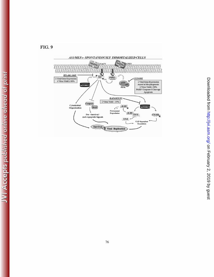
A schematic representation depicting the relevant findings of this study is shown in Fig. 9.
Soon after VACV and CPXV encounter a host spontaneously immortalized cell line such
as A31 or MEFs, signals from the PI3K/Akt pathway are triggered and required to release
translation and host survival, a function associated with the mTORC1, via upstream
phosphorylation of T308 by PDK1. Activation of mTORC2 (Akt473-P), which is required
on February 2, 2019 by guest
http://jvi.asm.org/
Dow
nloaded from
37
for cytoskeletal alterations and virus attachment/penetration, in conjunction with
phosphorylation of Akt on T308 via PI3K appears to boost kinase activities and, thus,
creates an intracellular environment that not only protects A31 and MEFs from undergoing
apoptosis, but also favors VACV and CPXV replication. Pharmacological or genetic
blockade of the PI3K/Akt pathway interrupt the early signals transmission and under this
circumstance the viral anti-apoptotic genes are not sufficient to prevent the cells from
undergoing apoptosis.
on February 2, 2019 by guest
http://jvi.asm.org/
Dow
nloaded from
38
ACKNOWLEDMENTS
The authors are grateful to Angela S. Lopes, Hilda M. V. Gama, João R. dos Santos,
Andreza A. Carvalho, Gisele O. L. Rodrigues and Jonas D. Albarnaz for their
secretarial/technical assistance. We also thank Drs. H. A. Armelin and M. C. Sogayar -
Department of Biochemistry - University of São Paulo, Brazil, Dr. Eileen D. Adamson -
The Burnham Institute, La Jolla, California, USA and Dr Richard D. Ye, Department of
Pharmacology – University of Illinois – Chicago - USA, who kindly provided us with the
A31 cell line, WT MEFs and plasmid expressing Akt dominant-negative mutation,
respectively. Dr. C. Ronald Kahn - Joslin Diabetes Center, USA, kindly provided us with
the SV40-LT immortalized MEFs. Viruses VACV (strain WR) and CPXV (strain BR)
were from Dr. C. Jungwirth, Universität Würzburg, Germany. We also thank Drs L. A.
Velloso (FMC - Unicamp) and J. A. Yunes (Centro I. Boldrini) - Campinas, for the Akt
antibody (T308). This work was supported by grants from Fundação de Amparo a Pesquisa
do Estado de Minas Gerais (FAPEMIG), CAPES (Coordenadoria de Aperfeiçoamento de
Pessoal de Nível Superior) - Brazilian Ministry of Culture, Science and Technology and
Conselho Nacional de Desenvolvimento Científico e Tecnológico (CNPq). Dr. JAPS was a
recipient of a pre-doctoral fellowship from CAPES and FGGL and LGA are recipient of
pre-doctoral fellowships from CNPq. Dr LSB holds a PRODOC research fellowship from
CAPES. CAB, EGK, PCPF, MMT and TSP are recipients of research fellowships from
CNPq. We also thank Bruno Brasil for critical reading of the manuscript.
on February 2, 2019 by guest
http://jvi.asm.org/
Dow
nloaded from
39
REFERENCES
1. Alcami, A. 2003. Viral mimicry of cytokines, chemokines and their receptors. Nat Rev
Immunol 3:36-50.
2. Alessi, D. R., M. Andjelkovic, B. Caudwell, P. Cron, N. Morrice, P. Cohen, and B. A.
Hemmings. 1996. Mechanism of activation of protein kinase B by insulin and IGF-1. Embo J
15:6541-51.
3. Amiri, A., F. Noei, S. Jeganathan, G. Kulkarni, D. E. Pinke, and J. M. Lee. 2007. eEF1A2
activates Akt and stimulates Akt-dependent actin remodeling, invasion and migration. Oncogene
26:3027-40.
4. Andrade, A. A., P. N. Silva, A. C. Pereira, L. P. De Sousa, P. C. Ferreira, R. T. Gazzinelli,
E. G. Kroon, C. Ropert, and C. A. Bonjardim. 2004. The vaccinia virus-stimulated mitogen-
activated protein kinase (MAPK) pathway is required for virus multiplication. Biochem J
381:437-46.
5. Avruch, J., K. Hara, Y. Lin, M. Liu, X. Long, S. Ortiz-Vega, and K. Yonezawa. 2006.
Insulin and amino-acid regulation of mTOR signaling and kinase activity through the Rheb
GTPase. Oncogene 25:6361-72.
6. Bablanian, R., G. Coppola, S. Scribani, and M. Esteban. 1981. Inhibition of protein
synthesis by vaccinia virus. III. The effect of ultraviolet-irradiated virus on the inhibition of
protein synthesis. Virology 112:1-12.
7. Benetti, L., and B. Roizman. 2006. Protein kinase B/Akt is present in activated form
throughout the entire replicative cycle of deltaU(S)3 mutant virus but only at early times after
infection with wild-type herpes simplex virus 1. J Virol 80:3341-8.
on February 2, 2019 by guest
http://jvi.asm.org/
Dow
nloaded from
40
8. Bondar, V. M., B. Sweeney-Gotsch, M. Andreeff, G. B. Mills, and D. J. McConkey. 2002.
Inhibition of the phosphatidylinositol 3'-kinase-AKT pathway induces apoptosis in pancreatic
carcinoma cells in vitro and in vivo. Mol Cancer Ther 1:989-97.
9. Buchkovich, N. J., Y. Yu, C. A. Zampieri, and J. C. Alwine. 2008. The TORrid affairs of
viruses: effects of mammalian DNA viruses on the PI3K-Akt-mTOR signalling pathway. Nat
Rev Microbiol 6:266-75.
10. Buttrick, G. J., and J. G. Wakefield. 2008. PI3-K and GSK-3: Akt-ing together with
microtubules. Cell Cycle 7:2621-5.
11. Cardone, M. H., N. Roy, H. R. Stennicke, G. S. Salvesen, T. F. Franke, E. Stanbridge, S.
Frisch, and J. C. Reed. 1998. Regulation of cell death protease caspase-9 by phosphorylation.
Science 282:1318-21.
12. Condit, R. C., N. Moussatche, and P. Traktman. 2006. In a nutshell: structure and
assembly of the vaccinia virion. Adv Virus Res 66:31-124.
13. Cooray, S. 2004. The pivotal role of phosphatidylinositol 3-kinase-Akt signal transduction in
virus survival. J Gen Virol 85:1065-76.
14. Cooray, S., M. W. Bahar, N. G. Abrescia, C. E. McVey, N. W. Bartlett, R. A. Chen, D. I.
Stuart, J. M. Grimes, and G. L. Smith. 2007. Functional and structural studies of the vaccinia
virus virulence factor N1 reveal a Bcl-2-like anti-apoptotic protein. J Gen Virol 88:1656-66.
15. da Fonseca, F. G., G. S. Trindade, R. L. Silva, C. A. Bonjardim, P. C. Ferreira, and E.
G. Kroon. 2002. Characterization of a vaccinia-like virus isolated in a Brazilian forest. J Gen
Virol 83:223-8.
16. Datta, S. R., H. Dudek, X. Tao, S. Masters, H. Fu, Y. Gotoh, and M. E. Greenberg. 1997.
Akt phosphorylation of BAD couples survival signals to the cell-intrinsic death machinery. Cell
91:231-41.
on February 2, 2019 by guest
http://jvi.asm.org/
Dow
nloaded from
41
17. Dawson, C. W., G. Tramountanis, A. G. Eliopoulos, and L. S. Young. 2003. Epstein-Barr
virus latent membrane protein 1 (LMP1) activates the phosphatidylinositol 3-kinase/Akt pathway
to promote cell survival and induce actin filament remodeling. J Biol Chem 278:3694-704.
18. de Magalhaes, J. C., A. A. Andrade, P. N. Silva, L. P. Sousa, C. Ropert, P. C. Ferreira,
E. G. Kroon, R. T. Gazzinelli, and C. A. Bonjardim. 2001. A mitogenic signal triggered at an
early stage of vaccinia virus infection: implication of MEK/ERK and protein kinase A in virus
multiplication. J Biol Chem 276:38353-60.
19. Esfandiarei, M., H. Luo, B. Yanagawa, A. Suarez, D. Dabiri, J. Zhang, and B. M.
McManus. 2004. Protein kinase B/Akt regulates coxsackievirus B3 replication through a
mechanism which is not caspase dependent. J Virol 78:4289-98.
20. Everett, H., M. Barry, X. Sun, S. F. Lee, C. Frantz, L. G. Berthiaume, G. McFadden,
and R. C. Bleackley. 2002. The myxoma poxvirus protein, M11L, prevents apoptosis by direct
interaction with the mitochondrial permeability transition pore. J Exp Med 196:1127-39.
21. Francois, F., and M. E. Klotman. 2003. Phosphatidylinositol 3-kinase regulates human
immunodeficiency virus type 1 replication following viral entry in primary CD4+ T lymphocytes
and macrophages. J Virol 77:2539-49.
22. Franke, T. F., S. I. Yang, T. O. Chan, K. Datta, A. Kazlauskas, D. K. Morrison, D. R.
Kaplan, and P. N. Tsichlis. 1995. The protein kinase encoded by the Akt proto-oncogene is a
target of the PDGF-activated phosphatidylinositol 3-kinase. Cell 81:727-36.
23. Fujiwara, Y., Y. Hosokawa, K. Watanabe, S. Tanimura, K. Ozaki, and M. Kohno. 2007.
Blockade of the phosphatidylinositol-3-kinase-Akt signaling pathway enhances the induction of
apoptosis by microtubule-destabilizing agents in tumor cells in which the pathway is
constitutively activated. Mol Cancer Ther 6:1133-42.
on February 2, 2019 by guest
http://jvi.asm.org/
Dow
nloaded from

42
24. Galetic, I., M. Andjelkovic, R. Meier, D. Brodbeck, J. Park, and B. A. Hemmings. 1999.
Mechanism of protein kinase B activation by insulin/insulin-like growth factor-1 revealed by
specific inhibitors of phosphoinositide 3-kinase--significance for diabetes and cancer. Pharmacol
Ther 82:409-25.
25. Galluzzi, L., C. Brenner, E. Morselli, Z. Touat, and G. Kroemer. 2008. Viral control of
mitochondrial apoptosis. PLoS Pathog 4:e1000018.
26. Hay, N., and N. Sonenberg. 2004. Upstream and downstream of mTOR. Genes Dev
18:1926-45.
27. He, Y., H. Nakao, S. L. Tan, S. J. Polyak, P. Neddermann, S. Vijaysri, B. L. Jacobs, and
M. G. Katze. 2002. Subversion of cell signaling pathways by hepatitis C virus nonstructural 5A
protein via interaction with Grb2 and P85 phosphatidylinositol 3-kinase. J Virol 76:9207-17.
28. Humlova, Z., M. Vokurka, M. Esteban, and Z. Melkova. 2002. Vaccinia virus induces
apoptosis of infected macrophages. J Gen Virol 83:2821-32.
29. Jacinto, E., R. Loewith, A. Schmidt, S. Lin, M. A. Ruegg, A. Hall, and M. N. Hall. 2004.
Mammalian TOR complex 2 controls the actin cytoskeleton and is rapamycin insensitive. Nat
Cell Biol 6:1122-8.
30. Johnson, R. A., X. Wang, X. L. Ma, S. M. Huong, and E. S. Huang. 2001. Human
cytomegalovirus up-regulates the phosphatidylinositol 3-kinase (PI3-K) pathway: inhibition of
PI3-K activity inhibits viral replication and virus-induced signaling. J Virol 75:6022-32.
31. Joklik, W. K. 1962. The purification fo four strains of poxvirus. Virology 18:9-18.
32. Katsafanas, G. C., and B. Moss. 2007. Colocalization of transcription and translation within
cytoplasmic poxvirus factories coordinates viral expression and subjugates host functions. Cell
Host Microbe 2:221-8.
on February 2, 2019 by guest
http://jvi.asm.org/
Dow
nloaded from
43
33. Kibler, K. V., T. Shors, K. B. Perkins, C. C. Zeman, M. P. Banaszak, J. Biesterfeldt, J.
O. Langland, and B. L. Jacobs. 1997. Double-stranded RNA is a trigger for apoptosis in
vaccinia virus-infected cells. J Virol 71:1992-2003.
34. Kim, D. H., D. D. Sarbassov, S. M. Ali, J. E. King, R. R. Latek, H. Erdjument-Bromage,
P. Tempst, and D. M. Sabatini. 2002. mTOR interacts with raptor to form a nutrient-sensitive
complex that signals to the cell growth machinery. Cell 110:163-75.
35. Kolsch, V., P. G. Charest, and R. A. Firtel. 2008. The regulation of cell motility and
chemotaxis by phospholipid signaling. J Cell Sci 121:551-9.
36. Kudchodkar, S. B., Y. Yu, T. G. Maguire, and J. C. Alwine. 2006. Human
cytomegalovirus infection alters the substrate specificities and rapamycin sensitivities of raptor-
and rictor-containing complexes. Proc Natl Acad Sci U S A 103:14182-7.
37. Kudchodkar, S. B., Y. Yu, T. G. Maguire, and J. C. Alwine. 2004. Human
cytomegalovirus infection induces rapamycin-insensitive phosphorylation of downstream
effectors of mTOR kinase. J Virol 78:11030-9.
38. Lee, C. J., C. L. Liao, and Y. L. Lin. 2005. Flavivirus activates phosphatidylinositol 3-
kinase signaling to block caspase-dependent apoptotic cell death at the early stage of virus
infection. J Virol 79:8388-99.
39. Lee, S. B., and M. Esteban. 1994. The interferon-induced double-stranded RNA-activated
protein kinase induces apoptosis. Virology 199:491-6.
40. Lin, C. L., C. S. Chung, H. G. Heine, and W. Chang. 2000. Vaccinia virus envelope H3L
protein binds to cell surface heparan sulfate and is important for intracellular mature virion
morphogenesis and virus infection in vitro and in vivo. J Virol 74:3353-65.
on February 2, 2019 by guest
http://jvi.asm.org/
Dow
nloaded from
44
41. Lin, S. Y., H. Cui, B. Yusta, and D. D. Belsham. 2004. IGF-I signaling prevents
dehydroepiandrosterone (DHEA)-induced apoptosis in hypothalamic neurons. Mol Cell
Endocrinol 214:127-35.
42. Linnemann, T., Y. H. Zheng, R. Mandic, and B. M. Peterlin. 2002. Interaction between
Nef and phosphatidylinositol-3-kinase leads to activation of p21-activated kinase and increased
production of HIV. Virology 294:246-55.
43. Locker, J. K., A. Kuehn, S. Schleich, G. Rutter, H. Hohenberg, R. Wepf, and G.
Griffiths. 2000. Entry of the two infectious forms of vaccinia virus at the plasma membane is
signaling-dependent for the IMV but not the EEV. Mol Biol Cell 11:2497-511.
44. Lu, Z., X. Hu, Y. Li, L. Zheng, Y. Zhou, H. Jiang, T. Ning, Z. Basang, C. Zhang, and Y.
Ke. 2004. Human papillomavirus 16 E6 oncoprotein interferences with insulin signaling pathway
by binding to tuberin. J Biol Chem 279:35664-70.
45. Macen, J. L., K. A. Graham, S. F. Lee, M. Schreiber, L. K. Boshkov, and G. McFadden.
1996. Expression of the myxoma virus tumor necrosis factor receptor homologue and M11L
genes is required to prevent virus-induced apoptosis in infected rabbit T lymphocytes. Virology
218:232-7.
46. Mallardo, M., S. Schleich, and J. Krijnse Locker. 2001. Microtubule-dependent
organization of vaccinia virus core-derived early mRNAs into distinct cytoplasmic structures.
Mol Biol Cell 12:3875-91.
47. Manning, B. D., and L. C. Cantley. 2007. AKT/PKB signaling: navigating downstream.
Cell 129:1261-74.
48. McFadden, G. 2005. Poxvirus tropism. Nat Rev Microbiol 3:201-13.
49. Mercer, J., and A. Helenius. 2008. Vaccinia virus uses macropinocytosis and apoptotic
mimicry to enter host cells. Science 320:531-5.
on February 2, 2019 by guest
http://jvi.asm.org/
Dow
nloaded from
45
50. Moody, C. A., R. S. Scott, N. Amirghahari, C. A. Nathan, L. S. Young, C. W. Dawson,
and J. W. Sixbey. 2005. Modulation of the cell growth regulator mTOR by Epstein-Barr virus-
encoded LMP2A. J Virol 79:5499-506.
51. Moss, B. 1996. Poxviridae, p. 2637-2671. In B. N. Fields, Knipe, D. M., Howley, P.M. (ed.),
Virology, 3rd ed, vol. 2. Lippincott-Raven, Philadelphia.
52. Moss, B. 2006. Poxviridae, p. 2905-2946. In B. N. Fields, Knipe, D. M., Howley, P.M. (ed.),
Virology, 5th ed, vol. 2. Lippincott-Raven, Philadelphia.
53. Munter, S., M. Way, and F. Frischknecht. 2006. Signaling during pathogen infection. Sci
STKE 2006:re5.
54. Onishi, K., M. Higuchi, T. Asakura, N. Masuyama, and Y. Gotoh. 2007. The PI3K-Akt
pathway promotes microtubule stabilization in migrating fibroblasts. Genes Cells 12:535-46.
55. O'Shea, C. C., S. Choi, F. McCormick, and D. Stokoe. 2005. Adenovirus overrides cellular
checkpoints for protein translation. Cell Cycle 4:883-8.
56. Pap, M., and G. M. Cooper. 1998. Role of glycogen synthase kinase-3 in the
phosphatidylinositol 3-Kinase/Akt cell survival pathway. J Biol Chem 273:19929-32.
57. Peruzzi, F., M. Prisco, M. Dews, P. Salomoni, E. Grassilli, G. Romano, B. Calabretta,
and R. Baserga. 1999. Multiple signaling pathways of the insulin-like growth factor 1 receptor
in protection from apoptosis. Mol Cell Biol 19:7203-15.
58. Pickup, D. J., B. S. Ink, W. Hu, C. A. Ray, and W. K. Joklik. 1986. Hemorrhage in lesions
caused by cowpox virus is induced by a viral protein that is related to plasma protein inhibitors of
serine proteases. Proc Natl Acad Sci U S A 83:7698-702.
59. Qian, Y., L. Corum, Q. Meng, J. Blenis, J. Z. Zheng, X. Shi, D. C. Flynn, and B. H.
Jiang. 2004. PI3K induced actin filament remodeling through Akt and p70S6K1: implication of
essential role in cell migration. Am J Physiol Cell Physiol 286:C153-63.
on February 2, 2019 by guest
http://jvi.asm.org/
Dow
nloaded from
46
60. Rahaus, M., N. Desloges, and M. H. Wolff. 2007. Varicella-zoster virus requires a
functional PI3K/Akt/GSK-3alpha/beta signaling cascade for efficient replication. Cell Signal
19:312-20.
61. Ray, C. A., R. A. Black, S. R. Kronheim, T. A. Greenstreet, P. R. Sleath, G. S. Salvesen,
and D. J. Pickup. 1992. Viral inhibition of inflammation: cowpox virus encodes an inhibitor of
the interleukin-1 beta converting enzyme. Cell 69:597-604.
62. Rodriguez, J. R., C. Risco, J. L. Carrascosa, M. Esteban, and D. Rodriguez. 1998.
Vaccinia virus 15-kilodalton (A14L) protein is essential for assembly and attachment of viral
crescents to virosomes. J Virol 72:1287-96.
63. Sarbassov, D. D., S. M. Ali, and D. M. Sabatini. 2005. Growing roles for the mTOR
pathway. Curr Opin Cell Biol 17:596-603.
64. Sarbassov, D. D., S. M. Ali, D. H. Kim, D. A. Guertin, R. R. Latek, H. Erdjument-
Bromage, P. Tempst, and D. M. Sabatini. 2004. Rictor, a novel binding partner of mTOR,
defines a rapamycin-insensitive and raptor-independent pathway that regulates the cytoskeleton.
Curr Biol 14:1296-302.
65. Sarbassov, D. D., S. M. Ali, S. Sengupta, J. H. Sheen, P. P. Hsu, A. F. Bagley, A. L.
Markhard, and D. M. Sabatini. 2006. Prolonged rapamycin treatment inhibits mTORC2
assembly and Akt/PKB. Mol Cell 22:159-68.
66. Sarbassov, D. D., D. A. Guertin, S. M. Ali, and D. M. Sabatini. 2005. Phosphorylation and
regulation of Akt/PKB by the rictor-mTOR complex. Science 307:1098-101.
67. Scheid, M. P., P. A. Marignani, and J. R. Woodgett. 2002. Multiple phosphoinositide 3-
kinase-dependent steps in activation of protein kinase B. Mol Cell Biol 22:6247-60.
68. Schepis, A., B. Schramm, C. A. de Haan, and J. K. Locker. 2006. Vaccinia virus-induced
microtubule-dependent cellular rearrangements. Traffic 7:308-23.
on February 2, 2019 by guest
http://jvi.asm.org/
Dow
nloaded from
47
69. Schwarzer, R., D. Tondera, W. Arnold, K. Giese, A. Klippel, and J. Kaufmann. 2005.
REDD1 integrates hypoxia-mediated survival signaling downstream of phosphatidylinositol 3-
kinase. Oncogene 24:1138-49.
70. Seet, B. T., J. B. Johnston, C. R. Brunetti, J. W. Barrett, H. Everett, C. Cameron, J.
Sypula, S. H. Nazarian, A. Lucas, and G. McFadden. 2003. Poxviruses and immune evasion.
Annu Rev Immunol 21:377-423.
71. Silva, P. N., J. A. Soares, B. S. Brasil, S. V. Nogueira, A. A. Andrade, J. C. de
Magalhaes, M. B. Bonjardim, P. C. Ferreira, E. G. Kroon, O. Bruna-Romero, and C. A.
Bonjardim. 2006. Differential role played by the MEK/ERK/EGR-1 pathway in orthopoxviruses
vaccinia and cowpox biology. Biochem J 398:83-95.
72. Soroceanu, L., A. Akhavan, and C. S. Cobbs. 2008. Platelet-derived growth factor-alpha
receptor activation is required for human cytomegalovirus infection. Nature 455:391-5.
73. Swart, R., I. K. Ruf, J. Sample, and R. Longnecker. 2000. Latent membrane protein 2A-
mediated effects on the phosphatidylinositol 3-Kinase/Akt pathway. J Virol 74:10838-45.
74. Taylor, J. M., and M. Barry. 2006. Near death experiences: poxvirus regulation of apoptotic
death. Virology 344:139-50.
75. Taylor, J. M., D. Quilty, L. Banadyga, and M. Barry. 2006. The vaccinia virus protein
F1L interacts with Bim and inhibits activation of the pro-apoptotic protein Bax. J Biol Chem
281:39728-39.
76. Testa, J. R., and A. Bellacosa. 2001. AKT plays a central role in tumorigenesis. Proc Natl
Acad Sci U S A 98:10983-5.
77. Thomas, K. W., M. M. Monick, J. M. Staber, T. Yarovinsky, A. B. Carter, and G. W.
Hunninghake. 2002. Respiratory syncytial virus inhibits apoptosis and induces NF-kappa B
activity through a phosphatidylinositol 3-kinase-dependent pathway. J Biol Chem 277:492-501.
on February 2, 2019 by guest
http://jvi.asm.org/
Dow
nloaded from
48
78. Tsuruta, F., N. Masuyama, and Y. Gotoh. 2002. The phosphatidylinositol 3-kinase (PI3K)-
Akt pathway suppresses Bax translocation to mitochondria. J Biol Chem 277:14040-7.
79. Vanhaesebroeck, B., and M. D. Waterfield. 1999. Signaling by distinct classes of
phosphoinositide 3-kinases. Exp Cell Res 253:239-54.
80. Vanhaesebroeck, B., S. J. Leevers, K. Ahmadi, J. Timms, R. Katso, P. C. Driscoll, R.
Woscholski, P. J. Parker, and M. D. Waterfield. 2001. Synthesis and function of 3-
phosphorylated inositol lipids. Annu Rev Biochem 70:535-602.
81. Virolle, T., E. D. Adamson, V. Baron, D. Birle, D. Mercola, T. Mustelin, and I. de Belle.
2001. The Egr-1 transcription factor directly activates PTEN during irradiation-induced
signalling. Nat Cell Biol 3:1124-8.
82. Vlahos, C. J., W. F. Matter, K. Y. Hui, and R. F. Brown. 1994. A specific inhibitor of
phosphatidylinositol 3-kinase, 2-(4-morpholinyl)-8-phenyl-4H-1-benzopyran-4-one (LY294002).
J Biol Chem 269:5241-8.
83. Walsh, D., and I. Mohr. 2004. Phosphorylation of eIF4E by Mnk-1 enhances HSV-1
translation and replication in quiescent cells. Genes Dev 18:660-72.
84. Walsh, D., and I. Mohr. 2006. Assembly of an active translation initiation factor complex by
a viral protein. Genes Dev 20:461-72.
85. Walsh, D., C. Arias, C. Perez, D. Halladin, M. Escandon, T. Ueda, R. Watanabe-
Fukunaga, R. Fukunaga, and I. Mohr. 2008. Eukaryotic translation initiation factor 4F
architectural alterations accompany translation initiation factor redistribution in poxvirus-infected
cells. Mol Cell Biol 28:2648-58.
86. Walsh, D., C. Perez, J. Notary, and I. Mohr. 2005. Regulation of the translation initiation
factor eIF4F by multiple mechanisms in human cytomegalovirus-infected cells. J Virol 79:8057-
64.
on February 2, 2019 by guest
http://jvi.asm.org/
Dow
nloaded from
49
87. Wang, F., Y. Ma, J. W. Barrett, X. Gao, J. Loh, E. Barton, H. W. Virgin, and G.
McFadden. 2004. Disruption of Erk-dependent type I interferon induction breaks the myxoma
virus species barrier. Nat Immunol 5:1266-74.
88. Wang, G., J. W. Barrett, M. Stanford, S. J. Werden, J. B. Johnston, X. Gao, M. Sun, J.
Q. Cheng, and G. McFadden. 2006. Infection of human cancer cells with myxoma virus
requires Akt activation via interaction with a viral ankyrin-repeat host range factor. Proc Natl
Acad Sci U S A 103:4640-5.
89. Wang, L., and B. Damania. 2008. Kaposi's sarcoma-associated herpesvirus confers a
survival advantage to endothelial cells. Cancer Res 68:4640-8.
90. Wasilenko, S. T., L. Banadyga, D. Bond, and M. Barry. 2005. The vaccinia virus F1L
protein interacts with the proapoptotic protein Bak and inhibits Bak activation. J Virol 79:14031-
43.
91. Wasilenko, S. T., T. L. Stewart, A. F. Meyers, and M. Barry. 2003. Vaccinia virus
encodes a previously uncharacterized mitochondrial-associated inhibitor of apoptosis. Proc Natl
Acad Sci U S A 100:14345-50.
92. Werden, S. J., and G. McFadden. 2008. The role of cell signaling in poxvirus tropism: the
case of the M-T5 host range protein of myxoma virus. Biochim Biophys Acta 1784:228-37.
93. Wolf, D., V. Witte, B. Laffert, K. Blume, E. Stromer, S. Trapp, P. d'Aloja, A.
Schurmann, and A. S. Baur. 2001. HIV-1 Nef associated PAK and PI3-kinases stimulate Akt-
independent Bad-phosphorylation to induce anti-apoptotic signals. Nat Med 7:1217-24.
94. Yang, J., P. Cron, V. Thompson, V. M. Good, D. Hess, B. A. Hemmings, and D. Barford.
2002. Molecular mechanism for the regulation of protein kinase B/Akt by hydrophobic motif
phosphorylation. Mol Cell 9:1227-40.
on February 2, 2019 by guest
http://jvi.asm.org/
Dow
nloaded from
50
95. Yu, Y., S. B. Kudchodkar, and J. C. Alwine. 2005. Effects of simian virus 40 large and
small tumor antigens on mammalian target of rapamycin signaling: small tumor antigen mediates
hypophosphorylation of eIF4E-binding protein 1 late in infection. J Virol 79:6882-9.
96. Zhang, B., D. F. Spandau, and A. Roman. 2002. E5 protein of human papillomavirus
type 16 protects human foreskin keratinocytes from UV B-irradiation-induced apoptosis. J Virol
76:220-31.
97. Zhang, P., B. L. Jacobs, and C. E. Samuel. 2008. Loss of protein kinase PKR expression in
human HeLa cells complements the vaccinia virus E3L deletion mutant phenotype by restoration
of viral protein synthesis. J Virol 82:840-8.
on February 2, 2019 by guest
http://jvi.asm.org/
Dow
nloaded from
51
FIGURE LEGENDS
Fig. 1 - VACV AND CPXV INDUCE AKT PHOSPHORYLATION AT (S473) AND
(T308). A) - A31 cells and (D) – MEFs, were either mock-infected or infected with VACV
or CPXV at an MOI of 10 for the indicated times. Infected-cell lysates were subjected to
western blot (WB) with anti-phospho-Akt (S473-P) or (T308-P) antibodies (top - upper
and lower panels) or with anti-β actin antibody (bottom - upper and lower panels) used as a
loading control. A) – Upper panel - cells were infected either in the absence (lanes 5-8, 11-
14) or in the presence (lanes 9-10, 15-16) of LY294002 (20 µM). As a control for Akt-P
(S473-P), cells were stimulated with EGF (100 nM) for 30 min (lanes 3-4). Viral induction
of Akt-P is independent of replication-competent virions. Cells were exposed to UV-
inactivated virus for one and three hours (Fig. 1A - lanes 17-18 – CPXV and lanes 19-20 –
VACV). Lower panel – cells were infected either in the absence (lanes 5-8, 11-14) or in the
presence (lanes 9-10) of LY294002 (20 µM) and subjected to WB using an anti Akt-T308-
P antibody as indicated. B) – VACV-induced Akt-P is dependent on post-DNA replication
events. A31 cells were infected with VACV at an MOI of 10 for the times shown, either in
the absence (lanes 3 and 4), or in the presence of Ara C (40 µg/ml) (lanes 5-6). Cell lysates
were then subjected to WB as described above. C) – Pharmacological inhibition of virus-
induced Akt-P is dose-dependent. A31 cells were mock-infected, infected with VACV or
incubated with 40, 20, 15, 10 or 5 µM LY294002 prior to virus infection for one hour as
indicated. The levels of phosphorylated Akt were quantified by densitometric analysis and
the phospho-Akt/β-actin ratio (arbitrary units) is shown. D) – Top – upper panel - MEFs
on February 2, 2019 by guest
http://jvi.asm.org/
Dow
nloaded from
52
were mock-infected (lanes 1-3), infected with CPXV (lanes 4-13), either in the presence
(lane 5) or in the absence of LY294002 (20 µM) (lanes 4, 6-13) or infected with VACV
(lanes 14-21), subject to WB using an anti Akt-S473-P antibody as indicated. Top – lower
panel – MEFs were mock-infected (lanes 1-3), infected with VACV (lanes 4-7), or with
CPXV (lanes 8-11), subject to WB using an anti Akt-T308-P antibody as indicated. The
molecular masses are indicated (kDa) on the left. Similar results were obtained in three
independent experiments.
Fig. 2 - THE PI3K/AKT SIGNALING PATHWAY IS REQUIRED FOR VIRAL
REPLICATION. A31 cells were either left untreated, treated for 30 min. with LY294002
(20 µM), rapamycin (50 nM), or Akt-X (15 µM) prior to virus infection and infected at an
MOI of 10 either in the absence or in the continued presence of LY294002 for 3, 6, 12, 24,
36 and 48 h, rapamycin for 12 h or Akt-X for 24 h, with CPXV or VACV. MEF cells,
either spontaneously immortalized (MEFs) or SV40-LT-immortalized cells (MEFs-LT),
were either left untreated or treated for 30 min. with LY294002 (20 µM) prior to VACV or
CPXV infection in the continued presence of the inhibitor for 24 h (Fig. 2C). Viruses were
then collected and the viral titers determined. Fig. 2D (Top) – Two representative cell lines
transfected with a dominant-negative form of Akt (Akt K179M) – DN-Akt (lane 2 and 3)
or A31 cells transfected with the empty vector (A31*) (lane 1) are shown. Cells were left
uninfected or infected with VACV or CPXV at an MOI of 10 for 24 h as indicated. Lysates
were collected and subjected to western blotting with a HA-specific antibody (upper left
panel), or with anti-Akt (S473-P) antibody (upper right panel). Lower panels – blots were
probed with β-actin as a loading control. 2D (bottom) – Viral infectivity assays. Data are
on February 2, 2019 by guest
http://jvi.asm.org/
Dow
nloaded from
53
representative of three independent experiments with identical results (Fig 2A-B), or are
means of triplicate experiments ± SD (Fig. 2C, 2D bottom and 2E). Fig. 2C-E: ** p < 0.01,
*** p < 0.001. Student's t-test was used in comparisons of A31* and DN-Akt (2D) or cell
lines untreated or treated with the indicated pharmacological inhibitor (2C and E).
Fig. 3 - THE INHIBITION OF THE PI3K/AKT PATHWAY INTERFERES WITH
THE EXPRESSION OF VIRAL GENES. A31 cells were left untreated or treated with
LY294002 (20 µM) for 30 min. prior to infection with VACV or CPXV at an MOI of 10,
either in the absence or in the continued presence of LY294002. At the various time points
shown, cell lysates were harvested and analyzed by western blotting using antibodies
raised against the viral proteins CrmA/SPI-2 (A), F18R (B), H3L (C), A14L (D) and D8L
(E). F – Anti-total ERK1/2 antibody was used as an internal control for protein loading. G-
H: A31 cells transfected with the empty vector (A31*) (lanes 1-3) or DN-Akt2 (lanes 4-6)
cells were infected with VACV or CPXV at an MOI of 10 for 24 h. Lysates were collected
and subjected to western blotting using anti viral H3L (G) and A14L (H) antibodies. The
levels of H3L and A14L were quantified by densitometric analysis and the H3L or
A14L/β-actin ratio (arbitrary units) is shown. Molecular masses (kDa) are indicated on the
left. Data are representative of at least three independent experiments with very similar
results.
Fig. 4 - ALTERED VIRAL EARLY/LATE GENE EXPRESSION IS FOLLOWED
BY AN ARREST IN ORTHOPOXVIRUS MORPHOGENESIS. A31 cells were
infected with VACV (A-B) or CPXV (C-D) at an MOI of 2 for 18 or 22 h, respectively,
on February 2, 2019 by guest
http://jvi.asm.org/
Dow
nloaded from
54
either in the absence (A, C) or in the presence of LY294002 (20 µM) (B, D). Cells were
then fixed and prepared for transmission electron microscopy. Electron micrographs are
shown with their scale indicated by the bars. Inset – detail showing the MV. Abbreviations:
IV – immature virus, IVN – IV with a nucleoid, MV – intracellular mature virus, N –
nucleus, M – mitochondria. Data is representative of at least two independent experiments
with identical results.
Fig. 5 - VIRAL STIMULATION OF THE PI3K/AKT PATHWAY HAS AN ANTI-
APOPTOTIC EFFECT ON HOST CELLS. A31 cells were either mock-infected (lanes
1-4), incubated with LY294002 (20 µM) alone (lanes 5-7), virus-infected at an MOI of 10
(lanes 8-15) or incubated with LY294002 prior to virus infection for 30 min. and then
virus-infected in the continued presence of the inhibitor (lanes 16-23) as indicated. At
various time points cell lysates (40 µg) were harvested and immunoblotted with anti-
caspase-3 (top panel) or with anti-PARP (middle panel) antibodies to detect the cleavage
of precursor forms. As a control for protein loading the middle panel membrane was
stripped and re-probed with an anti-ERK1/2 antibody (bottom panel). Molecular masses
are indicated on the left. Data are representative of at least two independent experiments
with identical results.
Fig. 6 - INHIBITION OF APOPTOSIS IS FOLLOWED BY AN ENHANCEMENT
OF THE VIRUS-STIMULATED SURVIVAL PATHWAY. A31 cells were either
infected with VACV or CPXV at an MOI of 10 for 3 h, incubated with LY294002 (20 µM)
alone or incubated simultaneously with zVAD.fmk prior to infection as indicated. Cell
lysates were collected (40 µg) and were subjected to western blotting with the indicated
on February 2, 2019 by guest
http://jvi.asm.org/
Dow
nloaded from
55
antibody. A) – Top panel - the pan-caspase inhibitor zVAD.fmk restores the levels of Akt-
P in a dose-dependent fashion. B) – Top panel - inhibition of apoptosis diminishes the
levels of cleaved caspase-3 in a dose-dependent manner. A) and B) – Bottom panel – An
anti β-actin antibody was used as an internal control for protein loading. Molecular masses
(kDa) are indicated on the left. The levels of phosphorylated Akt were quantified by
densitometric analysis and the phospho-Akt/ β -actin ratio (arbitrary units) is shown. C and
D) - Phase-contrast microscopy. A31 cells were either mock-infected (i), incubated with
zVAD.fmk (40 µM) alone (j), incubated with LY294002 (20 µM) alone (k), or incubated
with LY294002 or LY294002 plus zVAD.fmk as indicated prior to viral infection for 30
min. and then VACV- or CPXV- infected (C and D) (panels a – h) in the continued
presence of the inhibitor(s) for 8 and 24 h. Data were consistently reproduced in at least
three independent experiments with very similar results.
Fig. 7 - BLOCKADE OF VIRAL LATE PROTEIN EXPRESSION BY LY294002 IS
CASPASE-DEPENDENT. A31 cells were pre-incubated with LY294002 (20 µM) for 30
min. followed by treatment with zVAD.fmk (40 µM) for additional 30 min. prior to
infection at an MOI of 10 with VACV or CPXV for 24 h as indicated. Cell lysates were
collected (40 µg), subjected to western blotting using the anti-A14L or H3L antibodies as
indicated. Top and middle panels - the pan-caspase inhibitor zVAD.fmk reverses the
blockade of LY294002 on the viral late protein H3L and A14L expression (lanes 5 and
11). Bottom panel – anti β-actin antibody was used as an internal control for protein
loading. The levels of H3L and A14L were quantified by densitometric analysis and the
H3L or A14L/β-actin ratio (arbitrary units) is shown. Molecular masses are indicated on
on February 2, 2019 by guest
http://jvi.asm.org/
Dow
nloaded from
56
the left. Data were consistently reproduced in at least two independent experiments with
very similar results.
Fig. 8 - PHARMACOLOGICAL INHIBITION OF THE PI3K/AKT PATHWAY
RESULTS IN APOPTOSIS OF INFECTED CELLS. TUNEL staining for fragmented
DNA. A31 cells were left untreated (panels a and c) or treated with LY294002 (20 µM) for
30 min. (panels b and d), prior to the infection at an MOI of 10 for 4 or 6 h either with
VACV (b) or CPXV (d), respectively. MEFs were treated with LY294002 for 30 min.
prior to VACV (panel e) or CPXV (panel f) infection as described for A31 cells. Cells
were then processed for the TUNEL assay according to the manufacturer’s
recommendations (Calbiochem). Brown dots are TUNEL-positive nuclei. Magnification -
600x. Data is representative of an experiment performed in triplicate and repeated two
times with similar results.
Fig. 9 - SCHEMATIC REPRESENTATION OF THE VACV- AND CPXV-
STIMULATED PI3K/AKT PATHWAY OF BIOLOGICAL RELEVANCE IN
SPONTANEOUSLY IMMORTALIZED A31 AND MEF CELLS. Upon
attachement/penetration VACV and CPXV activate Akt-S473-P and Akt-T308-P via
mTORC2 and PI3K/PDK1, respectively. Thus, Akt is fully activated and transduces
signals associated with diverse biological activities to attend viral demands, such as
cytoskeleton alterations, survival, translation release, and microtubule stabilization, which
appear to benefit virus penetration, morphogenesis and release, thereby increasing viral
replication. Boxes highlight the biological consequences after pharmacological
(LY294002, rapamycin, Akt-X) and/or dominant negative (DN-Akt) blockade of the
on February 2, 2019 by guest
http://jvi.asm.org/
Dow
nloaded from
57
pathways. Both viral-stimulation of the PI3K/Akt pathway early during the infective cycle
and the viral anti-apoptotic genes (VAAG) are required to impede demise of the cells,
which is followed by an increase in virus titers. In contrast, upon blockade of the pathway
the VAAG alone are not sufficient to avoid the host cells to undergoing apoptosis, which is
accompanied by a decline in virus yield.
on February 2, 2019 by guest
http://jvi.asm.org/
Dow
nloaded from
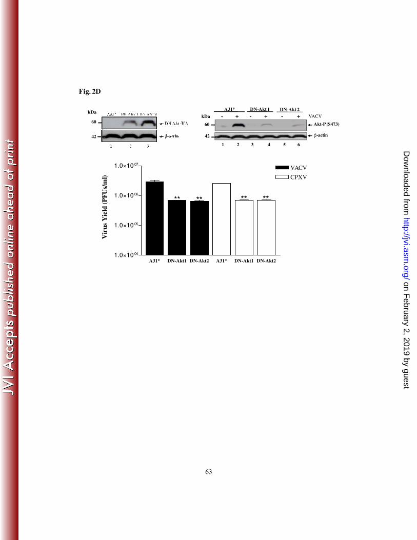
63
A31* DN-Akt1 DN-Akt2 A31* DN-Akt1 DN-Akt21.0×10 04
1.0×10 05
1.0×10 06
1.0×10 07
VACV
CPXV
**
Vir
us Y
ield
(P
FU
s/m
l)
**** **
Fig. 2D
60
42
kDa
42
60
kDa
A31* DN-Akt 1
- + - + - + VACV
1 2 3 4 5 6
Akt-P (S473)
β-actin
DN-Akt 2
on February 2, 2019 by guest
http://jvi.asm.org/
Dow
nloaded from