Embed Size (px)
Citation preview
8/12/2019 Action of Antimicrobials
http://slidepdf.com/reader/full/action-of-antimicrobials 1/14
At the basis of all antimicrobial chemotherapy lies the concept of selec-
tive toxicity. The necessary selectivity can be achieved in several ways:
vulnerable targets within the microbe may be absent from the cells
of the host or, alternatively, the analogous targets within the host cellsmay be sufficiently different, or at least sufficiently inaccessible, for
selective attack to be possible. With agents like the polymyxins, the
organic arsenicals used in trypanosomiasis, the antifungal polyenes
and many antiviral compounds, the gap between toxicity to the
microbe and to the host is small, but in most cases antimicrobial
agents are able to exploit fundamental differences in structure and
function within the microbial cell, and host toxicity generally results
from unexpected secondary effects.
ANTIBACTERIAL AGENTS
The minute size and capacity for very rapid multiplication of
bacteria ensures that they are structurally and metabolicallyvery different from mammalian cells and, in theory, there are
numerous ways in which bacteria can be selectively killed or
disabled. In the event, it turns out that only the bacterial cell
wall is structurally unique; other subcellular structures, includ-
ing the cytoplasmic membrane, ribosomes and DNA, are built
on the same pattern as those of mammalian cells, although suf-
ficient differences in construction and organization do exist at
these sites to make exploitation of the selective toxicity prin-
ciple feasible.
Antibacterial agents have been discovered – rarely designed
– that attack each of these vulnerable sites; the most success-
ful compounds seem to be those that interfere with the con-
struction of the bacterial cell wall, the synthesis of protein, or
the replication and transcription of DNA. Relatively few clin-
ically useful agents act at the level of the cell membrane or by
interfering with specific metabolic processes of the bacterial
cell (Table 2.1).
Unless the target is located on the outside of the bacterial
cell, antimicrobial agents must be able to penetrate to the site
of action. Access through the cytoplasmic membrane is usually
achieved by passive or facilitated diffusion, or (as, for example,
with aminoglycosides and tetracyclines) by active transport
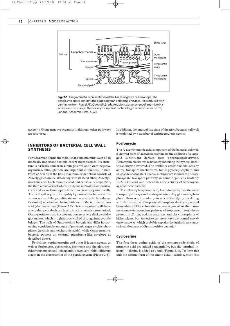
processes. In the case of Gram-negative organisms the anti-
biotic must also negotiate an outer membrane, consisting of a
characteristic lipopolysaccharide–lipoprotein complex, which
is responsible for preventing many antibiotics from reaching
an otherwise sensitive intracellular target. This lipophilic outer
membrane contains aqueous transmembrane channels
(porins), which selectively allow passage of hydrophilic mole-
cules depending on their molecular size and ionic charge
(Figure 2.1). Many antibacterial agents use porins to gain
D. Greenwood and R. Whitley
Modes of action
2
C H A P T E R
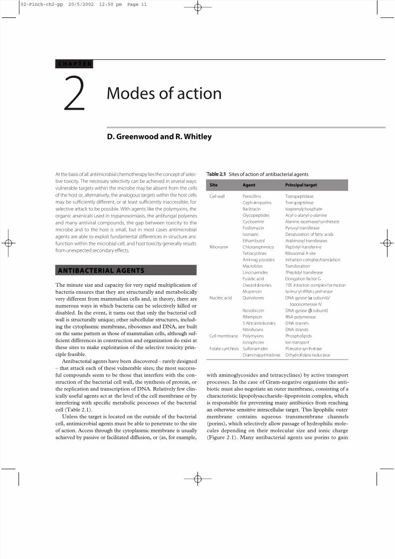
Table 2.1 Sites of action of antibacterial agents
Site Agent Principal target
Cell wall Penicillins TranspeptidaseCephalosporins Transpeptidase
Bacitracin Isoprenylphosphate
Glycopeptides Acyl-D-alanyl-D-alanine
Cycloserine Alanine racemase/synthetase
Fosfomycin Pyruvyl transferase
Isoniazid Desaturation of fatty acids
Ethambutol Arabinosyl transferases
Ribosome Chloramphenicol Peptidyl transferase
Tetracyclines Ribosomal A site
Aminoglycosides Initiation complex/translation
Macrolides Translocation
Lincosamides ?Peptidyl transferase
Fusidic acid Elongation factor G
Oxazolidinones 70S initiation complex formation
Mupirocin Isoleucyl-tRNA s ynthetase
Nucleic acid Quinolones DNA gyrase (a subunit)/
topoisomerase IV
Novobiocin DNA gyrase (b subunit)
Rifampicin RNA polymerase
5-Nitroimidazoles DNA strands
Nitrofurans DNA strands
Cell membrane Polymyxins Phospholipids
Ionophores Ion transport
Folate s ynthesis Sulfonamides Pteroate synthetase
Diaminopyrimidines Dihydrofolate reductase
02-Finch-ch2-pp 20/5/2002 12:50 pm Page 11
8/12/2019 Action of Antimicrobials
http://slidepdf.com/reader/full/action-of-antimicrobials 2/14
access to Gram-negative organisms, although other pathways
are also used.1
INHIBITORS OF BACTERIAL CELL WALLSYNTHESIS
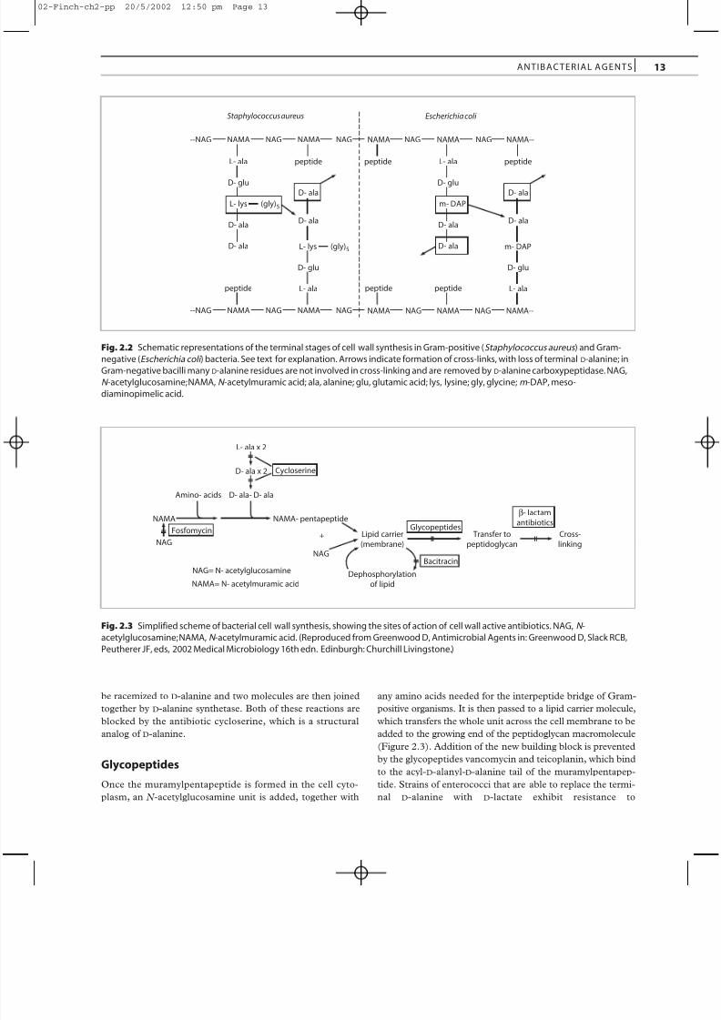
Peptidoglycan forms the rigid, shape-maintaining layer of all
medically important bacteria except mycoplasmas. Its struc-
ture is basically similar in Gram-positive and Gram-negative
organisms, although there are important differences. In bothtypes of organism the basic macromolecular chain consists of
N -acetylglucosamine alternating with its lactyl ether, N -acetyl-
muramic acid. Each muramic acid unit carries a pentapeptide,
the third amino acid of which is L -lysine in most Gram-positive
cocci and meso-diaminopimelic acid in Gram-negative bacilli.
The cell wall is given its rigidity by cross-links between this
amino acid and the penultimate amino acid (which is always
D-alanine) of adjacent chains, with loss of the terminal amino
acid (also D-alanine) (Figure 2.2). Gram-negative bacilli have
a very thin peptidoglycan layer, which is loosely cross-linked;
Gram-positive cocci, in contrast, possess a very thick peptido-
glycan coat, which is tightly cross-linked through interpeptide
bridges. The walls of Gram-positive bacteria also differ in con-
taining considerable amounts of polymeric sugar alcohol phos-
phates (teichoic and teichuronic acids), while Gram-negative
bacteria possess an external membrane-like envelope as
described above.
Penicillins, cephalosporins and other b-lactam agents, as
well as fosfomycin, cycloserine, bacitracin and the glycopep-
tides vancomycin and teicoplanin, selectively inhibit different
stages in the construction of the peptidoglycan (Figure 2.3).
In addition, the unusual structure of the mycobacterial cell wall
is exploited by a number of antituberculosis agents.
Fosfomycin
The N -acetylmuramic acid component of the bacterial cell wall
is derived from N -acetylglucosamine by the addition of a lactic
acid substituent derived from phosphoenolpyruvate.
Fosfomycin blocks this reaction by inhibiting the pyruvyl trans-
ferase enzyme involved. The antibiotic enters bacterial cells by
active transport mechanisms for a-glycerophosphate andglucose-6-phosphate. Glucose-6-phosphate induces the hexose
phosphate transport pathway in some organisms (notably
Escherichia coli ) and potentiates the activity of fosfomycin
against those bacteria.
The related phosphonic acid, fosmidomycin, uses the same
transport pathways and is also potentiated by glucose-6-phos-
phate. However, fosmidomycin acts differently by interfering
with the formation of isopentyl diphosphate during isoprenoid
biosynthesis.2 The vulnerable enzyme is part of an alternative
mevalonate-independent pathway of isoprenoid biosynthesis
present in E. coli , malaria parasites and the chloroplasts of
higher plants, but Staphylococcus aureus uses the normal meval-
onate pathway, which probably explains the intrinsic resistance
to fosmidomycin of Gram-positive bacteria.3
Cycloserine
The first three amino acids of the pentapeptide chain of
muramic acid are added sequentially, but the terminal D-
alanyl-D-alanine is added as a unit (Figure 2.3). To form this
unit the natural form of the amino acid, L -alanine, must first
12 CHAPTER 2 MODES OF ACTION
Protein
Phospholipid
Protein
Lipoprotein
LipopolysacchandaCell wall
Slime layer
Outer
membrane
Periplasmic
space
Cyloplasmic
membrane
Porin protein
Fig. 2.1 Diagrammatic representation of the Gram-negative cell envelope. The
periplasmic space contains the peptidoglycan and some enzymes. (Reproduced with
permission from Russel AD, Quesnel LB, eds, Antibiotics: assessment of antimicrobial
activity and resistance. The Society for Applied Bacteriology Technical Series no. 18,
London: Academic Press, p. 62.)
02-Finch-ch2-pp 20/5/2002 12:50 pm Page 12
8/12/2019 Action of Antimicrobials
http://slidepdf.com/reader/full/action-of-antimicrobials 3/14
be racemized to D-alanine and two molecules are then joined
together by D-alanine synthetase. Both of these reactions are
blocked by the antibiotic cycloserine, which is a structural
analog of D-alanine.
Glycopeptides
Once the muramylpentapeptide is formed in the cell cyto-
plasm, an N -acetylglucosamine unit is added, together with
any amino acids needed for the interpeptide bridge of Gram-
positive organisms. It is then passed to a lipid carrier molecule,
which transfers the whole unit across the cell membrane to be
added to the growing end of the peptidoglycan macromolecule
(Figure 2.3). Addition of the new building block is prevented
by the glycopeptides vancomycin and teicoplanin, which bind
to the acyl-D-alanyl-D-alanine tail of the muramylpentapep-
tide. Strains of enterococci that are able to replace the termi-
nal D-alanine with D-lactate exhibit resistance to
ANTIBACTER IAL AGENTS
Staphylococcus aureus
L- ala
D- glu
L- lys
D- ala
D- ala
peptide
D- ala
D- ala
D- glu
L- alapeptide
Escherichia coli
--NAG NAMA NAG NAMA NAG
(gly)5
L- lys (gly)5
peptide
--NAG NAMA NAG NAMA NAG
L- ala
D- glu
m- DAP
m- DAP
D- ala
D- ala
peptide
D- ala
D- ala
D- glu
L- alapeptide
NAMA NAG NAMA NAG NAMA--
NAMA NAG NAMA NAG NAMA--
peptide
Fig. 2.2 Schematic representations of the terminal stages of cell wall synthesis in Gram-positive (Staphylococcus aureus ) and Gram-
negative (Escherichia coli ) bacteria. See text for explanation. Arrows indicate formation of cross-links, with loss of terminal D-alanine; inGram-negative bacilli many D-alanine residues are not involved in cross-linking and are removed by D-alanine carboxypeptidase. NAG,
N -acetylglucosamine; NAMA, N -acetylmuramic acid; ala, alanine; glu, glutamic acid; lys, lysine; gly, glycine; m -DAP, meso-
diaminopimelic acid.
L- ala x 2
D- ala x 2
D- ala- D- alaAmino- acids
NAMA
NAG
NAMA- pentapeptide
Lipid carrier(membrane)
Transfer topeptidoglycan
Cross-linking
Fosfomycin
Cycloserine
Glycopeptides
Bacitracin
β- lactam
antibiotics
NAG
NAG= N- acetylglucosamine
NAMA= N- acetylmuramic acidDephosphorylation
of lipid
+
Fig. 2.3 Simplified scheme of bacterial cell wall synthesis, showing the sites of action of cell wall active antibiotics. NAG, N -
acetylglucosamine; NAMA, N -acetylmuramic acid. (Reproduced from Greenwood D, Antimicrobial Agents in: Greenwood D, Slack RCB,
Peutherer JF, eds, 2002 Medical Microbiology 16th edn. Edinburgh: Churchill Livingstone.)
02-Finch-ch2-pp 20/5/2002 12:50 pm Page 13
8/12/2019 Action of Antimicrobials
http://slidepdf.com/reader/full/action-of-antimicrobials 4/14
glycopeptides.4 Because these glycopeptides are large polar
molecules they cannot penetrate the outer membrane of Gram-
negative organisms, which explains their restricted spectrum
of activity.
Bacitracin
The lipid carrier involved in transporting the cell wall build-
ing block across the membrane has been characterized as a C 55
isoprenyl phosphate. The lipid acquires an additional phos-
phate group in the transport process and must be dephospho-
rylated in order to regenerate the native compound for another
round of transfer. The cyclic peptide antibiotic bacitracin binds
to the isoprenyl pyrophosphate and prevents this dephospho-
rylation. Unfortunately, analogous reactions in eukaryotic cells
are also inhibited by bacitracin, and this may be the basis of
the toxicity of the compound.
b-Lactam antibiotics
The final cross-linking reaction that gives the bacterial cell wallits characteristic rigidity was pinpointed many years ago as the
primary target of penicillin and other b-lactam agents. These
compounds were postulated to inhibit formation of the
transpeptide bond by virtue of their structural resemblance to
the terminal D-alanyl-D-alanine unit that participates in the
transpeptidation reaction. This knowledge had to be recon-
ciled with various concentration-dependent morphological
responses that Gram-negative bacilli undergo on exposure to
penicillin and other b-lactam compounds: filamentation
(caused by inhibition of division rather than growth of the bac-
teria) at low concentrations, and the formation of osmotically
fragile spheroplasts (peptidoglycan-deficient forms that have
lost their bacillary shape) at high concentrations.
Three observations suggested that these morphologicalevents could be dissociated:
● The oral cephalosporin cefalexin (and some other b-lactam
agents, including cefradine, temocillin and the monobac-
tam, aztreonam) causes the filamentation response alone
over an extremely wide range of concentrations.
● Mecillinam (amdinocillin) does not inhibit division (and
hence does not cause filamentation in Gram-negative
bacilli), but has a generalized effect on the bacterial cell
wall.
● Combining cefalexin and mecillinam evokes the ‘typical’
spheroplast response in E. coli that neither agent induces
when acting alone.5
It was subsequently shown that isolated membranes of bac-
teria contain a number of proteins that are able to bind peni-
cillin and other b-lactam antibiotics. These penicillin-binding
proteins (PBPs) are numbered in descending order of their
molecular weight according to their separation by polyacry-
lamide gel electrophoresis. The number found in bacterial cells
varies from species to species: E. coli has at least seven and
Staph. aureus four. b-Lactam agents that induce filamentation
in Gram-negative bacilli bind to PBP3; similarly, mecillinam
binds exclusively to PBP2. Most b-lactam antibiotics, when
present in suf ficient concentration, bind to both these sites and
to others (PBP1a and PBP1b) that participate in the rapidly
lytic response of Gram-negative bacilli to many penicillins and
cephalosporins.
The low-molecular-weight PBPs (4, 5 and 6) of E. coli arecarboxypeptidases, which may operate to control the extent of
cross-linking in the cell wall. Mutants lacking these enzymes
grow normally and have thus been ruled out as targets for the
inhibitory or lethal actions of b-lactam antibiotics. The PBPs
with higher molecular weights (PBPs 1a, 1b, 2 and 3) possess
transpeptidase activity, and it seems that these PBPs represent
different forms of the transpeptidase enzyme necessary to
arrange the complicated architecture of the cylindrical or
spherical bacterial cell during growth, septation and division.
The nature of the lethal event In Gram-negative bacilli, the bactericidal effect of b-lactam
antibiotics can be quantitatively prevented by providing suf fi-
cient osmotic protection. In these circumstances the bacteriasurvive as spheroplasts, which readily revert to the bacillary
shape on removal of the antibiotic. It thus seems clear that cell
death in Gram-negative bacilli is a direct consequence of
osmotic lysis of cells deprived of the protective peptidoglycan
coat.
The nature of the lethal event in Gram-positive cocci is
more complex. Since these bacteria possess a much thicker,
tougher peptidoglycan layer than that present in the Gram-
negative cell wall, much greater damage has to be inflicted
before death of the cell ensues. However, one of the first events
that occurs on exposure of Gram-positive cocci to b-lactam
antibiotics is a release of lipoteichoic acid, an event which
appears to trigger autolysis of the peptidoglycan.
Optimal dosage effect For many strains of Gram-positive cocci, an optimal bacteri-
cidal concentration of b-lactam antibiotics can be identified
above which the killing effect is reduced, sometimes very strik-
ingly. The basis of this effect, often called the ‘Eagle phenom-
enon’ after its discoverer, has never been satisfactorily
explained. A plausible hypothesis is that the lethal event is trig-
gered by low concentrations of the antibiotic as a consequence
of binding to one particular target protein; binding at higher
concentrations to other targets (PBPs) stops the bacterial cell
from growing and this antagonizes the lethal effect, which
requires continued cell growth.
PersistersAbout 1 in 105 bacteria in a culture exposed to b-lactam antibi-
otics survive, even on prolonged exposure to an optimal bac-
tericidal concentration. These ‘persisters’ have not acquired
resistance, since, if the antibiotic is removed and they are
allowed to grow, most of their immediate progeny are killed
on re-exposure, just as the parent culture is. These bacteria
may be cells in which the peptidoglycan exists transiently as a
14 CHAPTER 2 MODES OF ACTION
02-Finch-ch2-pp 20/5/2002 12:50 pm Page 14
8/12/2019 Action of Antimicrobials
http://slidepdf.com/reader/full/action-of-antimicrobials 5/14
complete covalently linked macromolecule. Inhibition by the
antibiotic of autolytic enzymes needed to create growth points
in the peptidoglycan would effectively trap the cells in a state
in which they could not grow (and therefore could not be
killed) until the antibiotic is removed.
ToleranceIn some Gram-positive cocci there may be a marked dissoci-
ation between the concentrations of b-lactam agents (and gly-
copeptides) required to achieve a bacteristatic and a
bactericidal effect. The organisms are not ‘resistant’ since they
remain fully susceptible to the inhibitory activity of the antibi-
otic, although the bactericidal effect is reduced. Defective
autolysins remain the most likely explanation of the effect,
which has some similarities to the persister phenomenon.6
Antimycobacterial agents
Agents acting specifically against Mycobacterium tuberculosis and
other mycobacteria have been less well characterized than other
antimicrobial drugs. However, it is thought that several of themowe their activity to selective effects on the unique structure
of the mycobacterial envelope.7 Thus, although isoniazid has
been found to interfere with various cellular functions of bac-
teria, it is likely that it owes its specific bactericidal activity
against M. tuberculosis to interference with mycolic acid syn-
thesis. The effect is achieved by inhibition of a fatty acid desat-
urase after intracellular oxidation of isoniazid to an active
product.8 Ethionamide, prothionamide and pyrazinamide,
which are related nicotinic acid derivatives, are also thought
to undergo intracellular modification and to act in a similar
fashion.
Ethambutol, a slow acting and primarily bacteriostatic
antimycobacterial agent, inhibits arabinosyl transferases. These
enzymes bring about the polymerization of arabinose to formarabinan, a polysaccharide component of the core polymers of
the mycobacterial cell wall.8
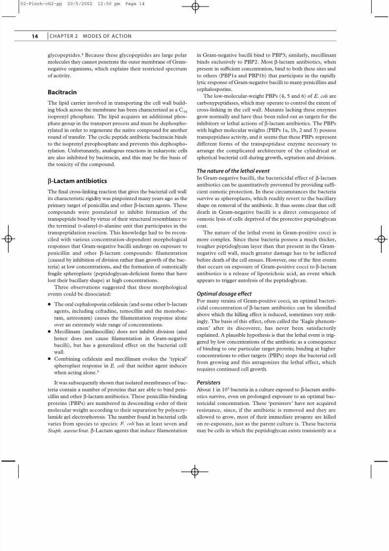
INHIBITORS OF BACTERIAL PROTEINSYNTHESIS
The amazing process by which the genetic message in DNA is
translated into large and unique protein molecules is univer-
sal; in prokaryotic, as in eukaryotic cells, the workbench is the
ribosome, composed of two distinct subunits, each a complex
of ribosomal RNA (rRNA) and numerous proteins. However,
bacterial ribosomes are open to selective attack because they
differ from their mammalian counterparts in both protein and
RNA content; indeed they can be readily distinguished in the
ultracentrifuge: bacterial ribosomes exhibit a sedimentation
coefficient of 70S (composed of 30S and 50S subunits),
whereas mammalian ribosomes display a coefficient of 80S
(composed of 40S and 60S subunits).
In the first stage of bacterial protein synthesis, messenger
RNA (mRNA), transcribed from the appropriate region of
DNA, binds to the smaller ribosomal subunit and attracts N -
formylmethionyl transfer RNA (fMet-tRNA) to the initiator
codon AUG. The larger subunit is then added to form a com-
plete initiation complex. fMet-tRNA occupies the P (peptidyl
donor) site; adjacent to it is the A (aminoacyl acceptor) site
aligned with the next trinucleotide codon of the mRNA.
Transfer RNA (tRNA) bearing the appropriate anticodon, andits specific amino acid, enters the A site, and a peptidyl trans-
ferase joins N -formylmethionine to the new amino acid with
loss (via an exit site) of the tRNA in the P site; the first peptide
bond of the protein has been formed. A translocation event
then moves the remaining tRNA with its dipeptide to the P site
and concomitantly aligns the next triplet codon of mRNA with
the now vacant A site. The appropriate aminoacyl-tRNA enters
the A site and the transfer process and subsequent transloca-
tion are repeated. In this way, the peptide chain is built up in
precise fashion, faithful to the original DNA blueprint, until a
so-called ‘nonsense’ codon is encountered on the mRNA that
signals chain termination and release of the peptide chain. The
mRNA is disengaged from the ribosome, which dissociates into
its two subunits ready to form a new initiation complex. Withinbacterial cells, many ribosomes are engaged in protein syn-
thesis during active growth, and a single strand of mRNA may
interact with many ribosomes along its length to form a
polysome.
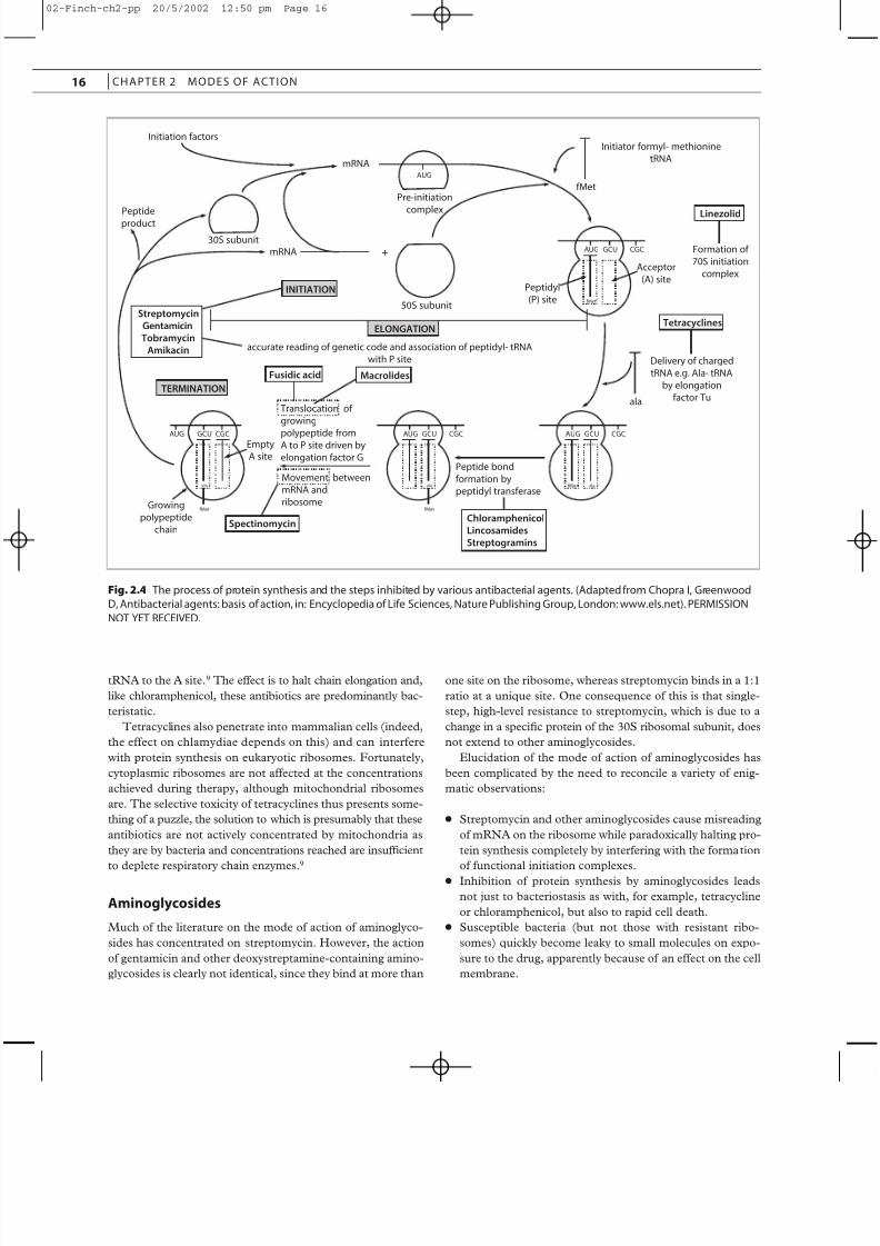
Many antibiotics interfere with the process of protein syn-
thesis (Figure 2.4). Some, like puromycin, which is an analog
of the aminoacyl tail of charged tRNA and causes premature
chain termination, act on bacterial and mammalian ribosomes
alike and are therefore unsuitable for systemic use in humans.
Therapeutically useful inhibitors of protein synthesis include
many of the naturally occurring antibiotics, such as chloram-
phenicol, tetracyclines, aminoglycosides, fusidic acid,
macrolides, lincosamides and streptogramins. Some newer
agents, including mupirocin and the oxazolidinones, also actat this stage.
Chloramphenicol
The molecular target for chloramphenicol is the peptidyl trans-
ferase enzyme that links amino acids in the growing peptide
chain. The effect of the antibiotic is thus to freeze the process
of chain elongation, bringing bacterial growth to an abrupt halt.
The process is completely reversible, and chloramphenicol is
fundamentally a bacteristatic agent. The binding of chloram-
phenicol to the 50S subunit of 70S ribosomes is highly specif-
ic. The basis for the rare, but fatal, marrow aplasia associated
with this compound is not therefore a generalized effect on
mammalian protein synthesis, although mitochondrial ribo-
somes, which are similar to those of bacteria, may be involved.
Tetracyclines
Antibiotics of the tetracycline group are actively transported
into bacterial cells and attach to 30S ribosomal subunits in
such a manner as to prevent binding of incoming aminoacyl-
ANTIBACTER IAL AGENTS
02-Finch-ch2-pp 20/5/2002 12:50 pm Page 15
8/12/2019 Action of Antimicrobials
http://slidepdf.com/reader/full/action-of-antimicrobials 6/14
tRNA to the A site.9 The effect is to halt chain elongation and,
like chloramphenicol, these antibiotics are predominantly bac-
teristatic.
Tetracyclines also penetrate into mammalian cells (indeed,
the effect on chlamydiae depends on this) and can interfere
with protein synthesis on eukaryotic ribosomes. Fortunately,
cytoplasmic ribosomes are not affected at the concentrations
achieved during therapy, although mitochondrial ribosomes
are. The selective toxicity of tetracyclines thus presents some-
thing of a puzzle, the solution to which is presumably that these
antibiotics are not actively concentrated by mitochondria as
they are by bacteria and concentrations reached are insuf ficient
to deplete respiratory chain enzymes.9
Aminoglycosides
Much of the literature on the mode of action of aminoglyco-
sides has concentrated on streptomycin. However, the action
of gentamicin and other deoxystreptamine-containing amino-
glycosides is clearly not identical, since they bind at more than
one site on the ribosome, whereas streptomycin binds in a 1:1
ratio at a unique site. One consequence of this is that single-
step, high-level resistance to streptomycin, which is due to a
change in a specific protein of the 30S ribosomal subunit, does
not extend to other aminoglycosides.
Elucidation of the mode of action of aminoglycosides has
been complicated by the need to reconcile a variety of enig-
matic observations:
● Streptomycin and other aminoglycosides cause misreading
of mRNA on the ribosome while paradoxically halting pro-
tein synthesis completely by interfering with the formation
of functional initiation complexes.
● Inhibition of protein synthesis by aminoglycosides leads
not just to bacteriostasis as with, for example, tetracycline
or chloramphenicol, but also to rapid cell death.
● Susceptible bacteria (but not those with resistant ribo-
somes) quickly become leaky to small molecules on expo-
sure to the drug, apparently because of an effect on the cell
membrane.
16 CHAPTER 2 MODES OF ACTION
INITIATION
accurate reading of genetic code and association of peptidyl- tRNA
with P site
ELONGATION
MacrolidesFusidic acid
Streptomycin
Gentamicin
Tobramycin
Amikacin
TERMINATION
AUG
Pre-initiation
complex
50S subunit
30S subunit
Initiation factors
AUG GCU CGC
fMet
AUG GCU CGC
fMet ala
AUG GCU CGC
ala
fMetfMet
AUG GCU CGC
ala
Peptidyl
(P) site
Acceptor
(A) site
mRNA
Initiator formyl- methionine
tRNA
Linezolid
Formation of
70S initiation
complex
Tetracyclines
Peptide
product
Delivery of charged
tRNA e.g. Ala- tRNA
by elongation
factor Tu
Empty
A site
Growing
polypeptide
chain Spectinomycin
Translocation of
growing
polypeptide fromA to P site driven by
elongation factor G
Movement between
mRNA and
ribosome
Peptide bond
formation by
peptidyl transferase
Chloramphenicol
Lincosamides
Streptogramins
mRNA
ala
fMet
+
Fig. 2.4 The process of protein synthesis and the steps inhibited by various antibacterial agents. (Adapted from Chopra I, Greenwood
D, Antibacterial agents: basis of action, in: Encyclopedia of Life Sciences, Nature Publishing Group, London: www.els.net). PERMISSION
NOT YET RECEIVED.
02-Finch-ch2-pp 20/5/2002 12:50 pm Page 16
8/12/2019 Action of Antimicrobials
http://slidepdf.com/reader/full/action-of-antimicrobials 7/14
● Mutations can occur that render the bacterial cell depen-
dent on streptomycin for growth.
● Susceptibility is dominant over resistance in merozygotes
that are diploid for the two allelic forms.
A well-lit path through this maze has not yet been defini-
tively charted, but the situation is slowly becoming clearer. Thetwo effects of aminoglycosides on initiation and misreading
may be explained by a concentration-dependent effect on ribo-
somes engaged in the formation of the initiation complex and
those in the process of chain elongation:10 in the presence of a
sufficiently high concentration of drug, protein synthesis is
completely halted once the mRNA is run off because re-initi-
ation is blocked; under these circumstances there is little or no
opportunity for misreading to occur. However, at concentra-
tions at which only a proportion of the ribosomes can be
blocked at initiation, some protein synthesis will take place and
the opportunity for misreading will be provided.
The dominance of susceptibility over resistance has been
tentatively explained by the fact that the non-functional initi-
ation complexes formed in the presence of aminoglycosidesare unstable, so that the ribosomes continuously dissociate
from mRNA and recycle in an inoperative form. These crip-
pled ribosomes (of which there are twice as many as there are
resistant ones in merozygotes) are hence continuously made
re-available to sequester newly formed mRNA and prevent the
resistant ribosomes from maintaining a supply of the polypep-
tides that the cell needs.10
The effects of aminoglycosides on membrane permeability,
and the potent bactericidal activity of these compounds,
remain enigmatic. However, the two phenomena may be
related.11 In bacteria, as in mammalian cells, some of the ribo-
somes (presumably those engaged in transmembrane protein
transfer) may be membrane bound. Moreover, aminoglyco-
sides enter bacteria by an active transport process (absent inanaerobes and streptococci, hence their inherent resistance)
that is dependent on protein synthesis. It is possible that site-
specific uptake of the drug at ribosomal attachment sites and
subsequent binding to the ribosome – membrane complex may
lead to membrane leakiness and cell death.12
SpectinomycinThe aminocyclitol antibiotic spectinomycin, often considered
alongside the aminoglycosides, binds in reversible fashion
(hence the bacteriostatic activity) to ribosomal RNA of the 30S
subunit. There it interrupts the translocation event that occurs
as the next codon of mRNA is aligned with the A site in readi-
ness for the incoming aminoacyl-tRNA.
Macrolides, lincosamides, streptogramins
These antibiotic groups are structurally very different, but bind
to closely related sites on the 50S ribosome of bacteria. One
consequence of this is that staphylococci exhibiting inducible
resistance to erythromycin, which is caused by methylation of
certain adenine residues in the rRNA, also become resistant
to other macrolides, lincosamides and streptogramin B in the
presence of erythromycin (p. 000).
The detailed mode of action of these antibiotics has not yet
been definitively worked out. Erythromycin (like type A strep-
togramins; see below) binds almost exclusively to free ribo-
somes and brings protein synthesis to a halt after formation of
the initiation complex, probably by interfering with the translo-cation reaction. The precise mechanism of action of lin-
cosamides is less clear, but they appear to interfere indirectly
with the peptidyl transferase reaction, possibly by blocking the
P site.
The streptogramins are composed of two interacting com-
ponents designated A and B (p. 000). The type A molecules
bind to 50S ribosomal subunits and affect both donor and
acceptor functions of peptidyl transferase by blocking attach-
ment of aminoacyl-tRNA to the catalytic site of the enzyme
and subsequent transfer of the growing peptide chain. Type B
streptogramins occupy an adjacent site on the ribosome and
prevent formation of the peptide bond, leading to the prema-
ture release of incomplete polypeptides.13 Type A molecules
bind to free ribosomes, but not to polysomes engaged inprotein synthesis, whereas type B can prevent further synthe-
sis during active synthesis. The bactericidal synergy between
the two components arises mainly from conformational
changes induced by Type A molecules that improve the
binding af finity of Type B compounds.14
Fusidic acid
Fusidic acid forms a stable complex with an elongation factor
(EF-G) involved in translocation and with guanosine triphos-
phate (GTP), which provides energy for the translocation
process. One round of translocation occurs, with hydrolysis of
GTP, but the fusidic acid – EF – G – GDP – ribosome complex
blocks further chain elongation, leaving peptidyl-tRNA in theP site.
Although protein synthesis in Gram-negative bacilli – and,
indeed, mammalian cells – is susceptible to fusidic acid, the
antibiotic penetrates poorly into these cells and the spectrum
of action is virtually restricted to Gram-positive bacteria,
notably staphylococci.
Oxazolidinones
Linezolid and other oxazolidinones are bacteriostatic agents
that act at an earlier stage than other inhibitors of protein syn-
thesis. They prevent the process by which the 50S ribosomal
subunit and the 30S unit (charged with mRNA and fMet-
tRNA) come together to form the 70S initiation complex.15,16
This is achieved by binding to the 50S subunit, at a site close
to that of chloramphenicol and lincosamides.17
Mupirocin
Mupirocin also has a unique mode of action. The epoxide-
containing monic acid tail of the molecule (see p. 000) is an
ANTIBACTER IAL AGENTS
02-Finch-ch2-pp 20/5/2002 12:50 pm Page 17
8/12/2019 Action of Antimicrobials
http://slidepdf.com/reader/full/action-of-antimicrobials 8/14
analog of isoleucine and, as such, is a competitive inhibitor of
isoleucyl-tRNA synthetase in bacterial cells.18 It may be a
bifunctional inhibitor of the synthetase, since it also binds to
the ATP-binding site of the enzyme.19 The corresponding
mammalian enzyme is unaffected.
INHIBITORS OF NUCLEIC ACIDSYNTHESIS
Compounds that bind directly to the double helix are gener-
ally highly toxic to mammalian cells and only a few – those that
interfere with DNA-associated enzymic processes – exhibit suf-
ficient selectivity for systemic use as antibacterial agents. These
compounds include antibacterial quinolones, novobiocin and
rifampicin (rifampin). Diaminopyrimidines, sulfonamides, 5-
nitroimidazoles and (probably) nitrofurans also affect DNA
synthesis and will be considered under this heading.
Quinolones
The problem of packaging the enormous circular chromosome
of bacteria (>1 mm long) into a microscopic cell, while making
adequate arrangements for transcription and replication, has
necessitated some considerable ingenuity on the part of the
microbe. The solution has been to condense the DNA down
and to twist it into a ‘supercoiled’ state – a process aided by
the natural strain imposed on a covalently closed double helix.
The twists are introduced in the opposite sense to those of the
double helix itself and the molecule is said to be negatively
supercoiled. Relaxation and re-establishment of the supercoiled
state involves precisely regulated nicking and resealing of the
DNA strands, accomplished by enzymes called topoisomeras-
es. One topoisomerase, DNA gyrase, is a tetramer composed
of two pairs of a and b subunits, and the primary target of theaction of nalidixic acid and other quinolones is the a subunit
of DNA gyrase, although another enzyme, topoisomerase IV,
is also affected.20 Indeed, in Gram-positive bacteria, topoiso-
merase IV seems to be the main target.21 This enzyme does
not have supercoiling activity; it appears to be involved in
relaxation of the DNA chain and chromosomal segregation.
Fortunately, the corresponding mammalian topoisomerases
are less susceptible to quinolone attack.
Curiously, the coumarin antibiotic novobiocin, which acts
in a complementary fashion by binding specifically to the b-
subunit of DNA gyrase, displays an exactly opposite spectrum
of activity to that of nalidixic acid.
RifampicinRifampicin and other compounds of the ansamycin group
specifically inhibit DNA-dependent RNA polymerase; that is,
they prevent the transcription of RNA species from the DNA
template. Rifampicin is an extremely ef ficient inhibitor of the
bacterial enzyme, but fortunately eukaryotic RNA polymerase
is not affected. RNA polymerase consists of a core enzyme
made up of four polypeptide subunits, and rifampicin specifi-
cally binds to the b subunit. However, since isolated b subunit
does not bind rifampicin, the precise configuration in which it
is locked into the core enzyme is important.
Sulfonamides and diaminopyrimidines
These agents act at separate stages in the pathway of folic acid
synthesis and thus act indirectly on DNA synthesis, since the
active form of the co-enzyme, tetrahydrofolic acid, serves as
intermediate in the transfer of methyl, formyl and other single-
carbon fragments in the biosynthesis of purine nucleotides and
thymidylic acid, as well as of some amino acids.
Sulfonamides are analogues of p-aminobenzoic acid. They
competitively inhibit dihydropteroate synthetase, the enzyme
which condenses p-aminobenzoic acid with dihydropteroic acid
in the early stages of folic acid synthesis. Most bacteria need
to synthesize folic acid and cannot use exogenous sources of
the vitamin. Mammalian cells, in contrast, require preformed
folate and this is the basis of the selective action of sulfon-
amides. The antileprotic sulfone dapsone, and the antituber-culosis drug p-aminosalicylic acid, act in a similar way; the
basis for their restricted spectrum may reside in differences of
af finity for variant forms of dihydropteroate synthetase in the
bacteria against which they act.
Diaminopyrimidines act later in the pathway of folate syn-
thesis. These compounds inhibit dihydrofolate reductase, the
enzyme that generates the active form of the co-enzyme
tetrahydrofolic acid. In most of the reactions in which tetrahy-
drofolate takes part it remains unchanged, but in the biosyn-
thesis of thymidylic acid tetrahydrofolate acts as hydrogen
donor as well as a methyl group carrier and is thus oxidized to
dihydrofolic acid in the process. Dihydrofolate reductase is
therefore crucial in recycling tetrahydrofolate, and diaminopy-
rimidines act relatively quickly to halt bacterial growth.Sulfonamides, in contrast, cut off the supply of folic acid at
source and act slowly, since the existing folate pool can satisfy
the needs of the cell for several generations.
The selective toxicity of diaminopyrimidines comes about
because of differential affinity of these compounds for dihy-
drofolate reductase from various sources. Thus trimethoprim
has a vastly greater af finity for the bacterial enzyme than for its
mammalian counterpart, pyrimethamine exhibits a particular-
ly high af finity for the plasmodial version of the enzyme and,
in keeping with its anticancer activity, methotrexate has high
af finity for the enzyme found in mammalian cells.
5-Nitroimidazoles
The most intensively investigated compound in this group is
metronidazole, but other 5-nitroimidazoles are thought to act
in a similar manner. Metronidazole siphons off electrons from
ferredoxin (or other electron transfer proteins with low redox
potential) causing the nitro group of the drug to be reduced.
It is this reduced and highly reactive intermediate that is
responsible for the antimicrobial effect, probably by binding
18 CHAPTER 2 MODES OF ACTION
02-Finch-ch2-pp 20/5/2002 12:50 pm Page 18
8/12/2019 Action of Antimicrobials
http://slidepdf.com/reader/full/action-of-antimicrobials 9/14
to DNA, which undergoes strand breakage.22 The requirement
for interaction with low redox systems restricts the activity
largely to anaerobic bacteria and certain protozoa that exhibit
anaerobic metabolism. The basis for activity against micro-
aerophilic species such as Helicobacter pylori and Gardnerella
vaginalis remains speculative, though a novel nitroreductase,
which is altered in metronidazole-resistant strains, is implicat-ed in H. pylori .23
Nitrofurans
As with nitroimidazoles, the reduction of the nitro group of
nitrofurantoin and other nitrofurans is a prerequisite for
antibacterial activity. Micro-organisms with appropriate
nitroreductases act on nitrofurans to produce a highly reactive
electrophilic intermediate and this is postulated to affect DNA
as the reduced intermediates of nitroimidazoles do. Other evi-
dence suggests that the reduced nitrofurans bind to bacterial
ribosomes and prevent protein synthesis;24 inducible enzyme
synthesis seems to be particularly susceptible. An effect on
DNA has the virtue of explaining the known mutagenicity of these compounds in vitro and any revised mechanism relating
to inhibition of protein synthesis needs to be reconciled with
this property.
AGENTS AFFECTING MEMBRANEPERMEABILITY
Agents acting on cell membranes do not normally discriminate
between microbial and mammalian membranes, although the
fungal cell membrane has proved more amenable to selective
attack (see below). The only membrane-active antibacterial
agents to be administered systemically in human medicine,
polymyxin B (now rarely used systemically) and the closelyrelated compound colistin (polymyxin E), act like cationic
detergents; they disrupt the cytoplasmic membrane of the cell,
probably by attacking the exposed phosphate groups of the
membrane phospholipid. They also have an effect on the exter-
nal membrane of Gram-negative bacilli, which might explain
their preferential action on these organisms. The end result is
leakage of cytoplasmic contents and death of the cell. Various
factors, including growth phase and incubation temperature,
alter the balance of fatty acids within the bacterial cell mem-
brane, and this can concomitantly affect the response to
polymyxins.25
Several antibiotics, known collectively as ionophores, inter-
fere with cation transport in cell membranes. These include
the topical antibiotic gramicidin A, and some agents used in
veterinary medicine, such as the macrotetralide monensin and
the depsipeptide valinomycin. Naturally occurring antimicro-
bial peptides, such as the cecropins, magainins and defensins,
as well as the lanthionine-containing lantibiotics, disrupt cell
membranes, sometimes in a selective manner; some of these
peptides appear to form aggregates with ionophoric proper-
ties.26,27
ANTIFUNGAL AGENTS
In view of the scarcity of antibacterial agents acting on the cyto-
plasmic membrane, it is surprising to find that the most suc-
cessful groups of antifungal agents – the polyenes, azoles, and
allylamines – all achieve their effects in this way.28,29 However,
the echinocandins, the latest addition to the antifungal arma-mentarium, differ in affecting the fungal cell wall.
POLYENES
The polyenes bind only to membranes containing sterols; ergos-
terol, the predominant sterol of fungal membranes, appears to
be particularly susceptible. The effect is to make the membrane
leaky, probably by the formation of transmembrane pores. Since
bacterial cell membranes (except those of mycoplasmas) do not
contain sterols, they are unaffected by polyenes, even in high
concentration; unfortunately, this immunity does not extend to
sterol-containing mammalian cells, and polyenes consequent-
ly exhibit a low therapeutic index.
AZOLES
The activity of the antifungal azoles is also dependent on the
presence of ergosterol in the fungal cell membrane. These
compounds block ergosterol synthesis by interfering with the
demethylation of its precursor, lanosterol.30 Lanosterol
demethylase is a cytochrome P450 enzyme and, although azole
antifungals have much less influence on analogous mammalian
systems, some of the side effects are attributable to such action.
Antifungal azole derivatives are predominantly fungistatic
but some compounds, notably miconazole and clotrimazole,
kill fungi at concentrations higher than those which merelyinhibit growth, apparently by causing direct membrane
damage. Other, less well characterized, effects of azoles on
fungal respiration have also been described.31
ALLYLAMINES
The antifungal allylamine derivatives terbinafine and naftifine
inhibit squalene epoxidase, another enzyme involved in the
biosynthesis of ergosterol.32 The fungicidal effect may be due
to accumulation of squalene rather than a deficiency of ergos-
terol. In Candida albicans the effect is fungistatic and the yeast
form is less susceptible than is mycelial growth. In this species
there is less accumulation of squalene than in dermatophytes,
and ergosterol deficiency may be the limiting factor.33
ECHINOCANDINS
Caspofungin and related compounds inhibit the formation of
glucan, an essential polysaccharide of the cell wall of many
ANTIFUNGAL AGENTS
02-Finch-ch2-pp 20/5/2002 12:50 pm Page 19
8/12/2019 Action of Antimicrobials
http://slidepdf.com/reader/full/action-of-antimicrobials 10/14
fungi, including Pneumocystis carinii . The vulnerable enzyme
is b-1,3-glucan synthase, which is located in the cell mem-
brane.34
FLUCYTOSINE
The spectrum of activity of flucytosine (5-fluorocytosine) is
virtually restricted to yeasts. In these fungi flucytosine is trans-
ported into the cell by a cytosine permease; a cytosine deam-
inase then converts flucytosine to 5-fluorouracil, which is
incorporated into RNA in place of uracil, leading to the for-
mation of abnormal proteins. There is also an effect on DNA
synthesis through inhibition of thymidylate synthetase.35
GRISEOFULVIN
The antidermatophyte antibiotic griseofulvin binds to the
microtubules of the mitotic spindle, interfering with their
assembly and function. However, the precise mechanism of action and the basis of the selectivity remain to be elucidated.
ANTIPROTOZOAL AGENTS
The actions of some antiprotozoal drugs overlap with, or are
analogous to, those seen with the antibacterial and antifungal
agents already discussed. Thus, the activity of 5-nitroimida-
zoles such as metronidazole extends to those protozoa that
exhibit an essentially anaerobic metabolism; the antimalarial
agents pyrimethamine and cycloguanil (the metabolic product
of proguanil), like trimethoprim, inhibit dihydrofolate reduc-
tase; some polyenes and antifungal imidazoles display suffi-
cient activity against Leishmania and certain other protozoa forthem to have received attention as potential therapeutic agents.
There seems to be deep uncertainty about how other
antiprotozoal agents actually work. Various sites of action have
been ascribed to many of them and, with a few notable excep-
tions, the literature reveals only desultory attempts to pin down
the primary target.
ANTIMALARIAL AGENTS
Quinoline antimalarials
Quinine and the various quinoline antimalarials were once
thought to achieve their effect by intercalation with plasmodi-
al DNA after concentration in parasitized erythrocytes.
However, these effects occur only at concentrations in excess
of those achieved in vivo; moreover, a non-specific effect on
DNA does not explain the selective action of these compounds
at precise points in the plasmodial life cycle, or the differential
activity of antimalarial quinolines.
Clarification of the mode of action of these compounds has
proved elusive, but it now seems likely that chloroquine and
related compounds act primarily by inhibiting haem poly-
merase, thus preventing detoxification of ferriprotoporphyrin
IX (heme), which is produced from the red cell hemoglobin
in the food vacuole of the parasites.36 Ferriprotoporphyrin IX
is a toxic metabolite which is normally rendered innocuous by
polymerization; malarial pigment consists of granules of thispolymer.
Chloroquine achieves a very high concentration within the
food vacuole of the parasite and this greatly aids its activity.
However, quinine and mefloquine are not concentrated to the
same extent, and have much less effect on heme polymeriza-
tion, raising the possibility that other (possibly multiple) targets
are involved in the action of these compounds.37,38
8-Aminoquinolines like primaquine, which, at therapeuti-
cally useful concentrations exhibit selective activity against
liver-stage parasites and gametocytes, possibly inhibit mito-
chondrial enzyme systems after undergoing hepatic metabo-
lism. However, the precise mechanism of action is unknown.
Artemisinin
Artemisinin, the active principle of the Chinese herbal remedy
qinghaosu, has several effects on malaria parasites, but the
activity appears to be due chiefly to the reactivity of the
endoperoxide bridge. This is cleaved in the presence of heme
or free iron within the parasitized red cell to form a short-lived,
but highly reactive, free radical that irreversibly alkylates
malaria proteins.39
Atovaquone
The hydroxynaphthoquinone atovaquone, which exhibits anti-
malarial and antipneumocystis activity, is an electron transport
inhibitor that causes depletion of the ATP pool. The primaryeffect is on the iron flavoprotein dihydro-orotate dehydroge-
nase, an essential enzyme in the production of pyrimidines.
Mammalian cells are able to avoid undue toxicity by use of
preformed pyrimidines.40 Dihydro-orotate dehydrogenase from
Plasmodium falciparum is inhibited by concentrations of ato-
vaquone that are very much lower than those needed to inhibit
the pneumocystis enzyme, raising the possibility that the
antimicrobial consequences might differ in the two organ-
isms.41
OTHER ANTIPROTOZOAL AGENTS
Arsenical compounds, which are still the mainstay of treatment
of African sleeping sickness, poison the cell by an effect on
glucose catabolism and are consequently very toxic to the host.
The mechanism by which this is achieved and the basis for any
selective action are not well understood, though they are
known to bind to essential thiol groups. The primary target
may be trypanothione, which substitutes for glutathione in try-
panosomes, and this may aid the selective toxicity.42
20 CHAPTER 2 MODES OF ACTION
02-Finch-ch2-pp 20/5/2002 12:50 pm Page 20
8/12/2019 Action of Antimicrobials
http://slidepdf.com/reader/full/action-of-antimicrobials 11/14
The actions of other agents with antitrypanosomal activity,
including suramin and pentamidine, are also poorly charac-
terized.43 Various cell processes, mainly those involved in gly-
colysis within the specialized glycosomes of protozoa of the
trypanosome family, have been implicated in the action of
suramin.44 Pentamidine and other diamidines disrupt the try-
panosomal kinetoplast, a specialized DNA-containingorganelle, probably by binding to DNA, though they also inter-
fere with polyamine synthesis.45
Laboratory studies of leishmania are hampered by the fact
that in-vitro culture yields promastigotes that are morpholog-
ically and metabolically different from the amastigotes involved
in disease. Such evidence as is available suggests that the pen-
tavalent antimonials commonly used for treatment inhibit gly-
colysis in leishmanial glycosomes. Antifungal azoles take
advantage of similarities in sterol biosynthesis among fungi and
leishmanial amastigotes.46
Eflornithine (difluoromethylornithine) is a selective inhibitor
of ornithine decarboxylase and achieves its effect by depleting
the biosynthesis of polyamines such as spermidine, a precur-
sor of trypanothione.47
The corresponding mammalian enzymehas a much shorter half-life than its trypanosomal counterpart,
and this may account for the apparent selectivity of action. The
preferential activity against Trypanosoma brucei gambiense rather
than the related rhodesiense form may be due to reduced drug
uptake or differences in polyamine metabolism in the latter
subspecies.48
Several of the drugs used in amebiasis, including the plant
alkaloid emetine and diloxanide furoate appear to interfere with
protein synthesis within amebic trophozoites or cysts.49
ANTHELMINTIC AGENTS
Just as the cell wall of bacteria is a prime target for selectiveagents and the cell membrane is peculiarly vulnerable in
fungi, so the neuromuscular system appears to be the
Achilles’ heel of parasitic worms. Despite the fact that present
understanding of the neurobiology of helminths is extremely
meagre, a considerable number of anthelmintic agents have
been shown to work by paralysing the neuromusculature in
various ways. Such compounds include piperazine, prazi-
quantel, levamisole, pyrantel pamoate, ivermectin, metri-
fonate (trichlorfon) and dichlorovos.50 – 52 Praziquantel
induces schistosomes to disengage from their intravascular
attachment site and migrate to the liver, but there is also a
profound effect on schistosome metabolism and disruption of
the tegument, causing exposure of parasite antigens. All these
effects appear to be referable to alterations in calcium home-
ostasis.53
A notable exception to the general rule that anthelmintic
agents act on the neuromuscular systems of worms is provid-
ed by the benzimidazole derivatives, including mebendazole
and albendazole. These broad-spectrum anthelmintic drugs
seem to have at least two effects on adult worms and larvae:
inhibition of the uptake of the chief energy source, glucose;
and binding to tubulin, the structural protein of micro-
tubules.54
The basis of the activity of the antifilarial drug diethylcar-
bamazine has long been a puzzle, since the drug has no effect
on microfilaria in vitro. Consequently it seems likely that the
effect of drug observed in vivo is due to alterations in the
surface coat, making them responsive to immunologicalprocesses from which they are normally protected.55 The
source of its effect on adult filarial worms is unknown.
ANTIVIRAL AGENTS
The prospects for the development of selectively toxic antivi-
ral agents were long thought to be poor, since the life cycle of
the virus is so closely bound to normal cellular processes.
However, closer scrutiny of the relationship of the virus to the
cell reveals several points at which the viral cycle might be
interrupted.56 These include:
● Adsorption to and penetration of the cell.
● Uncoating of the viral nucleic acid.
● The various stages of nucleic acid replication.
● Assembly of the new viral particles.
● Release of infectious virions (if the cell is not destroyed).
NUCLEOSIDE ANALOGS
In the event, it is the process of viral replication (which is
extremely rapid relative to most mammalian cells) that has
proved to be the most vulnerable point of attack, and most
clinically useful antiviral agents are nucleoside analogs.
Among these, only aciclovir (acycloguanosine) and penci-
clovir (the active product of the oral agent famciclovir) exhibita genuine selectivity. In order to achieve their antiviral effect,
nucleoside analogs have to be converted within the cell to the
triphosphate derivative. In the case of aciclovir and penci-
clovir the initial phosphorylation, yielding aciclovir or penci-
clovir monophosphate, is accomplished by a thymidine kinase
coded for by the virus itself. The corresponding cellular
thymidine kinase phosphorylates these compounds very inef-
ficiently and consequently only cells harbouring the virus are
affected. Moreover, the triphosphates of aciclovir and penci-
clovir inhibit viral DNA polymerase more ef ficiently than the
cellular enzyme; this is another feature of their selective activ-
ity. As well as inhibiting viral DNA polymerase, aciclovir and
penciclovir triphosphates are incorporated into the growing
DNA chain and cause premature termination of DNA syn-
thesis.
Other nucleoside analogues, including the anti-HIV agents
zidovudine, didanosine, zalcitabine, stavudine, lamivudine and
abacavir, and the anti-cytomegalovirus agents ganciclovir and
valganciclovir, act in a non-specific manner because they are
phosphorylated by cellular enzymes and/or are less selective
for viral versus host cell enzymes. Ribavirin is also a nucleo-
ANTIVIR AL AGENTS
02-Finch-ch2-pp 20/5/2002 12:50 pm Page 21
8/12/2019 Action of Antimicrobials
http://slidepdf.com/reader/full/action-of-antimicrobials 12/14
side analog that acts through the inosine monophosphate
pathway. The anti-HIV compounds are thought to act pri-
marily to inhibit reverse transcriptase activity by causing pre-
mature chain termination during the transcription of DNA
from the single-stranded RNA template. Similarly, ganciclovir
acts as a chain terminator and DNA polymerase inhibitor
during the transcription of cytomegalovirus DNA. Since thesecompounds lack a hydroxyl group on the deoxyribose ring,
they are unable to form phosphodiester linkages in the DNA
chain.57 Ribavirin, in contrast, allows DNA synthesis to occur,
but prevents the formation of viral proteins, probably by inter-
fering with capping of viral mRNA.58 In vitro, ribavirin antag-
onizes the action of zidovudine, probably by feedback
inhibition of thymidine kinase so that the zidovudine is not
phosphorylated.59
NON-NUCLEOSIDE REVERSETRANSCRIPTASE INHIBITORS
Although they are structurally unrelated, the non-nucleosidereverse transcriptase inhibitors nevirapine, delavirdine, and
efavirenz (p. 000) all bind to HIV-1 reverse transcriptase in a
non-competitive fashion.
PROTEASE INHIBITORS
An alternative tactic to disable HIV is to inhibit the enzyme
that cleaves the polypeptide precursor of several essential viral
proteins.60 Such protease inhibitors in therapeutic use include
saquinavir, ritonavir, indinavir, nelfinavir, amprenavir and
lopinavir/ritonavir (see Chapter 00).
NUCLEOTIDE ANALOGS
One nucleotide analog is licensed for the treatment of
cytomegalovirus disease in AIDS patients: hydrox-
ypropoxymethyl cytosine (cidofovir). It is phosphorylated by
cellular kinases to the triphosphate derivative, which then
becomes a competitive inhibitor of DNA polymerase.
PHOSPHONIC ACID DERIVATIVES
The simple phosphonoformate salt foscarnet and its close
analog phosphonoacetic acid inhibit DNA polymerase activi-
ty of herpes viruses by preventing pyrophosphate exchange.61
The action is selective in that the corresponding mammalian
polymerase is much less susceptible to inhibition. Activity of
foscarnet against HIV seems to be due to a different mecha-
nism. Like the nucleoside analogs, it inhibits reverse tran-
scriptase activity of retroviruses, but it binds to the enzyme at
a site distinct from that of the nucleoside triphosphates. The
effect is non-competitive and reversible.
AMANTADINE AND RIMANTIDINE
The anti-influenza A compound amantadine and its close rel-
ative rimantadine appear to act at the stage of viral uptake by
preventing membrane fusion; these compounds also interfere
with virus disassembly. Both effects may be due to specific
interaction of the drugs with a membrane-associated proteinof the virus.62,63
NEURAMINIDASE INHIBITORS
Two drugs target the neuraminidase of influenza A and B
viruses: zanamivir and oseltamivir. Both directly bind to the
neuraminidase enzyme and prevent the formation of infectious
progeny virions.64 – 66
ANTISENSE DRUGS
Fomivirsen is the only licensed antisense oligonucleotide forthe treatment of cytomegalovirus retinitis.67 The nucleotide
sequence of fomivirsen is complementary to a sequence in the
messenger RNA transcript of the major immediate early region
2 of cytomegalovirus, which is essential for production of infec-
tious virus. The binding is reversible.
Further Information
Detailed information on the mode of action of anti-infective agents can be found
in the following sources:
Campbell WC, Few RS (eds) 19 86 Chemotherapy of parasitic disease. Plenum, New
York
Dax SL 1997 Antibacterial chemotherapeutic agents. Blackie Academic, London
Franklin TJ, Snow GA 1998 Biochemistry and molecular biology of antimicrobial
action, 5th edn. Kluwer Academic Publishers, DordrechtFrayha GJ, Smyth JD, Gobert JG, Savel J 1997 The mechanism of action of antipro-
tozoal and anthelmintic drugs in man. General Pharmacology 28: 273–299
Gale EF, Cundliffe E, Reynolds PE, Richmond MH, Waring MJ 1981 The molecular
basis of antibiotic action, 2nd edn. Wiley, Chichester
Greenwood D, O’Grady F (eds) 1985 The scientific basis of antimicrobial chemo-
therapy. Cambridge University Press, Cambridge
James DH, Gilles HM 1985 Human antiparasitic drugs: Pharmacology and usage.
Wiley, Chichester
Lancini G, Parenti F, Gallo G-C 1995 Antibiotics: a multidisciplinary approach.
Plenum, New York.
Russell AD, Chopra I 1996 Understanding antibacterial action and resistance, 2nd
edn. Ellis Horwood, London
Scholar EM, Pratt WB 2000 The antimicrobial drugs 2nd edn. Oxford University
Press, Oxford
Williams RAD, Lambert PA, Singleton P 1996 Antimicrobial drug action. bios
Scientific Publishers, Oxford
References
1. Hancock REW, Bellido F 1992 Antibiotic uptake: unusual results for unusual
molecules. Journal of Antimicrobial Chemotherapy 29: 235–239.
2. Lichtenthaler 2000
22 CHAPTER 2 MODES OF ACTION
02-Finch-ch2-pp 20/5/2002 12:50 pm Page 22
8/12/2019 Action of Antimicrobials
http://slidepdf.com/reader/full/action-of-antimicrobials 13/14
8/12/2019 Action of Antimicrobials
http://slidepdf.com/reader/full/action-of-antimicrobials 14/14
62. Hay AJ, Wolstenholme AJ, Skehel JJ, Smith MH 1985 The molecular basis of the
specific anti-influenza action of amantadine. EMBO Journal 4: 3021–3024
63. Skehel JJ 1992 Amantadine blocks the channel. Nature 358: 110–111
64. Waghorn SL, Goa KL 1998 Zanamivir. Drugs 55: 721–725
65. Bardsley-Elliot A, Noble S 1999 Oseltamivir Drugs 58: 851–860
66. Shitara E, Nishimura Y, Nerome K et al 2000 Synthesis of 6-acetamido-5-amino-
and 5-guanidino-3, 4-dehydro-N-(2-ethylbutyrul)-3-piperideinecarboxylic
acids related to zanamivir and oseltamivir, inhibitors of influenza virus neu-
raminidases. Organic Letters 2: 3837–3840
67. Perry CM, Balfour JA 1999 Fomivirsen. Drugs 57: 375–380
Hooper DC 1993 Quinolone mode of action Drugs 49 (Suppl. 2): 10–15.
24 CHAPTER 2 MODES OF ACTION
02-Finch-ch2-pp 20/5/2002 12:50 pm Page 24