Embed Size (px)
Citation preview

ACTA ODONTOLOGICALATINOAMERICANAVol. 31 Nº 3 2018
ISSN 1852-4834 on line versionversión electrónica
AOL32018:32011 21/02/2019 11:39 Página 1

AOL32018:32011 21/02/2019 11:39 Página 1
Scientific EditorsEditores CientíficosMaría E. ItoizRicardo Macchi(Universidad de Buenos Aires, Argentina)
Associate EditorsEditores AsociadosAngela M. Ubios(Universidad de Buenos Aires, Argentina)Amanda E. Schwint(Comisión Nacional de Energía Atómica, Argentina)
Assistant EditorsEditores AsistentesPatricia MandalunisSandra J. Renou(Universidad de Buenos Aires, Argentina)
Technical and Scientific AdvisorsAsesores TécnicoCientíficosLilian Jara TracchiaLuciana M. SánchezTammy SteimetzDelia Takara(Universidad de Buenos Aires, Argentina)
Editorial BoardMesa EditorialEnri S. Borda (Universidad de Buenos Aires, Argentina)
Noemí E. Bordoni (Universidad de Buenos Aires, Argentina)
Fermín A. Carranza (University of California, Los Angeles, USA)
José Carlos Elgoyhen (Universidad del Salvador, Argentina)
Andrea Kaplan (Universidad de Buenos Aires, Argentina)
Andrés J.P. KleinSzanto (Fox Chase Cancer Center, Philadelphia, USA)
Susana Piovano (Universidad de Buenos Aires, Argentina)
Guillermo Raiden (Universidad Nacional de Tucumán, Argentina)
Sigmar de Mello Rode (Universidade Estadual Paulista,Brazil)
Hugo Romanelli (Universidad Maimónides, Argentina)
Cassiano K. Rösing (Federal University of Rio Grande do Sul, Brazil)
PublisherProducción Gráfica y PublicitariaImageGraf / email: [email protected]
Acta Odontológica Latinoamericana is the officialpublication of the Argentine Division of the InternationalAssociation for Dental Research.
Revista de edición argentina inscripta en el RegistroNacional de la Propiedad Intelectual bajo el N° 284335.Todos los derechos reservados.Copyright by:ACTA ODONTOLOGICA LATINOAMERICANAwww.actaodontologicalat.com
ACTA ODONTOLÓGICA LATINOAMERICANAAn International Journal of Applied and Basic Dental Research
POLÍTICA EDITORIAL
El objetivo de Acta OdontológicaLatinoamericana (AOL) es ofrecer a lacomunidad científica un medio adecuadopara la difusión internacional de los trabajos de investigación, realizados preferentemente en Latinoamérica, dentro delcampo odontológico y áreas estrechamente relacionadas. Publicará trabajos originales de investigación básica, clínica yepidemiológica, tanto del campo biológico como del área de materiales dentales ytécnicas especiales. La publicación de trabajos clínicos será considerada siempreque tengan contenido original y no seanmeras presentaciones de casos o series. Enprincipio, no se aceptarán trabajos de revisión bibliográfica, si bien los editorespodrán solicitar revisiones de temas departicular interés. Las ComunicacionesBreves, dentro del área de interés de AOL,serán consideradas para su publicación.Solamente se aceptarán trabajos no publicados anteriormente, los cuales no podránser luego publicados en otro medio sinexpreso consen timiento de los editores.
Dos revisores, seleccionados por lamesa editorial dentro de especialistas encada tema, harán el estudio crítico de losmanuscritos presentados, a fin de lograr elmejor nivel posible del contenido científico de la revista.
Para facilitar la difusión internacional,se publicarán los trabajos escritos eninglés, con un resumen en castellano o portugués. La revista publicará, dentro de laslimitaciones presupuestarias, toda información considerada de interés que se lehaga llegar relativa a actividades conexasa la investigación odontológica del árealatinoamericana.
EDITORIAL POLICY
Although Acta Odontológica Lati noamericana (AOL) will accept originalpapers from around the world, the principal aim of this journal is to be an instrumentof communication for and among LatinAmerican investigators in the field of dental research and closely related areas.
AOL will be devoted to original articlesdealing with basic, clinic and epidemiological research in biological areas or thoseconnected with dental materials and/orspecial techniques.
Clinical papers will be published aslong as their content is original and notrestricted to the presentation of singlecases or series.
Bibliographic reviews on subjects ofspecial interest will only be published byspecial request of the journal.
Short communications which fall within the scope of the journal may also besubmitted. Submission of a paper to thejournal will be taken to imply that it presents original unpublished work, not underconsideration for publication elsewhere.
By submitting a manuscript the authorsagree that the copyright for their article istransferred to the publisher if and whenthe article is accepted for publication. Toachieve the highest possible standard inscientific content, all articles will be refereed by two specialists appointed by theEditorial Board. To favour internationaldiffusion of the journal, articles will bepublished in English with an abstract inSpanish or Portuguese.
The journal will publish, within budgetlimitations, any data of interest in fieldsconnected with basic or clinical odontological research in the Latin America area.
Este número se terminó de editar el mes de Diciembre de 2018
Vol. 31 Nº 3 / 2018 ISSN 1852-4834 Acta Odontol. Latinoam. 2018
AOL32018:32011 21/02/2019 11:39 Página 123
CONTENTS / ÍNDICE
Maturation of cervical vertebrae and chronological age in children and adolescentsMaduración de vértebras cervicales y edad cronológica en niños y adolescentesMariela RamírezVelásquez, Tony J. Viloria Ávila, Dianiris A. Rodríguez, María E. Rojas, Olga Zambrano............................................................................................................................................................ 125
Study of epithelial rests of Malassez in an experimental periodontitis modelEstudio de los restos epiteliales de Malassez en un modelo de periodontitis experimentalGisela E. Pulitano Manisagian, Daniel Benedí, Juan A. Goya, Patricia M. Mandalunis .............................................................................................................................................................................................. 131
Evaluation of a protocol for reducing the microbial contamination of dental unit waterAvaliação de um protocolo para redução da contaminação microbiana da água de equipos odontológicosRachel M. Monteiro, Daniella M. Marques, Pedro C. A. Domingues, Viviane de C. Oliveira, Ana Paula Macedo, Ana M. Razaboni, Evandro Watanabe .......................................................................................................................................................................................................................................................................... 138
Description and characterization of an alternative technique for temporary crown cementation with calcium hydroxide cementDescrição e caracterização de uma técnica alternativa para a cementação de coroa temporária com cimento de hidróxido de cálcioFelipe Sczepanski, Claudia R. B. Sczepanski, Sandrine B. Berger, Lucineide L. Santos, Ricardo D. Guiraldo .......................................................................................................................................................... 144
Sliding resistance of rectangular vs. beveled archwires in two selfligating brackets: a finite element studyResistencia al deslizamiento entre arcos rectangulares y biselados en brackets de autoligado: estudio de elementos finitosArmando J. Iriarte, Susana Ortiz, Katerine CubidesFlechas, Catalina Olaya, Gustavo JaimesMonroy .................................................................................................................................................................... 149
Presence and count of S. mutans in children with dental caries: before, during and after a process of oral health educationPresencia y recuento de S. mutans en niños con caries dental: antes, durante y después de un proceso de educación en salud oralFredy Gamboa, Leandro Plazas, DabeibaAdriana García, Fabio Aristizabal, AnaLucía Sarralde, ClaudiaPatricia Lamby, Martin Abba .............................................................................................................. 156
Relation between periapical lesions and sinus membrane thickening assessed by Cone Beam Computed TomographyLesiones periapicales y engrosamiento de la membrana sinusal, su relación y valoración a través de Tomografía Computarizada de Haz CónicoGisela V. Brañas, Brenda G. Grisolia, Romina G. Iuliano, Ariel Gualtieri, Ariel Lenarduzzi, Sandra J. Renou, Pablo A. Rodríguez .......................................................................................................................... 164
Acta Odontol. Latinoam. 2018 ISSN 1852-4834 Vol. 31 Nº 3/ 2018
ACTA ODONTOLÓGICA LATINOAMERICANAAn International Journal of Applied and Basic Dental Research
Contact us Contactos: Cátedra de Anatomía Patológica, Facultad de Odontología, Universidad de Buenos Aires.M.T. de Alvear 2142 (C1122AAH) Buenos Aires, Argentina.http://www.actaodontologicalat.com/[email protected]
ACTA ODONTOLÓGICA LATINOAMERICANA
A partir del Volumen 27 (2014) AOL se edita en formato digital con el Sistema de Gestión de Revistas Electrónicas (Open Journal System, OJS). La revista es de accesoabierto (Open Access). Esta nueva modalidad no implica un aumento en los costos de publicación para los autores.
Comité Editorial
ACTA ODONTOLÓGICA LATINOAMERICANA
From volume 27 (2014) AOL is published in digital format with the Open Journal System (OJS). The journal is Open Access. This new modality does not implyan increase in the publication fees.
Editorial Board
AOL32018:32011 21/02/2019 11:39 Página 124

Maduración de vértebras cervicales y edad cronológica en niños y adolescentes
Maturation of cervical vertebrae and chronological age in children and adolescents
Mariela Ramírez-Velásquez1, Tony J. Viloria- Ávila2, Dianiris A. Rodríguez3, María E. Rojas3, Olga Zambrano3
1 Universidad Católica de Cuenca, Carrera de Odontología, Departamento de Investigación, Azogues, Ecuador.
2 Universidad Politécnica Salesiana, Carrera de Ingeniería Ambiental, Laboratorio de Radiactividad Ambiental y Toxicología, Cuenca, Ecuador.
3 Universidad del Zulia, Facultad de Odontología, Posgrado de Ortopedia Maxilar, Venezuela.
Vol. 31 Nº 3 / 2018 / 125-130 ISSN 1852-4834 Acta Odontol. Latinoam. 2018
125
ABSTRACTIn maxillary orthopedics and related areas, it is essential todetermine patient growth peak in order to provide timelydiagnosis and treatments. This requires the use of biologicalindicators that enable children and adolescents to be assignedto maturation stages. The aim of this study was to determinethe correlation between cervical vertebrae maturation stagesand chronological age in children and adolescents. In this studywere evaluated 93 lateral cranium radiographs of 6 to 17yearold patients who visited the Postgraduate MaxillaryOrthopedics Clinic at the School of Dentistry at Universidaddel Zulia. Two examiners made independent assessments of cervical vertebrae maturation stage using the methoddescribed by Baccetti et al. For each stage, descriptivestatistics for chronological age were evaluated, classifiedaccording to sex. In addition, parametric and nonparametrictests were performed in which p <0.05 was considered
significant. Mean age of the children and adolescents studiedwas 9.6 years, with standard deviation 2.5 years. Thecorrelation coefficient (r=0.771) certified a high positivecorrelation between bone maturation and chronological age.This correlation coefficient was highly positive for girls(r=0.858) and moderately positive for boys (r=0.688). Themodel obtained explains 59.4 % of the variation between bonematuration and chronological age, evidencing an average ageincrease of three years when maturation stage increases byapproximately 1 year. The results suggest that although thedegree of covariance between chronological age and matu ration stages was highly positive in this study, chronologicalage does not allow bone maturation to be determined precisely,since it may be influenced by genetic and/or environmentalfactors.
Key words: Cervical vertebrae, age, bone age.
RESUMENEn ortopedia maxilar y áreas afines resulta esencial determinarel pico de crecimiento de los pacientes para establecerdiagnósticos y tratamientos oportunos para lo cual es necesarioutilizar indicadores biológicos, que permiten ubicar a los niñosy adolescentes en estadios de maduración. El objetivo de esteestudio fue determinar la correlación de los estadios demaduración de las vértebras cervicales según la edadcronológica en niños y adolescentes. Se evaluaron 93 imágenesde radiografías lateral de cráneo, de pacientes entre 6 y 17 añosde edad que asistieron a la clínica del Posgrado de OrtopediaMaxilar de la Facultad de Odontología de La Universidad delZulia, dos examinadores estimaron de forma independiente elestadio de maduración de las vértebras cervicales, utilizando elmétodo descrito por Baccetti et al. y para cada estadio seevaluaron los estadísticos descriptivos de la edad cronológicacategorizando según sexo, además se realizaron pruebasparamétricas y no paramétricas donde un p <0,05 fue
considerado como significativo. La edad media de los niños yadolescentes estudiados resultó de 9,6 años y una desviacióntípica de 2,5 años. El coeficiente de correlación (r=0,771)certificó una correlación positiva alta entre maduración ósea yedad cronológica, igual producto se obtuvo en el caso de lasniños y adolescentes del sexo femenino (r=0,858), mientras losdel sexo masculino obtuvieron una correlación positivamoderada (r= 0,688). El modelo obtenido explica el 59,4 % dela variación entre maduración ósea y edad cronológica, lo cualevidencia el aumento de la edad promedio en tres años, cuandoel estadio de maduración aumenta 1 año aproximadamente. Losresultados registrados sugieren que, aunque el grado decovarianza entre edad cronológica y estadios de maduraciónen esta investigación fue positiva alta, la edad cronológica nopermite determinar con exactitud la maduración ósea, pudiendoestar influenciada por factores genéticos y/o ambientales.
Palabras clave: Vértebras cervicales, edad, edad ósea.
INTRODUCTION Throughout human life, the different periods of growth and development involve biologicaltransformations that characterize an individual and
are influenced by genetic and environmentalfactors. The study of craniofacial growth is essentialfor diagnosis and therapeutic planning in orthodon tics and maxillary orthopedics. Some authors15
AOL32018:32011 21/02/2019 11:39 Página 125

Acta Odontol. Latinoam. 2018 ISSN 1852-4834 Vol. 31 Nº 3 / 2018 / 125-130
126 Mariela Ramírez-Velásquez, et al.
highlight the need to identify the degree of skeletalmaturation in order to begin treatment of certaincraniofacial skeletal alterations in a timely manner.Treatments with removable appliances in functio nal or dentofacial orthopedics during the earlydevelopmental stage or during the pubertal phaseincrease the therapeutic efficacy of the treatment ofclass II dysgnathia, according to Perinnetti6 et al.and at dentoalveolar level7.Bone maturation is evaluated by means of differentbiological indicators such as height, weight,chronologi cal age, dental age, carpal xrays orcervical vertebrae.8,9
When the degree of biological maturity is evaluatedaccording to dental, skeletal and chronological age, there may be inconsistencies, and all threeindicators are often studied for patient diagnosis.Nevertheless, skeletal age is the most reliablemethod for determining an individual’s physicaldevelopment.The determination of growth periods through theevaluation of cervical vertebrae has been supportedby Baccetti10 et al., Haseel11 et al. and McNamara1
et al. Other studies criticize the subjective nature ofsuch evaluations12. Gray et al.13 claim that theanalysis of cervical vertebrae may be useful fordetermining whether mandibular growth spurt hasoccurred. Bone maturation can be studied in the cervicalvertebrae viewed in lateral cephalic radiographsinstead of by taking lefthand radiographs, therebyreducing patient exposure to radiation.14,15
The study of cervical vertebrae bone maturation iscontroversial, so further research is needed tocontribute more indepth knowledge to differentareas. The aim of the current study was to determinethe correlation between cervical vertebrae maturationstages and chronological age, categorized accordingto sex, in a group of children and adolescents aged 6to 17 years.
MATERIALS AND METHODSPatients One hundred and three (103) patients of both sexes,6 to 17yearold, who attended the clinic in theprogram “Atención integral del niño y adolescente”(Comprehensive child and adolescent care) of thePostgraduate Degree in Maxillary Orthopedics at the School of Dentistry of University del Zulia (FACOLUZ), Maracaibo City, Zulia State,
Venezuela, between March 2012 and June 2016,were selected for this study. Inclusion criteria were:children and adolescents born in Venezuela, withlateral cephalic xray of cranium taken at beginningof orthopedic treatment and showing up to fourthcervical vertebra, and complete clinical history.Exclu sion criteria were: patients undergoingorthodon tic treatment, patients with craniofacialsyndromes or malformations, patients with structuralproblems in cervical vertebrae or related diseases,patients with special healthcare needs, lateralcephalic radiographs with distortion. All parentsand/or representatives of the children who partici pated in the study signed an informed consent afterhaving the purpose of the study explained to themand in accordance with the Ethics Code for Life of the Ministry of Popular Power for Science,Technology and Intermediate Industries of theBolivarian Republic of Venezuela and the principlesof the Declaration of Helsinki.
Study DesignA crosssectional correlational study was conducted.The selected images were assessed by two raterswho made independent assessments of cervicalvertebral maturation stage using the methoddescribed by Baccetti10. A lateral cephalic cranial xray is requested as partof the examinations required for the FACOLUZmaxillary orthopedics postgraduate diagnosticprotocol. All radiographs were taken at the sameradiological center.
Estimation of bone maturationTo estimate the cervical vertebrae maturation, theselected radiographs were digitalized using a SONY DSCW220 digital camera. To obtain the images, the radiographs were placed on aconventional portable negatoscope, without flash. Subjects’ sex and age in years were recor ded. Digitalized images were evaluated using Adobe Photoshop software. Brightness, contrastand magnification features were used duringassessment. The two raters were trained andcalibrated following the method described byBaccetti10, by evaluating 10 lateral cephalicradiographs of children and adolescents. TheKappa coefficient was calculated, finding intrarater reliability 0.85 to 0.92 and interraterreliability 0.81.
AOL32018:32011 21/02/2019 11:39 Página 126

Table 1: Mean values for ages and cervical bone maturation stages according to sex.
Girls Boys P-value
Mean Standard deviation Mean Standard deviation
9.4 2.5 9.9 2.5 0.332
STAGE N % N %
I 17 33.3 14 33.3
II 13 25.5 13 31.0
III 11 21.6 12 28.6
IV 8 15.7 2 4.7
V 2 3.9 1 2.4
TOTAL 51 100.0 42 100.0
Vol. 31 Nº 3 / 2018 / 125-130 ISSN 1852-4834 Acta Odontol. Latinoam. 2018
Cervical vertebrae and chronological age 127
Statistical analysis of resultsAfter obtaining the database on an Excel file,statistical analysis was performed using the IBMSPSS Statistics 24 package. Descriptive statisticswere used to obtain frequency tables, contingencies,central tendency measures and dispersion, whichenabled adequate interpretation of the set ofqualitative and quantitative variables included inthe study. Normality assumptions for ages percomparison group were tested using ShapiroWilk’stest, and equality of variances were tested byLevene’s test. In both cases, rejection of the nullhypothesis indicated the need to use nonparametrictests to compare age means (KruskalWallis test).All tests used a statistical significance level of 0.05.
RESULTSA total of 93 child and adolescent lateral cephalicradiographs were included in the study. Ten radio graphs were excluded due to problems of imagedistortion. Of the cephalic radiographs included inthe study, 51 were females (54.8%) and 42 males(45.2%). Subject age ranged from 6 to 17 years,with mean age 9.6 years and standard deviation 2.5years, median and mode 9.0 years. Average age forfemales was 9.4 years with standard deviation 2.5years, and average age for males was 9.9 years withstandard deviation 2.5 years. MannWhitney’s U test for comparison of means between girls andboys indi cated no significant difference (p =0.332)(Table 1).Analysis of cervical bone maturation showed thatthe percentages of children and adolescents instages I, II, III, IV and V were 33.3%, 28.0%,24.7%, 10.8% and 3.2%, respectively.
Bone maturation stages according to sex weresimilar to those described above. For stage I,percentage was 33.3% for both girls and boys,decreasing progressively up to stage V, where it was3.9% for girls and 2.4% for boys (Table 1).The chisquare test applied to determine theassociation between the variables, ‘sex’ and ‘bonematuration stage’ showed independence betweenthese variables (p=0.489). This result needs to beinterpreted with caution considering that in crosstabulation, more than 20% of the expectedfrequencies were lower than 5.The average difference between the mean valuesfor chronological ages was 1.5 years between stagesI, II and III, and 2.4 years between stages III, IVand V (Table 2).Considering that at each stage (I, II, III, IV, V) thenumber of subjects is approximately 30 (in stage I)or lower than 30 (Table 2), ShapiroWilk’s test wasused to test the normality assumption for ages in each group, finding that age distribution is not normal at all stages. Levene’s test for theassumption of homogeneity of variances foundp=0.010, therefore the null hypothesis was rejectedand variances considered unequal.The KruskalWallis test to determine variability ofmean chronological ages among the different stagesshowed that they differ significantly (p<0.001). The dispersion diagram (Fig. 1) relating chronologi cal age to the different stages of cervical maturationshows that bone age increases as chronological age increases in the children and adolescents of both sexes studied, reflecting a positive correla tionbetween these variables. Pearson’s correlation coeffi cient (r=0.771) shows a positive correlation between
AOL32018:32011 21/02/2019 11:39 Página 127

these two variables, and was highly positive for girls (r=0.858), and moderately positive for boys (r= 0.688).A simple linear correlation model was obtainedbetween chronological age and bone age. The resultof the ANOVA test (p < 0.05) indicated thepossibility of constructing a linear regression modelwith these two variables, with the followingpredictive model:
STAGE = 1.07 + 0.34·AGE
According to Pearson’s correlation coefficient (r),the correlation between chronological age andcervical bone maturation stage in this model ismoderate, while the coefficient of determination(R2) indicates that the model can predict 59.4% ofthe variability of the stage through the variability ofchronological age. Moreover, the equation indicatesthat when chronological age is increased by oneyear, stage will vary by 0.34 years.For female subjects, the correlation betweenchronological age and bone age was highly positive,while for males it was moderately positive. Thelinear model obtained for females predicts 73.6%
of the variability of bone maturation throughchronological age, while for males it predicts47.4%. The linear regression model for females andmales can be seen in the following equations,respectively.
STAGE = 1.545 + 0.411·AGESTAGE = 0.277·AGE
DISCUSSION The results of this study on 93 children andadolescents show great variability regarding age and sex in the different maturation stages. The percentages for the whole sample decreasesuccessively as bone maturation increases, as do thepercentage of males and females independently.Similar results were reported in the study byPlazas16.The Chi square test applied to determine theassociation between the variables sex and bonematuration stage provided a nonstatisticallysignificant result (p=0.489), thereby demonstratingindependence between these variables. Similarresults were reported by Bedoya17 (p= 0.120), whofound no association between those variables. The results of the current study show that bone ageincreases as in chronological age of children and adolescents of both sexes increases, with ahighly positive correlation between the variables,expressed with the result of Pearson’s correlationcoefficient r=0.771. When the samples of bothsexes were studied separately, the correlation washighly positive for females, r=0.858 and moderatelypositive for males, r= 0.688. Considering the results reported by Bedoya17, witha highly positive correlation (r= 0.69), the resultsreported by Seyed18 in a study on 196 females aged9 to 14 years, with a low correlation (r=0.62)between chronological age and cervical maturation
Fig. 1: Dispersion diagram for the variables chronological ageand bone age.
Table 2: Maturation stages and chronological age in children and adolescents studied.
Stage N Mean chronological age Standard deviation
I 31 7.71 1.296
II 26 9.04 1.865
III 23 10.61 1.699
IV 10 13.00 1.491
V 3 15.33 2.082
Acta Odontol. Latinoam. 2018 ISSN 1852-4834 Vol. 31 Nº 3 / 2018 / 125-130
128 Mariela Ramírez-Velásquez, et al.
AOL32018:32011 21/02/2019 11:39 Página 128

FUNDINGNone
CORRESPONDENCEDr. Mariela RamírezCarrera Odontología Universidad Católica de Cuenca, Sede Azogues. Av 16 de abril, Azogues, [email protected]
REFERENCES1. McNamara JrJA, Franchi L. The cervical vertebral maturation
method: A user’s guide. The Angle Orthodontist 2018; 88:133143.
2. McNamara JrJA, Bookstein FL, Shaughnessy TG. Skeletaland dental changes following functional regulator therapyon Class II patients. Am J Orthod. 1985; 88:91110.
3. Franchi L, Baccetti T. New emphasis on the role ofmandibular skeletal maturity in dentofacial orthopedics. In:McNamara JA Jr, ed. The Enigma of the Vertical Dimension.Ann Arbor, Mich: Monograph 36, Craniofacial GrowthSeries, Center for Human Growth and Development,University of Michigan; 2000. URL: https://www.worldcat.org/title/enigmaoftheverticaldimension/oclc/44555829
4. Baccetti T, Franchi L, McNamara Jr JA. The CervicalVertebral Maturation (CVM) method for the assessment ofoptimal treatment timing in dentofacial orthopedics. SeminOrthod. 2005; 11: 119129. URL: https://www.dent.umich.edu/sites/default/files/departments/opd/193.pdf
5. Mehrmaz M., Nazanin B. Determination of developmentalstage of cervical vertebrae at menarche in a femaleorthodontic patients in Ahwaz. Int J Med Res Health Sci.2016; 5: 206210. URL: https://www.ijmrhs.com/medicalresearch/determinationofdevelopmentalstageofcervicalvertebraeatmenarcheinfemaleorthodonticpatientsinahwaz.pdf
6. Perinetti G, Primozic J, Franchi L, Contardo L. Treatmenteffects of removable functional appliances in prepubertaland pubertal Class II patients: a systematic review andmetaanalysis of controlled studies. PLoS One. 2015; 10:e0141198
7. Koretsi V, Zymperdikas VF, Papageorgiou SN, PapadopoulosMA. Treatment effects of removable functional appliances inpatients with Class II malocclusion: a systematic review andmetaanalysis. Eur J Orthod. 2015;37:41834.
8. Valizadeh S, Nakissa E, Sara E, Hooman B. CorrelationBetween Dental and Cervical Vertebral Maturation inIranian Females. Iran J Radiol 2013;10:17. URL: https://www.ncbi.nlm.nih.gov/pmc/articles/PMC3618898/
9. Litsas G, Lucchense A. Dental and Chronological Ages asDeterminants of Peak Growth period and Its Relationships withdental calcification stages. Open Dental J. 2016;10:99108.
10. Baccetti T, Franchi L, McNamara JA. An improved versionof the cervical vertebral maturation (CVM) method for theassessment of mandibular growth. Angle Orthod 2002; 72:316323.
11. Hassel B, Farman AG. Skeletal maturation evaluation usingcervical vertebrae. Am J Orthod Dentofacial Orthop 1995;107:5866.
12. Nestman TS, Marshall SD, Holton N, Franciscus RG, et al.Cervical vertebrae maturation method morphologic criteria:poor reproducibility. Am J Orthod Dentofacial Orthop2011;140:182188.
Vol. 31 Nº 3 / 2018 / 125-130 ISSN 1852-4834 Acta Odontol. Latinoam. 2018
Cervical vertebrae and chronological age 129
stage, and adding the results of the current studyregarding Pearson’s correlation coefficient, it can be inferred that even though the degree ofcovariance between the variables ‘chronologicalage’ and ‘maturation stages’ in the current study washighly positive, chronological age does not enablebone maturation to be determined precisely asreported by Bedoya17. The paper by Bedoya17 highlights the fact that thevalues of maturation stage increase as chronologicalage increases in both sexes, in agreement with thecurrent study. However, according to Bedoya17,Tukey’s posthoc test indicates that this occurs only up to stage 3. In the current study, the nonparametric tests used did not allow this type ofanalysis to be performed. The model obtained in the current study explains
59.4 % of the variation in maturation stage andchronological age, showing that when average age
increases by three years, bone maturation stageincreases by approximately 1 year, in contrast to other studies17 which explain 50.4 % of thevariability in chronological age in children andadolescents.The results of the current study show that femalechildren and adolescents attained higher maturationstages at earlier ages, in agreement with Bedoya17.
CONCLUSIONSThere is no association between sex and bonematuration stage.There is a positive correlation between chronologi cal age and cervical bone maturation stages. Boneage increases as chronological age increases. Forgirls, the correlation was highly positive, while forboys it was moderately positive.This study shows once again that girls attain bonematuration stages at earlier ages than boys do.
AOL32018:32011 21/02/2019 11:39 Página 129

Acta Odontol. Latinoam. 2018 ISSN 1852-4834 Vol. 31 Nº 3 / 2018 / 125-130
130 Mariela Ramírez-Velásquez, et al.
13. Gray S, Bennan H, Kieser JA., Farella M. Morphometricanalysis of cervical vertebrae in relation to mandibulargrowth. Am J Orthod Dentofacial Orthop , 2016;149: 9298.
14. Cericato GO, Bittencourt MA, Paranhos LR. Validity of the assessment method of skeletal maturation by cervical vertebrae: A systematic review and metaanalysis.Dentomaxillofac Radiol. 2015; 44:20140270. URL: https://www.ncbi.nlm.nih.gov/pmc/articles/ MC4628429/
15. Szemraj A, WojtaszekSłomińska A, RackaPilszak B. Isthe cervical vertebral maturation (CVM) method effectiveenough to replace the handwrist maturation (HWM)method in determining skeletal maturation? A systematicreview. Eur J Radiol. 2018; 102:125128.
16. Plazas RJE, Martínez BO, López PJ, Franco MT, et al.Determinación de los estadios de maduración esquelética
por medio de análisis de Bacceti. Salud, Barranquilla 2015; 31: 228233. URL: http://www.redalyc.org/pdf/817/81742138003.pdf
17. Bedoya RA, Osorio PJC, Tamayo CJA. Edad cronológica ymaduración ósea cervical en niños y adolescentes. Revcubana Estomatol. 2016; 53: 4353. URL:http://scielo.sld.cu/scielo.php?script=sci_arttext&pid=S003475072016000100006
18. Seyed MS, Hanie B, Raheleh H, Farnaz Y, et al. Correlationbetween cervical vertebral maturation and chronologicalage in a group of Iranian females. Dent Res J (Isfahan).2015; 12 (5):443448. URL: https://www.ncbi.nlm.nih.gov/pmc/articles/PMC4630708/
AOL32018:32011 21/02/2019 11:39 Página 130
Estudio de los restos epiteliales de Malassez en un modelo de periodontitis experimental
Study of epithelial rests of Malassez in an experimental periodontitis model
Gisela E. Pulitano Manisagian, Daniel Benedí, Juan A. Goya, Patricia M. Mandalunis
Universidad de Buenos Aires, Facultad de Odontología, Cátedra deHistología y Embriología, Buenos Aires, Argentina.
Vol. 31 Nº 3 / 2018 / 131-137 ISSN 1852-4834 Acta Odontol. Latinoam. 2018
131
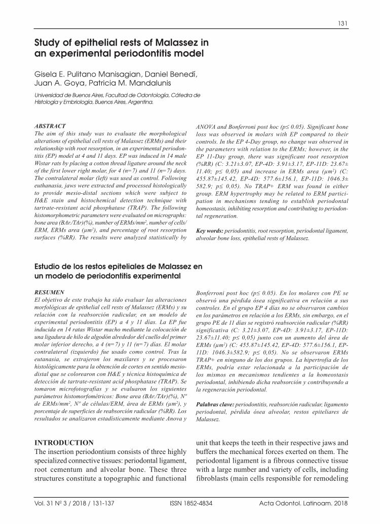
ABSTRACTThe aim of this study was to evaluate the morphologicalalterations of epithelial cell rests of Malassez (ERMs) and theirrela tionship with root resorption, in an experimental periodon titis (EP) model at 4 and 11 days. EP was induced in 14 maleWistar rats by placing a cotton thread ligature around the neckof the first lower right molar, for 4 (n=7) and 11 (n=7) days.The contralateral molar (left) was used as control. Followingeuthanasia, jaws were extracted and processed histologicallyto provide mesiodistal sections which were subject to H&E stain and histochemical detection technique withtartrateresistant acid phosphatase (TRAP). The followinghistomorphometric parameters were evaluated on micrographs:bone area (BAr./TAr)(%), number of ERMs/mm2, number of cells/ERM, ERMs area (µm2), and percentage of root resorptionsurfaces (%RR). The results were analyzed statistically by
ANOVA and Bonferroni post hoc (p£ 0.05). Significant boneloss was observed in molars with EP compared to theircontrols. In the EP 4Day group, no change was observed inthe parameters with relation to the ERMs; however, in theEP 11Day group, there was significant root resorption(%RR) (C: 3.21±3.07, EP4D: 3.91±3.17, EP11D: 23.67±11.40; p£ 0,05) and increase in ERMs area (µm2) (C:455.87±145.42, EP4D: 577.6±156.1, EP11D: 1046.3±582.9; p£ 0,05). No TRAP+ ERM was found in either group. ERM hypertrophy may be related to ERM partici pation in mechanisms tending to establish periodontalhomeostasis, inhibiting resorption and contributing to periodon tal regeneration.
Key words: periodontitis, root resorption, periodontal ligament,alveolar bone loss, epithelial rests of Malassez.
INTRODUCTIONThe insertion periodontium consists of three highlyspecialized connective tissues: periodontal ligament,root cementum and alveolar bone. These threestructures constitute a topographic and functional
unit that keeps the teeth in their respective jaws andbuffers the mechanical forces exerted on them. Theperiodontal ligament is a fibrous connective tissuewith a large number and variety of cells, includingfibroblasts (main cells responsible for remodeling
RESUMENEl objetivo de este trabajo ha sido evaluar las alteracionesmorfológicas de epithelial cell rests of Malassez (ERMs) y surelación con la reabsorción radicular, en un modelo deexperimental periodontitis (EP) a 4 y 11 días. La EP fueinducida en 14 ratas Wistar macho mediante la colocación deuna ligadura de hilo de algodón alrededor del cuello del primermolar inferior derecho, a 4 (n=7) y 11 (n=7) días. El molarcontralateral (izquierdo) fue usado como control. Tras laeutanasia, se extrajeron los maxilares y se procesaronhistológicamente para la obtención de cortes en sentido mesiodistal que se colorearon con H&E y técnica histoquímica dedetección de tartrateresistant acid phosphatase (TRAP). Setomaron microfotografías y se evaluaron los siguientesparámetros histomorfométricos: Bone area (BAr./TAr)(%), Nºde ERMs/mm2, Nº de células/ERM, área de ERMs (µm2), yporcentaje de superficies de reabsorción radicular (%RR). Losresultados se analizaron estadísticamente mediante Anova y
Bonferroni post hoc (p£ 0.05). En los molares con PE seobservó una pérdida ósea significativa en relación a suscontroles. En el grupo EP 4 días no se observaron cambiosen los parámetros en relación a los ERMs, sin embargo, en elgrupo PE de 11 días se registró reabsorción radicular (%RR)significativa (C: 3.21±3.07, EP4D: 3.91±3.17, EP11D:23.67±11.40; p£ 0,05) junto con un aumento del área deERMs (µm2) (C: 455.87±145.42, EP4D: 577.6±156.1, EP11D: 1046.3±582.9; p£ 0,05). No se observaron ERMsTRAP+ en ninguno de los dos grupos. La hipertrofia de losERMs, podría estar relacionada a la participación de los mismos en mecanismos tendientes a la homeostasisperiodontal, inhibiendo dicha reabsorción y contribuyendo ala regeneración periodontal.
Palabras clave: periodontitis, reabsorción radicular, ligamentoperiodontal, pérdida ósea alveolar, restos epiteliares deMalassez.
AOL32018:32011 21/02/2019 11:39 Página 131

the ligament), osteoblasts, cementoblasts, osteoclasts,macrophages, mastocytes, epithelial cell rests of Malassez (ERMs) and undifferentiatedectomesenchymal stem cells. Of all the above,fibroblasts and perivascular ectomesenchymal stemcells play an important part during the developmentand homeostasis of periodontal tissues. Severalsignaling factors modulate the activity of thesecells, which provide the machinery for tissuegrowth and regeneration1. The fibrillar organiccomponent consists of two types of fibers:collagenous fibers, mainly type I, inserted in thecementum and the alveolar bone, and collagen typesIII and XII and elastic fibers, which include threetypes: elastin, oxytalan and elaunin2.Within the study of periodontal components, it isworth highlighting the presence of the epithelialrests of Malassez (ERMs), which are groups of cellsthat appear during the formation of root hard tissueswhen the Hertwig epithelial root sheath breaksdown. They persist in the periodontal ligamentspace throughout the lifetime of the tooth. They areunique in that they are the only odontogenic cellsof epithelial nature within the periodontal structure.Studies on human ERMs3 have reported theircharacteristics: an irregular nucleus with denseheterochromatin and a halo of peripheral cytoplasm,small and scarcely distinguishable, and a highnucleus/cytoplasm ratio. A previous report4 describesthat in rat molars, ERMs undergo increase in size,signs of apoptosis and cell proliferation with age.The authors conclude that ERMs are maintained inthe periodontal ligament by cell turnover throughoutthe lifetime of the tooth.Regarding ultrastructure, different studies haveshown that a basal membrane separates the islandsof ERMs from the connective tissue5. The presenceof tonofilaments, desmosomes and hemidesmosomeshas also been demonstrated, all of which anchorERMS in the basal membrane6, 7. These characte ristics provide evidence of the epithelial nature ofthese cells. Rincon et al.8 reported the average distance fromthe cementum to the ERMs in three regions: apical21 microns, middle radicular: 33 microns, andcervical: 41 microns, indicating a coronal migrationof ERMs away from the root surface towardscoronal in human teeth. ERMs have been shown to express different types of proteins and macromolecules, including
cytokeratins9,10 and neuropeptides11,12,13. With regardto the expression of cytokeratins, CK 17 could be a marker to identify them14. Other studies15,16
also report the expression of cell surface proteins,including epidermal growth factor receptors.Despite their ectodermal origin, ERMs cansynthesize components frequently associated to cellsof mesenchymal origin, such as glycosaminoglycans,hyaluronic acid, dermatan sulfate and chondroitinsulfate17, as well as osteopontin (OPN), bonesialoprotein (BSP) and osteoprotegerin (OPG)8,18,and can also degrade collagen by synthesis ofcollagenases and proteinases19,20,21. It has thereforebeen suggested that they may make a considerablecontribution to periodontal regeneration bysynthesizing a series of proteins related to bone androot cementum22. The importance of ERMs in the etiopathology of odontogenic cysts and tumorsis well known. Yet there is still much to learnregarding their role in periodontal diseases,especially periodontitis, which is the most frequentoral inflammatory pathology.23, 24
Current scientific evidence suggests that thepossible roles of ERMs in the adult periodontalligament include maintenance of homeostasis of theperiodontal medium, thereby preventing ankylosis;maintenance of the periodontal ligament space,thereby inhibiting root resorption; and contributingto repairing periodontal cementum25, 26.The aim of this study was to conduct morphologicalevaluation of the epithelial rests of Malassez and toevaluate their relationship with root resorption inan experimental periodontitis model.
MATERIALS AND METHODSThe experimental protocol was approved by theEthics Committee of the School of Dentistry012/2016 CICUALODONTOFOUBA of BuenosAires Argentina, and is in keeping with the NationalInstitutes of Health Guidelines for the Care and Useof Laboratory Animals.
Experimental Periodontitis Experimental periodontitis (EP) was induced in 14male Wistar rats with body weight 280 g. On Day 1,all animals were anesthetized by intraperitonealadministration of 5% Ketamine (50 mg/Kg bodyweight) and 2% xylazine (5mg/kg body weight).Experimental periodontitis was induced by placing acotton thread ligature around the cervical region of the
132 Gisela E. Pulitano Manisagian, et al.
Acta Odontol. Latinoam. 2018 ISSN 1852-4834 Vol. 31 Nº 3 / 2018 / 131-137
AOL32018:32011 21/02/2019 11:39 Página 132

first lower right molar27 (Fig. 1). Two experimentaltimes were used: 4 days (n=7) and 11 days (n=7). Inboth groups, the contralateral molar was used as acontrol, thereby determining 3 groups: Control Group,EP Day 4 Group and EP Day 11 Group. The animalswere euthanized by intraperitoneal injection of sodiumthiopental solution (150 mg/kg body weight)(Pentovent, 49 Laboratorios Richmond, Buenos Aires,Argentina) and acepromazine maleate (3 mg/kg bodyweight) (Acedan, HollidayScott S.A., Buenos Aires,Argentina).
Histology and HistomorphometryThe section of each hemimandible corresponding tothe three lower molars was fixed 10% bufferedformalin (pH 7.4), decalcified in 10% EDTA solution,pH 7.0 for 25 days, and embedded in paraffin toprepare mesiodistally oriented histological sectionsof the first lower molar. The sections were a) stainedwith HematoxylinEosin and b) subjected tohistochemical determination with tartrateresistantacid phosphatase (TRAP) to evaluate whether thisenzyme, which is characteristic of cells that resorbmineralized tissues, is expressed in ERMs.The following histomorphometric parameters wereevaluated:· Using 40x digital micrographs and Image Pro Plus
software (Fig. 2): Bone Area (BAr/TAr)(%): Percentage of
total interradicular area occupied by bonetissue.
ERMs area (µm2) of the ERMS in theinterradicular space.
Root resorption (% RR): Percentage ofsurfaces in resorption, with or withoutpresence of odontoclasts on the root surface.
· Using direct 100x bright field microscopy: ERMs number/mm2: count of the number of
ERMs in the interradicular space (Fig. 2). Cell number / ERM: count of cell nuclei
observable in each ERM.
Results were analyzed statistically using ANOVAand Bonferroni post hoc, considering p£ 0.05 assignificant.
RESULTSBone loss in the interradicular space was observedat 4 and 11 days after induction of periodontitis, in
comparison to the contralateral molar (Fig. 3, Table 1).In 4day experimental periodontitis, no statisticallysignificant change was observed in the other studyparameters. However, at 11 days of periodontaldisease, there was a statistically significant increasein root resorption (%RR) and ERMs area (Fig.4).Table 2 shows the values recorded for histomorpho metric measurements. No TRAPpositive ERM wasfound in either of the study groups.
DISCUSSIONThe results of this study show that at both 4 and 11days after periodontitis was installed, there wasbone loss at the level of the root furcation. Inaddition, at 11 days there was marked rootresorption accompanied by a significant increase inthe size of the ERMs. This morphological change
Fig. 1: Placingcotton thread
ligature on firstlower molar
to induceexperimentalperiodontitis
(EP).
Fig. 2: Diagram ofrat molar in situ.
Above line a is thearea used to
calculate bone area(BAr/TAr)(%):.
Above line b, markedat the beginning of
acellular cementum,the periodontal area
is shown (shadedand hatched) used
for conducting ERMhistomorphometric
measurements.
Epithelial rests and periodontitis 133
Vol. 31 Nº 3 / 2018 / 131-137 ISSN 1852-4834 Acta Odontol. Latinoam. 2018
AOL32018:32011 21/02/2019 11:40 Página 133

occurred at a result of an increase in volume, not agreater number of cells. The data suggest anassociation between the onset of root resorption andthe morphological changes detected in the ERMs.To date, there is no published paper reporting ERMbehavior as a result of an infectious/inflammatorystimulus such as experimental periodontitis. Thequestion is whether ERMs are involved in theinduction of root resorption or whether theirpersistence and hypertrophy are related to theexpression of signaling factors associated to theirpotential participation in the regulation/inhibitionof said resorption, as part of the homeostasis in theperiodontal ligament.In this regard, the current study performed histo chemical determination of tartrateresistant acid
phosphatase (TRAP) with the aim of ascertainingwhether ERMs express that enzyme, whichcharacterizes cells that resorb mineralized tissues.These ERMs were found to be negative to histo chemical marking, suggesting that they do notparticipate directly in the root resorption observedin the group with experimental periodontitis at 11 days.Another study on rats subjected to in vivoorthodontic forces found that ERMs responded toexperimental orthodontics by proliferating andincreasing in size28. These findings partially agree with the current study, which found ERMhypertrophy but no increase in number. Other
134 Gisela E. Pulitano Manisagian, et al.
Acta Odontol. Latinoam. 2018 ISSN 1852-4834 Vol. 31 Nº 3 / 2018 / 131-137
Fig. 3: A, B, C: Bright fieldmicrographs. Decalcificationtechnique. H&E Stain. A Lowermolar in situ corresponding tocontrol group; B Molar with 4dayexperimental periodontitis. CMolar with 11day experimentalperiodontitis. Note the reductionin bone area (BAr/TAr) in thegroups with EP. 40X.
Fig. 4: Bright field micrographs. Decalcification technique.H&E Stain. Root furcation zone of first lower molar. A. Control; B. Molar with 11day experimental periodontitis.Note bone loss and ERMs. C. Control: ERMs visible (blackarrows). D Molar with 11day experimental periodontitis. Noteenlarged ERMs (black arrows) and root resorption areas(white arrows). A and B: 100 X; C and D: 400 X.
Table 1.
CONTROL EP 4D EP 11D
(BAr/TAr)(%) 40.44 ± 6.86 27.97 ± 7.61 27.26 ± 6.50
EP 4D: experimental periodontitis 4 DAYS; EP 11D: experimental periodontitis 11 DAYS
p£ 0.05. X ± SD of the parameters evaluated.
Table 2.
CONTROL EP 4D EP 11D
ERMs area (µm2) 455.87 ± 145.42 577.6 ± 156.1 1046.3 ± 582.9
Root Resorption 3.21 ± 3.07 3.91 ± 3.17 23.67 ± 11.40(%RR)
ERMs number/mm2 7.15 ± 3.53 6.14 ± 2.19 7.50 ± 6.28
Cell number/ERM 9.70 ± 2.86 8.12 ± 3.64 10.43 ± 5.28
EP 4D: experimental periodontitis 4 DAYS; EP 11D: experimental periodontitis 11 DAYS
p£ 0.05 X ± SD of the parameters evaluated
AOL32018:32011 21/02/2019 11:40 Página 134
papers28,29,30 have reported that orthodontics canstimulate ERMs to secrete different factors,contributing to maintaining normal periodontalstructure and function. Consolaro29 reported thatankylosis occurs when cementoblasts and ERMsare absent.Studies on mechanical forces in vitro have reportedthat ERMs express heat shock proteins, vascularendothelial growth factor (VEGF), osteopontin(OPN)26 and HSP 7031. The expression of theseproteins may contribute to the maintenance ofcementogenesis and osteogenesis, and in addition,particularly HSP 70 may play a part by protectingagainst different factors, including oxidants,inflamma tion, hypoxia, hyperthermia and mecha nical stimuli such as orthodontic forces32.Hasegawa et al.33 used Nakane’s root resorptionmodel (by mechanical injury) to study ERMs 7 days after injury, and observed ERMs adjacent to resorbed surfaces. Immunohistochemical testsrevealed that the ERMs expressed BMP2,osteopontin and ameloblastin, suggesting that theymay participate in periodontal repair.All of the aforesaid suggests that ERM hypertrophyin the current study at 11 days after induction ofexperimental periodontitis may be related to ERMparticipation in mechanisms that tend to maintainperiodontal homeostasis.As mentioned above, ERMs can express differentproteins of epithelial nature14 and typical of theenamel matrix. Nishio et al.34 identified two novelproteins, apin (APIN) and amelotin (AMTN),produced by ameloblasts and junctional epithelium,and assessed whether ERMs express them undernormal conditions and under disruption ofperiodontal integrity. They found that after a lesion,ERMs increased in size and they only obtainedimmunodetection of APIN, suggesting that its
expression may lead to the activation of ERMsduring periodontal healing and regeneration.More recently, new findings suggest that withinERMs there is a cell population with stem celllikecharacteristics and that in an adequate microenvi ronment, this cell population can differentiate intocells that produce mineralized matrices35.Takahashi et al.36 examined the expression ofamelogenin and ameloblastin, metallopeptidase(MMP 20) and kallikrein (KLK4) in ERMs andfibroblasts in culture, from samples of humanperiodontal ligament. Immunohistochemical analysisrevealed that those proteins were expressed weaklyby ERMs and were not detected in periodontalfibroblasts. Previous studies suggest that amelogeninand ameloblastin may both have activity as growthfactors, as well as participating in cell bonding,proliferation, migration and differentiation offibroblasts of the periodontal ligamentt37, 38. Giventhe reported capacity of amelogenin39 and itsclinical use, it is important to conduct furtherstudies on ERMs, considering their potential toinduce regeneration of periodontal tissues bysynthesizing proteins such as amelogenins.
CONCLUSIONSThis study found that the ERMs present in theperiodontium of rat teeth with experimentalperiodontitis with 11 days’ evolution reacted to rootresorption in those teeth, with evident increase insize. Taking into account the information currentlyavailable in the literature, this behavior may berelated to ERM participation in mechanisms thattend to maintain periodontal homeostasis byinhibiting the resorption process. Further studiesare needed to learn about the behavior of these cellgroups in response to stimuli of various origins, andtheir impact on periodontal regeneration.
Epithelial rests and periodontitis 135
Vol. 31 Nº 3 / 2018 / 131-137 ISSN 1852-4834 Acta Odontol. Latinoam. 2018
FUNDINGThis work was supported in part by a grant from the Universityof Buenos Aires, UBACyT Program N° 20020160100034
CORRESPONDENCEDr. Gisela Pulitano Manisagian, Cátedra de Histología y Embriología, Facultad de Odontología, Marcelo T. de Alvear 2142, 1ºA, (C1122AAH) CABA. [email protected]
REFERENCES1. Benatti BB, Silvério KG, Casati MZ, Sallum EA, Nociti NH Jr.
Physiological features of periodontal regeneration and approachesfor periodontal tissue engineering utilizing periodontal ligamentcells. J Biosci Bioeng 2007; 103:16.
2. Nanci A, Bosshardt DD. Structure of periodontal tissues in healthand desease. Periodontol 2000 2006; 40:1128.
3. Inove T, Enokiya Y, Hashimoto S, Fukumashi K, Shimono M.Homeostatic factors in periodontal ligament after wound healing.Effects of Malassez´s epithelial rests. Jpn J Oral Biol 1993; 37:5869.
AOL32018:32011 21/02/2019 11:40 Página 135

4. Oka K, Morokuma M, ImanakaYoshida K, SawaY, Isokawa K, Honda MJ. Cellular turnover in epithelialrests of Malassez in the periodontal ligament of the mousemolar 2012; 120:484494. doi: 10.1111/eos.12003
5. Valderhaug JP, Nylen MU. Function of epithelial rests assuggested by their ultrastructure. J Periodont Res 1966;1:6978.
6. Beertsen W, Everts V. Autodesmosomes in epithelial cellsof rests of Malassez in the incisor and molar periodontalligament of the mouse. Arch Oral Biol 1979; 24:239241.
7. Berkovitz BK, Whatling R, Barrett AW, Omar SS. Thestructure of bovine periodontal ligament with specialreference to the epithelial cell rests. J Periodontol 1997;68:905913.
8. Rincon JC, Young WG, Bartold PM. The epithelialcell rests of Malasseza role in periodontal regeneration? JPeriodontal Res 2006; 41:245252.
9. Gao Z, Mackenzie IC, Williams DM, Cruchley AT, Leigh I,Lane EB. Patterns of keratinexpression in rests ofMalassez and periapical lesions. J Oral Pathol 1988; 17:178185.
10. Peters BH, Peters JM, Kuhn C, Zoller J, Franke WW.Maintenance of celltypespecifi cytoskeletal character inepithelial cells out of epithelial context. Cytokeratins andother cytoskeletal proteins in the rest of Malassez of theperiodontal ligament. Differentiation 1995; 59:113126.
11. Heyeraas KJ, Kvinnsland I, Byers MR, Jacobsen EB. Nervefibers immunoreactive to protein gene product 9.5, calcitoningenerelated peptide, substance P, and neuropeptide Y inthe dental pulp, periodontal ligament, and gingiva in cats.Acta Odontol Scand 1993; 51:207221.
12. Beck F, Tucci J, Russell A, Senior PV, Ferguson MW. Theexpression of the gene coding for parathyroid hormonerelated protein (PTHrP) during tooth development in therat. Cell Tissue Res 1995; 280:283290.
13. Tadokoro O, Maeda T, Heyeraas KJ, Vandevska Radunovic V,Kozawa Y, Hals Kvinnsland I. Merkellike cells in Malassezepithelium in the periodontal ligament of cats: an immuno histo chemical, confocallaser scanning and immuno electronmicroscopic investigation. J Periodont Res 2002; 37:456463.
14. Li S, Ge S, Yang P. Expression of cytokeratins in enamelorgan, junctional epithelium and epithelial cell rests ofMalassez. J periodontal Res 2015; 50:846854.
15. Onishi T, Ooshima T, Sobue S, Tabata MJ, Maeda T, KurisuK, Wakisaka S. Immunohistochemical localization ofcalbindin D28k during root formation of rat molar teeth.Cell Tissue Res 1999; 297:503512.
16. Guajardo G, Okamoto Y, Gogen H, Shanfeld JL, Dobeck J,Herring AH, Davidovitch Z. Immunohistochemical locali zation of epidermal growth factor in cat paradental tissuesduring tooth movement. Am J Orthod Dentofacial Orthop2000; 118:210219.
17. Merrilees MJ, Sodek J, Aubin JE. Effects of cells ofepithelial rests of Malassez and endothelial cells onsynthesis of glycosaminoglycans by periodontal ligamentfibroblasts in vitro. Dev Biol 1983; 97:146153.
18. Rincon JC, Xiao Y, Young WG, Bartold PM. Production ofosteopontin by cultured porcine epitheliacell rests ofMalassez. J Periodont Res 2005; 40:417426.
19. Birek P, Wang HM, Brunette DM, Melcher AH. Epithelialrests of Malassez in vitro. Phagocytosis of collagen and thepossible role of their lysosomal enzymes in collagendegradation. Lab Invest 1980; 43:6172.
20. Firth JD, Putnins EE, Larjava H, Uitto VJ. Bacterialmatrixmetalloproteinase expression by cultured epithelial cells.Infect Immun 1997; 65:49314936.
21. Uitto VJ, Airola K, Vaalamo M, Johansson N, Putnins EE,Firth JD, Salonen J, LopezOtín C, SaarialhoKere U,Kahari VM. Collagenase3 (matrix metalloproteinase 13)expression is induced in oral mucosal epithelium duringchronic inflammation. Am J Pathol 1998; 152:14891499.
22. Yang Z, Li Y, Ma X, Shen L, Zhao Z, Jin F. Role of theEpithelial Cell Rests of Malassez in Periodontal Homeostasisand Regeneration A Review. Curr Stem Cell Res Ther 2015;10:398404.
23. Nazar Majeed Z, Philip K, Alabsi AM, Pushparajan S,Swaminathan D. Identification of Gingival CrevicularFluid Sampling, Analytical Methods, and Oral Biomarkersfor the Diagnosis and Monitoring of Periodontal Diseases:A Systematic Review. Dis Markers 2016; 2016:1804727.doi: 10.1155/2016/1804727
24. Savage A, Eaton KA, Moles DR, Needleman I. Asystematic review of definitions of periodontitis andmethods that have been used to identify this disease. J Clin Periodontol 2009; 36:458467. doi: 10.1111/j.1600051X.2009. 01408.x
25. Xiong J, Gronthos S, Bartold PM. Role of the epithelial cellrests of Malassez in the development, maintenance andregeneration of periodontal ligament tissues. Periodontol2000 2013; 63:217233.
26. Koshihara T, Matsuzaka K, Sato T, Inoue T. Effect ofstretching Force on the Cells of Epithelial Rests of MalassezIn Vitro. Int J Dent 2010; 2010:458408. doi: 10.1155/2010/458408
27. Ubios AM, Costa OR, Cabrini RL. Early steps in boneresorptionin experimental periodontitis: A histomorphometric study. ActaOdontol Latinoam 1993; 7:4550.
28. Talic NF, Evans CA, Daniel JC, Zaki AE. Proliferation ofepithelial rests of Malassez during experimental toothmovement. Am J Orthod Dentofacial Orthop 2003; 123:527533.
29. Consolaro A. The concept of root resorptions or rootresorptions are not multifactorial, complex, controversialor polemical! Dental Press J Orthod 2011; 16:1924.
30. Yamanaka T, Sakamoto A, Tanaka Y, Zhang Y, HayashidoY, Toratani S, Akagawa Y, Okamoto T. Isolation and serumfree culture of epithelial cells derived from epithelial restsof Malassez in human periodontal ligament. In Vitro CellDev Biol Anim 2000; 36:548553.
31. Kogai H , Nakajima K, SerOd T, AlWahabi A, MatsuzakaK, Nakagawa T, Inoue T. HSP70 mRNA expression by cellsof the epithelial rest of Malassez due to mechanical forcesin vitro. BMC Oral Health 2016; 16:22. doi: 10.1186/s1290301601814
32. Muraoka R, Nakano K, Matsuda H, Tomoda M, Okafuji N,Yamada K, Kawakami T, Kawakami T. A consideration onthe role of HSP70 appearing in the periodontal tissue dueto experimental orthodontic force. J Hard Tissue Biol 2011;20:275282.
136 Gisela E. Pulitano Manisagian, et al.
Acta Odontol. Latinoam. 2018 ISSN 1852-4834 Vol. 31 Nº 3 / 2018 / 131-137
AOL32018:32011 21/02/2019 11:40 Página 136

33. Hasegawa N, Kawaguchi H, Ogawa T, Uchida T, KuriharaH. Immunohistochemical characteristics of epithelial cellrests of Malassez during cementum repair. J Periodont Res2003; 38:5156.
34. Nishio C, Wazen R, Kuroda S, Moffatt P, Nanci A.Disruption of periodontal integrity induces expression ofapin by epithelial cell rests of Malassez. J Periodont Res2010; 45:709713.
35. Tsunematsu T, Fujiwara N, Yoshida M, TakayamaY, Kujiraoka S, Qi G, Kitagawa M, Kondo T, YamadaA, Arakaki R, Miyauchi M, Ogawa I, Abiko Y, NikawaH, Murakami S, Takata T, Ishimaru N, Kudo Y. Humanodontogenic epithelial cells derived from epithelial rests ofMalassez possess stem cell properties. Lab Invest 2016;96:10631075. doi: 10.1038/labinvest.2016.85
36. Takahashi K, Shimonishi M, Wang R, Watanabe H, KikuchiM. Epithelial– mesenchymal interactions induce enamel
matrix proteins and proteases in the epithelial cells of therests of Malassez in vitro. Eur J Oral Sci 2012; 120:475483.
37. ZeichnerDavid M, Chen LS, Hsu Z, Reyna J, Caton J,Bringas P. Amelogenin and ameloblastin show growthfactor like activity in periodontal ligament cells. Eur J OralSci 2006; 114:244253.
38. Kémoun P, Gronthos S, Snead ML, Rue J, Courtois B,Vaysse F, Salles JP, Brunel G. The role of cell surfacemarkers and enamel matrix derivatives on human periodontalligament mesenchymal progenitor responses in vitro.Biomaterials 2011; 32:73757388. doi: 10.1016/j.biomaterials.2011.06.043
39. Haruyama N, Hatakeyama J, Moriyama K, Kulkarni A.Amelogenins: Multifunctional enamel matrix proteinsand their binding partners. J Oral Biosci 2011; 53:257266.
Epithelial rests and periodontitis 137
Vol. 31 Nº 3 / 2018 / 131-137 ISSN 1852-4834 Acta Odontol. Latinoam. 2018
AOL32018:32011 21/02/2019 11:40 Página 137

138
Acta Odontol. Latinoam. 2018 ISSN 1852-4834 Vol. 31 Nº 3 / 2018 / 138-143
RESUMOBiofilme nas linhas d’água de equipos odontológicos podepropagar contaminação microbiana na água. O objetivo desteestudo foi investigar a contaminação microbiana da água deabastecimentos e equipos odontológicos antes e após a implemen tação de um protocolo para melhoria e manutenção da qualidademicrobiológica da água de equipos odontológicos. Avaliouse acarga microbiana da água de 27 torneiras e equipos (reservatórios,seringas tríplice e alta rotação sem as peças de mão) de uma clínicaodontológica por meio do sistema Petrifilm™ (bactérias aeróbiastotais e fungos) e meios de cultura convencionais (enterobactériase Legionella spp.). A carga bacteriana em amostras de água das
torneiras e reservatórios estava dentro do parâmetro estabelecidopela legislação brasileira (<500 UFC/mL), mas a carga bacte riana das seringas tríplices e das saídas dos alta rotação sem aspeças de mão não estava. A implementação do protocolo paramanutenção da qualidade da água dos equipos reduziu a cargabacteriana nas saídas dos alta rotação sem as peças de mão(p=0,004). Enterobactérias e Legionella spp. não foram isoladasde qualquer das amostras de água das torneiras e dos equiposodontológicos.
Palavras chave: Biofilmes, equipamento odontológico, microbiolo gia da água.
INTRODUCTIONOver the past century, dental units have evolved fromthe original pedalpowered pulley models to thecurrent versions with technology that provides safetyand reduces biological risk. The greatest transforma tion took place in the early 1950s, with the emergenceof airwater syringes and highspeed handpieces.Because this kind of equipment generates heat thatcan cause thermal injury, it requires watercooling, so
includes long, thin flexible tubes to channel water andair to it. But neither the inventors nor dental professio nals imagined that those long, thin waterlines couldconceal a great number of microorganisms fromwater, despite the implementation of basic principlesof asepsis.1
Dental units are supplied with drinking water,which contains small microbial load. In Brazil,Ministry of Health ordinance No. 2,914 establishes
ABSTRACTBiofilm on dental unit waterlines can spread microbialcontamination in the water. The aim of this study was toinvestigate microbial contamination of water from supplies anddental units before and after the implementation of a protocolfor microbial quality improvement and maintenance of dentalunit water. The microbial load was evaluated in water from 27taps and dental units (reservoirs, airwater syringes and highspeed outputs without handpieces) using the Petrifilm™ system(total aerobic bacteria and fungi) and conventional culturemedia (enterobacteria and Legionella spp.). The bacterial load
in water samples from taps and reservoirs was within theparameter established by Brazilian legislation (<500CFU/mL);but the bacterial load in samples from airwater syringes andhighspeed outputs without handpieces was not. The imple mentation of the protocol for the maintenance of microbialquality in dental unit water reduced bacterial load in highspeed outputs without handpieces (p=0.004). Enterobacteriaand Legionella spp. were not isolated from any of the watersamples from taps and dental units.
Key words: Biofilms, dental equipment, water microbiology.
Evaluation of a protocol for reducing the microbial contamination of dental unit water
Rachel M. Monteiro1, Daniella M. Marques1, Pedro C. A. Domingues1, Viviane de C. Oliveira2, Ana Paula Macedo2, Ana M. Razaboni3, Evandro Watanabe3
1 Universidade de São Paulo, Escola de Enfermagem de Ribeirão Preto, Ribeirão Preto, SP, Brazil
2 Universidade de São Paulo, Faculdade de Odontologia de Ribeirão Preto, Departamento de Materiais Dentários e Próteses, Ribeirão Preto, SP, Brazil
3 Universidade de São Paulo, Faculdade de Odontologia de Ribeirão Preto, Departamento de Odontologia Restauradora, Ribeirão Preto, SP, Brazil
Avaliação de um protocolo para redução da contaminação microbiana da água de equipos odontológicos
AOL32018:32011 21/02/2019 11:40 Página 138

the limit as 500 colonyforming units (CFU) permilliliter (mL) of water.2 In 1996, the AmericanDental Association (ADA)3 recommended that thewater in dental units should contain no more than200 CFU/mL.Dental unit water can pose a risk to oral and generalhealth due to microbial contamination and biofilmformation on waterlines.410 The first signs ofmicrobial contamination of dental unit water andbiofilm formation on waterlines were described byBlake11 and Kelstrup et al.12, respectively. Theliterature includes reports of infectious diseases dueto contamination in dental unit waterlines.13,14 Thisis a matter of concern, since the infections causedby microorganisms resistant to antimicrobials canbe fatal, mainly in immunocompromised patients.Once biofilm is formed, it is difficult to remove.Various strategies have been reported for controlling iton dental unit waterlines, such as development ofsurfaces with antibiofilm activity15, supply of sterilizedwater for dental units16, and physicalchemicaltreatments.17,18 However, most of the strategies usedfor biofilm formation control on waterlines havelimitations, often related to high cost and difficulty inimplementation. There is thus a need for thedevelopment and use of an efficient protocol forbiofilm control on dental unit waterlines based on easyimplementation, short execution times and low cost.The aim of this study was to evaluate the microbialload of water from taps and dental units (reservoirs,airwater syringes and highspeed outputs withouthandpieces) before and after the implementation ofa protocol for improvement and maintenance of themicrobiological quality of the water in dental units.
MATERIALS AND METHODSSamples were collected aseptically from dental unitwaterlines [reservoirs (R), airwater syringes(AWS) and highspeed outputs without handpieces(HSWH)] from 27 dental units at the School ofDentistry of Ribeirão Preto (SP, Brazil). Samples oftap water (TW) used to supply the dental units werealso collected. The samples were collected atbaseline (“T0”) and seven months after baseline(“T1”). The “T1” samples were collected after theimplementation of a protocol for improvement andmaintenance of the microbiological quality of waterin dental units as a daily routine. The protocolconsisted of supplying and draining the dental unitreservoirs at the beginning and end of the work,
respectively, and recommended flushing AWS andHSWH for 30 seconds before and after eachpatient.3,19 The protocol did not include anychemical agents for disinfecting reservoirs anddental unit waterlines.TW, AWS and HSWH samples were collected afterwater flushing (30s). In addition, samples werecollected from reservoirs after rinsing three timeswith TW. All samples were collected in anapproximate volume of 10mL in sterile test tubes(25x150mm). The samples were placed in a cooler,and microbiological processing began no longerthan 30 minutes after collection.The experiment was conducted in a Class II TypeA1 Biological Safety Cabinet (VECO, Campinas,SP, Brazil). A 50μL aliquot of 2% sodium thiosulfatewas added to each sample. The samples werehomogenized (Phoenix, Araraquara, SP, Brazil),diluted up to 104 and seeded on Petrifilm™ AC andYM (3M, St Paul, USA) plates to evaluate totalaerobic bacteria and fungi (filamentous fungi and yeasts), respectively. In addition, Petri plates(60x15mm) with conventional culture media fordetection of Legionella spp. (Legionella Agar Base®
supplemented with Legionella Agar Enrichment® –BD Difco, Sparks, MN, USA) and enterobacteria(MacConkey Agar – BD Difco, Sparks, MD, USA)were employed. The plates with water samples wereincubated at 37°C for 48 h (total aerobic bacteriaand enterobacteria), 23°C for 5 days (filamentousfungi and yeasts) and 37°C for 48 h (Legionella spp.).After the incubation periods, the colonies werecounted using a trinocular stereomicroscope(Tecnival) under reflected light. The number ofcolony forming units per milliliter (CFU/mL) ofwater in natura was determined.The statistical tests were performed using IBMSPSS Statistics 20.0 software (IBM Corp Armonk,NY, USA). As the distribution was nonnormal,nonparametric Wilcoxon test was used to compareT0 and T1. Differences between contamination ofthe TW, R, AWS and HSWH in T0 and T1 wereanalyzed by KruskalWallis. Since there was nocount of total aerobic bacteria in T1 for TW and R,MannWhitney test was used for the comparisonbetween AWS and HSWH. Relative frequency ofpresence and absence of contamination for theevaluated groups in T0 and T1 was performedthrough Pearson Chisquare test. The significancelevel was set at 0.05.
Microbial contamination of dental unit water 139
Vol. 31 Nº 3 / 2018 / 138-143 ISSN 1852-4834 Acta Odontol. Latinoam. 2018
AOL32018:32011 21/02/2019 11:40 Página 139

RESULTSTables 1 and 2 show the results of this study for loadsof total aerobic bacteria and fungi (filamentous fungiand yeasts).Of 27 TW samples, 6 (22.2%) were contaminatedby total aerobic bacteria at T0. No TW sample wascontaminated at T1, having a reduction of 16.0times the bacterial load (CFU/mL). Filamentousfungus and yeast counts showed that 8 (29.6%) ofTW samples were contaminated at T0, and 10
(37.0%) at T1, presenting an increase of 2.2 timesof CFU/mL (p=0.407).Of 27 reservoirs (R), 1 (3.7%) was contaminated bytotal aerobic bacteria at T0 and none at T1,presenting a reduction of 1.9 times of CFU/mL. Thefilamentous fungus and yeast count showed that 13(48.1%) R were contaminated at T0 and 8 (29.6%)at T1, a reduction of 1.8 times of CFU/mL(p=0.351). It is worth noting that only 2 R remainedcontaminated at T1, while the other 6 R had new
140 Rachel M. Monteiro, et al.
Acta Odontol. Latinoam. 2018 ISSN 1852-4834 Vol. 31 Nº 3 / 2018 / 138-143
Table 1: Median and confidence interval of CFU/mL for the evaluated groups: before (T0) and after (T1) the protocol implementation for reduction of the microbial contamination of dental unit water. Ribeirão Preto, SP, Brazil, 2018.
Total aerobic bacteria p*** Filamentous fungi and yeasts p***
T0 T1 T0 T1
TW 0.0 (0.0; 35.4)ab 0.0 (-;-) - 0.0 (0.0; 7.9)a,A 0.0 (0.6; 15.7)a,A 0.407
R 0.0 (0.0; 5.7)a 0.0 (-;-) - 0.0 (0.6; 2.6)a,A 0.0 (0.2; 1.5)a,A 0.351
AWS 0.0 (0.0; 294.4)a,A 0.0 (0.0; 0.7)a,A 0.225 0.0 (0.0; 60.7)a,A 0.0 (0.0; 1.3)a,A 0.098
HSWH 0.0 (0.0; 316.8)b,A 0.0 (0; 1.5)a,B 0.004 4.0 (0.0; 279.0)b,A 0.0 (0.0; 34.7)a,A 0.131
p 0.001* 0.096** 0.001* 0.133*
CFU/mL: colony forming units per milliliter of water; T0: baseline; T1: after implementation of the protocol for reduction of microbial contamination in dental unit water; TW: tap water; R: reservoirs; AWS: air-water syringes; HSWH: high-speed outputs without handpieces. *Kruskal-Wallis followedby Dunn test; **Mann-Whitney; ***Wilcoxon; ab Same lowercase letters indicate statistical similarity among collection sites; AB Same uppercase let-ters indicate statistical similarity between T0 and T1.
Table 2: Relative frequency of presence and absence of contamination for the evaluated groups in T0 and T1. Ribeirão Preto, SP, Brazil, 2018.
Total aerobic bacteria Filamentous fungi and yeasts
T0 T1 T0 T1
Absence Presence Absence Presence Absence Presence Absence Presence
TW 21 6 (22.2%) 27 0 (0.0%) 19 8 (29.8%) 17 (63.0%) 10
(77.8%) (100.0%) (70.4%) (37.0%)
R 26 1 (3.7%) 27 0 (0.0%) 14 13 19 (70.4%) 8 (29.6%)
(96.3%) (100.0%) (51.9%) (48.1%)
AWS 23 4 (14.8%) 26 (96.3%) 1 (3.7%) 16 11 22 (81.5%) 5 (18.5%)
(85.2%) (59.3%) (40.7%)
HSWH 14 13 (48.1%) 22 (81.5%) 5 (18.5%) 6 (22.2%) 21 16 (59.3%) 11
(51.9%) (77.8%) (40.7%)
Total 84 24 (22.2%) 102 6 (5.6%) 55 53 74 (68.5%) 34 (31.5%
(77.8%) (94.4%) (50.9%) (49.1%)
p* 0.001 0.007 0.003 0.307
T0: baseline; T1: after implementation of the protocol for reduction of microbial contamination in dental unit water; TW: tap water;R: reservoirs; AWS: air-water syringes; HSWH: high-speed outputs without handpieces; *Pearson Chi-square test.
AOL32018:32011 21/02/2019 11:40 Página 140

contamination. Consequently, the protocol forimprovement and maintenance of the microbiologicalquality of water in dental units as a daily routineshowed a reduction in fungal contamination in 11 R.Of 27 AWS, 4 (14.8%) were contaminated by totalaerobic bacteria at T0 and 1 (3.7%) at T1, with areduction of 539.7 times of CFU/mL (p=0.225).Moreover, this AWS contamination at T1 wasconsidered new. The filamentous fungus and yeastcount showed that 11 (40.7%) AWS were contami nated at T0 and 5 (18.5%) at T1, having a reductionof 143.7 CFU/mL (p=0.098). Thus, only 3 AWSremained contaminated at T1, and the other 2 AWShad new contamination, with a reduction in fungalcontamination of 3 AWS.Of 27 HSWH, 13 (48.15%) showed contaminationby total aerobic bacteria at T0 and 5 (18.52%) atT1, presenting a reduction of 33.6 times ofCFU/mL (p=0.004). Moreover, 4 HSWH remainedcontami nated at T1, and only one case of newcontamination was reported. The filamentousfungus and yeast count showed that 21 (77.8%) of HSWH were contaminated at T0 and 11(40.7%) at T1, with a reduction of 6.9 times ofCFU/mL (p=0.131). Thus, only 9 HSWH remainedcontaminated at T1, and the other 2 HSWH hadnew contamination, with a reduction in fungalcontamination of 12 HSWH.The comparison among loads of total aerobicbacteria and filamentous fungi and yeasts from thedifferent collection sites at T0 showed that thebacterial and fungal contamination in HSWH wasgreater than in AWS and R (p=0.001).In relation to the relative frequency of cases withpresence and absence of contamination at T0,HSWH contamination for total aerobic bacteria(48.1% / p=0.001) and filamentous fungi and yeasts(77.8% / p=0.003) was found to be greater than atthe other evaluated sites (TW, R and AWS).Moreover, in this study, the presence of enterobacteriaand Legionella spp. was not detected in any of thesamples (TW, R, AWS and HSWH) analyzed.
DISCUSSIONDental units consist of reservoirs that supply waterthrough waterlines (diameters 2 to 3 mm) to airwater syringes and highspeed handpieces.20 Biofilmon these thin waterlines is an alarming source ofmicrobial contamination of water7,9,10 and can spreadpathogenic microorganisms, thereby posing a threat
to public health by causing respiratory infectionsand surgical site infections. Moreover, dentists andprofessional staff may become infected by aerosolsgenerated in the dental office.21,22 Since the micro biological quality of water for human consumptionis directly related to human health, dental unit watermust meet the drinking standard determined orsuggested by national and interna tional legislationor organizations.In this study, water samples from AWS (7.4%) andHSWH (7.4%) presented a total aerobic bacterialload greater than 500CFU/mL. On the other hand,none of TW samples showed contamination abovethe limit permitted by Brazilian legislation.2
According to ADA recommendations (1996), at T0,2 AWS and 4 HSWH samples showed bacterialcontamination greater than 200CFU/mL. Inagreement with our results, other authors havereported contamination of water samples fromdental units with counts above 200CFU/mL and500CFU/mL.23,6
No national and/or international parameter has yetbeen established with regard to the fungal contami nation of water intended for human consumptionand dental units. Nevertheless, water should bemonitored and controlled frequently for biosafetyin dentistry, since filamentous fungi and yeasts havebeen isolated from dental unit water in otherstudies24, 25 as well as in the current study.Enterobacteria and Legionella spp. were notisolated from the water samples analyzed in thecurrent study. Although the traditional culturetechnique is the main evaluation method forbacterial contamination, falsenegative results orunderestimated counts have been reported forLegionella spp. Some authors have thereforesuggested the use of molecular techniques, such aspolymerase chain reaction (PCR), to avoid theseproblems.26,27
Biofilm is composed of microorganisms protectedby an extracellular polymeric matrix. When itdevelops on dental unit waterlines, it causesproblems which may be resolved by applyingphysical/ mechanical strategies such as flushingwater, as was done in the current study and inothers4, 28, 29, to partially remove microorganismsthat are loosely adhered to the biofilm. Antimicrobialchemical agents are also used for this purpose, butthey can compromise the structural integrity ofdental unit waterlines14, thereby increasing the
Microbial contamination of dental unit water 141
Vol. 31 Nº 3 / 2018 / 138-143 ISSN 1852-4834 Acta Odontol. Latinoam. 2018
AOL32018:32011 21/02/2019 11:40 Página 141

contact area for microbial adhesion and biofilmformation. Moreover, the use of chemical agents forbiofilm control may present limitations related tomicrobial phenotypic changes30, the difficulty ofreaching the innermost microbiota in the biofilm31,and residual toxic effects on individuals and theenvironment.In this study, a protocol was implemented toimprove and maintain the microbial quality of
dental unit water. The protocol was inexpensive andsimple to implement, created no risk to humanhealth or the environment, and provided a partialsolution to biofilm contamination on dental unitwaterlines. Nevertheless, the problem remains asone of the greatest challenges in dentistry andrequires further studies for better understanding,with the aim of providing a biologically safeenvironment in dentistry.
142 Rachel M. Monteiro, et al.
Acta Odontol. Latinoam. 2018 ISSN 1852-4834 Vol. 31 Nº 3 / 2018 / 138-143
FUNDINGThe authors would like to thank the São Paulo Research Foundation (FAPESP) for financial support of the project (grant#2013/029848).In addition, this study was partly financed by the Coordenaçãode Aperfeiçoamento de Pessoal de Nível Superior – Brazil(CAPES) – Finance Code 001.
CORRESPONDENCEDr. Evandro WatanabeFaculdade de Odontologia de Ribeirão PretoUniversidade de São Paulo.Departamento de Odontologia Restauradora.Avenida do Café s/ nº, Monte Alegre, Ribeirão Preto, SP, Brasil.CEP: 14.040[email protected]
REFERENCES1. Mills SE, Karpay RI. Dental waterlines and biofilm—
searching for solutions. Compend Contin Educ Dent 2002;23:237240.
2. Brazil. Ministry of Health. Ordinance No. 2,914, 12December 2011. Union Official Diary, Brasília, DF, 14December 2011. URL: http://bvsms.saude.gov.br/bvs/saudelegis/gm/2011/prt2914_12_12_2011.html
3. American dental association (ADA). ADA statement ondental unit waterlines. J Am Dent Assoc 1996; 127:185186.
4. Watanabe E, Agostinho AM, Matsumoto W, Ito IY. Dentalunit water: bacterial decontamination of old and new dentalunits by flushing water. Int J Dent Hyg 2008; 6:5662.
5. Bansal R, Puttaiah R, Harris R, Reddy A. Evaluation of twomethods in controlling dental treatment water contamination.J Contemp Dent Pract 2011; 12:7383.
6. Lin SM, Svoboda KKH, Giletto A, Seibert J, et al. Effectsof hydrogen peroxide on dental unit biofilms and treatmentwater contamination. Eur J Dent 2011; 5:4759.
7. O’Donnell MJ, Boyle MA, Russell RJ, Coleman DC.Management of dental unit waterline biofilms in the 21stcentury. Future Microbiol 2011; 6:12091226.
8. Puttaiah R, Seibert J, Spears R. Effects of iodine inmicrobial control of dental treatment water. J ContempDent Pract 2011; 12:143151.
9. Lal S, Singhrao SK, AchillesDay UE, Morton LHG, et al.Risk assessment for the spread of serratia marcescenswithin dentalunit waterline systems using vermamoebavermiformis. Curr Microbiol 2015; 71:434442.
10. Shajahan IF, Kandaswamy D, Lakshminarayanan L,Selvarajan R. Substantivity of hypochlorous acidbaseddisinfectant against biofilm formation in the dental unitwaterlines. J Conserv Dent 2017; 20:25.
11. Blake GC. The incidence and control of bacterial infectionin dental spray reservoirs. Br Dent J 1963; 115:413416.
12. Kelstrup J, FunderNielsen TD, Theilade J. Microbialaggregate contamination of water lines in dental equipmentand its control. Acta Pathol Microbiol Scand B 1977; 85:177183.
13. Ricci ML, Fontana S, Pinci F, Fiumana E, et al. Pneumoniaassociated with a dental unit waterline. Lancet 2012; 379:684.
14. Barbot V, Robert A, Rodier MH, Imbert C. Update oninfectious risks associated with dental unit waterlines.FEMS Immunol Med Microbiol 2012; 65:196204.
15. Luo J, Porteous N, Sun Y. Rechargeable biofilmcontrollingtubing materials for use in dental unit water lines. ACS ApplMater Interfaces 2011; 3:28952903.
16. Kettering JD, Stephens JA, MuñozViveros CA, NaylorWP. Reducing bacterial counts in dental unit waterlines:tap water versus distilled water. J Contemp Dent Pract2002; 3:19.
17. Liaqat I, Sabri AN. Effect of biocides on biofilm bacteriafrom dental unit water lines. Curr Microbiol 2008; 56:619624.
18. Stone ME, Kuehne JC, Cohen ME, Talbott JL, et al. Effect ofiodine on mercury concentrations in dentalunit wastewater.Dent Mater 2006; 22:119124.
19. Centers for Disease Control and Prevention (CDC).Recommended infectioncontrol practices for dentistry.MMWR Morb Mortal Wkly Rep 1993; 42:112.
20. Coleman DC, O’Donnell MJ, Shore AC, Russell RJ.Biofilm problems in dental unit water systems and itspractical control. J Appl Microbiol 2009; 106:14241437.
21. Lal S, Singhrao SK, Bricknell M, Pearce M, et al.Monitoring dentalunitwaterline output water by currentinoffice test kits. Curr Microbiol 2014; 69:135142.
22. Pankhurst CL, Scully C, Samaranayake L. Dental unitwater lines and their disinfection and management: AReview. Dent Update 2017; 44:284292.
23. Percival RS, Devine DA, Nattress B, Kite P, et al. Controlof microbial contamination in dental unit water systems
AOL32018:32011 21/02/2019 11:40 Página 142

using tetrasodium EDTA. J Appl Microbiol 2009; 107:10811088.
24. Agahi RH, Hashemipour MA, Kalantari M, AyatollahMosavi A, et al. Effect of 0.2% chlorhexidine on microbialand fungal contamination of dental unit waterlines. DentRes J (Isfahan) 2014; 11:351356.
25. Dallolio L, Scuderi A, Rini MS, Valente S, et al. Effect ofdifferent disinfection protocols on microbial and biofilmcontamination of dental unit waterlines in communitydental practices. Int J Environ Res Public Health 2014;11:20642076.
26. Ditommaso S, Giacomuzzi M, Ricciardi E, Zotti CM.Cultural and Molecular Evidence of Legionella spp.Colonization in Dental Unit Waterlines: Which Is the BestMethod for Risk Assessment? Int J Environ Res PublicHealth 2016; 13:211.
27. Carinci F, Scapoli L, Contaldo M, Santoro R, et al.Colonization of Legionella spp. In dental unit waterlines. JBiol Regul Homeost Agents 2018; 32:139142.
28. AlHiyasat AS, Ma’ayeh SY, Hindiyeh MY, Khader YS.The presence of Pseudomonas aeruginosa in the dental unitwaterline systems of teaching clinics. Int J Dent Hyg 2007;5:3644.
29. Abdallah SA, Khalil AI. Impact of cleaning regimes ondental water unit contamination. J Water Health 2011; 9:647652.
30. Liaqat I, Sabri AN. Isolation and characterization ofbiocides resistant bacteria from dental unit water linebiofilms. J Basic Microbiol 2009; 49:275284.
31. Soto, SM. Role of efflux pumps in the antibiotic resistanceof bacteria embedded in a biofilm. Virulence 2013; 4:223229.
Microbial contamination of dental unit water 143
Vol. 31 Nº 3 / 2018 / 138-143 ISSN 1852-4834 Acta Odontol. Latinoam. 2018
AOL32018:32011 21/02/2019 11:40 Página 143

144
Acta Odontol. Latinoam. 2018 ISSN 1852-4834 Vol. 31 Nº 3 / 2018 / 144-148
RESUMOO objetivo deste estudo foi descrever e caracterizar (utilizando oensaio de tração) uma técnica alternativa de manuseio para ocimento de hidróxido de cálcio na cimentação de coroatemporária. No grupo tratado com a técnica convencional (n=10),as pastas base e catalisadora de um cimento de hidróxido decálcio (Dycal) foram dispensadas na proporção de 1:1 emisturadas. O cimento foi então aplicado às superfícies cervicaisinternas da restauração provisória, e a restauração foi colocadasobre o dente preparado e mantido no local com pressão digital.No grupo tratado com a técnica alternativa (n=10), a pasta basedo cimento de hidróxido de cálcio (Dycal) foi colocada no dente ea pasta catalítica foi inserida na coroa provisória. A prótese
provisória foi colocada sobre o dente e mantida no local compressão digital. Os valores de tração foram avaliados comparadosentre os grupos pelo teste t de Student, com nível de significânciade 5% (α = 0,05). O uso desta técnica alternativa resultou emresistência à tração significativamente menor em comparaçãocom a técnica convencional (0,58 ± 0,12 vs. 1,08 ± 0,13 MPa; p<0,001). A técnica aqui apresentada (alternativa) poderia evitara remoção indesejada de pinos ou núcleos fundidos no momentoda remoção da prótese provisória e garantir a cimentação depróteses provisórias extensas com cimento de hidróxido de cálcio.
Palavras chave: Hidróxido de Cálcio. Cimentos Dentários.Resistência à Tração.
INTRODUCTIONThe use of provisional prostheses with temporarycements is necessary to meet the requirements ofpulp protection, periodontal protection, aesthetics,and occlusion prior to cementation of the final
prosthesis1. The operator’s familiarity with theprosthesis and cement materials optimizes theresults of provisional treatment. The cementingtechnique and the type of cement used playimportant roles. The retentive properties of a
ABSTRACTThe purpose of this study was to describe and characterize(using the tensile test) an alternative handling technique forcalcium hydroxide cement in temporary crown cementation. Inthe group treated with the conventional technique (n=10), thebase and catalyst pastes of a calcium hydroxide cement (Dycal)were dispensed at a 1:1 ratio and mixed. The cement was thenapplied to the internal cervical surfaces of the provisionalrestoration, and the restoration was placed on the preparedtooth and kept in place with digital pressure. In the grouptreated with the alternative technique (n=10), the base paste ofthe calcium hydroxide cement (Dycal) was placed on the toothand the catalyst paste was inserted into the temporary crown.
The provisional prosthesis was placed on the tooth and kept inplace with digital pressure. Tensile values were evaluated andcompared between groups using Student’s t test with a 5% levelof significance (α = 0.05). Use of this alternative techniqueresulted in significantly lower tensile strength compared to theconventional technique (0.58 ± 0.12 vs. 1.08 ± 0.13 MPa;p<0.001). The technique presented here (alternative) couldavoid the undesired removal of cemented cast posts or cores atthe time of provisional prosthesis removal and ensures thecementation of extensive provisional prostheses with calciumhydroxide cement.
Key words: Calcium hydroxide, dental cements, tensile strength.
Description and characterization of an alternative technique for temporary crown cementation with calcium hydroxide cement
Felipe Sczepanski1, Claudia R. B. Sczepanski2, Sandrine B. Berger3, Lucineide L. Santos3, Ricardo D. Guiraldo3
1 Universidade Estadual do Norte do Paraná – UENP, Faculdade de Odontologia, Departamento de Odontologia Restauradora, Jacarezinho, PR, Brasil
2 Universidade Estadual do Norte do Paraná – UENP, Faculdade de Fisioterapia, Departamento de Odontologia Fisioterapia, Jacarezinho, PR, Brasil
3 Universidade Norte do Paraná – UNOPAR, Faculdade de Odontologia, Departamento de Odontologia Restauradora, Londrina, PR, Brasil
Descrição e caracterização de uma técnica alternativa para a cementação de coroa temporária com cimento de hidróxido de cálcio
AOL32018:32011 21/02/2019 11:40 Página 144

temporary cement should be sufficient to preventpremature loss of the prosthesis, but should nothamper its removal when desired2.The choice of a cementing agent should be guidedby consideration of several factors, such as thedegree of tooth retention, duration of provisionalprosthesis use, prosthesis fabrication technique, andtooth vitality status3. Temporary cementing agentsshould be biocompatible, have low mechanicalstrength, and be easy to handle; however, no singlematerial meets all these requirements fully.Although calcium hydroxide cements weredeveloped for pulp capping, they are also suitablefor temporary cementation. They are among themost commonly used materials for the cementationof provisional prostheses4, but the time between themanipulation of the two pastes and insertion in the prosthesis is critical, and their mechanicalresistance can be high. The aim of this study was todescribe and characterize (using the tensile test) an alternative handling technique for calciumhydroxide cement in temporary crown cementation.
MATERIALS AND METHODSAn intact bovine incisor from which all debris hadbeen removed was used in this study. The toothwas embedded in selfcuring acrylic resin5, (Jet;Artigos Odontológicos Clássico Ltd., São Paulo,SP, Brazil) in a polyethylene tube (ReforplásIndústria e Comércio Ltd., São Paulo, SP, Brazil),with the cementoenamel junction projecting 5 mmabove the resin and the buccal surface orientedperpendicular to the tube. The tooth surfaces werecleaned for 10 s with a rubber cup and nonfluoridated pumice– water slurry (S.S. White,
Petrópolis, RJ, Brazil), rinsed with air–water sprayfor 10 s, and air dried for 10 s.A mold of the tooth was made withpolydimethylsiloxane impression material (Zetaplus/catalyst; Zhermack, Badia Polesine, RO, Italy),rinsed with 150 mL distilled water, and dried. Thetooth surfaces were cleaned again as describedpreviously. A polydimethylsiloxane index (Zetaplus/catalyst; Zhermack) and a scaled periodontal probe(S.S. White) were used to control tooth reduction.A 1.2mmdiameter diamond bur (No. 3216; KGSorensen, Barueri, SP, Brazil) was used to achievea 6° convergence angle and a circumferentialchamfer margin of 1.2 mm at the cementoenameljunction. The incisal edge was reduced by 2 mm,and the axiogingival and axioincisal angles wererounded and finished with a multilaminatedtungsten carbide bur (CF 375 R; Orthometric,Marília, SP, Brazil). After tooth reduction, selfcuring acrylic resin (Dencor; Artigos OdontológicosClássico Ltd.) was inserted into the mold. Theprovisional restoration was made with an Lshapedhandle in the incisal for tensile testing. Theprovisional restoration was finished and polished.From this restoration, 20 specimens were made of selfcuring acrylic resin by duplication inpolydimethylsiloxane (Zetaplus/catalyst; Zhermack).The specimens were divided randomly into twogroups (n=10) according to handling technique. Inthe group treated with the conventional technique,the base and catalyst pastes of a calcium hydroxidecement (Dycal; Dentsply, Petrópolis, RJ, Brazil)were dispensed at a 1:1 ratio (Fig. 1) and mixed(Fig. 2). The cement was then applied to the internalcervical surfaces of the provisional restoration
An alternative for temporary cementation 145
Vol. 31 Nº 3 / 2018 / 144-148 ISSN 1852-4834 Acta Odontol. Latinoam. 2018
Fig. 1: The base and catalyst pastes of a calcium hydroxidecement were dispensed at a 1:1 ratio (conventional technique).
Fig. 2: The base and catalyst pastes of a calcium hydroxidecement were mixed (conventional technique).
AOL32018:32011 21/02/2019 11:40 Página 145

(Fig. 3), and the restoration was placed on theprepared tooth and kept in place with digitalpressure (Fig. 4). In the group treated with thealternative technique, the base paste of the calciumhydroxide cement (Dycal; Dentsply) was placed
on the tooth (Fig. 5) and the catalyst paste wasinserted into the temporary crown (Fig. 6). Theprovisional prosthesis was placed on the tooth andkept in place with digital pressure (Fig. 7). A singleoperator performed all procedures, with the orderof specimens cemented by the two techniquesrandomized.After curing, excess cement was removed from allsurfaces of the provisional crown and the toothsurfaces were cleaned again as describedpreviously. After 10 min temporary cementation,the tensile test was performed with a universaltesting machine (EMIC DL2000; Instron BrasilEquipamentos Científicos Ltda., São José dosPinhais, PR, Brazil). The acrylic resin handle ofeach provisional crown was attached to the upperarm of the testing machine, which was attached to a1000 N load cell operated at 0.5 mm/min. Thus, theresults were obtained in N and divided by the area(200 mm2) to obtain tensile strength values in MPa.
146 Felipe Sczepanski, et al.
Acta Odontol. Latinoam. 2018 ISSN 1852-4834 Vol. 31 Nº 3 / 2018 / 144-148
Fig. 3: The cement was applied to the internal cervical surfacesof the provisional restoration (conventional technique).
Fig. 4: The provisional restoration was placed on the preparedtooth and kept in place with digital pressure (conventionaltechnique).
Fig. 5: The base paste of the calcium hydroxide cement wasplaced on the tooth (alternative technique).
Fig. 6: The catalyst paste was inserted into the temporarycrown (alternative technique).
Fig. 7: The provisional prosthesis was placed on the tooth andkept in place with digital pressure (alternative technique).
AOL32018:32011 21/02/2019 11:40 Página 146

Statistical analyses were performed using Minitab16 for Windows 8 (Minitab, State College, PA,USA). The normality of data distribution wasinvestigated with the Shapiro–Wilk test, followedby parametric testing. Tensile values were comparedbetween groups using Student’s t test with a 5%level of significance (α = 0.05).
RESULTSNone of the specimens presented cracks or fracturescaused by the tensile test; therefore, none wasdiscarded. The mean (± standard deviation) tensilevalues, in MPa, of the different techniques areshown in Table 1. The alternative technique showedsignificantly lower tensile strength compared to theconventional technique (0.58 ± 0.12 vs. 1.08 ± 0.13MPa; p<0.001).
DISCUSSIONA satisfactory temporary restoration must protectthe pulp from external stimuli, maintain toothposition and correct occlusion, and allow easycleaning by the patient6. In addition to these basicrequirements, the restoration must remain stable
in the mouth during the period required forfabrication of the final restoration, with nodislodgment, which could damage the restorationand cause issues such as pulpal and periodontalalteration, modification of tooth positioning, cariesdevelopment, and the patient’s social constraint.Thus, an adequate temporary cement must be usedfor provisional restoration7. Calcium hydroxidecement possesses most of the required characte ristics, but it has high mechanical strength and setsrapidly. Thus, an alternative handling technique forthis cement was tested in the current study. Use ofthis technique resulted in significantly lowertensile strength compared to the conventionaltechnique.Good retention and strength are required for atemporary restoration to meet functional andaesthetic requirements; the cementing techniqueand type of cement used play major roles. Theretentive properties of a temporary cement shouldbe sufficient to avoid premature loss of therestoration, but should not complicate its removalwhen desired2,7. Some calcium hydroxide–basedcements have greater mechanical retention thantemporary cements8,9, which could lead to theundesired removal of cemented cast posts or cores
at the time of provisional prosthesis removal. Atminimum, this issue entails the need to spendadditional time recementing the cast posts or cores;it may also lead to damage such as cracking orfracture of the tooth involved. Thus, a techniquethat reduces the mechanical strength of calciumhydroxide cement is needed. The alternativetechnique described in the present study reducedmechanical strength by 47%. This percentage isconsistent with the mechanical strength of otherzinc oxidebased noneugenol cements8.When used according to the manufacturer’sinstructions (conventional technique), the base andcatalyst pastes of calcium hydroxide cement aremixed for 30 s; the setting time is 2 min and theworking time is 1 min10,11. As this cement sets veryrapidly, it is not normally used for temporarycementation of extensive provisional prostheseswith several tooth elements. The alternativetechnique described in the present study would beof great value in such clinical situations and its usewith cemented cast posts or cores and for extensiveprovisional prostheses is feasible. Moreover, thistechnique has been used clinically to cementprovisional prostheses for three years at theUniversity of North Parana, with excellent results.However, further studies are needed to evaluateproperties other than the reduction in retention ofprovisional prostheses.
CONCLUSIONSBased on the methodology and materials used andthe results obtained in this study, the followingconclusion can be drawn:The technique presented here (alternative) couldavoid the undesired removal of cemented cast postsor cores at the time of provisional prosthesisremoval and it ensures the cementation of extensiveprovisional prostheses with calcium hydroxidecement.
An alternative for temporary cementation 147
Vol. 31 Nº 3 / 2018 / 144-148 ISSN 1852-4834 Acta Odontol. Latinoam. 2018
Table 1: Tensile strength means (MPa) for different techniques
Technique Tensile Strength
Conventional 1.08 (0.13)
Alternative 0.58 (0.12)
The difference between means is statistically significant(p<0.001). Standard deviations in parentheses.
AOL32018:32011 21/02/2019 11:40 Página 147

FUNDINGNone
CORRESPONDENCEProf. Dr. Ricardo Danil GuiraldoUniversidade Norte do Paraná – UNOPARRua Marselha, 18386041 140 Londrina, PR [email protected]
148 Felipe Sczepanski, et al.
Acta Odontol. Latinoam. 2018 ISSN 1852-4834 Vol. 31 Nº 3 / 2018 / 144-148
REFERENCES1. Altintas SH, Tak O, Secilmis A, Usumez A. Effect of
provisional cements on shear bond strength of porcelainlaminate veneers. Eur J Dent 2011; 5:373379.
2. Lepe X, Bales DJ, Johnson GH. Retention of provisionalcrowns fabricated from two materials with the use of fourtemporary cements. J Prosthet Dent 1999; 81:469475.
3. Brännström M. Reducing the risk of sensitivity and pulpalcomplications after the placement of crowns and fixed partialdentures. Quintessence Int 1996; 27:673678.
4. Willershausen B, Willershausen I, Ehlers V, Azaripour A, BriseñoB. A prospective clinical trial on the influence of a triamcinolone/demeclocycline and a calcium hydroxide based temporarycement on pain perception. Head Face Med 2012; 13:89.
5. Bakaus TE, Gruber YL, Reis A, Gomes OMM, Gomes GM.Bond strength values of fiberglass post to flared root canalsreinforced with different materials. Braz Oral Res 2018; 32:19.
6. Fisher DW, Shillingburg HT Jr, Dewhirst RB. Indirecttemporary restorations. J Am Dent Assoc 1971; 82:160163.
7. Lee SY, Wang CC, Chen DC, Lai YL. Retentive andcompressive strengths of modified zinc oxideeugenolcements. J Dent 2000; 28:6975.
8. Rego MR, Santiago LC. Retention of provisional crownscemented with eight temporary cements: comparativestudy. J Appl Oral Sci 2004; 12:209212.
9. Akashia AE, Francischone CE, Tokutsune E, da Silva W Jr.Effects of different types of temporary cements on the tensilestrength and marginal adaptation of crowns on implants. J Adhes Dent 2002; 4:309315.
10. Lim MY, McCabe JF. Lining materials for amalgamrestorations. Br Dent J. 1982; 152:313315.
11. Van Noort R, Barbour M. Introduction to dental materials,4th ed, Mosby Ltd, Elsevier, 2013.
AOL32018:32011 21/02/2019 11:40 Página 148

RESUMENEl objetivo de este artículo fue comparar con el métodotridimensional de elementos finitos (MEF) el comportamientode la resistencia al deslizamiento expresado en esfuerzos dearcos de acero inoxidable 0.019”x0.025” convencionales yarcos de acero inoxidable con bisel de 0.019”x0.025” enbrackets de autoligado slot 0.022” x 0.028” activo (InOvation® “R” Dentsply) y pasivo (SmartClip® 3M). Se diseñóun modelo de los brackets de autoligado InOvation® “R”Dentsply, SmartClip® 3M y de los arcos de acero inoxidableconvencionales y arcos de acero inoxidable con bisel, elanálisis se calculó con el coeficiente de fricción para el bracketde acero inoxidable con el del arco de acero inoxidable: µ 0.7.La relación de Poisson 0.3 para el arco de acero inoxidable.
El módulo de elasticidad del bracket: 205 GPa y del arco: 190GPa. La aplicación del método se realizó para un análisisestructural estático con condiciones de material homogéneas,lineales e isotrópicas y con contactos de tipo fricción entre elarco y el bracket. Se observó que el arco biselado generó menosesfuerzo que el arco rectangular en el bracket SmartClip® y enel bracket InOvation® “R”, siendo el SmartClip® en el que segeneró menor esfuerzo. Se encontró que existe una ventaja enel uso de los arcos rectangulares con bisel ya que presentanmenor resistencia al deslizamiento optimizando en algunassituaciones clínicas las mecánicas ortodónticas.
Palabras clave: Fricción, brackets, análisis de elementosfinitos.
INTRODUCTIONResistance to sliding occurs on a continuousarchwire as it slides through the bracket slot,producing frictional forces1. Frictional forces actparallel to the long axis of the archwire and areproduced by normal forces at 90° to the archwire1, 2.
The ligating method produces some frictionalforce36, mostly related to the normal forces requiredfor moving teeth1.Frictional forces are evident throughout all stages
of orthodontic treatment and involve mesiodistalsliding between the archwire and the bracket1.
ABSTRACTThe aim of this study was to use a 3D finite element method(FEM) to compare the sliding resistance of 0.019”x0.025”stainless steel conventional archwires versus 0.019”x0.025”stainless steel beveled archwires in active (InOvation®
Dentsply) and passive (SmartClip®, 3M) selfligating bracketswith 0.022” x 0.028” slots . A model was designed for each kindof bracketarchwire system and the following parameters wereintroduced in the models: friction coefficient calculated forstainless steel bracketwire: 0.7 µm; Poisson ratio for stainlesssteel wire: 0.3, and elastic module: 205 GPa for bracket and190 GPa for archwire. Static structural analysis was applied
for homogeneous, linear and isotropic properties consideringcontacts between wire and bracket as frictional. The resultsindicate that the beveled archwire generates less stress than therectangular wire in SmartClip® and InOvation® brackets.Comparing brackets, SmartClip® generated less stress than InOvation®. It is concluded that beveled rectangular arch wiresprovide the advantage of reduced sliding resistance, which is better in some clinical situations to improve orthodonticmechanics.
Key words: Friction, orthodontic brackets, finite elementanalysis.
149
Vol. 31 Nº 3 / 2018 / 149-155 ISSN 1852-4834 Acta Odontol. Latinoam. 2018
Sliding resistance of rectangular vs. beveled archwires in two self-ligating brackets: a finite element study
Armando J. Iriarte1, Susana Ortiz1, Katerine Cubides-Flechas1, Catalina Olaya1, Gustavo Jaimes-Monroy1,2,3
1 Universidad Antonio Nariño, Facultad de Odontología, Postgrado de Ortodoncia. Bogotá Colombia.
2 Universidad Antonio Nariño, Facultad de Odontología, Grupo Investigación en Salud Oral. Bogotá, Colombia.
3 Universidad Nacional Abierta y a Distancia. Escuela de Ciencias de la Salud. Bogotá, Colombia.
Resistencia al deslizamiento entre arcos rectangulares y biselados en brackets de autoligado: estudio de elementos finitos
AOL32018:32011 21/02/2019 11:40 Página 149

Twelve to sixty percent of the force applied in fixedbraces is lost through friction7. This happens notonly with useful sliding mechanics such as canineretraction, but also in alignment arches, where if thearch cannot slide, buccal or lingual forces may beattenuated1. Friction may also be employed to openspace in cases of discrepancies in arch length1.Although most orthodontic papers published referto evaluation of friction, in reality they evaluateresistance to sliding8. Frictional resistance reducesthe efficiency of orthodontic treatment due to theloss in total force applied2, 9, 10. Finite elementanalysis has shown that 60% to 80% of orthodonticforce applied is lost during retraction due to thesliding mechanics of a canine along a rectangulararchwire11. It is important to eliminate or minimize frictionalforces when planning orthodontic tooth movement12,13.During orthodontic treatment, frictional forcesshould be kept to a minimum in order to enableapplication of lower levels of force and optimalbiological response for efficient dental movement14
16. In orthodontics, friction occurs between thebracket, the archwire and ligature 1720. Resultingfrictional resistance reduces the efficacy oforthodontic treatment due to the total loss of theforce applied (21% to 60%) 21. Several studies have shown a significant reductionin friction by using selfligating brackets, with areduction in time required to move teeth 6, 2225.Stefanos et al. (2010) performed a comparativestudy of friction with passive and active brackets,and found that passive selfligating brackets haveless static and kinetic friction than active selfligating brackets when combined with 0.019”x0.025” stainless steel archwires 3. Huang et al.(2012) showed that passive selfligating bracketsare associated to lower static or kinetic friction thanconventional brackets20. Shumacher et al. (1998) evaluated frictional forceswhen using conventional and beveled rectangulararchwires, concluding that among the numerousparameters that affect the degree of frictionexerted, archwire beveling has a positive, thoughsecondary effect26. Comparison of all measure ments revealed that archwires with slightly bevelededges combined with steel ligature should bepreferred to an archwire without beveled edges,because a moderate bevel improves friction byapproximately 10%26.
To optimize sliding mechanics, rectangular archwirecross section has been modified by beveling theedges26. The 3M® company has developed beveledarchwires called hybrids for use with SmartClip®
3M selfligating appliance, but they have not yetbeen tested in other selfligating brands. Therefore,we propose a study to evaluate the benefits thatthose archwires may provide to other types ofbrackets. The aim of this study is to use the 3dimensional finite element method (FEM) tocompare resistance to sliding expressed as stress of0.019” x 0.025” conventional stainless steel wiresand 0.019” x 0.025” stainless steel wires withbeveled edges in active (InOvation® “R” Dentsply)and passive (SmartClip® 3M) selfligating bracketswith 0.022” x 0.028” slots .
MATERIALS AND METHODSA numerical simulation was performed using the 3dimensional finite element method (FEM). Modelswere designed of the active (InOvation® “R”Dentsply) and passive (SmartClip® 3M) selfligating brackets with 0.022” x 0.028” slots and ofthe conventional 0.019” x 0.025” stainless steelarchwires and 0.019” x 0.025” stainless steelarchwires with beveled edges, the latter using themeasurements of the 3M patented hybrid archwire.For the analysis, the friction coefficient for thestainless steel bracket with the stainless steelarchwire was calculated as µ=0.727, Poisson ratiowas 0.3 for the stainless steel archwire28, 29 andmodulus of elasticity was 205 GPa for the bracketand 190 GPa for the archwire10. The active bracketligating system uses a latch and the passive systemuses a clip. Slot size is the same for both brackets.The method presented herein was applied for astatic structural analysis with homogeneous, lineal,isotropic material conditions and frictiontypecontacts between archwire and bracket.The 3D model was designed according to thetechnical specifications of each manufacturer.Geometry, mesh and boundary conditions weredefined in each model and developed on AutodeskInventor® software (Fig. 1). The Autodesk Inventor® 3D geometric model wasexported and imported in Ansys Workbench®. Whenthe model was acquired by the FEM software, it wasmeshed and contact area refinement was employed(Table 1) in order to achieve a mesh that wouldenable the friction problem to be resolved.
150 Armando J. Iriarte, et al.
Acta Odontol. Latinoam. 2018 ISSN 1852-4834 Vol. 31 Nº 3 / 2018 / 149-155
AOL32018:32011 21/02/2019 11:40 Página 150

Static structural analysis was performed to determinethe difference in behavior with regard to frictionbetween a rectangular archwire and an archwire withbeveled edges for both types of brackets under thesame working conditions (Tables 23, Fig. 2).
RESULTSGeneral strainGeneral results are presented in kilopascal (kPa).For the passive bracket, maximum stress was 42.78kPa with rectangular archwire and 7.062 kPa withbeveled archwire. For the active bracket, stress was42.90 kPa with rectangular and 7.062 kPa withbeveled archwire (Fig. 3,Table 4).
Strain on archwiresResults for strain test on archwires are presented in kilopascal (kPa). For the rectangular archwire,maximum stress was 42.78 kPa with passive bracket
and 42.90 kPa with active bracket. For the beveledarchwire, maximum strain was 7.062 kPa withpassive bracket and 7.062 kPa with active bracket(Fig. 4,Tabla 4).
Stress on bracketsResults are presented in kilopascals (kPa). Forsliding in passive bracket, maximum stress was30.73 kPa for rectangular archwire and 2.966 kPafor beveled archwire. For active bracket it was42.90 kPa for rectangular archwire and 7.062 kPafor beveled archwire (Fig. 5,Table 4).
Strain in bracket contact areasResults in contact areas are presented in kilopascals(kPa). Maximum value for passive bracket was 9.66kPa with rectangular archwire and 0.599 kPa withbeveled archwire. For active bracket, values were
Sliding resistance in self-ligating brackets 151
Vol. 31 Nº 3 / 2018 / 149-155 ISSN 1852-4834 Acta Odontol. Latinoam. 2018
Fig. 1: Designs. (A) 0.019”x0.025” rectangular steel archwire, (B) 0.019”x0.025” beveled rectangular steel archwire, (C) Passiveselfligating bracket, (D) Active selfligating bracket.
Fig. 2: Boundary conditions. (A) Restriction of movement, (B)Application of sliding.
Table 1: Number of nodes and elements on which geometry was meshed.
Model Nodes Elements
Rectangular wire-SmartClip® 68849 21211
Rectangular wire-In-Ovation® “R” 92695 30087
Beveled wire-SmartClip® 76452 23094
Beveled wire-In-Ovation® “R” 104035 33215
Table 2: Working conditions.
Condition Value
Restriction of movement Base of bracket
Application of displacement One end of the wire
Displacement value 1 mm
Table 3: Material properties.
Material Modulus of elasticity(GPa) (4) Poisson’s coefficient (10)
Bracket 205 0.3
Wire 190 0.3
AOL32018:32011 21/02/2019 11:40 Página 151
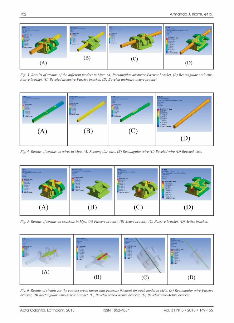
152 Armando J. Iriarte, et al.
Acta Odontol. Latinoam. 2018 ISSN 1852-4834 Vol. 31 Nº 3 / 2018 / 149-155
Fig. 3: Results of strains of the different models in Mpa. (A) Rectangular archwirePassive bracket, (B) Rectangular archwireActive bracket, (C) Beveled archwirePassive bracket, (D) Beveled archwireactive bracket.
Fig. 4: Results of strains on wires in Mpa. (A) Rectangular wire, (B) Rectangular wire (C) Beveled wire (D) Beveled wire.
Fig. 5: Results of strains on brackets in Mpa. (A) Passive bracket, (B) Active bracket, (C) Passive bracket, (D) Active bracket.
Fig. 6: Results of strains for the contact areas (areas that generate friction) for each model in MPa. (A) Rectangular wirePassivebracket, (B) Rectangular wireActive bracket, (C) Beveled wirePassive bracket, (D) Beveled wireActive bracket.
AOL32018:32011 21/02/2019 11:40 Página 152
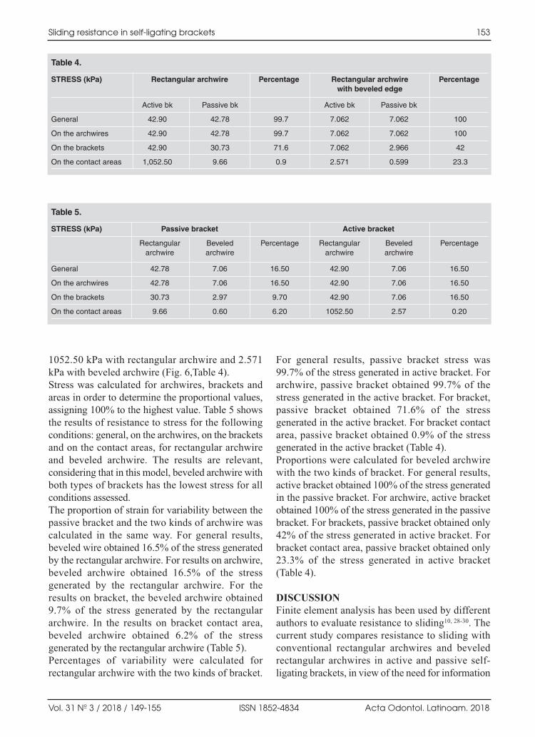
1052.50 kPa with rectangular archwire and 2.571kPa with beveled archwire (Fig. 6,Table 4).Stress was calculated for archwires, brackets andareas in order to determine the proportional values,assigning 100% to the highest value. Table 5 showsthe results of resistance to stress for the followingconditions: general, on the archwires, on the bracketsand on the contact areas, for rectangular archwire and beveled archwire. The results are relevant,considering that in this model, beveled archwire withboth types of brackets has the lowest stress for allconditions assessed.The proportion of strain for variability between thepassive bracket and the two kinds of archwire wascalculated in the same way. For general results,beveled wire obtained 16.5% of the stress generatedby the rectangular archwire. For results on archwire,beveled archwire obtained 16.5% of the stressgenerated by the rectangular archwire. For theresults on bracket, the beveled archwire obtained9.7% of the stress generated by the rectangulararchwire. In the results on bracket contact area,beveled archwire obtained 6.2% of the stressgenerated by the rectangular archwire (Table 5). Percentages of variability were calculated forrectangular archwire with the two kinds of bracket.
For general results, passive bracket stress was99.7% of the stress generated in active bracket. Forarchwire, passive bracket obtained 99.7% of thestress generated in the active bracket. For bracket,passive bracket obtained 71.6% of the stressgenerated in the active bracket. For bracket contactarea, passive bracket obtained 0.9% of the stressgenerated in the active bracket (Table 4). Proportions were calculated for beveled archwirewith the two kinds of bracket. For general results,active bracket obtained 100% of the stress generatedin the passive bracket. For archwire, active bracketobtained 100% of the stress generated in the passivebracket. For brackets, passive bracket obtained only42% of the stress generated in active bracket. Forbracket contact area, passive bracket obtained only23.3% of the stress generated in active bracket(Table 4).
DISCUSSIONFinite element analysis has been used by differentauthors to evaluate resistance to sliding10, 2830. Thecurrent study compares resistance to sliding withconventional rectangular archwires and beveledrectangular archwires in active and passive selfligating brackets, in view of the need for information
Sliding resistance in self-ligating brackets 153
Vol. 31 Nº 3 / 2018 / 149-155 ISSN 1852-4834 Acta Odontol. Latinoam. 2018
Table 4.
STRESS (kPa) Rectangular archwire Percentage Rectangular archwire Percentagewith beveled edge
Active bk Passive bk Active bk Passive bk
General 42.90 42.78 99.7 7.062 7.062 100
On the archwires 42.90 42.78 99.7 7.062 7.062 100
On the brackets 42.90 30.73 71.6 7.062 2.966 42
On the contact areas 1,052.50 9.66 0.9 2.571 0.599 23.3
Table 5.
STRESS (kPa) Passive bracket Active bracket
Rectangular Beveled Percentage Rectangular Beveled Percentagearchwire archwire archwire archwire
General 42.78 7.06 16.50 42.90 7.06 16.50
On the archwires 42.78 7.06 16.50 42.90 7.06 16.50
On the brackets 30.73 2.97 9.70 42.90 7.06 16.50
On the contact areas 9.66 0.60 6.20 1052.50 2.57 0.20
AOL32018:32011 21/02/2019 11:40 Página 153
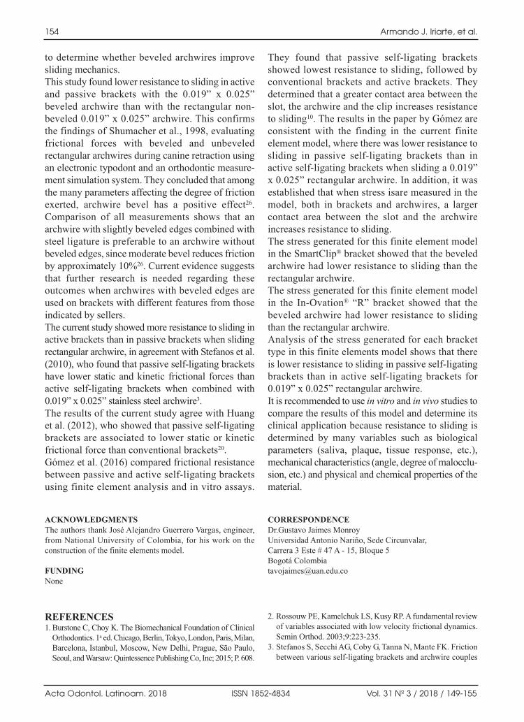
to determine whether beveled archwires improvesliding mechanics. This study found lower resistance to sliding in activeand passive brackets with the 0.019” x 0.025”beveled archwire than with the rectangular nonbeveled 0.019” x 0.025” archwire. This confirms the findings of Shumacher et al., 1998, evaluatingfrictional forces with beveled and unbeveledrectangular archwires during canine retraction usingan electronic typodont and an orthodontic measure ment simulation system. They concluded that amongthe many parameters affecting the degree of frictionexerted, archwire bevel has a positive effect26.Comparison of all measurements shows that anarchwire with slightly beveled edges combined withsteel ligature is preferable to an archwire withoutbeveled edges, since moderate bevel reduces frictionby approximately 10%26. Current evidence suggeststhat further research is needed regarding theseoutcomes when archwires with beveled edges areused on brackets with different features from thoseindicated by sellers.The current study showed more resistance to sliding inactive brackets than in passive brackets when slidingrectangular archwire, in agreement with Stefanos et al.(2010), who found that passive selfligating bracketshave lower static and kinetic frictional forces thanactive selfligating brackets when combined with0.019” x 0.025” stainless steel archwire3.The results of the current study agree with Huanget al. (2012), who showed that passive selfligatingbrackets are associated to lower static or kineticfrictional force than conventional brackets20. Gómez et al. (2016) compared frictional resistancebetween passive and active selfligating bracketsusing finite element analysis and in vitro assays.
They found that passive selfligating bracketsshowed lowest resistance to sliding, followed byconventional brackets and active brackets. Theydetermined that a greater contact area between theslot, the archwire and the clip increases resistanceto sliding10. The results in the paper by Gómez areconsistent with the finding in the current finiteelement model, where there was lower resistance tosliding in passive selfligating brackets than inactive selfligating brackets when sliding a 0.019”x 0.025” rectangular archwire. In addition, it wasestablished that when stress isare measured in themodel, both in brackets and archwires, a largercontact area between the slot and the archwireincreases resistance to sliding. The stress generated for this finite element modelin the SmartClip® bracket showed that the beveledarchwire had lower resistance to sliding than therectangular archwire. The stress generated for this finite element modelin the InOvation® “R” bracket showed that thebeveled archwire had lower resistance to slidingthan the rectangular archwire. Analysis of the stress generated for each brackettype in this finite elements model shows that thereis lower resistance to sliding in passive selfligatingbrackets than in active selfligating brackets for0.019” x 0.025” rectangular archwire.It is recommended to use in vitro and in vivo studies tocompare the results of this model and determine itsclinical application because resistance to sliding isdetermined by many variables such as biologicalparameters (saliva, plaque, tissue response, etc.),mechanical characteristics (angle, degree of malocclu sion, etc.) and physical and chemical properties of thematerial.
154 Armando J. Iriarte, et al.
Acta Odontol. Latinoam. 2018 ISSN 1852-4834 Vol. 31 Nº 3 / 2018 / 149-155
ACKNOWLEDGMENTSThe authors thank José Alejandro Guerrero Vargas, engineer,from National University of Colombia, for his work on the construction of the finite elements model.
FUNDINGNone
CORRESPONDENCEDr.Gustavo Jaimes MonroyUniversidad Antonio Nariño, Sede Circunvalar, Carrera 3 Este # 47 A 15, Bloque 5Bogotá [email protected]
REFERENCES1. Burstone C, Choy K. The Biomechanical Foundation of Clini cal
Orthodontics. 1a ed. Chicago, Berlin, Tokyo, London, Paris, Milan,Barcelona, Istanbul, Moscow, New Delhi, Prague, São Paulo,Seoul, and Warsaw: Quintessence Publishing Co, Inc; 2015; P. 608.
2. Rossouw PE, Kamelchuk LS, Kusy RP. A fundamental reviewof variables associated with low velocity frictional dynamics.Semin Orthod. 2003;9:223235.
3. Stefanos S, Secchi AG, Coby G, Tanna N, Mante FK. Frictionbetween various selfligating brackets and archwire couples
AOL32018:32011 21/02/2019 11:40 Página 154

during sliding mechanics. Am J Orthod Dentofac Orthop2010; 1379:463467.
4. Cacciafesta V, Sfondrini F, Ricciardi A. Evaluation offriction of stainless steel and esthetic selfligating bracketsin various bracketarchwire combinations. Am J OrthodDentofac Orthop 2003; 124:395402.
5. Gandini P, Orsi L, Bertoncini C, Massironi S, et al. In vitrofrictional forces generated by three different ligationmethods. Angle Orthod 2008; 78:917921.
6. Rozzi M, Mucedero M, Franchi L. Friction in a hybridsystem. An in vitro study. Roma Italia: Oral & implantology2010; p. 28.
7. Kusy RP, Whitley JQ. Friction between different wirebracket configurations and materials. Semin Orthod 1997;3:166177.
8. Ehsani S, Mandich MA, ElBialy TH, FloresMir C.Frictional resistance in selfligating orthodontic bracketsand conventionally ligated brackets a systematic review.Angle Orthod 2009; 79:592601.
9. Kusy RP. Influence of angulation on the resistance tosliding in fixed appliances. Am J Orthod Dentofacial Orthop1999; 115:3951.
10. Gómez SL, Montoya Y, Garcia NL, Virgen AL, Botero JE.Comparison of frictional resistance among conventional ,active and passive selfligating brackets with differentcombinations of arch wires : a finite elements study. ActaOdontol Latinoam 2016; 29:130136.
11. Kojima Y, Fukui H. Numerical simulation of canineretraction by sliding mechanics. Am J Orthod DentofacOrthop 2005; 127:542551.
12. Drescher D, Bourauel C, Schumacher HA. Frictional forcesbetween bracket and arch wire. Am J Orthod DentofacOrthop 1989; 96:397404.
13. Frank CA, Nikolai RJ. A comparative study of frictionalresistances between orthodontic bracket and arch wire. AmJ Orthod 1980; 78:593609.
14. Kapila S, Angolkar P V, Duncanson MG, Nanda RS.Evaluation of friction between edgewise stainless steelbrackets and orthodontic wires of four alloys. Am J OrthodDentofac Orthop 1990; 98:117126.
15. Ogata RH, Nanda RS, Duncanson JMG, Sinha PK, CurrierGF. Frictional resistances in stainless steel bracketwirecombinations with effects of vertical defections. Am JOrthod Dentofac Orthop 1996; 109:535542.
16. Park JH, Lee YK, Lim BS, Kim CW. Frictional forcesbetween lingual brackets and archwires measured by afriction tester. Angle Orthod 2004; 74:816824.
17. Bednar JR, Gruendeman GW, Sandrik JL. A comparativestudy of frictional forces between orthodontic brackets
and arch wires. Am J Orthod Dentofac Orthop 1991;100:513522.
18. Edwards GD, Davies EH, Jones SP. The ex vivo effect ofligation technique on the static frictional resistance ofstainless steel brackets and archwires. Br J Orthod 1995;22:145153.
19. Wichelhaus A, Geserick M, Hibst R, Sander FG. The effectof surface treatment and clinical use on friction in NiTiorthodontic wires. Dent Mater 2005; 21:938945.
20. Huang TH, Luk HS, Hsu YC, Kao CT. An in vitro comparisonof the frictional forces between archwires and selfligatingbrackets of passive and active types. Eur J Orthod 2012;34:625632.
21. Hain M, Dhopatkar A, Rock P. A comparison of differentligation methods on friction. Am J Orthod Dentofac Orthop2006; 130:666670.
22. Pizzoni L, Ravnholt G, Melsen B. Frictional forces relatedto selfligating brackets. Eur J Orthod 1998; 20:283291.
23. Thomas S, Sherriff M, Birnie D. A comparative in vitrostudy of the frictional characteristics of two types of selfligating brackets and two types of preadjusted edgewisebrackets tied with elastomeric ligatures. Eur J Orthod 1998;20:589596.
24. Shivapuja PK, Berger J. A comparative study of conventionalligation and selfligation bracket systems. Am J OrthodDentofac Orthop 1994; 106:472480.
25. Kahlon S, Rinchuse D, Robison JM, Close JM. Invitroevaluation of frictional resistance with 5 ligation methodsand Gianellytype working wires. Am J Orthod DentofacOrthop 2010; 138:6771.
26. Schumacher HA, Bourauel C, Drescher D. Frictional Forceswhen Rectangular Guiding Arches with Varying Edge BevelAre Employed . J Orofac Orthop 1998; 59:139149.
27. Serway RA, Kirkpatrick LD. Physics for Scientists andEngineers with Modern Physics. Vol. 26, The PhysicsTeacher 1988; p. 121.
28. Kojima Y, Fukui H, Miyajima K. The effects of friction andflexural rigidity of the archwire on canine movement insliding mechanics: A numerical simulation with a 3dimensional finite element method. Am J Orthod DentofacOrthop 2006; 130:110.
29. Kojima Y, Fukui H. Numeric simulations of enmasse spaceclosure with sliding mechanics. Am J Orthod DentofacOrthop 2010; 138:702.e1702.e6.
30. Hamanaka R, Yamaoka S, Anh TN, Tominaga JY, Koga Y,Yoshida N. Numeric simulation model for longtermorthodontic tooth movement with contact boundaryconditions using the finite element method. Am J OrthodDentofacial Orthop 2017; 152:601612.
Sliding resistance in self-ligating brackets 155
Vol. 31 Nº 3 / 2018 / 149-155 ISSN 1852-4834 Acta Odontol. Latinoam. 2018
AOL32018:32011 21/02/2019 11:40 Página 155

156
Acta Odontol. Latinoam. 2018 ISSN 1852-4834 Vol. 31 Nº 3 / 2018 / 156-163
RESUMENLa caries dental es un proceso infeccioso multifactorial,localizado y transmisible que se caracteriza por la destruccióndel tejido dental duro. Streptococcus mutans es considerado elprincipal microorganismo asociado al desarrollo de estaenfermedad. El objetivo de este estudio fue determinar lapresencia y recuento de S. mutans en saliva de niños con cariesdental antes y después de un proceso educativo. Con este fin setomó saliva no estimulada de 23 niños con caries dentalpertenecientes a un centro educativo con edades de 3 a 6 años.En todos los niños se tomaron 3 muestras de saliva: antes delproceso educativo y a los 3 y 6 meses de iniciado el procesoeducativo. El proceso educativo consistió en entrevistas,enseñanzas, conferencias, talleres educativos y actividadeslúdicas sobre la importancia del cuidado e higiene oral. Despuésde su recolección, las muestras de saliva fueron serialmentediluidas y sembradas en Agar Mitis Salivarius con bacitracina y
sacarosa al 20%. Los medios de cultivo sembrados se incubaronen anaerobiosis durante 2 días a 37ºC y los aislamientospresuntivos de S. mutans se identificaron con pruebasbioquímicas. Antes del inicio del proceso educativo, a los 3 y 6meses de iniciado el proceso educativo se encontró S. mutans,respectivamente, en 22 de los 23 niños (95.6%), en 15 de los 23niños (65.2%) y en 10 de los 23 niños (43.5%). En cuanto alrecuento de S. mutans, se encontró una reducción de 64.8 % y86.6% a los 3 y 6 meses, respectivamente, en comparación a losniveles encontrados antes del inicio del proceso educativo. Enconclusión, los resultados indican que la intervención educativarealizada produjo una reducción significativa en los niveles deS. mutans en saliva de niños con caries dental después de 3 y 6meses de iniciado el proceso educativo.
Palabras clave: Caries dental, niños, Streptococcus mutans,educación en salud.
ABSTRACTDental caries is an infectious, multifactorial, localized,transmissible process that leads to the destruction of hard dentaltissue. Streptococcus mutans is considered to be the mainmicroorganism associated with its development. The aim of thisstudy was to determine presence and count of S. mutans in salivasamples from children with dental caries before and after aneducational process including interviews, lessons, lectures,educational workshops and recreational activities on theimportance of oral care and hygiene. Twentythree 3 to 6yearold schoolchildren provided 3 unstimulated saliva samples: onebefore the educational process, one at 3 months and one at 6months into the educational process. The samples were seriallydiluted and plated on Mitis Salivarius agar supplemented with
bacitracin and 20% sucrose, and incubated anaerobically for 2days at 37oC. Presumptive S. mutans isolates were identified withbiochemical tests. Before the beginning of the educationalprocess, and at 3 and 6 months into the educational process, S.mutans was found, respectively, in 22 (95.6%), 15 (65.2%) and10 (43.5%) of the 23 children. The S. mutans count was reducedby 64.8% and 86.6% at 3 and 6 months, respectively, comparedto the levels found before the educational process. These resultsindicate that educational intervention produced a significantreduction in S. mutans levels in the saliva of children with dentalcaries at 3 and 6 months into the educational process.
Key words: Dental caries, children, Streptococcus Mutans,Health education.
Presence and count of S. mutans in children with dental caries: before, during and after a process of oral health education
Fredy Gamboa1,2, Leandro Plazas1, Dabeiba-Adriana García2, Fabio Aristizabal3, Ana-Lucía Sarralde2, Claudia-Patricia Lamby2, Martin Abba4
1 Pontificia Universidad Javeriana, Facultad de Ciencias, Departamento de Microbiología, Bogotá, Colombia.
2 Pontificia Universidad Javeriana, Facultad de Odontología,Grupo Centro de Investigaciones Odontológicas , Bogotá, Colombia.
3 Universidad Nacional de Colombia. Facultad de Ciencias, Departamento de Farmacia, Bogotá, Colombia.
4 Universidad Nacional de La Plata, Centro de Investigaciones Inmunológicas Básicas y Aplicadas (CINIBA), La Plata, Argentina.
Presencia y recuento de S. mutans en niños con caries dental: antes, durante y después de un proceso de educación en salud oral
AOL32018:32011 21/02/2019 11:40 Página 156
INTRODUCTIONDental caries is an infectious bacterial diseasewhich is multifactorial, chronic, localized, posteruptive and transmissible, and leads to destructionof hard dental tissue 13. Its development basicallyrequires three factors sustained over time: asusceptible host, cariogenic microflora in the dentalbiofilm, and an adequate substrate. Streptococcusmutans (primarily serotype C), and to a lesserextent, S. sobrinus and S. gordonii, as well asLactobacillus and Actinomyces species, in thatorder of frequency, are the primary microorganismsassociated to the development and progression ofdental caries 4,5.Different studies have found a strong correlationbetween number of S. mutans colonies in the oralcavity and prevalence and incidence of dentalcaries1,4,5. In addition, as a result of dental treatments,S. mutans can cause bacteremia, systemic infectionsand subacute endocarditis6. The fact that S. mutans has been clearly recognizedas the primary bacterial species involved in dentalcaries has led to the implementation of preventiveand control measures tending to eliminate or reduceits presence in the oral cavity3,68. Differentstrategies have been designed for such purpose, butbecause of lack of continuity, systematization,regulation and supervision, they have not been veryeffective8. Dental caries is currently one of the most frequentdiseases of the oral cavity, mainly affecting 5 to12year old children. Because it is a chronic disease,it progresses with age unless efforts are made tocontrol it9,10. The distribution and severity of dentalcaries varies among regions, and its onset isfrequently associated to nutritional, sociocultural,economic and environmental factors11,12. Different comparative clinical studies on 2.5 to 7yearold children with dental caries have shownreduction in oral levels of S. mutans andLactobacillus species1315 in dental biofilm andsaliva, during and after conventional and nonconventional restorative treatments1315. Althoughthe results in these studies suggest that S. mutansand Lactobacillus species counts are lower after 6months’ treatment than at baseline, they also recorda tendency for these microorganisms to becomereestablished over time1315. With regard to oralhealth educational strategies, very few studies todate present successful results of restorative dental
treatment in the reduction of microorganisms thatare important in dental caries1618.In Colombia, no study has been published evaluatingthe impact of oral health educational intervention onpresence and count of S. mutans in children withdental caries. The main aim of this study wastherefore to determine S. mutans presence and countin saliva from children with dental caries before,during and after a 6month oral health educationalprocess.
MATERIALS AND METHODSStudy populationThis was a longitudinal prospective study whichmet bioethical requirements for sampling andsample management. It included 23 3 to 7yearoldchildren with dental caries from the school CentroEducativo Fe y Alegría – José María Velaz in theSuba locality of Bogota city, Colombia. Exclusioncriteria were: use of topical or oral antimicrobialagents within 7 days prior to sampling; undergoingorthodontic treatment with fixed or removableappliances, and/or any kind of oral infectiousprocess different from dental caries. Each childunderwent an oral clinical examination to determinedental caries experience, performed by a singlecalibrated examiner using artificial light and adentist’s mirror. No Xray was taken. Dental cariesclinical stage was determined according to theInternational Caries Detection and AssessmentSystem (ICDAS), which distinguishes early noncavitated lesions from cavitated or dentinallesions19,20. Of the 23 children with dental cariesincluded in the study, 12 were diagnosed as ICDASscore 3 and 11 as ICDAS score 6.
S. mutans isolation, identification and countThree saliva samples were taken from each of the 23children: one before starting the educational process(baseline), one 3 months into the educational process,and a final sample 6 months into the educationalprocess. The samples were taken between 8:00 and10:00 a.m., with prior commitment from the childrennot to eat anything and not to brush their teeth beforesampling. Spontaneous saliva (0.21 ml) wassampled by gentle suction with a sterile plasticpipette21 and samples were immediately placed onice to be transferred to the bacteriological laboratory.At the laboratory, the saliva samples were vortexed
S. mutans in an educational process 157
Vol. 31 Nº 3 / 2018 / 156-163 ISSN 1852-4834 Acta Odontol. Latinoam. 2018
AOL32018:32011 21/02/2019 11:40 Página 157

for 30 seconds and serially diluted (1/10, 1/100 and1/1000) with 0.05M phosphate buffer. For S. mutansselective isolation and count, 35 µl of the serialdilutions were inoculated in duplicate in MitisSalivarius Bacitracin Agar (MSB; Difco Labora tories, Detroit, MI). MSB Agar contains digestedpancreatic casein, proteose peptone No.3, proteosepeptone, dextrose, 20% sucrose, dipotassiumphosphate, trypan blue, crystal violet, agar,Chapman’s tellurite and 0.2 U/ml bacitracin. ThePetri dishes with MSB agar were incubatedanaerobically (H2:CO2:N2; 10:10:80) for 2 days at 37oC. After bacterial growth in the MSB agar, colonies with S. mutans morphologicalcharacteristics22 were counted and expressed ascolonyforming units (CFU) per ml of unstimulatedsaliva. Then 5 to 10 colonies per sample with S.mutans characteristics were examined by Gram stain,catalase test and the following biochemical tests:fermentation of raffinose, mannitol, melibiose,trehalose and inulin; hydrolysis of esculin inpresence and absence of bile; urease; hydrolysis ofarginine; and resistance to 2 U of bacitracin. Thebiochemical profile for S. mutans is positivefermentation of raffinose, mannitol, melibiose,trehalose and inulin; negative hydrolysis of esculinin presence of bile and positive hydrolysis of esculinin absence of bile; negative urease; negativehydrolysis of arginine; and resistance to 2 U ofbacitracin. The commercial system Api 20S(bioMerieux, Marcylétoile, France) was also usedin the identification of strains.
Total aerobic microbial countTotal aerobes were counted in all saliva samples(before and after the beginning of the educationalprocess). To do so, 35 µl of all the serial dilutionsof saliva with phosphate buffer (as described above)were inoculated in blood agar (BHI agarsupplemented with 5% lamb´s blood). Afterinoculation, the dishes of blood agar were incubatedaerobically at 37oC for 48 hours. Total aerobicmicrobe CFU/ml were counted.
Oral health educational processThe educational process began with a survey todetermine parents’ and/or guardians’ knowledge,attitudes and practices regarding oral health,acquired previously in family and school settings.After the survey, the following activities were
conducted with parent/guardianchild pairs: 1.Talks and workshops for parents/guardians andchildren on the importance of good oral health; 2.Educational workshops on tooth brushing, evidenceof bacterial plaque or dental biofilm, and theimportance of good nutrition; 3. Workshops andfeedback with parents/guardians and children overthe 6 month educational period, on the importance ofgood oral hygiene; and 4. Recreationaleducationalworkshops for the children on dental care and thepractice of good oral health. Throughout the 6month educational process, oral hygiene kitsconsisting of 22 ml dentifrice and a child toothbrushwere delivered individually to each child every 15days. In addition, throughout the 6month study, thefour oral hygiene strategies described above werereinforced every 15 days.
Statistical analysisDescriptive statistics (frequency, mean, standarddeviation and maximum and minimum values) andpaired Student’s ttest were used to establishdifferences between S. mutans counts in the groupswith caries (ICDAS 3 and 6) before (baseline) andafter beginning the educational process (at 3 and 6months). Student’s ttest was performed with theprogram IBM SPSS Statistics version 22.0 and thelevel of statistical significance was set at p<0.05.
RESULTSTable 1 shows caries experience and demographicsfor the 23 children with dental caries included inthe study. The 11 children (5 female and 6 male)diagnosed with ICDAS score 3 were within an agerange of 3.54.5 years, average age 4.2 ± 0.24 yearsand dmft 3.7±3.1. The 12 children (5 female and 7male) diagnosed with ICDAS score 6 were withinan age range of 5.76.7 years, average age 6.5 ± 0.3years and dmft 5.3±3.9.Table 2 shows the frequency of S. mutans inbaseline, 3 and 6month samples in the 23 childrenwith caries (ICDAS groups 3 and 6). The 11children with ICDAS score 3 had S. mutansfrequencies of 91% (10/11), 27% (3/11) and 9%(1/11), at baseline, 3 and 6 months, respectively.The 12 children with ICDAS score 6 had S. mutans,frequencies of 100% (12/12), 100% (12/12) and 75% (9/12) at baseline, 3 and 6 months,respectively. Aggregate results for both groupsshowed S. mutans frequencies of 95.7% (22/23),
158 Fredy Gamboa, et al.
Acta Odontol. Latinoam. 2018 ISSN 1852-4834 Vol. 31 Nº 3 / 2018 / 156-163
AOL32018:32011 21/02/2019 11:40 Página 158

65.2% (15/23) and 43.5% (10/23) at baseline, 3 and6 months, respectively (Table 2).Fig. 1 shows average S. mutans values in saliva fromchildren at the two clinical stages of dental caries forthe three sampling times. For clinical stage ICDAS
3, there was a significant reduction in S. mutans frombaseline to month 3 (p=0.0221) and from baseline tomonth 6 (p= 0.0182). For reduction of S. mutans atclinical stage ICDAS 6, baseline to month 3 had p= 0.0526 and baseline to month 6 had p=0.0607.
S. mutans in an educational process 159
Vol. 31 Nº 3 / 2018 / 156-163 ISSN 1852-4834 Acta Odontol. Latinoam. 2018
Fig. 1: S. mutans count (CFU/ml)* insaliva of children classified by ICDAS into
two clinical stages of dental caries.*CFU/ml (mean ± SD) are expressed on
logarithmic scale. Paired Student ttest wasused to analyze the differences between
baseline and month 3, and betweenbaseline and month 6. There was a
significant reduction (p = 0.0221) forICDAS 3 between baseline and month 3.
There was also a significant reduction forICDAS 3 between baseline and month 6 (p
= 0.0182). No significant difference wasfound for ICDAS 6 between baseline andmonth 3 (p = 0.0526), between baseline
and month 6 (p = 0.0607) or betweenmonth 3 and month 6 (p=0.0569).
Table 1: Baseline: Caries experience and demographics of children included in the study.
Caries Status Children (n=) Age range in years Age* (years) Sex dmft*
ICDAS 3 11 3.5 – 4.5 4.2 ± 0.24Female: 21.7% (n= 5) 3.7 ± 3.1
Male: 26.1% (n= 6)
ICDAS 6 12 5.7 – 6.7 6.5 ± 0.3Female: 21.7% (n= 5) 5.3 ± 3.9
Male: 30.4% (n= 7)
Total 23 3.5 – 6.7 5.4 ± 1.2Female: 43.5% (n= 10) 4.2 ± 1.1
Male: 56.5% (n= 13)
* Values expressed as means (± standard deviation).
Table 2: Frequency of S. mutans in saliva samples at baseline and 3 and 6 months into the educational process.
Time Presence of S. mutans No. of children with: Total
ICDAS 3 ICDAS 6
Positive 10 12 22
Baseline Negative 1 0 1
Frequency 10/11 (91%) 12/12 (100%) 22/23(95.7%)
Positive 3 12 15
3 months Negative 8 0 8
Frequency 3/11 (27%) 12/12 (100%) 15/23(65.2%)
Positive 1 9 10
6 months Negative 10 3 13
Frequency 1/11 (9%) 9/12 (75%) 10/23(43.5%)
AOL32018:32011 21/02/2019 11:40 Página 159

Fig. 2 shows aggregate average values for S. mutanscount in saliva from all 23 children with dentalcaries (ICDAS 3 + ICDAS 6) at baseline, month 3and month 6. According to Student’s ttest, averagevalues for total count of S. mutans present in salivadecreased with statistically significant differences,comparing counts at baseline, month 3 and month 6(p˂0.05). Bacterial counts diminished significantlybetween baseline and month 3 (64.8%, p=0.007),and between baseline and month 6 (86%, p=0.02). Fig. 3 shows total aerobe count, expressed asCFU/ml, for saliva samples from the 23 childrenwith dental caries at baseline, 3 months and 6months. Total average value at baseline was 5.29 x10⁹ CFU/ml, increasing at 3 and 6 months by 11.6%(5.9 x 10⁹) and 21.8% (6.44 x 10⁹) respectively,without statistically significant differences (Studentttest p˃0.05, Fig. 3).
DISCUSSIONDental caries is considered to be a multifactorialdisease as a result of an imbalance in the oral
ecosystem leading to the predominance of florapreviously considered normal in the oral cavity, whichthen becomes pathogenic. Microbial imbalance, aswell as the presence of different external factors,including nutritional, sociocultural, economic,environmental and individual behavioral, impact thedevelopment of dental caries. For a long time, S.mutans has been considered the primary etiologicalagent involved in the development of dental caries,playing a very important part in initial stages of dentalenamel deterioration23,24. In children, S. mutans isassociated to a specific period, known as “window ofinfectivity”, which coincides with the eruption of teethand occurs between 6 and 30 months of age, withhigher risk from 18 to 30 months of age2426.Clear recognition of S. mutans as the primarybacterial species involved in dental caries has led tothe search for, and development and implemen tation of prevention and control measures aimed ateliminating or reducing S. mutans in the oral cavity 25.Previous clinical studies on children with dentalcaries have shown the positive effect of restorative
160 Fredy Gamboa, et al.
Acta Odontol. Latinoam. 2018 ISSN 1852-4834 Vol. 31 Nº 3 / 2018 / 156-163
Fig. 2: S. mutans count (CFU/ml)* found in saliva from children with dental caries (ICDAS 3 + ICDAS 6). * CFU/ml (mean ± SD) expressedon logarithmic scale. Paired student ttest wasused to analyze the differences. S. mutans wasreduced significantly between baseline and month3 (p = 0.007) and between baseline and month 6(p= 0.002).
Fig. 3: Total aerobic microorganism count* insaliva from children with dental caries (ICDAS 3 + ICDAS 6). * CFU/ml (mean ± SD) expressedon logarithmic scale. Paired student ttest wasused to analyze the differences. No difference inaerobic microorganisms was found betweenbaseline and month 3 (p=0.63417), betweenbaseline and month 6 (p= 0.5711) or betweenmonth 3 and month 6 (p= 0.6124).
AOL32018:32011 21/02/2019 11:40 Página 160

treatment on the reduction of S. mutans levels1315.On the other hand, with regard to educationalstrategies, very few studies conducted to datepresent similar results to those of restorative dentaltreatment for reduction of microorganisms whichare important in dental caries1618. In view of the fact that dental caries is basically acontrollable disease and considering the failure ofmany preventive and therapeutic actions, greaterefforts and resources need to be allocated topreventive and educational measures to reduce thepresence and quantity of cariogenic microorga nisms in the oral cavity, thereby reducing dentalcaries27, 28. Proper oral health education should be astrategy leading to a change in attitude in childrenand adults to achieve better individual andcollective health and wellbeing indicators27, 28. Inthe current study, the 23 children complied with allthe educational strategies proposed over the 6month educational process. They received relevantoral health and hygiene education in accordancewith the plans outlined: 1. Survey on knowledge oforal health; conferences and workshops forparents/guardians and children on the importanceof good oral health and hygiene; 2. Personalizededucational workshops on tooth brushing, identi fying bacterial plaque, and the importance ofhealthy eating; 3. Recreationaleducational workshopsfor the children on dental care and practicing goodoral health. The oral health educational strategieswere reinforced periodically, according to the pro posed schedule, and also by leaving in classroomsleaflets, guides, notice boards and documentsrelevant to oral health and hygiene education. Itshould be highlighted that all the activities requiredconstant, disciplined commitment of the dentists,preschool teachers and children’s parents and/orguardians who participated in this project. Regarding microbiological results, S. mutans countwas found to be directly proportional to cariesclinical stage, i.e., at higher levels of dental cariesthere were greater numbers of S. mutans colonies.The health and oral hygiene educational strategiesenabled a reduction in S. mutans levels at 3 and 6months, by 64.8% and 86% respectively, in childrenwith dental caries (ICDAS 3 + ICDAS 6, Fig. 2).The educational strategy had greater impact on thereduction of S. mutans at 6 months in the group ofchildren with ICDAS score 3, where S. mutans wasonly detected in 1 of the 11 children. The impact
was weaker in the children with ICDAS score 6,since at the end of the 6 months S. mutans persistedin 9 of the 12 children, at concentrations higher than500.000 CFU/ml. The high counts at the end of the 6montheducational process in children with ICDAS score6 may indicate the high risk for originating cariousstages and that educational strategies alone were insufficient. It is highly likely that futureeducational activities will need to cover longertimes, and/or strategies to achieve absence and/orreduction of S. mutans levels will need to bestrengthened, as proposed in other studies27 29, evenconsidering joint use of educational strategies anddentifrices with greater bactericidal power29. Astudy by Kumar et al. 29 analyzed the effect of oralhealth education and the use of fluoridated and nonfluoridated dentifrices on oral health status,using S. mutans and Lactobacillus counts beforebeginning the educational strategy, and 3, 6 and 12months posttreatment. They found posttreatmentreductions in the counts of both microorganisms,both in children who used fluoridated dentifricesand in those who used nonfluoridated dentifrices.However, the reduction was significantly greater at3, 6 and 12 months posttreatment in children whoused fluoridated dentifrices, and the authors suggestthat this may have been due to the bactericidalaction of fluoride. Another study on children foundthat the longterm use of fluoride mouthrinse led tolower levels of S. mutans30. However, longtermstudies have been proposed to clarify the effect ofchemotherapeutic agents on the reduction of S.mutans and their impact on caries incidence31. It is important to clarify that although the currentstudy focuses the microbiological risk factor, otherstudies have shown that longterm caries preventionneeds to act simultaneously on all risk factorsinvolved in this disease14, 19, 25.Different studies have shown the direct associationbetween dental caries incidence and S. mutanspresence and quantity in the oral cavity3235.Similarly, the current study found high frequency(95.7%, or 22 out of 23 children) and high S. mutanscolonization rate at baseline in both groups ofchildren with dental caries (ICDAS 3 and ICDAS 6).The current study also determined levels of aerobicmicroorganisms before and after the oral healtheduca tional strategies. In general, the countsremained stable, with slight, nonstatistically signi
S. mutans in an educational process 161
Vol. 31 Nº 3 / 2018 / 156-163 ISSN 1852-4834 Acta Odontol. Latinoam. 2018
AOL32018:32011 21/02/2019 11:40 Página 161

ficant increase over time. These levels of aerobicmicroorganisms may also be indicative of stableoral health, in which the place left by the reductionof S. mutans colonies achieved through educationalstrategies was taken over by aerobic microorganisms,which are compatible with the healthy microflorathat is part of the oral microbial ecology8.This study was intentionally sought children withdental caries. There was predominance of malechildren, with 56.5% (13/23), and overall averageage was 5.4 ±1.2 years. Oral examination detectedthat 47.8% (11/23) of the children had ICDAS score3 and 52.2% (12/23) had ICDAS score 6. Overallaverage dmft index was 4.2 ±1.1 for the 23 children.Other studies conducted in Colombia, which alsoused ICDAS and dmft diagnostic criteria, have
reported similar results3638. These results indicate ahigh rate of dental caries in the child populationstudied, which may serve as a sentinel and be anindicator of the results of the public health strategiesbeing implemented39. On the other hand, micro biolo gical analyses of saliva and monitoringchildren under 6 years old with dental caries maybe useful for identifying individual and communityrisk of caries, and for improving treatment anddelaying the process of S. mutans colonization andmultiplication in the oral cavity40. To conclude, the results of this study indicate thatthe oral health educational process conducted led toa significant reduction in S. mutans level in thesaliva of children with dental caries, 3 and 6 monthsinto the educational process.
162 Fredy Gamboa, et al.
Acta Odontol. Latinoam. 2018 ISSN 1852-4834 Vol. 31 Nº 3 / 2018 / 156-163
FUNDINGThis study was funded by the Office of the ViceRector ofResearch of Pontificia Universidad Javeriana within the proposal with ID No. 00007235.
CORRESPONDENCEDr. Fredy GamboaDepartamento de Microbiología (Facultad de Ciencias) y Centro de Investigaciones Odontológicas (Facultad deOdontología). Pontificia Universidad Javeriana. Carrera 7 No. 4062Bogotá, [email protected]
REFERENCES1. Acosta A, Agudelo C, Barrientos S, Chaves M, Cuellar A,
Durán C, Gamboa F, Gamboa L, et al. Fundamentos de cienciasbásicas aplicadas a la odontología. Bogotá, D.C., Colombia:Editorial Pontificia Universidad Javeriana, 2006:3037.
2. Mikx FH, van der Hoeven JS, König KG, Plasschaert AJ,Guggenheim B. Establishment of defined microbialecosystems in germfree rats. Caries Res 1972; 6:211223.
3. Beighton D, Manji F, Baelum V, Fejerskov O, Johnson NW,Wilton JM. Associations between salivary levels ofStreptococcus mutans, Streptococcus sobrinus, lactobacilli,and caries experience in Kenyan adolescents. J Dent Res1989; 68:12421246.
4. Lang NP, Hotz PR, Gusberti FA, Joss A. Longitudinalclinical and microbiological study on the relationshipbetween infection with Streptococcus mutans and thedevelopment of caries in humans. Oral Microbiol Immunol1987; 2:3937.
5. Hamada S, Slade HD. Biology, immunology, and cariogenicityof Streptococcus mutans. Microbiol Rev 1980; 44:331384.
6. Ullman RF, Miller SJ, Strampfer MJ, Cunha BA.Streptococcus mutans endocarditis: report of three cases andreview of the literature. Heart Lung 1988; 17:209212.
7. Schleifer K, KilpperBälz R. Molecular and chemotaxonomicapproaches to the classification of streptococci, enterococciand lactococci: a review. Syst Appl Microbiol 1987; 10:119.
8. Gamboa F, Herazo Acuña B, Martínez MC. Controlmicrobiológico sobre Streptococcus mutans y su acciónacidogénica. Univ Sci 2004; 9:455.
https://revistas.javeriana.edu.co/index.php/scientarium/article/view/5034
9. Cabrera D, Herrera N, Gispert E. Riesgo de caries dentalen niños atendidos en el hogar en el período 20062007.Rev Cub Estomatol 2009; 46:122. http://scielo.sld.cu/pdf/est/v46n2/est02209.pdf
10. Escobar G, Ortiz AC, Mejía LM. Caries dental en losmenores de veinte años en Colombia: un problema de saludpública. Rev Fac Nac Salud Pública 2003; 21:107108.http://aprendeenlinea.udea.edu.co/revistas/index.php/fnsp/article/view/759/655
11. Nithila A, Bourgeois D, Barmes D, Murtomaa H. WHOGlobal Oral Data Bank, 198696: an overview of oral healthsurveys at 12 years of age. Bull World Health Organ 1998;76:237244.
12. Tascón J, Cabrera G. Creencias sobre caries e higiene oralen adolescentes del Valle del Cauca. Colomb Med 2005;36:7378. http://colombiamedica.univalle.edu.co/index.php/comedica/article/view/335/1115
13. Wright JT, Cutter GR, Dasanayake AP, Stiles HM, CaufieldPW. Effect of conventional dental restorative treatment onbacteria in saliva. Community Dent Oral Epidemiol 1992;20: 138143.
14. Roshan NM, Shigli AL, Deshpande SD. Microbiologicalevaluation of salivary Streptococcus mutans from childrenof age 57 years, pre and postatraumatic restorativetreatment. Contemp Clin Dent 2010; 1:9497.
15. Twetman S, Fritzon B, Jensen B, Hallberg U, Stahl B. Preand posttreatment levels of salivary mutans streptococci
AOL32018:32011 21/02/2019 11:40 Página 162

and lactobacilli in preschool children. Int J Paediatr Dent1999; 9:9398.
16. Alvarez C, Perozo R, Rojas T. Componente educativorecreativoasociativo en estrategias promotoras de salud bucalen preescolares. Rev Cubana Estomatol 2006; 43:3338.http://scielo.sld.cu/scielo.php?script=sci_arttext&pid=S003475072006000200005
17. Ortiz B, SanchezBorjas P, Delgado E. Efectividad de unaintervención comunitaria en salud oral: resultados despuésde 18 meses. Rev Med Hered 2006; 17: 6773. http://www.upch.edu.pe/vrinve/dugic/revistas/index.php/RMH/article/view/874/840
18. de Freitas CN, Castelo PM, de Sousa KG, Alonso GC,Fonseca FLA, Klein MI, de Souza TS. Educationalstrategies and ART effect on salivary characteristics: acontrolled clinical trial. Oral Dis 2017, 23:11161126.
19. Ismail AI, Sohn W, Tellez M, Willem JM, Betz J,Lepkowski J. Risk indicators for dental caries using theInternational Caries Detection and Assessment System(ICDAS). Community Dent Oral Epidemiol 2008; 36:5558.
20. Shoaib L, Deery C, Ricketts DN, Nugent ZJ. Validity andreproducibility of ICDAS II in primary teeth. Caries Res2009; 43:442448.
21. Fure S. Tenyear incidence of tooth loss and dental caries inelderly Swedish individuals. Caries Res 2003; 37:462469.
22. Emilson CG, Carlsson P, Bratthall D. Strains of mutansstreptococci isolated in a population with extremely lowcaries prevalence are cariogenic in the hamster model. OralMicrobiol Immunol 1987; 2:183186.
23. Baca P, Castillo AM, Baca AP, Liébana MJ, Junco P,Liébana J. Genotypes of Streptococcus mutans in salivaversus dental plaque. Arch Oral Biol 2008; 53:751754.
24. Köhler B, Andreen I. Mutans streptococci and cariesprevalence in children after early maternal caries prevention:a followup at eleven and fifteen years of age. Caries Res2010; 44:453458.
25. Kishi M, Abe A, Kishi K, OharaNemoto Y, Kimura S,Yonemitsu M. Relationship of quantitative salivary levelsof Streptococcus mutans and S. sobrinus in mothers tocaries status and colonization of mutans streptococci inplaque in their 2.5yearold children. Community Dent OralEpidemiol 2009; 37:241249.
26. Douglas JM, Li Y, Tinanoff N. Association of mutansstreptococci between caregivers and their children. PediatrDent 2008; 30:375387.
27. Allen F. Embedding a population oral health perspective inthe dental curriculum. Community Dent Oral Epidemiol2012; 40:127133.
28. Kwan S, Petersen Poul, Pine C, Borutta A. Healthpromotingschools: an opportunity for oral health promotion. Bull WorldHealth Organ 2005; 83:677685.
29. Kumar S, Konde S, Raj S, Agarwal M. Effect of oral healtheducation and fluoridated dentifrices on the oral health
status of visually impaired children. Contemp Clin Dent2012; 3:398401.
30. Yoshihara A, Sakuma S, Kobayashi S, Miyazaki H.Antimicrobial effect of fluoride mouthrinse on mutansstreptococci and lactobacilli in saliva. Pediatr Dent 2001;23:113117.
31. Ersin NK, Eden E, Eronat N, Totu FI, Ates M. Effectivenessof 2year application of schoolbased chlorhexidinevarnish, sodium fluoride gel, and dental health educationprograms in highrisk adolescents. Quintessence Int 2008;39: e4551.
32. LoyolaRodriguez JP, MartinezMartinez RE, FloresFerreyra BI, PatinoMarin N, AlpucheSolis AG,ReyesMacias JF. Distribution of Streptococcus mutans andStreptococcus sobrinus in saliva of Mexican preschoolcariesfree and cariesactive children by microbial andmolecular (PCR) assays. J Clin Pediatr Dent 2008;32:121126.
33. Acevedo AM, Ray MV, Socorro M, RojasSanchez F.Frequency and distribution of Mutans Streptococci in dentalplaque from cariesfree and cariesaffected Venezuelanchildren. Acta Odontol Latinoam 2009; 22:1520.
34. Yasunaga A, Yoshida A, Morikawa K, Maki K, NakamuraS, Soh I, Awano S, Ansai T. Monitoring the prevalence ofviable and dead cariogenic bacteria in oral specimens andin vitro biofilms by qPCR combined with propidiummonoazide. BMC Microbiol 2013; 13:157. doi: 10.1186/1471218013157.
35. Zhang Q, van Palenstein Helderman WH. Caries expe rience variables as indicators in caries risk assessmentin 6–7yearold Chinese children. J Dent 2006; 34:676681.
36. Ramírez BS, Escobar G, Franco AM, Martínez MC, GómezL. Caries de la infancia temprana en niños de uno a cincoaños. Medellín, Colombia, 2008. Rev Fac Odontol UnivAntioq 2011; 22:164172.
37. DíazCárdenas S, GonzálezMartínez F. Prevalencia decaries dental y factores familiares en niños escolares deCartagena de Indias, Colombia. Rev Salud Pública 2010;12:843851. https://www.scielosp.org/article/rsap/2010.v12n5/843851/
38. Gamboa F, Estupiñan M, Galindo A. Presence ofStreptococcus mutans in saliva and its relationship withdental caries: Antimicrobial susceptibility of the isolates.Univ Sci 2004;9:2327 https://revistas.javeriana.edu.co/index.php/scientarium/article/view/5032
39. O’Sullivan DM, Thibodeau EA. Caries experience andmutans streptococci as indicators of caries incidence.Pediatr Dent 1996; 18:371374.
40. Köhler B, Andréen I Jonsson B. The earlier the colonizationby mutans streptococci, the higher the caries prevalence at4 years of age. Oral Microbiol Immunol 1998; 3:1417.
S. mutans in an educational process 163
Vol. 31 Nº 3 / 2018 / 156-163 ISSN 1852-4834 Acta Odontol. Latinoam. 2018
AOL32018:32011 21/02/2019 11:40 Página 163
164
Acta Odontol. Latinoam. 2018 ISSN 1852-4834 Vol. 31 Nº 3 / 2018 / 164-169
RESUMENEl objetivo del presente trabajo fue evaluar el engrosamiento de lamembrana de Schneider y determinar su asociación con patologíasperiapicales, mediante tomografía computarizada cone beam(CBCT). Se realizó un estudio observacional, analítico, retrospectivoy transversal. Un total de 179 senos maxilares fueron evaluadosutilizando CBCT. Se analizó la presencia de engrosamiento de lamembrana sinusal y su asociación con piezas dentarias sin vitalidadpulpar. Los resultados se muestran como porcentajes con intervalosde confianza del 95% (IC del 95%). Se utilizó la prueba de Chicuadrado con un nivel de significación del 5%. Se detectó engrosamiento de la membrana sinusal en 70 casos(39%; IC del 95% = 32% a 46%) y no se observó engrosamientode la membrana sinusal en 109 (61%; IC del 95% = 54% a 68%)(p < 0.05). Los 70 casos que mostraron engrosamiento de lamembrana sinusal incluyeron 46 de origen odontogénico (66%;IC del 95% = 54% a 76%) y 24 (34%; IC del 95% = 24% a 46%)de origen no odontogénico (p <0,05). La frecuencia de las causasodontogénicas siguió una distribución heterogénea (p <0.05):
caries penetrantes, tratamiento endodóntico deficiente, restosradiculares, restauraciones profundas, implantes, patolo gíaperiodontal. La principal causa fue la caries (46%; IC 95% = 32%a 60%), seguida por endodoncia deficiente (26%, IC 95% = 16%a 40%). La frecuencia se distribuyó en forma heterogénea entrelas distintas piezas (p<0,05). Las piezas más afectadas fueron la16 (33%; IC95=21% a 47%) y la 26 (30%; IC95=19% a 45%). La alta incidencia de patología sinusal de origen odontogénicoimplica la necesidad del trabajo interdisciplinario entreodontólogos y otorrinolaringólogos. Caries, restauracionesinadecuadas, lesiones periodontales, implantes y la presencia derestos radiculares son las principales causas del engrosamientode la membrana de Schneider. El uso de CBCT para eldiagnóstico y la planificación del tratamiento permite detectar elengrosamiento de la membrana del seno maxilar y determinar suasociación con una etiología odontogénica.
Palabras clave: seno maxilar, tejidos periapicales, tomografíacomputada cone beam, membrana de Schneider.
ABSTRACT The aim of this study was to evaluate thickening of theSchneiderian membrane and to determine its association withperiapical pathologies, using computerized cone beam tomo graphy. An observational, analytical, crosssectional retrospectivestudy was conducted. A total 179 maxillary sinuses were evaluatedusing CBCT. The presence of sinus membrane thickening and itsassociation with unhealthy teeth was analyzed. Results are shownas percentages with 95% confidence intervals (95%CI); Chisquare test was used with a significance level of 5%. Sinus membrane thickening was detected in 70 cases (39%;95%CI=32% to 46%) and no sinus membrane thickening wasobserved in 109 (61%; 95%CI =54% to 68%) (p<0.05). The 70cases showing sinus membrane thickening included 46 ofodontogenic origin (66%; 95%CI =54% to 76%) and 24 (34%;95%CI =24% to 46%) of non odontogenic origin (p<0.05). Thefrequency of odontogenic causes followed a heterogeneousdistribution (p<0.05): penetrating caries, failing endodontic
therapy, root remnants, deep restorations, implants, periodontalpathology. The main cause was caries (46%; 95%CI=32% to60%) followed by failing endodontic therapy (26%, 95%CI=16% to 40%). The frequency distribution of involved teethwas uneven (p<0.05), with tooth 16 (33%; 95%CI=21% to47%) being the most frequently involved, followed by tooth 26(30%; 95%CI=19% to 45%). The high incidence of sinus pathology of odontogenic originshows the need for interdisciplinary work involving dentists andearnosethroat specialists. Caries, inadequate restorations,periodontal lesions, implants, and the presence of root remnantsare the main causes of Schneiderian membrane thickening. Theuse of CBCT for diagnosis and treatment planning allowsdetecting maxillary sinus membrane thickening and determiningits association with an odontogenic etiology.
Key words: maxillary sinus, periapical tissues, cone beamcomputerized tomography, Schneiderian membrane.
Relation between periapical lesions and sinus membrane thickening assessed by Cone Beam Computed Tomography
Gisela V. Brañas1, Brenda G. Grisolia1, Romina G. Iuliano1, Ariel Gualtieri2, Ariel Lenarduzzi1, Sandra J. Renou3, Pablo A. Rodríguez1
1 Universidad de Buenos Aires, Facultad de Odontología, Cátedra de Endodoncia. Hospital Odontológico Universitario.
2 Universidad de Buenos Aires, Facultad de Odontología, Cátedra de Biofísica y Bioestadística. Hospital Odontológico Universitario.
3 Universidad de Buenos Aires, Facultad de Odontología, Cátedra de Anatomía Patológica, Hospital Odontológico Universitario.
Lesiones periapicales y engrosamiento de la membrana sinusal, su relación y valoración a través de Tomografía Computarizada de Haz Cónico
AOL32018:32011 21/02/2019 11:40 Página 164
INTRODUCTIONThe maxillary sinus is the main paranasal sinus, andis located in the body of the maxilla. At the end ofdentomaxillofacial growth, the floor of themaxilla presents its final anatomical features. Atthis point, the teeth and the sinus floor are in closeanatomical proximity, and this close anatomicalrelationship only differs among individuals1. Accurateknowledge of the anatomy of the maxillary sinus isparamount to the general dentist, since it allowsrecognizing the diversity of sinus pathologies oforal origin, and preventing iatrogenic complicationsassociated with dental and surgical procedures. The maxillary sinuses are anatomically located inan intermediate position between the nasal and oralcavities, and are therefore susceptible to invasionby pathogenic bacteria through the nasal ostium orthe oral cavity2. The maxillary sinuses consist of two pairs ofsymmetrical cavities that occupy the central part ofthe maxilla. The lower wall extends from the rootsof the upper premolars to the roots of the molars.The canine roots may occasionally project into thesinus. The premolar and molar roots are generallyimmediately below the floor of the maxillary sinus;this proximity explains the causal relationshipbetween dental pathology and pathologies of themaxillary sinus, such as sinusitis3. Sinus pathologyis common in patients presenting dental pathologiessuch as periapical lesions, cysts, and tumors4. Alterations in the Schneiderian membrane canmanifest as uniform thickening and hypertrophy,and/or the presence of polyps and solid or cysticmasses, which can be observed by computedtomography. The finding of mucosal thickening incone beam computerized tomography (CBCT) perse does not allow establishing differential diagnosisbetween acute and chronic disease, and clinicalevaluation is necessary for accurate diagnosis5. Radiographic techniques, such as panoramicradiographs, Waters radiograph, computed tomo graphy, magnetic resonance imaging, and CBCT,are frequently used to diagnose maxillary sinuspathology. Cone beam computed tomography isconsidered the “gold standard” for diagnosingmaxillary sinus pathology because of its highresolution and ability to visualize bone and softtissues4.Maxillary sinusitis is a common pathology worldwide,and has significant health impacts. A substantial
proportion of maxillary sinus cases is of odonto genic origin, given the proximity of the roots ofmaxillary posterior teeth to the sinus floor6. The bony wall that separates the maxillary sinusfrom the tooth roots can vary in thickness, reachingup to 12 mm. In some cases the wall is absent, andthe roots are only covered by a membrane7. Dental pathologies of infectious origin are verycommon, though they account for only 5 to 10% ofall cases of maxillary sinusitis. Sinus membraneintegrity can be disrupted in a number of situations,as is the case of trauma causing iatrogenicdisplacement of a tooth or implant, treatment ofperiapical lesions, periodontal pathologies, teethwith extensive caries, and teeth with apicalinfections. Certain materials, such as guttapercha,are inert and therefore cause no reaction in thesinus. Hence, no treatment is necessary in theabsence of symptoms. However, in the event thatchronic or acute sinusitis develops, the conditionwill not be resolved satisfactorily until the cause iseliminated8.The aims of the present work were to evaluatethickening of the Schneiderian membrane and to determine its association with periapicalpathologies, using computerized cone beamtomography.
MATERIALS AND METHODSCone beam computed tomography scans performedfor diagnosis of pathology and treatment planningof patients seen at the Department of Endodonticsof the School of Dentistry, University of BuenosAires, between March 2016 and September 2016were analyzed. The project was approved by theEthics Committee of the School of Dentistry of theUniversity of Buenos Aires (Res Nº 921/14).CBCT scans of male and female patients over theage of 18 years, requested for treatment (assessmentof missing teeth for implant treatment) or diagnosticpurposes (evaluation of existing pathologies,endodontic evaluation), and which allowed visuali zation of the entire maxillary sinus in the coronaland axial sections were included in the study. CBCTscans corresponding to patients who smoked, hadsystemic disease, abused drugs, were taking sometype of medication at the time of the CBCT, and/orfailed to sign the informed consent form toparticipate in the study were excluded from thestudy.
Sinus assessment associated with periapical lesions 165
Vol. 31 Nº 3 / 2018 / 164-169 ISSN 1852-4834 Acta Odontol. Latinoam. 2018
AOL32018:32011 21/02/2019 11:40 Página 165

All CBCT scans were taken with the same Scanner(Kodak 9000C 3D, FOV 5 X3.75 cm), at theImaging diagnosis Department of the School ofDentistry, University of Buenos Aires.The present observational, analytical, crosssectional, retrospective study was conducted by asingle operator, who evaluated the computerizedimages, first observing the panorex and thenthoroughly examining the paraaxial sections of theinvolved teeth. In keeping with the analyzedbibliography, sinus alteration was considered sinusmucosa thickening when membrane thickness wasgreater than 2mm 912.Membrane thickness was determined on coronalsections, taking the maximum thickness as reference(Fig. 1). Odontogenic origin was evaluated takinginto account the following 4 situations: 1) teeth withpenetrating caries, root remnants with periapicallesions, and teeth with endoperiodontal lesions(with severe attachment loss); 2) teeth withrestorations and recurrent caries; 3) endodonticallytreated teeth with unsuccessful treatment outcome(visible periapical lesions/lesions on CBCT scan),or with filling material inside the sinus; 4) no dentalcause; this group comprised all cases of nonodontogenic origin13, 14. The recorded data were analyzed to obtain absolutefrequencies and percentages. Ninetyfive percentconfidence intervals (95%CI) for percentages wereobtained using the Wilson score method15.Frequencies were compared using Chi square testStatistical significance was set at 5%.
RESULTSA total 179 CBCT scans were analyzed; significantsinus membrane thickening was observed in 70cases (39%; 95%CI = 32% to 46%, Chisquare =8.50; gl = 1; p < 0.05, Fig. 2). A significantly highernumber of cases of membrane thickening were ofodontogenic origin (66%; 95%CI = 54% to 76%),accounting for 46 cases as compared to 24 cases ofnonodontogenic etiology 34%; 95%CI = 24% to46%). With regard to cases of sinus pathology of odontogenic origin, an uneven frequencydistribution of the different causes was observed(Chisquare = 36.96; gl = 5; p < 0.05; Fig. 3).Specifically, the frequency of caries and endodontictreatment (72%; 95%CI = 57% to 83%) wassignificantly higher than that of the remainingcauses (Chisquare = 8.70; gl = 1; p < 0.05);however, no statistically significant differenceswere observed between caries and endodontictreatment (Chisquare = 2.45; gl = 1; p = 0.12), norwhen comparing root remnants, restorations,implants, and periodontal pathology (Chisquare =2.69; gl = 3; p = 0.44).An uneven frequency distribution was alsoobserved when analyzing the frequency of affectedteeth in cases of sinus pathology of odontogenicorigin (Chisquare = 42.00; gl = 7; p < 0.05; Fig.4). In this group of cases, the tooth most frequentlyassociated with sinus pathology was tooth 1.6(33%; 95%CI = 21% to 47%), and was significantlymore involved than teeth 1.4 (Chisquare = 15; gl =1; p < 0.05), 15 (Chisquare = 12.25, gl = 1; p <
166 Gisela V. Brañas, et al.
Acta Odontol. Latinoam. 2018 ISSN 1852-4834 Vol. 31 Nº 3 / 2018 / 164-169
Fig. 1: A Panorex shows membrane alteration in the right sector. B and C The images of the corresponding sections allowevaluating the close relation between the root and the maxillary sinus floor, and the increase in the thickness of the Schneiderianmembrane (↑).
AOL32018:32011 21/02/2019 11:40 Página 166
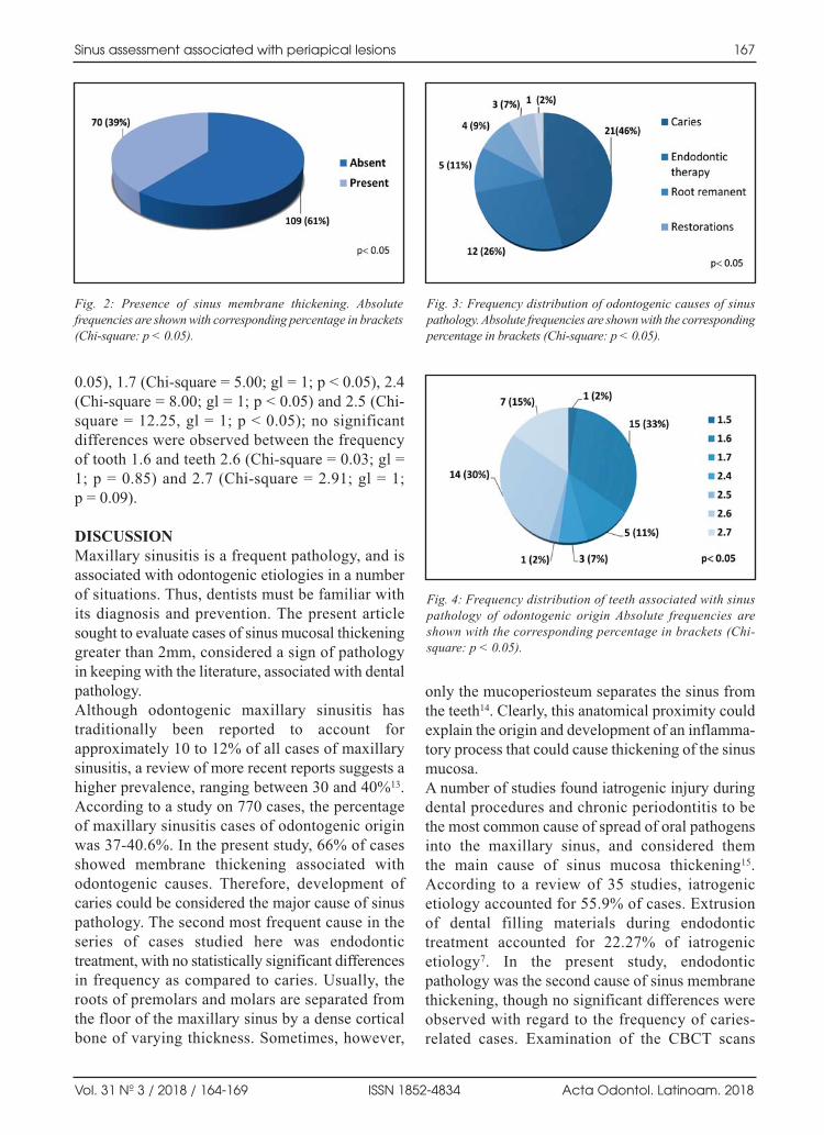
0.05), 1.7 (Chisquare = 5.00; gl = 1; p < 0.05), 2.4(Chisquare = 8.00; gl = 1; p < 0.05) and 2.5 (Chisquare = 12.25, gl = 1; p < 0.05); no significantdifferences were observed between the frequencyof tooth 1.6 and teeth 2.6 (Chisquare = 0.03; gl =1; p = 0.85) and 2.7 (Chisquare = 2.91; gl = 1; p = 0.09).
DISCUSSIONMaxillary sinusitis is a frequent pathology, and isassociated with odontogenic etiologies in a numberof situations. Thus, dentists must be familiar withits diagnosis and prevention. The present articlesought to evaluate cases of sinus mucosal thickeninggreater than 2mm, considered a sign of pathologyin keeping with the literature, associated with dentalpathology. Although odontogenic maxillary sinusitis hastraditionally been reported to account forapproximately 10 to 12% of all cases of maxillarysinusitis, a review of more recent reports suggests ahigher prevalence, ranging between 30 and 40%13.According to a study on 770 cases, the percentageof maxillary sinusitis cases of odontogenic originwas 3740.6%. In the present study, 66% of casesshowed membrane thickening associated withodontogenic causes. Therefore, development ofcaries could be considered the major cause of sinuspathology. The second most frequent cause in theseries of cases studied here was endodontictreatment, with no statistically significant differencesin frequency as compared to caries. Usually, theroots of premolars and molars are separated fromthe floor of the maxillary sinus by a dense corticalbone of varying thickness. Sometimes, however,
only the mucoperiosteum separates the sinus fromthe teeth14. Clearly, this anatomical proximity couldexplain the origin and development of an inflamma tory process that could cause thickening of the sinusmucosa. A number of studies found iatrogenic injury duringdental procedures and chronic periodontitis to bethe most common cause of spread of oral pathogensinto the maxillary sinus, and considered them the main cause of sinus mucosa thickening15.According to a review of 35 studies, iatrogenicetiology accounted for 55.9% of cases. Extrusionof dental filling materials during endodontictreatment accounted for 22.27% of iatrogenicetiology7. In the present study, endodonticpathology was the second cause of sinus membranethickening, though no significant differences wereobserved with regard to the frequency of cariesrelated cases. Examination of the CBCT scans
Sinus assessment associated with periapical lesions 167
Vol. 31 Nº 3 / 2018 / 164-169 ISSN 1852-4834 Acta Odontol. Latinoam. 2018
Fig. 2: Presence of sinus membrane thickening. Absolutefrequencies are shown with corresponding percentage in brackets(Chisquare: p < 0.05).
Fig. 3: Frequency distribution of odontogenic causes of sinuspathology. Absolute frequencies are shown with the correspondingpercentage in brackets (Chisquare: p < 0.05).
Fig. 4: Frequency distribution of teeth associated with sinuspathology of odontogenic origin Absolute frequencies areshown with the corresponding percentage in brackets (Chisquare: p < 0.05).
AOL32018:32011 21/02/2019 11:40 Página 167

included in the present study allowed detectingcases of overfilling with materials, such as guttapercha, with no sinus alteration. This could be anindication of the biocompatibility of said material,which would account for the absence of aninflammatory process at the apical level. Proper chemical preparation, maintaining apicalpermeability, and use of the coronoapical techniquefor endodontic treatment, contribute to decreasingthe likelihood of spread of microorganisms to themaxillary sinus. Periodontal disease has long been reported as anetiological factor of sinus mucosa inflammation.For example, in their 1943 study in humancadavers, Bauer et al. demonstrated direct diffusionof oral sepsis to the maxillary sinus. More recently,Abrahams et al.16 reported the incidence of sinusitisin patients with periodontal disease to be twofoldthat of patients without periodontal disease.Recognition of the close relationship betweeninflammation of periapical tissues and damage to the sinus membrane led to describing thepathological entity referred to as the “endoantralsyndrome. In recent years, however, the incidenceof these effects has decreased, likely due to factorssuch as improved oral hygiene and preventivetechniques related with periodontal disease. In thepresent study, periodontal lesions accounted for 2%of cases of sinus membrane thickening associatedwith direct spread of pathogens to the maxillarysinus. According to Panico and Adell17,18 development ofsinusitis associated with implant placement isinfrequent. Reviews published in the literaturefound that maxillary sinus floor augmentation priorto surgical placement of a dental implant resultedin sinus alteration in 4.17% of cases, and ininadequate implant position or implant migration in 0.92% of cases7. In the present study on a total179 maxillary sinuses analyzed using CBCT, sinus thickening was observed in 39% of cases, 6% of which were associated with the presence of implants in close proximity to the affectedmaxillary sinus. None of the cases studied here showed signs of
apicoectomy. It is of note that the latter procedurecan lead to a complication that is observed lessfrequently than expected, likely because mostprofessionals prefer extracting the tooth rather than
performing this technique in view of the associatedhigh risk of accidental perforation to the sinus dueto its proximity19. Nevertheless, reports such as thatby Freedman et al. involving 472 apicoectomies,none of which resulted in sinusitis, show that thereis no contraindication to performing apicoectomyin antral teeth, despite their proximity to themaxillary sinus 20. A review of 35 articles on maxillary sinusitis ofodontogenic origin showed the upper molar regionto be more frequently associated with maxillarysinus alterations (47.68%). The first upper molarwas the molar tooth most frequently associated withmaxillary sinusitis, with a 22.51% incidence,followed by the third molar tooth (17.21%) and thesecond molar tooth (3.97%). With regard to theupper premolar region, it was affected in 5.96% ofcases only; the second premolar was the mostaffected premolar tooth (1.98%), and the canine wasinvolved in only 0.66% of cases of maxillarysinusitis of odontogenic origin7.In keeping with the aforementioned study, theresults obtained here showed that the tooth most frequently involved in sinus alteration was tooth 1.6, accounting for 33% of cases. It wassignificantly more affected than teeth 1.4 (0%), 1.5(2%), 1.7 (11%), 2.4 (7%) and 2.5 (2%), but did notdiffer significantly compared to teeth 2.6 (30%) and2.7 (15%). Examination of sinus membrane thickening by conebeam computed tomography allows detecting thepresence of sinus pathology. Findings must becorrelated with the clinical condition of the patient.Thorough examination of the para nasal sinuses and visualization of the osteomeatal complex onaxial and coronal sections obtained by computedtomography without contrast is essential to evaluatepermeability of the ostium and for proper treatmentplanning. The retrospective nature of the presentstudy does not allow establishing a “causeeffect”relationship between periapical pathology andchanges in the maxillary sinus. Further prospectivestudies are necessary to correlate clinical andradiographic data and confirm the present findings.Lastly, a larger sample size and including casesclassified according to disease severity could revealmore significant associations between periapicaland/or periodontal pathologies and changes in thesinus mucosa.
168 Gisela V. Brañas, et al.
Acta Odontol. Latinoam. 2018 ISSN 1852-4834 Vol. 31 Nº 3 / 2018 / 164-169
AOL32018:32011 21/02/2019 11:40 Página 168

CONCLUSIONAccording to the results obtained in the present study,the high incidence of sinus pathology of odontogenicorigin implies the need for interdisci plinary workinvolving dentists and earthroat specialists. Cariousprocesses, poor restorations, periodontal lesions,
implants, and the presence of root remnants are themain causes of sinus mucosa thickening. The use of CBCT for diagnosis and treatmentplanning allows identifying the presence of maxillarysinus membrane thickening and determining whetherit is associated an odontogenic origin.
Sinus assessment associated with periapical lesions 169
Vol. 31 Nº 3 / 2018 / 164-169 ISSN 1852-4834 Acta Odontol. Latinoam. 2018
ACKNOWLEDGEMENTSThe authors thank Dr. Carlos Marino, Earthroat specialist andlecturer at the Specialization in Oralmaxillofacial prostheses,University of Buenos Aires, for his valuable assistance andguidance in performing the present study, and DDS JonathanSaiegh, from the Department of Imaging Diagnosis of theSchool of Dentistry of the University of Buenos Aires, for hisassistance in evaluating the samples.
FUNDINGNone
CORRESPONDENCEDr. Pablo A. RodríguezCátedra de Endodoncia, Facultad de OdontologíaMarcelo T. de Alvear 2142, 1ºA, (C1122AAH) CABA. [email protected]
REFERENCES1. Clavería R, Peña M, Gutiérrez I, Paredes M, Fouces Y.
Comunicación bucosinusal por extracciones dentales.Medisan 2010; 14: 346352. URL:http://scielo.sld.cu/scielo.php?script=sci_arttext&pid=S102930192010000300010&lng=es.
2. Nagar AC, Gautam A. Maxillary Sinusitis of OdontogenicOrigin: A Review. Int J Adv Res 2015; 3: 13231328.
3. Kilic C, Kamburoglu K, Yuksel SP, Ozen T. An Assessmentof the Relationship between the Maxillary Sinus Floor andthe Maxillary Posterior Teeth Root Tips Using DentalConebeam Computerized Tomography. Eur J Dent 2010;4: 462467.
4. Raghav M, Karjodkar FR, Sontakke S, Sansare K.Prevalence of incidental maxillary sinus pathologies indental patients on conebeam computed tomographicimages. Contemp Clin Dent 2014; 5: 361365.
5. PérezPiñas I, Sabaté J, Ruiz T, Jimeno L, Cueto L, VenturaJ. Semiología de la patología inflamatoria nasosinusalmediante TC. Patrones de afectación. Radiología 2001; 43:387393. https://doi.org/10.1016/S00338338(01)770014
6. Cymerman JJ, Cymerman DH, O’Dwyer RS. Evaluationof odontogenic maxillary sinusitis using conebeamcomputed tomography: three case reports. J Endod 2011;37: 14651469.
7. Simuntis R, Kubilius R, Vaitkus S. Odontogenic maxillarysinusitis: a review. Stomatologija 2014; 16: 3943.
8. Ferguson M. Rhinosinusitis in oral medicine and dentistry.Aust Dent J 2014; 59: 289295.
9. Shanbhag S, Karnik P, Shirke P, Shanbhag V. Associationbetween periapical lesions and maxillary sinus mucosalthickening: a retrospective conebeam computed tomo graphic study. J Endod 2013; 39: 853857.
10. GuerraPereira I, Vaz P, FariaAlmeida R, Braga AC, FelinoA. CT maxillary sinus evaluation—A retrospective cohortstudy. Med Oral Patol Oral Cir Bucal 2015; 20: e41926.
11. Khorramdel A, Shirmohammadi A, Sadighi A, et al.Association between demographic and radiographic
characteristics of the schneiderian membrane and periapicaland periodontal diseases using conebeam computedtomography scanning: A retrospective study. J Dent ResDent Clin Dent Prospects 2017; 11: 170176.
12. Rak KM, Newell JD 2nd, Yakes WF, Damiano MA,Luethke JM. Paranasal sinuses on MR images of the brain:significance of mucosal thickening. AJR Am J Roentgenol1991; 156: 381384.
13. AriasIrimia O, BaronaDorado C, SantosMarino JA,MartínezRodriguez N, MartínezGonzález JM. Metaanalysis of the etiology of odontogenic maxillary sinusitis.Med Oral Patol Oral Cir Bucal 2010; 15: e703.
14. García B, Martorell L, Martí E, Peñarrocha M. Periapicalsurgery of maxillary posterior teeth. A review of theliterature. Med Oral Patol Oral Cir Bucal 2006; 11: E146150.
15. Evwcombe R G., Merino Soto C. Intervalos de confianzapara las estimaciones de proporciones y las diferenciasentre ellas. Interdisciplinaria 2006; 23.URL:<http://www.redalyc.org/articulo.oa?id=18023201>ISSN 03258203
16. Abrahams JJ, Glassberg RM. Dental disease: a frequentlyunrecognized cause of maxillary sinus abnormalities? AJRAm J Roentgenol 1996; 166: 12191223.
17. Panico R, Fernandez Bodereau E, Sosa F. Dental implantmigration to maxillary sinus. Treatment and complications:case report. Rev Asoc Odontol Argent. 2014; 102: 2833.URL:https://www.aoa.org.ar/wpcontent/uploads/2017/03/V101N2M2014.pdf
18. Adell R, Lekholm U, Rockler B, Brånemark PI. A 15yearstudy of osseointegrated implants in the treatment of theedentulous jaw. Int J Oral Surg 1981; 1: 387416.
19. García B, Martorell L, Martí E, Peñarrocha M. Periapicalsurgery of maxillary posterior teeth. A review of theliterature. Med Oral Patol Oral Cir Bucal 2006; 11: E146150.
20. Freedman A, Horowitz I. Complications after apicoectomyin maxillary premolar and molar teeth. Int J Oral MaxillofacSurg. 1999; 28: 192194.
AOL32018:32011 21/02/2019 11:40 Página 169
Acta Odontol. Latinoam. 2018 ISSN 1852-4834 Vol. 31 Nº 3 / 2018
adolescence ............................................................3age......................................................................125alveolar bone loss ..............................................131apoptosis ............................................................110biofilms ............................................................138biomarkers ..........................................................97bone age ..........................................................125calcium hydroxide ............................................144capillary permeability ..........................................32carbonic anhydrase IX ........................................77cervical vertebrae ..............................................125child ..............................................................23, 82children ..............................................................156chronic periodontitis ............................................53complete denture..................................................91cone beam computed tomography ..............67, 164cytokines ..............................................................53dental......................................................................3dental arches orthodontics ................................104dental care............................................................58dental caries ....................................23, 58, 82, 156dental cements ..................................................144dental enamel hypoplasia ....................................38dental equipment................................................138dental fluorosis ....................................................82dentistry ............................................................117development ......................................................104diabetes mellitus ..................................................32diet ......................................................................97edema ..................................................................32EDTA ..................................................................11emergency service..............................................117epithelial rests of Malassez................................131esthetics..................................................................3facial pain ............................................................16fatty acids ............................................................97finite element analysis ......................................125friction................................................................149gingiva ................................................................32guttapercha ..........................................................45health education ................................................156health services accessibility ................................58hospital ..............................................................117
malocclusion ........................................................23maxillary sinus ..................................................164mechanical stress ..............................................110mechanotransduction ........................................110mesh pubmed ......................................................91mouth edentulous ................................................91nutritional status ..................................................58oral health ............................................................38oral leukoplakia ..................................................77oral lichen planus ................................................77orthodontic brackets ..........................................149orthodontic tooth movement ..............................110orthodontics ..........................................................3osteocytes ..........................................................110osteopontin ........................................................110oxidative stress ....................................................32pain ......................................................................45peracetic acid ......................................................11periapical tissues................................................164periodontal ligament ..........................................131periodontitis ................................................32, 131pregnancy ............................................................53preschool..............................................................23preterm birth ........................................................53preterm labor........................................................53quality of life................................23, 38, 58, 82, 91root canal ............................................................67root canal filling materials. ..................................45root resorption....................................................131saliva ....................................................................97schneiderian membrane ....................................164selfmedication....................................................117signs and symptoms ............................................16sodium hypochlorite ............................................11stomatognathic system ........................................91Streptococcus mutans ........................................156surveys and questionnaires ..................................16temporomandibular joint disorders ....................16tensile Strength ..................................................144tooth demineralization ........................................38tooth injuries ..................................................23, 82toxicity ................................................................11water microbiology............................................138
INDEXES TO VOLUME 31 (2018)
SUBJECT INDEX
AOL32018:32011 21/02/2019 11:40 Página 170

Vol. 31 Nº 3 / 2018 ISSN 1852-4834 Acta Odontol. Latinoam. 2018
Abanto, J ........................................................23, 82Abba, M ............................................................149Actis, AB..............................................................97Alchieri, JC ..........................................................91Álvarez, LV..........................................................38Alves, AC ............................................................91AlzateMendoza, D..............................................45Andornino, A ......................................................32Andrada, MM ....................................................117Arganiaraz, A ......................................................32Arias, C ..............................................................117Arias, E ................................................................53Aristizabal, F......................................................149Benedí, D ..........................................................131BenítezRogé, S ................................................104Berger, SB..........................................................144Boneckër, M ..................................................23, 82Bozal, CB ..........................................................110Brañas, GV ........................................................164Brasquet, N ..........................................................32Bueno, CES..........................................................11Calabrese, D ......................................................104Calderon, PS ........................................................91Catanzaro, OL......................................................32Cavalcanti, RVA ..................................................91Coelho, MS ..........................................................11Colussi, PRG..........................................................3Cortes, A ..............................................................58Cortés, F ..............................................................58CubidesFlechas, K............................................149de C. Oliveira, V ................................................138de Moraes, M ......................................................16Defagó, MD ........................................................97Di Martino, I ........................................................32Díaz, S............................................................23, 82Domingues, PCA ..............................................138Echeverri, J ..........................................................53EscobarArregoces, F ..........................................53Faes, L................................................................117Folco, A..............................................................104Fontana, CE ........................................................11Gabrielli, E ............................................................3Gamboa, F..........................................................156Gamboa, LF ........................................................58GamboaMartinez, LF ........................................45Gandolfo, MS ......................................................77
García, DA ........................................................140GonzálezCarrera, MC ........................................58Goulart, GP ..........................................................16Goya, JA ............................................................131Grisolia, BG ......................................................164Gualtieri, A ................................................117, 164Guiraldo, RD......................................................144Hassan, R ............................................................67Hecht, P..............................................................104HernándezMejia, D ............................................45Iglesias, M..........................................................104Iriarte, AJ ..........................................................149Issa, N ..................................................................67Itoiz, ME..............................................................77Iuliano, RG ........................................................164JacomeLievano, S ..............................................58JaimesMonroy, G..............................................149Kato, AS ..............................................................11Lamby, CP ........................................................149LasernaZuluaga, H ............................................45LatorreUriza, C ..................................................53Leandro Plazas, L ............................................149Lenarduzzi, A ....................................................164Lumi, M ............................................................104Macedo, AP........................................................138Mandalunis, PM ........................................110, 131Marques, DM ....................................................138Martigno, S ..........................................................58Martínez, LM ......................................................82Martins Paiva, SM ..............................................82Masquijo Bisio, P................................................77Mateu, ME ........................................................104Mejía, LP ............................................................38Mendez, CF........................................................117Merlo, GHS............................................................3Mondol, M ..........................................................23Monteiro, RM ....................................................138Muniz, FWMG ......................................................3NinoBarrera, JL ..................................................45Olaya, C ..............................................................45Olaya, C ............................................................149Ortiz, S ..............................................................149Paiva, SM ............................................................23Paparella, ML ......................................................77Pastore, GP ..........................................................16Pastore, PR ..........................................................16
INDEXES TO VOLUME 31 (2018)
AUTHOR INDEX
AOL32018:32011 21/02/2019 11:40 Página 171

PedroRocha, DG ................................................11Peñate, A ..............................................................23Pérez, M ..............................................................77Pernambuco, L ....................................................91Perovic, NR..........................................................97Piardi, CC ..............................................................3Prada, S ..............................................................117Prati, AJ................................................................16Puerta, G ..............................................................23Pulitano Manisagian, GE ..................................131RamírezVelásquez, M ......................................125Ramos, K ............................................................82Ramos, N ............................................................58Razaboni, AM....................................................138Renou, SJ ..........................................................164Repossi, G............................................................97RestrepoPerez, LF ..............................................58RoaMolina, R ....................................................53Rodríguez, D......................................................117Rodríguez, DA ..................................................125Rodríguez, MJ......................................................38Rodríguez, PA....................................................164Rojas, ME ..........................................................125Roscher, DF ......................................................117Roshdy, N ............................................................67
Rösing, CK ............................................................3Ruiz, AJ................................................................53RuizCarrizosa, JA ..............................................58Saiegh, J ............................................................117Sánchez, LM ......................................................110Santos, LL..........................................................144Sarralde, AL ......................................................149Sczepanski, CRB ..............................................144Sczepanski, F ....................................................144Silva, J..................................................................53Siragusa, C ........................................................117Solla, M..............................................................104Stolbizer, F ........................................................117Teixeira, PA..........................................................11Ubios, AM..........................................................110Unapanta, J ..........................................................45UsugaVacca, M ..................................................58Valentich, MA......................................................97Velandia, LM ......................................................38Vélez, MP ............................................................82VelosaPorras, J....................................................53Viloria Ávila, TJ ..............................................125Watanabe, E ......................................................138Zambrano, O ......................................................125
Acta Odontol. Latinoam. 2018 ISSN 1852-4834 Vol. 31 Nº 3 / 2018
INDEXES TO VOLUME 31 (2018)
AUTHOR INDEX
AOL32018:32011 21/02/2019 11:40 Página 172

AOL32018:32011 21/02/2019 11:40 Página 173

AOL32018:32011 21/02/2019 11:40 Página 174

AOL32018:32011 21/02/2019 11:40 Página 175

AOL32018:32011 21/02/2019 11:40 Página 176

AOL32018:32011 21/02/2019 11:40 Página 177