Embed Size (px)
Citation preview
© Acta Anæsthesiologica Belgica, 2019, 70, n° 4
Abstract : Horner Syndrome (HS) is a rare but benign complication with epidural analgesia in parturients. It is caused by the cephalad spread of local anesthetic with an overflow to the superior cervical sympathetic chain. We present 2 patients, a 28-year old woman and a 23-year old woman, both without any major medical comorbidity. Each patient received a lumbar epidural catheter with the conventional labor pain treatment of our hospital. The first patient developed HS after the administration of 10 ml of lidocaine 1% when a unilateral block occurred. At this time, the patient was placed in genupectoral position. The second patient developed HS directly after the first bolus of 10 ml of the standard epidural solution of our center when the patient was in supine position. A spontaneous resolution of HS was observed in both patients. The exact incidence of HS in parturients is not known and probably underreported by patients as well as by caregivers. Clinical vigilance and appropriate teaching of obstetrics caregivers is very important. We demonstrate that an accidental subdural placement of the catheter and the dose of the epidural solution can play a major role in the development of HS.
Keywords : Horner syndrome ; epidural analgesia ; obstetrics; complication ; local anesthetics.
IntroductIon
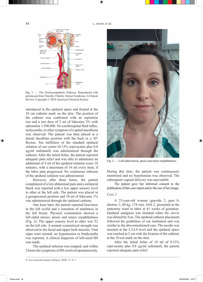
Horner syndrome (HS) is characterized by the classic triad of ipsilateral miosis, ptosis and anhidrosis. This clinical entity is associated with apparent ipsilateral enophthalmos and peripheral vasodilatation, resulting in a facial flush, conjunctival edema and a nasal obstruction. The clinical signs are the result of a unilateral block or lesion of the sympathetic neurons that innervate the head and neck region, more specifically the oculo-sympathetic pathway (Fig. 1). This pathway is a three-neuron chain that originates in the hypothalamus, travels down the spinal cord to the lower cervical and upper thoracic levels, then traverses the upper chest cavity and apex of the lung, traveling with the carotid artery into the cavernous sinus, traversing the orbit to innervate the pupillary sphincter; it also branches
to innervate accessory muscles for eyelid retraction (1). Any lesion along this course can present with HS.
The first case of HS associated with regional anesthesia was reported in 1972 by Kepes et al., as a complication after a caudal block (2). A recent systemic review of the literature by Chambers and Bhatia reported a total of 63 cases of HS after obstetric neuraxial blockade (3). This number is without a doubt an underestimation of the incidence of this clinical phenomenon.
case presentatIons
Case 1
A 28-year-old woman (gravida 1, para 0, abortus 0; 63kg, 166 cm), ASA 2, presented in labor at 39 weeks of gestation. Epidural analgesia was requested at 5 cm of cervical dilation. The patient was placed in a sitting position. A 17 Ga epidural needle (BBraun Perican Tuohy, 80 mm) was inserted at the L4-L5 level, after a subcutaneous infiltration of 3 ml lidocaine 2% with adrenaline 1/200,000. No pain nor paresthesia were reported during the insertion of the epidural needle. The epidural space was reached at 6 cm using the loss of resistance technique with saline. An epidural catheter was
(Acta Anaesth. Belg., 2020, 71, 43-47)
L. Peene, M.d. ; t. Vanneste, M.d. ; M. Beran, M.d. ; p. Vanelderen, M.d. ; J. Van Zundert, M.d. ; r. Heylen, M.d. ; d. Mesotten, M.d. ; M. Van de Velde, M.d.
(*) Department of Anesthesiology, Critical Care and Multidisciplinary Pain Center, Ziekenhuis Oost-Limburg, Genk/Lanaken, Belgium.
(**) Department of Anesthesiology and Pain Management, University Medical Centre Maastricht, Maastricht, The Netherlands.
(***) Department of Anesthesiology, University Hospitals Leuven, Leuven, Belgium.
Corresponding author : Laurens Peene, MD, Department of Anesthesiology, Ziekenhuis Oost-Limburg, Genk, Schiepse Bos 6, 3600 Genk, Belgium
E-mail : [email protected]
Paper submitted on May 14, 2019 and accepted on Nov 28, 2019. Conflict of interest: None
Horner syndrome following epidural analgesia for labor pain:two case reports
l. peene (*), t. Vanneste (*), M. Beran (*), p. Vanelderen (*), J. Van Zundert (*,**), r. Heylen (*), d. Mesotten (*), M. Van de Velde (***)
Peene.indd 43Peene.indd 43 23/03/2020 12:0723/03/2020 12:07
© Acta Anæsthesiologica Belgica, 2020, 71, n° 1
44 l. peene et al.
During this time, the patient was continuously monitored and no hypotension was observed. The subsequent vaginal delivery was uneventful.
The patient gave her informed consent to the publication of this case report and to the use of her image.
Case 2
A 23-year-old woman (gravida 2, para 0, abortus 1; 60 kg, 174 cm), ASA 2, presented at the maternity ward in labor at 41 weeks of gestation. Epidural analgesia was initiated when the cervix was dilated by 5cm. The epidural catheter placement followed the guidelines of our institution and was similar to the abovementioned case. The needle was inserted at the L3-L4 level and the epidural space was reached at 5 cm with the fixation of the catheter at the 10-cm mark on the skin.
After the initial bolus of 10 ml of 0.13% ropivacaine plus 0.8 µg/ml sufentanil, the patient reported adequate pain relief.
introduced in the epidural space and fixated at the 10 cm catheter mark on the skin. The position of the catheter was confirmed with an aspiration test and a test dose of 3 ml of lidocaine 2% with adrenaline 1/200,000. No cerebrospinal fluid reflux, tachycardia, or other symptom of a spinal anesthesia was observed. The patient was then placed in a dorsal decubitus position with the back in a 30° flexion. Ten milliliters of the standard epidural solution of our center (0.13% ropivacaine plus 0.8 µg/ml sufentanil) was administered through the catheter. After the initial bolus, the patient reported adequate pain relief and was able to administer an additional of 4 ml of the epidural solution every 10 minutes, with a maximum of 16 ml every hour, if the labor pain progressed. No continuous infusion of the epidural solution was administered.
However, after three hours, the patient complained of a low abdominal pain and a unilateral block was reported with a low upper sensory level to ether at the left side. The patient was placed in a genupectoral position and 10 ml of lidocaine 1% was administered through the epidural catheter.
One hour later, the patient reported heaviness in the left eyelid and a sensation of numbness in the left breast. Physical examination showed a left-sided miosis, ptosis and minor enophthalmos (Fig. 2). The upper sensory level to ether was T2 on the left side. A normal symmetrical strength was observed in the facial and upper limb muscles. Vital signs were normal: no hypotension or bradycardia was reported. A clinical diagnosis of left-sized HS was made.
The epidural infusion was stopped, and within 2 hours the symptoms of HS resolved spontaneously.
Fig. 1. — The Oculosympathetic Pathway. Reproduced with permission from Timothy J Martin. Horner Syndrome: A Clinical Review. Copyright © 2018 American Chemical Society.
Fig. 2. — Left-sided miosis, ptosis and minor enophthalmos.
Peene.indd 44Peene.indd 44 23/03/2020 12:0723/03/2020 12:07
© Acta Anæsthesiologica Belgica, 2020, 71, n° 1
Horner syndroMe followIng epIdural analgesIa for laBor paIn 45
section, as well as after epidural analgesia in non-obstetric populations. The exact incidence is unknown and varies amongst prospective studies. Clayton reported an incidence of HS in 1.33 % of 150 parturients in labor pain – compared to 4.0 % of parturients undergoing caesarean section (4). Rabinovich et al. reported an incidence of HS in 0.13 % of 4598 parturients who received epidural analgesia in a single center (5). However, the true incidence of HS in parturients receiving epidural analgesia is probably much higher. A plausible explanation is that the clinical signs are underreported by patients or not noticed by caregivers.
The development of HS in parturients who received epidural analgesia can be explained by an excessive cephalad spread of local anesthetic during supine position, with an overflow to the cervical sympathetic chain. This causes a disruption of the oculosympathetic pathway at the point where preganglionic neurons exit the spinal cord through the ventral roots on their path through the sympathetic chain to the superior cervical ganglion (6).
It has been suggested that high progesterone levels during pregnancy increase nerve fiber sensitivity, which may play a role in the higher risk of HS in parturient (7). This could explain the fact that the reported incidence of HS after epidural analgesia is much higher in obstetric neuraxial blockade compared with other patient populations (8).
In most cases, unilateral HS will manifest. Chambers and Bhatia reported only 3 cases with bilateral HS in their systematic review (3). Patient position and gravity are clear factors that affect the epidural spread of local anesthetic (9). The gravitational effect in the pathogenesis of HS becomes evident in the literature, where six parturients placed in lateral decubitus developed HS on the dependent side and only two developed HS on the contralateral side (3). Our first case supports the hypothesis of the gravitational effect, since HS developed when the patient was placed in a genupectoral position.
Chambers and Bhatia reported that HS occurs as well with bolus as infusion methods of epidural local anesthetic delivery and, that the medial volume of local anesthetic delivered before the onset was 18 ml (2). In our two cases, HS occurred after the administration of a bolus of local anesthetic of 10 ml of lidocaine 1% in case 1, and of 10 ml of 0.13% ropivacaine plus 0.8 µg/ml sufentanil associated to 3 ml of lidocaine 1% with adrenaline 1/200,000 as test dose in case 2. While there are some case reports that indicate the occurrence of HS after a bolus of 10 ml or less, in most cases a higher volume
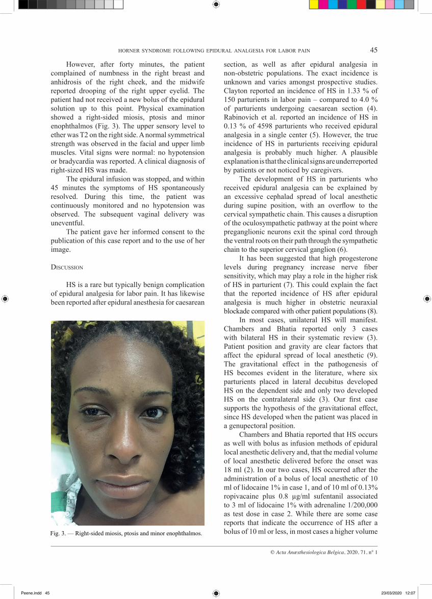
However, after forty minutes, the patient complained of numbness in the right breast and anhidrosis of the right cheek, and the midwife reported drooping of the right upper eyelid. The patient had not received a new bolus of the epidural solution up to this point. Physical examination showed a right-sided miosis, ptosis and minor enophthalmos (Fig. 3). The upper sensory level to ether was T2 on the right side. A normal symmetrical strength was observed in the facial and upper limb muscles. Vital signs were normal: no hypotension or bradycardia was reported. A clinical diagnosis of right-sized HS was made.
The epidural infusion was stopped, and within 45 minutes the symptoms of HS spontaneously resolved. During this time, the patient was continuously monitored and no hypotension was observed. The subsequent vaginal delivery was uneventful.
The patient gave her informed consent to the publication of this case report and to the use of her image.
dIscussIon
HS is a rare but typically benign complication of epidural analgesia for labor pain. It has likewise been reported after epidural anesthesia for caesarean
Fig. 3. — Right-sided miosis, ptosis and minor enophthalmos.
Peene.indd 45Peene.indd 45 23/03/2020 12:0723/03/2020 12:07
© Acta Anæsthesiologica Belgica, 2020, 71, n° 1
46 l. peene et al.
any case reports of HS following obstetric neuraxial blockade which met all of these inclusion criteria (3).
Our case report also demonstrates the point that HS is clinically heterogeneous: while figure 3 very clearly illustrates the features, figure 2 demonstrates more subtle ptosis, exhibits notable chemosis in addition to the classical signs of HS. Because of this heterogeneous presentation, signs can be subtle and often missed.
HS after epidural analgesia in parturients is a relatively benign and mostly transient condition that usually does not warrant further extensive investigations. However, systemic hypotension and subjective respiratory discomfort are associated with high sympathetic blockade (18). True respiratory compromise from a high block is a low risk but should always be considered. Recognition of the development of Horner’s syndrome is important for those involved in the care of obstetric patients. The signs and symptoms can be distressing in particular for the parturient. Continuation of regional anesthesia in the presence of HS is possible, but anesthetic vigilance is of the utmost importance (19). Appropriate teaching of all those involved in care of parturients is vital, and the symptomatic patient or anxious family members should receive appropriate reassurance when a diagnosis of HS is made in the obstetric setting.
References
1. Martin T. J. 2018. Horner Syndrome: A Clinical Review. ACS Chem. Neurosci. 9(2): 177–186.
2. Kepes E.R., Martinez L.R., Pantuck E. and Stark D.C.C. 1972. Horner’s syndrome following caudal anesthesia. NY State J Med. 72: 946–947.
3. Chambers D.J. and Bhatia K. 2018. Horner’s syndrome following obstetric neuraxial blockade – a systematic review of the literature. Int. J. Obstet. Anesth. 35: 75-87.
4. Clayton K. C. 1983. The incidence of Horner’s syndrome during lumbar extradural for elective Caesarean section and provision of analgesia during labour. Anaesthesia. 38: 583–585.
5. Rabinovich A., Abedelhady R., Mazor M., Piura B. and Margolin E. 2010. Horner’s syndrome following epidural analgesia during labor: report of six cases. Eur. J. Obstet. Gynecol. Reprod. Biol. 149: 225–231.
6. Thompson H. S., Miller N. R. and Newman N. J. 1998. Disorders of pupillary function, accommodation, and lacrimation. In: Walsh and Hoyt’s clinical neuro-ophthalmology. 5th ed, vol 1. p. 961-1040. Baltimore. Williams & Wilkins
7. Bromage P. R. 1978. Epidural Anesthesia for Obstetrics. In: Epidural Analgesia. p. 588. Philadelphia. W.B. Saunders Co.
8. Biousse V., Guevara R. A. and Newman N. J. 1998. Transient Horner’s syndrome after lumbar epidural anesthesia. Neurology. 51: 1473–1475.
9. Apostolou G. A., Zarmakoupis P. K. and Mastrokostopoulos G. T. 1981. Spread of epidural anesthesia and the lateral position. Anesth. Analg. 60: 584-586.
is reported. In the 78 cases of HS after neuraxial blockade reported by Chambers and Bhatia, only 11 cases occurred after a bolus of 10 ml or less (3). This phenomenon could be explained through unilateral cephalad spread of local anesthetic, which has been attributed to septation in the epidural cavity (10). This suggests that it might be possible to achieve a higher sensory level than predicted with a commonly used volume of local anesthetic, with or without the development of HS (11).
Current guidelines recommend the use of low-dose epidural solutions in parturient (12). The standard epidural solution used in our center attests to this directive. However, in case 1 a relative high dose solution (10 ml lidocaine 1%) was used as a top up for pain. We suggest that the dose of the epidural solution is an important factor in the pathogenesis of HS.
We postulate that the development of HS in case 2 can be explained by an accidental subdural injection of local anesthetic. An accidental insertion of the epidural catheter in the subdural space is a rare complication of attempted epidural placement. Reina et al. postulated that subdural spaces may be generated when the dura and arachnoid layers are pulled apart during catheter placement (13). The three most common features of an accidental subdural catheter placement are 1) an unusual high sensory block – which can cause the occurrence of HS –, 2) an unexpected motor block and 3) profound hypotension (14). Lubenow et al. reported an incidence of accidental subdural catheter placement of 0.82% (15). They also defined the major and minor criteria for the diagnosis of a positive subdural injection. The two major criteria are 1) a negative aspiration test and, 2) an unexpected widespread sensory block after epidural injection. The three minor criteria are 1) a delayed onset of 10 minutes or more of a sensory or motor nerve block, 2) a variable motor blockade occurring, despite use of low doses of bupivacaine and, 3) an out of proportion to the administered dose of local anesthetic sympatholysis (15). When a subdural catheter position is clinically presumed, the caregiver must always rule out an intradural catheter position (16). In each case, a catheter that was accidentally placed in the subdural space must always be removed, since it may rupture spontaneously through the arachnoid resulting in a total spinal block (17). A differential diagnosis between benign high spread of local anesthetics and a subdural catheter placement should always be made.
No significant hypotension was noted in our case, but our clinical findings were consistent with these criteria. Chambers and Bhatia did not identify
Peene.indd 46Peene.indd 46 23/03/2020 12:0723/03/2020 12:07
© Acta Anæsthesiologica Belgica, 2020, 71, n° 1
Horner syndroMe followIng epIdural analgesIa for laBor paIn 47
Subdural Block and Conversion Disorder. Case Rep Med. 751648. Online publication.
15. Lubenow T., Keh-Wong E., Kristof K., Ivankovich O. and Ivankovich A. D. 1998. Inadvert subdural injection: a complication of an epidural block. Anesth. Analg. 67: 175-179
16. Collier C. 2009. Most reported subdural injections are not in the subdural space, they are intradural! Reg. Anesth. Pain Med. 34(6): 613-615.
17. Scrutton M., Porter J., Russell R, and Reynolds F. 1996. Subdural or epidural? Anaesthesia. 51(7): 708-709.
18. Chandrasekhar S. and Peterfreund R. A. 2003. Horner’s syndrome following very low concentration bupivacaine infusion for labor epidural analgesia. Clin. Anesth. 15: 217–219.
19. Sharma R., Chatterjee J. and Edmonds K. 2010. Horner’s syndrome with epidural anaesthesia. BMJ Case Reports. bcr0120102698.
10. Goel S. and Burkat C. N. 2011. Unusual case of persistent Horner’s syndrome following epidural anaesthesia and caesarean section. Indian J. Ophthalmol. 59: 389–391.
11. Smith D. I., Chiem J. L., Burk S., Borovcanin Z. C. and Tran N. H. 2017. Hemodynamic instability and Horner’s syndrome following a labour lumbar neuraxial block: A warning sign of a potentially lethal event? J. R. Soc. Med. 110(6): 245-248.
12. Sng B. L. and Sia A. T. H. 2017. Maintenance of epidural labour analgesia: The old, the new and the future. Best Pract. Res. Clin. Anaesthesiol. 31(1): 15-22.
13. Reina M. A., Collier C. B., Prats-Galino A., Puigdellívol-Sánchez A., De Andrés J. A. et al. 2011. Unintentional subdural placement of epidural catheters during attempted epidural anesthesia: an anatomic study of spinal subdural compartment. Reg. Anesth. Pain Med. 36(6): 537-541.
14. Elsharkawy H., Khanna A. K. and Barsoum S. 2013. Caesarean Delivery Complicated by Unintentional
Peene.indd 47Peene.indd 47 23/03/2020 12:0723/03/2020 12:07