Embed Size (px)
Citation preview

Acquired Heart Diseases


DOI: 10.5644/PI2015-158

Publisher Academy of Sciences and Arts of Bosnia and Herzegovina
Editor-in-Chief Senka Mesihović-Dinarević
Reviewers Marko Bukša Samo Vesel
Katja Prokšelj Slavko Simeunović, Slavko Simeunović
Slobodan Ilić Emir Kabil
Technical Editor Zenaida Karavdić
Language Editor Juliet Walker
DTP Narcis Pozderac
Printed by Dobra knjiga d.o.o.
Circulation 100
Sarajevo 2015
ISBN 978-9958-501-98-2

5
CONTENTS
Senka Dinarević, Mirza Halimić, Almira Kadić ACQUIRED AND GENETICALLY PREDISPOSED HEART DISEASE IN CHILDREN . . . . . . . . . . . . . . . . . . . . . . . . . . . . . . . . . . . . . . . . . . . . . . . . . . . . 7
Aida Omerčahić-Dizdarević, Velma Selmanović, Adisa Čengić RHEUMATIC FEVER: A DISEASE THAT SHOULD NOT YET BE FORGOTTEN . . . . . . . . . . . . . . . . . . . . . . . . . . . . . . . . . . . . . . . . . . . . . 23
Fatima BegićACQUIRED VALVULAR HEART DISEASE IN CHILDREN: OUR EXAMPLES . . . . . . . . . . . . . . . . . . . . . . . . . . . . . . . . . . . . . . . . . . . . . . . . . . 27
Šejla Cerić, Elma Kučukalić-SelimovićTHE IMPORTANCE OF PHARMACOLOGICAL STRESS IN MYOCARDIAL PERFUSION SCINTIGRAPHY . . . . . . . . . . . . . . . . . . . . . . . . . . . . . . . . . . . . . . . 37
Zumreta Kušljugić, Katarina KovačevićBRUCELLA ENDOCARDITIS: A CASE STUDY . . . . . . . . . . . . . . . . . . . . . . . . 44
Mehmed Kulić, Ibrahim Terzić, Mirza Dilić, Elnur Tahirović, Muhamed SpužićPRIMARY PERCUTANEOUS CORONARY INTERVENTIONS NETWORK IN BOSNIA AND HERZEGOVINA . . . . . . . . . . . . . . . . . . . . . . . . . 52
Ilirijana Haxhibeqiri-Karabdić, Emir Kabil, Haris VranićISCHAEMIC HEART DISEASE – SURGICAL TREATMENT AND POST-OPERATIVE COMPLICATIONS. . . . . . . . . . . . . . . . . . . . . . . . . . . . 63
Sanko PandurREVIEW UP-TO-DATE CORONARY ARTERY BYPASS GRAFT SURGERY . . . . . . . . . 70


7
DOI: 10.5644/PI2015-158-01
ACQUIRED AND GENETICALLY PREDISPOSED HEART DISEASE IN CHILDREN
Senka Dinarević, Mirza Halimić, Almira Kadić Paediatric Clinic CCU Sarajevo
AbstractTypes of acquired heart disease among children include: cardiomyopathy, endocarditis, myocarditis, pericarditis, Kawasaki disease, arrhythmia, hypertension, rheumatic fever, and obesity. These are diseases that occur after birth, as opposed to congenital heart disease, which is present at birth. Acquired heart disease in children constitutes diagnoses which do not occur in adults (eg. Kawasaki disease), or which may be similar to conditions present in adolescents and adult patients (eg. dilated cardiomyopathy). Most frequently diagnosed are rheumatic fever and Kawasaki disease. Children who are diagnosed and treated for congenital heart disease are at higher risk of developing endocarditis and cardiomyopathy. Although he-art disease in childhood has a complex aetiology – and is often caused by unknown factors – a wide palette of diagnostic tests is required to determine its cause, including: genetic, echocar-diography, X-ray, MRI, CT, heart catheterisation, endomyocardial biopsy, and nuclear studies. Pathological process directly affects cardiac structures, causing myocardial dysfunction. If myocardinal function is suspected, it is necessary to take a detailed history of the disease, along with a physical examination, and apply a diagnostic and therapeutic algorithm in order to stabilise a haemodynamically compromised patient with acquired heart disease.
Key words: acquired, heart disease, children
IntroductionTypes of acquired heart disease in children include: cardiomyopathy, endocarditis, myocarditis, pericarditis, Kawasaki disease, abnormal heart rhythm, hypertension, rheumatic fever, and obesity. Some of these conditions will be reviewed in this pa-per, according to current references.
CardiomyopathyCardiomyopathy is defined as a primary myocardial disease of the heart muscle it-self, not associated with congenital, valvular, or coronary heart disease or systemic disorders.

8
Prevalence is 10/100000 in the neonatal period, 36/100000 for all age groups (Dilated Cardiomyopathy), and 2/100000 for all age groups (Hypertrophic Cardiomyopathy).
In this condition, pathological processes directly affect the heart’s structure, causing myocardial dysfunction. It is the third most common heart disease in the USA, and the second most common cause of sudden death in adolescence.
According to WHO classification:
Idiopathic (Primary):
- dilated cardiomyopathy- hypertrophic cardiomyopathy- restrictive cardiomyopathy - arrthymogenic right ventricular cardiomyopathy- specific cardiomyopathy - non-classified cardiomyopathies which include fibroelastosis and amyloidosis
hypertension
Specific (Secondary):
- inffective, metabolic, toxic, immune-cause, systemic disease, heredofamilial cause
Functional classificationDilated (congestive, DCM, IDC) cardiomyopathy
- ventricular dilatation with decreased contractile function Hypertrophic cardiomyopathy (IHSS, HCM, HOCM)
- massive ventricular hypertrophy with increased contractile function, ventricular filling is impaired by relaxation abnormalities
Restrictive (infiltrative) cardiomyopathy
- restriction of diastolic filling of the ventricles
Contractile function of the ventricle may be normal, but there is marked dilatation of both atria.
Dilated or Congestive Cardiomyopathy- the most common cause of dilated cardiomyopathy is idiopathic (80%)- dilatation of the left ventricle and weakening of systolic contraction, decrea-
sing cardiac output.
Myocarditis is a myocardial inflammation, in the absence of acute or chronic is-chaemia. Dilated cardiomyopathy is the result of primary myocardial damage and activation of the immune system, in 30-40% persons with a predisposition.

9
Histologic and genetic examinations from endomyocardial biopsies are one option for the distinction of acute myocardial damage from idiopathic cardiomyopathy de-veloping from a genetic predisposition.
Clinical-pathological classification - fulminant myocarditis:- acute myocarditis- chronic active myocarditis- chronic persistent myocarditis
Incidence: 3-10/100000; 20000 new cases per annum in the USA.
Deaths caused by progressive heart failure, systemic or pulmonary embolisation, and arrhythmia: over the course of 1 year, 25%; two years, 35-40%; and 5 years, 40-80%.
Stabilisation occurs in 20-50% cases, but complete recovery is very rare.
Causes of myocarditis
Infective Noninfective
Virus Bacterial Fungi Arsen
Influenza A, B Staphylococcus sp. Aspergillus sp. Anthacyclines
Adenovirus Streptococcus sp. Candida sp. CO
Coxsackie B Mycoplasma pneum. Hystoplasma sp. Ethanol
Epstein Barr Treponema pallidum Blastomycosis Fe
CMV, ECHO Mycobacteriae Protozoa IL 2
Hepatitis C Clostridium sp. Toxoplasmosis Cocaine
HIV Neisseria gonorrhoea Malaria
Parvovirus, HSV Larva migrans
Hypersensitivity Systemic disease
Antibiotics Collagen-vascular disease
Diuretics BID
Antiepileptics Giant cell myocarditis
Digoxin Thyrotoxicosis
Lithium
Dobutamin
PathogenesisAnimal research in viral myocarditis points to three important mechanisms:
1. Direct myocardial affection2. Local and systemic immunological activation

10
3. Cellular and humoral activation (activation of cellular CD4 and humoral B cell proliferation) response, leading to progression of local inflammation, production of antibodies against myocardium and, consequently, mionecrosis
Clinical manifestation- it can have a wide spectrum of presentation, and can imitate numerous noncardial
diseases;- clinical aspects vary from asymptomatic changes in ECG, to cardiogenic shock.
Key symptoms are: virus syndrome, autoimmune or infective disease, and adverse effects of medicaments or toxins (rarely). Additional signs are: chest pain, dyspnoea, fatigue, ablactation, agitation, peripheral hypoperfusion, hypotension, palpitations, fever, myalgia, respiratory and gastrointestinal symptoms, gallop rhythm, tachycar-dia, atrial and ventricular arrhythmia, syncope, and lymphadenopathy.
Based on 10 years’ retrospective study (from January 2001 to December 2011) in chil-dren with proven myocarditis in the Children’s Emergency Department of Kananga Kerbeau Hospital, Singapore (Canadian Journal of Emergency Medicine) M score was defined. M scores are obtained by gastrointestinal symptoms, and symptoms of hypoperfusion, hypoxia, respiratory distress, and hepatomegaly.
Investigations: Laboratory analysis of blood, stool, and urine samples; PCRSpecific cardiomarkers are: CK, CKMB (>22ng/ml), troponin (>0.9ng/ml), although when interpreting results of laboratory studies, it is important to note that the tro-ponin level is raised in less than 50% of patients with acute myocarditis; and BNP raised in response to ventricular compliance, which occurs in myocarditis and heart failure (a very high degree of sensitivity can predict the need for a heart pump).
ECG: Sinus tachycardia, microvoltage (QRS amplitude ≤5mm), LVH, and ST-T changes. Left or right atrial hypertrophy may be present (Fig. 1).
Figure 1. ECG: sinus tachycardia, microvoltage, changes in ST segment and left precordial leads
X-ray: bronchopneumonial focus, generalised cardiomegaly is usually present, with or without signs of pulmonary venous hypertension or pulmonary oedema. (Fig. 2).

11
Figure 2. X-ray: situs solitus, mezocardia, cardiomegaly, changes of bronchopulmonal markings
ECHO: LV dysfunction, dilatation of the left heart cavities, poor contractility, peri-cardial effusion (Fig. 3). Mitral inflow Doppler tracing demonstrates a reduced E velocity and a decreased E/A ratio.
Figure 3. 2D Echocardiography parasternal long axis and 4-chamber view: left ventricular disfunction, dilatation of left heart chambers, global hypokinesia, decreased mobility of
posterior wall
MRI in acute illness accurately demonstrates the oedema of the myocardium.
Endomyocardial biopsy is a criterion for differential diagnosis in a patient with di-lated cardiomyopathy. It is performed in circumstances of: unexplainable heart fail-ure within two weeks, with or without LV dilatation; suspected fulminant or giant cell myocarditis; patients who do not respond to therapy within two weeks; and patients with a positive family history. In 1984, the American College of Cardiology Foundation/American Heart Association/European Society of Cardiology (ACCF/AHA/ESC) published its recommendations for classification based on the results of endomyocardial biopsies. Clinical praxes regarding endomyocardial biopsy in acute myocarditis vary widely. Many centres have performed it only rarely, in few pa-tients. Mandatory, if chronic, persistent myocarditis (giant cell) is in question.

12
Dallas criteria: active myocarditis, borderline myocarditis, absence of myocarditisCriteria for diagnosis are:
1. heart failure, fever, virus syndrome, fatigue, dyspnoea, chest pain, palpitation, syncope
2. laboratory (CKMB, Troponin, BNP), ECG, ECHO (LV dilatation, paradoxal sep-tal movement)
3. MRI heart, endomyocardial biopsy, PCR of viral genome
Management - Antibiotics (suspected bacterial infection); - Inotropic support (dobutamine 2.5-15mcg/kg/min, dopamine 5-20 mcg/kg/min) is
often needed;- Digoxin – not in acute phase, and only in stable patients. First 20-30 mcg/kg di-
vided into three doses, then 8-10 mcg/kg divided into two doses. Some centres no longer use digoxin as an inotropic drug in children;
- Diuretics (furosemide 1-2mg/kg/dose, spironolactone 1-3mg/kg/day);- Control of fluid balance;- Angiotensin-converting enzyme (ACE) inhibitors (captopril, enalapril) are an in-
tegral part of therapy, as is bed rest or restriction of activity;- Immunoglobulines – data are controversial;- Critically ill children may require intubation and mechanical ventilation;- Beta adrenergic blocker therapy in children with chronic heart failure has been
shown to improve LV ejection fraction. Carvedilol is a beta-adrenergic blocker with additional vasodilation action;
- Antiplatelet agents (aspirin) should be initiated. The propensity for thrombus for-mation in patients with dilated cardiac chambers and blood stasis may prompt use of anticoagulation with warfarin. If thrombi are detected, they should be treated ag-gressively with heparin initially and later switched to long-term warfarin therapy;
- Patients with arrhythmia may be treated with amiodarone or other antiarrhythmic agents;
- Amiodarone is effective and relatively safe in children. For symptomatic bradycar-dia, a cardiac pacemaker may be necessary. An ICD may be considered, but there is limited experience with this device in children;
- Immunoglobulin;- Intracardial and intracoronal application of stem cells;- Supportive treatment: gastro protective, vitamins, high-energy nutrition;- Heart transplantation.
Doxorubicin cardiomyopathyDoxorubicin cardiomyopathy is becoming the most common cause of chronic heart failure in children. Its prevalence is nonlinearly dose related, occurring in 2-5% of

13
patients who have received a cumulative dose of 400 to 500 mg/m2, and up to 50% of patients who have received more than 1000 mg/m2 of doxorubicin. Risk factors include age (younger than 4 years), and a cumulative dose exceeding 400 to 600 mg/m2. A dosing regimen with larger and less frequent doses has been raised as a risk factor but not proved.
Hypertrophic Cardiomyopathy (HCM)The most characteristic abnormality is a hypertrophied left ventricle (LV), with the ventricular cavity usually small or normal in size, with increased contractility and abnormal relaxation. In about 50% of cases, HCM is inherited as a Mendelian auto-somal dominant trait and is caused by mutations in one of 10 genes encoding protein components of the cardiac sarcomere (chromosome 14, 11, 15, 11). Its prevalence is 1/500, with total mortality of 1% (in childhood 4-6%). Although asymmetrical septal hypertrophy, a condition formerly known as idiopathic hypertrophic subaortic stenosis (IHSS) is most common (70%), hypertrophy may be concentric (8-10%) or localised to a small segment of the septum (basal septal part 15-20%, apical or lateral wall less than 2%).
Pathophysiology- obstruction of the outflow tract- diastolic dysfunction- myocardial ischaemia- mitral regurgitation- arrhythmia
Physical examination- a sharp upstroke of the arterial pulse- a grade 1 to 3/6 ejection systolic murmur is most audible at the middle and lower
left sternal borders or at the apex. A soft holosystolic murmur of mitral regurgita-tion (MR) is often present.
ECG: LVH, ST-T changes, and abnormally deep Q waves (owing to septal hypertro-phy) with diminished or absent R waves in the left precordial leads, and arrhythmia.
X ray: Mild left ventricular enlargement with a globular-shaped heart may be pre-sent. The pulmonary vascularity is usually normal.
ECHO: concentric hypertrophy, localised segmental hypertrophy and asymmetrical septal hypertrophy
HCM: asymmetrical, wall thickness >15 mm, LA>40 mm, LVEDD<45 mm, dias-tolic function always abnormal
Athletic heart: concentric and regressive, wall thickness<15 mm, LA<40 mm, LVEDD>45 mm, diastolic function always normal

14
Restrictive CardiomyopathyVentricular diastolic filling is impaired, resulting from excessively stiff ventricular walls. It may be idiopathic, or it may be associated with a systemic disease such as scleroderma, amyloidosis, sarcoidosis, or an inborn error of metabolism (mucopoly-saccharidosis). This condition is characterised by markedly dilated atria and gener-ally normal ventricular dimensions and contractility.
Clinical manifestation: intolerance to exercise; weakness and dyspnoea; chest pain.
Physical examination: jugular venous distention; gallop rhythm; systolic murmur due to AV valve regurgitation.
Chest X-ray shows cardiomegaly, pulmonary venous congestion, and occasional pleural effusion.
ECG usually shows atrial hypertrophy. It may show atrial fibrillation and paroxysms of supraventricular tachycardia.
Right Ventricular DysplasiaThe myocardium of the right ventricle (RV) is partially or totally replaced by fibrous or adipose tissue. Onset occurs in infancy, childhood, or adulthood (but usually be-fore the age of 20), with a history of palpitation, syncopal episodes, or both. Sudden death may be the first sign of the disease. Chest X-rays usually show cardiomegaly, and ECG readings most often show tall P waves in lead II (RAH), decreased RV potentials, T-wave inversion in the right precordial leads, and premature ventricular contractions or ventricular tachycardia of LBBB morphologies.
Diagnostic algorithmI step:
- X-ray with esophagography- 24 hour ECG- radionucletide ventriculography- heart catheterisation, endomyocardial biopsy, microscopy, imunohystology,
immunohistochemistry- laboratory: ABS, lactate, CK, CKMB, LDH, AST, GGT, carnitine, saelen in serum
and urine, CRP, SE, electrophoresis, imunoelectrophoresis
II step:
- Laboratory: serology examination for viruses, ANA, C3, C4, complement, CIC, anti IgG antibody (Rose-Waaler, Latex tests), antimiolemal antibody (AMLAs-IgG, IgM, IgA), antisrcolemal antibody, T and B lymphocyte activity
- neurological findings (EMG, skeletal muscle biopsy)- ophthalmological findings (cataract in Kearns-Sayerov syndrome)

15
III step:
- myocardial perfusion scintigraphy with Ga 67 or marked antimiosin antibody- PET scan
TherapyDilated- anticongestive treatment (digoxin, diuretics, ACE inhibitor)- anticoagulants (coumarin, heparin)- beta blocker- pacemaker- ICD- heart transplantation
Hypertrophic- mild restriction activity- beta blocker- transaortic septal miotomy LV and miectomy (Morrow)- heart transplantation
Restrictive- diuretics- anticoagulants- antiarrhythmic agent- ICD- heart transplantation
Rheumatic CarditisClinical manifestation: tachycardia; murmur; cardiomegaly; signs of heart failure; hepatomegaly; peripheral oedema; lung oedema (Fig. 4. Echocardiography); detect-ed flow disturbance; decreased contractility; and pericardial effusion.
Figure 4. X-ray: situs solitus, cardiomegaly, pericardial efusion in patient with rheumatic fever

16
PericarditisThe parietal and visceral surfaces of the pericardium are inflamed. Isolated peri-carditis is very rare in a paediatric patient. Possible causes are rheumatic, bacterial, virus or tuberculosis, or complications associated with collagenases or open-heart surgery (postpericardial syndrome). Patients may complain of precordial pain (dull, aching, or stabbing) with occasional radiation to the shoulder and neck. This pain may be relieved by leaning forward and may be worse in a supine position, or with deep inhalation. Pericardial friction rub (a grating, to-and-fro sound in phase with the sounds of the heart) is the cardinal physical sign.
ECG: low-voltage QRS complex caused by pericardial effusion is characteristic but not a constant finding. Secondary to myocardial involvement, the following time-dependent change may occur after an initial ST-segment elevation: return of the ST segment to the baseline with inversion of T waves (2 to 4 weeks after onset [Fig. 5]). Echo is the most useful tool in establishing the diagnosis of pericardial effusion, which appears as an echo-free space between the epicardium (visceral pericardium) and the parietal pericardium. Pericardial effusion first appears posteriorly in the de-pendent portion of the pericardial sac. (Fig. 6.) The presence of a small amount of effusion posteriorly without anterior effusion suggests a small pericardial effusion. A small amount of fluid, which appears only in systole, is normal. With a larger ef-fusion, the fluid also appears anteriorly. The larger the echo-free space, the larger the pericardial effusion. With very large effusions, the swinging motion of the heart may be visualised.
Figure 5. ECG in pericarditis, (A) acute phase, (B) subacute, ST-segment elevation in acute phase in all standard leads
Figure 6. 2D echocardiogram, subcostal 4-chamber view, pericardial effusion (asterix)

17
Management: For cardiac tamponade, urgent decompression by surgical drainage or pericardiocentesis is indicated. When purulent pericarditis is suspected, urgent surgical drainage of the pericardium is needed. This must be followed by IV anti-biotic therapy for 4 to 6 weeks. There is no specific treatment for viral pericarditis. Although rare in children, constrictive pericarditis may be associated with: an early viral pericarditis; tuberculosis; incomplete drainage of purulent pericarditis; hemo-pericardium; mediastinal irradiation; neoplastic infiltration; or connective tissue dis-orders. In this condition, a fibrotic, thickened, and adherent pericardium restricts diastolic filling of the heart. The treatment for constrictive pericarditis is a complete resection of the pericardium. Symptomatic improvement occurs in 75% of patients.
Endocardial FibroelastosisPrimary endocardial fibroelastosis is a form of dilated cardiomyopathy with unknown cause, seen in infants and associated with prenatal viral endocarditis. The condition is characterised by diffuse changes in the endocardium with a white, opaque, glisten-ing appearance. The heart chambers, primarily the left atrium and LV, are notably dilated and hypertrophied. Symptoms and signs of heart failure (feeding difficul-ties, tachypnea, sweating, irritability, pallor, failure to thrive) develop in the first 10 months of life. These patients have tachycardia and tachypnea. No heart murmur is audible in the majority of patients, although gallop rhythm and hepatomegaly are usually present. ECG results showing LVH with “strain” are typical for this condi-tion. Occasionally, myocardial infarction patterns, arrhythmias, and varying degrees of AV block may be seen. Marked generalised cardiomegaly, with normal or con-gested pulmonary vascularity, is usually present on on chest X-rays.
Treatment: digoxin, diuretics, ACE inhibitors.
When proper treatment is given, about one third of patients deteriorate, and die of CHF. Another third survive, but exhibit persistent symptoms, while the remaining third make a complete recovery. Operative procedures are not available.
Kawasaki Disease (mucocutaneous lymph node syndrome)This is an acute, febrile disease, with skin manifestation in infants and children of preschool age. It comprises vasculitis of the coronary vessels with aneurismatic for-mation, leading to cicatrisation and calcification of affected arteries. Cause of death is heart infarction and high platelet levels, increasing the risk of coronary thrombo-sis. Kawasaki disease is a multisystemic disease, most likely driven by abnormalities of the immune system, and initiated by an infectious assault.
Clinical manifestation: The onset of illness is abrupt, with a high fever, usually above 39°C. Without treatment, fever persists for an average of 8 days, and is accompanied by: bilateral conjunctivitis; erythema; oedema; desquamation of hands and feet; and a rash. Changes in the lips and oral cavity include: erythema; dryness; fissuring,

18
peeling, cracking, and bleeding of the lips; “strawberry tongue” (indistinguishable from that of scarlet fever); and diffuse erythema of the oropharyngeal mucosa (Fig. 7). Oral ulceration and pharyngeal exudates are not evident. Cervical lymph node enlargement is the least common of the principal clinical features, occurring in ap-proximately 50% of patients. The firm swelling is usually unilateral, involves more than one node measuring more than 1.5 cm in diameter, and is confined to the ante-rior cervical triangle.
Figure 7. Typical “strawberry tongue” in patient with Kawasaki disease
In 10-40% of cases, a coronary artery aneurysm developed, with pancarditis, heart infarction, arrhythmia, and heart decompensation.
Laboratory: positive inflammatory parameters; protein and white blood cells in urine; increased activity of factor VIII; increased fibrinogen and beta thromboglobin.
Present in CBC tests: anaemia, thrombocytosis, and leucocytosis.
In the presence of four or more principal criteria plus fever, diagnosis of Kawasaki disease can be made on day 4 of the illness.
Multiple echo views should be obtained to visualise all major coronary artery seg-ments: left main coronary artery (LMCA), left anterior descending (LAD), left circumflex coronary artery (LCXA), and right coronary artery (RCA). (Fig. 8) According to the new guidelines, aneurysms are classified as secular (almost equal axial and lateral diameters), fusiform (symmetric dilatation with gradual proximal and distal tapering), and ectatic (dilated without segmental aneurysm). A “giant” aneurysm is present when the diameter of the aneurysm is 8 mm or more.
Figure 8. Two echocardiography short axis views: left coronary artery arises from aorta, saccular aneurysms of proximal part of left anterior descedens artery (LAD)

19
Differential diagnosisDiseases with similar manifestations must be ruled out through identification of ap-propriate cultures and the use of laboratory tests:
- Viral infections (e.g., measles, adenovirus, enterovirus, Epstein-Barr virus)- Scarlet fever- Staphylococcal scalded skin syndrome- Toxic shock syndrome- Bacterial cervical lymphadenopathy- Drug hypersensitivity reaction- Stevens-Johnson syndrome- Juvenile rheumatoid arthritis- Rocky Mountain spotted fever- Leptospirosis- Mercury hypersensitivity reaction (acrodynia)
TreatmentThe two main goals of treatment are the reduction of inflammation within the coro-nary artery and myocardium, and the prevention of thrombosis by inhibiting platelet aggregation.
Immunoglobulin is given in a dose of 2 g/kg IV, with Aspirin 60-100 mg/kg. Aspirin has an anti-inflammatory effect at high doses (80-100 mg/kg/day), and an antiplate-let action at low doses (3-5 kg/day). It should be administered for 6-8 weeks, with repeated ECHO examinations. Infliximab and steroids should be also considered as treatment options for these patients.
Coronarography – classical or MR/CT based – is indicated in infants with: giant aneurysms, signs of ischaemia, positive exercise tests or thallium studies, or heart infarctions.
References1. Akagi T, Benson LN, Lightfoot NE, et al. Natural history of dilated cardiomyopathy in
children. Am Heart J. May 1991;121(5):1502-6. [Medline].2. Richardson P, et al. Report of the 1995 World Health Organization: Task Force on the
Definition and Classification on Cardiomiopathies. Circulation; 1995.6. [Best Evidence] Andrews RE, Fenton MJ, Ridout DA, Burch M. New-onset heart failure
due to heart muscle disease in childhood: a prospective study in the United Kingdom and Ireland. Circulation. Jan 1 2008;117(1):79-84. [Medline].
7. Baughman KL. Diagnosis of Myocarditis. Circulation; 113:593-595.8. Arola A, Tuominen J, Ruuskanen O, Jokinen E. Idiopathic dilated cardiomyopathy in
children: prognostic indicators and outcome. Pediatrics. Mar 1998;101(3 Pt 1):369-76. [Medline].

20
9. Dimas VV, Denfield SW, Friedman RA, Cannon BC, Kim JJ, Smith EO, et al. Frequency of cardiac death in children with idiopathic dilated cardiomyopathy. Am J Cardiol. Dec 1 2009;104(11):1574-7. [Medline].
10. Friedberg MK, Roche SL, Balasingam M, et al. Evaluation of mechanical dyssynchrony in children with idiopathic dilated cardiomyopathy and associated clinical outcomes. Am J Cardiol. Apr 15 2008;101(8):1191-5. [Medline].
11. Griffin ML, Hernandez A, Martin TC, et al. Dilated cardiomyopathy in infants and children. J Am Coll Cardiol. Jan 1988;11(1):139-44. [Medline].
12. Hershberger RE, Lindenfeld J, Mestroni L, Seidman CE, Taylor MR, Towbin JA. Genetic evaluation of cardiomyopathy – a Heart Failure Society of America Practice Guideline. J Card Fail. Mar 2009;15(2):83-97. [Medline].
13. Janousek J, Gebauer RA. Cardiac resynchronization therapy in pediatric and congenital heart disease. Pacing Clin Electrophysiol. Feb 2008;31(Suppl 1):S21-3. [Medline].
14. John JB, Cron SG, Kung GC, Mott AR. Intracardiac thrombi in pediatric patients: presentation profiles and clinical outcomes. Pediatr Cardiol. May-Jun 2007;28(3):213-20. [Medline].
15. Pahl E, Sleeper LA, Canter CE, et al. Incidence of and risk factors for sudden cardiac death in children with dilated cardiomyopathy: a report from the pediatric cardiomyopathy registry. J Am Coll Cardiol. Feb 7 2012;59(6):607-15. [Medline].
16. Kimura A. Molecular etiology and pathogenesis of hereditary cardiomyopathy. Circ J. 2008;72 Suppl A:A38-48. [Medline].
17. McMahon CJ, Pignatelli RH, Nagueh SF, et al. Left ventricular non-compaction cardiomyopathy in children: characterisation of clinical status using tissue Doppler-derived indices of left ventricular diastolic relaxation. Heart. Jun 2007;93(6):676-81. [Medline].
18. Rhee EK, Canter CE, Basile S, Webber SA, Naftel DC. Sudden death prior to pediatric heart transplantation: would implantable defibrillators improve outcome?. J Heart Lung Transplant. May 2007;26(5):447-52. [Medline].
19. Shaddy RE, Boucek MM, Hsu DT, et al. Carvedilol for children and adolescents with heart failure: a randomized controlled trial. JAMA. Sep 12 2007;298(10):1171-9. [Medline].
20. Alexander PM, Daubeney PE, Nugent AW, Lee KJ, Turner C, Colan SD, et al. Long-term outcomes of dilated cardiomyopathy diagnosed during childhood: results from a national population-based study of childhood cardiomyopathy. Circulation. Sep 13 2013. [Medline].
21. Boggs W. Good Long-Term Outcomes for Childhood Dilated Cardiomyopathy. Available at http://www.medscape.com/viewarticle/812122. Accessed October 12, 2013.
22. 2011 ACCF/AHA Guideline for the Diagnosis and Treatment of Hypertrophic Cardiomyopathy A Report of the American College of Cardiology Foundation/American Heart Association Task Force on Practice Guidelines.
23. Classification of the cardiomyopathies: a position statement from the european society of cardiology working group on myocardial and pericardial diseases. Eur HeartJ. 2008;29(2):270-6.
24. Lipshultz SE, Orav EJ, Wilkinson JD, Towbin JA, Messere JE, Lowe AM, et al. Risk stratification at diagnosis for children with hypertrophic cardiomyopathy: an analysis of data from the Pediatric Cardiomyopathy Registry. Lancet. Sep 2 2013. [Medline].
25. Maron BJ, Spirito P, Ackerman MJ, Casey SA, Semsarian C, Estes NA 3rd, et al. Prevention of sudden cardiac death with implantable cardioverter-defibrillators in

21
children and adolescents with hypertrophic cardiomyopathy. J Am Coll Cardiol. Apr 9 2013;61(14):1527-35. [Medline].
26. Bolin E, Lam W. A review of sensitivity, specificity, and likelihood ratios: evaluating the utility of the electrocardiogram as a screening tool in hypertrophic cardiomyopathy. Congenit Heart Dis. Sep 2013;8(5):406-10. [Medline].
27. Sorajja P, Valeti U, Nishimura RA, Ommen SR, Rihal CS, Gersh BJ, et al. Outcome of alcohol septal ablation for obstructive hypertrophic cardiomyopathy. Circulation. Jul 8 2008;118(2):131-9. [Medline].
28. Ratnapalan S, Brown K, Benson L. Children presenting with acute pericarditis to the emergency department. Pediatr Emerg Care. Jul 2011;27(7):581-5. [Medline].
29. Kobayashi D, Aggarwal S, Kheiwa A, Shah N. Myopericarditis in children: elevated troponin I level does not predict outcome. Pediatr Cardiol. Oct 2012;33(7):1040-5. [Medline].
30. Pasquali SK, He X, Mohamad Z, McCrindle BW, Newburger JW, Li JS, et al. Trends in endocarditis hospitalizations at US children’s hospitals: impact of the 2007 American Heart Association Antibiotic Prophylaxis Guidelines. Am Heart J. May 2012;163(5):894-9. [Medline].
31. Johnson JA, Boyce TG, Cetta F, Steckelberg JM, Johnson JN. Infective endocarditis in the pediatric patient: a 60-year single-institution review. Mayo Clin Proc. Jul 2012;87(7):629-35. [Medline].
32. Russell HM, Johnson SL, Wurlitzer KC, Backer CL. Outcomes of surgical therapy for infective endocarditis in a pediatric population: a 21-year review. Ann Thorac Surg. Jul 2013;96(1):171-4: discussion 174-5. [Medline].
33. Molina KM, Garcia X, Denfield SW, Fan Y, Morrow WR, Towbin JA, et al. Parvovirus B19 myocarditis causes significant morbidity and mortality in children. Pediatr Cardiol. Feb 2013;34(2):390-7. [Medline].
34. Wood L, Tulloh R. Kawasaki disease: diagnosis, management and cardiac sequelae. Expert Rev Cardiovasc Ther. May 2007;5(3):553-61. [Medline].
35. Salguero JS, Durán DG, Peracaula CS, Iznardi CR, Tardío JO. Refractory Kawasaki disease with coronary aneurysms treated with infliximab. An Pediatr (Barc). Nov 2010;73(5):268-71. [Medline].
36. Printz BF, Sleeper LA, Newburger JW, Minich LL, Bradley T, Cohen MS, et al. Noncoronary cardiac abnormalities are associated with coronary artery dilation and with laboratory inflammatory markers in acute Kawasaki disease. J Am Coll Cardiol. Jan 4 2011;57(1):86-92. [Medline].
37. Phillips B. Review: corticosteroids with intravenous immunoglobulin reduced the incidence of coronary artery aneurysm formation in patients with Kawasaki disease. Arch Dis Child Educ Pract Ed. Feb 7, 2013; [Medline].
38. Kobayashi T, Morikawa A, et al. Efficacy of intravenous immunoglobulin combined with prednisolone following resistance to initial intravenous immunoglobulin treatment of acute Kawasaki disease. J Pediatr. Aug 2013;163(2):521-6.e1. [Medline].
39. Mayo Clin Proc. November 2009;84(11):1001-9; Diagnosis and Treatment of Viral Myocarditis.
40. Griffin ML, Hernandez A, Martin TC, et al. Dilated cardiomyopathy in infants and children. J Am Coll Cardiol. Jan 1988;11(1):139-44
41. Mayo Clin Proc. November 2009;84(11):1001-9; Diagnosis and Treatment of Viral Myocarditis.

22
42. Westermann D et al. Journal of Molecular and Cellular Cardiology. 2010;(48): 468-73. 43. Cooper LT Jr. Myocarditis N Engl J Med. 2009;(360):1526-38.44. Chest. 2005;128(2):881-895. doi:10.1378/chest.128.2.88145. Evaluation and Management of Pediatric Acute Infectious Myocarditis. April 2008;5(4).46. Chong SL, Bautista D. KK Women’s and Children’s Hospital CLINICAL presentation and
evaluation of pediatric myocarditis in the Asian population: the emergency physician’s perspective. Canadian Journal of Emergency medicine. 2012;14(Suppl. 1).

23
DOI: 10.5644/PI2015-158-02
RHEUMATIC FEVER: A DISEASE THAT SHOULD NOT YET BE FORGOTTEN
Aida Omerčahić-Dizdarević, Velma Selmanović, Adisa Čengić Department of Allergology, Rheumatology and Clinical Immunology, Children’s Hospital, University Clinical Center Sarajevo, Bosnia and Herzegovina, Phone: 033 566 417, 061 157 609, e-mail: aida.
Abstract Objective: Although the incidence of acute rheumatic fever (ARF) has significantly decre-ased, individually reported outbreaks of the disease still occur in developed countries. The aim of this report is to present three patients with an initial attack of ARF, treated in the Department for Allergology, Rheumatology and Clinical Immunology during 2012 and 2013.
Methods: The medical records of these three patients with ATF – who were treated in our department during the abovementioned period, and whose diagnoses were established accor-ding to the revised Jones criteria from 1992 – were reviewed.
Results: Of the three patients, two were female (13 and 17 years old) and one was male (9 years old). Clinical and laboratory data were: migrating arthritis (3); carditis (1); fever (3); raised inflammatory markers – erythrocyte sedimentation rate (ESR) and C-reactive protein (CRP) (3) – and a prolonged PR interval on electrocardiogram; and first-degree heart block (3). All patients had elevated antistreprolysin titres and all were evaluated by echocardio-graphy. One patient had mitral regurgitation. We introduced streptococcus eradication the-rapy with penicillin, therapeutic doses of aspirin, corticosteroids (for the patient with cardi-tis) and secondary prevention of streptococcal infections with 4-weekly doses of benzathine penicillin G. Patients have been monitored as outpatients, and as yet there is no evidence of recurrence of the disease or its complications.
Conclusion: ARF still occurs occasionally in developed countries. It is a disease that has not been eradicated, and this should not be forgotten in our efforts to reduce long-term morbidity and mortality.
Key words: polyarthritis, carditis, rheumatic fever, prophylaxis
24
Introduction Rheumatic fever was a common disease and the leading cause of valvular heart disease in the pre-antibiotic era.(12) Use of antibiotics in the treatment of streptococ-cal pharyngitis significantly reduces the incidence of ARF, but this disease should still be considered in the differential diagnosis of rheumatic and heart diseases. Rheumatic fever is characterised by the inflammation that occurs due to vasculitis, which is the result of an immune response to streptococcal infection. It occurs most often in school-age children, and has a known cause: tonsillopharyngitis caused by the β-hemolytic streptococcus group A. Streptococcal infection and the first signs of ARF are separated by a latency period of 2-3 weeks. In this period the patient is asymptomatic. The Jones criteria are the main diagnostic guide combining major and minor signs.(6) Major signs are: arthritis; carditis; chorea; erythema marginatum; and subcutaneous nodules. Minor signs are: fever and arthralgia; increased CRP values; and a prolonged PR interval on an ECG.(10) All manifestations of the disease can fully subside, apart from heart valve disease. Destruction of valves, which is char-acteristic of rheumatic heart problems, remains the most common cause of acquired heart disease globally. Rheumatic fever, as a nonpurulent complication of strepto-coccal disease, can be prevented by appropriate treatment of streptococcal pharyn-gitis. This paper aims to show the presentation of rheumatic fever in three patients treated in the Department of Allergology, Rheumatology and Clinical Immunology in 2012 and 2013.
Methods Three patients were suffering from the first attack of rheumatic fever. Diagnosis was established by two major criteria, or one major and three minor criteria. Historic data on gender, age and pharyngitis precedeing the disease were reviewed. At ad-mission ESR, CRP, antistreptolysin O titre (ASO) and throat swabs were taken. The ASO titre was considered elevated if values were >320 Todd u/mL. ECG and echo-cardiography were done routinely in all three patients. Polyarthritis was defined as joint swelling, local hyperemia, warmth, pain and limited range of motion in two or more joints. Carditis was diagnosed based on clinical findings, heart auscultation and echocardiography.
Results Three patients (one male and two female) with the first attack of rheumatic fever were treated in our department during 2012 and 2013. The average age of patients was 13 years. Clinical manifestations were as follows: two patients had arthritis, and one patient had arthritis and carditis (valvulitis detected by echocardiography). Chorea, erythema marginatum, and subcutaneous nodules were not present in this group of patients. Laboratory characteristics were that all patients had elevated ASO titre, and high ESR and CRP. All patients showed prolonged PR interval on ECG

25
(AV block I degree) and all had preceding pharyngitis. Two patients were insuf-ficiently and inadequately treated with antibiotics, and the third was not treated at all. Patients were treated according to the protocol, with acetilsalycil acid. The pa-tient with carditis received methylprednisolone. Primary prophylaxis was performed (streptococcus eradication therapy) folloved by secondary prophylaxis with benza-thine penicillin G every 28 days. To date there has been no recurrence of rheumatic fever, or appearance of any complications.
Discussion Rheumatic fever is a public health problem in developing countries, but still oc-curs occasionally in industrialised areas, where it can cause rheumatic heart disease.(1) Its aetiology is well-known: delayed diagnosis, and inadequate recognition and treatment of streptococcal pharyngitis. Rheumatic heart disease (the most serious form of ARF), is preventible, and knowledge of clinical manifestations allows early diagnosis and administration of appropriate therapy.(13) The average age in our case study was 13 years, which is higher than in other studies.(1,3) School children are at greatest risk of ARF, as they spend a great deal of time in poorly ventilated commu-nal spaces. This can lead to recurrent pharyngitis caused by β-hemolytic streptococ-cus. In our case study, more female children presented. This is in line with what was published in the Orun study (8), while other studies have shown a higher prevalence of male children.(1) In our study, arthritis was the most common major sign, which is in correlation with many other studies.(1,7) One patient had carditis, which was the second most common major characteristic of the disease. Some studies have reported carditis as the most common major sign, which can be explained by the routine application of an echocardiographic examination, leading to a better diagno-sis of heart changes in children with rheumatic fever.(1,2) Our patient with carditis had an affected mitral valve. Many other studies supported frequent involvement of the mitral valve, which was followed by involvement of the aortic valve.(7) Chorea prevalence varies from <2-30% (10). The patients in our series did not have chorea, subcutaneous nodules or erythema marginatum. In accordance with the Jones cri-teria, echocardiographic changes without clinical findings were not considered as rheumatic carditis. Australian and New Zealand ARF guides suggest that subclinical carditis, particularly echocardiography valvulitis without clinical course supported carditis, should be considered as part of the major characteristics of ARF.(10,12) In our study, all patients had a history of preceding pharyngitis. Two patients were insufficiently and inadequately treated, and the third was not treated with antibiot-ics. Throat swabs were negative on admission. Determination of the ASO titre has no diagnostic value for ARF, but represents evidence of a preceding streptococcal infection.(13) An elevated ASO titre can be expected in 85% of cases (11), and was present in all three of our patients. All patients are undergoing secondary prophylaxis with benzathine penicillin G. To date, there is no evidence of disease recurrence or complications.

26
Conclusion Our case study indicates that ARF is stil present, and can cause rheumatic heart disease. Paediatricians should routinely consider the diagnosis of streptococcal phar-yngitis and acute rheumatic fever, with an aim to reducing long-term morbidity and mortality.
References1. Breda L, Marzetti V, Gaspari S, Del Torto M, Vhiarelli F. Population-based study of
incidence and clinical characteristics of rheumatic fever in Abruzzo, Central Italy, 2000-2009.
2. Carceller A, Tapiero B, Rubin E, et al. Acute rheumatic fever: 27 year experience from the Montreal’s pediatric tertiary care centers. Ann pediatr (Barc). 2007;67(1):5-10.
3. Ozer S, Halliloglu O, Ozkutlu S, et al. Childhood acute rheumatic fever in Ankara, Turkey. Turk J. Pediatr. 2005;(47):120-4.
4. Giannoullia-Karantana A, Anagnostopoulos G, Kostaridou S, et al. Childhood acute rheumatic fever in Greece: experience in the past 18 years. Acta pediatrica. 2001;(90):809-12.
5. Carapetis JR, Currie BJ. Rheumatic fever in a high incidence population: the importance of monoarthritis and low grade fever. Arch Dis Child. Sep 2001;85(3):223-7.
6. Guidelines for the diagnosis of rheumatic fever. Jones Criteria, 1992 update. Special Writing Group of the Committee on Rheumatic Fever, Endocarditis, and Kawasaki Disease of the Council on Cardiovascular Disease in the Young of the American Heart Association. JAMA. Oct 21 1992;268(15):2069-73.
7. Nitin J, Deepak M, Ganesh S, Nelliyanil M et al. Clinical spectrum of rheumatic fever and rheumatic heart disease: a 10 year experience in an urban area of South India. North Am J of med. Science 2013;5(11):647-52.
8. Orun UA, Ceylan O, Bilici M, Karademir S, Ocal et al. Acute rheumatic fever in the Central Anatolia Region of Turkey: a 30-year experience in a single center. Eur J Pediatr. 2012;171(2):361-8.
9. Seckeler MD, Barton LL, Brownstein R. The persistent challenge of rheumatic fever in the Nothern Mariana Islands. Int J Infect Dis. Mar 2010;14(3):226-9.
10. Carapetis J, Brown A, Wilson NJ. An Australian guideline for rheumatic fever and rheumatic heart disease: an abridged outline. MJA. 2007;(186):581-6.
11. Sethi S, Kaushik K, Mohandus K, et al. O titers in normal healthy children of 5-15 years. Indian pediatr. 2002;(40):593-5.
12. Seckeler MD, Hoke TR. The worldwide epidemiology of acute rheumatic fever and rheumatic heart disease. Clinical Epidemiology. February 2011;3(1):67-84
13. Cassidy JT, Petty RE, Laxer RM, Lindsley CB. Textbook of Pediatric Rheumatology. 6th ed. Philadelphia; 2010.

27
DOI: 10.5644/PI2015-158-03
ACQUIRED VALVULAR HEART DISEASE IN CHILDREN: OUR EXAMPLES
Fatima BegićBihać Cantonal Hospital
AbstractThe most prevalent congenital heart defects for paediatric cardiologists to deal with have traditionally been cardiomyopathy (primary disease of the heart muscle), arrhythmia (irre-gular heartbeat), and acquired heart disease. More recently, most developed countries are also engaged in combatting fetal echocardiography.
Rheumatic heart disease and Kawasaki disease are the two most common types of acqu-ired disease in children. Rheumatic heart disease occurs in children aged 5-15 years, and Kawasaki disease is most common in those younger than 5 years. Both diseases represent inflammatory conditions. Kawasaki disease affects the lymph nodes, and blood vessels of the heart. Rheumatic disease affects the connective tissue, and causes inflammation of the joints, and especially inflammation of the heart.
Bacterial infections can affect not only the valves of the heart, but also the heart muscle, and the lining of the heart. Cardiac valves can be affected by systemic diseases, rheumatoid arthritis, and lupus.
Key words: heart valve, rheumatic heart disease, heart muscle, child
IntroductionHeart valves are elements of the cardiac apparatus, which enable it to work as a pump, supplying every cell with blood. When the valves are open, they allow blood to flow, and when they are closed they prevent it from returning. There are four heart valves: aortic, mitral, pulmonary and tricuspid. Heart valves do not work always properly. Congenital valvular disease is mainly evident immediately after birth, whereas more minor defects (such as bicuspid aortic valve disease), which are well-tolerated in childhood, are usually discovered later.

28
Figure 1. Normal Cardiac Valve
Acquired valvular heart diseases occur later in childhood. Almost all acquired heart diseases are rheumatic. Involvement of the mitral valve occurs in ¾, and aor-tic in ¼ of all cases with rheumatic heart disease. Stenosis and regurgitation of the same valve usually occur together. Isolated aortic stenosis of rheumatic genesis is extremely rare. Involvement of the tricuspid valve varies, and that of pulmonary valves almost never occurs.
Mitral regurgitation is the most common valve involvement in children with rheu-matic diseases. Children do not usually show symptoms; fatigue and palpitations are rarely present. The main symptom is a systolic murmur at the apex.
Mitral stenosis, although rare in children, is the most common valve involvement in adult rheumatic patients. Most patients have no visible symptoms, but the main symptom is a diastolic murmur over the apex.
Aortic regurgitation is less common than mitral regurgitation, as most patients have a disease associated with the mitral valve. Patients with mild regurgitation have no symptoms. Tolerance to this disease can be reduced with severe regurgitation or heart failure. The patient can be without symptoms for a long time, but if symptoms begin to develop, the condition of most patients will rapidly deteriorate. Anginal pain, premature ventricular contractions, and congestive heart failure are danger signs.
In adult patients, degenerative diseases – such as senile aortic calcification – are the main cause of changes to the valves.

29
Rheumatic heart disease is a result of idling rheumatic fever, and occurs after un-treated or improperly treated infection of the upper respiratory tracts, caused by the bacteria streptococcus. The main symptoms are manifested in the joints and the heart, but rarely in the skin and nervous system. This disease can lead to permanent damage of the heart. Since the introduction of penicillin to treat inflammation of the pharynx, there has been a sharp drop in the frequency of occurrence of rheumatic fever.
Rheumatic fever occurs in people who have a congenital tendency to the disease. After contact with bacteria, the immune system reacts not only against the bacteria, but also against the patient’s own tissues and organs. The most common sign – ar-thritis – leaves no lasting damage, but carditis (the most serious sign of rheumatic fever) if left untreated can have lasting effects on the heart. Rheumatic disease can affect any heart tissue, manifesting as pericarditis, myocarditis, or endocarditis.
Symptoms that indicate carditis are: tachycardia; quiet tones; cardiac murmur.
The doctor must assess the condition of the heart by listening to the patient’s heart-beat with a stethoscope. Discovery of a heart murmur is evidence that a rheumatic inflammation of the heart muscle is present.
Figure 2. Clinical examination of a child’s heart
ECG is also used to detect heartbeat irregularities.
A more accurate method for the diagnosis of carditis is ultrasound scanning, which shows the structure of the heart, and blood movement. If there is damage to the valves, ultrasound examination helps to detect the damage on time.
These methods are totally painless, and ultrasound scanning is harmless. There is no reason for the child not to be calm.

30
Rheumatic fever is treated with antibiotics. Streptococcus may remain hidden in the tonsils, and can stimulate the immune system.
In patients who have already had rheumatic fever, the use of the drug Extencillin is required every 3 weeks to prevent a relapse of the disease, which is common in the first 3-5 years. With each relapse the risk of damage to the heart increases. Antibiotics can be used for at least 5 years (or until 18 years of age), but no longer than this by those patients without heart damage. For patients with a damaged heart, it is recommended that antibiotic protection is taken until 40 years of age.
The prognosis of the disease depends on the severity of the clinical picture. If carditis was present at the first attack of the disease, the risk of later damage is increased. The most severe forms of heart failure require surgical intervention to replace the heart valves.
Case Study
Example 1:A girl born in 1997, and with the initials Č.A., comes to the Department of Paediatrics for the first time at the age of 14. She was referred from the Health Centre with Dg: Cor in obs.
Anamnesis: Denies having frequent sore throats. Present illness began about a month previously, with fatigue and exhaustion. A heart murmur, which was not pre-viously present, is auscultated by a pediatrician.
During a clinical examination, an asthenic material (afebrile, eupnoic) was found. The pharynx was calm. In the heart: hyperdynamic ictus; clear sounds; heartbeat ac-celerated to 120/min; holosystolic murmur over the ictus with an intensity III/6; qui-et diastolic murmur over III ICPO left; sat O2 97%; RR 110/55 mmHg. Stomach at chest level, soft; liver not palpably enlarged; no aedema; pulse a.radialis et Femoral +.
Laboratory findings: Se 17, CRP 5.6.L:6.9.E: 4.7 Hb: 130, hct 0.39 tromb.299. ASTO 1180.RF neg. Thyroid hormones normal.
RTG pulmo et cor: The heart shadow was not enlarged, with a straightened left contour of the heart.
EKG: Sinus rhythm, frequency 110/min, normogram, PR 0.18 sec, high R tines in left precordial drains, T wave positive from V2-V6.
Heart ECHO: Situs solitus, segmental structure of the heart tidy. Left cardiac cham-bers enlarged. LVDd 60 mm (up to 49 mm), LVSD 40 mm (up to 37 mm), S and PW 8 mm, FS 34.6%, ardila output normal. The mitral valve morphology had changed, and old post-inflammatory changes were visible, especially on the front cuspid, which slightly protruded with significant mitral regurgitation v=3.98 m/sec, PG

31
63,5 mmHg. Tricuspid regurgitation was not significant. The aorta was trivelar with a diameter of 27mm, LA enlarged, LA/AO 1.81. Significant aortic regurgitation v = 3.80 m/sec, PG 57,7 mmHg. Pericardial separation 2 mm, myocardial contractil-ity tidy. Septum continuous.
Figure 3. Mitral regurgitation Figure 4. Aortic regurgitation
Treatment includes Extencillin for 21 days, non-steroidal and anti-rheumatic drugs, and an ACE inhibitor. After a month the non-steroidal anti-rheumatic treatment is excluded, and other therapies are included.
Dg: Vitium cordis aorto – mitrale rheumatica.
Extencillin and ACE inhibitor continued.
The patient now visits a cardiologist every 6 months.
She has recently begun to complain of fatigue, and her needs tend towards refer-ral to a larger clinical centre. The patient has been examined by pediatric and adult cardiologists. In addition to an ultrasound of the heart, a Holter ECG was ordered (there were no rhythm disorders) and ergometry was undertaken, although it was subsequently discontinued due to fatigue.
Dg: Insuffitientio valvulae aortalis et mitralis gradus medii/gravis.Cor compensatum.Regular monitoring with ultrasound is recommended, every 6 months.
This patient is on the list for surgery for her heart defect, but it has not yet occurred.
When should surgery on valves be done, and which method should be applied? The treatment options for valvular heart disease are well-known: medicamentosa; bal-loon valvuloplasty; surgical replacement therapy; and surgical valve reconstruction.

32
Balloon valvuloplasty, or percutaneous mitral commisurotomy, is a method for treating mitral stenosis without mitral regurgitation and calcification.
In cases of mitral regurgitation, surgical correction of the mitral valve is preferable to its replacement. Anticoagulant therapy is not required, and the mortality rate is less than 1%. Replacing the valve is necessary if the valve is thickened, or signifi-cantly deformed.
Another possibility for surgical treatment is to install an artificial valve, which may be mechanical, biological or homograft. After installation of the mechanical valve, the patient must receive lifelong anticoagulation treatment, with regular control of co-agulation parameters to determine the optimum dose of the drug. After reconstruction of the valve or biological valve installation (i.e. bioprosthesis) such therapy is needed in the first three months after surgery, while maintaining a target value of INR 2.5
The goals in the treatment of valvular heart disease are: a reduction or elimination of the symptoms of the disease; avoiding damage to the left ventricular function; improvement to quality of life; prevention of complications; and mortality reduction.
Example 2:H. E. came to the department of Paediatrics in 2003 at the age of 7, with Dg: Tachicardia. Carditis susp.
Anamnesis: He had been ill for about a month, with fever, poor appetite, exhaustion, and sweating.
Clinical examination: well-developed for his age, subfebrile, distressed, sweaty, tachypneic.
His pharynx was calm, but breathing was weakened in the left basal lung. Heart action was rhythmic, tones quieter, no sign of a murmur, frequency 124/min, RR 121/71 mmHg. The liver was not palpably enlarged. No aedema.
Laboratory findings: SE 90/125, CRP 118 gr. 72%.L. 11.8. E: 3.95 Hb:196, hct 0,32. tromb.476. ASTO 1382, fibrinogen 7.6. CPK 15, CPK Mb8.Urea 3,7 GUK 6,6 Rf neg,
ECG: Normogram, sinus rhythm, frequency 120/min, voltage QRS of complex tidy, T flattened U II, AVF, a neg from V1 to V6.
RTG pulmo et cor: Significantly increased whole cardiac shadow without infiltra-tive opacities in the lung parenchyma.
Heart ECHO: Situs solitus, segmental structure of the heart tidy. Heart holes not enlarged. LVDd 38 mm, LVSD 24 mm, S 7 mm, PW 6 mm, FS 36.7%. Pulmonary and aortic flow rates tidy. Minor tricuspid regurgitation. Weaker kinetics of interven-tricular septum, smaller pericardial effusion.
Abdominal ultrasound tidy.

33
Figure 5. ECG
Figure 6. B and M Mod pericardial effusion Figure 7. B Mod pericardial effusion
Upon receipt of results, the treatment included: penicillin, a non-steroidal antirheu-matic, a diuretic, and corticosteroids.
The general condition of the patient is gradually improving, with less dyspnoea. Tachycardia is reduced. On the fourth day, he showed a systolic murmur and/6 aus-cultated with quieter tones. Pericardial effusion is gradually reducing.

34
Control findings: SE 24/38, CRP 38,6 ASTO 1023, fibrinogen 2,3. L. 20,28 (cor-ticosteroid therapy).
On the tenth day, penicillin was excluded, and Extencilin included. Corticosteroids are being reduced, as are diuretic doses.
Heart tones are clearer, their frequency decreased. In the ultrasound, pericardial ef-fusion has retreated, mobility of interventricular septum is better, and there are no changes to the heart valves, FS 38.1%. Control RTG of lungs and heart: cardiac shadow is smaller.
DiscussionThanks to the primary and secondary prophylaxis of streptococcal infections, rheu-matic heart disease is rare today.
If a child has a mild streptococcal infection, usually inflammation of the pharynx, and it is not treated, there is a chance of 1:1000 that rheumatic fever will develop. This figure increases to 3:100 if the infection is stronger.
Keeping in mind that a very small number of children suffer from rheumatic diseas-es, it is believed that other factors, such as a weakened immune system, participate in the development of the disease.
Clinical picture: Clinical symptoms and signs vary depening on the organ or system affected by the disease.
Some general symptoms are typical for an inflammatory process in the organism, and these are: fever, rapid heartbeat, fatigue and general malaise, loss of appetite accom-panied by weight loss, sweating, headaches, bleeding from the nose, and vomiting.
Localised rheumatic symptoms include: migratory polyarthritis, rheumatoid cardi-tis, chorea minor, subcutaneous rheumatoid nodules, and erythema marginatum, or anulare.
Diagnosis is based on the anamnesis of the idled streptococcal infection, clinical examination, laboratory findings, raised SE and CRP values, fibrinogen, and AST0 titre.
According to the Jones criteria, if there are two major or one major and two minor diagnostic criteria present, it can be assumed with high probability that the patient has acute rheumatic fever. If these criteria are accompanied with any of the above-mentioned symptoms, it is likely that an existing streptococcal infection preceded the appearance of the clinical picture of acute rheumatic fever, but without signs of carditis.
Often the clinical picture of rheumatic diseases pass asymptomatic, without arthritis. Carditis symptoms are often mild and can be anticipated, and the child is not treated.

35
The first symptom of idled rheumatic diseases can be a heart murmur that had not been present earlier. At this point, changes to the valves are already permanent, and over time they will require surgery.
When to operate on valves is a contentious question among cardiologists and cardiovascular surgeons Many cardiologists believe that a need to think about surgery exists at the moment when symptoms of heart failure develop. Cardiologists and cardiovascular surgeons calculate whether and when to intervene. This is crucial for a successful surgery and further RCA work. Rarely is a patient sent to surgery immediately after the discovery of a defect. Sometimes two or more years will pass until the intervention, depending on the individual case.
Another issue for cardiovascular surgeons is which surgical approach to apply. There are typically two methods to choose from. One is the so-called “plastic surgery of heart surgery”, where the valve is repaired rather than replaced, as is usually the case with mitral valves. This is the right method for a significant percentage of diseases, although all surgical interventions are very demanding. However, valve replacement surgery has the advantage of the patient not requiring long-term post-operative an-ticoagulant therapy. The second option is replacement of the valve. This is done with two types of artificial prosthesis. One type is a mechanical valve, made from titanium, which effectively lasts for a lifetime (at least 20-30 years). Another type, biological prosthesis, is made from porcine valves or bovine pericardium. These valves have the advantage of not requiring anticoagulant therapy, but there is also a drawback – they have a shorter lifetime, of only about ten years.
If the patient is treated surgically at the right time, he will have a normal life expec-tancy, provided he undergoes permanent anticoagulation treatment, and takes regular prothrombic time tests.
In another case, a patient is in the acute phase of heart disease, with symptoms of myocarditis and pericarditis.
Treatment can begin immediately, using antibiotics, a non-steroidal antirheumatic, and corticosteroids. Clinical symptoms of carditis will quickly retreat, and changes to the valves did not develop.
It is important to establish the aetiology of carditis. Is there arthritis? Is the disease infectious? Bacterial? Rheumatic? Idiopathic?
In long-term monitoring the prognosis is good, without congenital changes to the valves.
ConclusionAlthough heart defects in paediatric cardiac medicine are predominantly congeni-tal, paediatric cardiologists are also faced with cases of acquired heart disease. Almost all acquired heart disease is rheumatic. Thanks to the primary and secondary

36
prophylaxis of streptococcal infections, rheumatic heart diseases are becoming less frequent. The disease is often asymptomatic, and as a result is not treated. In this situation, heart disease can develop many years later.
When a heart defect forms, it becomes a question of when is the best time to perform surgery, and which method to use: valve replacement or valve correction.
References1. Grover A, Dhawer SD, Ivengar IS, Anand PL, Wahi PL, Ganguly NK. Epidemiology of
rheumatic fever and rheumatic heart disease in a rural community in northern India. Bull Morld Health Organ. 1993;71(1):59-66.
2. Radak-Perović M, Pilipović N. Reumatska groznica danas. Acta rheumatologica Belgradensia. 2002;32(1):46-51. University of Belgrade, Medical Faculty, Institute for Rheumatology.
3. Collins JA, Zhang Y, Burke AP. Pathologic findings in native infective endocarditis. Pathol Res Pract. 2014;May(16 pii):S0344-0338(14).
4. Ishimaru K, Nishigaki K, Kanaya T, Araki K, Shibata T. Modified commissural patch repair in a chill with active mitral endocarditis. Asien Cardiovasc. Thorac Ann. 2014; May19:231.
5. Qian W. Quantitative study of myocardial pathological lesions in rheumatic mitral valve disease: Correlations with changes in cardiac function after valve replacement. Zhonqhna Wai Ke Za Zhi, 1991;Nov29(11):691.4,719
6. Mesihović-Dinarević S. Children’s Cardiology. Sarajevo: Medical Faculty of the University of Sarajevo; 2000.
7. Gerber MA, Baltimore RS, Eaton CB, Gewitz M, Rowley AH, Shulman ST, Taubert KA. Prevention of Rheumatic Fever and Diagnosis and Treatment of Acute Streptococcal Pharyngiti. AHA Scientific Statement.
8. Chin TK, Berger, S. Pediatric Rheumatic Heart Disease Medication.9. Carapetis JR. F.R.A.C.P. Rheumatic Heart Disease in Developing Countries. N. Engl. J
Med. August 2007:357:439-41.10. Mesihović-Dinarević S. Diagnosis and therapy of heart disease in children. Sarajevo:
Medical Faculty of the University of Sarajevo; 201111. Swang S, Lange RA. Aortic regurgitation. Multimedia Library updated: Feb.11.2014.

37
DOI: 10.5644/PI2015-158-04
THE IMPORTANCE OF PHARMACOLOGICAL STRESS IN MYOCARDIAL PERFUSION SCINTIGRAPHY
Šejla Cerić, Elma Kučukalić-SelimovićClinic of Nuclear Medicine, University Clinical Centre of Sarajevo
Introduction Myocardial perfusion scintigraphy is a non-invasive diagnostic procedure for as-sessing coronary flow in the myocardium. It is used for the detection and assess-ment of the extent of ischaemia and necrosis, the assessment of myocardial viability, and prognosis and risk stratification of coronary disease. The radiopharmaceuticals that are routinely used for this procedure are technetium-labelled monovalent cat ion complex Tc-sestamibi and Tc-tetrophosmine for SPECT (Single photon emis-sion computed tomography), and Nitrogen-13 ammonium and Rubidium 82 for PET (Positron emission computed tomography).
After the radiopharmaceutical is administered intravenously, it accumulates in myo-cytes in proportion to the flow through the coronary artery. The two most important factors for uptake in the myocardium are the size of coronary flow and integrity of myocytes. In a healthy coronary artery, myocardium steadily accumulates the radi-opharmaceutical, while myocardial segments that supply the stenosed or occluded coronary arteries display reduced or absent uptake. This difference increases signifi-cantly during stress to the heart.(1) Myocardial perfusion scintigraphy consists of two parts: stress imaging (applications of radiopharmaceuticals after stress), and rest imaging (applications of radiopharmaceuticals when the heart is at rest).
In normal myocardial perfusion study, there is homogenous radiotracer distribution in both stress and rest images (Figure 1 and 2). If information on the scintigram is defective (or lacking) after stress repairs compared to the scinitgram at rest, a re-versible defect, or ischemia, is indicated. If the defect on the scintigram after stress persists on the scintigram at rest, this indicates an irreversible defect, occurring as a result of myocardial infarction and caused by the absence of myocytes. (Figure 3 and 4)

38
Figure 1 and 2. Normal myocardial perfusion SPECT – homogenous radiotracer distribution in both stress and rest images

39
Figure 3. and 4. Abnormal myocardial perfusion SPECT. Stress images show an absence of perfusion in the apex and apical segments of the inferior wall and septum. In the at rest images, defects persist but with a slight improvement to perfusion in the inferior wall and
septum. This indicates scarring with surrounding ischaemia

40
Single photon emission computed tomography Single photon emission computed tomography produces images of the heart in three planes: two parallel and one perpendicular to the long axis of the left ventricle. This procedure is performed using a tomographic gamma camera with one or more detec-tors. The patient lies on his back while the gamma camera rotates around him. To as-sess the systolic function of the left ventricle, the SPECT recording is synchronised with the patient’s ECG.(2)
Indications for myocardial perfusion scintigraphyMyocardial perfusion scintigraphy is indicated in: patients with atypical pain in the precordium; asymptomatic patients with risk factors; asymptomatic patients with positive ergometric findings; patients with symptoms but with negative ergometric findings; and patients with existing coronary disease. Before revascularisation, it is essential to: evaluate the extent of ischaemia; distinguish any consequent scarring; evaluate the functional significance of stenosis determined by coronary angiography; and monitor patients after coronary artery bypass or stent implantation. It is indicat-ed in the prognosis of coronary artery disease and assessment of viable myocardium.
Cardiac stress testsStress during myocardial perfusion scintigraphy may be dynamic-ergometric or pharmacological stress.(3)
Exercise testing is a physiological test and most accurately marks the state of the increased metabolic needs of the heart. It can constitute running on a treadmill, or riding a specially provided bike. For patients who cannot adequately perform ergo-metric stress, pharmacological stress is applied.(4,5) (Table 1.)
Table 1. Contraindications for adequate ergometric stress
Conditions that prevent the proper execution of the acute exercise stress of myocard
Peripheral vascular disease
Arthritis
cerebrovascular accident
Chronic obstructive pulmonary disease
Poor motivation for dynamic test
Application of β-blockers, long-acting nitrates, calcium antagonists (alleviate ischemia induced by load)
Left bundle branch block
The early post-infarction period

41
Contraindications for ergometric stress are: uncontrolled unstable angina pectoris; heart failure; life-threatening or untreated arrhythmias; uncontrolled hypertension (BP >200/115 mmHg); acute myocardial infarction; myocarditis; pericarditis; acute respiratory infections; severe pulmonary hypertension; pulmonary embolism; and severe aortic stenosis.
Pharmacological stress test Pharmacological stress testing involves the use of vasodilators, such as: dipyrida-mole; adenosine; regadenoson; or the inotropic agent, dobutamine. Dipyridamole and adenosine cause vasodilatation, which occurs in healthy coronary arteries, at an increased flow of 3-5 times compared with that at rest. In a stenosed coronary artery, vasodilatation does not occur after stenosis.
Dobutamine is a β-adrenergic agonist with both positive chronotropic and inotropic effects, resulting in coronary artery dilatation secondary to an increase in myocardial oxygen demand. It is used in patients for whom the use of vasodilators is contrain-dicated.(6)
Regadenoson is a selective coronary vasodilator (selective A2A adenosine receptor agonist) and is used as a pharmacological stress agent in perfusion scintigraphy. Activation of the A2A adenosine receptor causes coronary vasodilatation, and a sud-den but short-lived increase in coronary blood flow. It is used only for diagnostic purposes. The dose of regadenoson is fixed at 400g (5ml), and administered in a peripheral vein. There is no need to adjust the dose according to body weight, and it is not dependent on renal and hepatic function. Regadenoson is applied in the form of a rapid intravenous injection, making the procedure quicker and easier.(7) Before using regadenoson, patients should avoid the consumption of products containing methylxanthines (e.g. caffeine), as well as any medicine containing theophylline for at least 12 hours before the procedure. If it is possible, dipyridamole should not be taken for at least two days prior to regadenoson administration. Regadenoson causes a sudden increase in heart rate after injection, so patients should remain sitting or ly-ing down and should be monitored until ECG parameters, heart rate and blood pres-sure readings return to their pre-dosing values. Cardiac frequency returns to baseline within 10 minutes.
Contraindications for use of regadenoson are:
- Hypersensitivity to the active ingredient, or any other components- Atrioventricular (AV) block, second or third degree, or dysfunctional sinoatrial
node, unless the patient does not have a functioning pacemaker (pacemaker)- Unstable angina that has not been stabilised by drugs- Severe hypotension- Heart failure

42
Side effectsThe most commonly reported side effects during the clinical development of re-gadenoson were: dyspnoea (29%), headache (27%), redness and heating of the skin (23%), chest pain (19%), changes in the ST segment in the electrocardiogram (18%), discomfort in the gastrointestinal tract (15%), and dizziness (11%). Aminophylline may be used to relieve severe and/or persistent side effects of regadenoson.
Results of the Brinkert, Reyes et al. study demonstrate that regadenoson is well-tol-erated, and can be considered safe, with no deaths or severe cardiac complications. Its side effects are slight, and similar to those of adenosine (chest discomfort and dyspnoea), but less expressed, although regadenoson use has more frequent gastro-intestinal symptoms.(8) Regadenoson is safe for use in patients with chronic obstruc-tive pulmonary disease, and asthma.(9)
Jager PL et al. performed their research to compare the severity and duration of side effects of regadenoson and adenosine as a medicament for pharmacological cardiac stress. They have shown that regadenoson use has significantly fewer side effects, with shorter durations.(10)
ConclusionMyocardial perfusion scintigraphy as a non-invasive diagnostic procedure has a sig-nificant place in the algorithm of diagnostic tests in ischaemic heart disease.
Using pharmacological stress agents, this procedure can be performed on patients with a contraindication for ergometry.
With the development of new pharmacologic agents such as regadenoson, these di-agnostic procedures have become more efficient, safer, and have fewer side effects.
References1. Dodig D, Kusic Z. Klinička nuklearna medicina, drugo izdanje. Zagreb: medicinska
naklada, 2012. p. 127.2. Clark AN, Beller GA. The present role of nuclear cardiology in clinical practice. Q J Nucl
Med Mol Imaging. Mar 2005;49(1):43-58.3. http://emedicine.medscape.com/article/1827166-overview#aw2aab6b2b2. Accessed on
01.08.2014.4. Travain MI, Wexler JP. Pharmacological stress testing. Semin Nucl Med. Oct
1999;29(4):298-318.5. Mettler FA Jr, Guiberteau MJ. Essentials of Nuclear Medicine Imaging, Fourth edition.
W. B. Saunders Company; 1998.6. Biersack HJ, Freeman LM. Clinical Nuclear Medicine. Berlin Heidelberg: Springer-
Verlag; 2007. p. 95-114.7. Johnson SG, Peters S. Advances in Pharmacologic Stress Agents: Focus on Regadenoson.
J. Nucl. Med. Technol. September 1, 2010;38(3):163-71.

43
8. Brinket M, Reyes E, Walker S, Latus K, Maenhout A, Mizimuto R et al. Regadenoson in Europe: first-year experience of regadenoson stress combined with submaximal exercise in patients undergoing myocardial perfusion scintigraphy. Eur J Nucl Med Mol Imaging. 2014;(41):511-21.
9. Salgado Garcia S, Jimenez Heffernan A, Sanchez de Mora E, Ramos Front C, Lopez Martin J, Rivera de los Santos F, Yinfante Mila I. Comparative study of the safety of regadenoson between patients with mild/moderate chronic obstructive pulmonary disease and asthma. Eur J Nucl Med Mol Imaging. Jan 2014;41(1):119-25.
10. Jager PL et al. Regadenoson as a new stress agent in myocardial perfusion imaging. Initial experience in The Netherlands. Rev Esp Med Nucl Imagen Mol. 2014 May 23.

44
DOI: 10.5644/PI2015-158-05
BRUCELLA ENDOCARDITIS: A CASE STUDY
Zumreta Kušljugić, Katarina KovačevićUniversity Clinical Center Tuzla, Clinic for Internal Diseases
AbstractEndocarditis is a rare and serious complication of brucellosis and is the main cause of death in this pathology. Diagnosis requires a high level of attention, and is based on the association of epidemiological, clinical and serological elements. Echocardiography plays a crucial role in early diagnosis, as well as in identifying a predisposition to heart disease and local com-plications typical of this pathology. This case study follows a case of Brucella endocarditis involving the aortic valve, in a 39-year-old male, diagnosed in October 2010. Despite being given complete pharmaceutical treatment, in this instance emergency heart surgery was ne-cessary, due to an inability to control the infection and subsequent changes in the patient’s condition, which were leading to congestive heart failure. Aggressive treatment, with surgery performed during a period of active infection, produced good results in the eradication of infection, and in preventing fatal complications. The patient is now fully recovered, and was given triple therapy for brucellosis according to existing recommendations.
Key words: endocarditis, brucellosis, echocardiography
IntroductionBrucellosis is still a frequently occurring major infectious disease, particularly in the Mediterranean, the Middle East, and Central and South America.(1, 2) Although typical brucellosis is easily diagnosed in endemic areas, in other regions where the incidence of brucella is very low, definitive diagnosis of the infection is quite dif-ficult. A patient presenting with a heart murmur, who has a history of exposure to infected animals or animal products, and ingesting unpasteurised milk, should be considered to have cardiac manifestations of brucellosis until it is proven otherwise. In Brucella infective endocarditis, the most frequently isolated species are Brucella melitensis and B abortus. Despite having an incidence of less than 2%, these two spe-cies are the aetiologic factor in most mortal infections.(3) B suis and B canis are rare causative species.(4) Endocarditis and myocarditis are rare, but constitute the most serious complications of brucellosis.(5) Brucella appears to be a slowly destructive organism, with a marked tendency towards: tissue ulceration; the development of

45
large vegetations carrying significant risk of embolisation; and difficulty to eradicate with medical therapy alone. A case of Brucella endocarditis (BE), as well as a brief review, is presented below.
Case presentationA previously healthy 39-year-old male was hospitalised for the first time at the Clinic for Infectious Diseases, University Clinical Center Tuzla, on June 10, 2010. His symptoms included fever, sweating, weakness, loss of appetite and loss of body mass. These problems began one month before he was admitted, before which he had been healthy. From local epidemiological history we discovered that he was farmer, and was involved in cattle breeding. One month before his health problems occurred, he had been in contact with diseased sheep. During the physical examina-tion the patient was conscious, oriented, communicative, without fever, eupnoic, hydrated, and mobile. He had normal skin colour and turgor with no hives present, whitish sclera, smooth pharynx, and carious teeth. His lymph nodes in accessible regions were not palpable, and his lungs had presented normal findings. A cardiac examination revealed a rhythmic heart action, clear tones, and an audible systolic murmur at the ictus and Erb. His vital signs were RR 110/70, PR 90/min. His abdo-men was soft, without pain, and the liver and spleen were not palpable. His menin-geal signs were negative.
Initial laboratory work revealed: WTC 11,400/ul; RBC 5.070/ul; Hb 140 g/l; HCT 43.3%; platelets 319,000 u/l; C-reactive protein 69.3 mg/l; erythrocyte sedimenta-tion rate 40/60 mm/h; serum creatinine 58 μmol/l; alanin-aminotranspherase ALT 17 u/l; aspartat aminotranspherase 23 u/l; total serum protein 76 g/l; serum albumins 34 g/l; serum globulins 42 g/l; TSH 1.56 mIU/l; T3 1.66 pmol/l; T4 114.8 pmol/l; and urine normal.
Blood cultures were positive for Brucella species. The indirect Rose Bengal micro-agglutination test for Brucella was positive. The ELISA test for Brucella was: IgM positive (67 U/ml); IgG positive (272 U/ml); and IgA positive (230 U/ml).
Echocardiographic findings prepared in June show attributed hyperkineticism with an intergrown commissure between the right and left lobes of the velum, and a prac-tically bivelar aortic valve with subsequent mild AR. No visible vegetation. The other valve was competent, and the Pericardium normal. The first conclusion was a bivelar aortic valve. The ECG shows sinus rhythm, with a PQ interval of 0.20 sec., and an intermediate node, with no visible ischemia.
The patient was discharged from the hospital on June 22, 2010, with a diagnosis of brucellosis of the bivelar aortic valve. Medications administered to him during his hospitalisation were: gentamicin; doxycycline; vitamin B complex; atenolol; and ASA. It was recommended that he reduce physical activity until his next check-up, and in order to prevent bacterial endocarditis, he was instructed to repair his den-tures, and prescribed: Doxycycline 2x100 mg cps for 4 weeks; vitamin B complex

46
tablets 3x1; atenolol tablets 25 mg; and ASA tablets 100 1x1. It was recommended that he was checked by the doctor for infectious diseases after 14 days, with labora-tory tests for CBC, ESR, and CRP, and that he had a controlled echocardiographic examination within 6-12 months.
When presenting for his follow-up examination on October 18, 2010, the patient reported no particular symptoms.
Laboratory work conducted at that time revealed: WTC 10,910/ul; RBC 4,480/ul; Hb 127 g/l; HCT 37.0%; platelets 337,000 u/l; C-reactive protein 25.1 mg/l; eryth-rocyte sedimentation rate 48/63 mm/h; alanin-aminotranspherase ALT 9 u/l; as-partat aminotranspherase 19 u/l; total serum protein 78 g/l; serum albumins 38 g/l; serum globulins 40 g/l; serum iron 9.0 umol/l; RF 132; fibrinogen 4.45 g/l; and urine normal.
The patient was referred for a cardiac ultrasound (echocardiogram) on October 22, 2010, at the Clinic for Internal Diseases, University Clinical Center Tuzla. Echocardiographic findings on the doppler echocardiography show dilatation of the left heart cavities (LA 4.53 cm, LVDD 6.53 cm) (Fig.1), the initial spherical shape of the left ventricle, estimated EF 50%, and no segmental failure.
Figure 1. Dilatation of the left heart cavities and vegetations on the aortic valve in the parasternal long axis view
The aortic valve was three-foil (Fig. 2), with vegetation present (Fig. 3a, 3b).
Figure 2. Three-foil aortic valve in the parasternal short axis view

47
Figure 3a. Vegetations on the aortic valve in the apical four-chamber view
Figure 3b. Vegetations on the aortic valve in the parasternal short axis view
The registered transaortic regurgitation jet had an average velocity flow of about 200 m/sec, and accelerated systolic flow with V max 1.85 m/sec, with dilated root and ascending aorta (root area measured 4.41 cm, ascending aorta measured 3.48 cm) (Fig. 4, 5, 6).
Figure 4. Transaortic regurgitation jet and accelerated systolic flow

48
Figure 5. Dilated root of aorta in the parasternal long axis view
Figure 6. Dilated ascending aorta in the suprasternal long axis view.
The conclusion of these findings was a diagnosis of aortic valve endocarditis, with the development of severe aortic regurgitation. This indicated that surgery (the Benthal procedure) would be required. The patient was examined on November 4, 2010 in the Clinic for Cardiovascular Diseases, University Clinical Center Tuzla. From the results of subsequent transoesophageal echocardiography, a diagnosis of Brucella endocarditis with the involvement of a trivelar aortic valve and severe aor-tic regurgitation was reached. Surgery was recommended, with the previous preop-erative treatment. The patient underwent surgery on December 17, 2010 in the Clinic for Cardiovascular Diseases, University Clinical Center Tuzla. He is now fully re-covered, and was administered triple therapy for brucellosis according to existing recommendations.
DiscussionEndocarditis is an uncommon but devastating complication of brucellosis. The rate of BE has been accepted as lower than other causes of infective endocarditis (3), but its incidence was reported to be 10.9% by Al-Kasab and colleagues.(6) A review of 66 cases of native valve infective endocarditis at Istanbul’s Kosuyolu Heart and Research Hospital revealed a BE incidence of 9.1%. BE is a destructive process of

49
the native valve that commonly leads to heart failure occurring within 3 to 11 months after the onset of symptoms.(4) The aortic valve is the most commonly affected car-diac valve, reported in 75% of cases, and a particularly feared complication is the formation of aortic root abscesses.(7,8) The involvement rate of prosthetic valves was found to be 8%.(7) Myocarditis is another rare complication of adult brucellosis, which is initially associated with a prolonged PR interval and T segment changes.(5) Gross abscesses of myocardium or their consequent aneurysm are apparently more frequent in Brucella endocarditis than in endocarditis caused by any other bacteria.(8) Peery and Belter found endocarditis in 80% and myocardial abscess in 43% of cases in a study including 44 necropsies on cases of fatal brucellosis.(9) The diagno-sis of brucellosis is based on epidemiological evidence and positive culture or serol-ogy. The manifestations of the disease, especially in its chronic stage, make diagno-sis difficult, and most conventional diagnostic methods have their limitations. Blood culture is the only specific test; its sensitivity ranges from 17% to 85% depending on culture conditions, antibiotic therapy, and the length of the interval between the on-set of symptoms and diagnosis. Thus, diagnosis from the culture of Brucella species from body specimens is often unsuccessful.(2,4,10) Laboratory diagnosis is usually established by a positive blood culture and/or fourfold increase in Brucella titres. It can also be identified by a single serum agglutination test titre <1:320, or by an ELISA titre of IgM <1:800, either alone or with IgG <1:1600 and IgA <1:200.(5) It can often be difficult to differentiate acute brucellosis from acute rheumatic fever, especially if cardiac involvement is prominent. Both diseases may present with fe-ver, arthralgia, splenomegaly, and a high erythrocyte sedimentation rate, but acute brucellosis only occasionally shows leucocytosis.(7) Echocardiography is usually helpful in detecting valvular vegetations, and may increase the ability to establish a diagnosis of infective endocarditis. The accepted treatment for BE is a combina-tion of medical and surgical interventions.(6,8,11,12) Although conservative anti-biotic treatment alone is considered ineffective, and has not been recommended by most authors for this reason, a complete cure has been reported in a few cases of BE (13,14) using this method. Patients who may be suitable for this kind of treat-ment are those with congestive heart failure, relatively mild extravalvular involve-ment and a short symptomatic period before antibiotic therapy begins. In most other cases, surgery should be considered for patients with endocarditis and/or prosthetic heart valves, or abscesses that are antibiotic resistant.(15) The treatment of BE still raises the problem of selecting the correct antibiotic, and the duration of its use. Brucellosis treatments must effectively control the acute illness, and prevent com-plications and relapse. The choice of regimen and duration of antimicrobial therapy should be based on the presence of focal disease and underlying conditions that contraindicate certain specific antibiotics.(15) Indeed, requirements may be complex because one must select antibiotics that are active in vitro and that diffuse readily into tissues and cells without developing bacterial resistance. The microorganism’s intracellular localisation makes it refractory to the activity of various antibiotics.(1) Combinations of doxycycline and rifampicin or doxycycline and streptomycin have

50
been used with variable success rates.(15-17) Tetracycline and gentamicin as well as intravenous cotrimoxazole have been found to be effective.(1) The timing of surgical intervention and the duration of subsequent medical therapy remains controversial in the treatment of BE. There is no indication in published reports on how long an-tibiotic treatment should continue after surgery, but 6 months seems to be adequate if Brucella titres are decreasing. Optimal treatment of prosthetic valve endocarditis due to BE should achieve the sterilisation of infected cardiac tissue, and extensive surgical debridement without delay to control acute illness and prevent complica-tions and relapse.
ConclusionThe diagnosis of Brucella endocarditis requires a high amount of attention, as the symptoms of brucellosis may mimic many other diseases. In areas where this dis-ease is endemic, incorporating Brucella agglutination titres in the work of patients presenting with prolonged fever increases the chance of diagnosis. When endocar-ditis complicates Brucella, mortality is high. Therefore, Doppler echocardiography should be performed in patients early in the course of the disease, to identify vegeta-tions. Early treatment with the appropriate antibiotics results in a more favourable clinical response, and reduces the need for emergency surgical intervention. For an optimal outcome and prognosis, surgery should be performed after adequate sterili-sation has been achieved. The recommended antibiotic treatment consists of doxycy-cline, rifampin and aminoglycoside for 3-6 months. Even if symptoms improve with antibiotics, surgery is still necessary due to the embolic potential of residual vegeta-tions, or valvular obstruction. The timing of surgery is critical. In haemodynamically stable patients, administration of antibiotics for 6 weeks preoperatively is indicated in order to achieve sterilisation. However, the presence of vegetations and conges-tive heart failure are indications for early surgical intervention.
References1. Valliattu J, Shuhaiber H, Kiwan Y, Araj G, Chugh T. Brucella endocarditis: review of one
case and review of the literature. J Cardiovasc Surg. 1989;(30):782-5.2. Ruiz J, Lorente I, Perez J, Simarro E, Martinez-Campos L. Diagnosis of brucellosis by
using blood cultures. J Clin Microbiol. 1997;(35):2417-8.3. Berbari EF, Cockerill FR, Steckelberg JM. Infective endocarditis due to unusual or
fastidious microorganisms. Mayo Clin Proc. 1997;(72):532-42.4. Jeroudi MO, Halim MA, Harder EJ, et al. Brucella endocarditis. Br Heart J. 1987;(58):279-
83.5. Jubber AS, Gunawardana RL, Lulu AR. Acute pulmonary edema in Brucella myocarditis
and interstitial pneumonitis. Chest. 1990;(97):1008-9.6. Al-Kasab S, Al-Fagih MR, Al-Yousef S, et al. Brucella infective endocarditis: successful
combined medical and surgical therapy. J Thorac Cardiovasc Surg. 1988;(95):862-7.7. Lubani M, Sharda D, Helin I. Cardiac manifestations in brucellosis. Arch Dis Child.
1986;(61):569-72.

51
8. Al-Kasab S, Al-Fagih MR, Al-Rasheed A, et al. Management of Brucella endocarditis with aortic root abscess. Chest. 1990;(98):1532-34.
9. Peery TM, Belter LF. Brucellosis and heart disease. Fatal brucellosis: A review of the literature and report of new cases. Am J Pathol. 1960;(36):673-97.
10. Baldi PC, Miguel SE, Fossati CA, Wallach JC. Serological follow-up of human brucellosis by measuring IgG antibodies to lipopolysaccharide and cytoplasmic proteins of Brucella species. Clin Inf Dis. 1996;(22):446-55.
11. Uddin MJ, Sanyal SC, Mustafa AS, et al. The role of aggressive medical therapy along with early surgical intervention in the cure of Brucella endocarditis. Ann Thorac Cardiovasc Surg. 1998;(4):209-13.
12. Quiroga J, Miralles A, Farinola T, et al. Surgical treatment of Brucella endocarditis. Cardiovasc Surg. 1996;(4):227-30.
13. Cisneros JM, Pachon J, Cuello JA, Martinez A. Brucella endocarditis cured by medical treatment. J Inf Dis. 1989;(160):907.
14. Cohen N, Golik A, Alon, I, et al. Conservative treatment for Brucella endocarditis. Clin Cardiol. 1997;(20):291-4.
15. Solera J, Martinez Alfaro E, Espinosa A. Recognition and optimum treatment of brucellosis. Drugs. 1997;(53):245-56.
16. Cisneros JM, Viciana P, Colmenero J, Pachon J, Martinez C, Alarcon A. Multicenter prospective study of treatment of Brucella melitensis brucellosis with doxycycline for 6 weeks plus streptomycin for 2 weeks. Antimicrob Agents Chemother. 1990;(34):881-3.
17. Solera J, Rodriguez Zapata M, et al. Doxycycline-rifampin versus doxycycline-streptomycin in treatment of human brucellosis due to Brucella melitensis. The GECMEI Group. Grupo de Estudio de Castilla-la Mancha de Enfermedades Infecciosas. Antimicrob Agents Chemother. 1995;(39):2061-7.

52
DOI: 10.5644/PI2015-158-06
PRIMARY PERCUTANEOUS CORONARY INTERVENTIONS NETWORK IN BOSNIA AND HERZEGOVINA
Mehmed Kulić*, Ibrahim Terzić**, Mirza Dilić***, Elnur Tahirović*, Muhamed Spužić*
*Institute of Heart Diseases, Clinic of Cardiology, Clinical Center University of Sarajevo, 71000 Sarajevo, Bolnička 25, Bosnia and Herzegovina, e-mail: [email protected]
**BH Heart Center 2, 75000 Tuzla, A. Šantića 8, Bosnia and Herzegovina; ***Clinic of Vascular Diseases 3, 71000 Sarajevo, Bolnička 25, Bosnia and Herzegovina,
Clinical Center University of Sarajevo,
AbstractReperfusion therapy remains the most effective treatment for patients suffering from acute coronary syndrome. The start time of reperfusion therapy is an important factor, and has a positive influence in reducing the number of days of hospitalisation, occurrences of readmi-ssion, risk of reinfarction, and both short and long-term mortality. Several models of reper-fusion therapy are available: thrombolytic treatment (pre-hospital or in-hospital), primary percutaneous coronary intervention (primary PCI [pPCI]), or a combination of both. PPCI is the preferred treatment (and should be administered as early as possible) in centres with experienced teams, especially for patients in cardiogenic shock, or those with contraindicated fibrinolytic (TL) therapies. Many randomised clinical trials have shown that pPCI is superior to TL in reducing mortality, reinfarction, and stroke.Our aim is to describe the easiest and quickest way of establishing the primary PCI network in Bosnia and Herzegovina.It is possible, by combining the efforts of both entities of Bosnia and Herzegovina, to esta-blish a functional and effective PCI network, particularly since Bosnia and Herzegovina has become a participant in the Stent for Life initiative.
Key words: Acute coronary syndrome, primary percutaneous coronary intervention network, Stent for Life initiative
IntroductionReperfusion therapy is the most effective treatment for patients suffering from acute coronary syndrome (ACS). Early application of reperfusion therapy is an impor-tant factor, which has a positive impact on the overall outcome of ACS by reducing

53
short and long-term mortality, number of days’ hospitalisation, readmission, and risk of reinfarction. Today, several models of reperfusion therapy are available: throm-bolytic (TL) treatment (pre-hospital or in-hospital), primary percutaneous coronary intervention (primary PCI or pPCI), or a combination of both. Many randomised clinical trials (1-5) have shown that pPCI is superior to TL in reducing mortality, re-infarction, and stroke. PPCI is preferred (and should be administered as early as possible) in centres with experienced teams, especially for patients in cardiogenic shock, or those with contraindicated fibrinolytic therapy.
The same recommendations are pertinent to cases of unstable or recurrent angina associated with dynamic ST deviations, cardiac failure, life-threatening arrhythmia, or hemodynamic instability.(6-7) Although the European Cardiac Society (ESC) has made detailed guidelines for the primary PCI treatment of ACS, this treatment is not implemented in many countries (GRACE, EuroHeart Survey). For this reason, thrombolysis is still the most common reperfusion therapy in some European coun-tries, especially Eastern and South-Eastern Europe. Additionally, a large percentage of patients do not receive reperfusion treatment at all.
Data collected from national societies of cardiology/working groups for emergency and interventional cardiology are:
1. National STEMI and PCI registers 2. Epidemiology and treatment of STEMI3. The number of PCI and primary PCI procedures and number of PCI centers.
This data (8, 9) shows that the situation is better in countries where primary PCI is applied in the majority of ACS cases, such as in many Western and Central European countries. In order to improve the situation elsewhere, EAPC, EuroPCR, and the ESC Working Group on Acute Cardiac Care (in cooperation with Euromedic), cre-ated the Stent for Life (SFL) project.
SFL objectives are:
1. To increase the use of primary PCI over 70% of STEMI2. To reach 600 primary PCI per million/per year3. To provide 24/7 primary PCI services
Our goal is to describe the current situation of the pPCI network in Bosnia and Herzegovina, and to propose the easiest feasible way to properly establish it
Methods and resultsBosnia and Herzegovina has an area of 51.129 km2, with a GDP of $8,400 (nominal GDP $4940), and an estimated population of 3.900.000 (2014 census). According to this and other, broader economical data, it is categorised as a middle-income country.
Interventional cardiology is a relatively young discipline in clinical practice in Bosnia and Herzegovina. We have 5 interventional cardiology centers throughout

54
the country, completing more than 2500 interventional procedures and about 8000 diagnostic cardiology invasive procedures. There are only 18 independent trained interventional cardiologists in the country, although some young interventional car-diologists are currently finishing their training.
According to data collected from the Working Group for Interventional Cardiology of the Association of Cardiologists of Bosnia and Herzegovina in 2010, most STEMI patients received thrombolysis (40%) and medical treatment (>50%), while a very small number (<10%) were treated by pPCI procedures.(10) From 2012–2013, the number of pPCI procedures increased by up to 15%.
For the SFL survey, we conducted a study on myocardial infarction treatment in BiH in 2011. As a result of this survey, we found that more than 42% of patients with acute myocardial infarction (AMI) and ST elevation (STEMI), remained unreper-fused and were treated conservatively, with a high mortality rate (although there is no statistical data, estimates range from 12-15%). Only 15% (126 per million) pa-tients with STEMI, are treated by primary PCI, as required by ESC guidelines.(11) A major problem lies in transportation time to PCI centres. Most patients arrive at treatment centres with their own transportation, and not by EMS.
Dr Ibrahim Terzić, president of the Working Group for Interventional Cardiology of the Association of Cardiologists of Bosnia & Herzegovina, and champion of the SFL initiative for B&H, signed up to the Stent for Life initiative on behalf of the Association of Cardiologists of B&H in 2012, during the ESC congress in Munich.
Table 1. Data collected from the Working Group for Interventional Cardiology of the Association of Cardiologists of B&H, in 2013*.
Data* 2013Interventional cardiologists 24PCI centres 5PCI in total 2195PCI per centre 439PCI per operator 91Primary PCI /PCI in total 15%Radial access 20%
Table 2. PCI Centres and Procedures
PCI conditions
PCI centres 5
PCI/mil. 579 PPCI/mil. 149
Annual MI admissions 6954 Annual STEMIs 3122

55
Figure 3. Evolution of Primary PCI in Bosnia and Herzegovina (2007–2013)
Discussion
The Political Situation in B&HBosnia and Herzegovina has three main levels of government:
1. The Council of Ministers is the highest political State level, with 9 ministries. There is currently no Ministry of Health at State level.
2. The Government of the Federation of Bosnia and Herzegovina, with 17 ministries, 9 departments, 5 offices and services, and one special federal body.
3. The Government of Republika Srpska, with 16 ministries, 7 representative offices, 2 offices and services.
There are two Ministries of Health at entity level. Additionally, the Federation of B&H has ten Federal Cantons, each with its own Cantonal Ministry of Health.
Due to the complex political organisation and overall situation in the country, the main problem for the pPCI network is not medical, but political. Political consensus on the organisation of the pPCI must first be reached before the network can proceed. Unfortunately, no such consensus currently exists for the establishment of pPCI, which would cover the whole territory of the country.
Public Campaign and Patient Awareness of pPCIAlthough we try to educate our citizens, much more needs to be done. The greatest problem in this regard is late presentation of patients with chest pain. As a first step, relevant information should be disseminated in a public campaign through the media (newspapers, TV and radio). A broad-reaching public campaign must be started as soon as possible, in order to increase the number of citizens with awareness of chest pain, and with basic cardiopulmonary resuscitation skills.
Emergency Medical ServicesAn emergency phone number for all emergent medical situations in B&H has not yet been established. Most patients arrive at hospitals by EMS, which is required to

56
reach patients anywhere in B&H within 30 minutes. After the onset of symptoms, (i.e. chest pain), every patient should receive medical attention, on-site first aid, and urgent transportation. Awareness of the emergency phone number combined with rapid EMS response will result in greater success, and an increase of the pPCI rep-erfusion strategy.
EMS is currently provided by some hospitals and general practices. Its main problem is a loss of time in transportation, due to its disorganised nature, and the fact that the majority of patients are first transported to the local hospital, which often has no cath lab facilities. This problem can be overcome by reorganising EMS as part of the B&H pPCI network organisation.
Activity of the Association of Cardiologists of Bosnia & HerzegovinaIn the last five to six years, three new PCI centres have opened (a private centre in Tuzla, and state-run centres in Banja Luka, and, most recently, Mostar.) Not long ago, the Association of Cardiologists of Bosnia & Herzegovina acquired conditions to establish a pPCI network in B&H. To realise this goal, health care professionals must be supported by the Ministry of Health, and will need to organise a campaign on “chest pain and emergency telephone number” awareness. The relevant informa-tion should first be presented through newspapers, as well as on TV and the radio. At the moment, there is no national PCI registry or unique ACS call centre number.
Collaboration of the Association of Cardiologists of Bosnia & Herzegovina and the Ministry of Health To begin organisation of the pPCI network, the most effective strategy is to first implement a small, local pPCI network, with a good local database. The first big medical step is to shorten the amount of time from onset of chest pain to first medi-cal contact. Transportation from one well-organised regional hospital, with good personal relations between colleagues, to the nearest pPCI centre, is a good start in the solving of EMS problems. After the first successful year is completed, other collegues will be able to join the network. The Association of Cardiologists of B&H and the Ministry of Health should immediately organise national registries for ACS (or AMI), which will evaluate the incidence, treatment and outcome of hospitalised patients with potentially dangerous illnesses (STEMI, NSTEMI, IAP), and serve to improve the system. A national registry will also provide a useful and important tool for quality control. The Association of Cardiologists of B&H and the Ministry of Health should organise a campaign on “chest pain and emergency telephone num-ber” awareness. This should include a public campaign to inform the population about the PCI network, and to clarify that pPCI confers a significant additional sur-vival benefit.
Close cooperation between the Association of Cardiologists of B&H and the govern-ment (through the Ministry of Health) is absolutely necessary to achieve the effec-tive implementation of primary PCI programs.

57
Network and Infrastructure One primary PCI centre should cover around 0.5 million inhabitants (0.3-1 million). Regional networks must have a coordinating body that will organise annual meet-ings and education, and ensure protocols are followed. For these activities, we must respect the wishes of local hospitals and their cardiologists. A 24/7 service for PCI hospitals should be established, and those hospitals that cannot provide this service should not be part of the network. Non PCI hospitals must provide cardiologists with a 24/7 service for the adequate care of AMI patients. Without close relations with EMS and local hospitals, the pPCI network cannot be established. The key message is that we need to provide education for staff of primary health care centres and the EMS, and to develop appropriate transport protocols.(8,9)
Our proposal for the pPCI network in B&H includes four regions with five pPCI centres: Sarajevo, Tuzla (2 centres), Banja Luka and Mostar. Transportation routes should be established on the basis of medical principles and guidelines (meaning transport to the nearest pPCI centre) and not on the basis of entity borders (Figure 4).
Figure 4. Proposal of pPCI network at State level, where the north-west (Una Sana Canton) is directed to the PCI center in Banja Luka, the north-eastern part of Republika Srpska to PCI centres in Tuzla, the eastern part of RS to the PCI centre in Sarajevo, and east
Herzegovina to the PCI centre in Mostar.

58
Figure 5. Proposal of the pPCI network for the Federation of Bosnia and Herzegovina: pPCI centres in Sarajevo, Tuzla and Mostar cover most of the Federation’s territory, but
Una Sana Canton remains a problem because of long-distance transportation.
Different organisations of cath labs In a situation when one primary PCI center covers a population of between 0.3 and 1.1 million per PCI centre (non-stop, 24/7) there are approximately 200-800 primary PCI procedures/year/centre. The population per centre <0.3 million results in low numbers of STEMI, and thus the experience of the team may not be sufficient in low-volume centres. The optimal case volume would be between 50 and 100 pri-mary PCIs/operator/year (12,13). Our data from 2013 showed a total of 2195 PCI procedures per year, with a PCI/centre of 439/y, PCI/operator of 91/y, and finally, the number of primary PCI/total PCI at 15% (Fig 1.). Primary PCI numbers in B&H show a slightly lower rate of PCI and pPCI in 2013, compared with the period 2011- 2012, mostly because of financial heath care problems (Fig. 2, 3).
Variations exist in the organisation and model of cath lab team structures. The most economical of these is to have an interventional cardiologist on call, a nurse constantly in the cath lab (24/7), and to include staff from intensive care and coronary units where necessary. This means that two extra members of the team should be paid outside their

59
regular working hours. An expanded team should consist of one senior interventional cardiologist, an interventional cardiologist in training, plus two nurses and one techni-cian. Thus, five extra team members should be paid outside normal working hours. A specialist nurse should be permanently on-site (for preparation of materials, etc.), while other team members could arrive within 30 minutes of being called.
EMS, Transportation and Time Delays EMS should transport the patient directly to the cath lab, within 90 minutes. If the PCI centre is not the first port of call, transport to a PCI center usually requires an additional 30-60 minutes. If the patient is transported to the intensive care unit of the PCI center and then forwarded to the cath lab, another 20-40 minutes is lost.
Finance If using the International Refined Diagnosis Related Group (DRG) system, where each diagnosis and treatment has a specific code and corresponds to payment by insurance companies, funding is not a problem. In other cases, funding is a critical problem. Extra payment for out of hours staff in regional locations, and payments to motivate and increase the number of staff on the rota, must be established.
Barriers to the Implementation of Primary PCIReimbursement is only rarely a real problem. It is low staffing levels (including a lack of interventional cardiologists and/or nurses and other support staff) that prevent many smaller PCI hospitals running a non-stop (24/7) primary PCI service. Some conservative internists and even some noninvasive cardiologists, who still prefer to use TL instead of sending their patients to other cardiologists for pPCI, are most fre-quently the real barriers. Insufficient motivation of interventional cardiologists and/or nurses to run the non-stop (24/7) services (if they are not adequately paid for this activity), even when staffing is sufficient, is another barrier to the implementation of pPCI strategy.(13)
Conclusion A well-organised pPCI network influences positively on: the number of days of hos-pitalisation; readmission; risk of reinfarction; and both short- and long-term mortali-ty. A shorter symptom-onset-to-balloon time was associated with improved coronary flow, an increased likelihood of subsequent left ventricular systolic ejection fraction >40%, and greater 3-year survival in patients with ST-segment elevation myocardial infarction treated with pPCI.(14)
For a well-organised Primary Percutaneous Coronary Interventions network in Bosnia and Herzegovina we propose the following:
1. Agreement between the two Ministries of Health is very important, but so is a contract/agreement with the Public Health funds of both entities, and with insu-rance companies.

60
2. The definition of National Cardiology programs, protocols and guidelines for prehospital and hospital treatment, and transportation. The definition of primary PCI centers, with appropriate staffing, equipment, materials, and other needs. Recommendations on a national level are to combine and coordinate the efforts of all stakeholders in this program.
3. Public awareness of the symptoms of AMI and unstable angina, as well as the role of time in treatment (every minute is important).
4. An experienced EMS team has shown that a well-trained nurse can excellently serve to triage and transport patients with AMI. Training is more important than structure. The team must be equipped for resuscitation with necessary medication and 12-channel ECG.
5. An expanded team for pPCI procedures with one senior interventional cardiolo-gist, an interventional cardiologist in training, plus two nurses and one technician. Thus, five extra staff members should be paid outside their normal working hours.
6. Becoming a Stent for Life affiliate country will give us the capacity to increase pressure on overall stakeholders in the country, in order to have support for diffe-rent topics, including: funding for hospitals; additional fees for unmotivated PCI team members; and interventional cardiologists and technicians.
7. An increase in the education of young cardiologists to become independent inter-ventional cardiologists and facilitate the primary PCI implementation program.
8. Political support from the governments to create this national platform, and to establish a national registry for ACS and PCI patients. Establishment of a national level network composed of EMS, regional non-PCI hospitals and PCI centres is required, and will allow us to have primary PCI access for at least 70% of STEMI patients, as suggested in the ESC guidelines.
9. The Stent for Life Initiative gives us a great opportunity to use and share the experiences of the other Stent for Life countries, who have already attained many outstanding achievements. We hope to develop a national network in the tre-atment of STEMI patients in the near future (2014-2015), and reach the following objectives:- 70% of STEMIs to be treated by primary PCI,- 600 STEMI per million to be treated by primary PCI,- 100% STEMIs to access invasive approaches (coronarography, and eventually
PCI) within 24 hours.10. Our goals must be: shortening of symptom onset-to-door time; shortening of
door-to-balloon time; and shortening of symptom onset-to-ballon time, i.e. shor-tening of total ischaemic time.(15)
pPCI centres- Our proposal for the pPCI network in B&H includes four regions with five pPCI
centres. - Sarajevo pPCI centre would serve Canton Sarajevo, Zenica-Doboj Canton, and
the eastern part of the Republic of Srpska, nearest to Sarajevo.

61
- Tuzla (2 pPCI centres) for Canton Tuzla, District of Brčko, the northern part of Zenica-Doboj Canton, and the north-western part of RS, nearest to Tuzla.
- Banja Luka pPCI centre would serve the largest region of B&H, including Una Sana Canton, and parts of mid-Bosnia, nearest to Banja Luka.
- Mostar pPCI would serve Mostar, as the main PCI centre for the whole of Herzegovina.
In Bosnia and Herzegovina we still have a high proportion of patients left with-out reperfusion treatment. Countries in which all existing PCI centers offer 24/7 pPCI services appear to have the best pPCI results. In Bosnia and Herzegovina we have a substantial heterogenity, and problems of medical practice due to treatment of STEMI patients, but still we have much room for improvement.
References1. Vermeer F, Oude Ophuis AJ, van den Berg EJ, Brunninkhuis LG, Werter CJ, Boehmer
AG, et al. Prospective randomized comparison between thrombolytic, rescue PTCA, and primary PTCA in patients with extensive myocardial infarction admitted to a hospital without PTCA facilities: a safety and feasibility study. Heart. 1999;(82):426-31.
2. Widimsky P, Groch L, Zelizko M, Aschermann M, Bednar F, Suryapranata H. Multicenter randomized trial comparing transport to primary angioplasty vs immediate thrombolysis vs combined strategy for patients with acute myocardial infarction presenting to a community hospital without a catheterization laboratory. The PRAGUE study. Eur Heart J. 2000;(21):823-31.
3. Widimsky P, Budesinsky T, Vorac D, Groch L, Zelizko M, Aschermann M, et al. ‘PRAGUE’ Study Group Investigators. Long distance transport for primary angioplasty vs. immediate thrombolysis in acute myocardial infarction. Final results of the randomized national multicentre trial – PRAGUE-2. Eur Heart J. 2003;(24):94-104.
4. Andersen HR, Nielsen TT, Rasmussen K, Thuesen L, Kelbaek H, Thayssen P, et al. A comparison of coronary angioplasty with fibrinolytic therapy in acute myocardial infarction. N Engl J Med. 2003;(349):733-42.
5. Keeley EC, Boura JA, Grines C. Primary angioplasty versus intravenous thrombolytic therapy for acute myocardial infarction: a quantitative review of 23 randomized trials. Lancet. 2003;(361):13-20.
6. ESC Guidelines on the management of acute myocardial infarction in patients presenting with persistent ST segment elevation. EHJ 2008;(29):2909-45.
7. Silber S, Albertsson P, Aviles FF, et al. Guidelines for percutaneous coronary interventions. The Task Force for Percutaneous Coronary Interventions of the European Society of Cardiology. Eur Heart J. 2005;(26):804-47.
8. Widimsky P, Wijns W, Fajadet J, et al. How to set up an effective national primary angioplasty network: lessons learned from five European countries. Euro Intervention. Aug 2009;5(3):299,301-309.
9. Widimsky P, Budesinsky T, Vorac D, Groch L, Zelizko, M, Ascherman M, et al: Long distance transport for primary angioplasty vs immediate trombolysis in acute myocardial infarction. Final results of the randomized national multicentre trial-PRAGUE 2. Eur Heart J. 2003;(24):94-104.

62
10. Data collected from the Working Group for Interventional Cardiology of the Association of Cardiologists of Bosnia & Herzegovina, 2010.
11. Steg G, James S, Atar D, et al. ESC Guidelines for the management of acute myocardial infarction in patients presenting with ST-segment elevation. European Heart Journal. 2012;(33):2569-619.
12. Data collected from Working Group for Interventional Cardiology of the Association of Cardiologists of Bosnia & Herzegovina, 2013.
13. Widimsky P, Wijns W, Fajadet J, et al. Reperfusion therapy for ST elevation acute myocardial infarction in Europe: description of the current situation in 30 countries. Eur Heart J. April 2010;31(8):943-57.
14. Maeng M, Nielsen PH, Busk M, et al. Time to treatment and three-year mortality after primary percutaneous coronary intervention for ST-segment elevation myocardial infarction-a Danish trial in Acute Myocardial Infarction-2 (DANAMI-2) substudy. Am J Cardiol. Jun 1 2010;105(11):1528-34. Epub 2010 Apr 14.
15. Levine GM, Bates ER, Blankenship JC. 2011 ACCF/AHA/SCAI Guideline for Percutaneous Coronary Intervention A Report of the American College of Cardiology Foundation/American Heart Association Task Force on Practice Guidelines and the Society for Cardiovascular Angiography and Interventions J Am Coll Cardiol. 2011;(58):44-122. doi:10.1016/j.jacc.2011. 08.007 (Published online 7 November 2011).
63
DOI: 10.5644/PI2015-158-07
ISCHAEMIC HEART DISEASE – SURGICAL TREATMENT AND POST-OPERATIVE COMPLICATIONS
Ilirijana Haxhibeqiri-Karabdić*, Emir Kabil**, Haris Vranić**Heart Institute – Clinical Center University of Sarajevo, Bosnia and Herzegovina, Phone number:
+387 61 156 956, e-mail: [email protected], **BH Heart Center Tuzla
AbstractObjective: The aim of this study is to evaluate post-operative complications after myocardial revascularisation in patients with very severe ischaemic heart disease. Men typically have a higher incidence of coronary disease than women. Ischaemic heart disease is characterised by reduced blood supply to the heart. The coronary arteries supply blood to the heart muscle, and since no alternative blood supply exists, a blockage in the coronary arteries reduces the supply of blood to the heart muscle. Myocardial infarction can cause very severe dysfunction of the left ventricle. In spite of new medical and surgical treatments, the number of patients with ischaemic disease is constantly increasing.
Patients and Methods: A group of 80 patients with ischaemic heart and myocardial infarc-tion was observed, and 40 of them surgically treated in the period 2010–2013. In this study we evaluated only those surgically treated patients.
The results: Post-operative complications in patients surgically treated included: ICV 2.5% (1/40); bleeding 2.5% (1/40); arrhythmia 17.5% (7/40); pleural effusion 7.5% (3/40); media-stinitis 2.5% (1/40); sepsis 2.5% (1/40); acute renal insufficiency 2.5% (1/40); multiple organ failure 7.5% (3/40); and perioperative death 2.5% (1/40).
Conclusion: Surgical treatment of patients with severe ischaemic disease as a result of myocardial infarction can be performed with minimal operative and post-operative compli-cations, especially in elective cases.
Key words: myocardial infarction, ischaemic heart disease, myocardial revascularisation
IntroductionIschaemic heart disease is myocardial damage caused by a disproportion between coronary blood flow and myocardial oxygen demand, which occurs due to altera-tions in coronary circulation. These changes can constitute an acute or chronic con-dition.(1) Men are more likely to suffer from coronary heart disease than women.(2)

64
Myocardial infarction can be the result of coronary artery occlusion created by ath-eroma, embolisms or coronary artery spasms.(3-5) Infarction causes dysfunction of the left ventricle.(6-8) The problem of patients with impaired left ventricular func-tion has been present for a long time, and was intensively studied in Europe in the early 1970s.(9)
Initial research furing this period showed that left ventricular contraction dysfunc-tion in patients with coronary artery disease presents irrecersible damage to the heart muscle.(10) Patients with ischaemic heart disease often have other diseases that in-crease the risk of surgical treatment, and at the same time increase the mortality rate of this difficult group of patients.(11-14)
Cardiovascular disease (ischaemic heart disease) is the leading cause of death and hospitalisation in Bosnia and Herzegovina, at 53.4% (WHO 2009). Where cardio-myopathy was present (9.0%) and acute myocardial infarction 8.0%. Cardiovascular diseases are a global epidemic, both in Bosnia and Herzegovina and the rest of the world.
Methods and patientsThis retrospective study included 40 patients with ischemic heart disease, who were surgically treated with aorto-coronary bypass grafting in the period 2010–2013. Preoperative preparation of all patients consisted of anamnesis, clinical examination, invasive and non-invasive diagnostic methods (ECG, ergoemtry, echocardiogram, catheterisation and heart scintigraphy). Echocardiogram was performed using the Vivid 7 apparatus (General Motors). Ultrasound examination in all patients con-firmed the diagnosis of ischemic heart disease. This method determined the quality of left and right ventricular contraction, and abnormalities in myocardial wall mo-tion. All patients underwent coronary angiography, but scintigraphy was performed only in patients who were electively surgically treated.
All patients were examined qat the hospital, and supervised post-operatively in the intensive care unit of the Heart Center KCUS and BH Heart center Tuzla. Once indi-cation for surgery had been established, 40 patients underwent surgery of which 10 were urgent cases. This surgery comprised myocardial revascularisation treatment (coronary artery bypass grafting).
All patients underwent myocardial revascularisation using the Stockert S3 machine for extracorporeal circulation (ECC). The standard surgical technique of myocardial revascularisation – coronary artery bypass grafting – included midline sternotomy, harvesting of the internal thoracic artery and simultaneous preparation of vein grafts from the left or right leg (v. saphena magna). The internal thoracic artery and greater saphenous vein were used in all surgically treated patients. Most patients received three aorto-coronary bypasses.
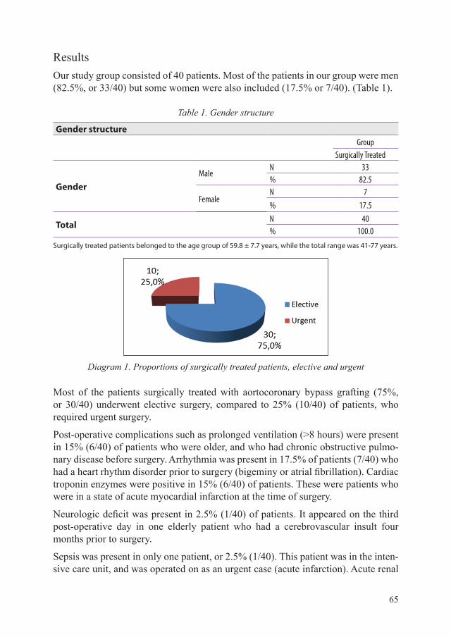
65
ResultsOur study group consisted of 40 patients. Most of the patients in our group were men (82.5%, or 33/40) but some women were also included (17.5% or 7/40). (Table 1).
Table 1. Gender structure
Gender structureGroup
Surgically Treated
GenderMale
N 33% 82.5
FemaleN 7% 17.5
TotalN 40% 100.0
Surgically treated patients belonged to the age group of 59.8 ± 7.7 years, while the total range was 41-77 years.
Diagram 1. Proportions of surgically treated patients, elective and urgent
Most of the patients surgically treated with aortocoronary bypass grafting (75%, or 30/40) underwent elective surgery, compared to 25% (10/40) of patients, who required urgent surgery.
Post-operative complications such as prolonged ventilation (>8 hours) were present in 15% (6/40) of patients who were older, and who had chronic obstructive pulmo-nary disease before surgery. Arrhythmia was present in 17.5% of patients (7/40) who had a heart rhythm disorder prior to surgery (bigeminy or atrial fibrillation). Cardiac troponin enzymes were positive in 15% (6/40) of patients. These were patients who were in a state of acute myocardial infarction at the time of surgery.
Neurologic deficit was present in 2.5% (1/40) of patients. It appeared on the third post-operative day in one elderly patient who had a cerebrovascular insult four months prior to surgery.
Sepsis was present in only one patient, or 2.5% (1/40). This patient was in the inten-sive care unit, and was operated on as an urgent case (acute infarction). Acute renal

66
failure was present in 2.5% (1/40) of the patients. Mediastinitis was present in 2.5% (1/40) of the patients. Multiple organ failure was present in 2.5% (1/40) of patients. All of these complications (sepsis, acute renal failure, mediastinitis, and multiple organ failure) were present in the same patient.
Pleural effusion was present in 7.5% (3/40) of patients. Only two patients required pleural puncture, as the third patient’s effusion was small. Pneumothorax was pre-sent in only one patient (2.5%, 1/40). This patient was drained on the fourth post-operative day.
Diagram 2. Post-operative complications
DiscussionReferral for coronary artery bypass grafting in patients with severe dysfunction of the left ventricle, and early after acute myocardial infarction, presents a dilemma to the cardiovascular team. It has long been maintained that surgery at this stage carries substantial operative risks. Recent reports, however, suggest that such estimates of operative mortality and complications may be unduly pessimistic.(15)
Patients with ischaemic heart disease and left ventricular dysfunction are typically very serious cases, with many factors of morbidity. In one study it was found that the majority of patients with ischaemic heart disease who have had myocardial in-farction are often smokers with chronic obstructive pulmonary disease, chronic re-nal insufficiency, gastrointestinal complications, and cerebrovascular insult. For this reason, medical treatment is recommended.(16-18)

67
This study supports the claim that the scale of myocardial viability determined by scintgraphy is a significant component of prognosis in patients with coronary artery disease and LV dysfunction, both in their functional status and post-operative results.(19) Most of the patients in our study who were treated surgically did so electively (75%, or 30/40 patients), while 25% (10/40) patients were urgent. Urgent surgery is itself hazardous, and increases the risk of operative mortality to 14.8%.(20-23)
Many studies have concluded that age is a factor: there is increased hospital mortal-ity in patients over 70, with left ventricular dysfunction and reduced ejection frac-tion.(24)
Revascularisation of the heart is the standard treatment for patients with severe three-vessel coronary artery disease, and LM stenosis reduces the risk of acute myocardial infarction and cerebrovascular insult. Surgical treatment of coronary artery bypass reduced the mortality rate as a result of acute myocardial infarction.(25)
In our study we had only one case of perioperative mortality (2.5%, or 1/40).
In some studies, 11% to 14.8% perioperative mortality is evidenced in patients with severe left ventricular dysfunction.(26-28)
Improved operating results are likely influenced by better myocardium protection and improved post-operative care.(29)
It is concluded that 63% (30) of patients who were surgically treated have a survival rate of 5 years.
ConclusionMyocardial revascularisation with aortocoronary bypass grafting can be performed safely in patients with severe ischemic heart disease and dysfunction of the left ven-tricle, with acceptable post-operative complications and mortality. Careful patient selection for revascularisation would confer maximum benefit to patients in this rela-tively high-risk group. We believe that improvements in cardiac anesthesia, surgical technique, extracorporeal perfusion, perioperative care, and post-operative manage-ment have contributed significantly to more encouraging outcomes.
References1. Camici PG, Rimoldi OE. Pathophysiology and diagnosis of hibernating myocardium
in patients with post-ischemic heart failure: The contribution of PET. Ann Nucl Med. 2003;(17):341-50.
2. Christakis GT, Weisel RD, Fremes SE, Ivanov J, David TE, Goldman BS, Salerno TA. Coronary artery bypass grafting in patients with poor ventricular function. Cardiovascular Surgeons of the University of Toronto. J Thorac Cardiovasc Surg. 1992;(103):1083-91.
3. DeRose JJ, Toumpoulis IK, Balaram SK. Preoperative prediction of long term survival after coronary artery bypass grafting in patients with low left ventricular ejection fraction. J Thorac Surg 2005;(129):314-21.

68
4. Dokainish H, Nguyen JS, Bobek J, Goswami R, Lakkis NM. Assessment of the American Society of Echocardiography-European Association of Echocardiography guidelines for diastolic function in patients with depressed ejection fraction: an echocardiographic and invasive haemodynamic study. Eur J Echocardiogr 2011;(12):857-64.
5. Durand DJ, Perler BA, Roseborough GS, et al. Mandatory versus selective preoperative carotid screening: a retrospective analysis. Ann Thorac Surg 2004;(78):159-66.
6. Kelder JC, Cramer MJ, van Wijngaarden J, van Tooren R, Mosterd A, Moons KG, et al. The diagnostic value of physical examination and additional testing in primary care patients with suspected heart failure. Circulation 2011;(124):2865-73.
7. Jones RH, White H, Velazquez EJ, et al. STICH (Surgical Treatment for Ischemic Heart Failure) trial enrollment. J Am Coll Cardiol. 2010;(56):490-5.
8. Lloyd-Jones D, Adams RJ, Brown TM, et al. Heart disease and stroke statistics – 2010 update: a report from the American Heart Association. Circulation. 2010;121(7):e46–e215-19.
9. Marwick TH, Raman SV, Carrio I, Bax JJ. Recent developments in heart failure imaging. JACC Cardiovasc Imaging. 2010;(3):429-439.
10. McMurray JJ. Clinical practice. Systolic heart failure. N Engl J Med. 2010;(362):228-238.
11. Eagle A, Guyton RA, Davidoff R. ACC/AHA 2004 guidelines update for coronary artery bypass surgery. Circulation. 2004;(110):340-437.
12. Eaker ED, Kronmal R, Kennedy JW, Davis K. Comparison of long-term, postsurgical survival of women and men in the coronary artery surgery study. Am Heart J. 1989;(117):71-81.
13. Youn YN, Chang BC, Hong YS, Kwak YL, Yoo KJ. Early and mid-term impacts of cardiopulmonary bypass on coronary artery bypass grafting in patients with poor left ventricular dysfunction: A propensity score analysis. Circulation. 2007;(71):1387-94.
14. Pfisterer M, Buser P, Osswald S, et al. Outcome of elderly patients with chronic symptomatic coronary artery disease with an invasive vs optimized medical treatment strategy: one-year results of the randomized TIME trial. JAMA. 2003;(289):1117.
15. Wang J, Xiao F, Ren J., et al. Risk factors for mortality after coronary artery bypass grafting in patients with low left ventricular ejection fraction. Chinese Medical Journal. 2007;120(4):317-22.
16. Shah AM, Mann DL. In search of new therapeutic targets and strategies heart failure: recent advances in basic science. Lancet. 2011;(378):704-12.
17. Topaz O, Warner M, Lanter P, Soffer A. Isolated significant left main coronary artery stenosis: Angiografic, haemodynamic and clinical findings in 16 patients. Am Heart J. 1991;(122):1308-14.
18. Schachner T, Zimmer A, Nagele G, Laufer G, Bonatti J. Risk factors for late stroke after coronary artery bypass grafting. J Thorac Cardiovasc Surg. 2005;(130):485-90.
19. Wijns W, Kolh P, Danchin N, et al. Guidelines on myocardial revascularization: Task Force on Myocardial Revascularization of the European Society of Cardiology (ESC) and the European Association for Cardio-Thoracic Surgery (EACTS). Eur Heart J. 2010;(31):2501-55.
20. Topkara VK, Cheema FH. Coronary artery bypass grafting in patients with low ejection fraction. Circulation. 2005;(112):344-50.
21. Underwood SR, Bax JJ, vom Dahl J, et al. Imaging techniques for the assessment of myocardial hibernation. Eur Heart J. 2004;(25):815-36.

69
22. Takai H, Kobayashi J, Tagusari O, Bando K, Niwaya K, Nakajima H, Yagihara T. Off-pump coronary artery bypass grafting for acute myocardial infarction. Circulation. 2006;(70):1303-6.
23. Kastelein JP, de Groot E. Ultrasound Imaging Techniques for the Evaluation of Cardiovascular Therapies. European Heart Journal. 15 Oct. 2008;29(7):849-58.
24. Nicholls SJ, Tuzcu EM, Kalidindi S, Wolski K. Effects of diabetes on progression of coronary atherosclerosis and arterial remodeling: a pooled analysis of 5 intravascular ultrasound trials. J Am Coll Cardiol. 2008;(52):255-62.
25. Lloyd-Jones D, Adams RJ, Brown TM, et al. Heart disease and stroke statistics – 2010 update: a report from the American Heart Association. Circulation. 2010;121(7):e46–e215-19.
26. Jones RH, Velazquez EJ, Michler RE, et al. Coronary bypass surgery with or without surgical ventricular reconstruction. N Engl J Med. 2009;(360):1705-12.
27. Mosterd A, Hoes AW. Clinical epidemiology of heart failure. Heart. 2007;(93):1137-46. 28. Kurki TS, Kataja M, Reich DL. Emergency and elective coronary artery bypass grafting:
comparisons of risk profiles postoperative outcomes, and resource requirements. J Cardiothorac Vasc Anesth. 2003;(17):594-7.
29. Mickleborough LL, Carson S, Tamariz M, Ivanov J. Results of revascularization in patients with severe left ventricular dysfunction. J Thorac Cardiovasc Surg. 2000;(119):550-7.
30. Mant J, Doust J, Roalfe A, Barton P, Cowie MR, Glasziou P, et al. Systematic review and individual patient data meta-analysis of diagnosis of heart failure, with modelling of implications of different diagnostic strategies in primary care. Health Technol Assess. 2009;(13):1-207.

70
DOI: 10.5644/PI2015-158-08
REVIEW UP-TO-DATE CORONARY ARTERY BYPASS GRAFT SURGERY
Sanko PandurCardiac Surgeon, Institute for Heart Disease – Clinical Center Sarajevo, Clinic for Cardiac Surgery,
Bolnička 25, 71000 Sarajevo
AbstractThe experimental and clinical efforts of Demikov and Kolesov between 1952 and 1964 rela-ted to coronary anastomosis between the internal thoracic artery and left anterior descending artery on a beating heart. Sones and Shirey (Cleveland, 1962) were pioneers of selective coronary angiography. The first coronary artery bypass grafting using a saphenous vein graft was performed by Garrett, Dennis, and DeBakey in Houston in 1964. Since that time, co-ronary surgery has become one of the most frequently performed surgical procedures in the world.
Coronarography can be considered the golden standard of diagnostics. Current supplements to coronarography are: functional assesment of the fractional flow reserve; intravascular ul-trasound; and optical coherence tomography, for the estimation of atherosclerotic plaque. When combined, these techniques provide high-quality diagnoses. Diagnostic coronaro-graphy is followed by percutaneous coronary procedures, the results of which are compara-ble to those of coronary surgery.
Prerequisites of coronary artery bypass grafting (CABG) are grafts and vascular conduits. Alternative flow into ischaemic myocardial territory takes place across these vascular con-duits. Usage of the left internal mammary artery is a proven superior standard in operative surgery, due to its better survival rates and prevention of massive myocardial reinfarction. Saphenous vein grafts are most frequently used in the myocardial territory that is not supplied by the left anterior descending artery. In 2018, we will have the final results of the ART trial, which is testing the usage of arterial grafts in coronary surgery. Preliminary results show superiority of the right internal mammary artery as a graft when compared with other arterial grafts (except the left internal mammary artery).
Nowadays, myocardial revascularisation with cardiopulmonary bypass is perfomed in 75-80% of cases. Off-pump coronary artery bypass (beating heart) surgery is performed in 20% of cases. Besides these procedures, there are less invasive procedures such as minimal inva-sive direct coronary bypass (MIDCAB), totally endoscopic coronary bypass (TECAB) and

71
myocardial laser revascularisation. Results of the perioperative application of stem cells and drugs for neoangiogenesis remain limited, in spite of some promising reports.
International guidelines from 2011, 2012 and 2013 give us an excellent framework for routi-ne daily practice, but trials such as SYNTAX I and II (2013) and FAME I and II (2013, 2015) will provide new inputs to diagnostic procedures, decision making processes, development, and the need for modern CABG.
Key words: coronary artery bypass grafting, diagnostics, conduits, OPCAB, ONCAB
Introduction After the pioneering works of Kolessov in 1964 to create anastomoses on a beating heart, the introduction of extracorporeal circulation as part of the operating arsenal has led to a progressive increase in the number of procedures. By the end of the 20th century, 500,000 operations were being performed in the United States each year. Improved diagnostic techniques, operative procedures, a system for extracor-poreal circulation, improved anesthesia techniques, intensive care, post-operative treatment, recovery, rehabilitation and resocialisation of the patient, makes CABG a resonably safe and promising procedure. In the 1990s percutaneous coronary pro-cedures, angioplasty, and stenting of the coronary blood vessels were established. These treatments are comparable with coronary surgery, and as a result the number of operations decreased in the period 1999-2003. Since then, the number of coro-nary surgerys (CABGs) has stabilised at around 300,000 in the USA and 165,000 in Europe, although this figure can be slightly higher from year to year. The main dif-ference between coronary surgery and percutaneous coronary cardiac procedures is that coronary surgery provides alternative blood flow in the ischaemic area while the PCI restitutes normal flow throughout the native vessel. By 2010, many cardiac sur-geons and cardiologists had created guidelines for the treatment of ischaemic disease individually, which caused unbalanced decision making in the treatment of coronary patients. A cooperative approach works better, as all professionals have limitations, and should have the opportunity to be complemented by others. Since then, common European guidelines were written on the principle of consensus between cardiolo-gists and cardiovascular surgeons.(1) Guidelines for the revascularisation treatment of coronary patients by cardiologists (2) and cardiovascular surgeons (3) were pub-lished in the USA in November 2011, with consensus among relevant professionals. The essence of the preamble is to guide the individual approach to each patient in terms of diagnostic conclusions and recommendations for treatment with the joint knowledge of cardiologists and cardiovascular surgeons, and the informed consent of patients for recommended procedures. In this paper, contemporary standards in coronary surgery in the fields of diagnostics, operative indications, graft choice and surgical methods will be presented.

72
Material and methods To explore these issues, we have used the MEDLINE and PubMed databases, and identified articles and studies for the period January 2009-July 2014. Keywords are: CABG, coronarography, vascular conduits, outcomes, clinical trials, fraction flow reserve, intravascular coronary imaging. Original articles, reviews, studies and guidelines were published in English. From the available literature, we have tried to present a good selection of information on diagnostic and therapeutic recommenda-tions in coronary artery bypass surgery at the beginning of the 21st century.
Epidemiology of coronary atherosclerotic disease In Bosnia and Herzegovina, morbidity and mortality from cardiovascular disease (CVD) began to rise after 1960, and rose three more times before 1990. Since the war of 1992-1995, the nation has seen a progressive increase in CVD morbidity and mortality rates. Morbidity has reached considerable proportions, with 253,367 patients diagnosed each year in the Federation, at an incidence of 10,880/100,000 (Institute of Public Health of Bosnia and Herzegovina from 2008 to 2010).
Figure 1. Epidemiology of cardiovascular mortality and morbidity in FBiH
Historical landmarks in the development of cardiac surgery Alexis Carrel, 1910. Experimental anastomosis between the descending aorta and left coronary artery of a dog.
Claude Beck, 1930, Cleveland. Indirect revascularisation, sewing mediastinal struc-tures or omentum to treat myocardial ischaemia.
Arthur Vinberg, 1946, Canada. Direct anastomosis of the left internal thoracic artery to the myocardium, resulting in the reversal of angina in the majority of patients.

73
Mason and Sones demonstrated a link between anastomosis and coronary flow by using angiography, and this operation became popular in the sixties.
Sones and Shirey, 1962, Cleveland. The first selective coronary angiography.
Longmire, 1958. First endarterectomy of coronary vessels. Poor results led to the abandonment of this technique, which included the first anastomosis of the right coronary artery (RIMA).
Garrett, Dennis, and DeBakey, 1964, Houston. First vein graft using the saphenous vein between the aorta and the LAD. The planned endarterectomy was proven to be too risky, so the operation is completed by bypass. In 1973 the patient was still alive, with an unobstructed bypass.
Vladimir Demikhov, 1952, Moscow. LIMA-LAD anastomoses on the beating heart of a dog.
Gibbon, 1952. Built the IBM Heart-Lung machine, the first successful use of extra-corporeal circulation.
Kolessov, 1964, Leningrad. The first anastomosis LIMA-LAD through a left thora-cotomy on a beating heart. A report on the experience of the 6 patients was published in 1967, in the American Journal of Surgery.
Green et al. Recommend the use of extracorporeal circulation in cardiac operations.
Bailey and Hirose, 1968. Recommended LIMA-LAD anastomosis and the use of a surgical loupe when creating anastomoses.
Rene Favaloro, 1968, Cleveland. Announced the results of 15 patients treated with aorto-coronary bypass graft using the saphenous vein.
Dudley Johnson, 1968. Published the first serious results of 301 patients treated in February 1967 with aorto-coronary bypass using saphenous vein grafts. 40% of pa-tients received two bypasses. Based on the results of the first, five rules were rec-ommended: do not limit the graft with the proximal portion of the artery; avoid the diseased part of the artery; the vein graft must be long enough, and should be grafted to the distal part of a healthy artery; always work end-to-side when performing anas-tomosis, and make sure the area is dry, as vascular anastomosis cannot be completed in a bloody environment; do not let the haematocrit fall below 35.
Thanks to the results and recommendations of Green, Loop and Crondina, LIMA becomes the graft of choice for coronary artery bypass surgery in the 1980s.
Carpantier, 1972, Paris. Proposed the radial artery as the preferred graft. This idea was quickly abandoned due to spasticity of the graft, but has been revisited in the past 15 years.
Denton Cooley et al, 1957, Houston. The first closure of a post-infarction ventricular septal defect.

74
Cardiac catheterisation – coronary angiography Coronary angiography is the gold standard in the diagnosis of coronary artery dis-ease, and the final level of its detection. It is also an important tool to assess the severity of the disease, and indication for revascularisation methods. The advantage of coronary angiography compared to other radiological methods is the possibility of simultaneous therapeutic intervention.
Negotiated assessment of coronary artery involvement divides segmental distribu-tion of the coronary artery system into 16 or 27 segments. Division into 16 segments is widely accepted, and, most importantly, forms the basis of angiographic analysis and evaluation of the results of the SYNTAX study.(4)
Figure 2. AHA segmental division of an artery
Angiography provides an overview of the frontal flow blood vessels. Stenosis can be concentric and eccentric, as well as in the lumen of the vessel. It was observed that angiographic imaging does not correlate with cross-sectional narrowing.
Figure 3. Relationship between angiographic and cross-sectional stenosis

75
Fractional flow reserve (FFR) FFR is the preferred method in modern cath labs for the evaluation of coronary stenosis5. FFR is defined as the maximum myocardial flow distal to the stenosis divided by the value at which there is no epicardial stenosis. FFR can be considered separately in relation to the myocardium, epicardial coronary artery, and collateral flow. The formula for FFR is simplified as pressure distal to the stenosis divided by the pressure proximal to the stenosis. In normal conditions, the value is 1.0. FFR. A value of 0.6 means that the maximum myocardial flow through the stenosis is only 60% of what would be expected in the absence of stenosis. An FFR of 0.9 after percutaneous coronary intervention means that 90% of the maximum flow has been achieved. FFR has a high reproducibility because it does not show any significant changes in the pulse pressure and LV contractility.
Figure 4. Fractional reserve blood flow
There is no strict correlation between myocardial stress tests and FFR. An FFR of less than 0.75 was defined as significant stenosis with inducible myocardial ischae-mia. This implies the following values: sensitivity 88%, specificity 100%, positive predictive value of 100%, and accuracy of 93%. Measurement of FFR is now consid-ered an extremely important indication tool, and is essential during coronary angiog-raphy. Tonino and his colleagues in the FAME study inaugurate the hypothesis that PCI with the measurement of FFR is superior to a conventional angiographic study in patients with three-vessel coronary artery disease. An FFR of less than 0.80 is an indication for intervention, and is of great importance for many studies.
Intravascular ultrasound (IVUS) IVUS catheters reflect sound waves to visualise the arterial wall-dimensional com-puted tomography in a format that is analogous to a histological section of the

76
vessel. IVUS uses a significantly higher frequency (20-40 MHz) than that of the non-invasive echocardiography (2-5 MHz). This ensures high image resolution and expansion of the limited penetration of sound waves (4-8mm from the tip of the catheter). Interpretation of the IVUS display begins with the identification of the intima, media and adventitia structures in the blood vessel. Within these differenti-ated boundaries atherosclerotic plaque can form, develop, and lead to complications. IVUS provides electronic caliper measurements of dimensions and restriction zones, determining maximum stenosis and assessment of the segment located proximal and distal to the lesion. Plaque with extensive infiltration of lipids has a low reflectivity echo, while plaques with predominantly fibrous tissue are more echogenic structures. Calcified plaques have a bright border covered by a dark shadow that extends radi-ally outwards. IVUS can detect calcified plaque in 60-80% of lesions, whereas only half these amounts can be detected by angiography. Clinical effectiveness of IVUS is also reflected in the identification of thrombotic masses and artery dissection, with or without intervention. IVUS provides accurate aterectomy, a clear display of stents and their position in relation to the arterial wall, and evaluation of in-stent stenosis.(6)
Figure 5. Intravascular ultrasound LMC
Optical coherence tomography (OCT) OCT generates real-time tomographic imaging by reflecting infrared light. This use of optical echo can be considered as an optical analogue version of IVUS. The big-gest advantage of a display based on the reflection of infrared light is that it has a higher resolution than conventional ultrasound. The intravascular OCT system consists of optical machinery which emits and receives infrared signals, a catheter for introducing light, a fibre optic catheter, and a computer processor. Experimental studies have shown that OCT can show structures measuring 10-50 microns, while

77
IVUS visualises structures of 150-200 microns. The diagnostic accuracy of OCT in-formation has proved effective, with a sensitivity of 79% and specificity of 98% for fibrotic plaques. The importance of OCT is particularly demonstrated in evaluation of the thickness of the fibrous cap of atherosclerotic plaque in the coronary arteries.(7)
Figure 6. OCT with histology showing vessels
Coronary CT The sensitivity and specificity of CT coronary angiography is over 95%, depending on the number of detectors (64, 128 or 256). However, at its current stage of tech-nological development CT coronary angiography does not offer the possibility of si-multaneous percutaneous coronary procedures. Heart rate must be less than 65 beats per minute. An elevated calcium score greater than 400 complicates the interpreta-tion of stenosis of the coronary arteries, and reduces the specificity of the examina-tion. The CT technique has excellent negative predictive value, ie. it can effectively exclude coronary disease.(8) It has become the method of choice in evaluating the patency of coronary stents and of grafts in bypass surgery. A bypass generally creates a wider lumen, which has less mobility during the cardiac cycle than the coronary arteries. Coronary anomalies are indications of choice for this type of test.
Treatment of ischaemic heart disease There are three modalities of treatment: optimal medical therapy, percutaneous coro-nary procedures, and surgical treatment.
Optimal medical therapy (OMT) OMT is the control of risk factors and the use of anti-ischaemic drugs as a non-aggressive method of treatment.(9)

78
Percutaneous coronary procedures In December 2011, recommendations were published for the first time to differenti-ate when percutaneous coronary intervention should be applied, and when surgi-cal intervention is more appropriate. Results and findings from individual and ran-domised trials show the following (10):
- PCI reduces the incidence of anginal disorders- PCI has not been shown to improve survival in stable patients with OMT - PCI has demonstrated its value in acute coronary syndrome - PCI did not reduce the long-term risk of myocardial infarction - PCI may increase the short-term risk of myocardial infarction - PCI can be a successful complement to CABG in hybrid procedures - PCI can be used in dealing with graft stenosis
Coronary artery bypass grafting (CABG) Indications for surgery
Since December 2011, indications for surgery have been determined by guidelines for surgical myocardial revascularisation. These are based on results and scores from the EBM SYNTAX study.(11) This paper will present only the class IA indications, about which there is consensus for surgical treatment (12):
- Left main stenosis (narrowing of the left main coronary artery) - Left main equivalent (proximal LAD and Cx) - Three-vessel disease - Two-vessel disease with proximal LAD and EF<50% - One-vessel and two-vessel disease without involvement of the LAD, and with a
large territory at risk - Severe refractory AP with full OMT - Failed PCI with haemodynamic, ischaemic ventricular dysfunction or electrical
instability - Persistent or recurrent ischaemia refractory to OMT with acceptable anatomy and
significant territory at risk - A condition that requires surgical repair post AMI, VSD, or acute mitral
regurgitation - Cardiogenic shock in patients younger than 75 years who have ST elevation,
LBBB, or posterior AMI within 18 hours of onset of symptoms. - Life-threatening ventricular arrhythmias in the presence of LM stenosis >50%,
and/or vessel disease - Impaired left ventricular function with LM and two-vessel or three-vessel disease - Life-threatening ventricular arrhythmias with LMS or LM equivalent - Foreign body in a critical position - Haemodynamic instability and coagulopathy without/with previous sternotomy - Previous CABG procedures

79
- Previous occlusion and bypass, and class I coronary artery disease
Methods of surgical treatment 1. Coronary artery bypass with the use of extracorporeal circulation (on-pump
CABG/ONCAB) (13) 2. Coronary artery bypass without the use of extracorporeal circulation (off-pump
CABG/OPCAB) (14)3. On-pump beating heart CABG 4. Minimally invasive coronary surgery (15)5. Transmyocardial revascularisation (16)6. CABG with simultaneous instillation of stem cells 7. Hybrid revascularisation (17)
The tactics and strategy of surgical intervention are focussed on two objectives: to achieve optimal revascularisation; and optimal intraoperative myocardial protection.
Prerequisites for vascular conduits (grafts) in coronary surgery Internal mammary artery (LIMA)
Using LIMA grafts to bypass the LAD provides proven superiority in short- and long-term survival.(18,19) Excellent biological characteristics, incomparable long-term patency and improved clinical results are some outcomes of LIMA graft use, which has become the first choice for myocardial revascularisation. LIMA shows considerable resistance to the development of atherosclerosis as well as to the pos-sible violation of the endothelium of the saphenous vein during preparation. Electron microscopy studies do not show thrombogenic intimal defects that are commonly seen in vein grafts. Non-fenestrated internal elastic lamina is likely to inhibit cel-lular migration by preventing intimal hyperplasia. The middle layer is thin, with few muscle cells showing a decreased proliferative response to mitogens and pulsatile mechanical force. The endothelium of the LIMA is a significant autoproducer of vasodilators such as nitric oxide and prostacyclin. The LIMA showed a favorable response to certain drugs used perioperatively. It is vasodilated under the influence of milrinone and does not tolerate vasoconstriction in response to norepinephrine. Nitroglycerine also causes LIMA vasodilation. Endogenous secretion of vasodilators carried in the blood stream has a positive effect on the coronary vasculature distal to the anastomosis. It is important to note significant remodelling of the LIMA repre-sents adaptation to flow demands. The consequence may be an increase in the lumen of the LIMA to 1.5 to 2.5 times its normal size. Two basic preparation techniques for LIMA are pedicled and skelotonised harvesting. In the case of using both internal mammary arteries (LIMA and RIMA), skeletonised harvesting is preferred because it provides better opportunities for sternum healing, especially among women, obese patients and patients with diabetes. RIMA is somewhat more prone to atherosclerosis compared to LIMA in patients of an advanced age (28% of 6%). RIMA is also sensi-tive to the effects of medication, which is important as it is more commonly used

80
as a free graft. The patency rate of skeletonised harvesting is undeniably high; more than 90% of LIMA grafts after 10 years, which is superior to that of all other grafts.
Figure 7. Pedicled and skeletonised LIMA
The radial artery
Use of radial artery grafts was first described in 1970. Artery spasm was common, and fixed by mechanical dilatation. Initial results were poor, so this graft was quickly abandoned. Akar postulated that the preparation technique is responsible for spas-ming and occlusion of the graft. Pedicled harvesting and pharmacological prepara-tion of the graft was recommended, making this type of graft gain importance. In some studies it proved superior to the saphenous vein graft, although is still open to discussion.(20) Graft patency was 90% for 9 months, and 65% after 10 years.
Gastroepiploic artery
Use of this artery is less common, although it can be used in redo surgery in the ab-sence of other acceptable grafts, or as a second or third arterial conduit in total arte-rial revascularisation of the heart. Preparation of the graft requires a longer operating time, increasing the possibility of abdominal complications. There is currently no consensus on the long-term benefits of total arterial revascularisation.
Great saphenous vein (GSV)
Despite the identification of its numerous negative characteristics, the GSV remains the most commonly used vein for grafts in bypass surgery, due to its ease of prepa-ration, rapid availability of sufficient length, resistance to spasm, and satisfactory long-term results.(21) Early use of aspirin and statins reduces occlusion of grafts in the first year after surgery. In the future, gene therapy is likely to be able to modify the venous vascular endothelium to avoid the development of intimal hyperplasia. Vein harvesting is performed by either open, semi-open, or endoscopic methods. The open method traumatises the skin, and has higher levels of complications and post-operative wound pain. The semi-open method, with its smaller cutdowns, can re-duce pain and wound complications but increases surgical manipulation of the graft.

81
Minimally invasive preparation of the GSV with endoscopic techniques is gaining popularity, although its effect on the morphology of the vein’s endothelial structure, function and long-term patency are questionable.(22) Conversion of endoscopic to open technique occurs in 5 to 7% of cases. Air and fat embolisms of the lungs have been reported. Long-term patency of the venous graft appears low, as 20% of grafts are occluded in the first year. Annual graft occlusion occurs at a level of 2%, mean-ing that after 10 years, 60% of grafts are patent, although recent studies using a non-touch preparation technique refute these results.(23,24,25)
Surgical revascularisation with the use of extracorporeal circulation (On-pump CABG/ONCAB) Preparation for cardiopulmonary bypass ends cannulation of the aorta and right atrium at a typical site. After this, lines for cardioplegia and venting are installed. The aorta is crossclamped, a cardioplegic solution is given, and the heart is arrest-ed in diastole. We can consider this the conventional approach to bypass surgery. Cardiopulmonary bypass now constitutes 75-80% of all bypass procedures in the world, and is seen as a proven treatment.(26)
Figure 8. CABG with the use of extracorporeal circulation
Figure 9. Conventional CABG (LIMA and SVG) and total arterial revascularisation

82
CABG without the use of extracorporeal circulation (off-pump CABG/OPCAB) CABG procedures have been documented to improve the patient’s chance of surviv-al and quality of life, and to eliminate symptoms. The number of OPCAB procedures was increasing until 2003, but has since stabilised at a level of 20%. The aims of these procedures are: graft patency at least equivalent to that of conventional techniques; to reduce morbidity and mortality, particularly in high-risk groups; ensure a quick re-turn to good functional capacity; and economic benefit. Selection of patients depends primarily on the technical skills of surgeons, and angiographic characteristics of the coronary anatomy. This includes patients with a satisfactory caliber of vessels, focal stenosis of the target vessel sufficiently distant from the AV groove, and vessels for which there is no probability of endarterectomy. High-risk patients who are likely to benefit from OPCAB are those with: severe LV dysfunction; renal insufficiency; severe atherosclerosis of the aorta; or COBP. Haemodynamically or electrically un-stable patients are often unable to tolerate the manipulation OPCAB requires, and are generally not considered candidates for this type of surgery. Maintaining tem-perature control is of great importance. There are conflicting reports about the safety of immediate extubation after surgery.
Optimal exposure of vessels is of paramount importance. The LIMA seam contrib-utes to initial exposure, while later techniques include additional seams, and are perfected by the use of mechanical stabilisers. The order of anastomosis creation in OPCAB surgery is crucial. Collateralised vessels are grafted before those that give collaterals. First, an anastomosis LIMA-LAD is created, then the proximal anasto-mosis, then the distal, followed by grafts, in this order: diagonal artery; RCA; PDA; PLA; first OM; and RIM.
Despite the fact that OPCAB eliminates CPR, it does not eliminate all the compli-cations that occur in coronary surgery, although some are significantly decreased. Non-touch aorta OPCAB avoids manipulation of the aorta, and reduces the pos-sibility of neurological deficit. Controlled studies showed no difference in mortality after one to three years. Randomised studies have shown a decreased need for blood transfusions, inotropic support, fewer respiratory infections, and a lower incidence of atrial fibrillation. However, we did not find statistical significance of differences in: post-operative ischaemia; use of IABP; ARF; wound infection or mediastinitis; or recurrence of angina. ONCAB conversions are reported to occur in an average of 8% of patients, with a subsequent mortality rate of 6-15%.
Surgical revascularisation with the use of extracorporeal circulation on the beating heart (on-pump beating heart CABG) Extracorporeal circulatory support for beating heart surgery is a useful tool. Unstable patients with: AMI; barely reduced EF; acute coronary occlusion; and coronary dis-section leading to cardiovascular collapse, are all ideal candidates for this type of

83
operation, where it is possible to simultaneously create anastomoses on a beating heart. Easier manipulation of the heart and greater control of blood loss during sur-gery are some advantages of extracorporeal circulation. This technique is most likely the future of cardiac surgery in severe cases.
Minimally invasive myocardial revascularisation (MIDCAB) MIDCAB is an insufficiently defined term, referring mainly to the exclusion of CPB surgical standards. Other important features are the practice of median sternotomy as a surgical approach, and the use of endoscopic methods of operation and preparation of grafts. OPCAB is partially MIDCAB, where a median sternotomy is the preferred approach. MIDCAB began as a single LIMA-LAD anastomosis through an antero-lateral thoracotomy, with the use of endoscopy in the preparation of grafts to avoid over-expansion of the ribs in the approach. Use of the harmonic scalpel in the prepa-ration of grafts represents a particular development in this technique. MIDCAB has been further developed via computer softwere that enables telemanipulation (robotic surgery). This is used in total endoscopic coronary artery bypass (TECAB) graft-ing. It is possibly the least invasive approach to revascularisation on a beating heart. Operators need access ports for scope and manipulation. The transition from the limited indication of the total endoscopic revascularisation will require more time, due to limitations in the adequacy of instruments, software, and computers, and the steep learning curve for surgeons. Despite its high technology, TECAB remains technically demanding, and is initially used on an arrested heart with femorofemoral bypass, aortic endo-clamping and cardioplegic arrest. It is shown that initial studies exhibit the same graft patency as standard procedures. The level of conversion to the standard procedure is high. Ignoring the surgical exposure in these experimental techniques in situations of three-vessel disease has led to complications, and ration-alisation of TECAB on one-vessel or two-vessel disease. Endoscopic preparation of the graft is introduced to minimise trauma, reduce wound complications, and add
Figure 10. Minimally invasive coronary bypass and totally endoscopic coronary
revascularisation

84
cosmetic benefits. Endoscopic preparation can be used for the radial artery, great saphenous vein, and less frequently-used grafts. The procedure is somewhat longer, and the level of conversion to the open method described is from 0-22%. The risk of early complications is significantly reduced, although the quality of the graft after these procedures has been questioned due to the appearance of some of its less fre-quent complications (such as air embolisms or thromboembolisms).
Transmyocardial revascularisation laser (TMR) Despite the success of various therapeutic options in the treatment of coronary ar-tery disease, there are a significant number of patients with refractory angina due to diffuse coronary disease who are not candidates for OMT, PCI or CABG. One technique developed for this group of patients is TMR. Three types of laser are used: carbon dioxide; holmium.Yag; and xenon chloride. The idea is to create vascular channels through the entire thickness of the left ventricular wall at a certain distance from the main blood vessels. Twelve to 20 channels should be created in this way. The mechanism is not completely clear, but it is believed that angiogenesis with redistribution of blood within the wall of the myocardium plays a role in freeing patients from angina and improving left ventricular perfusion. Patients with poor ejection fraction and acute ischaemia are not candidates for this technique. The cur-rent mortality rate is about 5%, and the one-year survival rate is 84-95%. Reduction of angina is seen more frequently in patients treated with TMR compared to those receiving OMT. Five-year survival was 65% for patients treated with TMR, and 52% for patients treated with OMT. TMR can be used as an adjunct to those CABG procedures in which one cannot achieve optimal revascularisation due to diffuse coronary disease.
CABG with instillation of angiogenic substances for angiogenesis and stem cells Experimental in vitro studies have shown that some proteins (VEGF, FGF, FGF1, FGF2) have angiogenic potential, and angiogenesis and new vascular channels have been observed in experiments on animal models. In limited clinical studies, these pro-teins were applied during CABG surgery. After 16 months, significantly better perfu-sion was observed in nuclear studies. Another option for bypass is the use of adult or embryonic stem cells. Embryonic angioblasts were found to be more potent in the formation of the primary capillary plexus. Cardiomyocytes are terminally differenti-ated, and can not regenerate. However, it was shown that a limited number of cardio-myocytes may be regenerated using stem cells. Clinical studies on a limited number of patients treated with stem cells during CABG showed functional improvement.
Hybrid procedures – Integrated coronary revascularisation In the case of older patients, diffuse atherosclerosis, significant comorbidity, and low ejection fraction are often present, and hybrid operations can have a major

85
effect in terms of relieving anginal disorders and increasing life expectancy. Hybrid operations are a combination of minimally invasive direct coronary artery bypass (MIDCAB) – incorporating LIMA-LAD via the left thoracotomy – and percutane-ous coronary interventions (PCI). The need to perform these procedures has led to the creation of a team of cardiologists and cardiovascular surgeons and a technically-equipped hybrid operating room, in which it is possible to undertake both types of procedure either simultaneously or in quick succession.
Outcome Operative mortality ranges from 2.6-4% (27), and is gradually falling due to constant improvement of surgical skills, myocardial protection and anesthetic techniques. In a study by Mack et al in 2002, the mortality rate in a group of 51,353 patients was 2.24%. Causes of death were: heart failure, 65%; neurological deficits, 7.3%; bleed-ing, 7%; respiratory failure, 5.5%; and dysrhythmias, 5.5%. Operative morbidity related to the development of myocardial infarction appears in 1-5% of patients with a poor prognosis for long-term survival. Post-operative myocardial dysfunction, pre-sented as syndrome of low cardiac output (28), has also been a common problem, and requires inotropic support. Its incidence is 9%, and in severe cases treatment requires an IABP.(29) LOS is a marker of increased operative mortality. Independent predictors of LOS in patients are: a poor EF<20%, reoperation, vital operation, fe-male gender, diabetes (30,31), age greater than 70 years (32,33), left main disease (34), recent AMI, and three-vessel disease.
Discussion Cardiovascular incidents remain the leading cause of morbidity and mortality in both the developed world and developing countries. In Europe and the USA, there have been around 2.5 million hospital admissions due to acute coronary syndrome. This large number of cases continues to dictate the efforts of the health system to preserve the population’s quality of life and increase its life expectancy. Although the intro-duction of percutaneous cardiac procedures has provided a remarkable reduction in the number of candidates for coronary surgery, the overall situation has not changed greatly. Vast improvements have occurred in the field of diagnosis of coronary artery disease, and in standardisation of indications. On the basis of angiographic charac-teristics, the SYNTAX study demonstrated that patients with higher scores are candi-dates for coronary surgery, and that compared with PCI, this surgery has lower rates of mortality and complications. The SYNTAX Score II combines the angiographic and clinical characteristics of a patient, and contributes to a much better preopera-tive assessment. Techniques for functional assessment of coronary stenosis (FAME study) leads to a further qualitative improvement in the indication for a particular type of treatment (OMT, PCI, or CABG). Intravascular visualisation techniques of atherosclerotic plaque further contribute to the knowledge of coronary artery dis-ease, and have an impact on clinical decision making. Technological development

86
in surgery occurs continuously, so that the improved capabilities of extracorporeal circulation, haemodynamic monitoring and the development of specific endoscopic systems constantly make surgery easier and more confident. The choice of graft in coronary surgery today is quite clear: the LIMA graft is used in more than 95% of coronary surgeries. This is followed by the RIMA graft, whose usability and quality is likely to be confirmed upon completion of the ARTS 2018 study. A venous graft with meticulous surgical preparation remains an essential factor in long-term paten-cy using antiagregants and statins. Radial artery and gastroepiploic artery applica-tion is useful in the case of subocclusion or native vessel occlusion, as the competing flow of native vessels leads quickly to spasm and thrombosis of the graft. Improving the results of treatment outcomes for coronary patients is based on an individual approach, and includes assement of indications, consideration of diagnostic possi-bilities and careful selection of treatment methods. Comorbidities and an ageing population can be seen to take treatment results to their limits in term of morbidity and mortality in coronary surgery. In Bosnia and Herzegovina it is necessary to make an extra effort for the early detection of coronary artery disease, as the population of patients with ischaemic heart disease is significantly younger compared to Western European countries and the USA.
References1. Wijns W, Kolho P, Danchin N, Di Mario C, et al. ESC/EACTS guidelines; Guidelines on
Myocardial Revascularisation. Eur Heart J. 2010;(31):2501-55. 2. Levine GN, Bates ER, Blankenship JC, Bailey SR, Bittl JA, Čerček B, et al. 2011 ACCF/
AHA/SCAI Guideline for Percutaneous coronary intervention: A Report of Cardiology Foundation/American Heart Association Task Force on Practice Guidelines and the Society for Cardiovascular Angiography and Interventions. Circulation. 2011;(124):e574-e651.
3. Hillis LD, Smith PK, Anderson JL, Bittl JA, Bridges CR, Byrne JG, et al. 2011 ACCF/AHA Guideline for Coronary Artery Bypass Graft Surgery: A Report of the American College of Cardiology Foundation / American Heart Association Task Force on Practice Guidelines. Circulation 2011. http://circ.ahajournals.org/by guest on March 22, 2012.
4. Mohr FW, Morice MC, Kappetein AP, Feldman TE, Stahle E, Colombo A, et al. Coronary artery bypass surgery versus percutaneous coronary intervention in patients with three-vessel disease and left main coronary disease: 5-year follow-up of a randomized, clinical trial SYNTAX. Lancet. 2013;(381):629-38.
5. De Bruyne B, Pijls NHJ, Kalesan B, Barbato E, Tonino PAL, Piróth Z, et al. Fractional Flow Reserve-Guided PCI versus Medical Therapy in Stable Coronary Disease. N Engl J Med. 2012;(367):991-1001. (FAME 2 Trial Investigators)
6. Garcia-Garcia HM, Costa MA, Serruys PW. Imaging of coronary atherosclerosis: Intravascular Ultrasound. N Engl J Med. 2010;(3):2456-69.
7. Terashima M, Kaneda H, Suzuki T. The role of optical coherence tomography in coronary interventions. Korean J Intern Med. 2012;27(1):1-12.
8. Sarno G, Decraemer I, Vanhoenacker PK, de Bruyne B, et al. On the inappropriateness of non invasive multidetector computed tomography coronary angiography to trigger

87
coronary revascularisation: a comparison with invasive angiography. JACC Cardiovasc Interv. 2009;(2):550-7.
9. Boden WE, O’Rourke RA, Teo KK, et al. COURAGE Trial Investigators. Impact of optimal medical therapy with or without percutaneous coronary intervention on long-term cardiovascular end points in patients with stable coronary artery disease (from the COURAGE Trial). J Am Coll Cardiol. 2009;104(1):1-4. [PMID: 19576311]
10. Simard T, Hibbert B, Ramirez FD, Froeschl M, Chen YX, O’Brien ER. The Evolution of Coronary Stents: a Brief Review. Can J Cardiol of 2014;30(1):35-45.
11. Farooq H, van Klaverern D, Steyerberg EW et al. Anatomical and clinical characteristics to guide decision making between coronary artery bypass surgery and percutaneous coronary intervention for individual patients: development and validation of SYNTAX score II. Lancet. 2013;(381):639-50.
12. Montalescot G, Sechtem U, Achenbach S, Andreotti F, Arden C, Budai A, et al. 2013 ESC Guidelines on the management of stable coronary artery disease: The Task Force on the management of stable coronary artery disease of the European Society of Cardiology. N Engl J Med. 2013;(34):2949-3003.
13. Greason KL, Sundt III TM, Myocardial Revascularisation with Cardiopulmonary Bypass. In: Cohn LH, editor. Cardiac Surgery in the Adult. Elsevier Saunders. 4th-Ed; 2012. p. 479-503.
14. Halkos ME, Puskas JD. Myocardial Revascularisation without Cardiopulmonary Bypass. In: Cohn LH, editor. Cardiac Surgery in the Adult. Elsevier Saunders. 4th-Ed; 2012. p. 505-20.
15. Holzhey DM, Rastan AJ, Falk V, Mohr FW. Minimally Invasive Myocardial Revascularisation. In: Cohn LH, editor. Cardiac Surgery in the Adult. Elsevier Saunders. 4th-Ed; 2012. p. 553-66.
16. Horvath KA, Zhou Y. Transmyocardial Laser Revascularization and Extravascular Angiogenetic Techniques to Increase Myocardial Blood Flow. In: Cohn LH, editor. Cardiac Surgery in the Adult. Elsevier Saunders. 4th-Ed; 2012. p. 585-601.
17. Popma JJ, Nathan S, Hagberg RC, Khabbaz KR. Hybrid myocardial revascularisation: An integrated approach to coronary revascularization. Cath and Cardiovasc Int. 2010;75(1):28-34.
18. Adams C, Burns DJP, Chu MWA, Jones AM, Shridar J, et al. Single-stage hybrid coronary revascularisation with long-term follow-up. Eur J Cardiovasc Surg. 2013:234-41.
19. Conradi L, Treed H, Reichenspurner H. Hybris coronary revascularisation: a task for the true heart team. Eur J Cardiovasc Surg. 2014;45(3):443-4.
20. Taggart DP. Current status of arterial grafts for coronary artery bypass grafting. Ann Cardiovasc Surg. 2013;2(4):427-30.
21. Cheng A, Slaughter MS. How I choose conduits and configure grafts for my patients – rationales and practices. Ann Cardiovasc Surg. 2013;2(4):527-32.
22. Barner HB. Conduits for Coronary Bypass Strategies. Korean J Thorac Cardiovasc Surg. 2013;(46):319-27.
23. Li J, Liu Y, Zheng J, et al. The patency of sequential and individual vein coronary bypass grafts: a systematic review. Surg. 2011;(92):1292-8.
24. Bello SOZ, Peng EWK, Sarkar PK. Conduits for coronary artery bypass surgery: the quest for second best. J Cardiovasc Med. 2011;(12):411-21.

88
25. Dreifaldt M, Mannion JD, Bodin L, Olsson H, Zagozdzon L, Souza D. The no-touch saphenous vein as the preferred second conduit for coronary artery bypass grafting. Surg. 2013;(96):105-11.
26. Shroyer AL, Grover FL, Hattler B, et al. On-pump versus off-pump coronary artery bypass surgery. N Engl J Med. 2009;(361):1827-37.
27. Ferguson TB Jr. Mortality in Coronary Artery Bypass Grafting: What’s next? Circulation. 2012;(125):2409-11.
28. Nagendran J, Norris CM, Graham MM, Ross DB, MacArthur RG, Keiser TM, et al. Coronary revascularisation for patients with severe left ventricular dysfunction. Surg. 2013;(96):2038-44.
29. Hamad MAS, van Straten AHM, Shonberger JPAM, ter Woorst JFJ, de Wolf AM, Martens EJ, van Zundert AAJ. Preoperative ejection fraction as predictor of survival after coronary artery bypass grafting: comparison with a matched general population. Journal of Cardiothoracic Surgery. 2010;(5):29.
30. Aggarwal B, Goel S, Sabik JF, Shishehbor MH. The FREEDOM trial: in appropriate patients with diabetes and multivessel coronary artery disease, CABG Beats PCI. Cliv Clin J Med. 2013;(80):515-23.
31. Farkouh ME, Domanski M, Sleeper LA, et al. FREEDOM Trial Investigators. Strategies for multivessel revascularisation in patients with diabetes. N Engl J Med. 2012;(367):2375-84.
32. Yanagawa B, Algarni KD, Yau TM, Brister SJ. Improving results for coronary artery bypass graft surgery in the elderly. Eur J Cardiovasc Surg. 2012;42(3):507-12.
33. Saito A, Motomura S, Miyata H, Takamoto S, Kyo S, Ono M, Database Organisation JC. Age-specific risk stratification in 13488 isolated coronary artery bypass grafting procedures. Interact Cardiovasc Thorac Surg. 2011;(12):575-81.
34. Ngaage DL, Sogliano F, Tang A. Early and late survival after surgical revascularisation for left main coronary artery stenosis in the stent era. Am J Cardiol. 2012;(19):134-8.

CIP - Katalogizacija u publikacijiNacionalna i univerzitetska bibliotekaBosne i Hercegovine, Sarajevo 616.12(063)(082) SCIENTIFIC Symposium Acquired HeartDiseases, World Heart Day (2014 ;Sarajevo) Proceedings / Scientific Symposium AcquiredHeart Diseases, World Heart Day, Sarajevo,September 29, 2014 ; editor-in-chief SenkaMesihović-Dinarević. - Sarajevo : Akademija naukai umjetnosti BiH = Academy of Sciences and Arts ofBosnia and Herzegovina, 2015. - 88 str. : ilustr.; 24 cm. - (Special editions ; vol. 168.Department of Medical Sciences ; vol. 45 /Academy and Arts of Bosnia and Herzegovina) Na prelim. nasl. str.: Zbornik radova. -Bibliografija i bilješke uz tekst. ISBN 978-9958-501-98-21. Mesihović-Dinarević, Senka. - I. Naučnisimpozij Stečene bolesti srca, Svjetski Dan srca(2014 ; Sarajevo) vidi Scientific SymposiumAcquired Heart Diseases, World Heart Day (2014 ;Sarajevo)COBISS.BH-ID 22051078