Embed Size (px)
Citation preview
ACP Internal Medicine 2016Clinical Pearls Hematology
Carrie Thompson, MDMayo Clinic College of Medicine
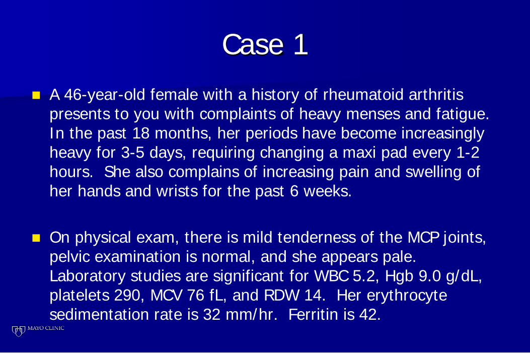
Case 1 A 46-year-old female with a history of rheumatoid arthritis
presents to you with complaints of heavy menses and fatigue. In the past 18 months, her periods have become increasingly heavy for 3-5 days, requiring changing a maxi pad every 1-2 hours. She also complains of increasing pain and swelling of her hands and wrists for the past 6 weeks.
On physical exam, there is mild tenderness of the MCP joints, pelvic examination is normal, and she appears pale. Laboratory studies are significant for WBC 5.2, Hgb 9.0 g/dL, platelets 290, MCV 76 fL, and RDW 14. Her erythrocyte sedimentation rate is 32 mm/hr. Ferritin is 42.
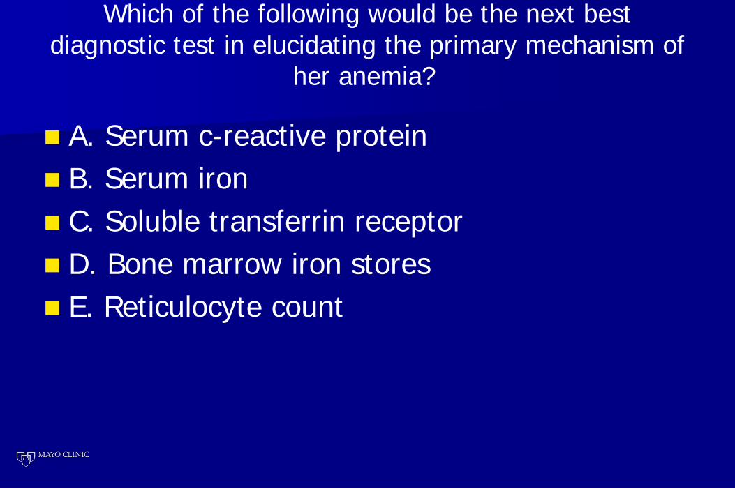
Which of the following would be the next best diagnostic test in elucidating the primary mechanism of
her anemia?
A. Serum c-reactive protein B. Serum iron C. Soluble transferrin receptor D. Bone marrow iron stores E. Reticulocyte count

Answer
C. Soluble transferrin receptor
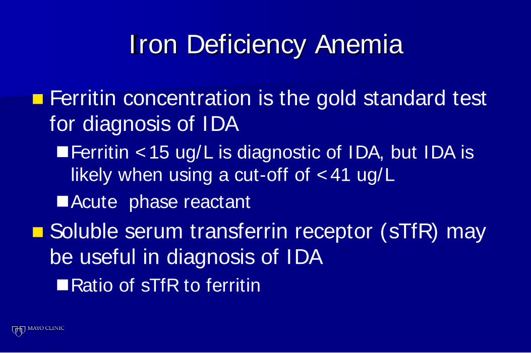
Iron Deficiency Anemia
Ferritin concentration is the gold standard test for diagnosis of IDA Ferritin <15 ug/L is diagnostic of IDA, but IDA is
likely when using a cut-off of <41 ug/LAcute phase reactant
Soluble serum transferrin receptor (sTfR) may be useful in diagnosis of IDA Ratio of sTfR to ferritin
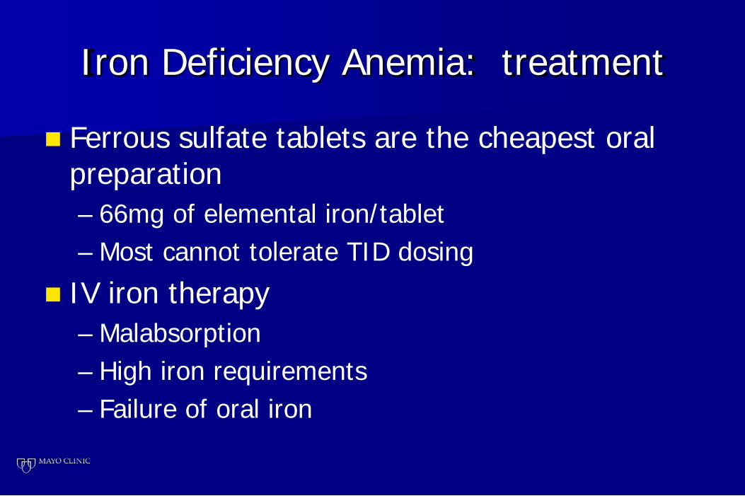
Iron Deficiency Anemia: treatment
Ferrous sulfate tablets are the cheapest oral preparation – 66mg of elemental iron/tablet – Most cannot tolerate TID dosing
IV iron therapy– Malabsorption– High iron requirements – Failure of oral iron
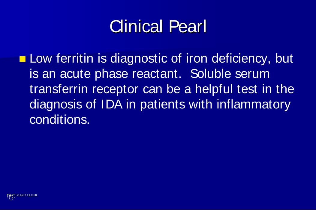
Clinical Pearl
Low ferritin is diagnostic of iron deficiency, but is an acute phase reactant. Soluble serum transferrin receptor can be a helpful test in the diagnosis of IDA in patients with inflammatory conditions.

Case 2
A 73 year old Caucasian male reports for a routine physical examination. Past medical history is significant for hypertension and osteoarthritis, and he is taking metoprolol, naproxen, and a multivitamin. CBC shows a normal WBC and platelets, but hemoglobin 11.8 g/dL and MCV of 106. B12 level is normal.

Which one of the following is the most likely cause of the anemia?
A. Anemia of chronic disease B. Folate deficiency C. Renal disease D. Alcohol use E. Thalassemia trait
Answer
D. Alcohol use

Macrocytic AnemiaMedication use
or alcohol excess?
NO
Homocysteine or B12 level
abnormal?
YES
Check serum MMA
NO
MCV 97-110 fl
Consider MDS or other
conditions
MCV >110
Consult hematology for probable MDS
YES - Manage meds and
alcohol

Macrocytosis
Medications Alcohol use
– 80 grams EtOH/day = 1 bottle of wine
Liver disease (from any cause) Reticulocytosis Hypothyroidism Copper deficiency

Clinical Pearl
Causes of macrocytosis include medications that interfere with DNA metabolism, B12 and folate deficiency, reticulocytosis, myelodysplastic syndrome, liver disease, hypothyroidism, and alcohol use disorder.
Case 3
A 65 year old male presents to establish medical care after recently moving to your area. He has not been seen by a physician in five years. He states that he takes no prescription medications and is generally healthy. He has never smoked. Exam is normal other than obesity. CBC shows a normal WBC and platelets, but hemoglobin is 18.8 g/dL. Repeat hemoglobin four weeks later is the same.
What would you recommend as the most appropriate next step in evaluation of his polycythemia?
A. Erythropoietin levels B. Overnight oximetry C. CT abdomen D. Arterial blood gas E. Testosterone level

Answer
A. Erythropoietin levels

Erythrocytosis workup
Step 1: repeat CBC in 1 month Step 2: determine if clonal vs. secondary
– Clonal Low epo level JAK2 V617F mutation + in 97% of PV

Secondary Erythrocytosis
Due to epo-response to hypoxia– High altitude, smoking, congenital heart disease,
carbon monoxide toxicity
Epo-producing tumors Drugs
– Androgens and erythropoietin
Clinical Pearl
The first step in evaluating persistent erythrocytosis is to determine if it is secondary or clonal via measurement of serum erythropoietin level.

Case 4
A 71 year old female was found to have lymphocytosis on routine CBC. Further work-up with flow cytometry revealed stage 0 chronic lymphocytic leukemia. After consultation with the hematologist, she returns to your office and shares that she does not require CLL treatment at this time.
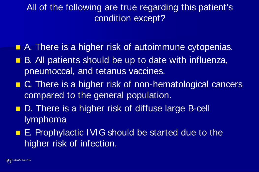
All of the following are true regarding this patient’s condition except?
A. There is a higher risk of autoimmune cytopenias. B. All patients should be up to date with influenza,
pneumoccal, and tetanus vaccines. C. There is a higher risk of non-hematological cancers
compared to the general population. D. There is a higher risk of diffuse large B-cell
lymphoma E. Prophylactic IVIG should be started due to the
higher risk of infection.

Answer
E. Prophylactic IVIG should be started due to the higher risk of infection.
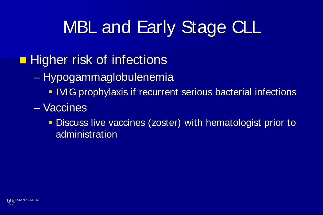
MBL and Early Stage CLL
Higher risk of infections– Hypogammaglobulenemia IVIG prophylaxis if recurrent serious bacterial infections
– Vaccines Discuss live vaccines (zoster) with hematologist prior to
administration
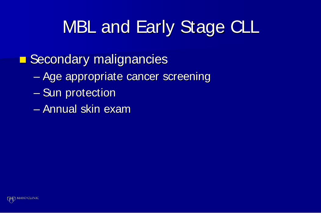
MBL and Early Stage CLL
Secondary malignancies– Age appropriate cancer screening– Sun protection– Annual skin exam

Clinical Pearl
Patients with MBL and CLL are at higher risk of infections and nonhematological cancers (particularly skin cancers) than the general population, even in the absence of CLL-directed therapy. Patients should be have an annual skin examination, adhere to age-appropriate cancer screening, and be up to date with vaccines.

Case 5
A 34 year old man is hospitalized after a motorcycle accident. He suffered multiple traumatic injuries, including splenic laceration requiring splenectomy and pneumothorax requiring chest tube placement. Today is post-op day 7, he is clinically improving, and hopes to be discharged today. Today’s CBC shows the following: WBC 10.5, hemoglobin is 12.5 g/dL, and platelets 832K. Platelet count was normal on the day of admission.

What is the next best step?
A. Low dose aspirin and clopidogrel B. Peripheral blood smear C. Dismiss home D. Plateletpheresis E. Start heparin therapy

Answer
C. Dismiss home

Reactive Thrombocytosis
Expected post-splenectomy– Peaks 1-3 weeks postop, normalizes within weeks– Generally not associated with thrombosis Consider ASA 81 mg
– If extreme (>1500K), risk of hemorrhage
Other causes– Inflammatory conditions, infection, trauma,
hemolysis, iron deficiency
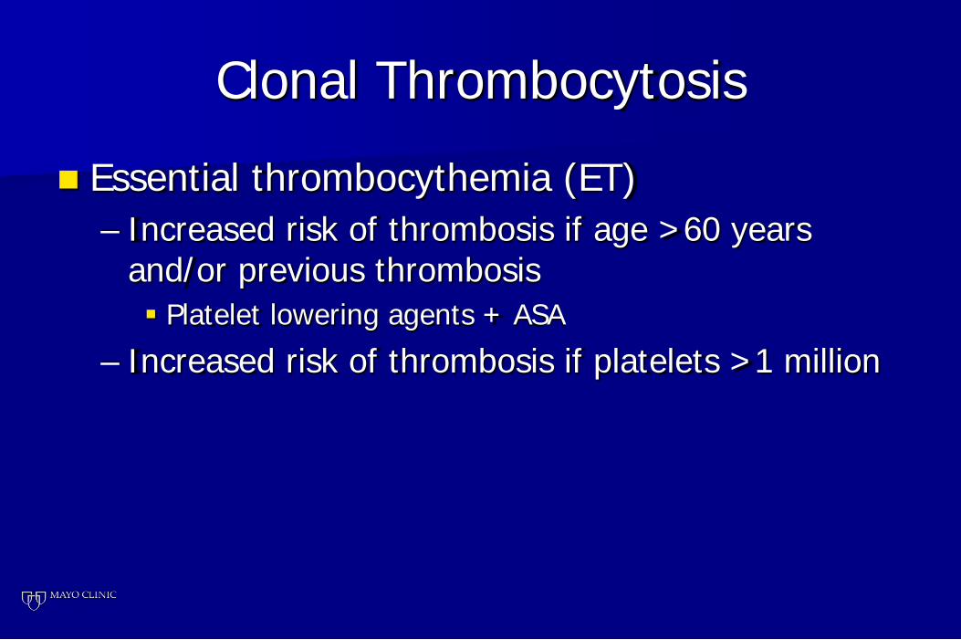
Clonal Thrombocytosis
Essential thrombocythemia (ET)– Increased risk of thrombosis if age >60 years
and/or previous thrombosis Platelet lowering agents + ASA
– Increased risk of thrombosis if platelets >1 million
Clinical Pearl
Reactive thrombocytosis can be due to inflammatory conditions, infection, trauma, hyposplenism, hemolysis, and iron deficiency and does not cause an increased risk of thrombosis. No specific therapy is recommended.

Case 6
A 63 year old Caucasian female reports for a routine physical examination. Past medical history is significant for hyperlipidemia, morbid obesity, diabetes type II, non-alcoholic steatohepatitis (NASH), hypertension, and osteoarthritis. Medication for the past three years have included aspirin, metoprolol, lisinopril, atorvastatin, metformin, and calcium/vitamin D. CBC shows a WBC 5.8, hemoglobin 12.8 g/dL and platelets 85K.
Which one of the following is the next best step for workup of the thrombocytopenia?
A. Anti-PF4 antibody B. Prothrombin time C. Ultrasound of the abdomen D. HIV E. Peripheral blood smear
Answer
E. Peripheral blood smear

Thrombocytopenia
Can be due to inadequate production, sequestration, or peripheral destruction
Step 1: is it real?– Peripheral smear– CBC in sodium citrate tube
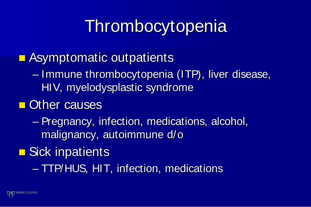
Thrombocytopenia
Asymptomatic outpatients– Immune thrombocytopenia (ITP), liver disease,
HIV, myelodysplastic syndrome
Other causes– Pregnancy, infection, medications, alcohol,
malignancy, autoimmune d/o
Sick inpatients– TTP/HUS, HIT, infection, medications
Clinical Pearl
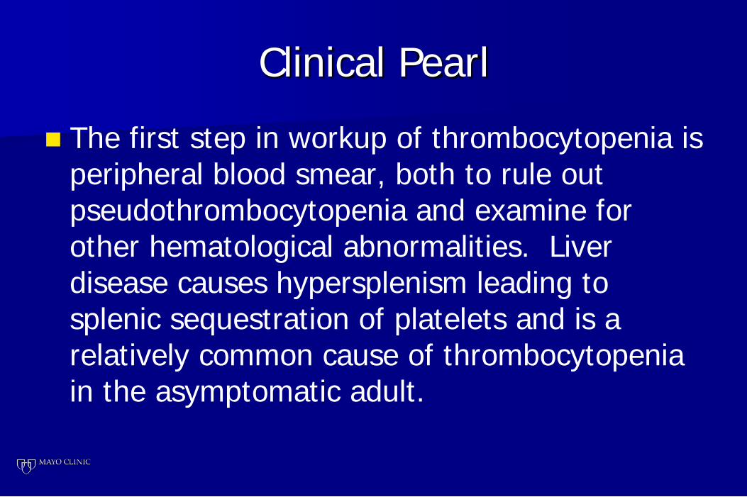
The first step in workup of thrombocytopenia is peripheral blood smear, both to rule out pseudothrombocytopenia and examine for other hematological abnormalities. Liver disease causes hypersplenism leading to splenic sequestration of platelets and is a relatively common cause of thrombocytopenia in the asymptomatic adult.

Case 7
A 39-year-old African American healthy male presents to you for life insurance screening examination. He takes no medications and has no significant symptoms or past medical history. Physical exam is unremarkable. Laboratory studies show normal chemistries and lipids, but CBC is flagged as abnormal -WBC is 2.1 with a low absolute neutrophil count (ANC) of 1350. Hemoglobin and platelets are within normal range.
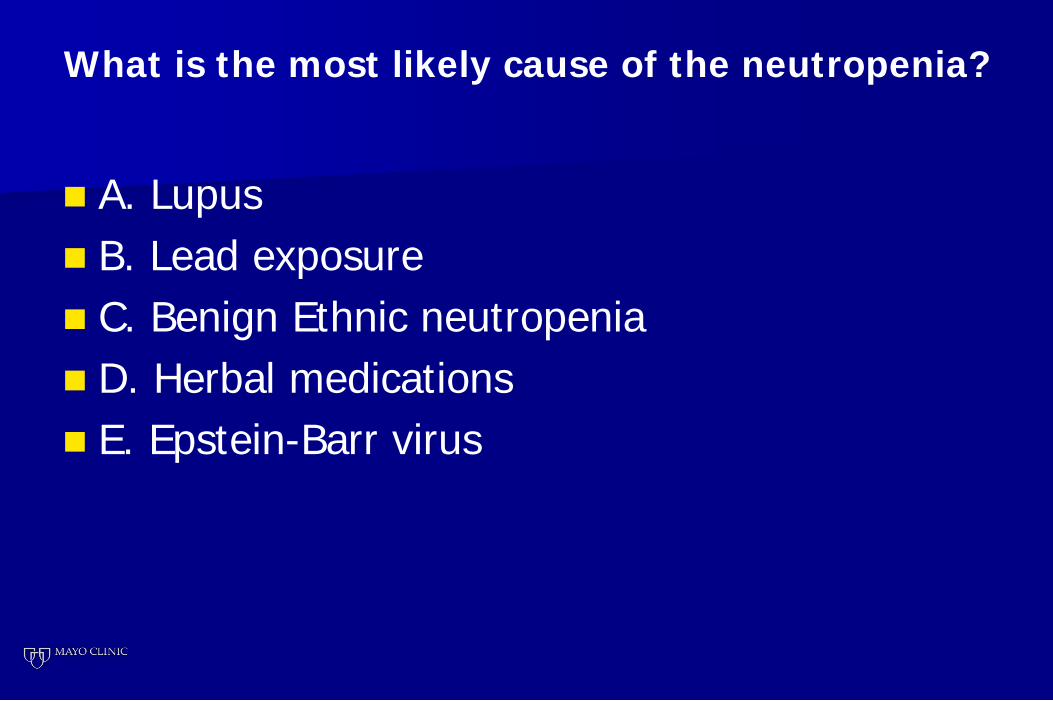
What is the most likely cause of the neutropenia?
A. Lupus B. Lead exposure C. Benign Ethnic neutropenia D. Herbal medications E. Epstein-Barr virus

Answer
C. Benign Ethnic neutropenia

Neutropenia
Benign ethnic neutropenia– African descent, Yemenites, West Indians, Arab
Jordanians– ANC generally 1000-1500– Normal bone marrow reserve no risk of infection
Neutropenia: Other Causes
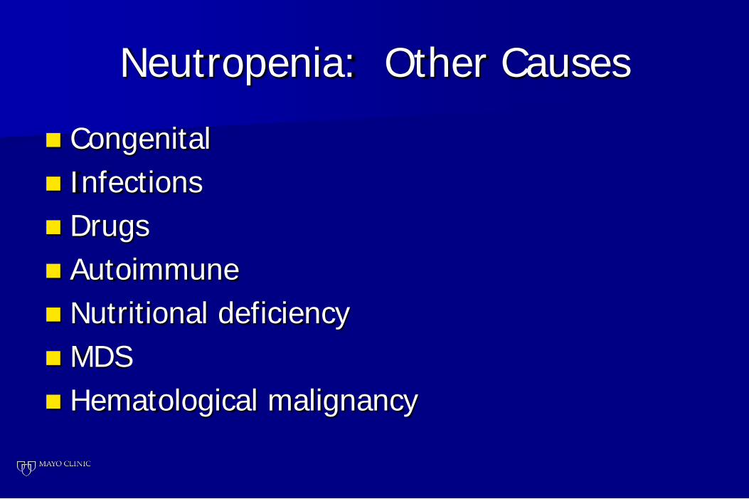
Congenital Infections Drugs Autoimmune Nutritional deficiency MDS Hematological malignancy

Clinical Pearl
Benign Ethnic neutropenia is an inherited condition common in individuals of African descent. It causes mild neutropenia and does not carry an increased risk of infection.
Case 8
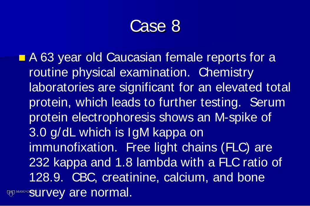
A 63 year old Caucasian female reports for a routine physical examination. Chemistry laboratories are significant for an elevated total protein, which leads to further testing. Serum protein electrophoresis shows an M-spike of 3.0 g/dL which is IgM kappa on immunofixation. Free light chains (FLC) are 232 kappa and 1.8 lambda with a FLC ratio of 128.9. CBC, creatinine, calcium, and bone survey are normal.
You give her the following diagnosis:
A. Monoclonal gammopathy of undetermined significance
B. Multiple myeloma C. AL amyloidosis D. Smoldering multiple myeloma E. Waldenstrom’s macroglobulenemia
Answer
B. Multiple myeloma
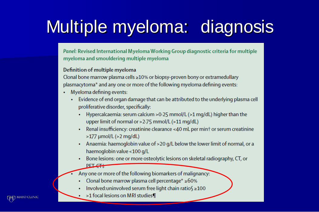
Multiple myeloma: diagnosis
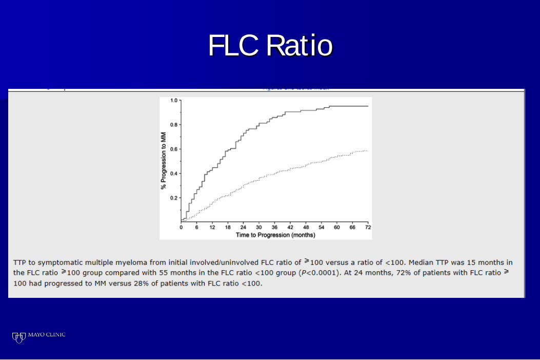
FLC Ratio
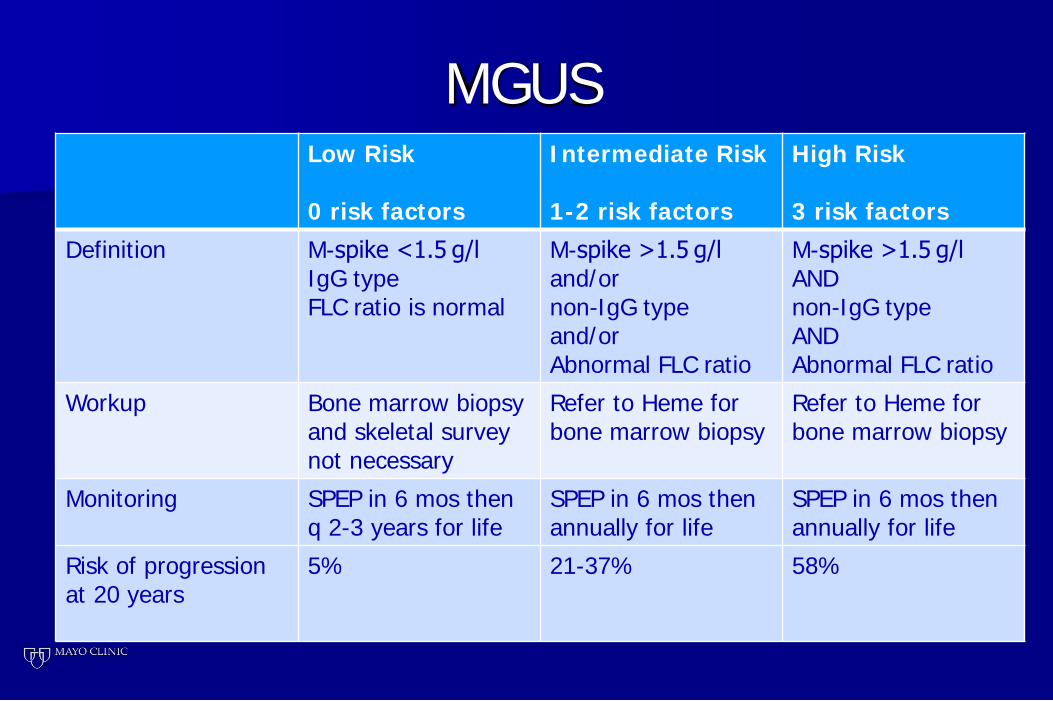
MGUSLow Risk
0 risk factors
Intermediate Risk
1-2 risk factors
High Risk
3 risk factorsDefinition M-spike <1.5 g/l
IgG typeFLC ratio is normal
M-spike >1.5 g/l and/ornon-IgG typeand/orAbnormal FLC ratio
M-spike >1.5 g/l ANDnon-IgG typeANDAbnormal FLC ratio
Workup Bone marrow biopsy and skeletal survey not necessary
Refer to Heme for bone marrow biopsy
Refer to Heme for bone marrow biopsy
Monitoring SPEP in 6 mos then q 2-3 years for life
SPEP in 6 mos then annually for life
SPEP in 6 mos then annually for life
Risk of progression at 20 years
5% 21-37% 58%
Clinical Pearl
A serum involved to uninvolved FLC of >100 is now one of the diagnostic criteria for multiple myeloma based on the high risk of progression to end-organ damage.