Embed Size (px)
Citation preview
Submitted 14 August 2015Accepted 2 September 2015Published 22 September 2015
Corresponding authorAndrew D. Vigotsky,[email protected]
Academic editorDavid Reser
Additional Information andDeclarations can be found onpage 8
DOI 10.7717/peerj.1261
Copyright2015 Contreras et al.
Distributed underCreative Commons CC-BY 4.0
OPEN ACCESS
A comparison of two gluteus maximusEMG maximum voluntary isometriccontraction positionsBret Contreras1, Andrew D. Vigotsky2, Brad J. Schoenfeld3,Chris Beardsley4 and John Cronin1,5
1 Auckland University of Technology, Sport Performance Research Institute New Zealand,Auckland, New Zealand
2 Kinesiology Program, Arizona State University, Phoenix, AZ, USA3 Department of Health Sciences, CUNY Lehman College, Bronx, NY, USA4 Strength and Conditioning Research Limited, London, UK5 School of Exercise, Biomedical and Health Science, Edith Cowan University, Perth, Australia
ABSTRACTBackground. The purpose of this study was to compare the peak electromyography(EMG) of the most commonly-used position in the literature, the prone bent-leg(90◦) hip extension against manual resistance applied to the distal thigh (PRONE), toa novel position, the standing glute squeeze (SQUEEZE).Methods. Surface EMG electrodes were placed on the upper and lower gluteusmaximus of thirteen recreationally active females (age = 28.9 years; height = 164 cm;body mass = 58.2 kg), before three maximum voluntary isometric contraction(MVIC) trials for each position were obtained in a randomized, counterbalancedfashion.Results. No statistically significant (p < 0.05) differences were observed betweenPRONE (upper: 91.94%; lower: 94.52%) and SQUEEZE (upper: 92.04%; lower:85.12%) for both the upper and lower gluteus maximus. Neither the PRONE norSQUEEZE was more effective between all subjects.Conclusions. In agreement with other studies, no single testing position is idealfor every participant. Therefore, it is recommended that investigators employmultiple MVIC positions, when possible, to ensure accuracy. Future research shouldinvestigate a variety of gluteus maximus MVIC positions in heterogeneous samples.
Subjects Anatomy and Physiology, Kinesiology, OrthopedicsKeywords MVC, MVIC, Electromyography, Neuromechanics, Normalization
INTRODUCTIONMaximum voluntary isometric contractions (MVIC) are often used to normalize
electromyography (EMG) signals. It is important to employ an MVIC position that
elicits the highest activation in order to increase the validity of EMG studies and decrease
incidents of abnormally high normalized mean and peak EMG data. In order for accurate
comparisons to be made between studies, it is also important for researchers to standardize
MVIC positions, or at least use positions that elicit similar magnitudes of EMG activity. A
number of MVIC positions have been used in the literature to assess the gluteus maximus,
How to cite this article Contreras et al. (2015), A comparison of two gluteus maximus EMG maximum voluntary isometric contractionpositions. PeerJ 3:e1261; DOI 10.7717/peerj.1261
including the Biering-Sorenson position (Cambridge et al., 2012; McGill, McDermott &
Fenwick, 2009), the prone straight leg hip extension position (Barton et al., 2014; Worrell et
al., 2001), the prone bent leg position (Jakobsen et al., 2013; Youdas et al., 2013), the prone
straight leg position with 70◦ of hip flexion (Simenz et al., 2012), and the standing bent leg
position (Boudreau et al., 2009). The most commonly used position, however, is the prone
bent-leg (90◦) hip extension with manual resistance applied to the distal thigh (PRONE)
(Choi et al., 2015; Emami, Arab & Ghamkhar, 2014; Hislop et al., 2014; Kang et al., 2013;
Kendall , 2005; Oh et al., 2007).
A recent study by Simenz et al. (2012) that used a prone gluteus maximus MVIC position
in 70◦ of hip flexion, demonstrates the importance of standardizing MVIC positions across
studies. Researchers have shown that lower gluteus maximus amplitude is elicited at higher
degrees of hip flexion and reaches a maximum EMG amplitude at end range hip extension
(Worrell et al., 2001). By employing an MVIC position that renders significantly lower
EMG activity than those values that are truly maximal, the normalized data of Simenz et al.
(2012) are most likely overestimated. For example, if the work of Worrell et al. (2001) is ex-
trapolated, the MVIC position used by Simenz et al. (2012) would only elicit approximately
80% of true MVIC, translating into 25% greater mean and peak values when compared to
the true MVIC position. The data reported by Simenz et al. (2012) therefore cannot be used
for comparison with exercises in other studies that utilized alternative MVIC positions
with smaller hip flexion angles, as the data would have overestimated how effectively the
gluteus maximus was activated. Therefore, it is apparent that researchers should only
compare EMG data that utilize positions that render similar values.
Since Worrell et al. (2001) found that full hip extension elicited the greatest amount
of gluteus maximus EMG activity, and this finding is corroborated by earlier work from
Wheatley & Jahnke (1951) and Fischer & Houtz (1968), it is postulated that the most ap-
propriate gluteus maximus MVIC position is at full hip extension, or hip hyperextension.
PRONE is currently the recommended position in several texts on muscle testing (Hislop
et al., 2014; Kendall , 2005), although to the authors’ knowledge, this position has not been
compared to others in the literature. In order to correct for individual variation, some
researchers have employed multiple MVIC positions. For example, McGill, McDermott &
Fenwick (2009) used both the Biering-Sorenson and PRONE positions; whichever position
elicited the greatest activity was used for normalization purposes. The authors, however,
are unaware of any existing research that quantitatively compares gluteus maximus MVIC
positions.
The gluteus maximus muscle appears to be segmented into at least two subdivisions,
which may display different EMG activity in response to certain muscle actions. McAndrew,
Gorelick & Brown (2006) used a laser-based mechanomyographic (MMG) technique to
measure the mean contraction time in six subdivisions of the gluteus maximus, both in
the sagittal plane (superior, middle, inferior) and in the frontal plane (medial and lateral).
The superior region displayed the longest contraction time followed by the middle region
and then the inferior region. On the basis of these findings, McAndrew, Gorelick & Brown
(2006) suggested that the superior region may contain more slow twitch fibers and be more
Contreras et al. (2015), PeerJ, DOI 10.7717/peerj.1261 2/10
involved in postural tasks compared to the inferior region, while the inferior region may
contain more fast twitch fibers and be more involved in dynamic tasks. This is further
substantiated by the work of Lyons et al. (1983) and Karlsson & Jonsson (1965), who found
differences between upper and lower gluteus maximus EMG during functional movement;
for example, load acceptance during stair ambulation better targets the lower gluteus
maximus (Lyons et al., 1983), while hip abduction better targets the upper gluteus maximus
(Karlsson & Jonsson, 1965).
Pilot data from our lab showed that some subjects were able to elicit greater EMG
activity during a standing glute squeeze (SQUEEZE) when compared to PRONE, and this
was especially true for the upper gluteus maximus. Given this observation and the findings
articulated in previous paragraphs, the purpose of this investigation was to compare upper
and lower gluteus maximus EMG activity in PRONE versus SQUEEZE. Based on our pilot
data, it was hypothesized that SQUEEZE would elicit greater upper gluteus maximus EMG
activity, while PRONE would elicit greater lower gluteus maximus EMG activity.
METHODSSubjectsThirteen healthy women (age = 28.9 ± 5.1 years; height = 164 ± 6.3 cm; body mass =
58.2 ± 6.4 kg) with 7.0 ± 5.8 years of resistance training experience participated in this
study. Inclusion criteria required subjects to be between 20 and 40 years of age and have at
least 3 years of consistent resistance training experience. All subjects were healthy and free
of any musculoskeletal or neuromuscular injuries, pain, or illnesses. Subjects completed an
Informed Consent form. Subjects were advised to refrain from training their lower body
for 72 h prior to testing. The study was approved by the Auckland University of Technology
Ethics Committee (AUTEC Reference number 13/375).
ProceduresSubjects first performed a 10-minute general warm-up consisting of various dynamic
stretches for the lower body musculature. Following warm-up, subjects practiced each
testing position several times, until they felt comfortable with the technique. Subjects were
asked to wear appropriate clothing for access to the EMG electrode placement sites. Before
placing the electrodes on the skin, excess hair was removed with a razor, and skin was
cleaned and abraded using an alcohol swab. After preparation, self-adhesive disposable
silver/silver chloride pre-gelled dual snap surface bipolar electrodes (Noraxon Product
#272, Noraxon USA Inc., Scottsdale, AZ) with a diameter of 1 centimeter (cm) and an
inter-electrode distance of 2 cm were attached in parallel to the fibers of the right upper
and lower gluteus maximus. More specifically, “[upper gluteus maximus] electrodes were
placed two finger’s width above the line just under the spina iliaca posterior superior and
the trochanter major; [lower gluteus maximus] electrodes were set below the same line”
(Fujisawa et al., 2014) (Figs. 1 and 2). After the electrodes were secured, a quality check was
performed to ensure EMG signal validity.
Following electrode placement, subjects completed three trials of PRONE then
SQUEEZE, or vice versa. The PRONE position was performed by having the subject
Contreras et al. (2015), PeerJ, DOI 10.7717/peerj.1261 3/10
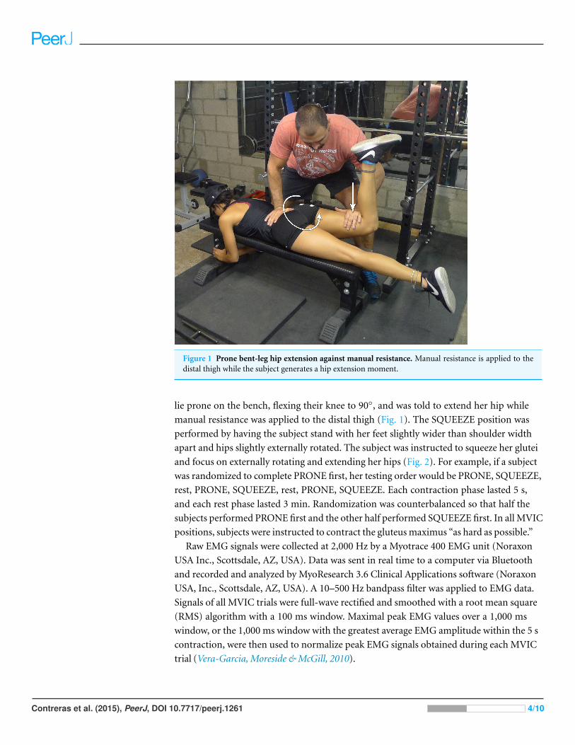
Figure 1 Prone bent-leg hip extension against manual resistance. Manual resistance is applied to thedistal thigh while the subject generates a hip extension moment.
lie prone on the bench, flexing their knee to 90◦, and was told to extend her hip while
manual resistance was applied to the distal thigh (Fig. 1). The SQUEEZE position was
performed by having the subject stand with her feet slightly wider than shoulder width
apart and hips slightly externally rotated. The subject was instructed to squeeze her glutei
and focus on externally rotating and extending her hips (Fig. 2). For example, if a subject
was randomized to complete PRONE first, her testing order would be PRONE, SQUEEZE,
rest, PRONE, SQUEEZE, rest, PRONE, SQUEEZE. Each contraction phase lasted 5 s,
and each rest phase lasted 3 min. Randomization was counterbalanced so that half the
subjects performed PRONE first and the other half performed SQUEEZE first. In all MVIC
positions, subjects were instructed to contract the gluteus maximus “as hard as possible.”
Raw EMG signals were collected at 2,000 Hz by a Myotrace 400 EMG unit (Noraxon
USA Inc., Scottsdale, AZ, USA). Data was sent in real time to a computer via Bluetooth
and recorded and analyzed by MyoResearch 3.6 Clinical Applications software (Noraxon
USA, Inc., Scottsdale, AZ, USA). A 10–500 Hz bandpass filter was applied to EMG data.
Signals of all MVIC trials were full-wave rectified and smoothed with a root mean square
(RMS) algorithm with a 100 ms window. Maximal peak EMG values over a 1,000 ms
window, or the 1,000 ms window with the greatest average EMG amplitude within the 5 s
contraction, were then used to normalize peak EMG signals obtained during each MVIC
trial (Vera-Garcia, Moreside & McGill, 2010).
Contreras et al. (2015), PeerJ, DOI 10.7717/peerj.1261 4/10
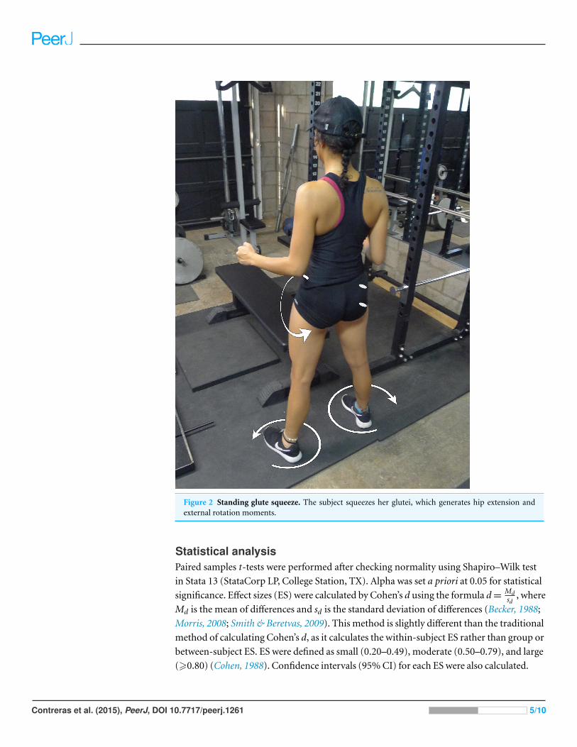
Figure 2 Standing glute squeeze. The subject squeezes her glutei, which generates hip extension andexternal rotation moments.
Statistical analysisPaired samples t-tests were performed after checking normality using Shapiro–Wilk test
in Stata 13 (StataCorp LP, College Station, TX). Alpha was set a priori at 0.05 for statistical
significance. Effect sizes (ES) were calculated by Cohen’s d using the formula d =Mdsd
, where
Md is the mean of differences and sd is the standard deviation of differences (Becker, 1988;
Morris, 2008; Smith & Beretvas, 2009). This method is slightly different than the traditional
method of calculating Cohen’s d, as it calculates the within-subject ES rather than group or
between-subject ES. ES were defined as small (0.20–0.49), moderate (0.50–0.79), and large
(>0.80) (Cohen, 1988). Confidence intervals (95% CI) for each ES were also calculated.
Contreras et al. (2015), PeerJ, DOI 10.7717/peerj.1261 5/10
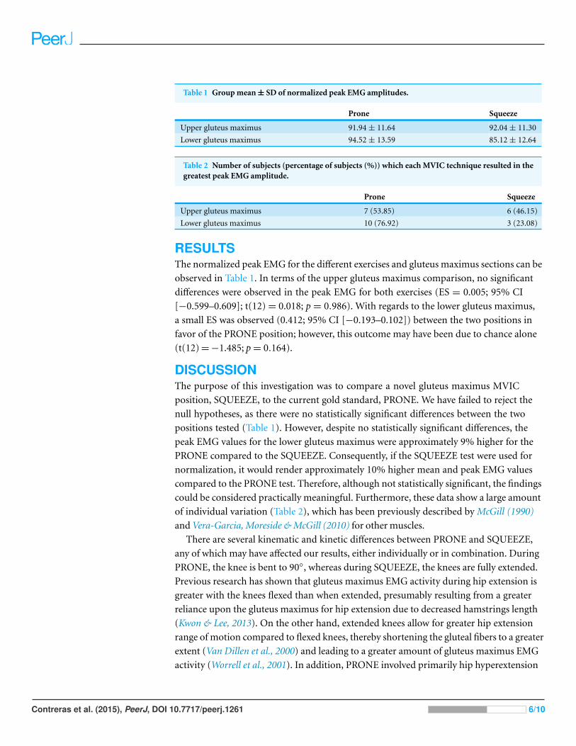
Table 1 Group mean ± SD of normalized peak EMG amplitudes.
Prone Squeeze
Upper gluteus maximus 91.94 ± 11.64 92.04 ± 11.30
Lower gluteus maximus 94.52 ± 13.59 85.12 ± 12.64
Table 2 Number of subjects (percentage of subjects (%)) which each MVIC technique resulted in thegreatest peak EMG amplitude.
Prone Squeeze
Upper gluteus maximus 7 (53.85) 6 (46.15)
Lower gluteus maximus 10 (76.92) 3 (23.08)
RESULTSThe normalized peak EMG for the different exercises and gluteus maximus sections can be
observed in Table 1. In terms of the upper gluteus maximus comparison, no significant
differences were observed in the peak EMG for both exercises (ES = 0.005; 95% CI
[−0.599–0.609]; t(12) = 0.018; p = 0.986). With regards to the lower gluteus maximus,
a small ES was observed (0.412; 95% CI [−0.193–0.102]) between the two positions in
favor of the PRONE position; however, this outcome may have been due to chance alone
(t(12) = −1.485; p = 0.164).
DISCUSSIONThe purpose of this investigation was to compare a novel gluteus maximus MVIC
position, SQUEEZE, to the current gold standard, PRONE. We have failed to reject the
null hypotheses, as there were no statistically significant differences between the two
positions tested (Table 1). However, despite no statistically significant differences, the
peak EMG values for the lower gluteus maximus were approximately 9% higher for the
PRONE compared to the SQUEEZE. Consequently, if the SQUEEZE test were used for
normalization, it would render approximately 10% higher mean and peak EMG values
compared to the PRONE test. Therefore, although not statistically significant, the findings
could be considered practically meaningful. Furthermore, these data show a large amount
of individual variation (Table 2), which has been previously described by McGill (1990)
and Vera-Garcia, Moreside & McGill (2010) for other muscles.
There are several kinematic and kinetic differences between PRONE and SQUEEZE,
any of which may have affected our results, either individually or in combination. During
PRONE, the knee is bent to 90◦, whereas during SQUEEZE, the knees are fully extended.
Previous research has shown that gluteus maximus EMG activity during hip extension is
greater with the knees flexed than when extended, presumably resulting from a greater
reliance upon the gluteus maximus for hip extension due to decreased hamstrings length
(Kwon & Lee, 2013). On the other hand, extended knees allow for greater hip extension
range of motion compared to flexed knees, thereby shortening the gluteal fibers to a greater
extent (Van Dillen et al., 2000) and leading to a greater amount of gluteus maximus EMG
activity (Worrell et al., 2001). In addition, PRONE involved primarily hip hyperextension
Contreras et al. (2015), PeerJ, DOI 10.7717/peerj.1261 6/10
since the pelvis was fixed, whereas SQUEEZE appeared to involve a combination of hip
extension and posterior pelvic tilt. Although posterior pelvic tilt mimics hip extension
(Neumann, 2010), it is unclear how each of these kinematic variables might affect gluteus
maximus EMG activity individually. To our knowledge, no study to date has investigated
gluteus maximus EMG activity with varying combinations of hip extension and posterior
pelvic tilt during MVIC actions. Moreover, PRONE is an open kinetic chain maneuver with
the torso stabilized onto a bench, whereas SQUEEZE is a closed kinetic chain maneuver
performed in a standing position. Stensdotter et al. (2003) investigated the EMG activity of
the quadriceps muscle group during open kinetic chain and closed kinetic chain positions
during MVIC actions and reported significant differences in EMG amplitude. The rectus
femoris displayed greater EMG activity during open kinetic chain maneuvers while the
vastus medialis displayed greater EMG activity during closed kinetic chain maneuvers. It
is therefore hard to predict whether the gluteus maximus would inherently display greater
or lesser EMG activity during either open or closed kinetic chain maneuvers. Finally,
PRONE required manual resistance, whereas SQUEEZE relied upon anatomical structures
surrounding the hip to provide resistance against hip extension. Whether this factor has
any effect on EMG activity recorded in a muscle is unclear, as the authors are unaware of
any previous investigations into the effect of squeezing a muscle whereby range of motion
is limited by anatomical structures on EMG activity rather than against external resistance.
This investigation was subject to several important limitations. Firstly, although we
observed what may have been a practically important difference between the MVIC
positions, this difference was not found to be statistically significant, which suggests that
our initial estimates for the appropriate sample size may have been too small. Secondly,
there were several kinematic differences between the two positions that were explored
(PRONE and SQUEEZE), including different pelvic, hip, and knee joint angles. There
were also kinetic differences between the two positions, in that PRONE was an open
kinetic chain maneuver and SQUEEZE was a closed kinetic chain maneuver. Moreover,
PRONE used external resistance and SQUEEZE utilized oppositional torques produced
by internal, anatomical structures. These multiple differences make it difficult to assess
whether our results arose from a combination of biomechanical factors acting in opposing
directions, heterogeneity, or genuinely no difference between the conditions. Thirdly,
we only compared two MVIC positions, and it is feasible that other positions might
result in superior or inferior levels of EMG activity. Fourthly, we only investigated two
subdivisions of the gluteus maximus muscle and there are indications that there may be
others, from proximal-to-distal, medial-to-lateral, and superficial-to-deep. Furthermore,
our statistical analysis was not designed to assess whether there was a difference between
the EMG activity of the upper and lower gluteus maximus during either MVIC position
and therefore this remains uncertain.
CONCLUSIONSAlthough these data are inconclusive as to which position is superior, they do provide
insight as to the complexity of MVIC positions for the gluteus maximus. More specifically,
Contreras et al. (2015), PeerJ, DOI 10.7717/peerj.1261 7/10
due to the large individual variations (Table 2), it is recommended that multiple MVIC
positions be utilized to ensure that the greatest possible EMG amplitude be the divisor
during normalization. These recommendations are well in line with other studies, which
have utilized or recommended multiple MVIC positions (McGill, McDermott & Fenwick,
2009; Vera-Garcia, Moreside & McGill, 2010). Future research should use heterogeneous
samples, such as athletic males, and also test more positions, such as the Biering-Sorenson
position, quadruped hip extension position, and top hip thrust position (Contreras, Cronin
& Schoenfeld, 2011), each with manual resistance, along with the tall kneeling position.
ADDITIONAL INFORMATION AND DECLARATIONS
FundingThe authors received no funding for this work.
Competing InterestsChris Beardsley is the founder and owner of Strength and Conditioning Research Limited.
All other authors declare that they have no competing interests.
Author Contributions• Bret Contreras conceived and designed the experiments, performed the experiments,
contributed reagents/materials/analysis tools, wrote the paper, reviewed drafts of the
paper.
• Andrew D. Vigotsky performed the experiments, analyzed the data, wrote the paper,
prepared figures and/or tables, reviewed drafts of the paper.
• Brad J. Schoenfeld, Chris Beardsley and John Cronin wrote the paper, reviewed drafts of
the paper.
Human EthicsThe following information was supplied relating to ethical approvals (i.e., approving body
and any reference numbers):
Auckland University of Technology Ethics Committee
AUTEC Reference number 13/375.
Supplemental InformationSupplemental information for this article can be found online at http://dx.doi.org/
10.7717/peerj.1261#supplemental-information.
REFERENCESBarton CJ, Kennedy A, Twycross-Lewis R, Woledge R, Malliaras P, Morrissey D. 2014. Gluteal
muscle activation during the isometric phase of squatting exercises with and without a Swissball. Physical Therapy in Sport 15:39–46 DOI 10.1016/j.ptsp.2013.02.006.
Becker BJ. 1988. Synthesizing standardized mean-change measures. British Journal ofMathematical and Statistical Psychology 41:257–278 DOI 10.1111/j.2044-8317.1988.tb00901.x.
Contreras et al. (2015), PeerJ, DOI 10.7717/peerj.1261 8/10
Boudreau SN, Dwyer MK, Mattacola CG, Lattermann C, Uhl TL, McKeon JM. 2009. Hip-muscleactivation during the lunge, single-leg squat, and step-up-and-over exercises. Journal of SportRehabilitation 18:91–103.
Cambridge ED, Sidorkewicz N, Ikeda DM, McGill SM. 2012. Progressive hip rehabilitation: theeffects of resistance band placement on gluteal activation during two common exercises. ClinicalBiomechanics 27:719–724 DOI 10.1016/j.clinbiomech.2012.03.002.
Choi SA, Cynn HS, Yi CH, Kwon OY, Yoon TL, Choi WJ, Lee JH. 2015. Isometric hip abductionusing a Thera-Band alters gluteus maximus muscle activity and the anterior pelvic tiltangle during bridging exercise. Journal of Electromyography and Kinesiology 25(2):310–315DOI 10.1016/j.jelekin.2014.09.005.
Cohen J. 1988. Statistical power analysis for the behavioral sciences. 2nd edition. New York:Lawrence Erlbaum Associates.
Contreras B, Cronin J, Schoenfeld B. 2011. Barbell hip thrust. Strength & Conditioning Journal33:58–61 DOI 10.1519/SSC.0b013e31822fa09d.
Emami M, Arab AM, Ghamkhar L. 2014. The activity pattern of the lumbo-pelvic muscles duringprone hip extension in athletes with and without hamstring strain injury. International Journalof Sports Physical Therapy 9:312–319.
Fischer FJ, Houtz SJ. 1968. Evaluation of the function of the gluteus maximus muscle. Anelectromyographic study. American Journal of Physical Medicine 47:182–191.
Fujisawa H, Suzuki H, Yamaguchi E, Yoshiki H, Wada Y, Watanabe A. 2014. Hip muscle activityduring isometric contraction of hip abduction. Journal of Physical Therapy Science 26:187–190DOI 10.1589/jpts.26.187.
Hislop H, Avers D, Brown M, Daniels L. 2014. Daniels and Worthingham’s muscle testing:techniques of manual examination and performance testing. St. Louis: Elsevier.
Jakobsen MD, Sundstrup E, Andersen CH, Aagaard P, Andersen LL. 2013. Muscle activityduring leg strengthening exercise using free weights and elastic resistance: effects of ballisticvs controlled contractions. Human Movement Science 32:65–78DOI 10.1016/j.humov.2012.07.002.
Kang SY, Jeon HS, Kwon O, Cynn HS, Choi B. 2013. Activation of the gluteus maximus andhamstring muscles during prone hip extension with knee flexion in three hip abductionpositions. Manual Therapy 18:303–307 DOI 10.1016/j.math.2012.11.006.
Karlsson E, Jonsson B. 1965. Function of the gluteus maximus muscle. An electromyographicstudy. Acta Morphologica Neerlando-Scandinavica 6:161–169.
Kendall F. 2005. Muscles: testing and function with posture and pain. Baltimore: LippincottWilliams & Wilkins.
Kwon YJ, Lee HO. 2013. How different knee flexion angles influence the hip extensor in the proneposition. Journal of Physical Therapy Science 25:1295–1297 DOI 10.1589/jpts.25.1295.
Lyons K, Perry J, Gronley JK, Barnes L, Antonelli D. 1983. Timing and relative intensity of hipextensor and abductor muscle action during level and stair ambulation. An EMG study. PhysicalTherapy 63:1597–1605.
McAndrew D, Gorelick M, Brown J. 2006. Muscles within muscles: a mechanomyographicanalysis of muscle segment contractile properties within human gluteus maximus. Journalof Musculoskeletal Research 10:23–35 DOI 10.1142/S0218957706001704.
McGill SM. 1990. Electromyographic activity of the abdominal and low back musculature duringthe generation of isometric and dynamic axial trunk torque: implications for lumbar mechanics.Journal of Orthopaedic Research 9:91–103 DOI 10.1002/jor.1100090112.
Contreras et al. (2015), PeerJ, DOI 10.7717/peerj.1261 9/10
McGill SM, McDermott A, Fenwick CM. 2009. Comparison of different strongman events:trunk muscle activation and lumbar spine motion, load, and stiffness. Journal of Strength andConditioning Research/National Strength & Conditioning Association 23:1148–1161DOI 10.1519/JSC.0b013e318198f8f7.
Morris SB. 2008. Estimating effect sizes from the pretest–posttest-control group designs.Organizational Research Methods 11(2):364–386 DOI 10.1177/1094428106291059.
Neumann DA. 2010. Kinesiology of the hip: a focus on muscular actions. Journal of Orthopaedicand Sports Physical Therapy 40:82–94 DOI 10.2519/jospt.2010.3025.
Oh JS, Cynn HS, Won JH, Kwon OY, Yi CH. 2007. Effects of performing an abdominal drawing-inmaneuver during prone hip extension exercises on hip and back extensor muscle activity andamount of anterior pelvic tilt. Journal of Orthopaedic and Sports Physical Therapy 37:320–324DOI 10.2519/jospt.2007.2435.
Simenz CJ, Garceau LR, Lutsch BN, Suchomel TJ, Ebben WP. 2012. Electromyographical analysisof lower extremity muscle activation during variations of the loaded step-up exercise. Journal ofStrength and Conditioning Research 26:3398–3405 DOI 10.1519/JSC.0b013e3182472fad.
Smith LJW, Beretvas SN. 2009. Estimation of the standardized mean difference for repeatedmeasures designs. Journal of Modern Applied Statistical Methods 8:27.
Stensdotter AK, Hodges PW, Mellor R, Sundelin G, Hager-Ross C. 2003. Quadriceps activationin closed and in open kinetic chain exercise. Medicine and Science in Sports and Exercise35:2043–2047 DOI 10.1249/01.MSS.0000099107.03704.AE.
Van Dillen LR, McDonnell MK, Fleming DA, Sahrmann SA. 2000. Effect of knee and hip positionon hip extension range of motion in individuals with and without low back pain. Journal ofOrthopaedic and Sports Physical Therapy 30:307–316 DOI 10.2519/jospt.2000.30.6.307.
Vera-Garcia FJ, Moreside JM, McGill SM. 2010. MVC techniques to normalize trunk muscle EMGin healthy women. Journal of Electromyography and Kinesiology 20:10–16DOI 10.1016/j.jelekin.2009.03.010.
Wheatley MD, Jahnke WD. 1951. Electromyographic study of the superficial thigh and hipmuscles in normal individuals. Archives of Physical Medicine and Rehabilitation 32:508–515.
Worrell TW, Karst G, Adamczyk D, Moore R, Stanley C, Steimel B, Steimel S. 2001. Influence ofjoint position on electromyographic and torque generation during maximal voluntary isometriccontractions of the hamstrings and gluteus maximus muscles. Journal of Orthopaedic and SportsPhysical Therapy 31:730–740 DOI 10.2519/jospt.2001.31.12.730.
Youdas JW, Foley BM, Kruger BL, Mangus JM, Tortorelli AM, Madson TJ, Hollman JH. 2013.Electromyographic analysis of trunk and hip muscles during resisted lateral band walking.Physiotherapy Theory and Practice 29:113–123 DOI 10.3109/09593985.2012.704492.
Contreras et al. (2015), PeerJ, DOI 10.7717/peerj.1261 10/10