Embed Size (px)
Citation preview
ACKNOWLEDGEMENTS This master thesis could not have been accomplished without the support of Prof. Dr. A. Timmermans
and Drs. T. Matheve. Their understanding and professional knowledge had an added value for this
thesis. Special thanks should be given to Drs. T. Matheve for answering our questions, providing new
insights and reviewing our texts. We also want to thank the ‘Jessa Hospital’, campus Virga Jessa,
under guidance of Dr. G. Claes and Dhr. E. Olivieri and the other physiotherapists who supervised the
patients during their rehabilitation. We would like to express our gratitude to the Hocoma company for
the use of the ValedoMotion system. We would also like to thank Mrs. L. Luyten and Mrs. L.
Vanspauwen for advising us in English grammar and spelling when needed.
Finally, we would like to extend our sincere gratitude to all others who supported us during this
research and writing process.
Mol, 25 June 2014
Bilzen, 25 June 2014 S.V.G. & L.V.

3
BACKGROUND This master thesis is a pilot study which can be situated in the musculoskeletal rehabilitation. It more
specifically discusses technology-supported rehabilitation in case of chronic non-specific low back pain
(CNSLBP). This study follows the literature study which was carried out last year. It’s part of a PhD by
Thomas Matheve.
The study has been carried out in the ‘Jessa Hospital’ (Hasselt) in cooperation with ‘Hocoma’ (Zurich)
by two students in Physiotherapy and rehabilitation sciences.
Despite the fact that there has already been a large number of treatment possibilities for CNSLBP, it
remains a common and widespread health related problem. Chronic non-specific low back pain has a
large influence on the daily activities and functioning. The prevalence is about 10-20%.1 In that
respect, it is important that further research and studies will be carried out regarding possible
treatments.
This study examines the influence of an 18 week lasting technology-supported rehabilitation
programme in which postural biofeedback was used in patients with CNSLBP, on pain, disability,
quality of life, motivation, credibility and expectancy. Rehabilitation is done in a game-like environment
through two motion sensors are attached on the low back.
If the results of the pilot study are promising and if there are no side effects, a randomized controlled
trial (RCT) will be carried out by Thomas Matheve.
In advance, the co-promoter had developed the study protocol. The students read this and completed
it if necessary. Remarks were formulated and adapted after consideration.
The recruitment of patients was done in association with the co-promoter. Under the supervision of
their co-promoter the students carried out the patient history and physical examination. Based on the
results, it was decided whether a patient was to be included or not.
Apart from that, the students also supervised and coached the patients during their rehabilitation.
The input of data happened independently of each other in order to prevent possible mistakes and
bias. The analysis and interpretation of the data was also described independently of each other.
After this phase, the students compared all the information and assembled it to one work. The
students were responsible for the complete writing process, being supervised by their co-promoter.

4
5
Technology-supported rehabilitation for patients
with chronic non-specific low back pain
Preliminary results of a pilot study
HASSELT, 2014
“Drawn up according to the guidelines of ‘Spine’: http://edmgr.ovid.com/spine/accounts/ifauth.htm”

6
7
ABSTRACT Study Design: Pilot study.
Objective: The aim of this study was to assess the influence of an 18 week lasting technology-
supported rehabilitation programme in which postural biofeedback was used in patients with CNSLBP.
Summary of Background Data: Previous studies on technology-supported rehabilitation for CNSLBP
mostly used analytical exercises. This study aims to incorporate technology into functional exercises.
Methods: Four patients participated in a standard therapy consisting of cardio training, learning
neutral position of the back, pelvic movements, strengthening and functional exercises and a
technology-supported rehabilitation programme consisting of playing games controlled by pelvic
movements in different directions. In addition functional exercises and posture corrections were
performed with feedback support.
Results: Patient one’s pain intensity, mental component of the Short Form-36 (SF-36), motivation,
credibility and expectancy stayed almost identical as compared with baseline. The patients’ physical
component of the SF-36 increased and experienced less disability.
Patient two had a minimal clinical important difference (MCID) in pain intensity and disability. The
patient improved in the physical component of the SF-36 and slightly increased in motivation. The
mental component of the SF-36, credibility and expectancy stayed almost the same.
Patient three had no improvements. The patients’ pain intensity, physical component of the SF-36,
motivation stayed almost the same. The patient had an increase in disability and decrease in the
mental component of the SF-36, credibility and expectancy.
Patient four’s pain intensity and quality of life stayed almost identical. The patient increased in
disability, credibility and expectancy and slightly increased in motivation.
Conclusions: There is a positive trend in several outcome measures and there are no side effects.
Therefore the criteria for an RCT are achieved.
Key Words: Chronic non-specific low back pain, technology-supported rehabilitation, exercise
therapy, postural biofeedback, sensor technology, motivation, quality of life, pain, disability, credibility,
expectancy
Level of evidence: 2C

8
9
INTRODUCTION Chronic low back pain (CLBP) is a common health related problem. Ten to twenty percent of the
population develops CLBP in the course of life.1 CLBP has a major impact on the daily functioning and
is one of the main reasons for work absenteeism, which leads to high economic and health expenses.2
In the rehabilitation of patients with CLBP, exercise therapy within a bio-psychosocial framework can
be considered as a very important aspect.3 This approach may contribute to positive effects on pain,
function and quality of life, resulting in a decrease of the financial costs related to long-term illness.4,5
Despite these positive effects, there still is only little evidence to conclude which sort of exercise
programme leads to the best results6 and thus, CLBP remains one of the most difficult clinical
problems to be treated.7 Due to the growing pressure on the health system, innovative approaches are
considered to be fundamental to meet the high and ever increasing care needs.4,5,8
In this respect, technology-supported rehabilitation may have an important role by offering an
individual exercise programme according to a functional approach. There are several forms of
technology-supported rehabilitation methods. For instance, whole body vibration9,10
and internet-
mediated programmes11,12
have been used in CLBP rehabilitation. In neurological rehabilitation
robotics, virtual reality en sensor technology13-20
are being used.
Technology can offer several possibilities in the rehabilitation of patients with CLBP. Doing so, more
training variability will be possible, the patient can receive specific and direct feedback from digital data
and there can be a better motivation.21
In most cases, patients with low back pain (LBP) have a disturbed intrinsic feedback system22-24
with a
changed muscle reaction25,26
, a disturbance in the postural control mechanisms27
and a decreased
lumbar-sacral position sense.28
These changed mechanisms can play a role in maintaining the
symptoms and the motor control problems.23,29,30
The rehabilitation of this sensorial feedback and
motor control can form an important part in the rehabilitation of patients with CLBP.31,32
A common strategy to restore motor control, is offering extrinsic feedback.33
“Extrinsic feedback is
defined as information from an external source such as another person or an instrument”, while
“Intrinsic feedback is defined as information of the sensory system”.34
Magnusson, Chow and Diamandopoulos et al35
for instance, does research on postural biofeedback
through a computer target programme. Patients receive information about three forms of feedback, i.e.
visual feedback, auditory feedback and success rates response.35
Using this feedback and by means
of movements of the back, the patients have to match an icon with a target on the computer screen.35
Henry and Teyhen36
does research on real-time ultrasound feedback in order to visualize the
contraction of the M.transversus abdominus and the M.multifidus.
Also electromyography (EMG) biofeedback is a possibility. By means of this feedback, patients learn
to decrease the tension of the lumbar paraspinal muscles37-39
or to strengthen these muscles.40
10
In most of the above mentioned studies, only analytical exercises had been used.9,10,36-40
As obtaining
motor control is a task specific skill, the isolated training of a component of the movement might not be
as useful as training of the functional task itself.41-43
In this respect, it is important to do additional research concerning technology-supported rehabilitation
by means of a functional approach.
The aim of this study is to assess the influence of an 18 week lasting technology-supported
rehabilitation programme in which postural biofeedback is used in patients with CNSLBP. The most
important outcome measures are pain, disability and quality of life. Additionally, the effects on
motivation of the patients and the credibility and expectancy of the programme will be measured. This
can be important because, according to Smeets et al44
, the treatment expectations and the credibility
can be linked to the treatment outcomes in case of LBP.
11
MATERIALS AND METHODS Recruitment
Patients were recruited in the ‘Jessa Hospital’, campus Virga Jessa at the department of Physical
Medicine and Rehabilitation. First of all, the patients were examined by a physical medicine specialist
and subsequently by a physiotherapist.
The physiotherapists carried out a patient history. The patient history consisted of specific questions in
order to form a clear picture of the patient. Also a clinical examination was carried out, consisting of an
inspection, active and passive examination and motor control tests. The motor control test protocol
according to Luomajoki et al45
consists of the waiter’s bow, posterior pelvic tilt, one leg stance, sitting
knee extension, backward and forward rocking and prone knee flexion. After the individual screening it
was decided whether the patients measured up to the inclusion criteria and whether they did not
include any exclusion criteria. These are described in table 1. If the patients met the inclusion criteria,
they were informed about the study.
All patients signed an informed consent before being included in the study.
Table 1. Inclusion and exclusion criteria
Inclusion criteria
Chronic non-specific low back pain defined as pain between the low ribs
and the gluteal region, with or without radiating pain in the legs for at
least 3 months
Age between 18-65 year
Sufficient knowledge of the Dutch language
≥ 3 positive motor control tests examined by the physiotherapist
according to test protocol Luomajoki.45
Exclusion criteria
Operation of the back in the past
Pregnant or recently been pregnant (till 1 year postpartum)
Signs or symptoms of nerve root compression
Confirmed or supposed severe pathology (i.e. fractures, tumors,
neurologic disorders, inflammatory illnesses)
Known allergy for tape
12
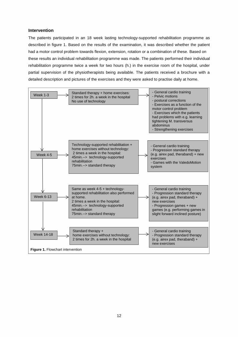
Week 1-3
Standard therapy + home exercises: 2 times for 2h. a week in the hospital No use of technology
Technology-supported rehabilitation + home exercises without technology: 2 times a week in the hospital: 45min.--> technology-supported rehabilitation 75min.--> standard therapy
- General cardio training - Progression standard therapy (e.g. airex pad, theraband) + new exercises - Games with the ValedoMotion system
Week 4-5
Same as week 4-5 + technology-supported rehabilitation also performed at home. 2 times a week in the hospital: 45min.--> technology-supported rehabilitation 75min.--> standard therapy
Week 6-13
Standard therapy + home exercises without technology: 2 times for 2h. a week in the hospital
- General cardio training - Progression standard therapy (e.g. airex pad, theraband) + new exercises
Week 14-18
- General cardio training - Pelvic motions - postural corrections - Exercises as a function of the motor control problem - Exercises which the patients had problems with e.g. learning tightening M. transversus abdominus - Strengthening exercises
- General cardio training - Progression standard therapy (e.g. airex pad, theraband) + new exercises - Progression games + new games (e.g. performing games in slight forward inclined posture)
Intervention
The patients participated in an 18 week lasting technology-supported rehabilitation programme as
described in figure 1. Based on the results of the examination, it was described whether the patient
had a motor control problem towards flexion, extension, rotation or a combination of these. Based on
these results an individual rehabilitation programme was made. The patients performed their individual
rehabilitation programme twice a week for two hours (h.) in the exercise room of the hospital, under
partial supervision of the physiotherapists being available. The patients received a brochure with a
detailed description and pictures of the exercises and they were asked to practise daily at home.
Figure 1. Flowchart intervention
13
STANDARD THERAPY was a multidisciplinary therapy that consisted of exercise therapy and back
school. This back school programme existed of five sessions in which psychological advice,
ergonomic advice and information on anatomy was given. If the patient required more information,
they could always rely on the advice of the complete team of therapists.
The exercise therapy consisted of general cardio training like cycling and cross trainer and an
individual programme. This exercise programme was composed after the individual screening and was
based on the principles of O’sullivan, Sahrmann and Comerford and Mottram.32,46,47
For each patient
the programme started with the awareness of a neutral position of the lumbar spine (LS), followed by
learning how to do pelvic movements. If the patient had become aware of the neutral position, specific
situations were dealt with such as reaching out to and picking up an object through segmentation.48
Consequently, specific exercises were chosen within the scope of the motor control problem. For
instance, with a motor control problem towards flexion, was worked by means of the standing bow. In
case of related problems such as difficulties with tightening the M. transversus abdominus, extra
exercises were taught. If necessary, also strengthening exercises were added to the programme.
In a further phase, progressions or new exercises were used. For example, a weight could be added
or exercises could be carried out on an unstable surface. It was individually evaluated in case of which
functional tasks the patient mentioned pain, for instance in case of cleaning the windows. The patient
was taught to keep the LS in a neutral position during these tasks.
Home exercises were also a part of the standard therapy. The patients were able to carry out the
exercises at home using the brochure.
TECHNOLOGY-SUPPORTED REHABILITATION was a continuation of the standard therapy by
means of playing games and providing feedback.
The technology used was the ValedoMotion system.49
The ValedoMotion system uses two motion
sensors that are attached with tape, respectively at the level of L1 and S1. These sensors are
connected to a computer that registers movements at the level of L1-S1.
The device offered two possibilities.
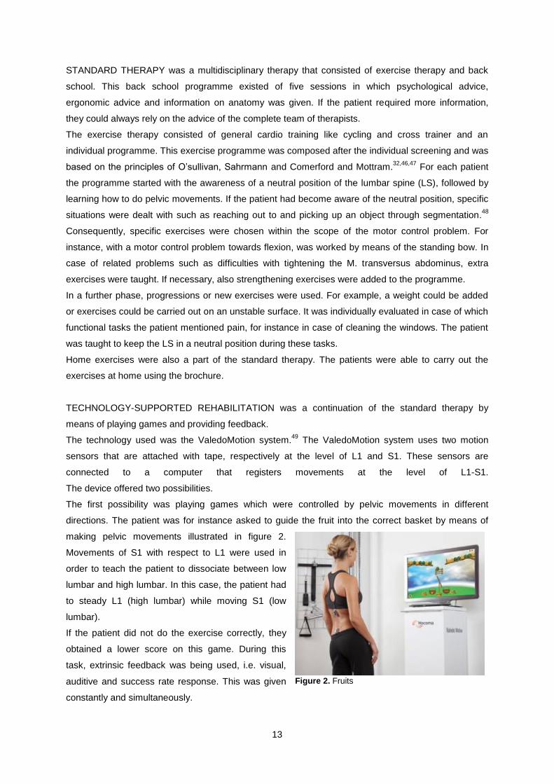
The first possibility was playing games which were controlled by pelvic movements in different
directions. The patient was for instance asked to guide the fruit into the correct basket by means of
making pelvic movements illustrated in figure 2.
Movements of S1 with respect to L1 were used in
order to teach the patient to dissociate between low
lumbar and high lumbar. In this case, the patient had
to steady L1 (high lumbar) while moving S1 (low
lumbar).
If the patient did not do the exercise correctly, they
obtained a lower score on this game. During this
task, extrinsic feedback was being used, i.e. visual,
auditive and success rate response. This was given
constantly and simultaneously.
Figure 2. Fruits
14
In a later phase, the degree of difficulty was increased or a more difficult position while carrying out the
game was asked. For example, the patient had to play the game in a slight forward inclined position.
Also more difficult games were added where the patient had to move in several planes, such as pelvic
movement in the sagittal and frontal plane.
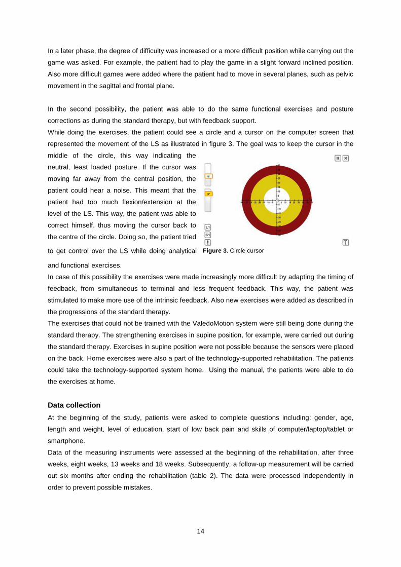
In the second possibility, the patient was able to do the same functional exercises and posture
corrections as during the standard therapy, but with feedback support.
While doing the exercises, the patient could see a circle and a cursor on the computer screen that
represented the movement of the LS as illustrated in figure 3. The goal was to keep the cursor in the
middle of the circle, this way indicating the
neutral, least loaded posture. If the cursor was
moving far away from the central position, the
patient could hear a noise. This meant that the
patient had too much flexion/extension at the
level of the LS. This way, the patient was able to
correct himself, thus moving the cursor back to
the centre of the circle. Doing so, the patient tried
to get control over the LS while doing analytical
and functional exercises.
In case of this possibility the exercises were made increasingly more difficult by adapting the timing of
feedback, from simultaneous to terminal and less frequent feedback. This way, the patient was
stimulated to make more use of the intrinsic feedback. Also new exercises were added as described in
the progressions of the standard therapy.
The exercises that could not be trained with the ValedoMotion system were still being done during the
standard therapy. The strengthening exercises in supine position, for example, were carried out during
the standard therapy. Exercises in supine position were not possible because the sensors were placed
on the back. Home exercises were also a part of the technology-supported rehabilitation. The patients
could take the technology-supported system home. Using the manual, the patients were able to do
the exercises at home.
Data collection
At the beginning of the study, patients were asked to complete questions including: gender, age,
length and weight, level of education, start of low back pain and skills of computer/laptop/tablet or
smartphone.
Data of the measuring instruments were assessed at the beginning of the rehabilitation, after three
weeks, eight weeks, 13 weeks and 18 weeks. Subsequently, a follow-up measurement will be carried
out six months after ending the rehabilitation (table 2). The data were processed independently in
order to prevent possible mistakes.
Figure 3. Circle cursor
15
Table 2. Measurements
T0 T1 T2 T3 T4 T5
NPRS x x x x x x
Patient satisfaction x x x x x
PSEQ x x x x x x
RMDQ x x x x x x
PSFS x x x x x x
TSK x x x x x x
CEQ* x x x x
IMI* x x x x
SF-36 x x x x x x
Drop-outs x x x x x
T0 = start rehabilitation; T1 = end week 3; T2 = end week 8; T3 = end week 13 (end technological-supported rehabilitation); T4 = end week 18 (end full rehabilitation); T5 = 6 months after the end of the rehabilitation. NPRS: Numeric pain rating scale; PSEQ: pain self-efficacy questionnaire; RMDQ: Roland morris disability questionnaire; PSFS: patient specific functioning scale; TSK: Tampa Scale of Kinesiophobia; CEQ: Credibility and expectancy questionnaire; IMI: Intrinsic motivation inventory; SF-36: Short-Form 36. *T0 en T1 were examined after the first treatment at week 1 and 4.
Outcome measures
Primary outcome measures
Numeric pain rating scale (NPRS)50,51
was used to measure the pain intensity on a scale of 0-10, 10
being the worst conceivable pain.
The satisfaction about the treatment was measured with the patient satisfaction scale of 0-10, 10
meaning very satisfied.
The self-efficacy questionnaire (PSEQ)52
measured the self-confidence of the patient. The patient
encircled the number that matched the best with their feeling, zero being completely not confident and
six meaning completely confident.
To measure the functional disability of the patients, the Roland morris disability questionnaire
(RMDQ)51,53
was used. This consists of 24 statements for which the patients have to indicate whether
they apply to them or not.
The patient specific functioning scale (PSFS)54
was also used to measure three to five specific
functional activities being important to the patient and which at the beginning could not be carried out
or could only be carried out with difficulty because of their CLBP.
The tampa scale of kinesiophobia (TSK)51,55,56
gave the impression of pain-related fear. It is a 17-items
questionnaire, where the patient have to fill in ‘agree’ or ‘disagree’.
16
Secondary outcome measures
The credibility and expectancy questionnaire (CEQ)44,57
measured the treatment expectancies and the
credibility of the rehabilitation programme. In total, six questions are asked about the confidence and
the feeling they had with the rehabilitation.
Intrinsic motivation was measured with the intrinsic motivation inventory (IMI).58
More specifically, it
measures by means of 35 questions the patient’s interest/enjoyment, perceived competence,
effort/importance, pressure/tension, value/usefulness and relatedness.
Also the quality of life and the physical/mental and social health were measured using the short form
36.51,59
The SF-36 consists of physical functioning, role limitations due to physical health, pain, general
health, role limitations due to emotional problems, energy/fatigue, emotional well-being and social
functioning. These criteria can be divided in a physical and mental component.60
The components are
calculated on the basis of average values.
The physical component consists of the categories: physical functioning, role limitations due to
physical health, pain and general health.
The mental component consists of the following categories: role limitations due to emotional
problems, energy/fatigue, emotional well-being and social functioning.
Finally, the drop outs were measured.
17
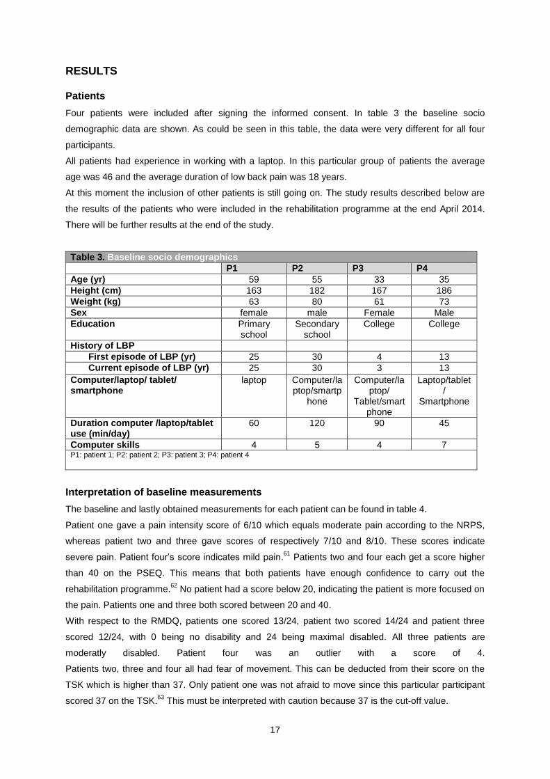
RESULTS Patients
Four patients were included after signing the informed consent. In table 3 the baseline socio
demographic data are shown. As could be seen in this table, the data were very different for all four
participants.
All patients had experience in working with a laptop. In this particular group of patients the average
age was 46 and the average duration of low back pain was 18 years.
At this moment the inclusion of other patients is still going on. The study results described below are
the results of the patients who were included in the rehabilitation programme at the end April 2014.
There will be further results at the end of the study.
Table 3. Baseline socio demographics
P1 P2 P3 P4
Age (yr) 59 55 33 35
Height (cm) 163 182 167 186
Weight (kg) 63 80 61 73
Sex female male Female Male
Education Primary school
Secondary school
College College
History of LBP
First episode of LBP (yr) 25 30 4 13
Current episode of LBP (yr) 25 30 3 13
Computer/laptop/ tablet/ smartphone
laptop Computer/laptop/smartp
hone
Computer/laptop/
Tablet/smartphone
Laptop/tablet/
Smartphone
Duration computer /laptop/tablet use (min/day)
60 120 90 45
Computer skills 4 5 4 7 P1: patient 1; P2: patient 2; P3: patient 3; P4: patient 4
Interpretation of baseline measurements
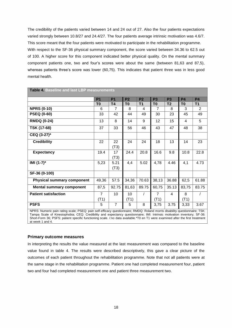
The baseline and lastly obtained measurements for each patient can be found in table 4.
Patient one gave a pain intensity score of 6/10 which equals moderate pain according to the NRPS,
whereas patient two and three gave scores of respectively 7/10 and 8/10. These scores indicate
severe pain. Patient four’s score indicates mild pain.61
Patients two and four each get a score higher
than 40 on the PSEQ. This means that both patients have enough confidence to carry out the
rehabilitation programme.62
No patient had a score below 20, indicating the patient is more focused on
the pain. Patients one and three both scored between 20 and 40.
With respect to the RMDQ, patients one scored 13/24, patient two scored 14/24 and patient three
scored 12/24, with 0 being no disability and 24 being maximal disabled. All three patients are
moderatly disabled. Patient four was an outlier with a score of 4.
Patients two, three and four all had fear of movement. This can be deducted from their score on the
TSK which is higher than 37. Only patient one was not afraid to move since this particular participant
scored 37 on the TSK.63
This must be interpreted with caution because 37 is the cut-off value.
18
The credibility of the patients varied between 14 and 24 out of 27. Also the four patients expectations
varied strongly between 10.8/27 and 24.4/27. The four patients average intrinsic motivation was 4.6/7.
This score meant that the four patients were motivated to participate in the rehabilitation programme.
With respect to the SF-36 physical summary component, the score varied between 34.36 to 62.5 out
of 100. A higher score for this component indicated better physical quality. On the mental summary
component patients one, two and four’s scores were about the same (between 81,63 and 87,5),
whereas patients three’s score was lower (60,75). This indicates that patient three was in less good
mental health.
Table 4. Baseline and last LBP measurements
P1 P1 P2 P2 P3 P3 P4 P4
T0 T4 T0 T1 T0 T2 T0 T1
NPRS (0-10) 6 7 8 4 7 8 3 2
PSEQ (0-60) 33 42 44 49 30 23 45 49
RMDQ (0-24) 13 8 14 9 12 15 4 5
TSK (17-68) 37 33 56 46 43 47 48 38
CEQ (3-27)*
Credibility 22 22
(T3)
24 24 18 13 14 23
Expectancy 19.4 17
(T3)
24.4 20.8 16.6 9.8 10.8 22.8
IMI (1-7)* 5,23 5.21
(T3)
4,4 5.02 4,78 4.46 4,1 4.73
SF-36 (0-100)
Physical summary component 49,36 57.5 34,36 70.63 38,13 36.88 62,5 61.88
Mental summary component 87,5 92.75 81,63 89.75 60,75 35.13 83,75 83.75
Patient satisfaction 7
(T1)
10 10
(T1)
/ 7
(T1)
4 8
(T1)
/
PSFS 5 7 5 8 3,75 3.75 3,33 3.67
NPRS: Numeric pain rating scale; PSEQ: pain self-efficacy questionnaire; RMDQ: Roland morris disability questionnaire; TSK: Tampa Scale of Kinesiophobia; CEQ: Credibility and expectancy questionnaire; IMI: Intrinsic motivation inventory; SF-36: Short-Form 36; PSFS: patient specific functioning scale. /:no data available.*T0 en T1 were examined after the first treatment at week 1 and 4.
Primary outcome measures
In interpreting the results the value measured at the last measurement was compared to the baseline
value found in table 4. The results were described descriptively, this gave a clear picture of the
outcomes of each patient throughout the rehabilitation programme. Note that not all patients were at
the same stage in the rehabilitation programme. Patient one had completed measurement four, patient
two and four had completed measurement one and patient three measurement two.
19
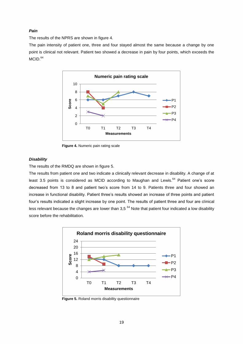
Pain
The results of the NPRS are shown in figure 4.
The pain intensity of patient one, three and four stayed almost the same because a change by one
point is clinical not relevant. Patient two showed a decrease in pain by four points, which exceeds the
MCID.64
Figure 4. Numeric pain rating scale
Disability
The results of the RMDQ are shown in figure 5.
The results from patient one and two indicate a clinically relevant decrease in disability. A change of at
least 3.5 points is considered as MCID according to Maughan and Lewis.64
Patient one’s score
decreased from 13 to 8 and patient two’s score from 14 to 9. Patients three and four showed an
increase in functional disability. Patient three’s results showed an increase of three points and patient
four’s results indicated a slight increase by one point. The results of patient three and four are cl inical
less relevant because the changes are lower than 3,5 64
Note that patient four indicated a low disability
score before the rehabilitation.
Figure 5. Roland morris disability questionnaire
0
2
4
6
8
10
T0 T1 T2 T3 T4
Sc
ore
Measurements
Numeric pain rating scale
P1
P2
P3
P4
0
4
8
12
16
20
24
T0 T1 T2 T3 T4
Sco
re
Measurements
Roland morris disability questionnaire
P1
P2
P3
P4
20
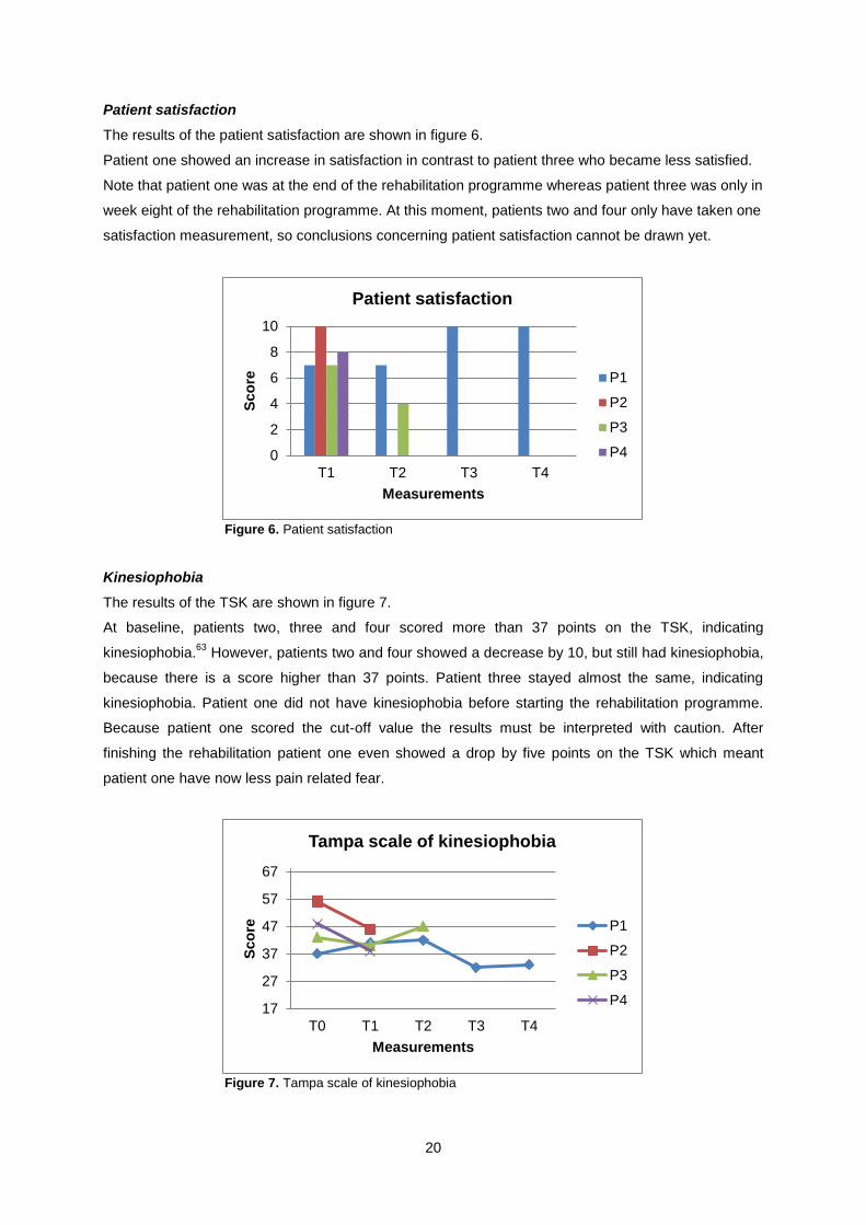
Patient satisfaction
The results of the patient satisfaction are shown in figure 6.
Patient one showed an increase in satisfaction in contrast to patient three who became less satisfied.
Note that patient one was at the end of the rehabilitation programme whereas patient three was only in
week eight of the rehabilitation programme. At this moment, patients two and four only have taken one
satisfaction measurement, so conclusions concerning patient satisfaction cannot be drawn yet.
Figure 6. Patient satisfaction
Kinesiophobia
The results of the TSK are shown in figure 7.
At baseline, patients two, three and four scored more than 37 points on the TSK, indicating
kinesiophobia.63
However, patients two and four showed a decrease by 10, but still had kinesiophobia,
because there is a score higher than 37 points. Patient three stayed almost the same, indicating
kinesiophobia. Patient one did not have kinesiophobia before starting the rehabilitation programme.
Because patient one scored the cut-off value the results must be interpreted with caution. After
finishing the rehabilitation patient one even showed a drop by five points on the TSK which meant
patient one have now less pain related fear.
Figure 7. Tampa scale of kinesiophobia
0
2
4
6
8
10
T1 T2 T3 T4
Sco
re
Measurements
Patient satisfaction
P1
P2
P3
P4
17
27
37
47
57
67
T0 T1 T2 T3 T4
Sco
re
Measurements
Tampa scale of kinesiophobia
P1
P2
P3
P4
21
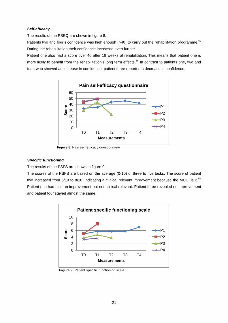
Self-efficacy
The results of the PSEQ are shown in figure 8.
Patients two and four’s confidence was high enough (>40) to carry out the rehabilitation programme.62
During the rehabilitation their confidence increased even further.
Patient one also had a score over 40 after 18 weeks of rehabilitation. This means that patient one is
more likely to benefit from the rehabilitation’s long term effects.65
In contrast to patients one, two and
four, who showed an increase in confidence, patient three reported a decrease in confidence.
Figure 8. Pain self-efficacy questionnaire
Specific functioning
The results of the PSFS are shown in figure 9.
The scores of the PSFS are based on the average (0-10) of three to five tasks. The score of patient
two increased from 5/10 to 8/10, indicating a clinical relevant improvement because the MCID is 2.54
Patient one had also an improvement but not clinical relevant. Patient three revealed no improvement
and patient four stayed almost the same.
Figure 9. Patient specific functioning scale
0
10
20
30
40
50
60
T0 T1 T2 T3 T4
Sco
re
Measurements
Pain self-efficacy questionnaire
P1
P2
P3
P4
0
2
4
6
8
10
T0 T1 T2 T3 T4
Sco
re
Measurements
Patient specific functioning scale
P1
P2
P3
P4
22
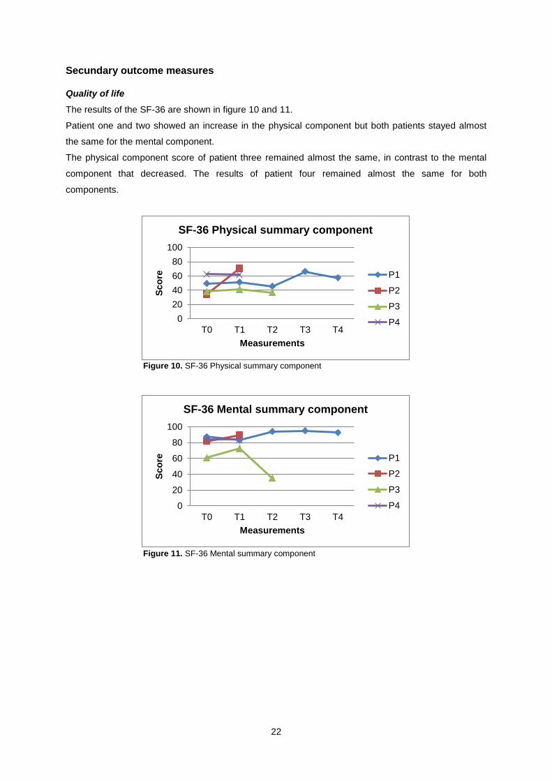
Secundary outcome measures Quality of life
The results of the SF-36 are shown in figure 10 and 11.
Patient one and two showed an increase in the physical component but both patients stayed almost
the same for the mental component.
The physical component score of patient three remained almost the same, in contrast to the mental
component that decreased. The results of patient four remained almost the same for both
components.
Figure 10. SF-36 Physical summary component
Figure 11. SF-36 Mental summary component
0
20
40
60
80
100
T0 T1 T2 T3 T4
Sco
re
Measurements
SF-36 Physical summary component
P1
P2
P3
P4
0
20
40
60
80
100
T0 T1 T2 T3 T4
Sco
re
Measurements
SF-36 Mental summary component
P1
P2
P3
P4
23
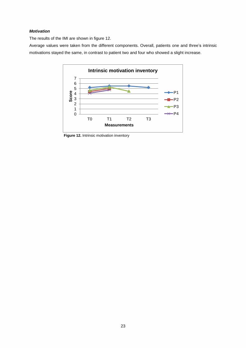
Motivation
The results of the IMI are shown in figure 12.
Average values were taken from the different components. Overall, patients one and three’s intrinsic
motivations stayed the same, in contrast to patient two and four who showed a slight increase.
Figure 12. Intrinsic motivation inventory
0
1
2
3
4
5
6
7
T0 T1 T2 T3
Sco
re
Measurements
Intrinsic motivation inventory
P1
P2
P3
P4
24
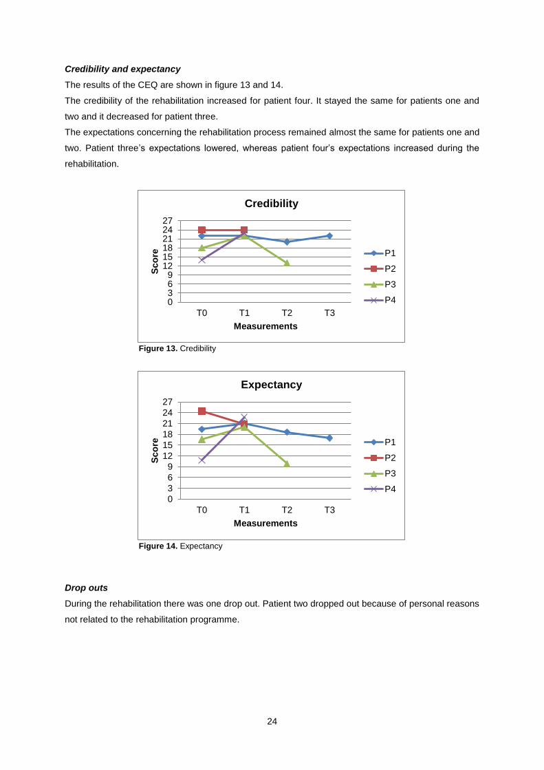
Credibility and expectancy
The results of the CEQ are shown in figure 13 and 14.
The credibility of the rehabilitation increased for patient four. It stayed the same for patients one and
two and it decreased for patient three.
The expectations concerning the rehabilitation process remained almost the same for patients one and
two. Patient three’s expectations lowered, whereas patient four’s expectations increased during the
rehabilitation.
Figure 13. Credibility
Figure 14. Expectancy
Drop outs
During the rehabilitation there was one drop out. Patient two dropped out because of personal reasons
not related to the rehabilitation programme.
0369
121518212427
T0 T1 T2 T3
Sco
re
Measurements
Credibility
P1
P2
P3
P4
0
3
6
9
12
15
18
21
24
27
T0 T1 T2 T3
Sco
re
Measurements
Expectancy
P1
P2
P3
P4
25
DISCUSSION The purpose of this study was to find out what the influence was of an 18 week lasting technology-
supported rehabilitation programme in which postural biofeedback was used in patients with CNSLBP,
on pain, disability and quality of life. In addition, the programs effects on motivation and the patients’
credibility and expectancy of the programme were examined.
The technology-supported programme used the ValedoMotion system in which patients rehabilitate
using games and feedback focused on functional activities. These functional activities are the main
difference with other studies that can be found in the literature. Many other studies use analytical
exercises in their rehabilitation programme.
A first technology-supported rehabilitation programme is real-time ultrasound feedback. Henry and
Teyhen36
does research about real-time ultrasound feedback to visualize the contraction of the
M.transversus abdominus and M.multifidus. This research suggests that it is a useful tool to improve
motor learning of the Transversus Abdominus and Multifidus muscles in patients with LBP. However,
further research is needed for more results. Another technology-supported rehabilitation programme is
the use of EMG biofeedback. EMG biofeedback can offer feedback to help patients reduce the tension
on their lumbar paraspinal muscles.37,38,39
Or EMG biofeedback can be used to strengthen these
muscle groups.40
This particular form of feedback shows a significant reduction in pain and depression
in patients suffering from CLBP.37,38,39
However, some studies also show that EMG biofeedback is not
better than cognitive behaviour therapy or no therapy at all.37,39
The studies mentioned above used technology-supported rehabilitation programs and feedback but in
contrast to the current study, they used another technology device and used feedback in analytical
exercises. The technology used in the current study provide feedback during functional activities. The
tasks and games the patients had to carry out were chosen in function of their transfer to everyday life
and activities. In doing so it was easier for patients to keep motor control over their everyday activities.
Further research is needed to confirm these findings.
Magnusson, Chow and Diamandopoulos et al35
does use a similar technology concept as the current
study but with analytical exercises. This study does research on postural biofeedback by using a
computer target programme.35
Patients are given three kinds of feedback: visual feedback, auditory
feedback and succes rates response. While using these kinds of feedback, patients have to match an
icon seen on their computer screen by moving their back. The study results indicate that using
postural biofeedback gives better outcomes than conventional CLBP therapy. More specifically,
postural biofeedback means an improvement in kinematic measurements and visual analogue scale.
In contrast to the study of Magnusson, Chow and Diamandopoulos et al35
, a relevant decrease in pain
was found by one of the four patient in the current study. A possible explanation for this could be that
not all patients have completed the rehabilitation programme yet. Further research is needed to draw
conclusions as to the evolution of patients pain.

26
Nevertheless there were some findings to present, such as patient one’s results indicated that even
though the patient’s pain did not improve, there were some positive effects. For example, the patients’
confidence increased and was more satisfied with the treatment. Furthermore patients’ functional
disabilities were reduced, kinesiophobia decreased and overall the quality of life improved. These
results indicated that, in order to achieve positive results through this kind of rehabilitation programme,
it was not necessary to reduce the pain intensity.
Furthermore, patient three’s pain scores showed a decrease at first, but later in the programme the
patients’ pain increased again. This trend was also seen in other outcome measures. A possible
explanation can be found in the amount of work load the patient was having at the time of the
measurement. At the time patient three was filling in the questionnaires for T2, the patient was
experiencing a higher work load. This might have made patient three’s experience the pain as worse
compared to moment T1. Work load and the stress could influence measurement results.
All patients were at different stages of the rehabilitation. In this respect patient one’s results gave a
clearer picture of the programs final outcomes than patients two, three and four.
At the moment patient two has interruped the programme because of personal reasons not related to
the rehabilitation programme. Apart from patient two there were no other drop outs or interruptions of
the programme.
In general there was a positive trend in several outcome measures and there were no side effects.
The criteria for an RCT were reached.
However there were some strengths and limitations of the current study. A strength of the current
study was that the patients already worked with the technology in the hospital before using it at home.
Due to the fact that the patients already used the technology, there were no problems in using it.
The limitations should be adapted with regards to an RCT. The main limitation was the small sample
size. There were some difficulties recruiting patients. In this study patients were sent by a physical
medicine specialist to a physiotherapist who performed a patient history and a clinical examination. A
lot of patients could not be included in the study because of exclusion criteria, such as previous
operations. Part of this problem could be solved if the physical medicine specialist would be better
informed as to which patients were suitable for the programme.
Another limitation was that there was no use of statistics, but only descriptive data.
Because of the small number of participants in this particular study it is hard to determine what the
results will be for a larger population. But, even though further research is needed to generalize the
findings of the current study.
27
CONCLUSION So far in this pilot study there is a positive trend in several outcome measures and there are no side
effects.
Key Points
This study was important for further research because there are rarely results available
for technology-supported rehabilitation with functional exercises.
There was a positive trend on several outcome measures.
Further research with larger sample size is necessary to generalize the findings of the
current study.

28
29
Reference background
1. Maher CG. Effective physical treatment for chronic low back pain. The Orthopedic clinics of North America
2004;35:57-64.
References article 1. Maher CG. Effective physical treatment for chronic low back pain. The Orthopedic clinics of North America
2004;35:57-64. 2. Dagenais S, Caro J, Haldeman S. A systematic review of low back pain cost of illness studies in the United
States and internationally. The spine journal : official journal of the North American Spine Society 2008;8:8-20. 3. Airaksinen O, Brox JI, Cedraschi C, et al. Chapter 4. European guidelines for the management of chronic non-
specific low back pain. European spine journal : official publication of the European Spine Society, the European Spinal Deformity Society, and the European Section of the Cervical Spine Research Society 2006;15 Suppl 2:S192-300.
4. Bevan S. MR, and Quadrello T. Musculoskeletale aandoeningen en de Belgische arbeidsmarkt Fit For Work 2009.
5. van Zundert J, van Kleef M. Low back pain: from algorithm to cost-effectiveness? Pain practice : the official journal of World Institute of Pain 2005;5:179-89.
6. Garcia AN, Gondo FL, Costa RA, et al. Effectiveness of the back school and mckenzie techniques in patients with chronic non-specific low back pain: a protocol of a randomised controlled trial. BMC musculoskeletal disorders 2011;12:179.
7. Leclere H, Beaulieu MD, Bordage G, et al. Why are clinical problems difficult? General practitioners' opinions concerning 24 clinical problems. CMAJ : Canadian Medical Association journal = journal de l'Association medicale canadienne 1990;143:1305-15.
8. Hermens HJ, Vollenbroek-Hutten MM. Towards remote monitoring and remotely supervised training. Journal of electromyography and kinesiology : official journal of the International Society of Electrophysiological Kinesiology 2008;18:908-19.
9. Perraton L, Machotka Z, Kumar S. Whole-body vibration to treat low back pain: fact or fad? Physiotherapy Canada. Physiotherapie Canada 2011;63:88-93.
10. del Pozo-Cruz B, Hernandez Mocholi MA, Adsuar JC, et al. Effects of whole body vibration therapy on main outcome measures for chronic non-specific low back pain: a single-blind randomized controlled trial. Journal of rehabilitation medicine : official journal of the UEMS European Board of Physical and Rehabilitation Medicine
2011;43:689-94. 11. Krein SL, Kadri R, Hughes M, et al. Pedometer-based internet-mediated intervention for adults with chronic
low back pain: randomized controlled trial. Journal of medical Internet research 2013;15:e181. 12. Del Pozo-Cruz B, Adsuar JC, Parraca J, et al. A web-based intervention to improve and prevent low back pain
among office workers: a randomized controlled trial. The Journal of orthopaedic and sports physical therapy 2012;42:831-41.
13. Brewer BR, McDowell SK, Worthen-Chaudhari LC. Poststroke upper extremity rehabilitation: a review of robotic systems and clinical results. Topics in stroke rehabilitation 2007;14:22-44.
14. Norouzi-Gheidari N, Archambault PS, Fung J. Effects of robot-assisted therapy on stroke rehabilitation in upper limbs: systematic review and meta-analysis of the literature. Journal of rehabilitation research and development 2012;49:479-96.
15. Hesse S, Tomelleri C, Bardeleben A, et al. Robot-assisted practice of gait and stair climbing in non-ambulatory stroke patients. Journal of rehabilitation research and development 2012;49:613-22.
16. Holden MK. Virtual environments for motor rehabilitation: review. Cyberpsychology & behavior : the impact of the Internet, multimedia and virtual reality on behavior and society 2005;8:187-211; discussion 2-9.
17. Timmermans AA, Lemmens RJ, Geers RP, et al. A comparison of treatment effects after sensor- and robot-based task-oriented arm training in highly functional stroke patients. Conference proceedings : ... Annual International Conference of the IEEE Engineering in Medicine and Biology Society. IEEE Engineering in Medicine and Biology Society. Conference 2011;2011:3507-10.
18. Timmermans AA, Seelen HA, Geers RP, et al. Sensor-based arm skill training in chronic stroke patients: results on treatment outcome, patient motivation, and system usability. IEEE transactions on neural systems and rehabilitation engineering : a publication of the IEEE Engineering in Medicine and Biology Society 2010;18:284-92.
19. Lange B, Chang CY, Suma E, et al. Development and evaluation of low cost game-based balance rehabilitation tool using the Microsoft Kinect sensor. Conference proceedings : ... Annual International Conference of the IEEE Engineering in Medicine and Biology Society. IEEE Engineering in Medicine and Biology Society. Conference 2011;2011:1831-4.
20. Oess NP, Wanek J, van Hedel HJ. Enhancement of bend sensor properties as applied in a glove for use in neurorehabilitation settings. Conference proceedings : ... Annual International Conference of the IEEE Engineering in Medicine and Biology Society. IEEE Engineering in Medicine and Biology Society. Conference 2010;2010:5903-6.
30
21. Wade E, Winstein CJ. Virtual reality and robotics for stroke rehabilitation: where do we go from here? Topics in stroke rehabilitation 2011;18:685-700.
22. Descarreaux M, Blouin JS, Teasdale N. Repositioning accuracy and movement parameters in low back pain subjects and healthy control subjects. European spine journal : official publication of the European Spine Society, the European Spinal Deformity Society, and the European Section of the Cervical Spine Research Society 2005;14:185-91.
23. Panjabi MM. A hypothesis of chronic back pain: ligament subfailure injuries lead to muscle control dysfunction. European spine journal : official publication of the European Spine Society, the European Spinal Deformity Society, and the European Section of the Cervical Spine Research Society 2006;15:668-76.
24. Brumagne S, Janssens L, Knapen S, et al. Persons with recurrent low back pain exhibit a rigid postural control strategy. European spine journal : official publication of the European Spine Society, the European Spinal Deformity Society, and the European Section of the Cervical Spine Research Society 2008;17:1177-84.
25. Sterling M, Jull G, Wright A. The effect of musculoskeletal pain on motor activity and control. The journal of pain : official journal of the American Pain Society 2001;2:135-45.
26. Jacobs JV, Henry SM, Nagle KJ. People with chronic low back pain exhibit decreased variability in the timing of their anticipatory postural adjustments. Behavioral neuroscience 2009;123:455-8.
27. Hodges PW. Changes in motor planning of feedforward postural responses of the trunk muscles in low back pain. Experimental brain research 2001;141:261-6.
28. Brumagne S, Lysens R, Spaepen A. Lumbosacral repositioning accuracy in standing posture: a combined electrogoniometric and videographic evaluation. Clinical biomechanics 1999;14:361-3.
29. O'Sullivan PB, Burnett A, Floyd AN, et al. Lumbar repositioning deficit in a specific low back pain population. Spine 2003;28:1074-9.
30. Dolan KJ, Green A. Lumbar spine reposition sense: the effect of a 'slouched' posture. Manual therapy 2006;11:202-7.
31. Hides JA, Richardson CA, Jull GA. Multifidus muscle recovery is not automatic after resolution of acute, first-episode low back pain. Spine 1996;21:2763-9.
32. O'Sullivan P. Diagnosis and classification of chronic low back pain disorders: maladaptive movement and motor control impairments as underlying mechanism. Manual therapy 2005;10:242-55.
33. Ribeiro DC, Sole G, Abbott JH, et al. Extrinsic feedback and management of low back pain: A critical review of the literature. Manual therapy 2011;16:231-9.
34. Richard A, Wrisberg SaCA. Motor learning and performance: a situation-based learning approach., 2008. 35. Magnusson ML, Chow DH, Diamandopoulos Z, et al. Motor control learning in chronic low back pain. Spine
2008;33:E532-8. 36. Henry S, Teyhen D. Ultrasound Imaging as a Feedback Tool in the Rehabilitation of Trunk Muscle Dysfunction
for People With Low Back Pain. journal of orthopaedic & sports physical therapy 2007;37:627e34. 37. Newton-John TR, Spence SH, Schotte D. Cognitive-behavioural therapy versus EMG biofeedback in the
treatment of chronic low back pain. Behaviour research and therapy 1995;33:691-7. 38. Donaldson S, Romney D, Donaldson M, et al. Randomized study of the application of single motor unit
biofeedback training to chronic low back pain. Journal of occupational rehabilitation 1994;4:23-37. 39. Bush C, Ditto B, Feuerstein M. A controlled evaluation of paraspinal EMG biofeedback in the treatment of
chronic low back pain. Health psychology : official journal of the Division of Health Psychology, American Psychological Association 1985;4:307-21.
40. Asfour SS, Khalil TM, Waly SM, et al. Biofeedback in back muscle strengthening. Spine 1990;15:510-3. 41. Shepherd RB. Exercise and training to optimize functional motor performance in stroke: driving neural
reorganization? Neural plasticity 2001;8:121-9. 42. van Vliet PM, Heneghan NR. Motor control and the management of musculoskeletal dysfunction. Manual
therapy 2006;11:208-13. 43. Shumway-Cook A, Woollacott MH. Motor control: translating researchinto clinical practice ed. px. Philadelphia,
PA; London: LippincottWilliams &Wilkins, 2007. 44. Smeets RJ, Beelen S, Goossens ME, et al. Treatment expectancy and credibility are associated with the
outcome of both physical and cognitive-behavioral treatment in chronic low back pain. The Clinical journal of pain 2008;24:305-15.
45. Luomajoki H, Kool J, de Bruin ED, et al. Movement control tests of the low back; evaluation of the difference between patients with low back pain and healthy controls. BMC musculoskeletal disorders 2008;9:170.
46. Sahrmann S. Diagnosis and treatment of movement impairment syndromesed. St. Louis Mosby, 2001. 47. Comerford M, Mottram S. Kinetic Control: The Management of Uncontrolled Movement ed. Australia: Elsevier
Health, 2012 48. Hodges PW. Core stability exercise in chronic low back pain. The Orthopedic clinics of North America
2003;34:245-54. 49. Bauer C, Baumgartner L, Schelldorfer S et al. Technical validation of a new movement therapy system for
treatment of low back pain. Gait Posture 2012 36:40-1. 50. Von Korff M, Jensen MP, Karoly P. Assessing global pain severity by self-report in clinical and health services
research. Spine 2000;25:3140-51.
51. Chapman JR, Norvell DC, Hermsmeyer JT, et al. Evaluating common outcomes for measuring treatment success for chronic low back pain. Spine 2011;36:S54-68.
52. Nicholas MK. The pain self-efficacy questionnaire: Taking pain into account. European journal of pain 2007;11:153-63.
31
53. Roland M, Morris R. A study of the natural history of back pain. Part I: development of a reliable and sensitive measure of disability in low-back pain. Spine 1983;8:141-4.
54. Stratford PW, Gill C, Westaway M, et al. Assessing disability and change on individual patients: a report of patient-specific measure. Physiotherapy Canada 1995 47:258-63.
55. Vlaeyen JW, Kole-Snijders AM, Boeren RG, et al. Fear of movement/(re)injury in chronic low back pain and its relation to behavioral performance. Pain 1995;62:363-72.
56. Lame IE, Peters ML, Kessels AG, et al. Test--retest stability of the Pain Catastrophizing Scale and the Tampa Scale for Kinesiophobia in chronic pain over a longer period of time. Journal of health psychology 2008;13:820-6.
57. Devilly GJ, Borkovec TD. Psychometric properties of the credibility/expectancy questionnaire. Journal of behavior therapy and experimental psychiatry 2000;31:73-86.
58. Deci EL, Ryan R. The “What” and “Why” of goal pursuits: Human needs and the self-detremination of behavior. Psychol Inq. 2000 11:227-68.
59. Walsh TL, Hanscom B, Lurie JD, et al. Is a condition-specific instrument for patients with low back pain/leg symptoms really necessary? The responsiveness of the Oswestry Disability Index, MODEMS, and the SF-36. Spine 2003;28:607-15.
60. Ware JE, Kosinski M, Keller SD. SF-36 Physical and Mental Health Summary Scales: A User’s Manual.ed. Boston, MA: The Health Institute: QualityMetric 1994.
61. MacCaffery M, Beebe A. Pain: Clinical manual for nursing practiceed: Mosby St. Louis, 1989. 62. Frost H, Klaber MJ, Moser J, et al. Evaluation of a fitness programme for patients with chronic low back pain.
Brit Med J 1993;310:151-4. 63. Miller RP, Kori SH, Todd DD. Tampa Scale. Tampa, FL 1991. 64. Maughan EF, Lewis JS. Outcome measures in chronic low back pain. European spine journal : official
publication of the European Spine Society, the European Spinal Deformity Society, and the European Section of the Cervical Spine Research Society 2010;19:1484-94.
65. Keefe FJ, Rumble ME, Scipio CD, et al. Psychological aspects of persistent pain: current state of the science. The journal of pain : official journal of the American Pain Society 2004;5:195-211.
Auteursrechtelijke overeenkomst
Ik/wij verlenen het wereldwijde auteursrecht voor de ingediende eindverhandeling:
Technology-supported rehabilitation for patients with chronic non-specific
low back pain. Preliminary results of a pilot study
R i c h t i n g : m a s t e r i n d e r e v a l i d a t i e w e t e n s c h a p p e n e n d e
k i n e s i t h e r a p i e - r e v a l i d a t i e w e t e n s c h a p p e n e n k i n e s i t h e r a p i e b i j
musculoskeletale aandoeningen
Jaar: 2014
in alle mogelijke mediaformaten, - bestaande en in de toekomst te ontwikkelen - , aan de
Universiteit Hasselt.
Niet tegenstaand deze toekenning van het auteursrecht aan de Universiteit Hasselt
behoud ik als auteur het recht om de eindverhandeling, - in zijn geheel of gedeeltelijk -,
vrij te reproduceren, (her)publiceren of distribueren zonder de toelating te moeten
verkrijgen van de Universiteit Hasselt.
Ik bevestig dat de eindverhandeling mijn origineel werk is, en dat ik het recht heb om de
rechten te verlenen die in deze overeenkomst worden beschreven. Ik verklaar tevens dat
de eindverhandeling, naar mijn weten, het auteursrecht van anderen niet overtreedt.
Ik verklaar tevens dat ik voor het materiaal in de eindverhandeling dat beschermd wordt
door het auteursrecht, de nodige toelatingen heb verkregen zodat ik deze ook aan de
Universiteit Hasselt kan overdragen en dat dit duidelijk in de tekst en inhoud van de
eindverhandeling werd genotificeerd.
Universiteit Hasselt zal mij als auteur(s) van de eindverhandeling identificeren en zal geen
wijzigingen aanbrengen aan de eindverhandeling, uitgezonderd deze toegelaten door deze
overeenkomst.
Voor akkoord,
Vanherle, Lien Van Genechten, Sanne

![presentation SCHOLARSHIP 2018-2019 - Universiteit Hasselt · 2018. 10. 26. · Microsoft PowerPoint - presentation SCHOLARSHIP 2018-2019 [Compatibility Mode] Author: lucp7915 Created](https://img.pdfslide.us/doc/110x75/5fe6401521a41642cb6df792/presentation-scholarship-2018-2019-universiteit-hasselt-2018-10-26-microsoft.jpg)