Embed Size (px)
Citation preview
W550–W556 Nucleic Acids Research, 2016, Vol. 44, Web Server issue Published online 5 May 2016doi: 10.1093/nar/gkw393
ACFIS: a web server for fragment-based drugdiscoveryGe-Fei Hao1,†, Wen Jiang1,†, Yuan-Nong Ye2, Feng-Xu Wu1, Xiao-Lei Zhu1, Feng-Biao Guo2,*
and Guang-Fu Yang1,3,*
1Key Laboratory of Pesticide & Chemical Biology, Ministry of Education, College of Chemistry, Central China NormalUniversity, Wuhan 430079, P.R.China, 2Center of Bioinformatics and Key Laboratory for NeuroInformation of theMinistry of Education, University of Electronic Science and Technology of China, Chengdu 610054, P.R. China and3Collaborative Innovation Center of Chemical Science and Engineering, Tianjing 300072, P.R.China
Received January 31, 2016; Revised April 20, 2016; Accepted April 28, 2016
ABSTRACT
In order to foster innovation and improve the effec-tiveness of drug discovery, there is a considerableinterest in exploring unknown ‘chemical space’ toidentify new bioactive compounds with novel anddiverse scaffolds. Hence, fragment-based drug dis-covery (FBDD) was developed rapidly due to its ad-vanced expansive search for ‘chemical space’, whichcan lead to a higher hit rate and ligand efficiency(LE). However, computational screening of fragmentsis always hampered by the promiscuous bindingmodel. In this study, we developed a new web serverAuto Core Fragment in silico Screening (ACFIS). Itincludes three computational modules, PARA GEN,CORE GEN and CAND GEN. ACFIS can generatecore fragment structure from the active molecule us-ing fragment deconstruction analysis and perform insilico screening by growing fragments to the junc-tion of core fragment structure. An integrated en-ergy calculation rapidly identifies which fragmentsfit the binding site of a protein. We constructed asimple interface to enable users to view top-rankingmolecules in 2D and the binding mode in 3D for fur-ther experimental exploration. This makes the AC-FIS a highly valuable tool for drug discovery. TheACFIS web server is free and open to all users athttp://chemyang.ccnu.edu.cn/ccb/server/ACFIS/.
INTRODUCTION
It is widely recognized that drug discovery is a time-consuming, expensive and complex process. The search fornew drugs is mainly plagued by the rising cost and low suc-cess rate in Research and Development (R&D) (1). The goal
of drug lead discovery is to identify candidate molecule withimproved biological potency and physiochemical proper-ties. However, the number of compounds needed to be syn-thesized to deliver one new lead is continuously rising inrecent years (2). Both quality and quantity of hit classesavailable to medicinal chemists are primary drives for dis-covering best-in-class leads, which makes hit identificationa crucial step to reduce attrition and therefore improve theoverall R&D productivity (3). Thus, how to improve the ef-ficiency and reduce the time required for successful hit iden-tification will contribute greatly to drug discovery.
Over the past decade, high-throughput screening (HTS)of corporate compound decks has become the majorparadigm for hit discovery in big pharma. Up to a few mil-lion compounds can be screened against the target of in-terest. But challenges facing traditional HTS technologiesinclude low hit rates and hit molecule with low syntheticfeasibility and nondrug-like properties are often identified.Compared with HTS, fragment-based drug design (FBDD)has some significant advantages (4). First, FBDD identi-fies ligand using ligand efficiency (LE) rather than potencyalone to judge the relative order of molecules with differ-ent sizes, which offers more efficient and fruitful optimiza-tion campaigns. Second, FBDD only requires screening asmall number of fragments to reach a more expansive chem-ical space, which leads to a much broader range of activity.Hence, the hit rate of FBDD is typically much higher thanthat observed with HTS (5). However, the specialized andexpensive instruments, such as NMR spectroscopy, X-raydiffraction, surface plasmon resonance or mass spectrom-etry are always required to detect fragment binding due tothe relative weak affinity (6,7). In addition, a large amountof purified proteins (>10 mg) and high concentrations offragment molecules are always essential to achieve this task,which are very difficult to achieve in most cases (8,9).
*To whom correspondence should be addressed. Tel: +86 27 67867800; Fax: +86 27 67867141; Email: [email protected] may also be addressed to Feng-Biao Guo. Email: [email protected]†
These authors contributed equally to the work as first authors.
C© The Author(s) 2016. Published by Oxford University Press on behalf of Nucleic Acids Research.This is an Open Access article distributed under the terms of the Creative Commons Attribution License (http://creativecommons.org/licenses/by-nc/4.0/), whichpermits non-commercial re-use, distribution, and reproduction in any medium, provided the original work is properly cited. For commercial re-use, please [email protected]
Downloaded from https://academic.oup.com/nar/article-abstract/44/W1/W550/2499367by gueston 12 February 2018
Nucleic Acids Research, 2016, Vol. 44, Web Server issue W551
Hence, various computational methods have been devel-oped to perform FBDD (10–12). Molecule docking hasbeen used as a potentially attractive way to prioritize frag-ments from the much larger commercially available data set(11). Nevertheless, the problem of fitting small fragmentstargeting sub-pockets within the active site of the target pro-tein is different from fitting a larger ‘druglike’ molecule.The binding mode of a fragment is relatively more diffi-cult to be predicted by docking due to the smaller size.Meanwhile, scoring functions optimized according to drug-like molecules may be less accurate for predicting fragmentaffinities, which makes fragment docking problematic andtriggers critical discussions (11). Furthermore, commercialsoftwares like LUDI (13), GLIDE (14), etc. are proved to bepowerful enough to place fragments into the correct pocketof the active site. In addition, there is more and more freesoftwares dedicated to de novo drug design compared toother cheminformatic tools. For example, LigBuilder wasdeveloped for structure-based de novo drug design and opti-mization (15), and S4MPLE was developed as a conforma-tional sampling tool for in silico FBDD (16). However, mostof these tools are not web server facility, which makes themnot easily practicable for non computational scientists. Onlya minority of the web servers are dedicated to in silico drugdiscovery such as (17): MTiOpenScreen (http://bioserv.rpbs.univ-paris-diderot.fr/services/MTiOpenScreen/) (18),e-LEA3D (http://bioinfo.ipmc.cnrs.fr/lea.html) (19), iS-creen (http://iScreen.cmu.edu.tw/) (20), VSDocker (http://www.bio.nnov.ru/projects/vsdocker2/) (21), however, theyperform virtual screening of molecular libraries rather thanfragments using docking algorithm. This prompts us to de-velop a web server to perform FBDD.
Herein, we present Auto Core Fragment in silico Screen-ing (ACFIS), the first web server to perform computer-aided fragment-based drug discovery. It can generatecore fragment structure based on the primary activemolecule and perform automated fragment linking andvirtual screening. The computation is based on thepharmacophore-linked fragment virtual screening (PFVS)method (22). Several libraries containing fragments ex-tracted from known bioactive compounds are provided forscreening. The core fragment generation tool uses fragmentdeconstruction analysis to evaluate the contribution of eachfragment of the primary active molecule to the bindingaffinity. Another tool assists the user in the linking of newfragments to the selected core fragment and in the evaluat-ing of the contribution to the binding potency. Thus, ACFISenables researcher to perform computer-aided fragment-based drug discovery on traditional or more challengingprotein targets.
MATERIALS AND METHODS
Fragment library
Fragment library design is crucial for the effectiveness offragment-based virtual screening. The virtual library canbe designed as a set of compounds consisting of buildingblocks for synthetic feasibility, a set of structures obtainedby decomposition of larger molecules, or a set of commer-cially available molecules. The Rule of Three (Ro3) is ac-knowledged as the best definition of a fragment, i.e. a low
molecular weight (MW) ≤ 300 Da, a reduced lipophily in-dex clogP ≤ 3, a number of hydrogen bond donors and ac-ceptors ≤3, low flexibility represented by a number of ro-tatable bonds ≤3 and a polar surface area (PSA) ≤ 60 A2
(23,24).The decomposition approach was used to design combi-
natorial fragment libraries by the deconstruction of knownbioactive compounds (FDA approved drug and commer-cial pesticide molecules). Decomposition and Identificationof Molecules (DAIM) program was used to perform de-composition of small molecules (25). The fragment size iscontrolled by the number of bond breakages. The breakingrules are based on retrosynthetic analysis. The fragment li-brary derived from decomposition was filtered by Ro3. Inaddition to certain physicochemical property filters, the as-pect of availability is also important and should be checkedindividually. However, the fragments from decompositionshould already feature certain synthetic accessibility. De-pending on the application, two fragment libraries (drugand pesticide fragment database) are supplied and will becontinuously improved to expand its diversity.
Fragment deconstruction analysis
The FBDD approach can create novel lead compoundsand allow the exploration of a larger chemical space. Iteither grows functional fragments from the starting frag-ment or pieces several fragments together into novel scaf-fold, which are generally defined as growing or linking al-gorithms. Growing algorithms start from a core fragmentplaced in the binding site and other fragments which arelinked to improve activity. Linking algorithms, on the otherhand, involve docking of functional fragments to each sub-pockets followed by linking adjacent fragments together.The ACFIS web server creates new molecules based ongrowing algorithm. First, the core fragment structure is de-termined. Then, automatic fragment linking is performedto improve the binding affinity. Hence, how to select a corefragment structure is a major challenge.
Studies of drugs entering the market show that knowndrugs, clinical candidates and bioactive compounds rep-resent very attractive and valuable starting point for newlead compound discovery (26). Therefore, selecting corefragment structures from the known bioactive compoundsseems to be an effective strategy. The fragment decon-struction analysis is performed with a three-step compu-tational protocol shown in Supplementary Figure S1: Athree-step minimization procedure was performed on thebinding conformation of protein–ligand complex. (i) First,movement was allowed only for the ligand molecule witha harmonic constraints (100 kcal/mol•A2) applied to thecomplex. Second, the mainchain atoms of the protein werefixed and other atoms were allowed to move. Finally, allatoms were minimized with no restraint to a convergenceof 0.01 kcal/(mol·A). (ii) Ligand structure binding in thepocket is deconstructed into fragments according to the ret-rosynthetic analysis by using DAIM software. Single bondis broken and hydrogen is used to link with heavy atom tomake the total charge value of each ‘piece’ integer. In addi-tion, everything will be reparameterized including the par-tial charges according to new generated protein-fragment
Downloaded from https://academic.oup.com/nar/article-abstract/44/W1/W550/2499367by gueston 12 February 2018
W552 Nucleic Acids Research, 2016, Vol. 44, Web Server issue
structure. (iii) The binding free energy (�G) is calculated aspreviously described by the combination of the MM PBSAmethod (27) for the enthalpy and an empirical method forthe entropy (see details in the Supplementary Material) (28)for each protein–fragment structure. The computationalprocedure used to evaluate the binding free energy includesbinding energy, solvation entropy and conformational en-tropy (Equation 1), which is the same as that described inour previous publication (29).
�Gbind = �Ebind − T�Ssolv − T�Sconf =�Ebind − T�Ssolv + w(�Nrot)
(1)
The ranking of fragments is sorted according to LE de-fined as �G divided by the heavy atom count (HAC), LE= −�Gcal/HAC. The core fragment is derived from the re-assembly of the adjacent fragments based on the bindingcontribution. Hence, the obtained core fragment structurehas a highly conserved binding conformation and efficientcontribution to the entire binding affinity. Last, junction onthe core fragment is determined according to the surround-ing space (volume ≥ 50 A3).
Auto core fragment in silico screening
New fragment is linked to the junction of core fragmentstructure placed in the binding site using a modified versionof AutoGrow (30). The orientation of growing fragmentwas optimized with minimum steric clashes (overlap vol-ume ≤ 4 A3) to the surrounding residues. Because the PFVSmethod reasonably relies on the resemblance assumptionthat the structural perturbations associated with fragmentchanging are relatively small, which would only lead to localchanges of the protein structure and would not significantlychange the backbone structure of protein. This procedurepreserves the binding conformation of the core fragment.The energy minimization of each new ligand is achievedin four steps using the Sander module of Amber program(31). First, the fragment is minimized with the core frag-ment and the protein fixed. Then, the ligand is minimizedwith the protein fixed. Subsequently, the backbone atoms ofthe protein are fixed and other atoms are relaxed. The finalminimization is performed with both the ligand and proteinrelaxed. In each step, the energy minimization is executedby using the steepest descent method for the first 2000 cy-cles and the conjugated gradient method for the subsequent3000 cycles with a convergence criterion of 0.1 kcal mol−1
A−1. Finally, the MD simulation is performed using the gen-eralized Born continuum solvent model to further relax theconformation of the growing fragment. The last snapshotof the MD simulation was minimized to a convergence cri-terion of 0.1 kcal mol−1A−1 and used for �G calculation.Finally, hit candidates were selected according to the �Gvalue.
WEB SERVER
Web server configuration
ACFIS contains three computational modules.PARA GEN is a tool to generate parameters for AC-FIS, CORE GEN is a tool to derive core fragment
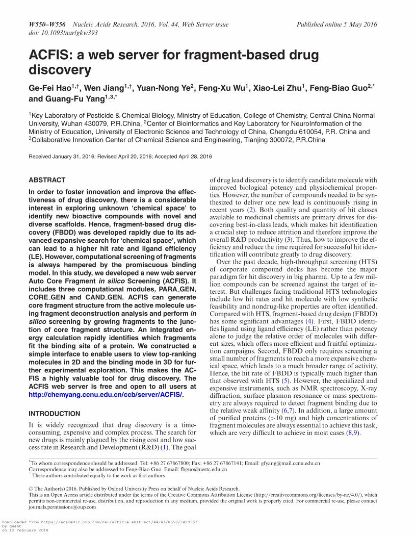
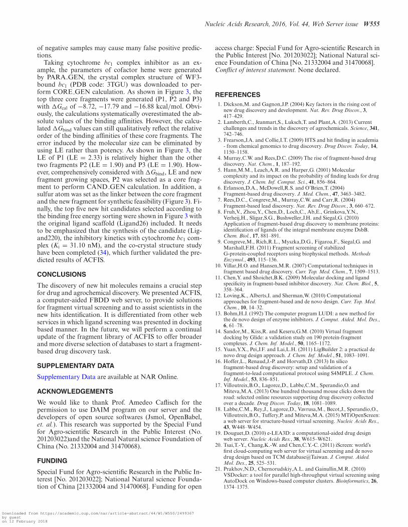
Figure 1. The interplay of the three ACFIS modules. PARA GEN runsin a serial mode, whereas CORE GEN and CAND GEN run in parallelmode.
structure from a bioactive molecule, and CAND GEN is atool to link fragments to the core fragment structure andgenerate candidates. The inputs of all modules are easy toprepare and some items are optional to meet the purpose ofindividual projects. Output structure files and related datacan be downloaded. Figure 1 shows how PARA GEN,CORE GEN and CAND GEN work together to makeonline FBDD possible. In order to make this server moreuser friendly, these modules are connected in the ‘primarymode’. After the submission of a complex pdb file, theserver will generate several cores and select the top-rankedcore fragment to perform fragment virtual screeningautomatically. To avoid the bugs of the uploaded pdb file,an initial file checking module is also developed in ‘primarymode’. The three modules can also be independently usedin the ‘Advanced Mode’. ACFIS server runs on a dedicatedLinux machine at the Supercomputer Cluster. The webapplication uses PHP (version 5.0), HTML and Java scriptto serve web pages. Related messages and results of eachtask are stored in a database implemented using MySQL(version 14.12). The web server runs on the apache HTTPserver version 2.0.51, and the JSmol interactive molecularviewer applet (http://www.jmol.org/) is used for structurevisualization. Chrome and Firefox are recommendedexplorers for our server. Screen with resolution higher than1440 × 900 are needed for the web pages.
For PARA GEN, ‘your job’ will start to run after yousubmit your job with a runtime of seconds to minutesbased on the size of your molecule and the charge method.CORE GEN will also manage your job immediately andfinish in a few hours. CAND GEN is a lengthy step de-pending on the size of the protein, Some tasks may takea few days. A job management system based on PHP andMYSQL has been developed to manage all the submittedjobs in this server.
Downloaded from https://academic.oup.com/nar/article-abstract/44/W1/W550/2499367by gueston 12 February 2018
Nucleic Acids Research, 2016, Vol. 44, Web Server issue W553
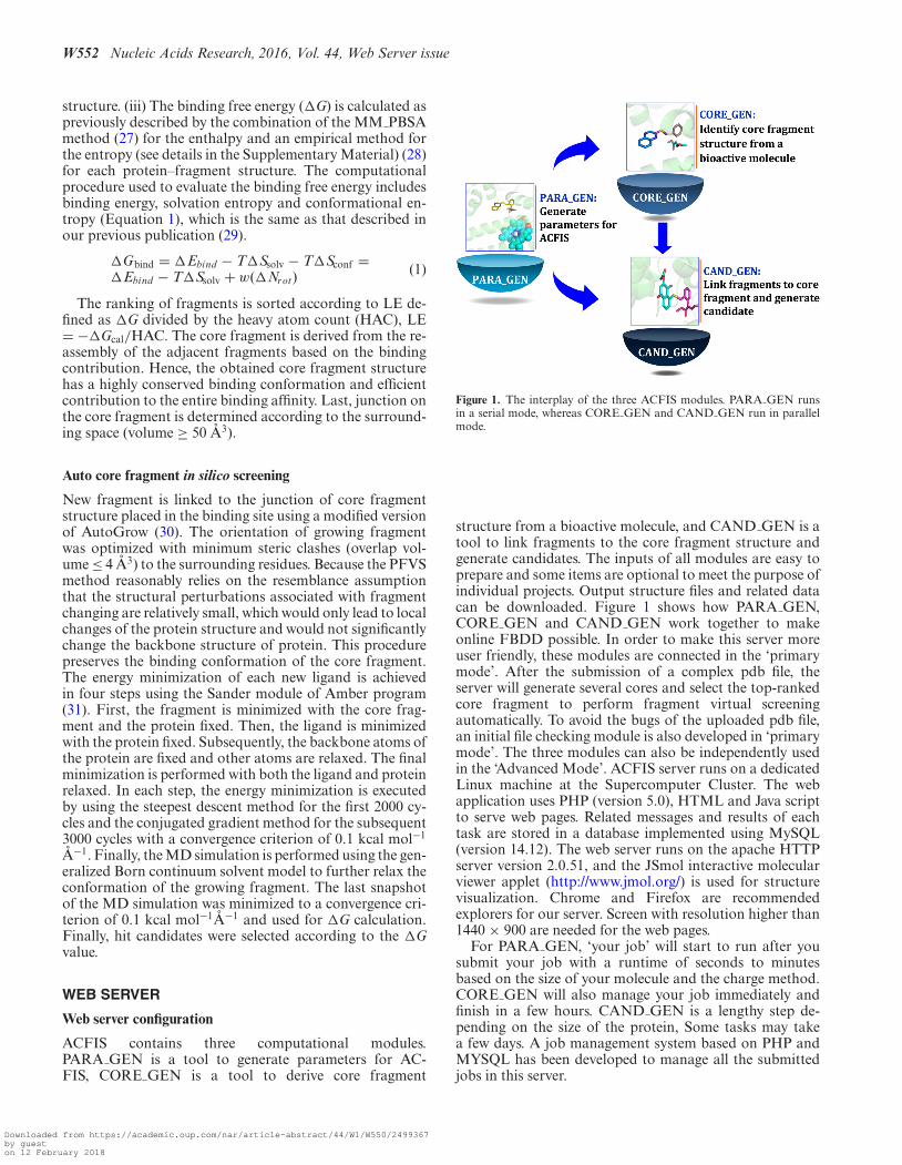
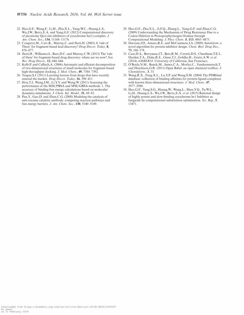
Figure 2. Workflow of ACFIS. The user input is shown in blue. The arrows denote the computational process.
PARA GEN: a parameter generation tool
Input. In the front-page, the user is invited to upload astructure file of a protein cofactor in mol2 or pdb for-mat. If there is no cofactor, this module can be skipped.A guideline at the bottom of the page explains how to usePARA GEN to set exact parameters for a selected ligand.Hydrogen atoms should be correctly assigned, which can bedone with most visualization software like AutoDock Tools,CHIMERA and PYMOL. Total formal charge should becorrectly assigned, which is an essential parameter for thecalculation of atomic charge. If the total formal charge isuncertain, it can be assigned automatically. Then a chargemethod from AM1-BCC, Muliken and Gasteiger optionscan be selected. And finally, an email address may be sub-mitted to receive the final result files.
Server workflow. Once a valid structure file of cofactor isuploaded, PARA GEN program starts to run (Figure 2).First, your uploaded pdb file will be converted into mol2 fileby using openbabel2.3.1 (32). Then, the total formal chargewill be calculated, if it is not already specified. Parameterfiles for AMBER force field are then prepared by AMBER-TOOLS.
Output. Result of PARA GEN contains all the parame-ter files of AMBER force field required by MD simulationand a log file recording the calculation process, which willbe available after your job successfully finished. If the up-loaded structure file is invalid, the job will be terminatedand an ‘error’ will be reported on the web page. Moreover,it is also linked with CORE GEN and CAND GEN mod-ule in the result page to start the following computation di-rectly.
CORE GEN: a core fragment generation tool
Input. A protein–ligand complex in pdb format is requiredas an input of CORE GEN. This can be obtained fromRCSB protein data bank (http://www.rcsb.org/pdb/home/
home.do) or acquired from docking calculation. Addition-ally, the name of ligand in the complex structure file shouldbe referred. Parameters for the cofactor will be required, ifit is in the complex structure. At last, a password should beassigned to make your job confidential and an email noticeis optional.
Server workflow. After the initial data validation,CORE GEN started to run with a three-step computa-tional protocol (Figure 2): (i) The minimization procedurewas performed for the complex structure. (ii) Ligand struc-ture was deconstructed into fragments by using DAIMprogram. (iii) The �G was calculated for each protein-fragment complexes. The ranking of fragments is sortedaccording to LE. Comparison based on LE rather thanpotency alone could be useful in deciding the potential offragments.
Output. The users are guided to the result page after theysubmit the job successfully. Messages about all submittedjobs are printed in this page. Once the job is completed, re-sults of CORE GEN are presented to the user via a webpage by a click on the ID of the job (the user is invitedto insert a password if it was assigned before). The sum-mary table shows the information related to each gener-ated core fragment structure (results shown in Supplemen-tary Figure S2 in the Supplementary Material). Due to theimportance of physicochemical properties for fragment se-lection, a link to mol-inspiration prediction server (http://www.molinspiration.com/) is created. The 3D structure ofeach protein-core fragment complex is shown with JSmolby a click on the structure, which can also be downloadedby clicking on the link. The junction of the core fragmentstructure is marked with a hydrogen atom shown as a ball inthe 3D view mode. In addition, CORE GEN is also linkedwith CAND GEN module in the result page to start thefollowing computation directly.
CAND GEN: a hit generation tool
Downloaded from https://academic.oup.com/nar/article-abstract/44/W1/W550/2499367by gueston 12 February 2018
W554 Nucleic Acids Research, 2016, Vol. 44, Web Server issue
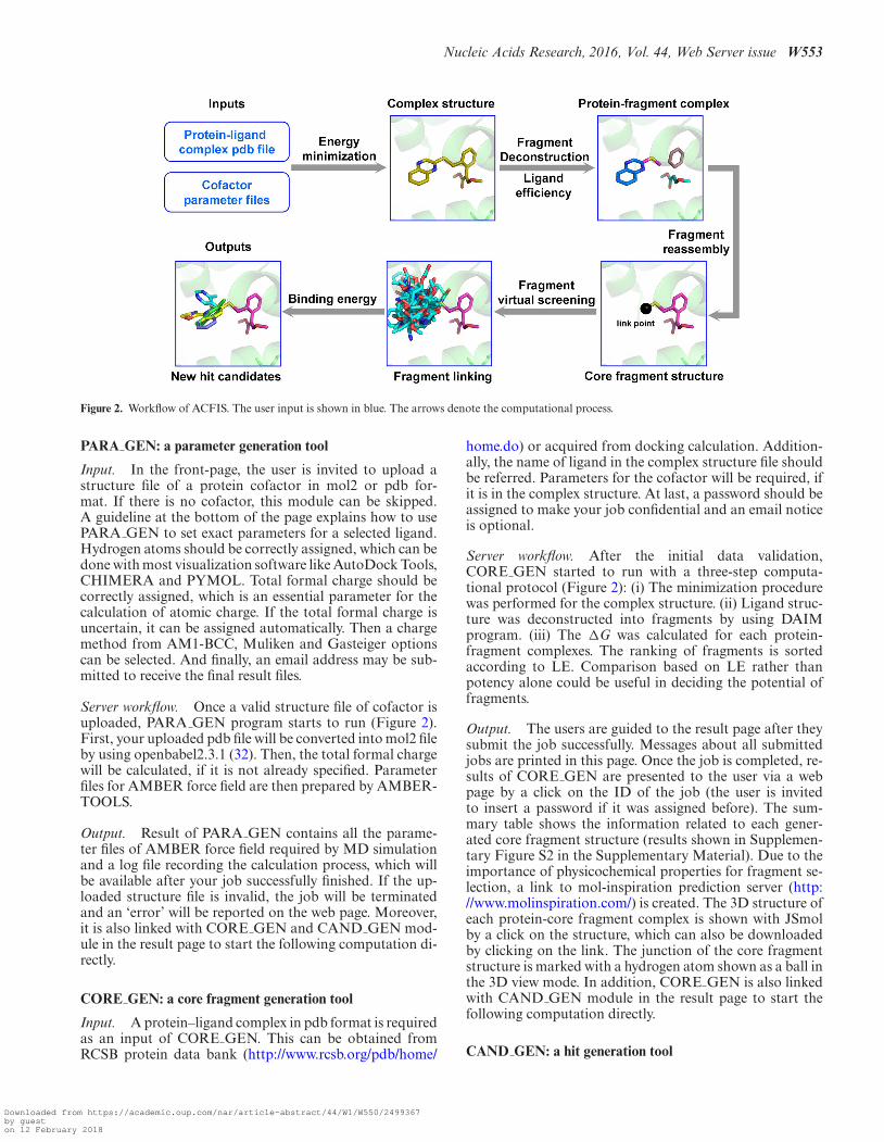
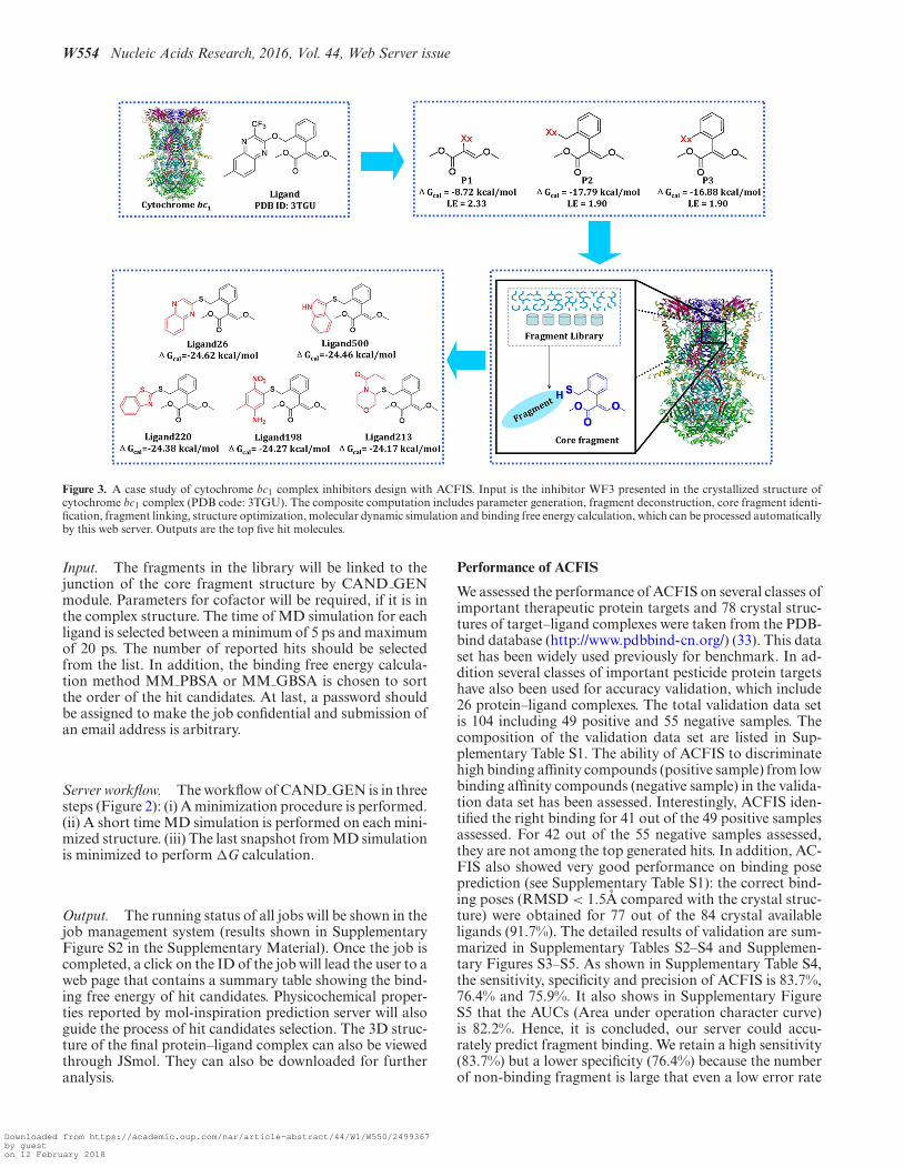
Figure 3. A case study of cytochrome bc1 complex inhibitors design with ACFIS. Input is the inhibitor WF3 presented in the crystallized structure ofcytochrome bc1 complex (PDB code: 3TGU). The composite computation includes parameter generation, fragment deconstruction, core fragment identi-fication, fragment linking, structure optimization, molecular dynamic simulation and binding free energy calculation, which can be processed automaticallyby this web server. Outputs are the top five hit molecules.
Input. The fragments in the library will be linked to thejunction of the core fragment structure by CAND GENmodule. Parameters for cofactor will be required, if it is inthe complex structure. The time of MD simulation for eachligand is selected between a minimum of 5 ps and maximumof 20 ps. The number of reported hits should be selectedfrom the list. In addition, the binding free energy calcula-tion method MM PBSA or MM GBSA is chosen to sortthe order of the hit candidates. At last, a password shouldbe assigned to make the job confidential and submission ofan email address is arbitrary.
Server workflow. The workflow of CAND GEN is in threesteps (Figure 2): (i) A minimization procedure is performed.(ii) A short time MD simulation is performed on each mini-mized structure. (iii) The last snapshot from MD simulationis minimized to perform �G calculation.
Output. The running status of all jobs will be shown in thejob management system (results shown in SupplementaryFigure S2 in the Supplementary Material). Once the job iscompleted, a click on the ID of the job will lead the user to aweb page that contains a summary table showing the bind-ing free energy of hit candidates. Physicochemical proper-ties reported by mol-inspiration prediction server will alsoguide the process of hit candidates selection. The 3D struc-ture of the final protein–ligand complex can also be viewedthrough JSmol. They can also be downloaded for furtheranalysis.
Performance of ACFIS
We assessed the performance of ACFIS on several classes ofimportant therapeutic protein targets and 78 crystal struc-tures of target–ligand complexes were taken from the PDB-bind database (http://www.pdbbind-cn.org/) (33). This dataset has been widely used previously for benchmark. In ad-dition several classes of important pesticide protein targetshave also been used for accuracy validation, which include26 protein–ligand complexes. The total validation data setis 104 including 49 positive and 55 negative samples. Thecomposition of the validation data set are listed in Sup-plementary Table S1. The ability of ACFIS to discriminatehigh binding affinity compounds (positive sample) from lowbinding affinity compounds (negative sample) in the valida-tion data set has been assessed. Interestingly, ACFIS iden-tified the right binding for 41 out of the 49 positive samplesassessed. For 42 out of the 55 negative samples assessed,they are not among the top generated hits. In addition, AC-FIS also showed very good performance on binding poseprediction (see Supplementary Table S1): the correct bind-ing poses (RMSD < 1.5A compared with the crystal struc-ture) were obtained for 77 out of the 84 crystal availableligands (91.7%). The detailed results of validation are sum-marized in Supplementary Tables S2–S4 and Supplemen-tary Figures S3–S5. As shown in Supplementary Table S4,the sensitivity, specificity and precision of ACFIS is 83.7%,76.4% and 75.9%. It also shows in Supplementary FigureS5 that the AUCs (Area under operation character curve)is 82.2%. Hence, it is concluded, our server could accu-rately predict fragment binding. We retain a high sensitivity(83.7%) but a lower specificity (76.4%) because the numberof non-binding fragment is large that even a low error rate
Downloaded from https://academic.oup.com/nar/article-abstract/44/W1/W550/2499367by gueston 12 February 2018
Nucleic Acids Research, 2016, Vol. 44, Web Server issue W555
of negative samples may cause many false positive predic-tions.
Taking cytochrome bc1 complex inhibitor as an ex-ample, the parameters of cofactor heme were generatedby PARA GEN, the crystal complex structure of WF3-bound bc1 (PDB code: 3TGU) was downloaded to per-form CORE GEN calculation. As shown in Figure 3, thetop three core fragments were generated (P1, P2 and P3)with �Gcal of −8.72, −17.79 and −16.88 kcal/mol. Obvi-ously, the calculations systematically overestimated the ab-solute values of the binding affinities. However, the calcu-lated �Gbind values can still qualitatively reflect the relativeorder of the binding affinities of these core fragments. Theerror induced by the molecular size can be eliminated byusing LE rather than potency. As shown in Figure 3, theLE of P1 (LE = 2.33) is relatively higher than the othertwo fragments P2 (LE = 1.90) and P3 (LE = 1.90). How-ever, comprehensively considered with �Gbind, LE and newfragment growing spaces, P2 was selected as a core frag-ment to perform CAND GEN calculation. In addition, asulfur atom was set as the linker between the core fragmentand the new fragment for synthetic feasibility (Figure 3). Fi-nally, the top five new hit candidates selected according tothe binding free energy sorting were shown in Figure 3 withthe original ligand scaffold (Ligand26) included. It needsto be emphasized that the synthesis of the candidate (Lig-and220), the inhibitory kinetics with cytochrome bc1 com-plex (Ki = 31.10 nM), and the co-crystal structure studyhave been completed (34), which further validated the pre-dicted results of ACFIS.
CONCLUSIONS
The discovery of new hit molecules remains a crucial stepfor drug and agrochemical discovery. We presented ACFIS,a computer-aided FBDD web server, to provide solutionsfor fragment virtual screening and to assist scientists in thenew hits identification. It is differentiated from other webservices in which ligand screening was presented in dockingbased manner. In the future, we will perform a continualupdate of the fragment library of ACFIS to offer broaderand more diverse selection of databases to start a fragment-based drug discovery task.
SUPPLEMENTARY DATA
Supplementary Data are available at NAR Online.
ACKNOWLEDGEMENTS
We would like to thank Prof. Amedeo Caflisch for thepermission to use DAIM program on our server and thedevelopers of open source softwares (Jsmol, OpenBabel,et. al.). This research was supported by the Special Fundfor Agro-scientific Research in the Public Interest (No.201203022)and the National Natural science Foundation ofChina (No. 21332004 and 31470068).
FUNDING
Special Fund for Agro-scientific Research in the Public In-terest [No. 201203022]; National Natural science Founda-tion of China [21332004 and 31470068]. Funding for open
access charge: Special Fund for Agro-scientific Research inthe Public Interest [No. 201203022]; National Natural sci-ence Foundation of China [No. 21332004 and 31470068].Conflict of interest statement. None declared.
REFERENCES1. Dickson,M. and Gagnon,J.P. (2004) Key factors in the rising cost of
new drug discovery and development. Nat. Rev. Drug Discov., 3,417–429.
2. Lamberth,C., Jeanmart,S., Luksch,T. and Plant,A. (2013) Currentchallenges and trends in the discovery of agrochemicals. Science, 341,742–746.
3. Frearson,J.A. and Collie,I.T. (2009) HTS and hit finding in academia- from chemical genomics to drug discovery. Drug Discov. Today, 14,1150–1158.
4. Murray,C.W. and Rees,D.C. (2009) The rise of fragment-based drugdiscovery. Nat. Chem., 1, 187–192.
5. Hann,M.M., Leach,A.R. and Harper,G. (2001) Molecularcomplexity and its impact on the probability of finding leads for drugdiscovery. J. Chem. Inf. Comput. Sci., 41, 856–864.
6. Erlanson,D.A., McDowell,R.S. and O’Brien,T. (2004)Fragment-based drug discovery. J. Med. Chem., 47, 3463–3482.
7. Rees,D.C., Congreve,M., Murray,C.W. and Carr,R. (2004)Fragment-based lead discovery. Nat. Rev. Drug Discov., 3, 660–672.
8. Fruh,V., Zhou,Y., Chen,D., Loch,C., Ab,E., Grinkova,Y.N.,Verheij,H., Sligar,S.G., Bushweller,J.H. and Siegal,G. (2010)Application of fragment-based drug discovery to membrane proteins:identification of ligands of the integral membrane enzyme DsbB.Chem. Biol., 17, 881–891.
9. Congreve,M., Rich,R.L., Myszka,D.G., Figaroa,F., Siegal,G. andMarshall,F.H. (2011) Fragment screening of stabilizedG-protein-coupled receptors using biophysical methods. MethodsEnzymol., 493, 115–136.
10. Villar,H.O. and Hansen,M.R. (2007) Computational techniques infragment based drug discovery. Curr. Top. Med. Chem., 7, 1509–1513.
11. Chen,Y. and Shoichet,B.K. (2009) Molecular docking and ligandspecificity in fragment-based inhibitor discovery. Nat. Chem. Biol., 5,358–364.
12. Loving,K., Alberts,I. and Sherman,W. (2010) Computationalapproaches for fragment-based and de novo design. Curr. Top. Med.Chem., 10, 14–32.
13. Bohm,H.J. (1992) The computer program LUDI: a new method forthe de novo design of enzyme inhibitors. J. Comput. Aided. Mol. Des.,6, 61–78.
14. Sandor,M., Kiss,R. and Keseru,G.M. (2010) Virtual fragmentdocking by Glide: a validation study on 190 protein-fragmentcomplexes. J. Chem. Inf. Model., 50, 1165–1172.
15. Yuan,Y.X., Pei,J.F. and Lai,L.H. (2011) LigBuilder 2: a practical denovo drug design approach. J. Chem. Inf. Model., 51, 1083–1091.
16. Hoffer,L., Renaud,J.-P. and Horvath,D. (2013) In silicofragment-based drug discovery: setup and validation of afragment-to-lead computational protocol using S4MPLE. J. Chem.Inf. Model., 53, 836–851.
17. Villoutreix,B.O., Lagorce,D., Labbe,C.M., Sperandio,O. andMiteva,M.A. (2013) One hundred thousand mouse clicks down theroad: selected online resources supporting drug discovery collectedover a decade. Drug Discov. Today, 18, 1081–1089.
18. Labbe,C.M., Rey,J., Lagorce,D., Vavrusa,M., Becot,J., Sperandio,O.,Villoutreix,B.O., Tuffery,P. and Miteva,M.A. (2015) MTiOpenScreen:a web server for structure-based virtual screening. Nucleic Acids Res.,43, W448–W454.
19. Douguet,D. (2010) e-LEA3D: a computational-aided drug designweb server. Nucleic Acids Res., 38, W615–W621.
20. Tsai,T.-Y., Chang,K.-W. and Chen,C.Y.-C. (2011) iScreen: world’sfirst cloud-computing web server for virtual screening and de novodrug design based on TCM database@Taiwan. J. Comput. Aided.Mol. Des., 25, 525–531.
21. Prakhov,N.D., Chernorudskiy,A.L. and Gainullin,M.R. (2010)VSDocker: a tool for parallel high-throughput virtual screening usingAutoDock on Windows-based computer clusters. Bioinformatics, 26,1374–1375.
Downloaded from https://academic.oup.com/nar/article-abstract/44/W1/W550/2499367by gueston 12 February 2018
W556 Nucleic Acids Research, 2016, Vol. 44, Web Server issue
22. Hao,G.F., Wang,F., Li,H., Zhu,X.L., Yang,W.C., Huang,L.S.,Wu,J.W., Berry,E.A. and Yang,G.F. (2012) Computational discoveryof picomolar Q(o) site inhibitors of cytochrome bc(1) complex. J.Am. Chem. Soc., 134, 11168–11176.
23. Congreve,M., Carr,R., Murray,C. and Jhoti,H. (2003) A ’rule ofThree’ for fragment-based lead discovery? Drug Discov. Today, 8,876–877.
24. Jhoti,H., Williams,G., Rees,D.C. and Murray,C.W. (2013) The ’ruleof three’ for fragment-based drug discovery: where are we now? Nat.Rev. Drug Discov., 12, 644–644.
25. Kolb,P. and Caflisch,A. (2006) Automatic and efficient decompositionof two-dimensional structures of small molecules for fragment-basedhigh-throughput docking. J. Med. Chem., 49, 7384–7392.
26. Teague,S.J. (2011) Learning lessons from drugs that have recentlyentered the market. Drug Discov. Today, 16, 398–411.
27. Hou,T.J., Wang,J.M., Li,Y.Y. and Wang,W. (2011) Assessing theperformance of the MM/PBSA and MM/GBSA methods. 1. Theaccuracy of binding free energy calculations based on moleculardynamics simulations. J. Chem. Inf. Model., 51, 69–82.
28. Pan,Y., Gao,D. and Zhan,C.G. (2008) Modeling the catalysis ofanti-cocaine catalytic antibody: competing reaction pathways andfree energy barriers. J. Am. Chem. Soc., 130, 5140–5149.
29. Hao,G.F., Zhu,X.L., Ji,F.Q., Zhang,L., Yang,G.F. and Zhan,C.G.(2009) Understanding the Mechanism of Drug Resistance Due to aCodon Deletion in Protoporphyrinogen Oxidase throughComputational Modeling. J. Phys. Chem. B, 113, 4865–4875.
30. Durrant,J.D., Amaro,R.E. and McCammon,J.A. (2009) AutoGrow: anovel algorithm for protein inhibitor design. Chem. Biol. Drug Des.,73, 168–178.
31. Case,D.A., Berryman,J.T., Betz,R.M., Cerutti,D.S., Cheatham,T.E.I.,Darden,T.A., Duke,R.E., Giese,T.J., Gohlke,H., Goetz,A.W. et al.(2014) AMBER14. University of California, San Francisco.
32. O’Boyle,N.M., Banck,M., James,C.A., Morley,C., Vandermeersch,T.and Hutchison,G.R. (2011) Open Babel: an open chemical toolbox. J.Cheminform., 3, 33.
33. Wang,R.X., Fang,X.L., Lu,Y.P. and Wang,S.M. (2004) The PDBbinddatabase: collection of binding affinities for protein-ligand complexeswith known three-dimensional structures. J. Med. Chem., 47,2977–2980.
34. Hao,G.F., Yang,S.G., Huang,W., Wang,L., Shen,Y.Q., Tu,W.L.,Li,H., Huang,L.S., Wu,J.W., Berry,E.A. et al. (2015) Rational designof highly potent and slow-binding cytochrome bc1 Inhibitor asfungicide by computational substitution optimization. Sci. Rep., 5,13471.
Downloaded from https://academic.oup.com/nar/article-abstract/44/W1/W550/2499367by gueston 12 February 2018