Embed Size (px)
Citation preview
e u r o p e a n u r o l o g y 5 1 ( 2 0 0 7 ) 1 0 4 2 – 1 0 5 3
avai lable at www.sc iencedi rect .com
journal homepage: www.europeanurology.com
Neuro-urology
Acetylcholine and Molecular Components of its Synthesis andRelease Machinery in the Urothelium
Katrin S. Lips a, Julia Wunsch a, Shirin Zarghooni a, Thomas Bschleipfer b,Konstantin Schukowski b, Wolfgang Weidner b, Ignaz Wessler c,Ulrich Schwantes d, Hermann Koepsell e, Wolfgang Kummer a,*a Institute for Anatomy and Cell Biology, Justus-Liebig-University, Giessen, GermanybClinic for Urology and Pedriatic Urology, Justus-Liebig-University, Giessen, GermanycDepartment of Pathology, University of Mainz, GermanydDr. R. Pfleger Chemische Fabrik GmbH, Bamberg, Germanye Institute for Anatomy and Cell Biology, Julius-Maximilians-University, Wurzburg, Germany
Article info
Article history:Accepted October 16, 2006Published online ahead ofprint on October 27, 2006
Keywords:AceytlcholineCarnitine acetyltransferaseCholine acetyltransferaseOrganic cation transportersVesicular acetylcholinetransporterUrothelium
Abstract
Objectives: Previous studies provided indirect evidence for urothelial synthesis and
release of acetylcholine (ACh). We aimed to determine directly the ACh content in the
urothelium and to characterize the molecular components of its synthesis and release
machinery.
Methods: The study was performed on mouse bladder and abraded urothelium, and
human mucosal bladder biopsies. ACh content was measured by high-performance
liquid chromatography-electrochemical. Reverse transcriptase–polymerase chain
reaction (RT-PCR) and immunohistochemistry served to investigate expression of
ACh-synthesizing enzymes—choline acetyltransferase (ChAT) and carnitine acetyl-
transferase (CarAT)—vesicular ACh transporter (VAChT), and polyspecific organic
cation transporters (OCTs; isoforms 1–3). Transfected cells served to investigate
whether the anticholinergic drug trospium chloride interferes with ACh-transporting
OCTs.
Results: ACh is present in the urothelium in a nanomolar range per gram of
wet weight. RT-PCR data support the presence of CarAT but not ChAT. VAChT, used
by neurons to shuffle ACh into synaptic vesicles, is detected in subepithelial
cholinergic nerve fibres, but not by RT-PCR or immunohistochemistry in the urothe-
lium. OCT1 and OCT3 are expressed by the urothelium. The quarternary ammonium
base trospium chloride inhibits cation transport by OCTs with a potency rank order
of OCT2 (IC50 = 0.67 � 0.42 mmol/l) > OCT1 (IC50 = 6.2 � 2.1 mmol/l) > OCT3 (IC50 = 871 �177 mmol/l).
Conclusions: This study demonstrates a urothelial non-neuronal cholinergic system
om that of neurons with respect to molecular components of the
release machinery. Consequently, these two systems might be
that differs widely fr
ACh synthesis and
differentially targeted by pharmacologic approaches.
sociation of Urology. Published by Elsevier B.V. All rights reserved.
# 2006 European As* Corresponding author. Institute for Anatomy and Cell Biology, Justus-Liebig-University,Aulweg 123, 35385 Giessen, Germany. Tel. 49 641 9947000; Fax: +49 641 9947009.E-mail address: [email protected] (W. Kummer).
0302-2838/$ – see back matter # 2006 European Association of Urology. Published by Elsevier B.V. All rights reserved. doi:10.1016/j.eururo.2006.10.028
e u r o p e a n u r o l o g y 5 1 ( 2 0 0 7 ) 1 0 4 2 – 1 0 5 3 1043
1. Introduction
Synthesis and release of acteylcholine (ACh) is notrestricted to specialized subsets of neurons but alsooccurs in a broad variety of non-neuronal cells, inparticular in surface epithelia [1,2]. Recent studiessuggest that this may apply for the bladderurothelium as well. Urothelial ACh has beenproposed to be released into the bladder lumen toaddress nicotinic receptors on the luminal mem-brane of umbrella cells or to be released basally toact upon the detrusor and nerve fibres [3–6]. So far,however, there is only indirect experimental evi-dence for urothelial ACh. Yoshida et al. [6,7]measured ACh release from isolated human bladderstrips and demonstrated a reduced release inpreparations in which the epithelium had beenremoved. These findings are compatible with aurothelial release of ACh, but also with urothelialrelease of another factor that might trigger AChrelease from deeper structures. In addition, immu-nolabelling of the urothelium with an antiserumdirected against the ACh-synthesizing enzyme cho-line acetyltransferase (ChAT) has been reported[6,7], although these data still await validation bypreabsorption experiments or other independentmethods, such as Western blotting, determinationof ChAT activity, or reverse transcriptase–polymer-ase chain reaction (RT-PCR).
On this background, we set out to determinethe molecular components of a putative cholinergicsystem in the urothelium in more detail. First,ACh was detected in the urothelial cell layer by ahigh-performance liquid chromatography-electro-chemical (HPLC-EC) method. Next, we employedRT-PCR and immunohistochemistry to investigateurothelial expression of (1) ChAT, the classicalAch-synthesizing enzyme in the nervous systemand several non-neuronal cells; (2) carnitineacetyltransferase (CarAT), which is responsiblefor ACh synthesis in muscle cells [8]; (3) thevesicular ACh transporter (VAChT), which shufflesACh from the cytoplasm into synaptic vesicle incholinergic nerve terminals [9]; and (4) polyspecificorganic cation transporters (OCTs; isoforms 1–3),which are able to translocate ACh directly acrossthe plasma membrane [10]. In view of their poly-specific properties [11], we tested whether acommonly used anticholinergic drug, trospiumchloride, interferes with ACh transport by OCTs.The study was performed on human and murineurothelium in parallel to evaluate whether themouse may serve as a suitable model for experi-mental approaches addressing this system in thefuture.
2. Materials and methods
2.1. ACh assay
FVB mice were killed by inhalation of an overdose of isoflurane
(Abbott, Wiesbaden, Germany). The bladder was carefully
dissected, opened, and fixed in a Petri dish with the luminal
surface facing upwards. A cotton-tipped applicator (Q-tip) was
gently rubbed along the luminal surface and thereafter placed
in 1 ml 15% formic acid in acetone (v/v). Two samples were
taken from the luminal surface (about 2 cm2) of human
urinary bladders obtained from surgery. After standing on ice
(30 min), specimens (material swiped off by the Q-tip) were
frozen in liquid nitrogen. The bladder was then fixed in
buffered 4% paraformaldehyde and processed as described for
immunohistochemistry; then serial cryosections were stained
with hematoxylin-eosin and evaluated by light microscopy.
Only those specimens were included in the further analysis in
which the basal lamina was not disrupted.
ACh was measured by cationic exchange HPLC combined
with bioreactors and electrochemical detection with a detec-
tion limit of 10 fmol ACh per injection (20 ml) as described in
detail elsewhere [12].
2.2. Reverse transcriptase–polymerase chain reaction
Murine samples were obtained by scraping off the urothelium
as described above, but specimen holders were then placed in
lysis buffer (RLT-buffer; Qiagen, Hilden, Germany) instead of
formic acid/acetone. Further sample processing was as
described in detail earlier [10]. Primer sequences are provided
in Table 1.
NS20Y cells—a cholinergic murine forebrain neuroblastoma
cell line (GermanCollection ofMicroorganismsand CellCulture,
Braunschweig, Germany)—were used as a positive control for
expression of components of the murine cholinergic system.
Human bladder mucosal biopsies were obtained from six
female patients aged between 57 and 76 yr (mean: 67). All
patients underwent endoscopic surgery primarily to remove
bladder cancer or to examine the bladder wall by biopsies for
carcinoma in situ or interstitial cystitis. Four additional
mucosal biopsies were taken out of the trigonum (four
patients) or the bottom (two patients) of the bladder with
the use of endoscopic forceps. This procedure was approved
by the local ethics committee, and informed patients gave
signed consent. Processing of these specimens was identical to
that of murine specimens, except that human specific primers
were used (Table 1).
Caco cells—a human colonic epithelial cell line (German
Collection of Microorganisms and Cell Culture)—served as a
positive control for human epithelial ChAT expression. Addi-
tional complementary DNAs generated from airways during a
previous study [10] were also used as positive controls.
2.3. Immunohistochemistry
Mice were killed by isoflurane inhalation; then the bladder was
rapidly dissected, embedded in optimal-cutting-temperature
compound, and shock-frozen in melting isopentane. For ChAT
e u r o p e a n u r o l o g y 5 1 ( 2 0 0 7 ) 1 0 4 2 – 1 0 5 31044
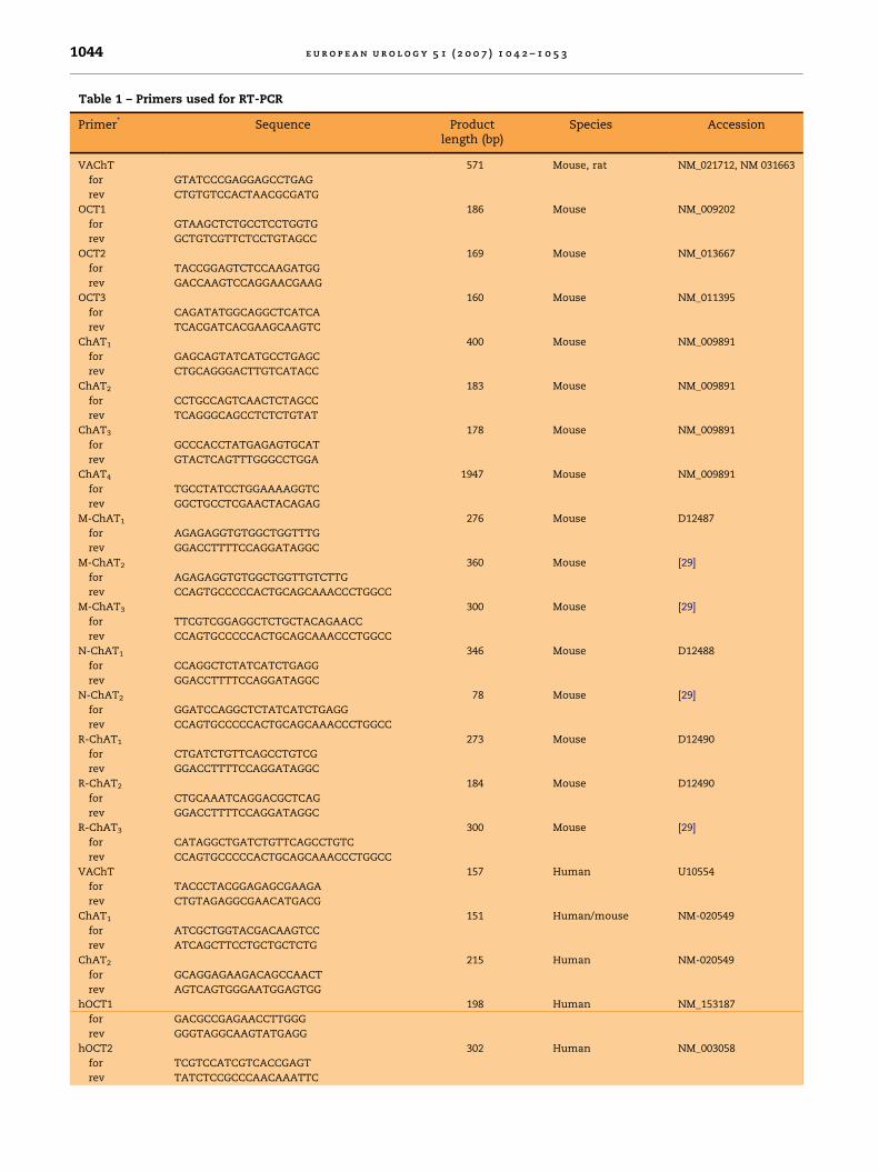
Table 1 – Primers used for RT-PCR
Primer* Sequence Productlength (bp)
Species Accession
VAChT 571 Mouse, rat NM_021712, NM 031663
for GTATCCCGAGGAGCCTGAG
rev CTGTGTCCACTAACGCGATG
OCT1 186 Mouse NM_009202
for GTAAGCTCTGCCTCCTGGTG
rev GCTGTCGTTCTCCTGTAGCC
OCT2 169 Mouse NM_013667
for TACCGGAGTCTCCAAGATGG
rev GACCAAGTCCAGGAACGAAG
OCT3 160 Mouse NM_011395
for CAGATATGGCAGGCTCATCA
rev TCACGATCACGAAGCAAGTC
ChAT1 400 Mouse NM_009891
for GAGCAGTATCATGCCTGAGC
rev CTGCAGGGACTTGTCATACC
ChAT2 183 Mouse NM_009891
for CCTGCCAGTCAACTCTAGCC
rev TCAGGGCAGCCTCTCTGTAT
ChAT3 178 Mouse NM_009891
for GCCCACCTATGAGAGTGCAT
rev GTACTCAGTTTGGGCCTGGA
ChAT4 1947 Mouse NM_009891
for TGCCTATCCTGGAAAAGGTC
rev GGCTGCCTCGAACTACAGAG
M-ChAT1 276 Mouse D12487
for AGAGAGGTGTGGCTGGTTTG
rev GGACCTTTTCCAGGATAGGC
M-ChAT2 360 Mouse [29]
for AGAGAGGTGTGGCTGGTTGTCTTG
rev CCAGTGCCCCCACTGCAGCAAACCCTGGCC
M-ChAT3 300 Mouse [29]
for TTCGTCGGAGGCTCTGCTACAGAACC
rev CCAGTGCCCCCACTGCAGCAAACCCTGGCC
N-ChAT1 346 Mouse D12488
for CCAGGCTCTATCATCTGAGG
rev GGACCTTTTCCAGGATAGGC
N-ChAT2 78 Mouse [29]
for GGATCCAGGCTCTATCATCTGAGG
rev CCAGTGCCCCCACTGCAGCAAACCCTGGCC
R-ChAT1 273 Mouse D12490
for CTGATCTGTTCAGCCTGTCG
rev GGACCTTTTCCAGGATAGGC
R-ChAT2 184 Mouse D12490
for CTGCAAATCAGGACGCTCAG
rev GGACCTTTTCCAGGATAGGC
R-ChAT3 300 Mouse [29]
for CATAGGCTGATCTGTTCAGCCTGTC
rev CCAGTGCCCCCACTGCAGCAAACCCTGGCC
VAChT 157 Human U10554
for TACCCTACGGAGAGCGAAGA
rev CTGTAGAGGCGAACATGACG
ChAT1 151 Human/mouse NM-020549
for ATCGCTGGTACGACAAGTCC
rev ATCAGCTTCCTGCTGCTCTG
ChAT2 215 Human NM-020549
for GCAGGAGAAGACAGCCAACT
rev AGTCAGTGGGAATGGAGTGG
hOCT1 198 Human NM_153187
for GACGCCGAGAACCTTGGG
rev GGGTAGGCAAGTATGAGG
hOCT2 302 Human NM_003058
for TCGTCCATCGTCACCGAGT
rev TATCTCCGCCCAACAAATTC
e u r o p e a n u r o l o g y 5 1 ( 2 0 0 7 ) 1 0 4 2 – 1 0 5 3 1045
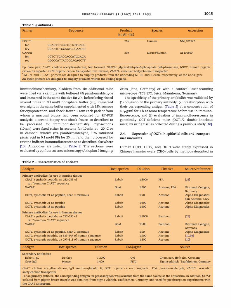
Table 1 (Continued )
Primer* Sequence Productlength (bp)
Species Accession
hOCT3 216 Human NM_021977
for GGAGTTTCGCTCTGTTCAGG
rev GGAATGTGGACTGCCAAGTT
GAPDH 299 Mouse/human AF106860
for CGTCTTCACCACCATGGAGA
rev CGGCCATCACGCCACAGCTT
bp: base pair; ChAT: choline acetyltransferase; for: forward; GAPDH: glyceraldehyde-3-phosphate dehydrogenase; hOCT; human organic
cation transporter; OCT: organic cation transporter; rev: reverse; VAChT: vesicular acetylcholine transporter.* M-, N- and R-ChAT primers are designed to amplify products from the noncoding M-, N- and R-exon, respectively, of the ChAT gene.
All other primers are designed to amplify products within the coding regions.
immunohistochemistry, bladders from six additional mice
were filled via a cannula with buffered 4% paraformaldehyde
and immersed in the same fixative for 2 h, before being rinsed
several times in 0.1 mol/l phosphate buffer (PB), immersed
overnight in the same buffer supplemented with 18% sucrose
for cryoprotection, and shock-frozen. From each patient from
whom a mucosal biopsy had been obtained for RT-PCR
analysis, a second biopsy was shock-frozen as described to
be processed for immunohistochemistry. Cryosections
(10 mm) were fixed either in acetone for 10 min at �20 8C or
in Zamboni fixative (2% paraformaldehyde, 15% saturated
picric acid in 0.1 mol/l PB) for 20 min and then processed for
routine indirect immunofluorescence as described elsewhere
[10]. Antibodies are listed in Table 2. The sections were
evaluated by epifluorescence microscopy (Axioplan 2 imaging;
Table 2 – Characteristics of antisera
Antigen Host s
Primary antibodies for use in murine tissues
ChAT, synthetic peptide, aa 282–295 of
rat ‘‘common ChAT’’ sequence
Rab
VAChT Goa
OCT1, synthetic 21 aa peptide, near C-terminus Rab
OCT2, synthetic 21 aa peptide Rab
OCT3, synthetic 18 aa peptide Rab
Primary antibodies for use in human tissues
ChAT; synthetic peptide, aa 282–295 of
rat ‘‘common ChAT’’ sequence
Rab
VAChT Goa
OCT1, synthetic 21 aa peptide, near C-terminus Rab
OCT2, synthetic peptide, aa 533–547 of human sequence Rab
OCT3, synthetic peptide, aa 297–313 of human sequence Rab
Antigen Host species Dilution
Secondary antibodies
Rabbit-IgG Donkey 1:2000
Goat-IgG Mouse 1:400
ChAT: choline acetyltransferase; IgG: immunoglobulin G; OCT: organ
acetylcholine transporter.
For all primary antisera, the corresponding antigen for preabsorption was
isolated from pigeon breast muscle was obtained from Sigma-Aldrich, T
the ChAT antiserum.
Zeiss, Jena, Germany) or with a confocal laser-scanning
microscope (TCS SP2; Leica, Mannheim, Germany).
The specificity of the primary antibodies was validated by
(1) omission of the primary antibody, (2) preabsorption with
their corresponding antigen (Table 2) at a concentration of
40 mg/ml for 1 h at room temperature before use in immuno-
fluorescence, and (3) evaluation of immunofluorescence in
genetically OCT-deficient mice (OCT1/2 double-knockout
mice) by using tissues collected during a previous study [10].
2.4. Expression of OCTs in epithelial cells and transportmeasurements
Human OCT1, OCT2, and OCT3 were stably expressed in
Chinese hamster ovary (CHO) cells by methods described in
pecies Dilution Fixative Source/reference
bit 1:8000 PFA [23]
t 1:800 Acetone, PFA Biotrend, Cologne,
Germany
bit 1:20 Acetone Alpha Diagnostics,
San Antonio, USA
bit 1:400 Acetone Alpha Diagnostics
bit 1:400 Acetone Alpha Diagnostics
bit 1:8000 Zamboni [23]
t 1:500 Zamboni Biotrend, Cologne,
Germany
bit 1:20 Acetone Alpha Diagnostics
bit 1:200 Acetone [10,30]
bit 1:500 Acetone [10]
Conjugate Source
Cy3 Chemicon, Hofheim, Germany
FITC Sigma-Aldrich, Taufkirchen, Germany
ic cation transporter; PFA: paraformaldehyde; VAChT: vesicular
available from the same source as the antiserum. In addition, CarAT
aufkirchen, Germany, and used for preabsorption experiments with
e u r o p e a n u r o l o g y 5 1 ( 2 0 0 7 ) 1 0 4 2 – 1 0 5 31046
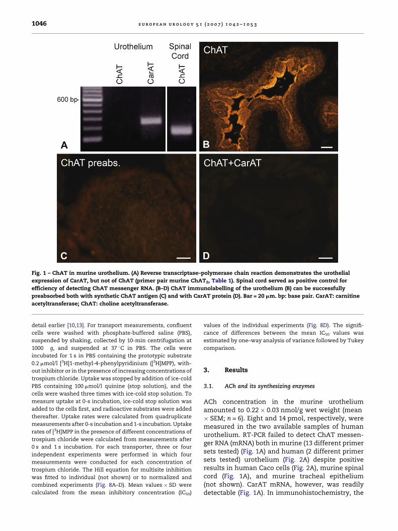
Fig. 1 – ChAT in murine urothelium. (A) Reverse transcriptase–polymerase chain reaction demonstrates the urothelial
expression of CarAT, but not of ChAT (primer pair murine ChAT2, Table 1). Spinal cord served as positive control for
efficiency of detecting ChAT messenger RNA. (B–D) ChAT immunolabelling of the urothelium (B) can be successfully
preabsorbed both with synthetic ChAT antigen (C) and with CarAT protein (D). Bar = 20 mm. bp: base pair. CarAT: carnitine
acetyltransferase; ChAT: choline acetyltransferase.
detail earlier [10,13]. For transport measurements, confluent
cells were washed with phosphate-buffered saline (PBS),
suspended by shaking, collected by 10-min centrifugation at
1000 � g, and suspended at 37 8C in PBS. The cells were
incubated for 1 s in PBS containing the prototypic substrate
0.2 mmol/l [3H]1-methyl-4-phenylpyridinium ([3H]MPP), with-
out inhibitor or in the presence of increasing concentrations of
trospium chloride. Uptake was stopped by addition of ice-cold
PBS containing 100 mmol/l quinine (stop solution), and the
cells were washed three times with ice-cold stop solution. To
measure uptake at 0-s incubation, ice-cold stop solution was
added to the cells first, and radioactive substrates were added
thereafter. Uptake rates were calculated from quadruplicate
measurements after 0-s incubation and 1-s incubation. Uptake
rates of [3H]MPP in the presence of different concentrations of
trospium chloride were calculated from measurements after
0 s and 1 s incubation. For each transporter, three or four
independent experiments were performed in which four
measurements were conducted for each concentration of
trospium chloride. The Hill equation for multisite inhibition
was fitted to individual (not shown) or to normalized and
combined experiments (Fig. 8A–D). Mean values � SD were
calculated from the mean inhibitory concentration (IC50)
values of the individual experiments (Fig. 8D). The signifi-
cance of differences between the mean IC50 values was
estimated by one–way analysis of variance followed by Tukey
comparison.
3. Results
3.1. ACh and its synthesizing enzymes
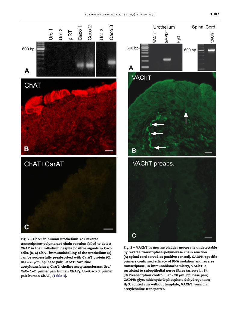
ACh concentration in the murine urotheliumamounted to 0.22 � 0.03 nmol/g wet weight (mean� SEM; n = 6). Eight and 14 pmol, respectively, weremeasured in the two available samples of humanurothelium. RT-PCR failed to detect ChAT messen-ger RNA (mRNA) both in murine (13 different primersets tested) (Fig. 1A) and human (2 different primersets tested) urothelium (Fig. 2A) despite positiveresults in human Caco cells (Fig. 2A), murine spinalcord (Fig. 1A), and murine tracheal epithelium(not shown). CarAT mRNA, however, was readilydetectable (Fig. 1A). In immunohistochemistry, the
e u r o p e a n u r o l o g y 5 1 ( 2 0 0 7 ) 1 0 4 2 – 1 0 5 3 1047
Fig. 2 – ChAT in human urothelium. (A) Reverse
transcriptase–polymerase chain reaction failed to detect
ChAT in the urothelium despite positive signals in Caco
cells. (B, C) ChAT immunolabelling of the urothelium (B)
can be successfully preabsorbed with CarAT protein (C).
Bar = 20 mm. bp: base pair; CarAT: carnitine
acetyltransferase; ChAT: choline acetyltransferase; Uro/
CaCo 1+2: primer pair human ChAT1; Uro/Caco 3: primer
pair human ChAT2 (Table 1).
Fig. 3 – VAChT in murine bladder mucosa is undetectable
by reverse transcriptase–polymerase chain reaction
(A; spinal cord served as positive control). GADPH-specific
primers confirmed efficacy of RNA isolation and reverse
transcriptase. In immunohistochemistry, VAChT is
restricted to subepithelial nerve fibres (arrows in B).
(C) Preabsorption control. Bar = 20 mm. bp: base pair;
GADPH: glyceraldehyde-3-phosphate dehydrogenase;
H2O: control run without template; VAChT: vesicular
acetylcholine transporter.
e u r o p e a n u r o l o g y 5 1 ( 2 0 0 7 ) 1 0 4 2 – 1 0 5 31048
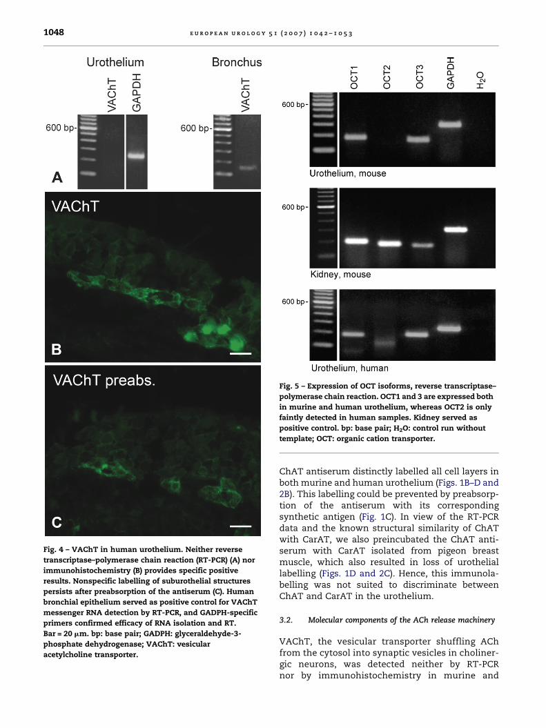
Fig. 4 – VAChT in human urothelium. Neither reverse
transcriptase–polymerase chain reaction (RT-PCR) (A) nor
immunohistochemistry (B) provides specific positive
results. Nonspecific labelling of suburothelial structures
persists after preabsorption of the antiserum (C). Human
bronchial epithelium served as positive control for VAChT
messenger RNA detection by RT-PCR, and GADPH-specific
primers confirmed efficacy of RNA isolation and RT.
Bar = 20 mm. bp: base pair; GADPH: glyceraldehyde-3-
phosphate dehydrogenase; VAChT: vesicular
acetylcholine transporter.
Fig. 5 – Expression of OCT isoforms, reverse transcriptase–
polymerase chain reaction. OCT1 and 3 are expressed both
in murine and human urothelium, whereas OCT2 is only
faintly detected in human samples. Kidney served as
positive control. bp: base pair; H2O: control run without
template; OCT: organic cation transporter.
ChAT antiserum distinctly labelled all cell layers inboth murine and human urothelium (Figs. 1B–D and2B). This labelling could be prevented by preabsorp-tion of the antiserum with its correspondingsynthetic antigen (Fig. 1C). In view of the RT-PCRdata and the known structural similarity of ChATwith CarAT, we also preincubated the ChAT anti-serum with CarAT isolated from pigeon breastmuscle, which also resulted in loss of urotheliallabelling (Figs. 1D and 2C). Hence, this immunola-belling was not suited to discriminate betweenChAT and CarAT in the urothelium.
3.2. Molecular components of the ACh release machinery
VAChT, the vesicular transporter shuffling AChfrom the cytosol into synaptic vesicles in choliner-gic neurons, was detected neither by RT-PCRnor by immunohistochemistry in murine and
e u r o p e a n u r o l o g y 5 1 ( 2 0 0 7 ) 1 0 4 2 – 1 0 5 3 1049
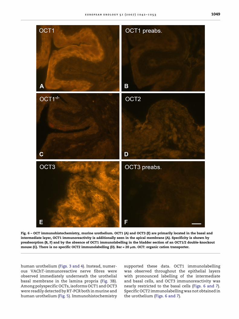
Fig. 6 – OCT immunohistochemistry, murine urothelium. OCT1 (A) and OCT3 (E) are primarily located in the basal and
intermediate layer, OCT1 immunoreactivity is additionally seen in the apical membrane (A). Specificity is shown by
preabsorption (B, F) and by the absence of OCT1 immunolabelling in the bladder section of an OCT1/2 double-knockout
mouse (C). There is no specific OCT2 immunolabelling (D). Bar = 20 mm. OCT: organic cation transporter.
human urothelium (Figs. 3 and 4). Instead, numer-ous VAChT-immunoreactive nerve fibres wereobserved immediately underneath the urothelialbasal membrane in the lamina propria (Fig. 3B).Among polyspecific OCTs, isoforms OCT1 and OCT3were readily detected by RT-PCR both in murine andhuman urothelium (Fig. 5). Immunohistochemistry
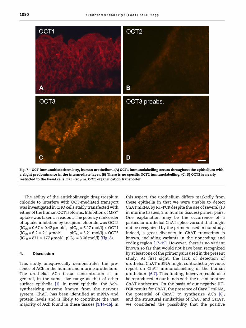
supported these data. OCT1 immunolabellingwas observed throughout the epithelial layerswith pronounced labelling of the intermediateand basal cells, and OCT3 immunoreactivity wasnearly restricted to the basal cells (Figs. 6 and 7).Specific OCT2 immunolabelling was not obtained inthe urothelium (Figs. 6 and 7).
e u r o p e a n u r o l o g y 5 1 ( 2 0 0 7 ) 1 0 4 2 – 1 0 5 31050
Fig. 7 – OCT immunohistochemistry, human urothelium. (A) OCT1 immunolabelling occurs throughout the epithelium with
a slight predominance in the intermediate layer. (B) There is no specific OCT2 immunolabelling. (C, D) OCT3 is nearly
restricted to the basal cells. Bar = 20 mm. OCT: organic cation transporter.
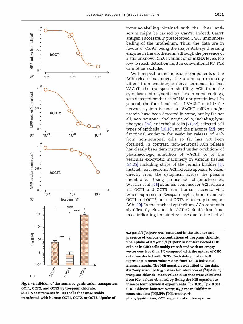
The ability of the anticholinergic drug trospiumchloride to interfere with OCT-mediated transportwas investigated in CHO cells stably transfected witheither of the human OCT isoforms. Inhibition of MPP+
uptake was taken as readout. The potency rank orderof uptake inhibition by trospium chloride was OCT2(IC50 = 0.67 � 0.42 mmol/l, pIC50 = 6.17 mol/l) > OCT1(IC50 = 6.2 � 2.1 mmol/l, pIC50 = 5.21 mol/l) > OCT3(IC50 = 871 � 177 mmol/l, pIC50 = 3.06 mol/l) (Fig. 8).
4. Discussion
This study unequivocally demonstrates the pre-sence of ACh in the human and murine urothelium.The urothelial ACh tissue concentration is, ingeneral, in the same size range as that of othersurface epithelia [1]. In most epithelia, the Ach-synthesizing enzyme known from the nervoussystem, ChAT, has been identified at mRNA andprotein levels and is likely to contribute the vastmajority of ACh found in these tissues [1,14–16]. In
this aspect, the urothelium differs markedly fromthese epithelia in that we were unable to detectChAT mRNA by RT-PCR despite the use of several (13in murine tissues, 2 in human tissues) primer pairs.One explanation may be the occurrence of aparticular urothelial ChAT splice variant that mightnot be recognized by the primers used in our study.Indeed, a great diversity in ChAT transcripts isknown, including variants in the noncoding andcoding region [17–19]. However, there is no variantknown so far that would not have been recognizedby at least one of the primer pairs used in the presentstudy. At first sight, the lack of detection ofurothelial ChAT mRNA might contradict a previousreport on ChAT immunolabelling of the humanurothelium [6,7]. This finding, however, could alsobe reproduced in our hands with the use of anotherChAT antiserum. On the basis of our negative RT-PCR results for ChAT, the presence of CarAT mRNA,the potential of CarAT to synthesize ACh [8],and the structural similarities of ChAT and CarAT,we considered the possibility that the positive
e u r o p e a n u r o l o g y 5 1 ( 2 0 0 7 ) 1 0 4 2 – 1 0 5 3 1051
Fig. 8 – Inhibition of the human organic cation transporters
OCT1, OCT2, and OCT3 by trospium chloride.
(A–C) Measurements in CHO cells that were stably
transfected with human OCT1, OCT2, or OCT3. Uptake of
immunolabelling obtained with the ChAT anti-serum might be caused by CarAT. Indeed, CarATantigen successfully preabsorbed ChAT immunola-belling of the urothelium. Thus, the data are infavour of CarAT being the major Ach-synthesizingenzyme in the urothelium, although the presence ofa still unknown ChAT variant or of mRNA levels toolow to reach detection limit in conventional RT-PCRcannot be excluded.
With respect to the molecular components of theACh release machinery, the urothelium markedlydiffers from cholinergic nerve terminals in thatVAChT, the transporter shuffling ACh from thecytoplasm into synaptic vesicles in nerve endings,was detected neither at mRNA nor protein level. Ingeneral, the functional role of VAChT outside thenervous system is unclear. VAChT mRNA and/orprotein have been detected in some, but by far notall, non-neuronal cholinergic cells, including lym-phocytes [20], endothelial cells [21,22], selected celltypes of epithelia [10,16], and the placenta [23], butfunctional evidence for vesicular release of AChfrom non-neuronal cells so far has not beenobtained. In contrast, non-neuronal ACh releasehas clearly been demonstrated under conditions ofpharmacologic inhibition of VAChT or of thevesicular exocytotic machinery in various tissues[24,25] including strips of the human bladder [6].Instead, non-neuronal ACh release appears to occurdirectly from the cytoplasm across the plasmamembrane. Using antisense oligonucleotides,Wessler et al. [26] obtained evidence for ACh releasevia OCT1 and OCT3 from human placenta villi.When expressed in Xenopus oocytes, human and ratOCT1 and OCT2, but not OCT3, efficiently transportACh [10]. In the tracheal epithelium, ACh content issignificantly elevated in OCT1/2 double-knockoutmice indicating impaired release due to the lack of
0.2 mmol/l [3H]MPP was measured in the absence and
presence of various concentrations of trospium chloride.
The uptake of 0.2 mmol/l [3H]MPP in nontransfected CHO
cells or in CHO cells stably transfected with an empty
vector was less than 5% compared with the uptake of CHO
cells transfected with OCTs. Each data point in A–C
represents a mean value W SEM from 12–16 individual
measurements. The Hill equation was fitted to the data.
(D) Comparison of IC50 values for inhibition of [3H]MPP by
trospium chloride. Mean values W SD that were calculated
from IC50 values obtained by fitting the Hill equation to
three or four individual experiments. **p < 0.01, ***p < 0.001.
CHO: Chinese hamster ovary; IC50: mean inhibitory
concentration; [3H]MPP: [3H]1-methyl-4-
phenylpyridinium; OCT: organic cation transporter.
e u r o p e a n u r o l o g y 5 1 ( 2 0 0 7 ) 1 0 4 2 – 1 0 5 31052
these transporters [27]. Hence, it can be expectedthat OCTs, particularly OCT1, which we havedemonstrated to occur in the human and murineurothelium, represent a major ACh release mechan-ism from the bladder mucosa as well. This findingdoes not exclude the simultaneous occurrence ofadditional ACh transporters because recent micro-dialysis studies on human skin revealed ACh releasethat was resistant to inhibition of both vesicularexocytosis and OCTs [28].
In view of the bidirectionality of OCTs, it also hasto be considered that OCT1 residing in the urothelialplasma membrane mediates ACh uptake into thecell. Cation transport by OCTs is electrogenic, anddirection of transport is determined by membranepotential and substrate concentration [11]. Directevidence for concentration-dependent bidirectionaltransport of ACh has indeed been presented forOCT1-expressing Xenopus oocytes [10]. In view of thepresence of cholinergic nerve terminals immedi-ately underneath the urothelium, which we demon-strated by VAChT immunohistochemistry, it can beexpected that a locally high ACh concentration canbe reached upon stimulation of these nerve endings,and may lead to ACh loading of neighbouringepithelial cells via OCT1.
Because the molecular components of the urothe-lial ACh release machinery differ from those inthe nervous system, the option arises to addressthese systems differentially by pharmacologicapproaches. A characteristic feature of OCTs is theirpolyspecificity, and accordingly, many widely useddrugs are also OCT substrates or interfere with OCT-mediated transport [11]. Here we show that thisfeature also applies to trospium chloride, an anti-cholinergic compound commonly used for treat-ment of overactive bladder. In view of the presentdata, it has to be considered that, in addition to thewell-established muscarinic receptor antagonistproperties, this and chemically similar compoundsalso may act as inhibitors of ACh release from theepithelium.
In conclusion, the present study demonstrates aurothelial non-neuronal cholinergic system thatdiffers widely from that found in neurons withrespect to the molecular components of the AChsynthesis and release machinery. Consequently,these two systems can be differentially targetedby pharmacologic approaches.
Conflicts of interest
This study was supported by the DFG, grant SFB 487/A4 to H.K. and Li 1051/1-1 to K.S.L., and by a project
bound donation of the Dr. R. Pfleger ChemischeFabrik GmbH.
U. Schwantes is employed by Dr. R. PflegerChemische Fabrik GmbH, Bamberg, Germany.
Acknowledgements
The authors thank M. Bodenbenner for experttechnical assistance and K. Michael for preparingthe figures.
References
[1] Klapproth H, Reinheimer T, Metzen J, et al. Non-neuronal
acetylcholine, a signalling molecule synthesized by sur-
face cells of rat and man. Naunyn Schmiedebergs Arch
Pharmacol 1997;355:515–23.
[2] Wessler I, Kirkpatrick J, Racke K. The cholinergic ‘‘pitfall’’:
acetylcholine, a universal cell molecule in biological sys-
tems, including humans. Clin Exp Pharmacol Physiol
1999;26:198–205.
[3] de Groat WC. The urothelium in overactive bladder:
passive bystander or active participant? Urology 2004;
64:7–11.
[4] Birder LA. More than just a barrier: urothelium as a drug
target for urinary bladder pain. Am J Physiol Renal Physiol
2005;289:F489–95.
[5] Beckel JM, Kana A, Lee SJ, de Groat WC, Birder LA. Expres-
sion of functional nicotinic acetylcholine receptors in rat
urinary bladder epithelial cells. Am J Physiol Renal Physiol
2006;290:F103–10.
[6] Yoshida M, Inadome A, Maeda Y, et al. Non-neuronal
cholinergic system in human bladder urothelium. Urol-
ogy 2006;67:425–30.
[7] Yoshida M, Miyamae K, Iwashita H, Otani M, Inadome A.
Management of detrusor dysfunction in the elderly:
changes in acetylcholine and adenosine triphosphate
release during aging. Urology 2004;63(3 suppl 1):17–23.
[8] Tucek S. The synthesis and release of acetylcholine in
normal and denervated rat diaphragms during incuba-
tions in vitro. J Physiol 1982;322:53–69.
[9] Eiden LE. The cholinergic gene locus. J Neurochem
1998;70:2227–40.
[10] Lips KS, Volk C, Schmitt BM, et al. Polyspecific cation
transporters mediate luminal release of acetylcholine
from bronchial epithelium. Am J Respir Cell Mol Biol
2005;33:79–88.
[11] Koepsell H, Schmitt BM, Gorboulev V. Organic cation
transporters. Rev Physiol Biochem Pharmacol 2003;150:
36–90.
[12] Reinheimer T, Bernedo P, Klapproth H, et al. Acetylcholine
in isolated airways of rat, guinea pig, and human: species
differences in role of airway mucosa. Am J Physiol
1996;270:L722–8.
[13] Busch AE, Karbach U, Miska D, et al. Human neurons
express the polyspecific cation transporter hOCT2,
e u r o p e a n u r o l o g y 5 1 ( 2 0 0 7 ) 1 0 4 2 – 1 0 5 3 1053
which translocates monoamine neurotransmitters,
amantadine, and memantine. Mol Pharmacol 1998;54:
342–52.
[14] Grando SA, Kist DA, Qi M, Dahl MV. Human keratinocytes
synthesize, secrete, and degrade acetylcholine. Invest
Dermatol 1993;101:32–6.
[15] Wessler I, Kirkpatrick CJ, Racke K. Non-neuronal acetyl-
choline, a locally acting molecule, widely distributed in
biological systems: expression and function in humans.
Pharmacol Ther 1998;77:59–79.
[16] Proskocil BJ, Sekhon HS, Jia Y, et al. Acetylcholine is an
autocrine or paracrine hormone synthesized and secreted
by airway bronchial epithelial cells. Endocrinology 2004;
145:2498–506.
[17] Oda Y. Choline acetyltransferase: The structure, distribu-
tion and pathologic changes in the central nervous sys-
tem. Pathol Int 1999;49:921–37.
[18] Tooyama I, Kimura H. A protein encoded by an alternative
splice variant of choline acetyltransferase mRNA is loca-
lized preferentially in peripheral nerve cells and fibers.
J Chem Neuroanat 2000;17:217–26.
[19] Robert I, Quirin-Stricker C. A novel untranslated ‘‘exon H’’
of the human choline acetyltransferase gene in placenta.
J Neurochem 2001;79:9–16.
[20] Tayebati SK, El-Assouad D, Ricci A, Amenta F. Immuno-
chemical and immunocytochemical characterization of
cholinergic markers in human peripheral blood lympho-
cytes. J Neuroimmunol 2002;132:147–55.
[21] Haberberger RV, Bodenbenner M, Kummer W. Expression
of the cholinergic gene locus in pulmonary arterial
endothelial cells. Histochem Cell Biol 2000;113:379–87.
[22] Kirkpatrick DJ, Bittinger F, Unger RE, Kriegsmann J,
Kilbinger H, Wessler I. The non-neuronal cholinergic
system in the endothelium: evidence and possible patho-
biological significance. Jpn J Pharmacol 2001;85:24–8.
[23] Pfeil U, Vollerthun R, Kummer W, Lips KS. Expression of
the cholinergic gene locus in the rat placenta. Histochem
Cell Biol 2004;122:121–30.
[24] Wessler I, Roth E, Schwarze S, et al. Release of non-
neuronal acetylcholine from the human placenta: differ-
ence to neuronal acetylcholine. Naunyn Schmiederbergs
Arch Pharmacol 2001;364:205–12.
[25] Moffat JD, Cocks TM, Page CP. Role of the epithelium and
acetylcholine in mediating the contraction to 5-hydroxy-
tryptamine in the mouse isolated trachea. Br J Pharmacol
2004;141:1159–66.
[26] Wessler I, Roth E, Deutsch C, et al. Release of non-
neuronal acetylcholine from the isolated human placenta
is mediated by organic cation transporters. Br J Pharmacol
2001;134:951–6.
[27] Kummer W, Wiegand S, Akinci S, et al. Role of acetylcho-
line and polyspecific cation transporters in serotonin-
induced bronchoconstriction in the mouse. Respir Res
2006;7:65.
[28] Schlereth T, Birklein F, Haack KA, et al. In vivo release of
non-neuronal acetylcholine from the human skin as mea-
sured by dermal microdialysis: effect of botulinum toxin.
Br J Pharmacol 2006;147:183–7.
[29] Misawa H, Ishii K, Deguchi T. Gene expression of mouse
choline acetyltransferase. Alternative splicing and iden-
tification of a highly active promoter region. J Biol Chem
1992;267:20392–9.
[30] Gorboulev V, Ulzheimer JC, Akhoundova A, et al. Cloning
and characterization of two human polyspecific orga-
nic cation transporters. DNA Cell Biol 1997;16:
871–81.