Embed Size (px)
Citation preview
ACCF/ACR/SCCT/SCMR/ASNC/NASCI/SCAI/SIR APPROPRIATENESS CRITERIA
ACCF/ACR/SCCT/SCMR/ASNC/NASCI/
SCAI/SIR Appropriateness Criteria for Cardiac
Computed Tomography and Cardiac Magnetic
Resonance Imaging – Online Appendix
A Report of the American College of Cardiology Foundation Quality Strategic
Directions Committee Appropriateness Criteria Working Group, American College of
Radiology, Society of Cardiovascular Computed Tomography, Society for
Cardiovascular Magnetic Resonance, American Society of Nuclear Cardiology, North
American Society for Cardiac Imaging, Society for Cardiovascular Angiography and
Interventions, and Society of Interventional Radiology
CCT/CMR WRITING GROUP
Robert C. Hendel, MD, FACC, FAHA*
Manesh R. Patel, MD*
Christopher M. Kramer, MD, FACC, FAHA‡
Michael Poon, MD, FACC†
Representative: *ACCF, †SCCT, ‡SCMR
TECHNICAL PANEL MEMBERS
Robert C. Hendel, MD, FACC, FAHA, Moderator**
James C. Carr, MD, BCh, BAO§§
Nancy A. Gerstad, MD
Linda D. Gillam, MD, FACC, FAHA
John McB. Hodgson, MD, FSACI, FACC‡‡
Raymond J. Kim, MD, FACC
Christopher M. Kramer, MD, FACC, FAHA‡
John R. Lesser, MD, FACC
Edward T. Martin, MD, FACC, FACP, FAHA
Joseph V. Messer, MD, MACC, FAHA, FSCAI
Rita F. Redberg, MD, MSc, FACC¶
Geoffrey D. Rubin, MD, FSCBTMR§
John S. Rumsfeld, MD, PhD, FACC
Allen J. Taylor, MD, FACC, FAHA
Wm. Guy Weigold, MD, FACC†
Pamela K. Woodard, MD, FAHA††
Representative: §ACR, ¶AHA, **ASNC, ††NASCI, ‡‡SCAI, †SCCT, ‡SCMR, §§SIR
ACCF APPROPRIATENESS CRITERIA WORKING GROUP
Ralph G. Brindis, MD, MPH, FACC, Chair
Robert C. Hendel, MD, FACC, FAHA, Pamela S. Douglas, MD, MACC, FAHA
Eric D. Peterson MD, FACC, FAHA, Michael J. Wolk, MD, MACC,
Joseph M. Allen, MA, Manesh R. Patel, MD
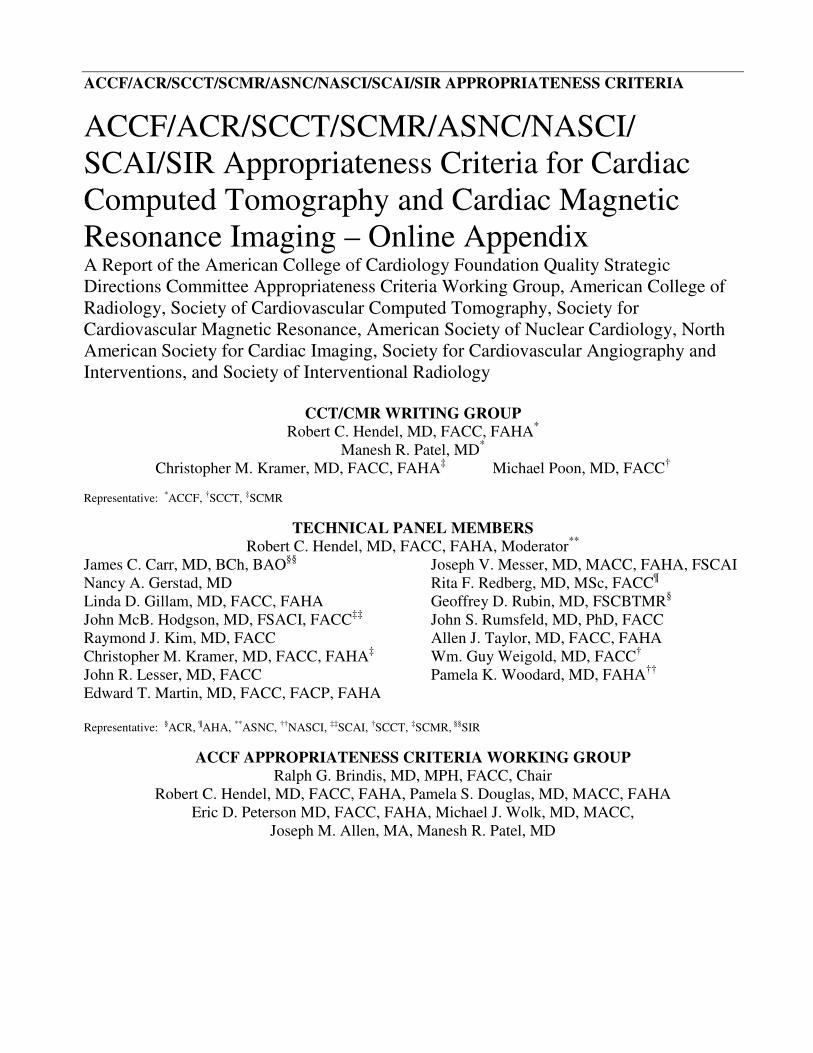
APPENDIX A. CCT Appropriateness Criteria Ratings
1 2 3 4 5 6 7 8 9 10 11 12 13 14 15 Median MADM R
Table 1. Detection of CAD: SymptomaticEvaluation of Chest Pain Syndrome (Use of CT Angiogram)
� Intermediate pre-test probability 4 7 8 5 8 7 1 1 1 1 6 1 2 6 5 2.6 U -
� ECG interpretable AND able to exercise
� Intermediate pre-test probability 9 5 7 9 7 8 9 5 5 2 7 7 4 2 9 7 1.9 A
� ECG uninterpretable OR unable to
exercise
3 � High pre-test probability of CAD 5 4 3 2 1 2 2 3 1 2 2 3 1 2 5 2 0.9 I +
4 � Evaluation of suspected coronary
anomalies
9 9 9 9 9 9 9 8 9 7 8 8 7 7 9 9 0.6 A +
� Low pre-test probability of CAD 6 4 9 9 6 5 7 1 5 2 2 6 1 2 6 5 2.1 U
� No ECG changes and serial enzymes
negative
� Intermediate pre-test probability of CAD 7 8 6 9 7 7 9 4 6 2 7 6 2 4 9 7 1.7 A
� No ECG changes and serial enzymes
negative
7 � High pre-test probability of CAD 6 3 6 1 2 8 5 4 7 7 6 6 1 1 9 6 2.1 U
� No ECG changes and serial enzymes
negative
� High pre-test probability of CAD 1 4 2 1 1 1 1 1 1 2 1 2 1 1 1 1 0.4 I +
� ECG – ST elevation and/or positive
cardiac enzymes
� “Triple rule out” – exclude obstructive
CAD, aortic dissection, and pulmonary
embolism
5 2 8 5 2 6 8 1 7 6 4 3 1 2 4 4 2.0 U
� Intermediate pre-test probability for one
of the above
� ECG – no ST elevation and initial
enzymes negative
1 8
Indication
5
2
Acute Chest Pain (Use of CT Angiogram)
Evaluation of Intra Cardiac Structures (Use of CT Angiogram)
6
8
9
Agree
2
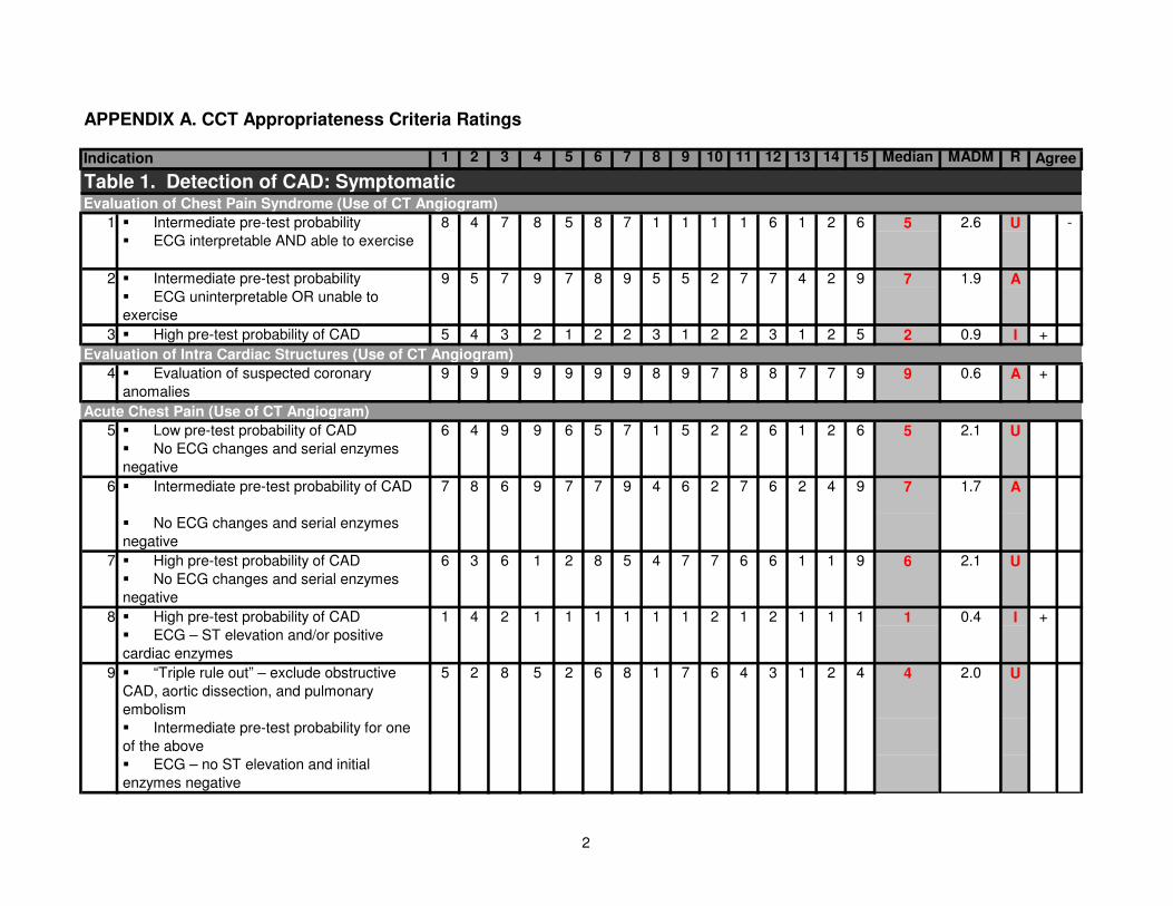
1 2 3 4 5 6 7 8 9 10 11 12 13 14 15 Median MADM RIndication Agree
10 � Low CHD Risk (Framingham Risk
Criteria)
2 2 1 1 1 1 2 1 1 1 1 1 1 1 1 1 0.2 I +
11 � Moderate CHD risk (Framingham) 5 3 2 1 1 5 4 1 1 1 4 3 1 1 3 2 1.3 I +
12 � High CHD risk (Framingham) 5 5 2 5 1 5 6 1 5 1 2 4 1 1 4 4 1.7 U
Asymptomatic (Calcium Scoring)
13 � Low CHD Risk (Framingham) 3 3 1 2 2 3 3 1 1 1 1 1 1 5 1 1 0.9 I +
14 � Moderate CHD Risk (Framingham) 8 8 6 8 7 6 8 3 5 1 7 5 1 8 3 6 2.0 U
15 � High CHD Risk (Framingham) 7 5 7 2 2 7 9 1 6 1 5 6 1 5 4 5 2.1 U
Table 4. Detection of CAD with Prior Test ResultsEvaluation of Chest Pain Syndrome (Use of CT Angiogram)
16 � Uninterpretable or equivocal stress test
(exercise, perfusion or stress echo)
8 7 9 9 8 9 8 8 7 7 7 7 3 5 9 8 1.1 A +
17 � Evidence of moderate to severe ischemia
on stress test (exercise, perfusion or stress
echo)
3 4 3 2 1 3 3 1 1 2 2 2 1 2 1 2 0.7 I +
Asymptomatic (Use of CT Angiogram)
Table 2: Detection of CAD: Asymptomatic (without Chest Pain Syndrome)
Table 3. Risk Assessment: General Population
3
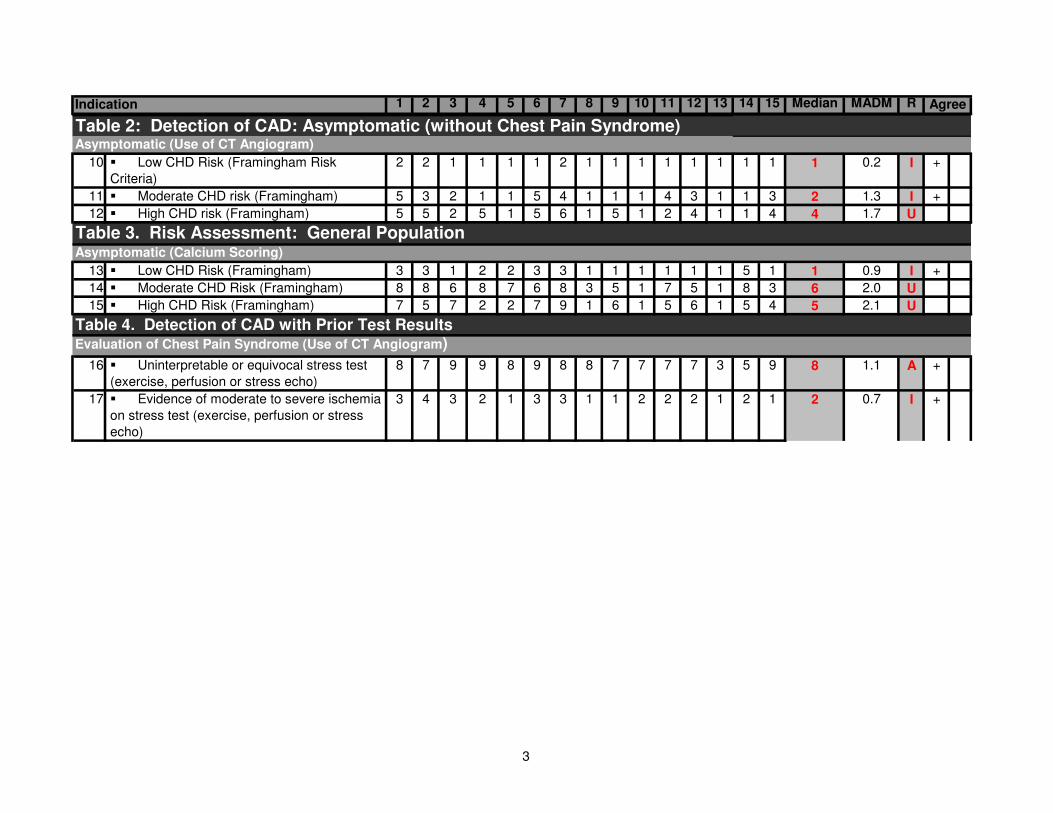
1 2 3 4 5 6 7 8 9 10 11 12 13 14 15 Median MADM RIndication Agree
18 � Prior calcium score within previous 5
years
1 4 2 2 1 4 1 1 1 1 2 5 1 1 4 1 1.1 I +
� High CHD Risk (Framingham) 1 2 1 3 2 1 2 1 2 4 1 1 2 2 0.7 I +
� Within 2 years prior cardiac CT
angiogram or invasive angiogram without
significant obstructive disease
� High CHD Risk (Framingham) 4 1 3 1 5 4 1 5 4 4 4 1 1 2 3 1.5 I
� Prior calcium score greater than or equal
to 400
Asymptomatic (Use of CT Angiogram)
1
1
19 3
20
Asymptomatic (Calcium Scoring)
Table 5. Risk Assessment with Prior Test Results
4
1 2 3 4 5 6 7 8 9 10 11 12 13 14 15 Median MADM RIndication Agree
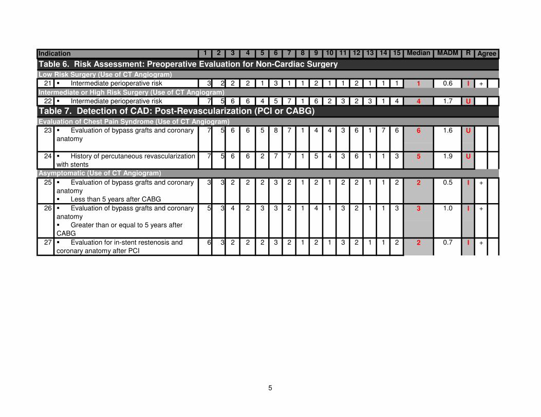
Table 6. Risk Assessment: Preoperative Evaluation for Non-Cardiac Surgery
21 � Intermediate perioperative risk 3 2 2 2 1 3 1 1 2 1 1 2 1 1 1 1 0.6 I +
22 � Intermediate perioperative risk 7 5 6 6 4 5 7 1 6 2 3 2 3 1 4 4 1.7 U
� Evaluation of bypass grafts and coronary
anatomy
6 6 5 8 7 1 4 4 3 6 1 7 6 6 1.6 U
24 � History of percutaneous revascularization
with stents
7 5 6 6 2 7 7 1 5 4 3 6 1 1 3 5 1.9 U
� Evaluation of bypass grafts and coronary
anatomy
2 2 2 3 2 1 2 1 2 2 1 1 2 2 0.5 I +
� Less than 5 years after CABG
� Evaluation of bypass grafts and coronary
anatomy
4 2 3 3 2 1 4 1 3 2 1 1 3 3 1.0 I +
� Greater than or equal to 5 years after
CABG
27 � Evaluation for in-stent restenosis and
coronary anatomy after PCI
6 3 2 2 2 3 2 1 2 1 3 2 1 1 2 2 0.7 I +
Asymptomatic (Use of CT Angiogram)
Low Risk Surgery (Use of CT Angiogram)
Intermediate or High Risk Surgery (Use of CT Angiogram)
Table 7. Detection of CAD: Post-Revascularization (PCI or CABG)
3
Evaluation of Chest Pain Syndrome (Use of CT Angiogram)
25 3
23 7 5
5
3
26
5
1 2 3 4 5 6 7 8 9 10 11 12 13 14 15 Median MADM RIndication Agree
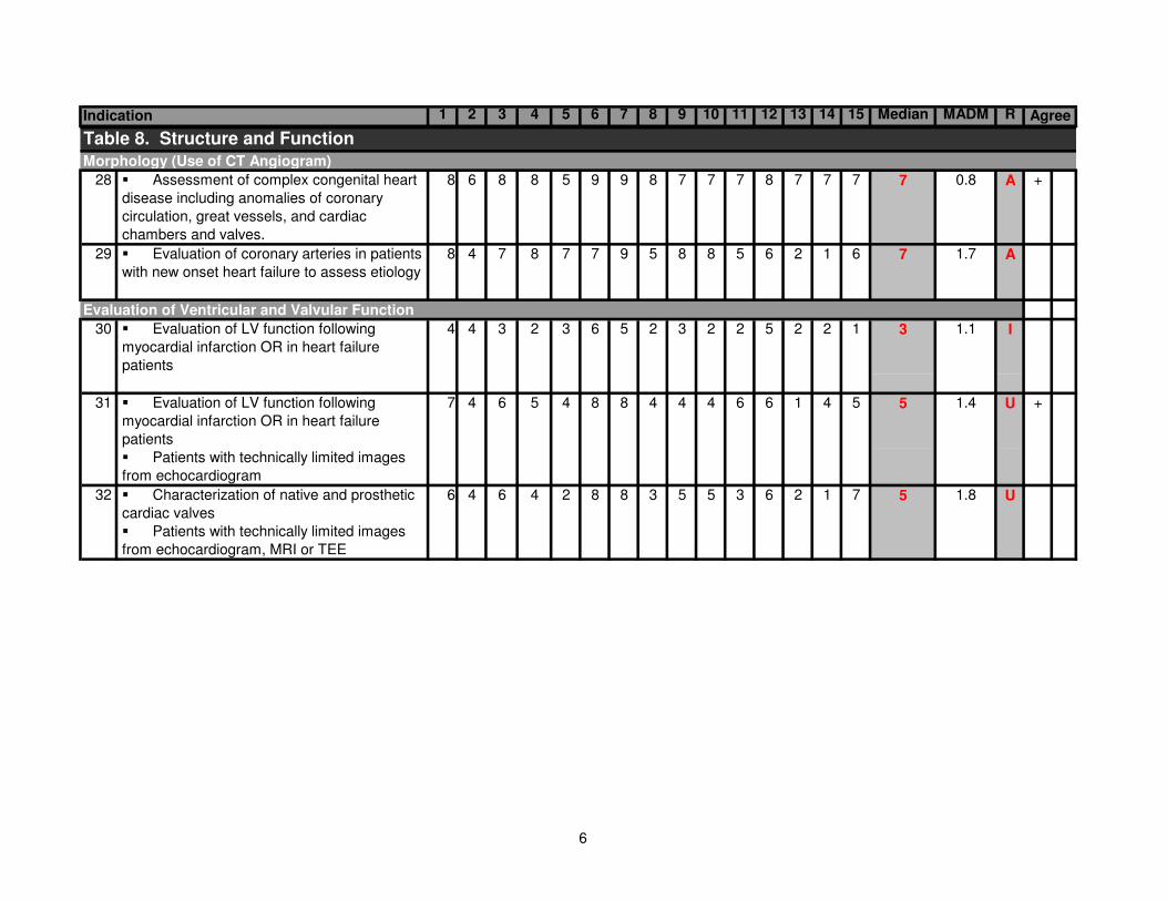
Table 8. Structure and Function
28 � Assessment of complex congenital heart
disease including anomalies of coronary
circulation, great vessels, and cardiac
chambers and valves.
8 6 8 8 5 9 9 8 7 7 7 8 7 7 7 7 0.8 A +
29 � Evaluation of coronary arteries in patients
with new onset heart failure to assess etiology
8 4 7 8 7 7 9 5 8 8 5 6 2 1 6 7 1.7 A
� Evaluation of LV function following
myocardial infarction OR in heart failure
patients
4 3 2 3 6 5 2 3 2 2 5 2 2 1 3 1.1 I
� Evaluation of LV function following
myocardial infarction OR in heart failure
patients
4 6 5 4 8 8 4 4 4 6 6 1 4 5 5 1.4 U +
� Patients with technically limited images
from echocardiogram
� Characterization of native and prosthetic
cardiac valves
4 6 4 2 8 8 3 5 5 3 6 2 1 7 5 1.8 U
� Patients with technically limited images
from echocardiogram, MRI or TEE
31 7
Evaluation of Ventricular and Valvular Function
Morphology (Use of CT Angiogram)
30 4
32 6
6
1 2 3 4 5 6 7 8 9 10 11 12 13 14 15 Median MADM RIndication Agree
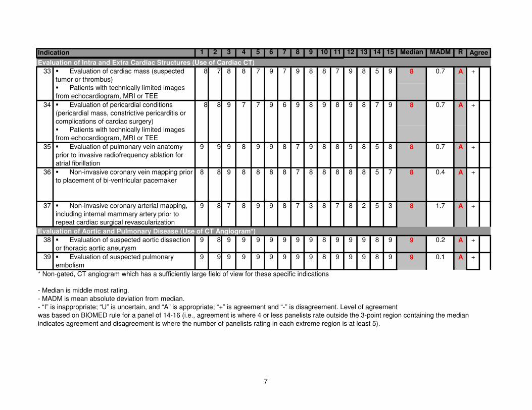
� Evaluation of cardiac mass (suspected
tumor or thrombus)
8 8 7 9 7 9 8 8 7 9 8 5 9 8 0.7 A +
� Patients with technically limited images
from echocardiogram, MRI or TEE
� Evaluation of pericardial conditions
(pericardial mass, constrictive pericarditis or
complications of cardiac surgery)
9 7 7 9 6 9 8 9 8 9 8 7 9 8 0.7 A +
� Patients with technically limited images
from echocardiogram, MRI or TEE
35 � Evaluation of pulmonary vein anatomy
prior to invasive radiofrequency ablation for
atrial fibrillation
9 9 9 8 9 9 8 7 9 8 8 9 8 5 8 8 0.7 A +
36 � Non-invasive coronary vein mapping prior
to placement of bi-ventricular pacemaker
8 8 9 8 8 8 8 7 8 8 8 8 8 5 7 8 0.4 A +
37 � Non-invasive coronary arterial mapping,
including internal mammary artery prior to
repeat cardiac surgical revascularization
9 8 7 8 9 9 8 7 3 8 7 8 2 5 3 8 1.7 A +
38 � Evaluation of suspected aortic dissection
or thoracic aortic aneurysm
9 8 9 9 9 9 9 9 9 8 9 9 9 8 9 9 0.2 A +
39 � Evaluation of suspected pulmonary
embolism
9 9 9 9 9 9 9 9 9 8 9 9 9 8 9 9 0.1 A +
* Non-gated, CT angiogram which has a sufficiently large field of view for these specific indications
- Median is middle most rating.
- MADM is mean absolute deviation from median.
- “I” is inappropriate; “U” is uncertain, and “A” is appropriate; “+” is agreement and “-” is disagreement. Level of agreement
was based on BIOMED rule for a panel of 14-16 (i.e., agreement is where 4 or less panelists rate outside the 3-point region containing the median
indicates agreement and disagreement is where the number of panelists rating in each extreme region is at least 5).
34 8 8
7
Evaluation of Intra and Extra Cardiac Structures (Use of Cardiac CT)
Evaluation of Aortic and Pulmonary Disease (Use of CT Angiogram*)
33 8
7
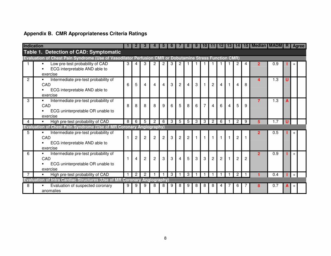
Appendix B. CMR Appropriateness Criteria Ratings
1 2 3 4 5 6 7 8 9 10 11 12 13 14 15 Median MADM R
Table 1. Detection of CAD: Symptomatic
� Low pre-test probability of CAD 3 4 3 2 2 3 2 1 1 1 1 1 1 2 4 2 0.9 I +
� ECG interpretable AND able to
exercise
� Intermediate pre-test probability of
CAD 6 5 4 4 4 3 2 4 3 1 2 4 1 4 84 1.3 U
� ECG interpretable AND able to
exercise
� Intermediate pre-test probability of
CAD 8 8 8 8 9 6 5 8 6 7 4 6 4 5 97 1.3 A
� ECG uninterpretable OR unable to
exercise
4 � High pre-test probability of CAD 8 6 5 2 6 3 5 5 3 3 2 6 1 2 9 5 1.7 U
Evaluation of Chest Pain Syndrome (Use of MR Coronary Angiography)
� Intermediate pre-test probability of
CAD 1 2 2 2 2 3 2 2 1 1 1 1 1 2 12 0.5 I +
� ECG interpretable AND able to
exercise
� Intermediate pre-test probability of
CAD 1 4 2 2 3 3 4 5 3 3 2 2 1 2 22 0.9 I +
� ECG uninterpretable OR unable to
exercise
7 � High pre-test probability of CAD 1 2 2 1 1 3 1 3 1 1 1 1 1 2 1 1 0.4 I +
Evaluation of Intra Cardiac Structures (Use of MR Coronary Angiography)
9 9 9 8 8 9 8 9 8 8 8 4 7 6 7 8 0.7 A +� Evaluation of suspected coronary
anomalies
8
Indication Agree
5
6
1
2
3
Evaluation of Chest Pain Syndrome (Use of Vasodilator Perfusion CMR or Dobutamine Stress Function CMR)
8
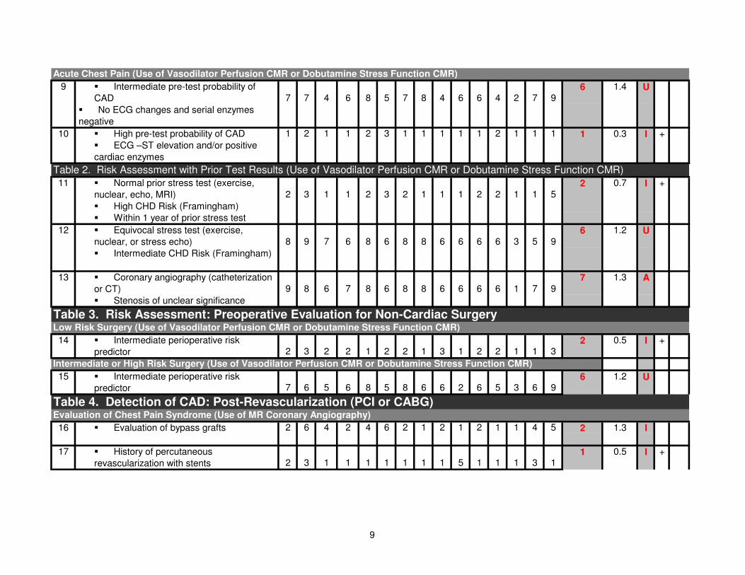
Acute Chest Pain (Use of Vasodilator Perfusion CMR or Dobutamine Stress Function CMR)
� Intermediate pre-test probability of
CAD 7 7 4 6 8 5 7 8 4 6 6 4 2 7 96 1.4 U
� No ECG changes and serial enzymes
negative
� High pre-test probability of CAD 1 2 1 1 2 3 1 1 1 1 1 2 1 1 1 1 0.3 I +
� ECG –ST elevation and/or positive
cardiac enzymes
� Normal prior stress test (exercise,
nuclear, echo, MRI) 2 3 1 1 2 3 2 1 1 1 2 2 1 1 52 0.7 I +
� High CHD Risk (Framingham)
� Within 1 year of prior stress test
� Equivocal stress test (exercise,
nuclear, or stress echo) 8 9 7 6 8 6 8 8 6 6 6 6 3 5 96 1.2 U
� Intermediate CHD Risk (Framingham)
� Coronary angiography (catheterization
or CT) 9 8 6 7 8 6 8 8 6 6 6 6 1 7 97 1.3 A
� Stenosis of unclear significance
Low Risk Surgery (Use of Vasodilator Perfusion CMR or Dobutamine Stress Function CMR)
14 � Intermediate perioperative risk
predictor 2 3 2 2 1 2 2 1 3 1 2 2 1 1 32 0.5 I +
15 � Intermediate perioperative risk
predictor 7 6 5 6 8 5 8 6 6 2 6 5 3 6 96 1.2 U
Evaluation of Chest Pain Syndrome (Use of MR Coronary Angiography)
� Evaluation of bypass grafts 2 6 4 2 4 6 2 1 2 1 2 1 1 4 5 2 1.3 I
17 � History of percutaneous
revascularization with stents 2 3 1 1 1 1 1 1 1 5 1 1 1 3 11 0.5 I +
Table 4. Detection of CAD: Post-Revascularization (PCI or CABG)
Table 3. Risk Assessment: Preoperative Evaluation for Non-Cardiac Surgery
16
11
12
13
Intermediate or High Risk Surgery (Use of Vasodilator Perfusion CMR or Dobutamine Stress Function CMR)
9
10
Table 2. Risk Assessment with Prior Test Results (Use of Vasodilator Perfusion CMR or Dobutamine Stress Function CMR)
9
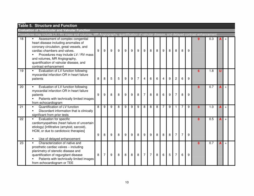
Table 5. Structure and Function Evaluation of Ventricular and Valvular Function
Procedures may include LV / RV mass and volumes, MR Angiography, quantification of valvular disease, and delayed contrast enhancement
� Assessment of complex congenital
heart disease including anomalies of
coronary circulation, great vessels, and
cardiac chambers and valves. 9 9 9 9 9 9 9 9 8 8 9 8 8 8 9
9 0.3 A +
� Procedures may include LV / RV mass
and volumes, MR Angiography,
quantification of valvular disease, and
contrast enhancement
� Evaluation of LV function following
myocardial infarction OR in heart failure
patients 8 8 5 5 9 9 7 4 6 6 4 9 2 6 9
6 1.8 U
� Evaluation of LV function following
myocardial infarction OR in heart failure
patients 9 9 8 8 9 9 8 7 8 8 6 9 7 8 9
8 0.7 A +
� Patients with technically limited images
from echocardiogram
21 � Quantification of LV function 8 9 9 8 9 9 9 8 8 8 7 9 1 7 9 8 1.0 A +
� Discordant information that is clinically
significant from prior tests
� Evaluation for specific
cardiomyopathies (heart failure of uncertain
etiology) [infiltrative (amyloid, sarcoid),
HCM, or due to cardiotoxic therapies]
9 8 9 8 9 9 8 9 9 8 8 8 7 7 9
8 0.5 A +
� Use of delayed enhancement
� Characterization of native and
prosthetic cardiac valves – including
planimetry of stenotic disease and
quantification of regurgitant disease 8 7 9 8 8 8 8 7 7 8 6 5 7 6 9
8 0.7 A +
� Patients with technically limited images
from echocardiogram or TEE
22
23
18
19
20
10
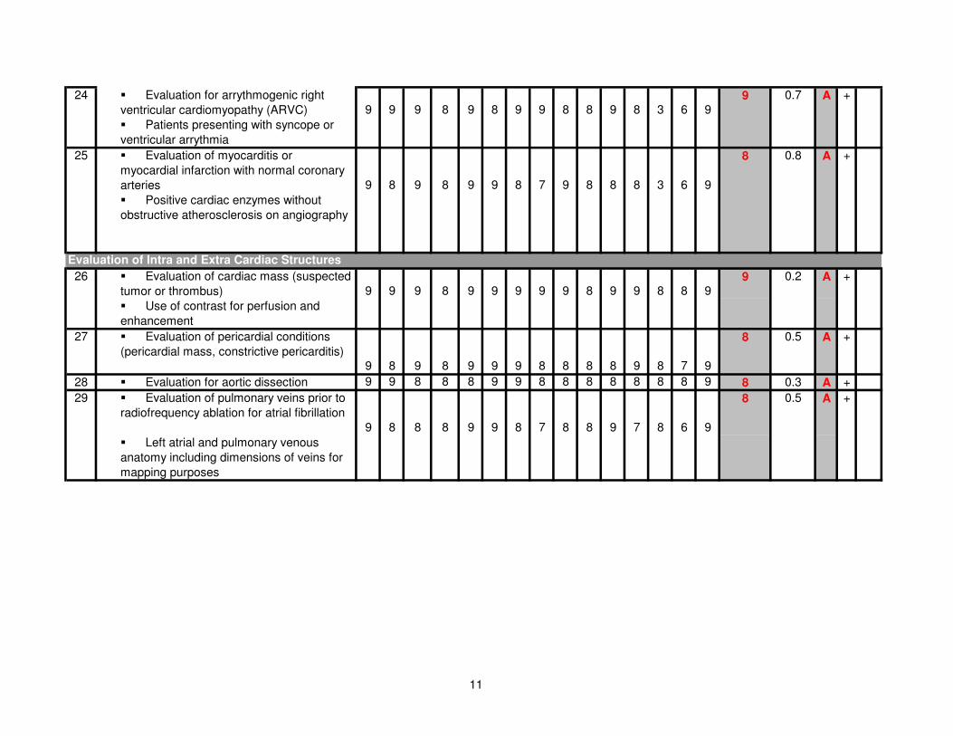
� Evaluation for arrythmogenic right
ventricular cardiomyopathy (ARVC) 9 9 9 8 9 8 9 9 8 8 9 8 3 6 99 0.7 A +
� Patients presenting with syncope or
ventricular arrythmia
� Evaluation of myocarditis or
myocardial infarction with normal coronary
arteries 9 8 9 8 9 9 8 7 9 8 8 8 3 6 9
8 0.8 A +
� Positive cardiac enzymes without
obstructive atherosclerosis on angiography
Evaluation of Intra and Extra Cardiac Structures
� Evaluation of cardiac mass (suspected
tumor or thrombus) 9 9 9 8 9 9 9 9 9 8 9 9 8 8 99 0.2 A +
� Use of contrast for perfusion and
enhancement
27 � Evaluation of pericardial conditions
(pericardial mass, constrictive pericarditis)9 8 9 8 9 9 9 8 8 8 8 9 8 7 9
8 0.5 A +
28 � Evaluation for aortic dissection 9 9 8 8 8 9 9 8 8 8 8 8 8 8 9 8 0.3 A +
� Evaluation of pulmonary veins prior to
radiofrequency ablation for atrial fibrillation
9 8 8 8 9 9 8 7 8 8 9 7 8 6 9
8 0.5 A +
� Left atrial and pulmonary venous
anatomy including dimensions of veins for
mapping purposes
26
29
25
24
11
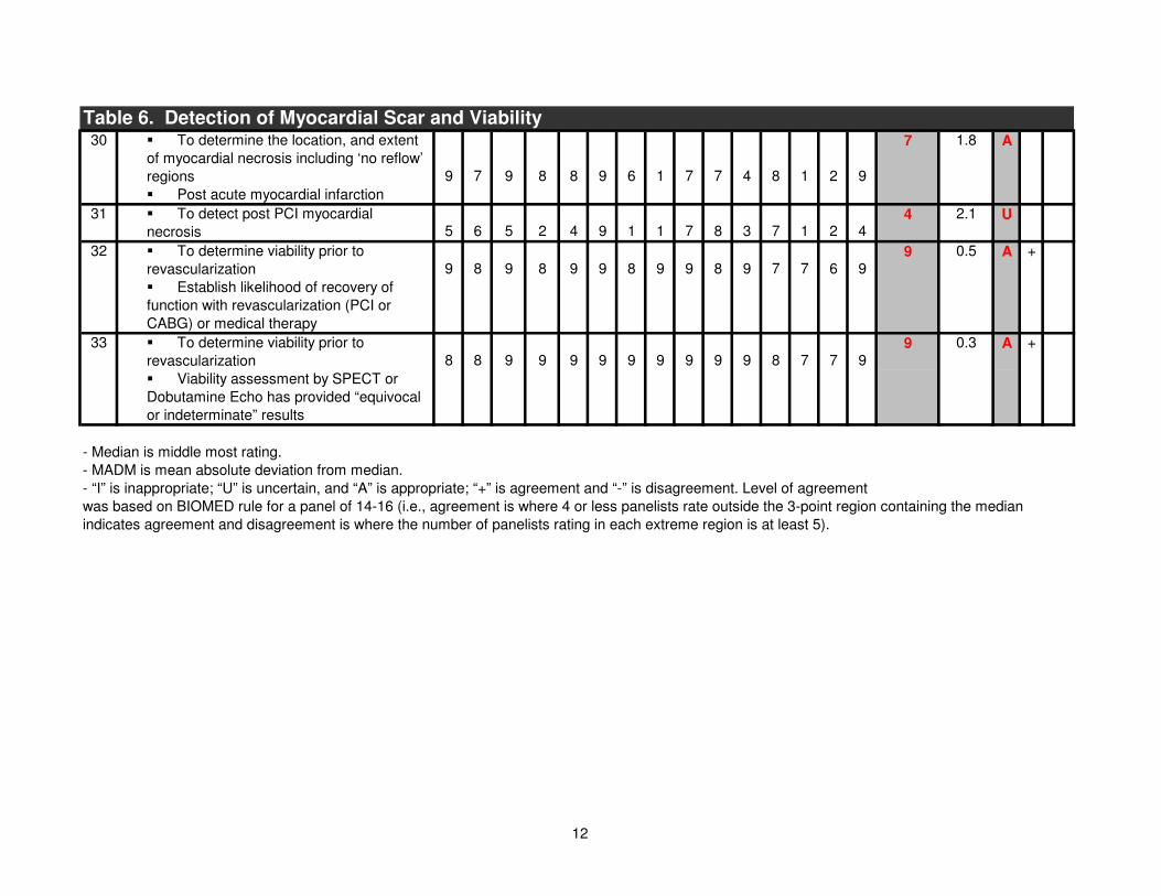
� To determine the location, and extent
of myocardial necrosis including ‘no reflow’
regions 9 7 9 8 8 9 6 1 7 7 4 8 1 2 9
7 1.8 A
� Post acute myocardial infarction
31 � To detect post PCI myocardial
necrosis 5 6 5 2 4 9 1 1 7 8 3 7 1 2 44 2.1 U
� To determine viability prior to
revascularization 9 8 9 8 9 9 8 9 9 8 9 7 7 6 99 0.5 A +
� Establish likelihood of recovery of
function with revascularization (PCI or
CABG) or medical therapy
� To determine viability prior to
revascularization 8 8 9 9 9 9 9 9 9 9 9 8 7 7 99 0.3 A +
� Viability assessment by SPECT or
Dobutamine Echo has provided “equivocal
or indeterminate” results
- Median is middle most rating.
- MADM is mean absolute deviation from median.
- “I” is inappropriate; “U” is uncertain, and “A” is appropriate; “+” is agreement and “-” is disagreement. Level of agreement
was based on BIOMED rule for a panel of 14-16 (i.e., agreement is where 4 or less panelists rate outside the 3-point region containing the median
indicates agreement and disagreement is where the number of panelists rating in each extreme region is at least 5).
33
Table 6. Detection of Myocardial Scar and Viability30
32
12
13
Appendix C. CCT Evidence Summary and Tables
The sensitivity of advanced MDCT technology with a slice collimation less than 1.0 mm
for the detection of hemodynamically significant coronary artery stenosis has been
demonstrated to be very high (i.e. higher than 95% in most of all currently available
published reports using 16 or more detector row CTs) provided that image quality is
adequate, evaluation is performed by CCT experts, and the patients are properly chosen
and prepared prior to the study.
CCT has been used for the diagnosis of hemodynamically significant coronary artery
disease in patients with a low to intermediate likelihood of having significant stenosis
(Tables 1 and 2*).
CCT also has been used to facilitate a decision for or against invasive coronary
angiography in patients who had an uninterpretable or equivocal stress ECG or stress
myocardial perfusion study.
CCT has been used for the assessment of coronary anomalies, pulmonary veins and left
atrium prior to radiofrequency ablation of atrial fibrillation, and coronary vein mapping
prior to the placement of pacemaker leads for cardiac resynchronization therapy.
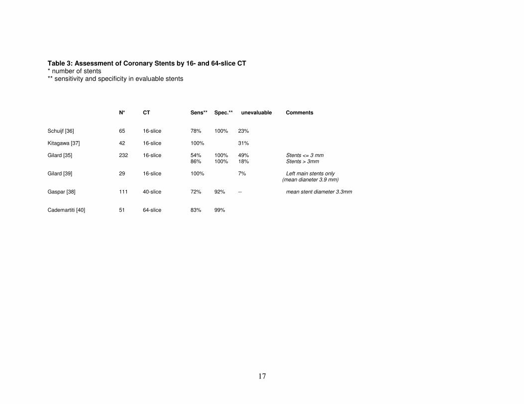
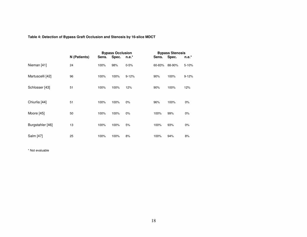
The use of CCT for stent occlusion and patency as well as bypass graft patency continues
to be under investigation (Tables 3 and 4).
CT imaging has been used to detect and rule out aortic dissection and pulmonary
embolism.
*
Publication Note: Studies cited in Tables 1 and 2 generally can be inferred to be of patients at
intermediate risk of disease. The sensitivity and specificity data cited are primarily for studies in which
patients had a high prevalence of disease.
14
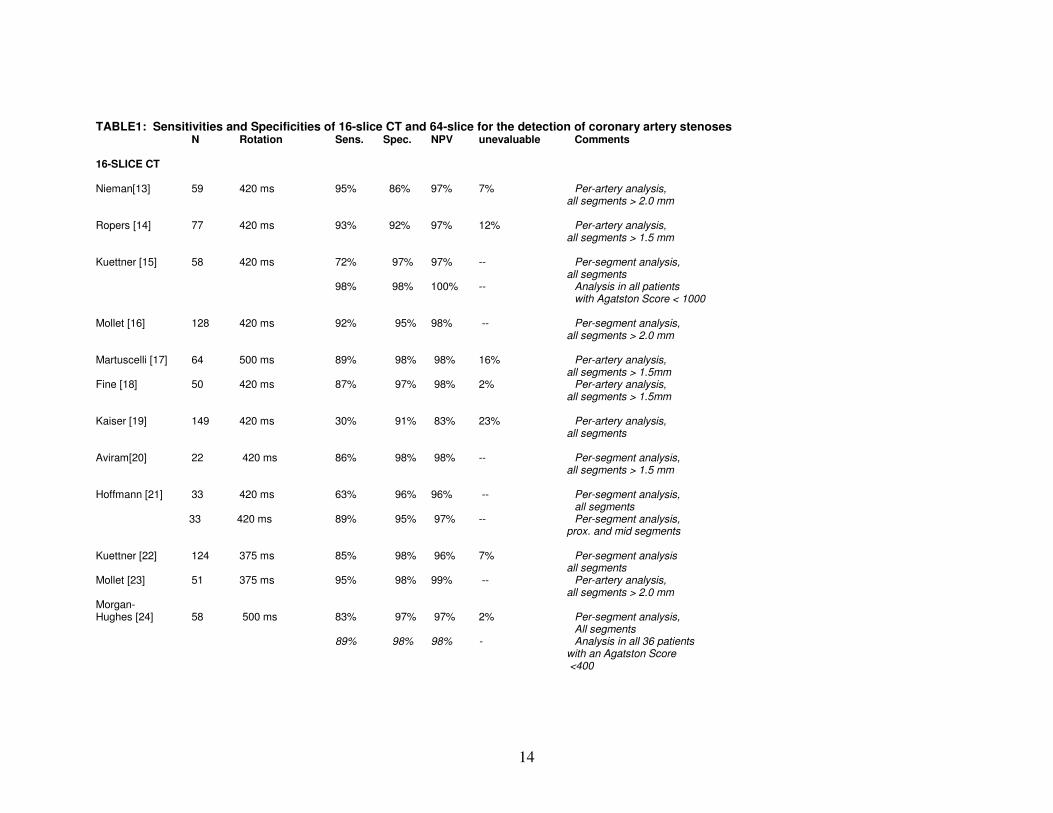
TABLE1: Sensitivities and Specificities of 16-slice CT and 64-slice for the detection of coronary artery stenoses N Rotation Sens. Spec. NPV unevaluable Comments 16-SLICE CT Nieman[13] 59 420 ms 95% 86% 97% 7% Per-artery analysis,
all segments > 2.0 mm
Ropers [14] 77 420 ms 93% 92% 97% 12% Per-artery analysis, all segments > 1.5 mm
Kuettner [15] 58 420 ms 72% 97% 97% -- Per-segment analysis,
all segments 98% 98% 100% -- Analysis in all patients with Agatston Score < 1000 Mollet [16] 128 420 ms 92% 95% 98% -- Per-segment analysis,
all segments > 2.0 mm
Martuscelli [17] 64 500 ms 89% 98% 98% 16% Per-artery analysis, all segments > 1.5mm
Fine [18] 50 420 ms 87% 97% 98% 2% Per-artery analysis, all segments > 1.5mm
Kaiser [19] 149 420 ms 30% 91% 83% 23% Per-artery analysis,
all segments Aviram[20] 22 420 ms 86% 98% 98% -- Per-segment analysis,
all segments > 1.5 mm Hoffmann [21] 33 420 ms 63% 96% 96% -- Per-segment analysis, all segments
33 420 ms 89% 95% 97% -- Per-segment analysis, prox. and mid segments
Kuettner [22] 124 375 ms 85% 98% 96% 7% Per-segment analysis
all segments Mollet [23] 51 375 ms 95% 98% 99% -- Per-artery analysis,
all segments > 2.0 mm Morgan- Hughes [24] 58 500 ms 83% 97% 97% 2% Per-segment analysis, All segments 89% 98% 98% - Analysis in all 36 patients
with an Agatston Score <400
15
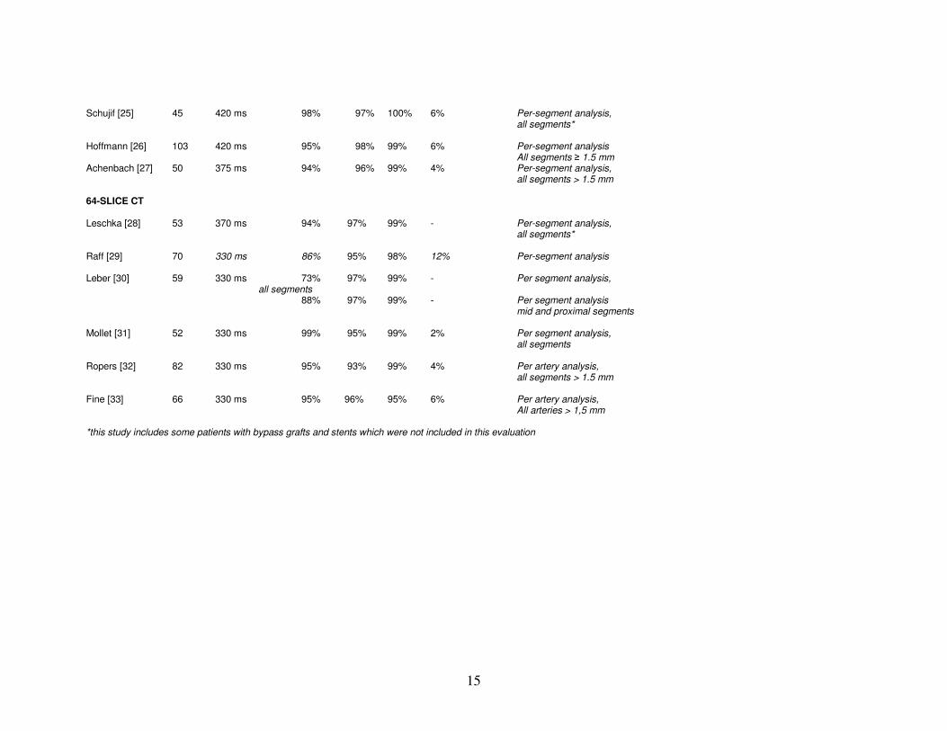
Schujif [25] 45 420 ms 98% 97% 100% 6% Per-segment analysis, all segments* Hoffmann [26] 103 420 ms 95% 98% 99% 6% Per-segment analysis All segments ≥ 1.5 mm Achenbach [27] 50 375 ms 94% 96% 99% 4% Per-segment analysis, all segments > 1.5 mm 64-SLICE CT Leschka [28] 53 370 ms 94% 97% 99% - Per-segment analysis, all segments* Raff [29] 70 330 ms 86% 95% 98% 12% Per-segment analysis Leber [30] 59 330 ms 73% 97% 99% - Per segment analysis, all segments 88% 97% 99% - Per segment analysis mid and proximal segments Mollet [31] 52 330 ms 99% 95% 99% 2% Per segment analysis, all segments Ropers [32] 82 330 ms 95% 93% 99% 4% Per artery analysis, all segments > 1.5 mm Fine [33] 66 330 ms 95% 96% 95% 6% Per artery analysis, All arteries > 1,5 mm *this study includes some patients with bypass grafts and stents which were not included in this evaluation
16
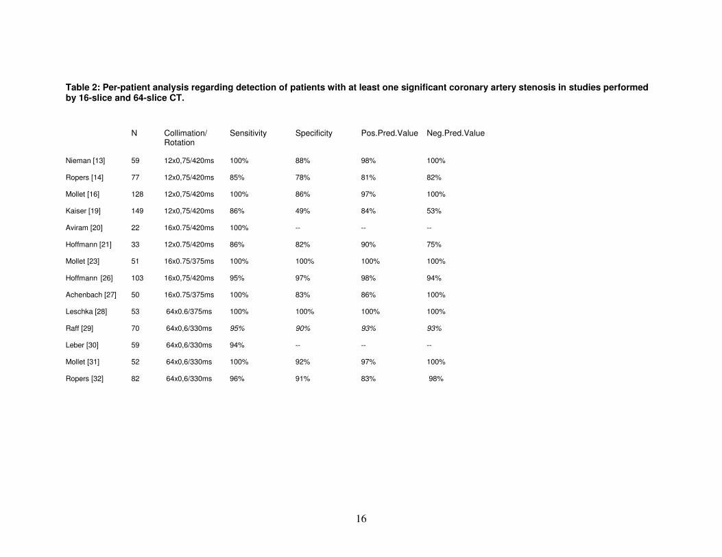
Table 2: Per-patient analysis regarding detection of patients with at least one significant coronary artery stenosis in studies performed by 16-slice and 64-slice CT.
N Collimation/ Sensitivity Specificity Pos.Pred.Value Neg.Pred.Value Rotation Nieman [13] 59 12x0,75/420ms 100% 88% 98% 100% Ropers [14] 77 12x0,75/420ms 85% 78% 81% 82% Mollet [16] 128 12x0,75/420ms 100% 86% 97% 100% Kaiser [19] 149 12x0,75/420ms 86% 49% 84% 53% Aviram [20] 22 16x0.75/420ms 100% -- -- -- Hoffmann [21] 33 12x0.75/420ms 86% 82% 90% 75% Mollet [23] 51 16x0.75/375ms 100% 100% 100% 100% Hoffmann [26] 103 16x0,75/420ms 95% 97% 98% 94% Achenbach [27] 50 16x0.75/375ms 100% 83% 86% 100% Leschka [28] 53 64x0.6/375ms 100% 100% 100% 100% Raff [29] 70 64x0,6/330ms 95% 90% 93% 93% Leber [30] 59 64x0,6/330ms 94% -- -- -- Mollet [31] 52 64x0,6/330ms 100% 92% 97% 100% Ropers [32] 82 64x0,6/330ms 96% 91% 83% 98%
17
Table 3: Assessment of Coronary Stents by 16- and 64-slice CT * number of stents ** sensitivity and specificity in evaluable stents
N* CT Sens** Spec.** unevaluable Comments Schuijf [36] 65 16-slice 78% 100% 23% Kitagawa [37] 42 16-slice 100% 31% Gilard [35] 232 16-slice 54% 100% 49% Stents <= 3 mm 86% 100% 18% Stents > 3mm Gilard [39] 29 16-slice 100% 7% Left main stents only
(mean dianeter 3.9 mm) Gaspar [38] 111 40-slice 72% 92% -- mean stent diameter 3.3mm Cademartiti [40] 51 64-slice 83% 99%
18
Table 4: Detection of Bypass Graft Occlusion and Stenosis by 16-slice MDCT Bypass Occlusion Bypass Stenosis N (Patients) Sens. Spec. n.e.* Sens. Spec. n.e.* Nieman [41] 24 100% 98% 0-5% 60-83% 88-90% 5-10%
Martuscelli [42] 96 100% 100% 9-12% 90% 100% 9-12%
Schlosser [43] 51 100% 100% 12% 90% 100% 12%
Chiurlia [44] 51 100% 100% 0% 96% 100% 0%
Moore [45] 50 100% 100% 0% 100% 99% 0%
Burgstahler [46] 13 100% 100% 5% 100% 93% 0%
Salm [47] 25 100% 100% 8% 100% 94% 8% * Not evaluable
19
REFERENCES
1. Becker CR, Knez A, Leber A, et al. Initial experiences with multi-slice detector spiral
CT in diagnosis of arteriosclerosis of coronary vessels. Radiologe 2000;40:118-122
2. Ohnesorge B, Flohr T, Becker C, et al. Cardiac imaging by means of
electrocardiographically gated multisection spiral CT: initial experience. Radiology
2000;217:564-571
3. Achenbach S, Ulzheimer S, Baum U, et al. Noninvasive coronary angiography by
retrospectively ECG-gated multislice spiral CT. Circulation. 2000;102:2823-2828.
4. Nieman K, Oudkerk M, Rensing BJ, et al. Coronary angiography with multi-slice
computed tomography. Lancet 2001; 357:599-603
5. Achenbach S, Giesler T, Ropers D, et al. Detection of coronary artery stenoses by
contrast-enhanced, retrospectively ECG-gated, multi-slice spiral CT. Circulation
2001;103:2535-2538
6. Knez A, Becker CR, Leber A, et al. Usefulness of multislice spiral computed
tomography angiography for determination of coronary artery stenoses. Am J Cardiol
2001;88:1191-1194
7. Herzog C, Abolmaali N, Balzer JO, et al. Heart-rate-adapted image reconstruction in
multidetector-row cardiac CT: influence of physiological and technical prerequesite on
image quality. Eur Radiol 2002;12:1670-1678
8. Kopp AF, Schroeder S, Kuettner A, et al. Non-invasive coronary angiography with
high resolution multidetector-row computed tomography. Results in 102 patients. Eur
Heart J 2002;23:1714-1725
9. Nieman K, Rensing BJ, van Geuns RJ, et al. Usefulness of multislice computed
tomography for detecting obstructive coronary artery disease. Am J Cardiol 2002;89:913-
918
10. Becker CR, Knez A, Leber A, et al. Detection of coronary artery stenoses with
multislice helical CT angiography. J Comp Assist Tomogr 2002;26:250-255
11. Morgan-Hughes GJ, Marshall AJ, Roobottom CA. Multislice computed tomography
coronary angiography: Experience in a UK centre. Clinical Radiology 2003;58:378-383
12. Sato Y, Matsumoto N, Kato M, et al. Noninvasive assessment of coronary artery
disease by multislice spiral computed tomography using a new retrospectively ECG-
gated image reconstruction technique. Comparison with angiographic results. Circ J
2003;67:401-405
13. Nieman K, Cademartiri F, Lemos PA, et al. Reliable noninvasive coronary
angiography with fast submillimeter multislice spiral computed tomography. Circulation
2002; 106:2051-2054
14. Ropers D, Baum U, Pohle K, et al. Detection of coronary artery stenoses with thin-
slice
20
multi-detector row spiral computed tomography and multiplanar reconstruction.
Circulation 2003; 107: 664-666
15. Kuettner A, Trabold T, Schroeder S, Feyer A, Beck T, Brueckner A, Heuschmid M,
Burgstahler C, Kopp F, Claussen C. Noninvasive detecction of coronary lesions using 16-
detector multislice spiral computed tomography technology. Initial Clinical Results. J Am
Coll Cardiol 2004;44:1230-1237
16. Mollet NR, Cademartiri F, Nieman K, et al. Multislice spiral computed tomography
coronary angiography in patients with stable angina pectoris. J Am Coll Cardiol. 2004;
43: 2265-2270
17. Martuscelli E, Romagnoli A, D'Eliseo A, Razzini C, Tomassini M, Sperandio M,
Simonetti G, Romeo F. Accuracy of thin-slice computed tomography in the detection of
coronary stenoses. Eur Heart J. 2004;25:1043-1048
18. Fine JJ, Hopkins CB, Hall BAX, Delphie RE, Atteberry TW, Newton C. Noninvasive
coronary angiography: agreement of multi-slice spiral computed tomography and
selective catheter angiography. Int J Cardiovasc Imaging 2004;20:549-552
19. Kaiser C, Bremerich J, Haller S, Brunner-La Roca HP, Bongartz G, Pfisterer M,
Buser P. Limited diagnostic yield of non-invasive coronary angiography by 16-slice
multidetector spiral computed tomography in routine patients referred for evaluation of
coronary artery disease. Eur Heart J. 2005;26:1987-1992
20. Aviram G, Finkelstein A, Herz I, Lessick J, Miller H, Graif M, Keren G. Clinical
value of 16-slice multi-detector CT compared to invasive coronary angiography. Int J
Cardiovasc Intervent. 2005;7(1):21-8.
21. Hoffmann U, Moselewski F, Cury RC, Ferencik M, Jank IK, Diaz LJ, Abbara S, Brady
TJ, Achenbach S. Predictive value of 16-slice multidetector spiral computed tomography to
detect significant obstructive coronary artery disease in patients at high risk for coronary
disease. Patient versus segment-based analysis. Circulation 2004;110:2638-2643
22. Kuettner A, Beck T, Drosch T, Kettering K, Heuschmid M, Burgstahler C, Claussen
CD, Kopp AF, Schroeder S. Image quality and diagnostic accuracy of non-invasive
coronary imaging with 16-detector slice spiral computed tomography with 188 ms
temporal resolution. Heart 2005;91:938-941
23. Mollet NR, Cademartiri F, Krestin GP, McFadden EP, Arampatzis CA, Serruys PW,
de Feyter PJ. Improved diagnostic accuracy with 16-row multi-slice computed
tomography coronary angiography. J Am Coll Cardiol 2005;45:128-132
24. Morgan-Hughes GJ, Roobottom CA, Owens PE, Marshall AJ. Highly accurate
coronary angiography with submillimetre, 16 slice computed tomography. Heart. 2005
Mar;91(3):308-13
25. Schuijf JD, Bax JJ, Salm LP, Jukema JW, Lanb HJ, van der Wall EE, de Roos A,
Noninvasive coronary imaging and assessment of left ventricular function using 16-slice
computed tomography. Am J Cardiol 2005;95:571-574.
21
26. Hoffmann MHK, Shi H, Schmitz BL, Schmid FT, Lieberknecht M, Schulze R,
Ludwig B, Kroschel U, Jahnke N, Haerer W, Brambs HJ, Aschoff AJ. Noninvasive
coronary angiography with multislice computed tomography. JAMA 2005; 293:2471-
2478
27. Achenbach S, Ropers D, Pohle FK, Raaz D, von Erffa J, Yilmaz A, Muschiol G,
Daniel WG. Detection of coronary artery stenoses using multi-detector CT with 16x0.75
mm collimation and 375 ms rotation. Eur Heart J 2005;26:1978-1986
28. Leschka S, Alkadhi H, Plass A, Desbiolles L, Grünenfelder J, Marincek B,
Wildermuth S.
Accuracy of MSCT coronary angiography with 64-slice technology: first experience
Eur Heart J. 2005;26:1482-1487
29. Raff GJ, Gallagher MJ, O´Neill WW, Goldstein JA. Diagnostic accuracy of
noninvasive angiography using 64-slice spiral computed tomography. J Am Coll Cardiol.
2005 Aug 2;46:552-557
30. Leber AW, Knez A, von Ziegler F, Becker A, Nikolaou K, Paul S, Wintersperger B,
Reiser M, Becker CR, Steinbeck G, Boekstegers P. Quantification of obstructive and
nonobstructive coronary lesions by 64-slice computed tomography. A comparative study
with quantitative coronary angiography and intravascular ultrasound. J Am Coll Cardiol
2005;46:147-154
31. Mollet NR, Cademartiri F, van Mieghem CA, Runza G, McFadden EP, Baks T,
Serruys PW, Krestin GP, de Feyter PJ. High-resolution spiral computed tomography
coronary angiography in patients referred for diagnostic conventional coronary
angiography.
Circulation. 2005;112:2318-2323.
32. Ropers D, Rixe J, Anders K, Küttner A, Baum U, Bautz W, Daniel WG, Achenbach
S. Usefulness of multidetector row computed tomography with 64 x 0.6 mm collimation
and 330-ms rotation for the noninvasive detection of significant coronary artery stenoses.
Am J Cardiol 2006, in press (available online).
33. Fine JJ, Hopkins CB, Ruff N, Newton FC. Comparison of accuracy of 64-slice
cardiovascular computed tomography with coronary angiography in patients with
suspected coronary artery disease. Am J Cadiol 2006; in press (available online).
34. Maintz D, Seifarth H, Raupach R, Flohr T, Rink M, Sommer T, Özgün M, Heindel
W, Fischbach R. 64-slice multidetector coronary CT angiography: in vitro evaluation of
68 different stents. Eur Radiol 2006, in press (available online)
35. Gilard M, Cornily JC, Pennec PY, Le Gal G, Nonent M, Mansourati J, Blanc JJ,
Boschat J. Assessment of coronary artery stents by 16 slice computed tomography. Heart.
2006 Jan;92:58-61.
36. Schuijf JD, Bax JJ, Jukema JW, Lamb HJ, Warda HM, Vliegen HW, de Roos A, van
der Wall EE. Feasibility of assessment of coronary stent patency using 16-slice computed
22
tomography.
Am J Cardiol. 2004 Aug 15;94(4):427-30.
37. Kitagawa T, Fujii T, Tomohiro Y, Maeda K, Kobayashi M, Kunita E, Sekiguchi Y.
Noninvasive assessment of coronary stents in patients by 16-slice computed tomography.
Int J Cardiol. 2006, in press (available online)
38. Gaspar T, Halon DA, Lewis BS, Adawi S, Schliamser JE, Rubinshtein R, Flugelman
MY, Peled N. Diagnosis of coronary in-stent restenosis with multidetector row spiral
computed tomography. J Am Coll Cardiol 2005;46:1573-1579
39. Gilard M, Cornily JC, Rioufol G, Finet G, Pennec PY, Mansourati J, Blanc JJ,
Boschat J. Noninvasive assessment of left main coronary stent patency with 16-slice
computed tomography.
Am J Cardiol. 2005 Jan 1;95(1):110-2.
40. Cademartiri F, Mollet N, Lemos PA, Pugliese F, Baks T, McFadden EP, Krestin GP,
de Feyter PJ. Usefulness of multislice computed tomographic coronary angiography to
assess in-stent restenosis. Am J Cardiol 2005;96:799-802.
41. Nieman K, Pattynama PMT, Rensing BJ, van Geins RJM, de Feyter PJ. Evaluation of
patients after coronary artery bypass surgery: CT angiographic assessment of grafts and
coronary arteries. Radiology. 2003;229:749-756
42. Martuscelli E, Romagnoli A, D'Eliseo A, Tomassini M, Razzini C, Sperandio M,
Simonetti G, Romeo F, Mehta J. Evaluation of venous and arterial conduit patency by 16-
slice spiral computed tomography. Circulation. 2004;110:3234-3238
43. Schlosser T, Konorza T, Hunold P, Kuhl H, Schmermund A, Barkhausen J.
Noninvasive visualization of coronary artery bypass grafts using 16-detector row
computed tomography. J Am Coll Cardiol. 2004 Sep 15;44(6):1224-1229
44. Chiurlia E, Menozzi M, Ratti C, Romagnoli R, Modena MG. Follow-up of coronary
artery bypass graft patency by multislice computed tomography. Am J Cardiol
2005;95:1094-1097
45. Moore RKG, Sampson C, Mac Donald S, Moynahan C, Groves D, Chester MR.
Coronary artery bypass graft imaging using ECG-gated multislice computed tomography:
Comparison with catheter angiography. Clin Radiol 2005;60:990-998
46. Burgstahler C, Beck T, Kuettner A, Drosch T, Kopp AF, Heuschmid M, Claussen
CD, Schroeder S. Non-invasive evaluation of coronary artery bypass grafts using 16-row
mutli-slice computed tomography with 188 ms temporal resolution. Int J Cardiol
2006;106:244-249.
47. Salm LP, Bax JJ, Jukema JW, Schuijf JD, Vliegen HW, Lamb HJ, van der Wall EE,
de Roos A.Comprehensive assessment of patients after coronary artery bypass grafting by
16-detector-row computed tomography. Am Heart J 2005;150:775-781
23
48. Ropers D, Moshage W, Daniel WG, et al. Visualization of coronary artery anomalies
and their course by contrast-enhanced electron beam tomography and three-dimensional
reconstruction. Am J Cardiol 2001;87:193-197
49. Deibler AR, Kuzo RS, Vohringer M, Page EE, Safford RE, Patron JN, Lane GE, Morin
RL, Gerber TC. Imaging of congenital coronary anomalies with multislice computed
tomography. Mayo Clin Proc 2004;79:1017-1023
50. Datta J, White CS, Gilkeson RC, Meyer CA, Kansal S, Jani ML, Arildsen RC, Read
K. Anomalous coronary arteries in adults: depiction at multi-detector row CT
angiography. Radiology. 2005 Jun;235(3):812-8
51. Lessick J, Kumar G, Beyar R, Lorber A, Engel A. Anomalous origin of a posterior
descending artery from the right pulmonary artery: report of a rare case diagnosed by
multidetector computed tomography angiography. J Comput Assist Tomogr
2004;28:857-859
52. Schmid M, Achenbach S, Ludwig J, Baum U, Anders K, Pohle K, Daniel WG,
Ropers D. Visualization of coronary artery anomalies by contrast-enhanced multi-
detector row spiral computed tomography.Int J Cardiol. 2006, in print (available online)
53. van Ooijen PM, Dorgelo J, Zijlstra F, Oudkerk M. Detection, visualization and
evaluation of anomalous coronary anatomy on 16-slice multidetector-row CT. Eur
Radiol. 2004;14:2163-2171
54. Memisoglu E, Hobikoglu G, Tepe MS, Morganz T, Bilsel T. Congenital anomalies in
adults: Comparison of anatomic course visualization by catheter angiography and
electron beam CT. Catheter Cardiovasc Interv. 2005; 66:34-42
55. Manghat NE, Morgan-Hughes GJ, Marshall AJ, Roobottom CA. Multidetector row
comuted tomography: imaging congenital coronary artery anomalies in adults. Heart
2005;91:1515-1522
56. Achenbach S, Moselewski F, Ropers D, Ferencik M, Hoffmann U, MacNeill B, Pohle
K, Baum U, Anders K, Jang IK, Daniel WG, Brady TJ. Detection of calcified and
noncalcified coronary atherosclerotic plaque by contrast-enhanced, submillimeter
multidetector spiral computed tomography: a segment-based comparison with
intravascular ultrasound. Circulation 109:14-17, 2004
57. Achenbach S, Ropers D, Hoffmann U, MacNeill B, Baum U, Pohle K, Brady TJ,
Pomerantsev E, Ludwig J, Flachskampf FA, Wicky S, Jang IK, Daniel WG. Assessment
of coronary remodeling in stenotic and non-stenotic coronary atherosclerotic lesions by
multi-detector spiral CT.
J Am Coll Cardiol. 43:842-847, 2004
58. Moselewski F, Ropers D, Pohle K, Hoffmann U, Ferencik M, Chan RC, Cury RC,
Abbara S, Jang IK, Brady TJ, Daniel WG, Achenbach S. Comparison of measurement of
cross-sectional coronary atherosclerotic plaque and vessel areas by 16-slice multidetector
computed tomography versus intravascular ultrasound. Am J Cardiol 2004;94:1294-1297
24
59. Becker CR, Knez A, Ohnesorge B, et al. Imaging of noncalcified coronary plaques
using helical CT with retrospective ECG gating. AJR 175:423-424, 2000.
60. Becker CR, Nikolaou K, Muders M, et al. Ex vivo coronary atherosclerotic plaque
characterization with multi-detector-row CT. Eur Radiol 13:2094-2098, 2003
61. Caussin C, Ohanessian A, Ghostine S, Jacq L, Lancelin B, Dambrin G, Sigal-
Cinqualbre A, Angel CY, Paul JF. Characterization of vulnerable nonstenotic plaque with
16-slice computed tomography compared with intravascular ultrasound. Am J Cardiol
94:99-100;2004
62. Leber AW, Knez A, Becker A, Becker C, von Ziegler F, Nikolaou K, Rist C, Reiser
M, White C, Steinbeck G, Boekstegers P. Accuracy of multidetector spiral computed
tomography in identifying and differenitating the composition of coronary atherosclerotic
plaques: a comparative study with intracoronary ultrasound. J Am Coll Cardiol
2004;43:1241-1247
63. Schoenhagen P, Tuzcu EM, Stillman AE, Moliterno DJ, Halliburton SS, Kuzmiak
SA; Kasper SA, Magyar WA, Lieber ML, Nissen SE, White RD. Non-invasive
assessment of plaque morphology and remodeling in mildly stenostic coronary artery
segments: comparison of 16-slice computed tomography and intravascular ultrasound.
Coron Artery Dis 14:459-462, 2003
64. Schroeder S, Kopp AF, Baumbach A, et al. Noninvasive detection and evaluation of
atherosclerotic coronary plaques with multislice computed tomography. J Am Coll
Cardiol 37:1430-1435, 2001
65. Giesler T, Baum U, Ropers D, et al. Noninvasive Visualization of Coronary Arteries
Using Contrast-Enhanced Multidetector CT: Influence of Heart Rate on Image Quality
and Stenosis Detection Am J Roentgenol 2002;179: 911-916
66. Schroeder S, Kopp AF, Kuettner A, et al . Influence of heart rate on vessel visibility
in noninvasive coronary angiography using new multislice computed tomography:
experience in 94 patients. Clin Imaging 2002;26:106-111
67. Hoffmann MH, Shi H, Manzke R, Schmid FT, De Vries L, Grass M, Brambs HJ,
Aschoff AJ. Noninvasive coronary angiography with 16-detector row CT: effect of heart
rate. Radiology 2005;234:86-97
68. Herzog C, Abolmaali N, Balzer JO, et al. Heart-rate-adapted image reconstruction in
multidetector-row cardiac CT: influence of physiological and technical prerequisite on
image quality. Eur Radiol 2002;12:2670-2678
69. Herzog C, Arning-Erb M, Zangos S, Eichler K, Hammerstingl R, Dogan S,
Ackermann H, Vogl TJ. Multi-detector row CT coronary angiography: Influence of
reconstruction technique and heart rate on image quality. Radiology 2006;238:75-86
70. Cademartiri F, Mollet NR, Runza G, Belgrano M, Malagutti P, Meijboom BW, et al.
Diagnostic accuracy of multislice computed tomographic coronary angiography is
improved at low heart rates. Int J Cardiovasc Imaging 2006, in press
25
71. Henneman MM, Bax JJ, Schuijf JD, van der Wall EE. Noninvasive visualization of
the coronary arteries with multi-slice computed tomography: influence of heart rate on
diagnostic accuracy. Int J Cardiovasc Imaging 2006, in press
72. Gerber TC, Stratmann BP, Kuzo RS, Kantor B, Morin RL. Effect of acquisition
technique on radiation dose and image quality in multidetector row computed
tomography coronary angiography with submillimeter collimation. Invest Radiol. 2005
Aug;40(8):556-63
26
Appendix D. Cardiac MRI (CMR) Evidence Summary
Cardiovascular MRI (CMR) utilizes magnetic resonance imaging with or without contrast
infusion (gadolinium based agents) to provide detailed analysis of cardiac and vascular
structure and function when performed by experienced operators/readers(1) in patients
without contraindications. The Society for Cardiovascular Magnetic Resonance Imaging
(SCMR) has published clinical indications for CMR.(2) CMR in patients with
pacemakers and implantable cardioverter defibrillators requires careful consideration of
potential risks and benefits. Patients with intracoronary stents are safe to image even
immediately after placement. Gadolinium based contrast agents have an excellent side
effect profile. In contrast to iodinated contrast media, they are not nephrotoxic and the
incidence of serious allergic reactions is less than 0.01%
CMR offers detailed evaluation of cardiac anatomy.
� This includes evaluation of congenital heart disease in both children and adults
with congenital heart disease including post surgical follow up. CMR provides a
radiation free method for assessing overall structure and great vessel anatomy,
evaluating the right ventricle, quantifying valvular regurgitation and shunts, and
identifying areas of fibrosis with contrast enhancement.(3-9)
� CMR has been studied in patients with valvular disease and compared to
invasive(10) and echocardiographic assessment(11).
� CMR has been used to identify a cardiac mass concerning for tumor, and may
help differentiate ventricular thrombus.(12)
� CMR also has been used for evaluation of patients with specific
cardiomyopathies, specifically non-ischemic cardiomyopathies such as infiltrative
cardiomyopathies [sarcoid(13,14), amyloid(15)], hypertrophic
cardiomyopathy(16-18), cardiomyopathies dues to iron overload or other
cardiotoxins(19-24), arrythmogenic right ventricular cardiomyopathy
(ARVC)(25-29), myocarditis(30-33), and several rarer forms of
cardiomyopathy(34-39).
� CMR also has been used in extra-cardiac evaluation of structures such as the
pericardium (i.e. constriction)(40), aortic diseases(41), and pulmonary veins prior
to ablation.(42,43)
For patients in whom repeated measurements of ventricular parameters are required,
CMR has been noted to have a higher inter-study reproducibility for left and right
ventricular volumes, ejection fraction, and mass in patients with normal, dilated, and
hypertrophied hearts.(44,45)
CMR has been used for infarct detection and viability.
� Delayed enhancement with gadolinium contrast CMR (DE-CMR) has been shown
to be a reproducible technique(46) with high resolution for detecting minute
amounts of myocardial damage following infarction.(47,48), and is more sensitive
than SPECT for detecting subendocardial infarcts.(49) In acute infarcts, DE-CMR
27
identifies the transmural extent of infarction and predicts long-term contractile
improvement(50).
� Therefore, CMR has been used as a test of myocardial viability to identify
patients that will respond to coronary revascularization(51-53) and medical
therapy such as beta-blockers.(54,55)
� CMR also has been used to identify areas of infarction following percutaneous
intervention and bypass surgery.(56-59)
Dobutamine stress CMR has been used to diagnose CAD and establish prognosis,
especially in patients not suitable for stress echocardiography.(60-62)
Detection of CAD with perfusion remains a technically evolving field. In general, stress
perfusion CMR has been used to diagnose hemodynamically significant coronary artery
disease in patients with intermediate to high likelihood of having significant stenosis.
Numerous studies have been performed evaluating the diagnostic accuracy of stress
perfusion CMR(63-69), including recent multi-center dose ranging studies.(70,71) (See
Table 1 – Stress CMR)
MRA of coronary arteries has been used for identifying anomalous coronary arteries(72).
The techniques remain in development. Varying sensitivity, specificity, and accuracy
have been noted in studies using existing techniques.(73) (Table 2 - MR detection of
coronary artery stenoses).
In patients with acute chest pain in the emergency room, the combination approach of
cine, rest perfusion, and delayed enhancement CMR has been used in the diagnosis of
acute coronary syndromes(67) and for patients with NSTEMI(74).
CMR has been used for the evaluation of bypass graft and stent occlusion and
patency(75). The visualization of coronary stent lumen is influenced substantially both by
scanner technology as well as size and type of stent.
28
References
1. Budoff MJ, Cohen MC, Garcia MJ, et al. ACCF/AHA clinical competence
statement on cardiac imaging with computed tomography and magnetic
resonance: a report of the American College of Cardiology Foundation/American
Heart Association/American College of Physicians Task Force on Clinical
Competence and Training. [Review] [39 refs]. Journal of the American College of
Cardiology. 46(2):383-402, 2005 Jul 19. 2005.
2. Pennell DJ, Sechtem UP, Higgins CB, et al. Clinical indications for
cardiovascular magnetic resonance (CMR): Consensus Panel report. [Review]
[399 refs]. European Heart Journal. 25(21):1940-65, 2004 Nov. 2004.
3. Oosterhof T, Mulder BJ, Vliegen HW, de Roos A. Corrected tetralogy of Fallot:
delayed enhancement in right ventricular outflow tract. Radiology. 237(3):868-71,
2005 Dec. 2005.
4. van Huysduynen BH, van Straten A, Swenne CA, et al. Reduction of QRS
duration after pulmonary valve replacement in adult Fallot patients is related to
reduction of right ventricular volume.[see comment]. European Heart Journal.
26(9):928-32, 2005 May. 2005.
5. Prasad SK, Soukias N, Hornung T, et al. Role of magnetic resonance angiography
in the diagnosis of major aortopulmonary collateral arteries and partial anomalous
pulmonary venous drainage. Circulation. 109(2):207-14, 2004 Jan 20. 2004.
6. Korperich H, Gieseke J, Barth P, et al. Flow volume and shunt quantification in
pediatric congenital heart disease by real-time magnetic resonance velocity
mapping: a validation study. Circulation. 109(16):1987-93, 2004 Apr 27. 2004.
7. Davlouros PA, Kilner PJ, Hornung TS, et al. Right ventricular function in adults
with repaired tetralogy of Fallot assessed with cardiovascular magnetic resonance
imaging: detrimental role of right ventricular outflow aneurysms or akinesia and
adverse right-to-left ventricular interaction. Journal of the American College of
Cardiology. 40(11):2044-52, 2002 Dec 4. 2003.
8. Beerbaum P, Korperich H, Gieseke J, Barth P, Peuster M, Meyer H. Rapid left-to-
right shunt quantification in children by phase-contrast magnetic resonance
imaging combined with sensitivity encoding (SENSE). Circulation.
108(11):1355-61, 2003 Sep 16. 2003.
9. Hornung TS, Anagnostopoulos C, Bhardwaj P, et al. Comparison of equilibrium
radionuclide ventriculography with cardiovascular magnetic resonance for
assessing the systemic right ventricle after Mustard or Senning procedures for
complete transposition of the great arteries. American Journal of Cardiology.
92(5):640-3, 2003 Sep 1. 2003.
10. Hundley WG, Li HF, Willard JE, et al. Magnetic resonance imaging assessment
of the severity of mitral regurgitation. Comparison with invasive techniques.
Circulation. 92(5):1151-8, 1995 Sep 1. 1995.
29
11. Caruthers SD, Lin SJ, Brown P, et al. Practical value of cardiac magnetic
resonance imaging for clinical quantification of aortic valve stenosis: comparison
with echocardiography. Circulation. 108(18):2236-43, 2003 Nov 4. 2003.
12. Mollet NR, Dymarkowski S, Volders W, et al. Visualization of ventricular
thrombi with contrast-enhanced magnetic resonance imaging in patients with
ischemic heart disease. Circulation. 106(23):2873-6, 2002 Dec 3. 2002.
13. Smedema JP, Snoep G, van Kroonenburgh MP, et al. Evaluation of the accuracy
of gadolinium-enhanced cardiovascular magnetic resonance in the diagnosis of
cardiac sarcoidosis. Journal of the American College of Cardiology. 45(10):1683-
90, 2005 May 17. 2005.
14. Vignaux O, Dhote R, Duboc D, et al. Clinical significance of myocardial
magnetic resonance abnormalities in patients with sarcoidosis: a 1-year follow-up
study.[see comment]. Chest. 122(6):1895-901, 2002 Dec. 2002.
15. Maceira AM, Joshi J, Prasad SK, et al. Cardiovascular magnetic resonance in
cardiac amyloidosis.[see comment]. Circulation. 111(2):186-93, 2005 Jan 18.
2005.
16. Moon JC, Fisher NG, McKenna WJ, Pennell DJ. Detection of apical hypertrophic
cardiomyopathy by cardiovascular magnetic resonance in patients with non-
diagnostic echocardiography. Heart (British Cardiac Society). 90(6):645-9, 2004
Jun. 2004.
17. Moon JC, McKenna WJ, McCrohon JA, Elliott PM, Smith GC, Pennell DJ.
Toward clinical risk assessment in hypertrophic cardiomyopathy with gadolinium
cardiovascular magnetic resonance.[see comment]. Journal of the American
College of Cardiology. 41(9):1561-7, 2003 May 7. 2003.
18. Rickers C, Wilke NM, Jerosch-Herlold M, et al. Utility of cardiac magnetic
resonance imaging in the diagnosis of hypertrophic cardiomyopathy. Circulation.
112(6):855-61, 2005 Aug 9 2005;2005.
19. Galia M, Midiri M, Bartolotta V, et al. Potential myocardial iron content
evaluation by magnetic resonance imaging in thalassemia major patients treated
with Deferoxamine or Deferiprone during a randomized multicenter prospective
clinical study.[see comment]. Hemoglobin. 27(2):63-76, 2003 May. 2003.
20. Westwood M, Anderson LJ, Firmin DN, et al. A single breath-hold multiecho T2*
cardiovascular magnetic resonance technique for diagnosis of myocardial iron
overload. Journal of Magnetic Resonance Imaging. 18(1):33-9, 2003 Jul. 2003.
21. Westwood MA, Anderson LJ, Firmin DN, et al. Interscanner reproducibility of
cardiovascular magnetic resonance T2* measurements of tissue iron in
thalassemia. Journal of Magnetic Resonance Imaging. 18(5):616-20, 2003 Nov.
2003.
22. Anderson LJ, Westwood MA, Holden S, et al. Myocardial iron clearance during
reversal of siderotic cardiomyopathy with intravenous desferrioxamine: a
prospective study using T2* cardiovascular magnetic resonance. British Journal
of Haematology. 127(3):348-55, 2004 Nov. 2004.
23. Westwood MA, Sheppard MN, Awogbade M, Ellis G, Stephens AD, Pennell DJ.
Myocardial biopsy and T2* magnetic resonance in heart failure due to
thalassaemia. British Journal of Haematology. 128(1):2, 2005 Jan. 2005.
30
24. Mavrogeni SI, Markussis V, Kaklamanis L, et al. A comparison of magnetic
resonance imaging and cardiac biopsy in the evaluation of heart iron overload in
patients with beta-thalassemia major. European Journal of Haematology.
75(3):241-7, 2005 Sep. 2005.
25. Keller DI, Osswald S, Bremerich J, et al. Arrhythmogenic right ventricular
cardiomyopathy: diagnostic and prognostic value of the cardiac MRI in relation to
arrhythmia-free survival.[see comment]. The International Journal of
Cardiovascular Imaging. 19(6):537-43; discussion 545-7, 2003 Dec. 2003.
26. Tandri H, Saranathan M, Rodriguez ER, et al. Noninvasive detection of
myocardial fibrosis in arrhythmogenic right ventricular cardiomyopathy using
delayed-enhancement magnetic resonance imaging. Journal of the American
College of Cardiology. 45(1):98-103, 2005 Jan 4. 2005.
27. Bluemke DA, Krupinski EA, Ovitt T, et al. MR Imaging of arrhythmogenic right
ventricular cardiomyopathy: morphologic findings and interobserver reliability.
Cardiology. 99(3):153-62, 2003. 2003.
28. Bomma C, Dalal D, Tandri H, et al. Regional differences in systolic and diastolic
function in arrhythmogenic right ventricular dysplasia/cardiomyopathy using
magnetic resonance imaging. American Journal of Cardiology. 95(12):1507-11,
2005 Jun 15. 2005.
29. Castillo E, Tandri H, Rodriguez ER, et al. Arrhythmogenic right ventricular
dysplasia: ex vivo and in vivo fat detection with black-blood MR imaging.
Radiology. 232(1):38-48, 2004 Jul. 2004.
30. Mahrholdt H, Goedecke C, Wagner A, et al. Cardiovascular magnetic resonance
assessment of human myocarditis: a comparison to histology and molecular
pathology. Circulation. 109(10):1250-8, 2004 Mar 16. 2004.
31. Roditi GH, Hartnell GG, Cohen MC. MRI changes in myocarditis--evaluation
with spin echo, cine MR angiography and contrast enhanced spin echo imaging.
Clinical Radiology. 55(10):752-8, 2000 Oct. 2000.
32. Laissy JP, Hyafil F, Feldman LJ, et al. Differentiating acute myocardial infarction
from myocarditis: diagnostic value of early- and delayed-perfusion cardiac MR
imaging. Radiology. 237(1):75-82, 2005 Oct. 2005.
33. Abdel-Aty H, Boye P, Zagrosek A, et al. Diagnostic performance of
cardiovascular magnetic resonance in patients with suspected acute myocarditis:
comparison of different approaches.[see comment]. Journal of the American
College of Cardiology. 45(11):1815-22, 2005 Jun 7. 2005.
34. Smedema JP, van Paassen P, van Kroonenburgh MJ, Snoep G, Crijns HJ,
Tervaert JW. Cardiac involvement of Churg Strauss syndrome demonstrated by
magnetic resonance imaging. Clinical & Experimental Rheumatology. 22(6 Suppl
36):S75-8, 2004. 2004.
35. Mukherjee B, Chir B, Moon JC, Sandrasagra M, Pennell DJ. Endomyocardial
fibrosis in Churg-Strauss syndrome. Clinical Cardiology. 27(1):21, 2004 Jan.
2004.
36. Moon JC, Sachdev B, Elkington AG, et al. Gadolinium enhanced cardiovascular
magnetic resonance in Anderson-Fabry disease. Evidence for a disease specific
abnormality of the myocardial interstitium. European Heart Journal. 24(23):2151-
5, 2003 Dec. 2003.
31
37. Chun W, Grist TM, Kamp TJ, Warner TF, Christian TF. Images in cardiovascular
medicine. Infiltrative eosinophilic myocarditis diagnosed and localized by cardiac
magnetic resonance imaging. Circulation. 110(3):e19, 2004 Jul 20. 2004.
38. Varghese A, Fisher NG, Pennell DJ. Late recognition of left ventricular non-
compaction by cardiovascular magnetic resonance. Heart (British Cardiac
Society). 91(3):282, 2005 Mar. 2005.
39. Varghese A, Pennell DJ. Late gadolinium enhanced cardiovascular magnetic
resonance in Becker muscular dystrophy. Heart (British Cardiac Society).
90(9):e59, 2004 Sep. 2004.
40. Axel L. Assessment of pericardial disease by magnetic resonance and computed
tomography. [Review] [11 refs]. Journal of Magnetic Resonance Imaging.
19(6):816-26, 2004 Jun. 2004.
41. Macura KJ, Szarf G, Fishman EK, Bluemke DA. Role of computed tomography
and magnetic resonance imaging in assessment of acute aortic syndromes.
[Review] [58 refs]. Seminars in Ultrasound, CT & MR. 24(4):232-54, 2003 Aug.
2003.
42. Kato R, Lickfett L, Meininger G, et al. Pulmonary vein anatomy in patients
undergoing catheter ablation of atrial fibrillation: lessons learned by use of
magnetic resonance imaging. Circulation. 107(15):2004-10, 2003 Apr 22. 2003.
43. Dill T, Neumann T, Ekinci O, et al. Pulmonary vein diameter reduction after
radiofrequency catheter ablation for paroxysmal atrial fibrillation evaluated by
contrast-enhanced three-dimensional magnetic resonance imaging. Circulation.
107(6):845-50, 2003 Feb 18. 2003.
44. Grothues F, Smith GC, Moon JC, et al. Comparison of interstudy reproducibility
of cardiovascular magnetic resonance with two-dimensional echocardiography in
normal subjects and in patients with heart failure or left ventricular hypertrophy.
American Journal of Cardiology. 90(1):29-34, 2002 Jul 1. 2002.
45. Grothues F, Moon JC, Bellenger NG, Smith GS, Klein HU, Pennell DJ. Interstudy
reproducibility of right ventricular volumes, function, and mass with
cardiovascular magnetic resonance. American Heart Journal. 147(2):218-23, 2004
Feb. 2004.
46. Mahrholdt H, Wagner A, Holly TA, et al. Reproducibility of chronic infarct size
measurement by contrast-enhanced magnetic resonance imaging. Circulation.
106(18):2322-7, 2002 Oct 29. 2002.
47. Simonetti OP, Kim RJ, Fieno DS, et al. An improved MR imaging technique for
the visualization of myocardial infarction. Radiology. 218(1):215-23, 2001 Jan.
2001.
48. Wu E, Judd RM, Vargas JD, Klocke FJ, Bonow RO, Kim RJ. Visualisation of
presence, location, and transmural extent of healed Q-wave and non-Q-wave
myocardial infarction. Lancet. 357(9249):21-8, 2001 Jan 6. 2001.
49. Wagner A, Mahrholdt H, Holly TA, et al. Contrast-enhanced MRI and routine
single photon emission computed tomography (SPECT) perfusion imaging for
detection of subendocardial myocardial infarcts: an imaging study.[see comment].
Lancet. 361(9355):374-9, 2003 Feb 1. 2003.
32
50. Choi KM, Kim RJ, Gubernikoff G, Vargas JD, Parker M, Judd RM. Transmural
extent of acute myocardial infarction predicts long-term improvement in
contractile function. Circulation. 104(10):1101-7, 2001 Sep 4. 2001.
51. Kim RJ, Wu E, Rafael A, et al. The use of contrast-enhanced magnetic resonance
imaging to identify reversible myocardial dysfunction.[see comment]. New
England Journal of Medicine. 343(20):1445-53, 2000 Nov 16. 2000.
52. Knuesel PR, Nanz D, Wyss C, et al. Characterization of dysfunctional
myocardium by positron emission tomography and magnetic resonance: relation
to functional outcome after revascularization. Circulation. 108(9):1095-100, 2003
Sep 2. 2003.
53. Selvanayagam JB, Kardos A, Francis JM, et al. Value of delayed-enhancement
cardiovascular magnetic resonance imaging in predicting myocardial viability
after surgical revascularization.[see comment]. Circulation. 110(12):1535-41,
2004 Sep 21. 2004.
54. Bello D, Shah DJ, Farah GM, et al. Gadolinium cardiovascular magnetic
resonance predicts reversible myocardial dysfunction and remodeling in patients
with heart failure undergoing beta-blocker therapy. Circulation. 108(16):1945-53,
2003 Oct 21. 2003.
55. Cleland JG, Pennell DJ, Ray SG, et al. Myocardial viability as a determinant of
the ejection fraction response to carvedilol in patients with heart failure
(CHRISTMAS trial): randomised controlled trial.[see comment]. Lancet.
362(9377):14-21, 2003 Jul 5. 2003.
56. Ricciardi MJ, Wu E, Davidson CJ, et al. Visualization of discrete microinfarction
after percutaneous coronary intervention associated with mild creatine kinase-MB
elevation. Circulation. 103(23):2780-3, 2001 Jun 12. 2001.
57. Steuer J, Bjerner T, Duvernoy O, et al. Visualisation and quantification of peri-
operative myocardial infarction after coronary artery bypass surgery with
contrast-enhanced magnetic resonance imaging.[see comment]. European Heart
Journal. 25(15):1293-9, 2004 Aug. 2004.
58. Selvanayagam JB, Porto I, Channon K, et al. Troponin elevation after
percutaneous coronary intervention directly represents the extent of irreversible
myocardial injury: insights from cardiovascular magnetic resonance imaging.
Circulation. 111(8):1027-32, 2005 Mar 1. 2005.
59. Selvanayagam JB, Pigott D, Balacumaraswami L, Petersen SE, Neubauer S,
Taggart DP. Relationship of irreversible myocardial injury to troponin I and
creatine kinase-MB elevation after coronary artery bypass surgery: insights from
cardiovascular magnetic resonance imaging. Journal of the American College of
Cardiology. 45(4):629-31, 2005 Feb 15. 2005.
60. Hundley WG, Hamilton CA, Thomas MS, et al. Utility of fast cine magnetic
resonance imaging and display for the detection of myocardial ischemia in
patients not well suited for second harmonic stress echocardiography.[see
comment]. Circulation. 100(16):1697-702, 1999 Oct 19. 1999.
61. Nagel E, Lehmkuhl HB, Bocksch W, et al. Noninvasive diagnosis of ischemia-
induced wall motion abnormalities with the use of high-dose dobutamine stress
MRI: comparison with dobutamine stress echocardiography.[see comment].
Circulation. 99(6):763-70, 1999 Feb 16. 1999.
33
62. Hundley WG, Morgan TM, Neagle CM, Hamilton CA, Rerkpattanapipat P, Link
KM. Magnetic resonance imaging determination of cardiac prognosis.
Circulation. 106(18):2328-33, 2002 Oct 29. 2002.
63. Al-Saadi N, Nagel E, Gross M, et al. Noninvasive detection of myocardial
ischemia from perfusion reserve based on cardiovascular magnetic resonance.
Circulation. 101(12):1379-83, 2000 Mar 28. 2000.
64. Schwitter J, Nanz D, Kneifel S, et al. Assessment of myocardial perfusion in
coronary artery disease by magnetic resonance: a comparison with positron
emission tomography and coronary angiography. Circulation. 103(18):2230-5,
2001 May 8. 2001.
65. Schwitter J, DeMarco T, Kneifel S, et al. Magnetic resonance-based assessment
of global coronary flow and flow reserve and its relation to left ventricular
functional parameters: a comparison with positron emission tomography.
Circulation. 101(23):2696-702, 2000 Jun 13. 2000.
66. Al-Saadi N, Gross M, Paetsch I, et al. Dobutamine induced myocardial perfusion
reserve index with cardiovascular MR in patients with coronary artery disease.
Journal of Cardiovascular Magnetic Resonance. 4(4):471-80, 2002. 2002.
67. Kwong RY, Schussheim AE, Rekhraj S, et al. Detecting acute coronary syndrome
in the emergency department with cardiac magnetic resonance imaging.
Circulation. 107(4):531-7, 2003 Feb 4. 2003.
68. Nagel E, Klein C, Paetsch I, et al. Magnetic resonance perfusion measurements
for the noninvasive detection of coronary artery disease. Circulation. 108(4):432-
7, 2003 Jul 29. 2003.
69. Paetsch I, Jahnke C, Wahl A, et al. Comparison of dobutamine stress magnetic
resonance, adenosine stress magnetic resonance, and adenosine stress magnetic
resonance perfusion. Circulation. 110(7):835-42, 2004 Aug 17. 2004.
70. Wolff SD, Schwitter J, Coulden R, et al. Myocardial first-pass perfusion magnetic
resonance imaging: a multicenter dose-ranging study. Circulation. 110(6):732-7,
2004 Aug 10. 2004.
71. Giang TH, Nanz D, Coulden R, et al. Detection of coronary artery disease by
magnetic resonance myocardial perfusion imaging with various contrast medium
doses: first European multi-centre experience. European Heart Journal.
25(18):1657-65, 2004 Sep. 2004.
72. McConnell MV, Ganz P, Selwyn AP, Li W, Edelman RR, Manning WJ.
Identification of anomalous coronary arteries and their anatomic course by
magnetic resonance coronary angiography. Circulation. 92(11):3158-62, 1995
Dec 1. 1995.
73. Kim WY, Danias PG, Stuber M, et al. Coronary magnetic resonance angiography
for the detection of coronary stenoses.[see comment]. New England Journal of
Medicine. 345(26):1863-9, 2001 Dec 27. 2001.
74. Plein S, Greenwood JP, Ridgway JP, Cranny G, Ball SG, Sivananthan MU.
Assessment of non-ST-segment elevation acute coronary syndromes with cardiac
magnetic resonance imaging.[see comment]. Journal of the American College of
Cardiology. 44(11):2173-81, 2004 Dec 7. 2004.

34
75. Langerak SE, Vliegen HW, de Roos A, et al. Detection of vein graft disease using
high-resolution magnetic resonance angiography.[see comment]. Circulation.
105(3):328-33, 2002 Jan 22. 2002.