Embed Size (px)
Citation preview

Accepted author version posted online: 3 August 2018
Isolation and Partial Characterization of Thermophilic Cellulolytic Bacteria from North Malaysian
Tropical Mangrove Soil
Sandrasekaran Naresh, Balakrishnan Kunasundari*, Ahmad Anas Nagoor Gunny, Yi Peng Teoh, Siew
Hoong Shuit, Qi Hwa Ng and Peng Yong Hoo
Faculty of Engineering Technology, Universiti Malaysia Perlis (UniMAP), P.O Box 77, D/A Pejabat Pos
Besar Kangar, Perlis, 01000, Malaysia.
*Corresponding author: [email protected]
Running head: Thermophilic Cellulolytic Bacteria in Malaysia
Abstrak. Kajian ini melaporkan biodiversiti strain bakteria selulosa termofilik yang terdapat di ekosistem
bakau utara Malaysia. Sampel tanah dikumpulkan dari empat negeri paling utara di Malaysia (Perak,
Pulau Pinang, Kedah, dan Perlis). Sampel yang diperolehi diperkaya terlebih dahulu dengan brot
nutrien pada suhu 45°C dan 55°C sebelum dibiak dalam medium agar karboksimetilselulosa (CMC).
Teknik corek digunakan untuk mendapatkan strain tulen setiap bakteria dan bakteria tersebut diuji untuk
kapasiti hidrolisis. Strain bakteria yang menunjukkan zon sellulosa (halozone) telah dihantar untuk
penjujukan 16S rRNA. Secara keseluruhan, tujuh strain bakteria (2 dari Perak, 3 dari Kedah, 1 dari
Pulau Pinang dan 1 lagi dari Perlis) menunjukkan zon sellulosa. KFX-40 (strain dari Kedah)
menunjukkan zon sellulosa yang terbesar sebanyak 3.42 ± 0.58, manakala, AFZ-0 (strain dari Perak)
menunjukkan zon sellulosa yang terkecil sebanyak 2.61 ± 0.10. Hasil penjujukan 16S rRNA
menunjukkan bahawa lima (AFY-40, AFZ-0, KFX-40, RFY-20, and PFX-40) daripada tujuh strain yang
diperolehi adalah Anoxybacillus sp. Sementara itu, dua strain yang lain adalah Bacillus subtilis (KFY-
40) dan Paenibacillus dendritiformis (KFX-0). Analisis keluk pertumbuhan menunjukkan masa ganda
dua bagi bakteria Anoxybacillus sp. UniMAP-KB06 adalah 32.3 minit. Suhu dan pH optimum untuk
strain tersebut adalah 55°C dan 6.0. Penambahan ion Mg2+ dan Ca2+ meningkatkan aktiviti selulase,
manakala, Fe3+ membantut aktiviti selulase.
Kata Kunci: Bakau, Selulase, Termofil, Bacillus
Abstract. This study reports the biodiversity of thermophilic cellulolytic bacterial strains that present in
the north Malaysian mangrove ecosystem. Soil samples were collected at the four most northern state
of Malaysia (Perak, Penang, Kedah, and Perlis). The samples obtained were first enriched in nutrient
broth at 45°C and 55°C prior culturing in the carboxymethylcellulose (CMC) agar medium. Repeated
streaking was performed on the CMC agar to obtain a pure culture of each isolate prior subjecting it to
hydrolysis capacity testing. The isolates that showing the cellulolytic zone (halozone) were sent for 16S
rRNA sequencing. Total seven isolates (2 from Perak, 3 from Kedah, another 2 were from Perlis and
Penang each) showed halozone. The isolate (KFX-40) from Kedah exhibited highest halozone of 3.42
± 0.58, meanwhile, the one obtained from Perak (AFZ-0) showed the lowest hydrolysis capacity (2.61
± 0.10. Based on 16S rRNA sequencing results, 5 isolates (AFY-40, AFZ-0, KFX-40, RFY-20, and PFX-
40) were determined to be Anoxybacillus sp. The other two isolates were identified as Bacillus subtilis
(KFY-40) and Paenibacillus dendritiformis (KFX-0). Based on growth curve, doubling time of
Anoxybacillus sp. UniMAP-KB06 was calculated to be 32.3 minute. Optimal cellulose hydrolysis
temperature and pH of this strain were determined to be 55°C and 6.0 respectively. Addition of Mg2+
and Ca2+ were found to enhance the cellulase activity while Fe3+ acted as an enzyme inhibitor.
Keywords: Mangrove, Cellulase, Thermophiles, Bacillus

INTRODUCTION
Mangrove is an ecosystem that protects the land along the seashore from the soil erosion. It is also
associated with the defense of shoreline against the flood mainly caused by heavy rainfall and tidal
waves. Due to the root system which has a large spread out, the mangrove capable to promote the
sedimentation besides claiming land from the sea. Mangrove plays an important role as a natural
defense against ecological disasters (Kathiresan 2012). Moreover, mangrove homes variety of marine
organisms such as fish, prawns, crabs, amphibians and many more. Mangrove also involved in the
purification of water by captivating impurities and heavy metals as well as absorbing the pollutants from
the air.
A study carried out by Donato et al. (2011) showed that mangroves are the most carbon-rich
forests in the tropics. It contains 1,023 Mg carbon per hectare on average, which 49-98% of carbon is
stored in organic-rich soil with the depth ranged from 0.5 m to 3.0 m. Mangrove tree has a very
advanced morphological and physiological adaptation. For instance, the pneumatophore is the roots of
the mangrove which grows upward to allow the gaseous exchange. Besides that, the mangrove has a
cytological pump mechanism which enables them to secrete out excess salts out from their cell (Donato
et al. 2011). In Malaysia, there are at least 70 mangroves species from 28 families. Out of these 28
families, Rhizophoraceae family is the most dominant in this country. Figure 1 shows the distribution of
the mangrove forest in Malaysia (Kanniah et al. 2015).
Organic matters available abundantly in mangrove ecosystem which enables bacteria to bio-
mineralize. Leaves and wood of the mangrove plants to the soil are primitively degraded by various
microorganisms which contribute to the heterotrophic food chain (Alongi 1994). More than 50% of the
organic matters in the mangrove leaves are characterized as water soluble compounds such as tannins
and sugars. Nevertheless, the residual of it is made of lignocelluloses which primarily consist of
cellulose (Behera et al. 2016). Cellulose is a linear chain polymer of D-glucose linked by β-(1,4)-
glycosidic bonds and it is the most abundant carbohydrate source in the world.
Cellulose can be broken down using cellulase which is an enzyme. Cellulase can be divided
into three types namely endoglucanase (EC 3.2.1.4), exoglucanase (EC 3.2.1.91) and β-glucosidase
(EC 3.2.1.21) (Zhang & Zhang 2013). Carboxymethylcellulase is an example of endoglucanase.
Cellulase can be produced by fungi, bacteria, and plants. Numerous studies were carried out on the
degradation of cellulose using fungi due to their capability to produce a large amount of cellulolytic
enzyme. Nonetheless, recent studies are more focused on bacteria due to some obvious advantages
over the fungal enzyme. Bacteria are considered as potent and functional enzyme producer due to their
high growth rate, stability at the harsh condition, and availability of multi-enzyme complexes (Ladeira et
al. 2015). There are several studies that prove the potential of cellulolytic bacteria isolated from
mangroves forest (Behera et al. 2014; Soares et al. 2013; Das 2012; Tabao & Monsalud 2010)
However, limited studies were available on the isolation of cellulolytic bacteria from Malaysian’ northern
states mangrove forests. Thus, in this study, soil samples from the mangrove forests at Perlis, Kedah,
Penang, and Perak were used to isolate cellulose degrading bacteria.
MATERIALS AND METHOD
Collection of Mangrove Soil
Soil sample collection was carried out at tropical mangrove ecosystem located in northern states of
Malaysia which are Perlis, Kedah, Pulau Pinang, and Perak. The locations for the soil collection from
these as stated in Table 1. Three random soil collection points were chosen for each location and the
soil samples were collected at three different depths for each point. The depths were 0 cm (at the
surface), 20 cm, and 40 cm. Prior to the sample collection, the temperature of the soil was measured.

About 20 g of soil was collected using a sterile spatula and placed into a 50 mL of sterile Falcon tubes.
The soil samples were added with 20 mL of sterile distilled water (pH 7.0) and were shaken before
measuring the pH and dissolved oxygen content. Then, the Falcon tubes containing soil samples were
stored at – 20°C until further use (Rastogi et al. 2010).
Culture Enrichment
Ten grams of each soil sample was transferred into a 250 mL Erlenmeyer flask containing 100 mL of
sterile distilled water. Subsequently, the flasks were shaken using an incubator shaker (Infors,
Switzerland) for 15 minutes at 150 RPM. For each sample, 1.0 mL of the suspension was pipetted into
two different flasks containing 99.0 mL of sterile nutrient broth. The flasks were incubated for two days
using incubator shaker at 45°C and 55°C, 150 RPM.
Screening for Cellulolytic Microorganisms
CMC agar plates were prepared using the composition as stated in Table 2 (Kunasundari et al. 2016).
After the incubation period, 150 µL of culture from each flask was transferred to the CMC agar plate. A
sterile hockey stick was used to spread the culture evenly on the plates. The cultured plates were
incubated at their respective temperatures of 45°C (KFX-0 and KFY-40) and 55°C (AFY-40, AFZ-0,
KFX-40, RFY-20 and PFX-40) for 24 hours. Then, single colonies from the plates were picked and
streaked onto the new CMC agar plates. This procedure was repeated a few times until there was no
morphological differences observed among the colonies on the same plate.
Hydrolysis Capacity Testing
Each petri dish was divided into 4 quadrants. A single colony was picked from the cultured plate and
placed in the middle of each quadrant. The plates were then incubated for 24 hours at their respective
temperatures (45°C or 55°C). After incubation, the plates were flooded with Gram’s iodine solution (2 g
potassium iodide and 1 g iodine in 300 ml water) for 5 min (Ferbiyanto et al. 2015; Bradner et al. 1999).
The cellulolytic index for each culture was calculated using the following Equation 1 (Bradner et al.
1999)
Cellulolytic Index = (Diameter of clear zone - Diameter of bacterial colony)
Diameter of bacterial colony (1)
Strain Identification and Phylogenetic Tree Construction
Morphological identification was carried out by observing the colony appearance on the CMC agar
plates. The 16S rRNA gene sequence analysis was done by Macrogen, Inc. Korea. The data obtained
from 16S rRNA sequencing were analyzed by a similarity search in the GenBank database using the
BLAST-N program at National Center Biotechnology Information (http://www.ncbi.nlm.nih.gov). The
gene sequences were aligned and the phylogenetic tree was established using a neighbor-joining
method at 1000X bootstraps with MEGA 7.0 software (Gusakov et al. 2011).
The primers 27F 5' (AGA GTT TGA TCM TGG CTC AG) 3' and 1492R 5' (TAC GGY TAC CTT
GTT ACG ACT T) 3' were used for the PCR. The PCR reaction was performed with 20 ng of genomic
DNA as the template in a 30 µL reaction mixture by using a EF-Taq (SolGent, Korea) as follows:
activation of Taq polymerase at 95°C for 2 minutes, 35 cycles of denaturation at 95°C for 1 minute,
annealing at 55°C, and extension at 72°C for 1 minute. Final extension was done for 10 minute at 72°C.
The amplification products were purified with a multiscreen filter plate (Millipore Corp., Bedford, MA,
USA). Sequencing reaction was performed using a PRISM BigDye Terminator v3.1 Cycle sequencing
Kit. The DNA samples containing the extension products were added to Hi-Di formamide (Applied

Biosystems, Foster City, CA). The mixture was incubated at 95°C for 5 min, followed by 5 minutes on
ice and then analyzed by ABI Prism 3730XL DNA analyzer (Applied Biosystems, Foster City, CA).
Growth Profile Analysis
Growth profile was established using the strain that showed the highest cellulolytic capacity (KFX-40).
Two loopful of pure culture was transferred into 100 mL of sterile nutrient broth in a 250 mL Erlenmeyer
flask and shaken at 55°C, 150 RPM using an incubator shaker (Infors, Switzerland). A control containing
100 mL nutrient broth in a 250 mL Erlenmeyer flask was also prepared. Sampling was carried with an
interval of 1 hour. The cell concentration was obtained by measuring the optical density (OD600nm) using
a UV-Vis Spectrophotometer (UV-1800, Shimadzu). The measurements were taken until three
consecutive readings were observed to decrease. A growth curve of cell concentration against time was
plotted. The growth kinetics was analyzed using Polymath software to determine the specific growth
rate and doubling time.
Enzyme Production and Recovery
Two loopful of Anoxybacillus sp. UniMAP-KB06 were transferred from CMC agar plate into a 250 mL
flask containing 100 mL of nutrient broth, pH 7.0. The flask was incubated at 55°C, 150 RPM using the
incubator shaker until the culture reached its log phase (6-8 hours). Subsequently, 3% of the culture
(OD600nm ~ 0.9) was transferred to the 97 mL of cellulase production broth and incubated for 12 hours
at 55°C, 150 RPM (Kunasundari et al. 2017). Production broth was modified with the addition of 0.1 g/L
of CaCl2.2H2O (Table 1) as described by Liang et al. (2010). Then, the cultures were transferred into
50 mL centrifuge tubes and centrifuged using Gyrogen centrifuge at room temperature and 4,000 RPM
for 20 minutes. The cells pellets were discarded whereas the supernatant was used as the crude
enzyme.
Enzyme Assay
Five hundred microliter of crude enzyme was added to 500 µL of 2.0% CMC (substrate) prepared in
0.05 M citrate buffer at pH 4.8 and incubated at 50°C in a water bath for 30 minutes. Subsequently, 3.0
mL of DNS reagent was added to stop the reaction. The solution was boiled for another 5 minutes for
colour development. Next, it was cooled using an ice bath for 2 minutes to stabilize the colour. Two
hundred microliter of sample was diluted with 2.5 mL of distilled water and subjected to absorbance
determination at 540 nm against a blank containing all the reagents excluding the enzyme. The
absorbance reading was used to determine the cellulase activity by determining the glucose
concentration based on the glucose standard curve (Ghose 1987).
Enzyme Activity and Stability
Effect of temperature on enzyme activity and stability
The enzyme stability under different incubation temperatures (40°C, 50°C, 60°C, 70°C, 80°C,
and 90°C) was determined using standard assay condition (2% CMC in 0.05 M citrate buffer with pH
4.8) (Ghose 1987). The stability study was carried out for 3 hours and 0.5 mL of sample was withdrawn
at an interval of 1 hour to test for the residual enzyme activity.
Effect of pH on enzyme activity and stability

The cellulase activities at different pH were experimented using citrate (4.8, 5.0 and 6.0) and
Tris-HCl (7.0, 8.0, and 9.0) buffers under standard assay conditions (Ghose 1987). Stability study was
performed by incubating the enzyme mixed with respective buffer solution at 50°C for 3 hours. With
every hour interval, 0.5 mL was withdrawn to measure the residual enzyme activity (Ghose 1987).
Effect of metal ions on enzyme activity and stability
The effects of metal ions on enzyme activity were studied by dissolving the respective salts
(FeSO4.7H2O, CaCl2.2H2O, and MgSO4.7H2O) separately in 0.05 M citrate buffer pH 4.8 under standard
assay conditions (Ghose 1987). The metal ion-citrate buffer solution was mixed with enzyme and
incubated at 50°C for 3 hours. To test for residual enzyme activity, 0.5 mL of sample was withdrawn at
an interval of 1 hour (Ghose 1987).
RESULTS AND DISCUSSIONS
Collection of Mangrove Soil
Soil samples were collected from the mangrove swamp. The information on the temperature, dissolved
oxygen content, and pH of the collected soil samples at various mangrove ecosystems from northern
states of Malaysia are shown in Table 3. Figure 2 shows the surroundings of mangrove ecosystems
where the soil samples were obtained for this research. The pH in the range of 6.2 – 6.8 was recorded
from all the sampling locations. It can be deduced that the mangrove ecosystems in northern states of
Malaysia are slightly acidic. Unlike the popular belief that the mangrove water to be alkaline due to the
dissolved calcium of the shells and corals, the presence of sulphur reducing bacteria causes the water
to be slightly acidic to neutral (Gusakov et al. 2011).
The highest temperature recorded was 36.8°C at Kuala Trong, Perak. Meanwhile, the lowest
temperature was 27.1°C at Taman Paya Bakau, Perak. However, these readings are also influenced
by the weather and temperature during the sampling day (Table 1). Besides pH and temperature, the
dissolved oxygen was measured during sampling. The dissolved oxygen was found to be in the range
of 15.7 – 19.6%. The dissolved oxygen content in soil gave some rough idea whether the
microorganisms surrounding the specific ecosystem are mainly aerobes or anaerobes. These readings
revealed that the mangrove soils among the sampling locations did not show much variation in terms
of temperature, pH, and dissolved oxygen.
Enrichment and Screening of the Cellulolytic Bacterial Strain
The enrichment of the soil sample was carried out to enhance the growth of all the microorganisms
present in the ecosystem. This is important to enable the non-dominant microorganisms to have equal
survival chances before using selective media. Subsequently, the culture was grown on the CMC agar
plates to screen for the microorganisms that producing cellulase. Figure 3 (A) shows the streak plate
that was obtained from the culture on spread plate using the soil sample from Kedah (KFX-40). As two
distinct colonies were observed, further subculturing was performed by streaking a single colony of
different morphology appearance. Repeated streaking was performed to obtain pure culture as in Figure
3 (B) and (C). The same method was applied to all the samples. A total of twelve isolates were obtained
based on the morphological characterization. Four cultures were isolated from Perak (AFX-0, AFY-40,
AFZ-0, and ASY-20), four from Kedah (KFX-0, KFX-40, KFY-40, and KFZ-20), two from Penang (PFX-
0 and PFX-40), and two from Perlis (RFY-20 and RFY-40). The pure cultures obtained were tested for
hydrolysis capacity to evaluate the cellulolytic properties.
Hydrolysis Capacity Testing
Table 4 and Table 5 show the cellulolytic capacity of each isolate after staining with Gram’s iodine at
45°C and 55°C, respectively. It can be observed that the Isolate KFY-40 has the highest cellulolytic
index of 3.40 ± 0.01 for the temperature of 45°C and the lowest was KFX-0 with the cellulolytic index of
3.21 ± 0.58 in the same temperature group. Meanwhile, for 55°C, isolate KFX-40 has the highest
cellulolytic index of 3.42 ± 0.58 and the lowest was by isolate AFZ-0 (2.61 ± 0.10). The larger the
hydrolysis capacity means the higher the production of the cellulase enzyme by the culture. Hence,
isolate AFX-40 was selected as the potential cellulase producing strain at 55°C. Similar findings were
documented by Hatami and Alikhani (2008) where the cellulolytic bacteria was detected based on
halozone formation.
Strain Identification
Only seven out of twelve cultures have shown good hydrolysis capacity testing which were then
subjected to genotypic identification. The 16S rRNA sequences obtained from Macrogen, Inc. were
analyzed using BLAST-N approach. Table 6 shows the identified isolates by 16S rRNA sequencing with
respective designated names. An isolate was assigned to the corresponding species based on the 99%
identity with 16S rRNA sequences deposited in GenBank. It was determined that all the isolates were
from Bacillaceae family. Bacillaceae is a member of the phylum Firmicutes, gram-positive, rod- or
coccus-shaped bacteria which produces endospore. Most of the isolates belong to the relatively new
genus Anoxybacillus, which emerged in the year 2000. Anoxybacillus spp. can be either aerobes or
facultative anaerobes. These bacteria are moderately thermophile and exhibited ability to withstand
high alkaline conditions. Hence, Anoxybacillus spp. strains have been proposed as suitable candidates
for numerous bioprocessing applications (Goh et al. 2013). Various lignocellulosic enzymes such as
xylanases and cellulases were found to be secreted by the members of the bacterial genera of Bacillus
and Anoxybacillus, and possess high temperature tolerance. For instance, a study documented that the
extracellular xylanase activity Anoxybacillus flavithermus TWXYL3 showed thermostability up to 85°C,
meanwhile, the optimum temperature was found to be 65°C (Ellis & Magnuson 2012).
Bacillus subtilis is an aerobic bacterium and capable to produce spores in extreme conditions
for survival. There were a few studies carried out on B. subtilis for cellulase production (Ma et al. 2015;
Andriani & Park 2006; Mawadza et al. 2000) which indicated the potentiality of the bacterium. B. subtilis
was said to be the best studied Gram-positive bacterium and model organism for various other studies.
The genus Paenibacillus comprises facultative anaerobic, endospore-forming bacteria (Ash et al. 1993).
Paenibacillus dendritiformis is known for its capability as a phosphate solubilizing bacterium. It is used
in the production of biofertilizer which is safer compared to the chemically synthesized fertilizer which
emits hydrogen fluoride gas. It can form endospore just like Anoxybacillus sp. and B. subtilis in order to
thrive the harsh conditions (Grady et al. 2016). Paenibacillus terrae ME27-1 was isolated by Liang et
al. (2014) which showed enzyme activity (CMCase) of 2.08 U/mL at optimal pH of 5.5 and optimal
temperature of 50°C.
Phylogenetic Tree Construction
Neighbor-Joining method was employed to construct the phylogenetic tree for the identified isolates.
The Maximum Composite Likelihood method was employed to compute the evolutionary distances and
expressed in the units of the number of base substitutions per site. From the phylogenetic tree (Figure
4), it can be observed that the isolate Anoxybacillus sp. UniMAP-KB03, KB04, and KB06 are very
closely related to each other. Comparatively, it can be seen based on the phylogenetic tree that all the
Anoxybacillus sp. isolated in this study are similar to the Anoxybacillus rupiensis strain R270. A.
rupiensis strain R270 is a large rod-shaped gram-positive bacterium which is highly motile. This
endospore forming bacterium is an obligate thermophile which grows between 35°C to 67°C with an
optimal growth temperature of 55°C. It can grow in a wide range of pH (5.5 – 8.5). The optimal growth

pH is in a slightly acidic range (6.0 – 6.5) (Derekova et al. 2007). However, there is a slight variation in
the growth temperature of the isolated strain Anoxybacillus sp. UniMAP-KB06. This particular strain
does not grow at the temperature of 35°C. Besides, to our knowledge, there are limited studies reported
on the cellulolytic activity of A. rupiensis (Ghaffari et al. 2011). Thus, the stability of cellulase produced
by this strain was tested. On the other hand, the Bacillus subtilis UniMAP-KB01 and Paenibacillus
dendritiformis UniMAP-KB01 were found to be distinct when compared to the existing strains in NCBI
database.
Growth Profile Analysis
Figure 5 shows the growth profile of Anoxybacillus sp. UniMAP-KB06 in nutrient broth. The exponential
phase lasted for 6 hours which started at 2nd and ended at the 8th hour. The determination of the
exponential phase is crucial to ensure the cells are active prior testing for cellulolytic activities. Thus,
the inoculum used throughout this study was prepared by growing it in the nutrient broth for 6 to 8 hours
with the optical density in the range of 0.8 to 0.9. The Equation 1 was used to calculate the specific
growth rate prior determining the doubling time.
Xt = X
0∙exp
m∙t
(1)
where,
Xt = cell concentration at particular time
X0 = initial cell concentration
m = specific growth rate
t = time
The initial cell concentration was 0.017. Subsequently, the specific growth rate of this bacterium
was determined to be 1.29 hr-1. A regression plot (Figure 6) was established to analyze the data using
POLYMATH software. Xt calc indicates the concentration of biomass at the specific time calculated
based on the model equation (Equation 1). Meanwhile, the Xt exp represents the experimental values
obtained for the cell concentrations. The plot was verified by using the linear regression (R2) value which
was computed to be 0.9113. This suggests only 8.87% of the total variation could not be explained by
the model. Besides that, the adjusted R2 value was determined to be 0.8936 which is very near to
experimental R2. This indicate a high correlation between adjusted and experimental values.
Meanwhile, root mean square deviation (RMSD) and variance were calculated to be 0.0395 and 0.0153,
respectively. This small values of RMSD and variance denoted the error for the data obtained is
insignificant. The doubling time of the bacterium was determined to be 32.3 minutes using the following
Equation 2. It can be deduced that the Anoxybacillus sp. UniMAP-KB06 has a short doubling time
indicates the potential of this strain to yield a larger amount of biomass in a shorter time period. This
property can be exploited to produce cellulase in a shorter duration.
td=ln 2
m (2)
where,
td = doubling time
ln = log base e
m = specific growth rate
Enzyme Activity and Stability at Different Temperatures
The sample at 50°C was fixed as a control as it was the standard condition for cellulase assay. The
relative enzyme activity was appointed as 100% to function as a basis for calculating the cellulolytic
activity of other samples. It was observed from Figure 7(A) that there was an increase in relative enzyme
activity about 30.43% when the temperature raised from 40°C to 50°C. The highest relative activity was
obtained at 50°C, implying that is the optimum temperature for this strain. The relative enzyme activity
at 40°C and 60°C were 69.57% and 73.37% suggesting the temperature required for cellulose
hydrolysis by this strain is comparatively higher than mesophile-secreted enzymes. There was a
negative slope in enzyme activity beyond 50°C. At 90°C, the residual relative enzyme activity was only
17.93%. This obeys the theory that the enzyme-catalyzed reaction will only rose to a certain limited
temperature which was known as optimum temperature, and decrease above the temperature as
denaturation occurred. Similar findings reported by Lee et al. (2008) on cellulase produced by B.
amyoliquefaciens DL-3 which also showed an optimum enzyme activity at 50°C.
For the stability test, the initial enzyme activity at 0 hour for every temperature was appointed
as 100%, to act as a basis for relative enzyme activity evaluation during the 3 hours incubation period.
A declining trend was observed as the period of incubation increased in all the temperatures ranging
from 40°C – 90°C (Figure 7B). This indicates that the cellulase synthesized by Anoxybacillus sp.
UniMAP-KB06 could not able to withstand high temperature for a prolonged duration. The enzyme
relative activity for 40°C shows a tremendous drop of 47.37% after 1 hour of incubation. Yet, the relative
activity for 50°C only decreased by 25.58% after 1 hour of the incubation period. This gives evidence
that the enzyme produced was more stable at their optimum temperature. Besides, for the stability test
at 90°C, the relative activity has significantly reduced up to 89.36% for the 1st hour and no enzyme
activity was detected for subsequent sampling. This indicated that the enzymes had denatured and
were not stable at 90°C. After 3 hours of incubation, the relative activities were 21.05% (40°C), 24.42%
(50°C), 20.00% (60°C), 5.61% (70°C) while 0.00% for 80°C and 90°C. These depict cellulase produced
by Anoxybacillus sp. UniMAP-KB06 is moderately thermostable.
Enzyme Activity and Stability of Crude Enzyme at Different pH
Test at pH 4.8 was selected as a control where the relative enzyme activity was appointed as 100% to
serve as a basis for calculating the remaining relative enzyme activity at different pH. The results from
Figure 8A illustrates that the enzyme activities at pH 5.0, 6.0 and 7.0 were higher than the control. The
cellulase secreted by Anoxybacillus sp UniMAP-KB06 can withstand a broader range of pH when
comparing with standard assay conditions. There were reduction in enzyme activity at pH 8.0 (49.85 ±
1.85 mU/mL) and 9.0 (30.16 ± 2.13 mU/mL) when compared to the control (57.85 ± 2.82 mU/mL).
Variation of pH of the medium could change the ionic form of the active site, which led to the decreased
enzyme affinity towards the substrate. Besides, alteration in pH usually causes the enzyme to lose its
three-dimensional structure. This isolate showed the highest relative activity of 117.02% at pH 6.0
indicating that is the optimal condition for the cellulase produced.
The results are similar to the finding made by Ariffin et al. (2006) where endoglucanase
synthesized by B. pumilus EB3 demonstrated the highest activity at pH 6.0. Besides, the study carried
out by Ellis and Magnuson (2012) on the xylanase synthesized by A. flavithermus TWXYL3 states that
the enzymes possessed a bimodal pH optimum, with maximal activity at pH 6.0 and pH 8.0. As a whole,
more than 50% of relative enzymes activity was demonstrated from the range of pH 4.8 to pH 9.0.
Therefore, the enzyme synthesized by Anoxybacillus sp. UniMAP-KB06 was considered functional at a
broad range of pH conditions.
For stability study, the initial enzyme activity at 0 hour for every tested pH was appointed to be
100%, functioned as a basis for the relative enzyme activity evaluation for the 3 hours incubation period.
Figure 8B revealed that this enzyme functioned at its best at pH 6.0 as it able to retain the activity for a
minimum of 2 hours incubation. Increase in relative enzyme activity from 0 to 1st hour incubation by
26.79% (from 34.46 ± 2.82 mU/mL to 43.70 ± 2.82 mU/mL) was detected. The CMCase activity still
remained high (40.62 ± 1.8 mU/mL) at the 2nd hour where the relative enzyme activity is 17.86%, higher
than the 0 hour. The stability of the enzyme synthesized by Anoxybacillus sp. UniMAP-KB06 drop
drastically after the 2nd hour of incubation for pH 6.0. At end of the 3rd hour, the relative enzyme activity
recorded was 17.98% with enzyme activity of 16.62 ± 1.85 mU/mL.
Based on the graph plotted, it can be deduced that the enzymes were stable at the range of pH
6.0 to pH 8.0 for the duration of 2 hours as the relative enzyme activity was more than 50% for the entire
period of incubation. The result is in agreement with research done by Lin et al. (2015) where the
cellulase produced by B. subtilis YJ1 were stable at pH 6.0 – 7.5. The stability test for different pH is
essential to determine the ability of cellulase for being utilized for industrial application. For instance,
Wang et al. (2010) reported that new xylanase thermoalkaline Anoxybacillus sp. E2 expressed in E.
coli BL21 (DE3) could reach 90% of maximal enzyme activity at pH 6.6 – pH 8.6 while maintaining more
than 80% of catalytic property at pH 4.6 – pH 12.0. These properties make the newly obtained xylanase
as a suitable candidate in the pulp and paper industries, especially for the paper deinking process.
Enzyme Activity and Stability at Different Concentrations of Various Metal Ions
The control used was the enzyme that incubated at standard condition of temperature 50°C, pH 4.8.
The enzyme activity for control was set at 100% which serves as a basis for calculating the remaining
relative enzyme activity. Different metal ions provide different effects on the reaction catalyzed by
enzymes. Calcium chloride (CaCl2.2H2O), magnesium sulphate (MgSO4.7H2O) and iron sulphate
(FeSO4.7H2O) at 1 mM and 5 mM were added to CMC substrate medium separately.
From Figure 9A, the relative enzyme activity was increased by 18.99% and 32.91% in the
presence of 1 mM and 5 mM of CaCl2.2H2O compare to the control. The highest enzymatic activity of
141.77% was achieved using 5mM MgSO4.7H2O. Therefore, it can be deduced that the presence of 5
mM of Mg2+ promotes the enzyme activity to function optimally in the medium. The relative enzyme
activity in 1 mM of Mg2+ was slightly lower (139.24%; 67.70 ± 2.13 mU/mL). The increase in Mg2+ ions
concentration from 1 mM to 5 mM only enhanced the enzyme reaction by 2.53%. Trace concentration
of metal ions are believed to improve the substrate binding affinity of the enzyme while providing
conformation stability in the catalytic site.
Lin et al. (2015) and Zeng et al. (2016) found out that the metal cations at 1 mM and 5 mM act
as enhancer or activator of cellulase produced by B. subtilis YJ1 and T. virens. Another study recorded
that metal ions such as Na+ (sodium ion), K+ (potassium ion), Ca2+ (Calcium ion), Mg2+ (Magnesium ion)
and Mn2+ (Manganese ion) improved the cellulase activity produced by B. amyoliquefaciens DL-3.
Besides, the presence of Ca2+ for Clostridium thermocellum, D-endoglucanase is reported to stabilize
the enzyme against thermal denaturation and increase substrate binding affinity (Champasri et al.
2015). The results documented by Zeng et al. (2016) also showed that Ca2+ is an activator which can
significantly promote cellulase activity.
From of the three metal ions tested, FeSO4.7H2O repressed the enzyme activity by 27.85% and
49.37% at the concentrations of at 1 mM and 5 mM, respectively. This indicates that Fe3+ ion act as an
inhibitor of the crude cellulase of this isolate. This statement can be reinforced with the study
documented by Tejirian and Xu (2010) on inhibition of cellulase-catalyzed lignocellulosic hydrolysis by
iron and two other oxidative metal ions and complexes.
The initial enzyme activity at 0 hour for every tested metal ion with respected concentration was
appointed to be 100%, functioned as a basis to evaluate the relative enzyme activity for 3 hours
incubation period. As illustrated in Figure 9B, the enzyme activity remained stable in CaCl2.2H2O which
were 55.39 ± 1.85 mU/mL and 36.31 ± 2.82 mU/mL at concentration of 1 mM, 59.70 ± 2.82 mU/mL and
38.16 ± 2.82 mU/mL for concentration of 5 mM at the first 2 hours of incubation period. The enzyme
activity decreased during the 3rd hour to 18.46 ± 1.85 mU/mL (1 mM) and 20.31 ± 1.85 mU/mL (5 mM).
This indicates that the presence of Ca2+ ion could maintain the enzyme activity for certain duration.
Relative activity of 30.00% and 29.20% remained after 3 hours of incubation for both 1 mM and 5 mM
concentration of CaCl2.2H2O.
The relative enzyme activity for MgSO4.7H2O for the first 2 hours incubation was maintained
high with respect to 1 mM and 5 mM concentration and reduced slightly to 57.14% and 60.63% at the
end of the 3rd hour. Medium supplemented with Mg2+ was determined to retain the relative activity of
cellulase produced by Anoxybacillus sp. UniMAP-KB06 up to 50% while FeSO4.7H2O was observed to

be an inhibitor. The initial CMCase activity in 1 mM and 5 mM of FeSO4.7H2O recorded were 31.39 ±
1.85 mU/mL and 28.93 ± 2.13 mU/mL, which dropped drastically to 66.67% and 55.32% following 1
hour of incubation respectively. For both concentrations, the relative activity remained at the end of
experiments were very only 21.57% and 19.15%.
Gaur and Tiwari (2015) documented that the organic-solvent-thermostable alkalophilic
cellulase excreted from Bacillus vallismortis RG-07 showed higher residual activity in the presence of 5
mM Ca2+ (125.4%), Mg2+ (115.6%) and Fe3+ (100.8%). It is essential to investigate the stability of
cellulase with metal ions as production medium usually contain traces of CaCl2.2H2O, MgSO4.7H2O,
and FeSO4.7H2O. As metal possess complex action towards enzyme activity, determination of optimum
concentration is crucial for better medium preparation and storage stability as they act as a prominent
factor for commercialization of enzyme (Sayem et al. 2006).
CONCLUSION
Soil samples were collected from five different locations in four northern states of Malaysia to isolate
thermophilic cellulose degrading bacteria. Seven isolates were screened and subjected to 16S rRNA
sequencing. Five isolates were found belong to the genus of Anoxybacillus, while the others were
Bacillus subtilis, and Paenibacillus dendritiformis. Anoxybacillus sp. strain UniMAP-KB06 exhibited
highest hydrolysis capacity. Analysis of growth profile revealed that the doubling time was 32.3 minutes.
Activity and stability studies showed that the enzyme produced by the Anoxybacillus sp. UniMAP-KB06
attained maximum activity and stability at temperature of 50°C and pH 6.0. The effect of metal ions
study indicated that Mg2+ and Ca2+ promote the cellulase activity while Fe3+ acted as an inhibitor.
ACKNOWLEDGEMENTS
This study was supported by Fundamental Research Grant Scheme (FRGS), Grant No: 9003-
00530[FRGS/1/2015/SG05/UNIMAP/02/4] entitled: Identification of New Cellulolytic Bacterial Strains
from Tropical Mangrove Soil from the Ministry of Higher Education Malaysia.
REFERENCES
Alongi D M. (1994). The role of bacteria in nutrient recycling in tropical mangrove and other coastal
benthic ecosystems. Hydrobiologia 285:19–32. doi:10.1007/BF00005650
Andriani D and Park DH. (2006). Screening and optimization of cellulase production of Bacillus subtilis
TD6 isolated from Takifugu rubripes fish. Ann. Bogor. 14:31–37.
Ariffin H, Abdullah N, Umi KMS, Shirai Y and Hassan M. (2006). Production and characterization of
cellulase by Bacillus pumilus EB3. Int. J. Eng. Technol. 3:47–53.
Ash C, Priest F G and Collins M D. (1993). Molecular identification of rRNA group 3 bacilli (Ash, Farrow,
Wallbanks and Collins) using a PCR probe test - Proposal for the creation of a new genus
Paenibacillus. Antonie Van Leeuwenhoek 64:253–260. doi:10.1007/BF00873085
Behera B C, Mishra R R, Singh S K, Dutta S K and Thatoi H. (2016). Cellulase from Bacillus
licheniformis and Brucella sp. isolated from mangrove soils of Mahanadi river delta, Odisha, India.
Biocatal. Biotransformation 34:44–53. doi:10.1080/10242422.2016.1212846
Behera B C, Patra M, Dutta S K and Thatoi H N. (2014). Isolation and identification of cellulose
degrading bacteria from mangrove soil of Mahanadi river delta and their cellulase production
Ability. Am. J. Microbiol. Res. 2:41–46. doi:10.12691/jaem-2-1-1
Bradner J R, Gillings M and Nevalainen K M H. (1999). Qualitative assessment of hydrolytic activities
in Antarctic microfungi grown at different temperatures on solid media. World J. Microbiol.
Biotechnol. 15:131–132.
Champasri C, Champasri T and Woranam K. (2015). Purification, Biochemical Characterization of a

Macrotermes gilvus Cellulase and Zymogram Analysis. Asian J. Biochem. 10:190–204.
doi:10.3923/ajb.2015.190.204
Das S. (2012). Depth integrated microbial community and physico-chemical properties in mangrove soil
of Sundarban, India. Adv. Microbiol. 2:234–240. doi:10.4236/aim.2012.23028
Derekova A, Sjøholm C, Mandeva R and Kambourova M. (2007). Anoxybacillus rupiensis sp. Nov., a
novel thermophilic bacterium isolated from Rupi basin (Bulgaria). Extremophiles 11:577–583.
doi:10.1007/s00792-007-0071-4
Donato D C, Kauffman J B, Murdiyarso D, Kurnianto S, Stidham M and Kanninen M. (2011). Mangroves
among the most carbon-rich forests in the tropics. Nat. Geosci. 4:293–297. doi:10.1038/ngeo1123
Ellis J T and Magnuson T S. (2012). Thermostable and alkalistable xylanases produced by the
thermophilic bacterium Anoxybacillus flavithermus TWXYL3. ISRN Microbiol 2012, 517524.
doi:10.5402/2012/517524
Ferbiyanto A, Rusmana I and Raffiudin R. (2015). Characterization and identification of cellulolytic
bacteria from gut of worker Macrotermes gilvus. HAYATI J. Biosci. 22:197–200.
doi:10.1016/j.hjb.2015.07.001
Gaur R and Tiwari S. (2015). Isolation, production, purification and characterization of an organic-
solvent-thermostable alkalophilic cellulase from Bacillus vallismortis RG-07. BMC Biotechnol.
15:19. doi:10.1186/s12896-015-0129-9
Ghaffari S, Sepahi A.A, Razavi M R, Malekzadeh F and Haydarian H. (2011). Effectiveness of
inoculation with isolated Anoxybacillus sp MGA110 on municipal solid waste composting process.
African J. Microbiol. Res. 5:5373–5378. doi:10.5897/AJMR11.864
Ghose T K. (1987). Measurement of cellulase activities. Pure Appl. Chem. 59.
doi:10.1351/pac198759020257
Goh K M, Kahar U M, Chai Y Y, Chong C S, Chai K P, Ranjani V, Illias R M and Chan K G. (2013).
Recent discoveries and applications of Anoxybacillus. Appl. Microbiol. Biotechnol. 97:1475–1488.
doi:10.1007/s00253-012-4663-2
Grady E N, MacDonald J, Liu L, Richman A and Yuan Z-C. (2016). Current knowledge and perspectives
of Paenibacillus: a review. Microb. Cell Fact. 15:203. doi:10.1186/s12934-016-0603-7
Gusakov A V, Kondratyeva E G and Sinitsyn A P. (2011). Comparison of two methods for assaying
reducing sugars in the determination of carbohydrase activities. Int. J. Anal. Chem. 2011:1–4.
doi:10.1155/2011/283658
Hatami S and Alikhani H. (2008). Investigation on aerobic cellulolytic bacteria in some of north forest
and farming soils. American-Eurasian J. Agric. & Environ. Sci. 3:713–716.
Kanniah K D, Sheikhi A, Cracknell A P, Goh H C, Tan K P, Ho C S and Rasli F N. (2015). Satellite
images for monitoring mangrove cover changes in a fast growing economic region in southern
Peninsular Malaysia. Remote Sens. 7:14360–14385. doi:10.3390/rs71114360
Kathiresan K. (2012). Importance of mangrove ecosystem. International Journal of Marine Science
2(10):70-89.
Kunasundari B, Naresh S and Zakaria N Z C. (2017). Isolation and Characterization of Cellulase
Producing Bacteria from Tropical Mangrove Soil. Int. Conf. Biomed. Eng. Bioinforma. 34–37.
Kunasundari B, Teoh Y P and Roshita I. (2016). Isolation of cellulase producing thermophilic bacterial
from Hot Spring. Int. J. Adv. Sci. Eng. Technol. 4:155–157.
Ladeira S A, Cruz E, Delatorre A B, Barbosa J B and Martins M L L. (2015). Cellulase production by
thermophilic Bacillus sp. SMIA-2 and its detergent compatibility. Electron. J. Biotechnol. 18:110–
115. doi:10.1016/j.ejbt.2014.12.008
Lee Y J, Kim B K, Lee B H, Jo K I, Lee N K, Chung C H, Lee Y C and Lee, J W. (2008). Purification and
characterization of cellulase produced by Bacillus amyoliquefaciens DL-3 utilizing rice hull.
Bioresour. Technol. 99:378–386. doi:10.1016/j.biortech.2006.12.013
Liang Y, Feng Z, Yesuf J and Blackburn JW. (2010). Optimization of growth medium and enzyme assay
conditions for crude cellulases produced by a novel thermophilic and cellulolytic bacterium,
Anoxybacillus sp. 527. Appl. Biochem. Biotechnol. 160:1841–1852. doi:10.1007/s12010-009-
8677-x
Liang Y L, Zhang Z, Wu M, Wu Y and Feng J X. (2014). Isolation, screening, and identification of
cellulolytic bacteria from natural reserves in the subtropical region of China and optimization of
cellulase production by Paenibacillus terrae ME27-1. Biomed Res. Int. 1–13.
doi:10.1155/2014/512497
Lin H, Yin L, Lin H and Xiao Z. (2015). Purification and characterization of a cellulase from Bacillus
subtilis YJ1. J. Mar. Sci. Technol. 18:466–471.
Ma L, Yang W, Meng F, Ji S, Xin H and Cao B. (2015). Characterization of an acidic cellulase produced
by Bacillus subtilis BY-4 isolated from gastrointestinal tract of Tibetan pig. J. Taiwan Inst. Chem.
Eng. 56:67–72. doi:10.1016/j.jtice.2015.04.025
Mawadza C, Hatti-Kaul R, Zvauya Ra and Mattiasson B. (2000). Purification and characterization of
cellulases produced by two Bacillus strains. J. Biotechnol. 83:177–187. doi:10.1016/S0168-
1656(00)00305-9
Rastogi G, Bhalla A, Adhikari A, Bischoff K M, Hughes S R, Christopher L P and Sani R K. (2010).
Characterization of thermostable cellulases produced by Bacillus and Geobacillus strains.
Bioresour. Technol. 101:8798–8806. doi:10.1016/j.biortech.2010.06.001
Sayem S M A, Alam M J and Hoq M. (2006). Effect of Temperature, pH and Metal Ions in the Activity
and Stability of Alkaline Protease from Novel Bacillus licheniformis MZK03. Science 43(80):257–
262.
Soares Júnior F L, Dias A C F, Fasanella C C, Taketani R G, Lima A O de S, Melo I S and Andreote F
D. 2013. Endo-and exoglucanase activities in bacteria from mangrove sediment. Brazilian J.
Microbiol. 44:969–976. doi:10.1590/S1517-83822013000300048
Tabao N I K S C and Monsalud R G. (2010). Screening and optimization of cellulase production of
Bacillus strains isolated from Philippine mangroves. Philipp. J. Syst. Biol. 4: 79–87.
doi:10.3860/pjsb.v4i0.1566
Tejirian A and Xu F. (2010). Inhibition of cellulase-catalyzed lignocellulosic hydrolysis by iron and
oxidative metal ions and complexes. Appl. Environ. Microbiol. 76: 7673–7682.
doi:10.1128/AEM.01376-10
Wang J, Bai Y, Yang P, Shi P, Luo H, Meng K, Huang H, Yin J and Yao B. (2010). A new xylanase from
thermoalkaline Anoxybacillus sp. E2 with high activity and stability over a broad pH range. World
J. Microbiol. Biotechnol. 26:917–924. doi:10.1007/s11274-009-0254-5
Zeng R, Yin X-Y, Ruan T, Hu Q, Hou Y.L, Zuo Z.Y, Huang H and Yang Z-H. (2016). A Novel Cellulase
Produced by a Newly Isolated Trichoderma virens. Bioengineering 3:13.
doi:10.3390/bioengineering3020013
Zhang X and Zhang Y P. (2013). Bioprocessing Technologies in Biorefinery for Sustainable Production
of Fuels, Chemicals, and Polymers. 1st ed. New Jersey: John Wiley & Sons, Inc.
doi:10.7150/ijbs.5.500

Table 1. Soil sampling locations and conditions.
State Location Latitude Longitude Weather Surrounding Temperature
Perlis Kuala Perlis N6°23’28.5” E100°07’59.5” Light rain 23°C – 28°C Kedah Kuala Kedah N6°06’17.6” E100°17’04.8” Mostly cloudy 29°C – 32°C Penang Balik Pulau N5°20’21.5” E100°11’50.0” Passing clouds 24°C – 25°C Perak Kuala Trong N4°42’45.1” E100°41’12.8” Light rain 30°C – 31°C
Taman Paya Bakau, Lumut
N4°12’40.9” E100°38’48.6” Overcast 25°C – 30°C
Table 2. Composition of Basal Medium (Kunasundari et al. 2016).
No. Compounds Amount (g/L)
1 KH2PO4 1.36 g 2 MgSO4.7H2O 0.20 g 3 NaCl 2.00 g 4 (NH4)2SO4 1.00 g 5 FeSO4.7H2O 0.01 g 6 CMC 3.00 g 7 Yeast Extract 1.00 g 8 Agar Powder 15.00 g
Table 3. Summary of soil sample analysis data.
Locations Sampling Point Label
Temperature (°C) Dissolved Oxygen (%)
pH
Taman Paya Bakau, Lumut, Perak
AFX 27.1 – 29.1 18.7 – 19.5 6.6 – 6.8 AFY 29.9 – 30.1 17.8 – 19.2 6.3 – 6.6 AFZ 29.8 – 30.9 18.9 – 19.3 6.2 – 6.7
Kuala Trong, Terong, Perak
ASX 31.6 – 36.8 16.9 – 19.6 6.2 – 6.7 ASY 32.0 – 32.7 15.7 – 19.4 6.7 – 6.8 ASZ 31.6 – 31.9 18.0 – 18.7 6.5 – 6.7
Balik Pulau, Penang
PFX 35.8 – 36.3 16.2 – 19.3 6.5 – 6.7 PFY 34.6 – 36.1 17.8 – 18.9 6.7 – 6.8 PFZ 35.4 – 36.1 17.6 – 18.6 6.7 – 6.8
Kuala Perlis, Perlis RFX 35.1 – 36.2 17.3 – 18.6 6.5 – 6.6 RFY 34.9 – 36.1 16.9 – 17.2 6.6 – 6.8 RFZ 35.3 – 36.1 18.6 – 19.1 6.6 – 6.7
Kuala Kedah, Kedah
KFX 34.6 – 36.1 16.9 – 19.2 6.2 – 6.4 KFY 31.2 – 33.9 17.5 – 19.4 6.4 – 6.9 KFZ 31.6 – 32.3 17.8 – 18.4 6.5 – 6.7
Note: For each sampling points, the range of the temperature, pH, and dissolved oxygen were reported for three different depths of soil (0, 20, and 40 cm). Table 4. Cellulolytics of isolated strains grown at 45°C
Isolates Cellulolytics index* Figures
KFX-0 3.21 ± 0.58
KFY-40 3.40 ± 0.00
*Calculated based on the average of quadruplicate
Table 5. Cellulolytic indexes of isolated strains grown at 55°C.
Isolates Cellulolytic index* Figures
AFY-40 3.02 ± 0.31
AFZ-0 2.61 ± 0.10
KFX-40 3.42 ± 0.58
RFY-20 3.06 ± 0.53
PFX-40 3.21 ± 0.10
*Calculated based on the average of quadruplicate.

Table 6. Identified isolates by 16S rRNA sequencing with respective designated names.
Isolates Species name Designation
AFY-40 Anoxybacillus sp. UniMAP-KB04 AFZ-0 Anoxybacillus sp. UniMAP-KB05 KFX-0 Paenibacillus dendritiformis UniMAP-KB01 KFX-40 Anoxybacillus sp. UniMAP-KB06 KFY-40 Bacillus subtilis UniMAP-KB01 RFY-20 Anoxybacillus sp. UniMAP-KB03 PFX-40 Anoxybacillus sp. UniMAP-KB02
Figure 1. Malaysian mangrove forest distribution (Kanniah et al. 2015).

Figure 2. (A), (B), (C), and (D) shows the mangrove soils where the sampling were done.
Figure 3. (A) Initial screening for cellulolytic microorganisms, (B) and (C) are pure isolates.
Figure 4. Evolutionary relationships of taxa generated using MEGA 7.0 software.
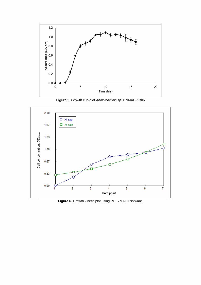
Figure 5. Growth curve of Anoxybacillus sp. UniMAP-KB06
Figure 6. Growth kinetic plot using POLYMATH sotware.
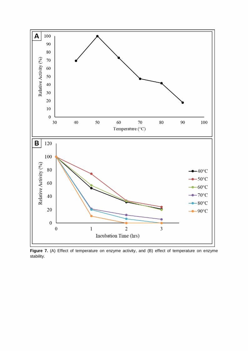
Figure 7. (A) Effect of temperature on enzyme activity, and (B) effect of temperature on enzyme
stability.

Figure 8. (A) Effect of pH on enzyme activity, and (B) effect of pH on enzyme stability.
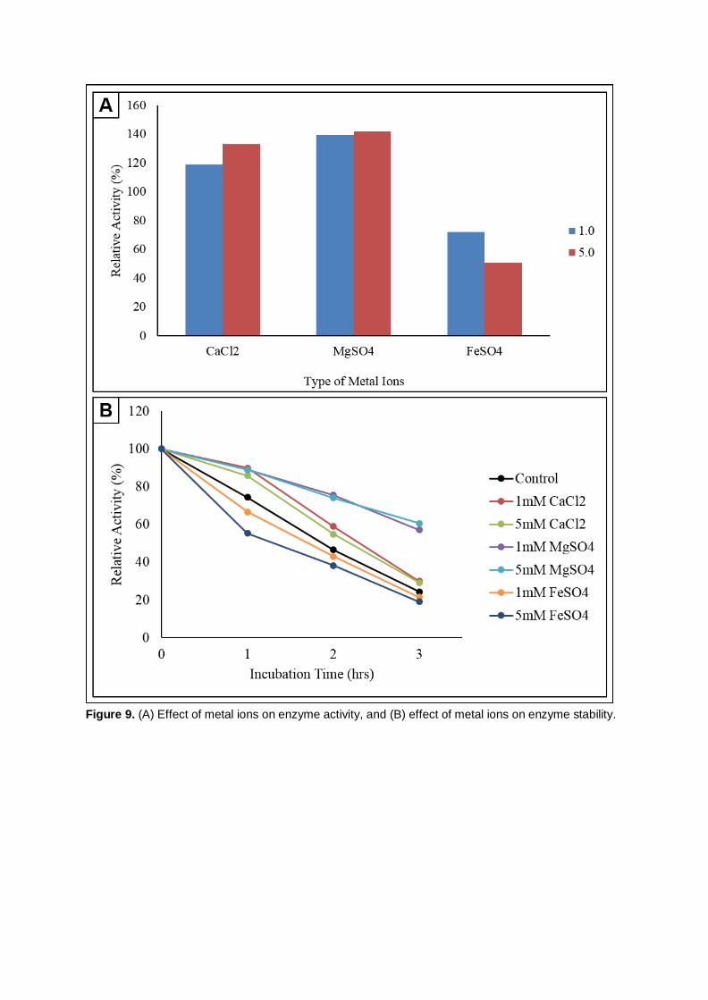
Figure 9. (A) Effect of metal ions on enzyme activity, and (B) effect of metal ions on enzyme stability.