Embed Size (px)
Citation preview
Research Article
EVALUATION OF IN-VITRO ANTIOXIDANT ACTIVITIES OF PTERIS BIAURITA L.
NISHIKA JAISHEE, USHA CHAKRABORTY*
Plant Biochemistry Laboratory, Department of Botany, University of North Bengal, Siliguri 734013
Email: [email protected]
Received: 28 Nov 2013, Revised and Accepted: 03 Feb 2014
ABSTRACT Objectives: The aim of the present study was to investigate the antioxidant activity of Pteris biaurita L. along with the qualitative analysis and quantification of various natural metabolites.
Methods: The antioxidant activities of hot water (HW), methanol (MeOH) and ethanol (EtOH) extracts were evaluated by DPPH free radical scavenging activity, Hydrogen peroxide scavenging activity assay (H202), Nitric oxide scavenging activity assay (NO), Superoxide scavenging activity and Ferric Reducing Antioxidant Power assay (FRAP). Qualitative and quantitative analysis of phytochemicals were screened/done by using standard methods.
Results: The crude extract revealed vast array of phytochemicals and considerable amount of protein, total and reducing sugars, phenol, flavonoid, carotenoid and vitamin C and E. All the three extracts i.e, HW, MeOH and EtOH showed appreciable antioxidant activity in a dose-dependent manner. HW extract showed highest DPPH activity while MeOH extract revealed highest H2O2, NO, superoxide and FRAP activity. Correlations analysis of flavonoid content with DPPH activity and FRAP activity showed good relation with R2=0.993 and R2=0.866 value respectively, then of total phenol with the antioxidant activities.
Conclusion: Overall methanolic extract showed higher antioxidative activity in comparison to the other extracts. Good correlation was obtained between flavonoid content and antioxidant activity signifying that flavonols may be responsible for the antioxidant activity of the plant.
Keywords: Pteris biaurita, Phytochemicals, Phenol, Flavonoid, Antioxidants, DPPH, FRAP.
INTRODUCTION
Plants have always been known to possess medicinal properties due to the fact that they produce various compounds, which must have perhaps developed to protect themselves against different biotic and abiotic environmental factors [1]. The most commonly found secondary metabolites in plants extracts are phenolic acids, flavonoids and tannins [2,3]. Their role as the powerful scavengers of reactive oxygen species (ROS) have been stated by many authors [4,5]. Free radicals or ROS are generated from the cellular reduction and oxidation processes, basically when cells happen to utilize oxygen to produce energy. The commonly formed reactive oxygen species are superoxide anions, hydrogen peroxide, hydroxyl radical, nitric oxide and peroxynitrite radicals [6-8]. Free radicals are extremely unstable as they contain one or more unpaired electrons and can donate or receive single electron, which in turn accounts for their high reactivity with other molecules [9]. Although, the existence of extremely developed antioxidative defense mechanism in healthy person balances the production of free radicals, but sometimes as a consequence of deficit in antioxidant levels, free radicals are generated which leads to oxidative stresses [10,11]. There are reports of beneficial effects of ROS on various cellular and immunological responses at lower concentrations. However, higher concentrations have resulted in oxidative stress that causes detrimental diseases such as cancer, autoimmune disorders, rheumatoid arthritis, cataract, aging, cardiovascular and neurodegenerative diseases [6,12]. Thus, sometimes when body fails to overcome the stress, antioxidants can be taken externally in the form of food or other types of supplements [6]. Antioxidants counteract the damages caused due to the higher levels of ROS [13]. Huge numbers of plants have been used ethno-medicinally to treat various diseases related to oxidative stresses [14].
For a long time, ferns have been used as traditional medicines to treat many diseases like ascarid disease, cold, diarrhea, burn, trauma bleeding etc yet they are applied at lesser rates than the flowering plants in modern chemotherapy [15-17]. But in recent years, many workers have explored the biological and medicinal properties of pteridophytes [18-22]. Pteris biaurita L. commonly known as Brake fern belongs to family Pteridaceae and is available abundantly in various habitats and has been used as traditional medicine from ages. Ethno-medicinally, the rhizome
and fronds decoction of the plant is used to treat chronic disorders [23,24] and the rhizome paste is applied to relief the body pain [25]. Antibacterial activity of P.biaurita L. was reported earlier [26], and compounds such as eicosenes and heptadecanes have also been isolated [27]. Phytochemical analysis, both qualitative and quantitative of Pteris biaurita L. along with other ferns has also been done previously [26, 28]. Antioxidant activity of other pteridophytic species has been reported by many authors [5, 29-35]. However, the detailed study on antioxidant status of P.biaurita L. has not been reported so far. Therefore the aim of the present study was to analyze the antioxidant property and evaluation of bioactive compounds of P.biaurita L.
MATERIAL AND METHODS
Chemicals
2,2-Diphenyl-1-picrylhydrazyl(H), acetic acid(M), chloroform(M), H2SO4(M), methanol(M), aluminium chloride, NaNO2(M), catechin, BSA, L-ascorbate acid, ferulic acid(H), hydrogen peroxide(SDFCL), acetone(M), NaOH(M), ethanol, Nelson’s arseno molybdate, 2,2’– Bipyridyl (H), hexane(M), 2,4-dintrophenylhydrazine(H), thiourea(H), Folin-Ciocalteau reagent(M), Na2CO3(H), disodium hydrogen phosphate(H), sodium dihydrogen phosphate di-hydrate(M), NaOH, potassium ferricyanide, trichloroacetic acid (TCA), gallic acid monohydrate(H), FeCl3, sodium nitroprusside(H), sulphanilic acid(M), glacial acetic acid , N-(1- napthyl)ethylenediamine dihydrochloride(H), nicotinamide adenine dinucleotide (NADH)(H), nitroblue tetrazolium chloride (NBT)(H), phenazine methosulphate (PMS)(H) , mercury (II) chloride(M), potassium iodide. All chemicals used including solvents were of analytical grade obtained from Merck (M) Himedia (H) and SDFCL India Ltd, Mumbai.
Plant collection
The disease free mature fronds of Pteris biaurita L. was collected from the campus of University of North Bengal. Plants were identified and the voucher specimen has been deposited and preserved in the North Bengal University Herbarium, Department of Botany, University of North Bengal, India.
International Journal of Pharmacy and Pharmaceutical Sciences
ISSN- 0975-1491 Vol 6 suppl 2, 2014
AAccaaddeemmiicc SScciieenncceess
Chakraborty et al. Int J Pharm Pharm Sci, Vol 6, Suppl 2, 413-421
414
Preparation of Extracts
The fronds were washed thoroughly initially with tap water then with double distilled water and dried using blotting paper. The air and shade dried plant material were ground to obtain fine powder and was stored at 40C for further use. The sample was extracted using three different solvents: ethanol (EtOH), methanol (MeOH) and hot water (HW). Method described by Okwori et al. (2006) [36] with slight modification was used for ethanol and methanol extraction. 10g each of the powdered samples were soaked in 100 ml of ethanol or methanol for 72 hr at room temperature stirring with magnetic stirrer at 24 hr interval. The soaked samples were filtered using blotting paper for 3-4times followed by filter paper (Whatman No.1). The filtrates were concentrated using rotary evaporator at 400C and were lyophilized for complete solvent removal. For hot water extraction, method of Coban and Konuklugil (2005) [37] was followed with slight modification. The dried powder was mixed with boiling double distilled water in the ratio of 1:10 and kept for 15mins.The mixture was kept overnight and filtered and concentrated as mentioned above. The extractive values as yield percentage were determined on dry weight basis using the formula:
100% tionnforextracweighttake
yextractweightofdryield
The extracts were stored at -200C, which was made to desired concentrations before use.
DPPH free radical scavenging activity
The free radical scavenging activity of the plant extract and reference substance was determined using the method described by Lim & Quah (2007) [38]. Different concentration of each extract (hot water, methanol and ethanol) and reference sample was mixed with equal volume of DPPH methanolic solution (100µM). The mixture was incubated in dark condition at room temperature for 30 minutes, after which the absorbance was recorded at 517nm against blank solution. The control was prepared taking all the reagents except the extract. L-ascorbic acid was used as antioxidant standard. The percentage inhibition was calculated according to the formula:
100
0
10%
A
AAtionDPPHinhibi
Where, A0 was the absorbance of the control and A1 was the absorbance of the extract/standard.
Hydrogen peroxide scavenging activity assay
The scavenging activity of the extracts was measured using the method of Ruch et al. (1989) [39] with minor changes. 5ml of the plant extract (50-500 µg/ml) was mixed with a solution of H2O2 (1 ml, 2mM) prepared in phosphate buffer (0.1M, pH 7.4) and incubated for 10 minutes at room temperature. The absorbance was determined at 230nm against a blank solution containing phosphate buffer without hydrogen peroxide. Ascorbic acid was used as positive control. The percentage of hydrogen peroxide scavenged was calculated using the following formula:
100
0
10)(% 22
A
AAOHscavenged
Where, A0 was the absorbance of the control and A1 was the absorbance of the extract/standard.
Nitric oxide scavenging activity assay
The ability of the extracts to scavenge nitric oxide was determined using the protocol described by Jagetia et al. (2004) [40] and Packer et al. (1998) [41]. The principle of this procedure lies on the competition of scavengers of nitric oxide with oxygen leading to the reduced production of nitric oxide. Sodium nitroprusside in an aqueous solution at physiological pH spontaneously produce nitric
oxide which interacts with oxygen to generate nitrite ions that can be estimated by the Griess reagent. Nitric oxide scavengers compete with oxygen reducing the production of nitric oxide. The reaction mixture containing 2ml of sodium nitroprusside (10mM), 0.5ml of phosphate buffer saline ( pH:7.4,0.1M) and 0.5ml of the extract was incubated at 25oC for 150mins. Then, 0.5ml of the incubated solution was mixed with 1ml of sulphanilic acid (0.33% in 20% glacial acetic acid) and allowed to stand for 5min. After 5min, 1ml of naphthylethylene diamine dihydrochloride (NED) was added, mixed thoroughly and incubated for another 30mins at 25◦ c. The absorbance of the pink chromophore was taken at 540nm.L-ascorbic acid was taken as the reference standard. The nitric oxide scavenging percentage was calculated according to the formula:
1000
10%
A
AANOescavengednitricoxid
Where, A0 was the absorbance of the control and A1 was the absorbance of the extract/standard.
Superoxide scavenging activity
Measurement of superoxide anion radicals (generated in a non-enzymatic PMS-NADH system) scavenging activity was based on the method described by Nishikimi et al. (1972) [42] with slight changes. The superoxide anions generated in a non-enzymatic system through the reaction of PMS, NADH and oxygen was detected by the reduction of nitro blue tetrazolium (NBT). The reaction mixture contained 1ml sample, 1ml of NBT (312µM prepared in phosphate buffer pH 7.4) and 1ml of NADH (936 µM in phosphate buffer pH 7.4). The reaction was accelerated by adding 0.2ml of PMS (120 µM). Reaction mixture was incubated for 5mins at 25◦ C and the absorbance was read at 560nm against blank samples taking vitamin-C as a positive control. Percentage of superoxide anion radical scavenged was measured using the equation as follows:
100
0
10)%(sup
A
AAscavengedoneroxideani
Where, A0 was the absorbance of the control and A1 was the absorbance of the extract/standard.
Ferric Reducing Antioxidant Power (FRAP) assay
The ferric reducing power of ethanol, methanol and water extracts were evaluated using an assay described by Oyaizu (1986) [43] with slight modification.1ml (20-100µg/ml) of extracts was mixed with 2.5ml (0.2M,pH 6.6) of phosphate buffer and 2.5 ml of 1% potassium ferricyanide. The mixture was incubated for 20 minutes at 500C. The solution was allowed to cool at room temperature after which 2.5ml of Tri-carboxylic acid (10%) was added and centrifuged at 3000rpm for 10 minutes. Upper layer of the centrifuged solution was taken and mixed with equal volume of double distilled water. To this, 0.5ml of 0.1% ferric chloride was added and incubated for 10min.Absorbance was taken against appropriate blank solution at 700nm after allowing the solution to stand for 10 minutes at room temperature. Vitamin C was taken as a positive control. Results were expressed as mg gallic acid equivalent (GAE)/g dry weight using standard graph of gallic acid.
Phytochemicals analysis
The small fraction of crude powder was analyzed for the detection of various secondary metabolites using various standard methods [44-53].
Total flavonoids estimation
Total soluble flavonoids were quantified using the method Sultana et al. (2009) [54]. A 500μl aliquot of solution was mixed with 4ml of distilled water and 300μl of NaNO2 (5%). After incubation for 5 min at room temperature 300μl of 10% AlCl3.6H2O was added. At 6th min 2ml of NaOH, followed by 2.4ml of distilled water was added. The absorbance of the mixture was read at 510 nm.
Chakraborty et al. Int J Pharm Pharm Sci, Vol 6, Suppl 2, 413-421
415
Total phenol determination
Extraction method given by Mahadevan and Sridhar (1982) [55] was followed for total phenolic content. For estimation, 1ml of extract,
1ml of 1N Folin ciocalteau’s phenol reagent and 2ml of 20% Na2CO3 solution was mixed thoroughly and boiled in water bath for 1min. The reaction mixture was cooled under running tap water and then diluted with distilled water to make the final volume up to 25ml. The absorbance was recorded at 650nm in a colorimeter against appropriate blank solution [56].
Estimation of total Protein content
Soluble protein was extracted using the standard protocol given by Chakraborty et al. (1995) [57]. Fresh sample (1g) was homogenized with 5ml of sodium phosphate buffer (pH-7.2) and polyvinyl-pyrrolidone which was then centrifuged at 10,000 rpm for 15 minutes. The supernatant was collected and used for further estimation. Estimation of protein content in the extract was done according to Lowry et al. (1951) [58]. The reaction mixture containing 1ml of extract and 5ml alkaline reagent (2% Na2CO3 in 0.1N NaOH, 1% CuSO4 and 2% Na+ - K+ tartarate) was mixed thoroughly and allowed to stand for 15mins. Then, Folin Ciocalteau’s phenol reagent was added which was further incubated for 20 minutes and the absorbance was read at 690 nm.
Total and reducing sugar estimation
The method described by Harborne (1998) [59] with slight modifications was used for total and reducing sugar extraction.
Total sugar estimation was done according to the Anthrone’s method explained by Plummer (1978) [60]. In 1ml of test solution, 4ml of Anthrone’s reagent was added (0.2% Anthrone in conc. H2SO4), which after mixing thoroughly was placed in boiling water for 10mins (precaution were taken to prevent the water loss). The reaction mixture was cooled under running tap water before measuring the absorbance in a colorimeter at 620nm against a suitable blank solution. A calibration curve was constructed using D-glucose and results were expressed as mg glucose equivalent (GA)/g dry weight.
For the estimation of reducing sugar, Somogyi-Nelsons method as described by Plummer (1978) [60] was used in which 1ml of the test solution was mixed with 1ml of Alkaline copper tartarate solution (4g-CuSO4, 24g- Na2CO3 anhydrous, 16g- Na+-K+ tartarate, 180g- Na2SO4 anhydrous-in 1000ml of distilled water) and was heated over a boiling water bath for 20mins (taking necessary precautions).The reaction mixture was cooled under running tap water and 1ml of commercially available Nelson’s arseno molybdate reagent and 2ml of distilled water was added sequentially. Absorbance at 515nm was taken after mixing the reaction mixture properly in a colorimeter and the reducing sugar content was calculated from a glucose standard curve.
Chlorophyll content
Chlorophyll was extracted and estimated according to the procedure given by Harborne (1973) [59] and Arnon (1949) [61] respectively. 1g of fresh leaf tissue was homogenized in 80% acetone and filtered through Whatman No.1 filter paper. The residue was repeatedly re-extracted using 80% acetone until suitable volume was obtained. The filtrate was taken directly for recording absorbance at 663nm and 645nm in a spectrophotometer. The total chlorophyll, chlorophyll a and b content were expressed as mg/g fresh tissue.
Ascorbate
The fresh sample (1g) was homogenised using 6% Trichloroacetic acid under chilled condition and filtered. The filtrate (4ml), 2ml (2% Dinitrophenylhydrazine) and 1 drop of 10% Thiourea was mixed properly and kept in boiling water bath for 15mins. After cooling, 5ml of 80% (v/v) sulphuric acid (H2SO4) was added at O0C and the absorbance was observed at 530nm against blank solution and quantified from the standard curve of ascorbic acid [62].
Carotenoids
Carotenoids extraction and estimation was done according to the procedure of Lichtenthaler (1987) [63]. 1g of the fresh tissue was extracted with methanol and filtered through Whatman filter paper 1.The absorbance of the filtrate was noted at 480nm, 645nm and 663nm and contents was calculated using standard formula.
ɑ-Tocopherol (Vitamin E) content
Vitamin E (ɑ-Tocopherol) was estimated following the method of Jayaraman (1996) [64] with minor modifications. Leaf tissue was homogenized in 5ml of hexane and was shaken vigorously and filtered. Then to 2ml of the extract, 2ml of absolute ethanol was added and mixed thoroughly followed by the addition 0.2ml of 2,2’- Bipyridyl solution (0.5% in ethanol) and 0.2ml of ferric chloride solution (0.2% in ethanol). The mixture was shaken properly and then incubated in dark for 15mins. After the development of red colour 4ml of distilled water was added and mixed well. Two layers were formed which was then separated out by a separating funnel and the red coloured aqueous layer was collected which was stable for 30mins. The absorbance was measured against a blank at 520nm and quantified using a standard curve of ɑ-tocopherol.
Statistical analysis
All analyses were carried out in triplicate and data are expressed as mean ± standard deviation (SD). The mean, standard deviations and correlation analysis were calculated using MS-Excel spreadsheet. IC50 values were calculated using KyPlot (v2beta15) software.
RESULTS & DISCUSSION
In the present study, preliminary phytochemical analysis of the dried plant sample of P.biaurita showed the presence of flavonoids, phenols, alkaloid, tannins, terpenes, steroid, cholesterol and cardiac glycosides. The test showed negative result for saponin and anthraquinone (Table 1). In an earlier study carried out with several Pteris species [28], authors had obtained maximum positive results for phytochemicals in methanol extracts of P. biaurita (including saponin and anthraquinone) followed by P. vittata, P. argyreae and P. confusa. Minimum tests were positive in P. multiaurita extract. The difference in the result obtained for the same plant may be due to the variation in the methods used particularly the extraction procedures and the amount of the samples used.
The presence of various bioactive compounds signifies that the plant can be beneficial [65]. Misra et al. (2008) [66] has reported that the secondary metabolites are responsible for the antioxidant activity of the plants. Phytochemical screening of the methanolic root extract of Gentiana kurroo Royle (Gentianaceae) revealed the presence of tannins, alkaloids, saponins, cardiac glycosides, terpenes, flavonoids, phenolics, and carbohydrates [67]. Kumudhavalli and Jaykar (2012) [68] evaluated the petroleum ether, chloroform, acetone, ethanol and aqueous extracts of the fern Hemionitis arifolia for preliminary phytochemical screening. The ethanol and aqueous extracts showed the presence of flavonoids, carbohydrates, phenolic compounds and sterols were the major phyto constituents.
The dried powder of P.biaurita L. showed highest extractability in alcohol followed by hot water and methanol. The yields as percentage of dry whole plant powder of alcohol, hot water and methanol were 29.77, 21.14 and 14.28 respectively (Table 2).Quantification of bioactive compounds revealed 10.399 mg/g dwt of total phenol, 14.539 mg/g dwt of flavonoid, 37.09 mg/g fwt of total protein, 1.62 mg/g fwt of ascorbate, 0.94 mg/g dwt of α-Tocopherol, 125 mg/g dwt of total sugar, 110.505 mg/g dwt of reducing sugar, 0.895 mg/g fwt of total chlorophyll, 0.604 mg/g fwt of chlorophyll a (chl.a), 0.291 mg/g fwt of chlorophyll b (chl.b) and 62.26µg/g fwt of carotenoid (Table 3). Rasool et al. (2010) [69] confirmed the earlier report of Prunella vulgaris been consumed for its taste and nutritional value as it good content of carbohydrate, protein and fat. Moreover, there are polysaccharides which have shown to be immunomodulatory (Jayabalan et al. 1994) [70].
The plant sample tested can be considered nutritional as it has good amount of carbohydrate and protein. A considerable quantity of vitamin has been detected in the fronds of Pteris biaurita. Vitamins play an important role in many biochemical functions
Chakraborty et al. Int J Pharm Pharm Sci, Vol 6, Suppl 2, 413-421
416
Table 1: Phytochemical compounds of P. biaurita
Phytochemical compounds Present/absent Flavonoids + Phenols + Tannins + Alkaloid + Cardiac glycosides + Saponins - Terpenes + Steroid + Cholesterol + Anthraquinone -
+ = present, - = absent
Table 2: Percent yield of HW, MeOH and EtOH extract from P.biaurita
Sample (extract) Yield (%) HW 21.14 ±0.413 MeOH 14.28±0.035 EtOH 29.77± 0.321
HW = Hot water extract, MeOH = Methanol extract
EtOH = Ethanol extract, ± = SD
Table 3: Quantitative analysis of phytochemicals in P.biaurita
Chemicals Content
Flavonoids 14.539± 0.00
Total phenol (mg/g dwt) 10.399±0.052
Ascorbate (mg/g dwt) 1.620±0.002
α -Tocopherol (mg/g dwt) 0.94±0.003
Total sugar (mg/g dwt) 125.00±0.011
Reducing sugar (mg/g dwt) 37.09±0.038
Total protein (mg/g fwt) 110.51±0.015
Total chl (mg/g fwt) 0.90±0.035
Chl a (mg/g fwt) 0.60±0.070
Chl b (mg/g fwt) 0.29±0.037
Carotenoid (g/g fwt) 62.26±0.001
Dwt= dry weight tissue, fwt=fresh weight tissue, chl=chlorophyll, ± = SD
We must obtain vitamins from foods/fruits, as it cannot be produced in-vivo (Peter, 1990) [71]. Vitamin C has been reported to prevent arteriosclerosis and, to protect against harmful reactive oxygen species produced during normal metabolic processes [72]. Tocopherols (α, β, γ, and δ) are also involved in scavenging oxygen free radicals, lipid peroxy radicals, and O2 [73]. Carotenoids are one of the major non-enzymatic anti-oxidants that have a role in defense against water stress by scavenging of singlet oxygen and suppressing lipid peroxidation in all photosynthetic organisms [74]. Naik et al. (2005) [3] reported that the aqueous extract from the different parts of the four medicinal plants, Momordica charantia, Glycyrrhiza glabra, Acacia catechu and Terminalia chebula are the rich sources of enzymatic and non-enzymatic antioxidants. The amount of phenol and flavonoid obtained in the present study is in accordance with the finding of Gracelin et al. (2013) [28]. Polyphenolic compounds like flavonoids and phenolic acids commonly found in plants have been reported to contribute significantly to the total antioxidant activity of plants [75].There are reports of phenols having good anti-oxidative, anti-mutagenic and anti-cancerous properties [76].
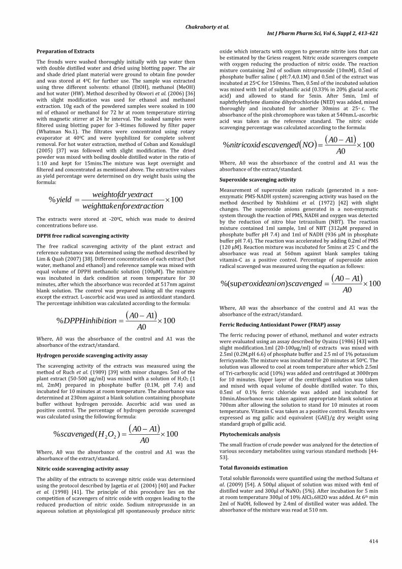
Determination of antioxidant potential of the extract was carried out next. DPPH is a stable free radical containing an odd electron having a characteristic absorption at 517nm (deep purple colour). The deep purple colour usually gets decolorized when exposed to antioxidant in the solution. Lower the absorption, higher is the radical
scavenging activity of the extract [77]. Results obtained from DPPH radical scavenging activity showed dose-dependent inhibition. It was observed that hot water extract showed significantly higher DPPH inhibition activity compared to methanol and ethanol. However, ascorbic acid which was used as positive control showed highest inhibition at the same concentration. The IC50 values for HW, MeOH, EtOH and ascorbic acid are 1.107, 1.84, 5.09 and 0.142 mg/ml respectively (Figure 1). Chai and Wong (2012) [78], reported the concentration-dependent DPPH radical scavenging activities in the aqueous extract of Selaginella willdenowii. Likewise, it was reported that the aqueous extract of D. solida rhizome contained a high phenolic compound and showed a strong DPPH scavenging activity [79]. In the study conducted by Chang et al. (2007) [5] with six folk medicinal ferns “Gusuibi”, they found that all the samples irrespective of solvent showed scavenging activity in a concentration dependent manner.
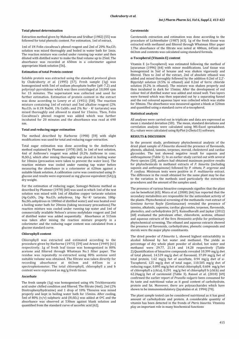
Phenolics (which can donate electrons to H2O2 thus reducing it to water) present in the extract, may be responsible for the H2O2
scavenging ability of the sample [80,81]. The hydrogen peroxide scavenging activity of EtOH extract was the least among the three extract. MeOH extract showed higher hydrogen peroxide scavenging activity than HW and EtOH extract, but the activity is lesser than positive controls at the same concentration. IC50 values of Vitamin C, MeOH, HW and EtOH extract was found to be 71.749, 146.608, 152.351 and 398.759µg/ml respectively (Figure 2).
Chakraborty et al. Int J Pharm Pharm Sci, Vol 6, Suppl 2, 413-421
417
Fig. 1: DPPH activity of HW, MeOH and EtOH extracts from Pteris biaurita. Ascorbic acid was used as the positive control.
Fig. 2: Hydrogen peroxide scavenging activity of HW, MeOH and EtOH extracts from Pteris biaurita.
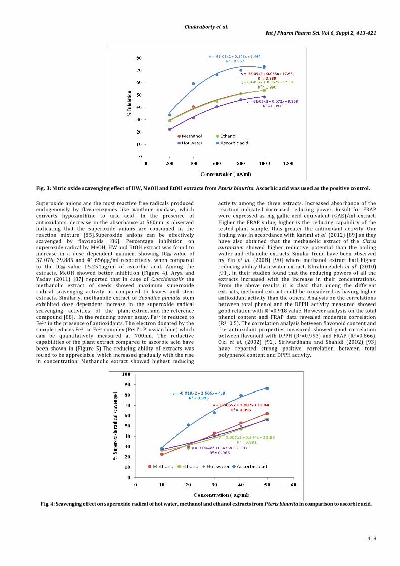
Nitric oxide is a ROS which is associated with inflammation, cancer and other pathological conditions [80,82]. Thus the ability of the extracts to reduce/scavenge nitric oxide may be considered advantageous for health as it can evade the ill effects of excessive NO generation. In case of nitric oxide scavenging activity, MeOH extract exhibited slightly higher activity than EtOH extract with IC50 values of 694.825µg/ml and 708.007µg/ml respectively. HW extract showed very less activity with IC50 value of 915.137µg/ml. However, the activity of
ascorbic acid was more prominent than the other three extracts with IC50 value of 329.246µg/ml. The percentages of inhibition increased appreciably with increasing concentration of the extracts (Figure 3). Banerjee et al. (2011) [83] reported the dose dependent increase of NO scavenging activity in the ethanolic extracts of Ixora coccinea. Methanolic extract in comparison to petroleum ether and chloroform extract from leaves of Limonia acidissima Linn. (Rutaceae) were reported to show higher NO scavenging [84].
Chakraborty et al. Int J Pharm Pharm Sci, Vol 6, Suppl 2, 413-421
418
Fig. 3: Nitric oxide scavenging effect of HW, MeOH and EtOH extracts from Pteris biaurita. Ascorbic acid was used as the positive control.
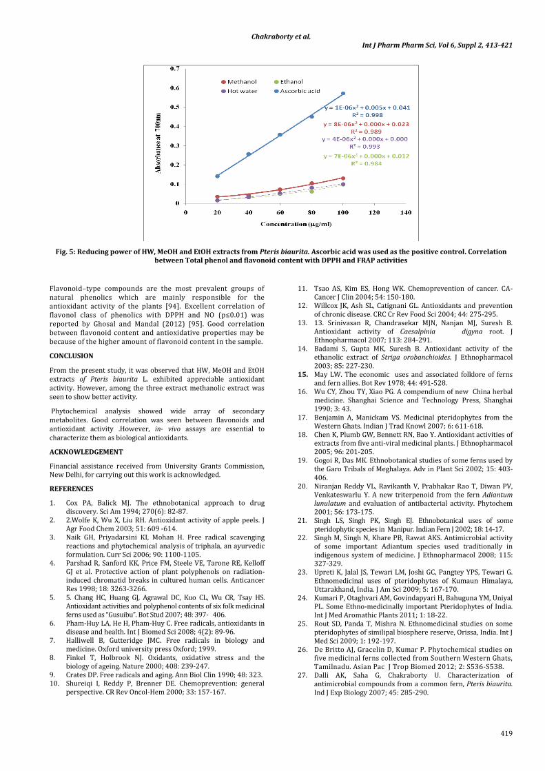
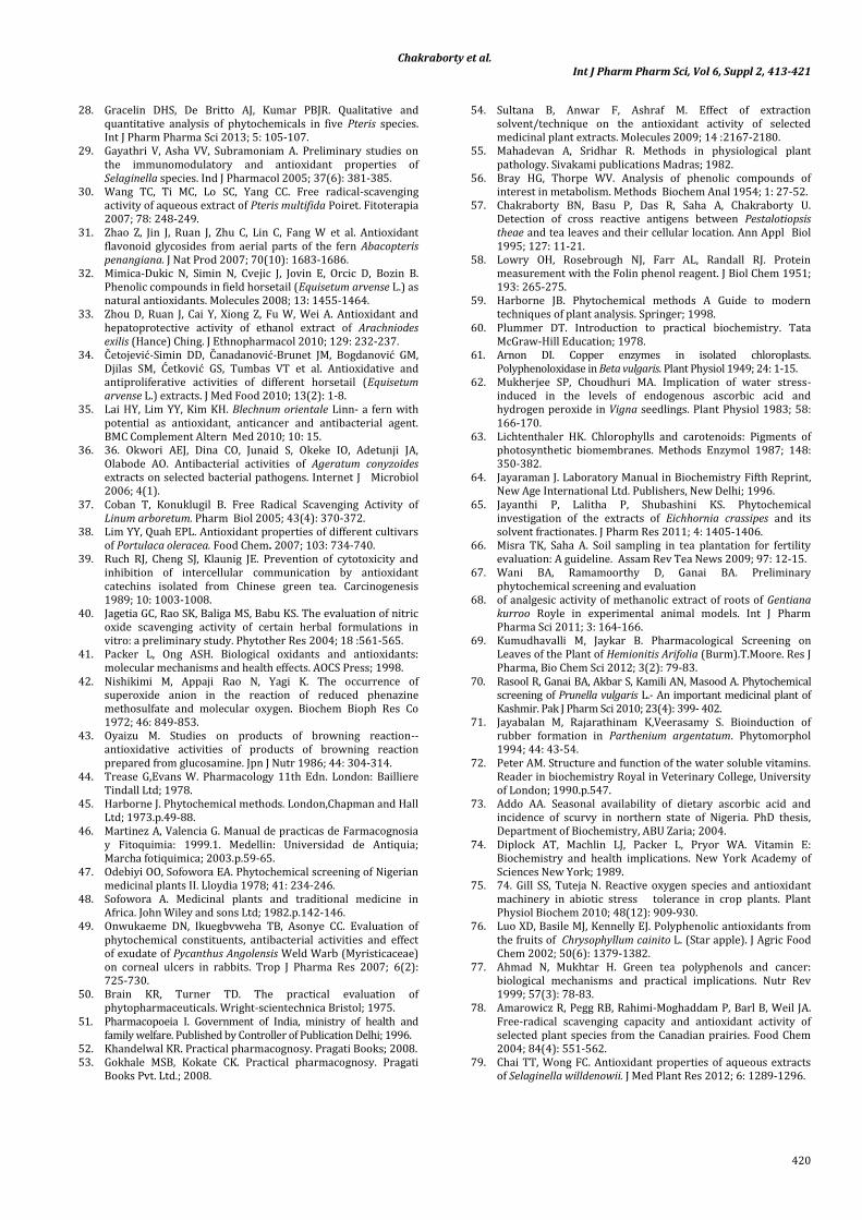
Superoxide anions are the most reactive free radicals produced endogenously by flavo-enzymes like xanthine oxidase, which converts hypoxanthine to uric acid. In the presence of antioxidants, decrease in the absorbance at 560nm is observed indicating that the superoxide anions are consumed in the reaction mixture [85].Superoxide anions can be effectively scavenged by flavonoids [86]. Percentage inhibition on superoxide radical by MeOH, HW and EtOH extract was found to increase in a dose dependent manner, showing IC50 value of 37.076, 39.885 and 41.656µg/ml respectively, when compared to the IC50 value 16.254µg/ml of ascorbic acid. Among the extracts, MeOH showed better inhibition (Figure 4). Arya and Yadav (2011) [87] reported that in case of C.occidentalis the methanolic extract of seeds showed maximum superoxide radical scavenging activity as compared to leaves and stem extracts. Similarly, methanolic extract of Spondias pinnata stem exhibited dose dependent increase in the superoxide radical scavenging activities of the plant extract and the reference compound [88]. In the reducing power assay, Fe3+ is reduced to Fe2+ in the presence of antioxidants. The electron donated by the sample reduces Fe3+ to Fe2+ complex (Perl’s Prussian blue) which can be quantitatively measured at 700nm. The reductive capabilities of the plant extract compared to ascorbic acid have been shown in (Figure 5).The reducing ability of extracts was found to be appreciable, which increased gradually with the rise in concentration. Methanolic extract showed highest reducing
activity among the three extracts. Increased absorbance of the reaction indicated increased reducing power. Result for FRAP were expressed as mg gallic acid equivalent (GAE)/ml extract. Higher the FRAP value, higher is the reducing capability of the tested plant sample, thus greater the antioxidant activity. Our finding was in accordance with Karimi et al. (2012) [89] as they have also obtained that the methanolic extract of the Citrus aurantium showed higher reductive potential than the boiling water and ethanolic extracts. Similar trend have been observed by Yin et al. (2008) [90] where methanol extract had higher reducing ability than water extract. Ebrahimzadeh et al. (2010) [91], in their studies found that the reducing powers of all the extracts increased with the increase in their concentrations. From the above results it is clear that among the different extracts, methanol extract could be considered as having higher antioxidant activity than the others. Analysis on the correlations between total phenol and the DPPH activity measured showed good relation with R2=0.918 value. However analysis on the total phenol content and FRAP data revealed moderate correlation (R2=0.5). The correlation analysis between flavonoid content and the antioxidant properties measured showed good correlation between flavonoid with DPPH (R2=0.993) and FRAP (R2=0.866). Oki et al. (2002) [92], Siriwardhana and Shahidi (2002) [93] have reported strong positive correlation between total polyphenol content and DPPH activity.
Fig. 4: Scavenging effect on superoxide radical of hot water, methanol and ethanol extracts from Pteris biaurita in comparison to ascorbic acid.
Chakraborty et al. Int J Pharm Pharm Sci, Vol 6, Suppl 2, 413-421
419
Fig. 5: Reducing power of HW, MeOH and EtOH extracts from Pteris biaurita. Ascorbic acid was used as the positive control. Correlation between Total phenol and flavonoid content with DPPH and FRAP activities
Flavonoid–type compounds are the most prevalent groups of natural phenolics which are mainly responsible for the antioxidant activity of the plants [94]. Excellent correlation of flavonol class of phenolics with DPPH and NO (p≤0.01) was reported by Ghosal and Mandal (2012) [95]. Good correlation between flavonoid content and antioxidative properties may be because of the higher amount of flavonoid content in the sample.
CONCLUSION
From the present study, it was observed that HW, MeOH and EtOH extracts of Pteris biaurita L. exhibited appreciable antioxidant activity. However, among the three extract methanolic extract was seen to show better activity.
Phytochemical analysis showed wide array of secondary metabolites. Good correlation was seen between flavonoids and antioxidant activity .However, in- vivo assays are essential to characterize them as biological antioxidants.
ACKNOWLEDGEMENT
Financial assistance received from University Grants Commission, New Delhi, for carrying out this work is acknowledged.
REFERENCES
1. Cox PA, Balick MJ. The ethnobotanical approach to drug discovery. Sci Am 1994; 270(6): 82-87.
2. 2.Wolfe K, Wu X, Liu RH. Antioxidant activity of apple peels. J Agr Food Chem 2003; 51: 609 -614.
3. Naik GH, Priyadarsini KI, Mohan H. Free radical scavenging reactions and phytochemical analysis of triphala, an ayurvedic formulation. Curr Sci 2006; 90: 1100-1105.
4. Parshad R, Sanford KK, Price FM, Steele VE, Tarone RE, Kelloff GJ et al. Protective action of plant polyphenols on radiation-induced chromatid breaks in cultured human cells. Anticancer Res 1998; 18: 3263-3266.
5. 5. Chang HC, Huang GJ, Agrawal DC, Kuo CL, Wu CR, Tsay HS. Antioxidant activities and polyphenol contents of six folk medicinal ferns used as “Gusuibu”. Bot Stud 2007; 48: 397- 406.
6. Pham-Huy LA, He H, Pham-Huy C. Free radicals, antioxidants in disease and health. Int J Biomed Sci 2008; 4(2): 89-96.
7. Halliwell B, Gutteridge JMC. Free radicals in biology and medicine. Oxford university press Oxford; 1999.
8. Finkel T, Holbrook NJ. Oxidants, oxidative stress and the biology of ageing. Nature 2000; 408: 239-247.
9. Crates DP. Free radicals and aging. Ann Biol Clin 1990; 48: 323. 10. Shureiqi I, Reddy P, Brenner DE. Chemoprevention: general
perspective. CR Rev Oncol-Hem 2000; 33: 157-167.
11. Tsao AS, Kim ES, Hong WK. Chemoprevention of cancer. CA-Cancer J Clin 2004; 54: 150-180.
12. Willcox JK, Ash SL, Catignani GL. Antioxidants and prevention of chronic disease. CRC Cr Rev Food Sci 2004; 44: 275-295.
13. 13. Srinivasan R, Chandrasekar MJN, Nanjan MJ, Suresh B. Antioxidant activity of Caesalpinia digyna root. J Ethnopharmacol 2007; 113: 284-291.
14. Badami S, Gupta MK, Suresh B. Antioxidant activity of the ethanolic extract of Striga orobanchioides. J Ethnopharmacol 2003; 85: 227-230.
15. May LW. The economic uses and associated folklore of ferns and fern allies. Bot Rev 1978; 44: 491-528.
16. Wu CY, Zhou TY, Xiao PG. A compendium of new China herbal medicine. Shanghai Science and Technology Press, Shanghai 1990; 3: 43.
17. Benjamin A, Manickam VS. Medicinal pteridophytes from the Western Ghats. Indian J Trad Knowl 2007; 6: 611-618.
18. Chen K, Plumb GW, Bennett RN, Bao Y. Antioxidant activities of extracts from five anti-viral medicinal plants. J Ethnopharmacol 2005; 96: 201-205.
19. Gogoi R, Das MK. Ethnobotanical studies of some ferns used by the Garo Tribals of Meghalaya. Adv in Plant Sci 2002; 15: 403-406.
20. Niranjan Reddy VL, Ravikanth V, Prabhakar Rao T, Diwan PV, Venkateswarlu Y. A new triterpenoid from the fern Adiantum lunulatum and evaluation of antibacterial activity. Phytochem 2001; 56: 173-175.
21. Singh LS, Singh PK, Singh EJ. Ethnobotanical uses of some pteridophytic species in Manipur. Indian Fern J 2002; 18: 14-17.
22. Singh M, Singh N, Khare PB, Rawat AKS. Antimicrobial activity of some important Adiantum species used traditionally in indigenous system of medicine. J Ethnopharmacol 2008; 115: 327-329.
23. Upreti K, Jalal JS, Tewari LM, Joshi GC, Pangtey YPS, Tewari G. Ethnomedicinal uses of pteridophytes of Kumaun Himalaya, Uttarakhand, India. J Am Sci 2009; 5: 167-170.
24. Kumari P, Otaghvari AM, Govindapyari H, Bahuguna YM, Uniyal PL. Some Ethno-medicinally important Pteridophytes of India. Int J Med Aromathic Plants 2011; 1: 18-22.
25. Rout SD, Panda T, Mishra N. Ethnomedicinal studies on some pteridophytes of similipal biosphere reserve, Orissa, India. Int J Med Sci 2009; 1: 192-197.
26. De Britto AJ, Gracelin D, Kumar P. Phytochemical studies on five medicinal ferns collected from Southern Western Ghats, Tamilnadu. Asian Pac J Trop Biomed 2012; 2: S536-S538.
27. Dalli AK, Saha G, Chakraborty U. Characterization of antimicrobial compounds from a common fern, Pteris biaurita. Ind J Exp Biology 2007; 45: 285-290.
Chakraborty et al. Int J Pharm Pharm Sci, Vol 6, Suppl 2, 413-421
420
28. Gracelin DHS, De Britto AJ, Kumar PBJR. Qualitative and quantitative analysis of phytochemicals in five Pteris species. Int J Pharm Pharma Sci 2013; 5: 105-107.
29. Gayathri V, Asha VV, Subramoniam A. Preliminary studies on the immunomodulatory and antioxidant properties of Selaginella species. Ind J Pharmacol 2005; 37(6): 381-385.
30. Wang TC, Ti MC, Lo SC, Yang CC. Free radical-scavenging activity of aqueous extract of Pteris multifida Poiret. Fitoterapia 2007; 78: 248-249.
31. Zhao Z, Jin J, Ruan J, Zhu C, Lin C, Fang W et al. Antioxidant flavonoid glycosides from aerial parts of the fern Abacopteris penangiana. J Nat Prod 2007; 70(10): 1683-1686.
32. Mimica-Dukic N, Simin N, Cvejic J, Jovin E, Orcic D, Bozin B. Phenolic compounds in field horsetail (Equisetum arvense L.) as natural antioxidants. Molecules 2008; 13: 1455-1464.
33. Zhou D, Ruan J, Cai Y, Xiong Z, Fu W, Wei A. Antioxidant and hepatoprotective activity of ethanol extract of Arachniodes exilis (Hance) Ching. J Ethnopharmacol 2010; 129: 232-237.
34. Četojević-Simin DD, Čanadanović-Brunet JM, Bogdanović GM, Djilas SM, Ćetković GS, Tumbas VT et al. Antioxidative and antiproliferative activities of different horsetail (Equisetum arvense L.) extracts. J Med Food 2010; 13(2): 1-8.
35. Lai HY, Lim YY, Kim KH. Blechnum orientale Linn- a fern with potential as antioxidant, anticancer and antibacterial agent. BMC Complement Altern Med 2010; 10: 15.
36. 36. Okwori AEJ, Dina CO, Junaid S, Okeke IO, Adetunji JA, Olabode AO. Antibacterial activities of Ageratum conyzoides extracts on selected bacterial pathogens. Internet J Microbiol 2006; 4(1).
37. Coban T, Konuklugil B. Free Radical Scavenging Activity of Linum arboretum. Pharm Biol 2005; 43(4): 370-372.
38. Lim YY, Quah EPL. Antioxidant properties of different cultivars of Portulaca oleracea. Food Chem. 2007; 103: 734-740.
39. Ruch RJ, Cheng SJ, Klaunig JE. Prevention of cytotoxicity and inhibition of intercellular communication by antioxidant catechins isolated from Chinese green tea. Carcinogenesis 1989; 10: 1003-1008.
40. Jagetia GC, Rao SK, Baliga MS, Babu KS. The evaluation of nitric oxide scavenging activity of certain herbal formulations in vitro: a preliminary study. Phytother Res 2004; 18 :561-565.
41. Packer L, Ong ASH. Biological oxidants and antioxidants: molecular mechanisms and health effects. AOCS Press; 1998.
42. Nishikimi M, Appaji Rao N, Yagi K. The occurrence of superoxide anion in the reaction of reduced phenazine methosulfate and molecular oxygen. Biochem Bioph Res Co 1972; 46: 849-853.
43. Oyaizu M. Studies on products of browning reaction--antioxidative activities of products of browning reaction prepared from glucosamine. Jpn J Nutr 1986; 44: 304-314.
44. Trease G,Evans W. Pharmacology 11th Edn. London: Bailliere Tindall Ltd; 1978.
45. Harborne J. Phytochemical methods. London,Chapman and Hall Ltd; 1973.p.49-88.
46. Martinez A, Valencia G. Manual de practicas de Farmacognosia y Fitoquimia: 1999.1. Medellin: Universidad de Antiquia; Marcha fotiquimica; 2003.p.59-65.
47. Odebiyi OO, Sofowora EA. Phytochemical screening of Nigerian medicinal plants II. Lloydia 1978; 41: 234-246.
48. Sofowora A. Medicinal plants and traditional medicine in Africa. John Wiley and sons Ltd; 1982.p.142-146.
49. Onwukaeme DN, Ikuegbvweha TB, Asonye CC. Evaluation of phytochemical constituents, antibacterial activities and effect of exudate of Pycanthus Angolensis Weld Warb (Myristicaceae) on corneal ulcers in rabbits. Trop J Pharma Res 2007; 6(2): 725-730.
50. Brain KR, Turner TD. The practical evaluation of phytopharmaceuticals. Wright-scientechnica Bristol; 1975.
51. Pharmacopoeia I. Government of India, ministry of health and family welfare. Published by Controller of Publication Delhi; 1996.
52. Khandelwal KR. Practical pharmacognosy. Pragati Books; 2008. 53. Gokhale MSB, Kokate CK. Practical pharmacognosy. Pragati
Books Pvt. Ltd.; 2008.
54. Sultana B, Anwar F, Ashraf M. Effect of extraction solvent/technique on the antioxidant activity of selected medicinal plant extracts. Molecules 2009; 14 :2167-2180.
55. Mahadevan A, Sridhar R. Methods in physiological plant pathology. Sivakami publications Madras; 1982.
56. Bray HG, Thorpe WV. Analysis of phenolic compounds of interest in metabolism. Methods Biochem Anal 1954; 1: 27-52.
57. Chakraborty BN, Basu P, Das R, Saha A, Chakraborty U. Detection of cross reactive antigens between Pestalotiopsis theae and tea leaves and their cellular location. Ann Appl Biol 1995; 127: 11-21.
58. Lowry OH, Rosebrough NJ, Farr AL, Randall RJ. Protein measurement with the Folin phenol reagent. J Biol Chem 1951; 193: 265-275.
59. Harborne JB. Phytochemical methods A Guide to modern techniques of plant analysis. Springer; 1998.
60. Plummer DT. Introduction to practical biochemistry. Tata McGraw-Hill Education; 1978.
61. Arnon DI. Copper enzymes in isolated chloroplasts. Polyphenoloxidase in Beta vulgaris. Plant Physiol 1949; 24: 1-15.
62. Mukherjee SP, Choudhuri MA. Implication of water stress-induced in the levels of endogenous ascorbic acid and hydrogen peroxide in Vigna seedlings. Plant Physiol 1983; 58: 166-170.
63. Lichtenthaler HK. Chlorophylls and carotenoids: Pigments of photosynthetic biomembranes. Methods Enzymol 1987; 148: 350-382.
64. Jayaraman J. Laboratory Manual in Biochemistry Fifth Reprint, New Age International Ltd. Publishers, New Delhi; 1996.
65. Jayanthi P, Lalitha P, Shubashini KS. Phytochemical investigation of the extracts of Eichhornia crassipes and its solvent fractionates. J Pharm Res 2011; 4: 1405-1406.
66. Misra TK, Saha A. Soil sampling in tea plantation for fertility evaluation: A guideline. Assam Rev Tea News 2009; 97: 12-15.
67. Wani BA, Ramamoorthy D, Ganai BA. Preliminary phytochemical screening and evaluation
68. of analgesic activity of methanolic extract of roots of Gentiana kurroo Royle in experimental animal models. Int J Pharm Pharma Sci 2011; 3: 164-166.
69. Kumudhavalli M, Jaykar B. Pharmacological Screening on Leaves of the Plant of Hemionitis Arifolia (Burm).T.Moore. Res J Pharma, Bio Chem Sci 2012; 3(2): 79-83.
70. Rasool R, Ganai BA, Akbar S, Kamili AN, Masood A. Phytochemical screening of Prunella vulgaris L.- An important medicinal plant of Kashmir. Pak J Pharm Sci 2010; 23(4): 399- 402.
71. Jayabalan M, Rajarathinam K,Veerasamy S. Bioinduction of rubber formation in Parthenium argentatum. Phytomorphol 1994; 44: 43-54.
72. Peter AM. Structure and function of the water soluble vitamins. Reader in biochemistry Royal in Veterinary College, University of London; 1990.p.547.
73. Addo AA. Seasonal availability of dietary ascorbic acid and incidence of scurvy in northern state of Nigeria. PhD thesis, Department of Biochemistry, ABU Zaria; 2004.
74. Diplock AT, Machlin LJ, Packer L, Pryor WA. Vitamin E: Biochemistry and health implications. New York Academy of Sciences New York; 1989.
75. 74. Gill SS, Tuteja N. Reactive oxygen species and antioxidant machinery in abiotic stress tolerance in crop plants. Plant Physiol Biochem 2010; 48(12): 909-930.
76. Luo XD, Basile MJ, Kennelly EJ. Polyphenolic antioxidants from the fruits of Chrysophyllum cainito L. (Star apple). J Agric Food Chem 2002; 50(6): 1379-1382.
77. Ahmad N, Mukhtar H. Green tea polyphenols and cancer: biological mechanisms and practical implications. Nutr Rev 1999; 57(3): 78-83.
78. Amarowicz R, Pegg RB, Rahimi-Moghaddam P, Barl B, Weil JA. Free-radical scavenging capacity and antioxidant activity of selected plant species from the Canadian prairies. Food Chem 2004; 84(4): 551-562.
79. Chai TT, Wong FC. Antioxidant properties of aqueous extracts of Selaginella willdenowii. J Med Plant Res 2012; 6: 1289-1296.
Chakraborty et al. Int J Pharm Pharm Sci, Vol 6, Suppl 2, 413-421
421
80. Chen YH, Chang FR, Lin YJ, Hsieh PW, Wu MJ, Wu YC. Identification of antioxidants from rhizome of Davallia solida. Food Chem 2008; 107: 684-691.
81. 80. Nabavi S, Ebrahimzadeh M, Nabavi S, Jafari M. Free radical scavenging activity and antioxidant capacity of Eryngium caucasicum Trautv and Froripia subpinnata. Pharmacol online 2008; 3: 19-25.
82. Ebrahimzadeh MA, Nabavi SF, Nabavi SM. Antioxidant activities of methanol extract of Sambucus ebulus L. flower. Pak J Biol Sci 2009d; 12(5): 447-450.
83. Nabavi SM, Ebrahimzadeh MA, Nabavi SF, Hamidinia A, Bekhradnia AR. Determination of antioxidant activity, phenol and flavonoids content of Parrotia persica Mey. Pharmacol online 2008; 2: 560-567.
84. Banerjee S, Chanda A, Ghoshal A, Debnath R, Chakraborty S, Saha R et al. Nitric Oxide Scavenging Activity Study of Ethanolic Extracts of from Two Different Areas of Kolkata. Asian J Exp Biol Sci 2011; 2: 595-599.
85. Attarde DL, Chaudhari B, Bhambar RS. Phytochemical investigation and in vitro antioxidant activity of extracts from leaves of Limonia acidissima Linn.(Rutaceae). J Pharm Res 2011; 4: 766-768.
86. Bora K, Sharma A. In Vitro Antioxidant and Free Radical Scavenging Potential of Medicago sativa Linn. J Pharma Res 2010; 3: 1206-1210.
87. Robak J, Gryglewski RJ. Flavonoids are scavengers of superoxide anions. Biochem Pharmacol 1988; 37: 837-841.
88. Arya V, Yadav JP. Antioxidant properties of the methanol extracts of the leaves, seeds and stem of Cassia occidentalis. Res J of Med Plant 2011; 5: 547-556.
89. Hazra B, Biswas S, Mandal N. Antioxidant and free radical scavenging activity of Spondias pinnata. BMC Complement Altern Med 2008; 8: 63.
90. Karimi E, Oskoueian E, Hendra R, Oskoueian A, Jaafar HZE. Phenolic compounds characterization and biological activities of Citrus aurantium Bloom. Molecules 2012; 17: 1203-1218.
91. Yin J, Heo SI, Wang MH. Antioxidant and antidiabetic activities of extracts from Cirsium japonicum roots. Nutr Res Pract 2008; 2: 247-251.
92. Ebrahimzadeh MA, Nabavi SM, Nabavi SF, Bahramian F, Bekhradnia AR. Antioxidant and free radical scavenging activity of H. officinalis L. var. angustifolius, V. odorata, B. hyrcana and C. speciosum. Pak J Pharm Sci 2010; 23: 29-34.
93. Oki T, Masuda M, Furuta S, Nishiba Y, Terahara N, Suda I. Involvement of anthocyanins and other phenolic compounds in radical scavenging activity of purple-fleshed sweet potato cultivars. J Food Sci 2002; 67: 1752-1756.
94. Siriwardhana SSKW, Shahidi F. Antiradical activity of extracts of almond and its by-products. J Am Oil Chem Soc 2002; 79: 903-908.
95. Cakir A, Mavi A, Yıldırım A, Duru ME, Harmandar M, Kazaz C. Isolation and characterization of antioxidant phenolic compounds from the aerial parts of Hypericum hyssopifolium L. by activity-guided fractionation. J Ethnopharmacol 2003; 87(1): 73-83.
96. Ghosal M, Mandal P. Phytochemical screening and antioxidant activities of two selected ‘BIHI’ Fruits used as vegetables in Darjeeling Himalaya. Int J Pharm Pharma Sci 2012; 4: 567-574.

![Simulations and Tests of a Danish Smartgrid – The Cell ... · 0.000 100.00 200.0 300.0 400.0 [s] 500.0 1.005 1.002 0.999 0.996 0.993 0.990 Grid Breaker: Electrical Frequency/Terminal](https://img.pdfslide.us/doc/110x75/5fc3074cea0c6a21f22e4e08/simulations-and-tests-of-a-danish-smartgrid-a-the-cell-0000-10000-2000.jpg)