Embed Size (px)
Citation preview

Academic Press is an imprint of Elsevier 32 Jamestown Road, London NW1 7BY, UK 30 Corporate Drive, Suite 400, Burlington, MA 01803, USA 525 B Street, Suite 1900, San Diego, CA 92101-4495, USA
Copyright © 2010 Elsevier Inc. All rights reserved
No part of this publication may be reproduced, stored in a retrieval system or transmitted in any form or by any means electronic, mechanical, photocopying, recording or otherwise without the prior written permission of the publisher
Permissions may be sought directly from Elsevier’s Science & Technology Rights Department in Oxford, UK: phone ( � 44)(0) 1865 843830; fax ( � 44) (0) 1865 853333; email: [email protected] . Alternatively you can submit your request online by visiting the Elsevier web site at ( http://elsevier.com/locate/permissions ), and selecting Obtaining permissions to use Elsevier material
Notice No responsibility is assumed by the publisher for any injury and/or damage to persons or property as a matter of products liability, negligence or otherwise, or from any use or operation of any methods, products, instructions or ideas contained in the material herein, Because of rapid advances in the medical sciences, in particular, independent verification of diagnoses and drug dosages should be made
British Library Cataloguing in Publication Data A catalogue record for this book is available from the British Library
Library of Congress Catalog Number: 2009923451
ISBN : 978-0-12-374767-9
For information on all Elsevier publications visit our website at www.elsevierdirect.com
Typeset by MPS Limited, a Macmillan Company www.macmillansolutions.com
Printed and bound in the USA
10 11 12 13 10 9 8 7 6 5 4 3 2 1
Cover art: D1 and D2 dopamine receptor-expressing medium spiny projection neurons of the striatum and their terminal fields, labeled by bacterial artificial chromosome (BAC) vector-driven expression of enhanced green fluorescent protein. Illustration is a composite of images from Chapter 6 by Surmeier et al., “D1 and D2 dopamine receptor modulation of glutamatergic signaling in striatal medium spiny neurons”, and Chapter 28 by Gerfen, “D1 dopamine receptor supersensitivity in the dopamine-depleted striatum: Aberrant ERK1/2 signaling”. Overlaid in red is an activity trace of a medium spiny neuron recorded intracellularly in vivo in a dopamine-depleted rat by Kuei Tseng.

xiContents
15. The Subthalamic Nucleus: From In Vitro to In Vivo Mechanisms 259
Stephane Charpier, Corinne Beurrier and Jeanne T. Paz
I. Introduction 259 II. Synaptic Organization of the Subthalamic
Nucleus and Responses to Cortical Stimulation 261A. Inputs 261B. Outputs 261C. Responses to Cortical Stimulation 262
III. Cellular Basis of Single-Spike and Burst Firing in Subthalamic Nucleus Neurons In Vitro 262A. Burst Firing 262B. Single-Spike Activity 263C. In Vivo Activities of STN Neurons and
their Relation to Cortical Patterns 265D. Anesthesia-Dependent Slow Oscillations 265E. Natural Patterns 266
IV. Subthalamic Nucleus, Dopamine and Parkinsonism 266A. Dopaminergic Control of STN Activity 266B. Aberrant Oscillations in the GPe-STN
Network in Parkinsonism 266 V. The Subthalamic Nucleus as a Remote
Control System for Cortical Seizures 267A. Pharmacological and Deep-Brain
Stimulation Studies in Generalized Epilepsy 267
B. Propagation of SWDs in Basal Ganglia Networks: Functional Imbalance Between Cortico-Subthalamo-Nigral and Cortico-Striato-Nigral Pathways 269
C. Rhythmic Bursting in STN and GPe Neurons During Seizures and its Repercussion on SNr Cells 269
D. Control of Ictogenesis by the Subthalamo-Nigro-Thalamo-Cortical Pathway 270
E. Is There an On-line Control of Cortical Seizures by the STN? 271
Acknowledgments 271References 271
16. Neurophysiology of Substantia Nigra Dopamine Neurons: Modulation by GABA 275
James M. Tepper
I. Introduction 275 II. Neurocytology of Nigrostriatal Dopamine
Neurons 276
III. Electrophysiological Properties of Nigrostriatal Dopamine Neurons 277A. Extracellular Recordings 277B. Intracellular Recordings 279
IV. Neuroanatomy of GABA Afferents to Nigral Dopamine Neurons 280
V. Neurophysiology of GABA Afferents 281A. Responses to Striatal Stimulation 281B. Responses to Pallidal Stimulation 281C. Responses to SNr Stimulation 284D. Why are SNr Neurons so Much
More Sensitive to GABA than Nigrostriatal Neurons? 285
E. Pharmacology of GABAergic Synaptic Responses in Nigrostriatal Neurons In Vivo 285
F. Why are Postsynaptic GABAB Responses Seen in Response to Stimulation of GABA Afferents in Mice In Vivo, but not in Rats? 286
G. Effects of GABA Receptor Antagonists on Spontaneous Activity in Nigrostriatal Neurons 287
H. Afferent Regulation of Burst Firing in Nigrostriatal Neurons 289
VI. Concluding Remarks 290Acknowledgments 291References 291
17. Regulation of Extracellular Dopamine: Release and Reuptake 297
David Sulzer, Hui Zhang, Marianne Benoit-Marand and Francois Gonon
I. Introduction 297 II. Regulation of Dopamine Release 297
A. Exocytotic Processes 297B. Regulation of Quantal Size 299C. Regulation of Release by Autoreceptors 301D. Regulation of Release by Heteroreceptors 303E. Relationship Between Impulse Flow and
Vesicular Release 305 III. Dopamine Reuptake 307
A. Reuptake Replenishes the Releasable Pool 307B. Extracellular Elimination of the Released
Dopamine is Achieved by Reuptake 307C. Reuptake Limits Dopamine Diffusion
in the Extracellular Fluid 307D. Regulation of Dopamine Reuptake by
D2 Autoreceptors 310 IV. Relationship Between the Firing of
Dopamine Neurons and Extracellular Dopamine 310
Contentsxii
A. The Tonic Extracellular Dopamine Level 310B. Phasic Changes in Extracellular
Dopamine 310 V. Conclusions 312
References 312
Part DNetwork Integration
18. Organization of Corticostriatal Projection Neuron Types 323
Anton Reiner
I. Introduction 323 II. Cortical Projections to Basal Ganglia –
Historical Overview 323 III. Corticostriatal Neuron Types 325 IV. Ultrastructure of Cortical Input
to Striatum 328 V. Differential Input of Cortex to Striatal
Neurons 330A. Anatomical Evidence 330B. Electrophysiological Evidence 333C. Open Questions 334
VI. Functional Considerations 334A. Motor Control 334B. Motor Learning, Corticostriatal
Plasticity and the Differential Cortical Input to Striatum 335
Acknowledgments 337References 337
19. Gating of Cortical Input to the Striatum 341
Jeffery R. Wickens and Gordon W. Arbuthnott
I. Introduction 341 II. Anatomy of Corticostriatal Input Pathways 342 III. Corticostriatal Mapping 342 IV. Cortical Cells of Origin 342 V. Terminal Distribution of Corticostriatal
Axons 343 VI. Significance of Corticostriatal Statistics 344
A. Lack of Output Flexibility 345B. Broad Tuning 345
VII. Synaptic Plasticity in the Corticostriatal Pathway 346
VIII. Synthesis and Conclusions 348 Acknowledgment 348
References 348
20. Organization of Prefrontal-Striatal Connections 353
Henk J. Groenewegen and Harry B.M. Uylings
I. Introduction: Prefrontal Cortex-Basal Ganglia Circuits 353
II. Prefrontal Cortex and Striatum 354 III. Topographical Organization of
Prefrontal-Striatal Projections 355A. Medial Prefrontal and Agranular Insular
Projections to the Striatum 355B. Orbital-Prefrontal Projections to
the Striatum 357 IV. Relationships of the Prefrontal-Striatal
Projections with the Compartmental Structure of the Striatum 357A. Striatal Compartments: Patch-Matrix
and Shell-Core 357B. Prefrontal Cortical Lamination and
Striatal Compartments 358 V. Cortico-Cortical and Corticostriatal
Relationships 359 VI. Relationships of the Prefrontal-Striatal
Topography with Other Striatal Inputs 361A. Triadic Relationships of the Thalamic
and Limbic Projections with the Prefrontal-Striatal System 361
VII. Medium-Sized Spiny Projection Neurons: Integrators of Striatal Inputs 363References 363
21. Gating of Limbic Input to the Ventral Striatum 367
Patricio O’Donnell
I. Introduction 367 II. The Nucleus Accumbens: A Forebrain Gateway 368 III. Electrophysiological Properties of MSNs
that Shape Input Integration 368A. Up and Down Membrane Potential
States and Ensemble Coding in the NAc 368B. Up States Depend on Glutamatergic
Inputs 370C. Dopamine Modulation of Up States 371
IV. Hippocampal Gating of Prefrontocortical Throughput 373
V. Other Inputs Can also Drive Up States and Command Neuronal Activity in the Nucleus Accumbens 374
VI. The Nucleus Accumbens, a Behavioral Switchboard 375References 377

297Handbook of Basal Ganglia Structure and FunctionCopyright © 2010 Elsevier B.V. All rights reserved.
Regulation of Extracellular Dopamine: Release and Reuptake
Chapter 17
David Sulzer 1 , Hui Zhang 1 , Marianne Benoit-Marand 2 and Francois Gonon 3 1 Departments of Psychiatry, Neurology and Pharmacology, Columbia University, New York, USA;
2 Institut de Biologie et Physiologie Cellulaires, University of Poitiers, Poitiers, France;
3 University of Bordeaux, CNRS UMR 5227, France
I. INTRODUCTION
This review deals with one central question: How is the discharge activity of dopamine neurons translated into a chemical signal, i.e., into dynamic changes in the extra-cellular dopamine level? The extracellular dopamine level results from an equilibrium between dopamine release and dopamine clearance. In the first section, we discuss how dopamine release is achieved and regulated. The second section focuses on dopamine reuptake, which is the main mechanism of dopamine clearance. Finally, both release and reuptake are integrated to describe the relationship between the firing of dopamine neurons and the extra-cellular dopamine level. We do not discuss here how the discharge activity of dopamine neurons is generated and regulated (recently reviewed in Grace et al., 2007 ; see Chapter 16) .
I. Introduction II. Regulation of Dopamine Release
A. Exocytotic Processes B. Regulation of Quantal Size C. Regulation of Release by
Autoreceptors D. Regulation of Release by
Heteroreceptors E. Relationship Between Impulse
Flow and Vesicular Release III. Dopamine Reuptake
A. Reuptake Replenishes the Releasable Pool
B. Extracellular Elimination of the Released Dopamine is Achieved by Reuptake
C. Reuptake Limits Dopamine Diffusion in the Extracellular Fluid
D. Regulation of Dopamine Reuptake by D2 Autoreceptors
IV. Relationship Between the Firing of Dopamine Neurons and Extracellular Dopamine A. The Tonic Extracellular
Dopamine Level B. Phasic Changes in
Extracellular Dopamine V. Conclusions References
II. REGULATION OF DOPAMINE RELEASE
Dopamine neurotransmission is generally initiated by syn-aptic vesicle fusion, which can be modulated at different levels including dopamine synthesis, uptake and vesicular transport as well as Ca 2 � homeostasis and exocytotic pro-teins. In addition, dopamine autoreceptors expressed on dopamine neurons and presynaptic axon terminals provide feedback and regulate dopamine release.
A. Exocytotic Processes
1. Quantal Dopamine Release
In 1950, Bernard Katz and Paul Fatt at University College London published recordings of random electrical “ noise ” consisting of spontaneous small, rapid “ action potentials ”

Handbook of Basal Ganglia Structure and Function298
at the frog neuromuscular junction they compared to “ fluc-tuations in the number of light quanta which strike the [photo]receptor cells ” ( Fatt and Katz, 1950 ). These “ mini-ature end plate potentials ” required extracellular calcium and were exacerbated in high osmolarity solution ( Fatt and Katz, 1952 ). The events fitted well with a Poisson distri-bution, which simulates the probability of random occur-rences of a multiple basic event ( Del Castillo and Katz, 1954 ), indicating that neurotransmission occurs in multi-ples of a basic “ quantal ” unit. From these data and others, Fatt conjectured that “ the apparatus for the release of ace-tylcholine at a junction is subdivided into a large number of units (at least 100), each of which is able to operate independently of the rest ” ( Fatt, 1954 ).
While adrenal cell extracts provided the subject of the original studies of secretory transmission ( Oliver and Schafer, 1895 ), catecholamines activate G protein-coupled receptors that do not produce rapid potential changes, and four decades transpired until the measure of the quantal catecholamine release event. In 1990, Mark Wightman and colleagues ( Leszczyszyn et al., 1990 ) used carbon fiber electrodes, originally designed by Gonon et al. (1978) , to provide amperometric detection of quantal catecholamine release from adrenal cells. In contrast to postsynaptic record-ing, amperometric recordings directly measure the number of molecules released and the duration of the quantal release event, which in adrenal chromaffin cells were found to release about � 10 6 molecules over the course of � 10 � 1 sec.
Amperometric recordings were adapted to record from the axonal terminals of cultured midbrain dopamine neu-rons, which produced quantal events that were about three orders of magnitude smaller and of shorter duration than that from adrenal cells ( Pothos et al., 1998a ; Staal et al., 2004 ). The material measured by the amperometric record-ings was identified as dopamine based on (1) reserpine blockade; (2) colocalization with tyrosine hydroxylase immunolabel; (3) dependence on sufficient oxidation poten-tial; (4) absence of events recorded in neurons that lack dopamine; (5) elevation of quantal size following l -DOPA or increased vesicular catecholamine transporter (VMAT2) expression. The shape of the majority of quantal dopamine events in neurons closely fit a simulation of transmitter dif-fusion through a pore ( Sulzer and Pothos, 2000 ), but there are events that deviate from such simple shapes (see below).
In cultured dopamine neurons, quantal events have been observed to date in axons and not from cell bodies, but similar events have been recorded from acutely dissociated substantia nigra neuronal cell bodies ( Kim et al., 2008 ),
which may represent either normal dendritic quantal release events or synaptic vesicles that would have otherwise been trafficked to axons. Similar events have been found in acute midbrain slices, although it is difficult to exclude release from nearby dopamine or serotonergic terminals ( Jaffe et al., 1998 ). In any case, while dopamine is well established to be released from dendrites, normal dendrites have few obvious synaptic vesicles in electron microscopy, and VMAT2 is mostly found in tubular structures ( Nirenberg et al., 1996b ). Quantal dopamine release from synaptic vesicles has also been recorded by amperometry from retinal bipolar cells ( Puopolo et al., 2001 ) and invertebrate neuronal cell bodies ( Chen et al., 1995 ; Sulzer et al., 1995 ).
2. The Synaptic Vesicle Cycle
The fundamental difference between the quantal release of catecholamines from adrenal and other secretory glands and central dopamine neurons is due to differences in the storage vesicle. In adrenal medullary cells, the large (150 – 300 nm diameter) “ chromaffin granules ” ( Cramer, 1918 ) that accumu-late catecholamines ( Blaschko and Welch, 1953 ; Hillarp et al., 1953 ) fuse with the plasma membrane to exocytose transmit-ter, but do not recycle locally to produce new storage vesicles.
In contrast, the relatively complex “ cycle ” of synap-tic vesicles leads to a range of means to regulate synaptic transmission. Soon after early electron microscope images of synapses demonstrated the presence of small ( � 40 nm diameter) “ synaptic vesicles ” in axonal terminals ( Palade, 1954 ; De Robertis and Bennett, 1955 ), Sanford Palay made a link between Fatt’s conjecture from quantal recording, writing, “ The heretofore unrecognized structure demanded by these physiological data may be the small vesicles which crowd the axon terminals, cluster at the junctional sur-face, and open onto the intrasynaptic space ” ( Palay, 1956 ). Indeed, catecholamine synaptic vesicles played an impor-tant role in confirming the hypothesis that synaptic vesi-cles store and release neurotransmitter, as they accumulate osmophilic catecholamine reaction products ( Wood, 1966 ) and the osmophilic false transmitter 5-hydroxydopamine ( Tranzer and Thoenen, 1967 ).
Eric Holtzman ( Holtzman et al., 1971 ) and subsequently Bruno Ceccarelli ( Ceccarelli et al., 1972 ) showed that fluid phase endocytotic tracers such as horseradish peroxidase are accumulated by synaptic vesicles during stimulation, and that after the tracer is removed, further stimulation eliminates the label. This demonstrated that small synaptic vesicle mem-brane was endocytosed from the plasma membrane following

299Chapter | 17 Regulation of Extracellular Dopamine
full fusion and that synaptic vesicles were reformed (i.e., recy-cled) and then underwent further bouts of fusion to release the tracer. Early studies indicated that some vesicles may fuse transiently via a fusion pore without full fusion ( Ceccarelli et al., 1973 ; Heuser and Reese, 1973 ; Valtorta et al., 2001 ), a process typically known as ‘ kiss-and-run ” fusion, and although clearly demonstrated for large dense core vesicles ( Williams and Webb, 2000 ), this continues to be controversial for small synaptic vesicles ( Klyachko and Jackson, 2002 ; Gandhi and Stevens, 2003 ; Mitchell and Ryan, 2004 ; Staal et al., 2004 ; Zhang et al., 2009a ), although amperometric recordings of quantal dopamine release are consistent with “ flickering ” reversible fusion pore formation (see following section).
The synaptic vesicle cycle introduced by Holtzman and Ceccarelli can be modeled as a series of kinetic steps, including the uptake of neurotransmitter by specific trans-porters that utilize an energy gradient formed by an ATP driven proton pump, trafficking of vesicles to a presynap-tic release site, a “ docking ” step which tethers the vesicle to its eventual site of fusion with the plasma membrane, a “ priming ” step during which the docked vesicle is placed in a fusion-ready state, a fusion step which may proceed via full fusion with the membrane or transient fusion, and a series of recycling steps leading to vesicle reformation ( Edwards, 2007 ).
3. Dopamine Vesicle Fusion Events and Quantal Size
Amperometric recordings from chromaffin and mast cells suggest that the fusion pore during large dense cored vesi-cle fusion can exist in at least two states, a “ foot ” that rep-resents a reversible fusion pore, and a full event that often indicates full fusion ( Alvarez de Toledo and Fernandez, 1990 ; Chow et al., 1992 ; Albillos et al., 1997 ; Xu and Tse, 1999 ). Such findings suggest that fusion pore modulation is capable of affecting the amount and kinetics of transmit-ter release.
Amperometric recordings at high time resolution ( � 50 μ sec) demonstrate that dopamine small synaptic vesicle fusion pores flicker either once ( simple events) or multiple times in rapid succession ( complex events), with each flicker releasing on average � 25 – 30% of total vesicu-lar dopamine. The type of event is apparently regulated by PKC activity ( Staal et al., 2004 ), as drugs that enhance PKC activity increase the number of events per stimulus but decrease the fraction of complex events, whereas staurospo-rine, a broad spectrum kinase inhibitor, decreases the num-ber of events but enhances the fraction of complex events.
Complex events may provide a means by which neurons can rapidly reuse vesicles without undergoing the compara-tively slow process of recycling. As complex events release a higher quantal size, complex events could regulate the spillover of neurotransmitter (see below). Transient flick-ering of the fusion pore also appears to occur in adrenal chromaffin and other large dense cored vesicles ( Alvarez de Toledo and Fernandez, 1990 ; Zhou et al., 1996 ) but the duration of dopamine synaptic vesicles subunits is consid-erably shorter (100 – 150 μ s vs. 10,000 – 500,000 μ s respec-tively), occurs at a much higher frequency than in LDCVs (4000 Hz vs. 170 Hz) ( Zhou et al., 1996 ), and releases a far greater fraction of the vesicle’s neurotransmitter (25 – 30% vs. 1%) ( Zhou et al., 1996 ).
B . Regulation of Quantal Size
In addition to the mode of fusion, a variety of means to regulate steps in synaptic vesicle cycling that modify the quantal size of dopamine neurotransmission have been identified: detail is provided in extensive reviews ( Sulzer and Pothos, 2000 ; Edwards, 2007 ). Pertinently, there has been a long-standing parallel effort on the part of William Van der Kloot to detail presynaptic mechanisms that lead to altered quantal size in the neuromuscular junction, and his reviews, which precede the advent of amperometric quantal detection in the CNS, are highly recommended ( Van der Kloot, 1991 ; Van der Kloot and Molgo, 1994 ).
1. Altered Free Energy for Vesicular Dopamine Sequestration
In the 1980s, work by several groups characterized how chro-maffin granules maintain high levels of monoamines against a large concentration gradient ( Njus et al., 1986 ; Johnson, 1988 ). In isolated chromaffin granules, monoamines (A) distribute according to the electrochemical gradient composed of the voltage gradient DY and pH gradient as:
log([ ] [ ] )A / A F/RT pHin out � �� � �2
In chromaffin granules, granule pH is often estimated to be � 5.6, the cytosolic pH � 7.2, and DY � � 80 mV. Assuming RT/F � 59 mV, this indicates an equilibrium transvesicular catecholamine gradient of � 36,000:1.
This relationship hints at multiple interventions that might alter quantal size. First, we will discuss effects on the right hand side of the equation the pH and electrical gradients. Then, we will discuss effects of the left side of

Handbook of Basal Ganglia Structure and Function300
the equation, the dopamine concentration gradient across the membrane.
2 . pH, Electrical Gradients, and Amphetamines
The pH gradient is provided by the vacuolar H � -ATPase, which consists of V0 and V1 subunits ( Drory and Nelson, 2006 ; Nakanishi-Matsui and Futai, 2006 ). [Some data implicate the V0 domain in the process of vesicle fusion as well ( Peters et al., 2001 ; Hiesinger et al., 2005 ).]
Mani and Ryan have used “ synaptopHlorin ” , a fluores-cent pH sensitive mutation of the synaptic vesicle protein synaptobrevin, to determine that the internal pH of dopa-mine synaptic vesicles in situ is about 5.6 ( Mani and Ryan, 2009 ): while the pKa of the protein is neutral, they took advantage of the dependence of total fluorescence on the contribution of plasma membrane synaptopHlorin to solve a simultaneous equation that would describe changes due to weak base induced synaptic vesicle pH collapse and quenching of the external signal using acidic buffer.
Exposure of isolated catecholamine vesicles to protono-phores collapses the pH gradient and rapidly redistributes transmitter from inside to outside the vesicle ( Johnson, 1988 ; Sulzer and Rayport, 1990 ), while the proton pump inhibitor bafilomycin reduces quantal size in chromaffin cells ( Pothos et al., 2002 ). Lipophilic weak bases such as chloroquine are distributed across membranes according to the pH gradient ( Maron et al., 1983 ). As their concentration becomes suffi-ciently high, they exceed the buffering capacity of the ves-icle interior and collapse the pH gradient. Thus, lipophilic weak bases collapse the pH gradient, leading to decreases in quantal size ( Sulzer et al., 1995 ). The amphetamines are weak base compounds that are the only widely used class of drugs that elicit transmitter release by a non-exocytic mechanism ( Sulzer et al., 2005 ). It seems likely that as both VMAT and DAT substrates ( Amara and Kuhar, 1993 ; Pifl et al., 1995 ), amphetamines are effectively sequestered in vesicles. Amphetamine provided a first instance of pharma-cological manipulation of dopamine quantal size, as seen in an adrenal derived cell line (PC12 cells) ( Sulzer et al., 1995 ). Cyclic voltammetry (CV) recordings in the acute striatal slice strongly suggest that similar actions occur in intact tis-sue ( Jones et al., 1998a ; Schmitz et al., 2001 ). Interestingly, two classes of dopamine vesicles are detected in the giant dopamine neuron of Planorbis corneus , and they are differ-entially depleted by amphetamine ( Anderson et al., 1998 ).
An unexpected effect of prolonged amphetamine or weak base exposure, at least in adrenal vesicles, is a
delayed rebound hyperacidification that eventually leads to an enhanced quantal size ( Markov et al., 2008 ), although this has not been explored for small synaptic vesicles. Likewise, extensive depolarization also acidifies chromaf-fin vesicles ( Pothos et al., 2002 ) and increases quantal size ( Finnegan and et al., 1996 ; Pothos et al., 2002 ), in tandem with a greater proportion of larger “ active ” vesicles that contain a halo around the dense core ( Colliver et al., 2000 ; Pothos et al., 2002 ), [see also ( Han et al., 1999 ; Elhamdani et al., 2001 ; Camacho et al., 2006 ; Camacho et al., 2008 )]. The means by which prolonged weak base exposure or stimulation regulate enhanced acidification are unknown, although PKA and PKC effects on quantal size and exo-cytosis ( Machado et al., 2001 ; Staal et al., 2008 ) may be involved, as these kinases can be regulated by activity, via by calcium-dependent mechanisms. Calcium gradients may further regulate vesicle trafficking ( Camacho et al., 2008 ).
It appears that additional regulation of ionic conduc-tances, particularly via chloride channels ( Jentsch et al., 2005 ), trp channels ( Krapivinsky et al., 2006 ) and glutamate accumulated by a vesicular vGluT transporter (R.H. Edwards et al., under submission) across the synaptic vesicle also con-trol the net accumulation of dopamine, by regulating the elec-trical gradient, although this has to date been explored mostly in adrenal chromaffin and other large dense cored vesicles ( Barasch et al., 1988 ; Tamir et al., 1996 ; Pothos et al., 2002 ).
3. Transmitter Concentration Gradients
If the electrochemical gradient is unchanged, a 2-fold increase in cytosolic levels of transmitter should produce a 2-fold increase in quantal size. Typically, the synthesis of l -DOPA from tyrosine via tyrosine hydroxylase provides the rate-limiting step in catecholamine synthesis, and so the dopamine precursor l -DOPA is the most widely used clinical intervention for Parkinson’s disease. Amperometric recordings demonstrate that l -DOPA rapidly elevates quan-tal size in midbrain neurons ( Pothos et al., 1998a ; Puopolo et al., 2001 ; Kim et al., 2008 ) and secretory cells ( Pothos et al., 1996 ) ( Pothos et al., 2002 ). The effects on quantal size appear consistent with in vivo cyclic voltammetry results, where l -DOPA rapidly increased evoked dopamine release ( Garris et al., 1994 ). Tyrosine hydroxylase activity appears to underlie changes in quantal size mediated by D2 autorecep-tors in PC12 cells ( Pothos et al., 1998b ) in addition to autore-ceptor-mediated effects on the number of events released (see below).
301Chapter | 17 Regulation of Extracellular Dopamine
4. VMAT2 Activity
The level of VMAT expression further regulates dopamine accumulation. As originally reported by Carlsson and Kirshner in chromaffin vesicles, inhibition of VMAT1 with reserpine decreases catecholamine content ( Carlsson et al., 1962 ; Kirshner, 1962 ), and consistently, amperometry showed that it decreased quantal size ( Kozminski et al., 1998 ). More surprisingly, reserpine decreased the volume of large dense cored vesicles, while l -DOPA exposure increased the vesi-cle volume, with the resulting catecholamine concentration apparently remaining constant ( Colliver et al., 2000 ; Pothos et al., 2002 ; Gong et al., 2003 ). The means by which large dense cored vesicle volume changes occur remain obscure.
Expression of the CNS transporter, VMAT2, can convert even hippocampal neurons to secrete dopamine in the pres-ence of l -DOPA ( Li et al., 2005 ). In cultured dopamine neu-rons, overexpression of VMAT2 markedly increases quantal size, and also increases the number of events per stimulus, likely by revealing events that were otherwise buried in the noise ( Pothos et al., 2000 ). While quantal recordings have not been conducted in neurons underexpressing VMAT2, mutants with low VMAT2 activity release less dopamine ( Patel et al., 2003 ; Croft et al., 2005 ), whereas VMAT2 over-expressing mice release more (H. Zhang, R. Edwards, et al., unpublished results), suggesting that corresponding changes in quantal size occur. Interestingly, VMAT2 knockout mice appear to recycle synaptic vesicles in dopamine terminals normally, indicating that recycling steps may be independent of the rate of transmitter accumulation ( Croft et al., 2005 ).
5. Growth Factors
Exposure to glial-derived neurotrophic factor (GDNF) increases the quantal size of dopamine release neurons ( Pothos et al., 1998a ), although whether this occurs by altering energy or substrate concentration gradients, trans-porter activity, vesicle fusion, or vesicle volume remains unknown.
6. Regulation of the Number of Quanta Released
As apparent from the steps involved in the synaptic vesicle cycle, there are likely to be multiple presynaptic means to reg-ulate the number of dopamine quantal events evoked per stim-ulus. As demonstrated by Fatt and Katz (1952) , extracellular calcium regulates quantal release; the role of presynaptic cal-cium channels in dopamine release has been characterized in
axon terminals ( Phillips and Stamford, 2000 ), which contain N and P/Q type calcium channels, as well as dendrites ( Chen et al., 2006b ; Beckstead et al., 2007 ), where CaV1.3 channels in the substantial nigra ( Nedergaard et al., 1993 ; Chan et al., 2007 ) regulate pacemaking. A variety of heteroreceptors and the D2 autoreceptor appear to regulate these calcium currents ( Cardozo and Bean, 1995 ) (see below).
While there has been little analysis of the regulation of dopamine synaptic vesicle recycling and reacidification, there is evidence suggesting that this occurs at somewhat higher rates than for hippocampal terminals under high stimu-lus ( Mani and Ryan, 2009 ). The fusion of chromaffin large dense cored vesicles is regulated by the adaptor protein AP-3 ( Grabner et al., 2005 ; Grabner et al., 2006 ) and over-expression of the neuronal isoform reduces quantal size while the loss of AP-3 increases quantal size with consistent changes in vesicle volume. Although AP-2 and AP-3 are involved in synaptic vesicle recycling ( Voglmaier et al., 2006 ) their effects on dopamine quantal size are unreported.
A relatively surprising means to regulate quantal dopamine release is via alpha-synuclein, which inhib-its dopamine release. The inhibition of dopamine release by alpha-synuclein is attenuated with high activity and high calcium ( Abeliovich et al., 2000 ; Yavich et al., 2004 ; Yavich et al., 2005 ), possibly as high calcium redistributes alpha-synuclein away from synaptic vesicles ( Fortin et al., 2004 ). In chromaffin cells, this inhibitory effect appears to take place at a late pre-fusion “ priming ” step ( Larsen et al., 2006 ).
Relatively little research has examined quantal dopa-mine release for mutations of proteins directly involved in exocytosis, such as the SNARE proteins or synaptotagmin, as most efforts have examined such roles in adrenal cells or the adrenal-derived PC12 cell line ( Wang et al., 2003 ). Examination of synapsin I/II/III triple knock-out mice, however, revealed that these proteins regulate dopamine synaptic vesicle “ reserve pools ” ( Venton et al., 2006 ).
C. Regulation of Release by Autoreceptors
Dopamine autoreceptors expressed on dopamine neurons and presynaptic axon terminals provide feedback and regulate dopamine release. While activation of dopamine autorecep-tors decrease release by inhibiting dopamine synthesis and enhancing dopamine reuptake by the dopamine transporter as well as regulating VMAT expression ( Schmitz et al., 2003 ), here we specifically review data on effects on the probability of release events.

Handbook of Basal Ganglia Structure and Function302
Dopamine autoreceptors belong to the D2-family (D2, D3, D4) of dopamine receptors that are coupled to inhibi-tory G proteins and modulate ion channel activity and/or inhibit adenylyl cyclase. D2 receptors are expressed along the somatodendritic extent of midbrain dopamine neurons, as well as at their axon terminals in the striatum and nucleus accumbens ( Sesack et al., 1994 ). Roles for D2 autoreceptors in presynaptic regulation are clearly established, but a role for D3 autoreceptors has been controversial. D3 immunore-activity was found in almost all midbrain dopamine neurons, but is undetectable in the terminal regions ( Diaz et al., 2000 ). While it has been suggested that both D2 and D3 receptors function as autoreceptors ( Sokoloff et al., 1990 ; Gainetdinov et al., 1994 ; Tepper et al., 1997 ; Zapata et al., 2001 ), no defi-cits in autoreceptor functions were apparent in D3 receptor knockout mice, although extracellular dopamine was elevated in the ventral striatum ( Koeltzow et al., 1998 ). D2 receptor knockout mice exhibited no detectable autoreceptor response to D2-class receptor agonists in firing rate, dopamine release or dopamine synthesis. Thus, results implicated the D2 receptor as the only functional autoreceptor in the D2-family ( Mercuri et al., 1997 ; L’Hirondel et al., 1998 ; Benoit-Marand et al., 2001 ; Schmitz et al., 2002 ). Nevertheless, one study of D3 KO mice using CV in striatal slices has demonstrated a small D3 role for regulation of secretion, but not synthesis, in the striatum ( Joseph et al., 2002 ).
Studies on transfected cells demonstrated that D3 recep-tors can modulate the same ion channels G protein-activated inwardly rectifying potassium channels (GIRKs) as D2 receptors ( Kuzhikandathil et al., 1998 ; Kuzhikandathil and Oxford, 1999, 2000 ), and have the potential to affect dopa-mine release ( Tang et al., 1994 ). However, a study on acutely dissociated midbrain neurons from D2 receptor knockout mice did not find evidence for D3 receptor-activation of GIRK currents ( Davila et al., 2003 ).
There is little evidence supporting a role of D4 recep-tors as autoreceptors beyond an immunohistochemistry study that demonstrated presynaptic D4 receptor localiza-tion in a subset of mesoaccumbal terminals in the nucleus accumbens shell ( Svingos et al., 2000 ).
In summary, it appears that dopamine autoreceptor func-tion is predominantly carried out by D2 receptors. In recent studies, it has been suggested that of two isoforms of the D2 receptor generated by alternative splicing, D2S and D2L, D2S serves presynaptic autoreceptor functions regulating dopamine release while D2L acts mainly at postsynaptic sites ( Usiello et al., 2000 ; Wang et al., 2000 ; Centonze et al., 2002 ; Rouge-Pont et al., 2002 ).
1. In Vitro Studies
The regulation of dopamine release by dopamine autore-ceptors was initially studied in vitro with neurochemical approaches ( Starke et al., 1989 ). D2 autoreceptor activa-tion inhibits axon terminal ( Starke K et al., 1978 ; Cubeddu and Hoffmann, 1982 ; Dwoskin and Zahniser, 1986 ; Mayer et al., 1988 ; Palij et al., 1990 ; Kennedy et al., 1992 ; Cragg and Greenfield, 1997 ) and somatodendritic dopamine release ( Cragg and Greenfield, 1997 ).
The molecular mechanism underlying the inhibition of dopamine release through terminal D2 autoreceptor is unknown. One possibility is that D2 autoreceptors inhibit the voltage-gated Ca 2 � channels in axon terminals, thus directly inhibiting Ca 2 � -dependent release of dopamine. Patch clamp studies on dopamine midbrain neurons in vitro have revealed a D2 receptor regulation of voltage-gated cal-cium currents ( Cardozo and Bean, 1995 ) and axon terminal dopamine release is dependent on N and P/Q type calcium channels ( Phillips and Stamford, 2000 ; Chen et al., 2006a ). This has not been proven directly, however, and a study on autapses (i.e., synapses that a neuron makes on itself) of midbrain neurons in culture found no evidence for a D2 autoreceptor regulation of calcium influx ( Congar et al., 2002 ) but that 4-aminopyridine-sensitive K � channels acting downstream from calcium influx are involved. Autapses of cultured dopamine neurons are glutamatergic ( Sulzer et al., 1998 ), however, and it is not known whether this finding can be extrapolated to dopamine release. Nevertheless, the broad-spectrum K � channel blockers 4-aminopyridine and tetraethylammonium limit quinpirole ability to inhibit evoked dopamine release in slices ( Cass and Zahniser, 1991 ), supporting a role for presynaptic K � channels.
2. In Vivo Studies
The regulation of dopamine autoreceptors was subse-quently shown in vivo with microdialysis. Indeed, systemic administration of D2 antagonists enhances the extracel-lular dopamine level ( Imperato and Di Chiara, 1985 ). Moreover, intrastriatal infusion of D2 agonists or antago-nists decreases or enhances extracellular dopamine, respec-tively ( Imperato and Di Chiara, 1988 ). This autoregulation acts on the impulse flow-dependent dopamine release ( Imperato and Di Chiara, 1985 ) and is not mediated by an indirect action on striatal neurons ( Westerink and De Vries, 1989 ). However, the extracellular dopamine level results from a dynamic equilibrium between dopamine release and

303Chapter | 17 Regulation of Extracellular Dopamine
dopamine reuptake, and microdialysis is not suitable for distinguishing between changes in release and reuptake.
In vivo electrochemical studies have shed light on the D2 autoregulation of dopamine release and of dopamine reuptake. These studies confirmed that the tonic level of extracellular dopamine concentration is high enough to exert a tonic stimulation of D2 autoreceptors, which inhib-its the impulse flow-dependent dopamine release ( May and Wightman, 1989 ; Suaud-Chagny et al., 1991 ). However, the electrochemical techniques used in these initial studies were too slow to describe the kinetics of the D2 autoregu-lation. The use of a faster technique and of control experi-ments with mice lacking D2 receptors made it possible to describe the dynamic characteristics of D2 autoregulation ( Benoit-Marand et al., 2001 ). The amplitude of the dopa-mine release per pulse was tested with brief electrical stim-ulations (three pulses at 100 Hz) and the inhibitory effect of conditioning stimulation on the response to the test stimula-tion was investigated. The onset of the D2 effect is between 50 ms and 100 ms. The D2 inhibition reaches a maximum between 150 and 300 ms after the end of the conditioning stimulation and disappears within 800 ms ( Benoit-Marand et al., 2001 ). Similar kinetics have been described with in vitro experiments ( Phillips et al., 2002 ; Schmitz et al., 2002 ) with the maximum effect around 500 ms and a duration of less than 5 sec. The slightly longer time course determined in the in vitro studies is likely due to the larger amount of dopamine released per stimulation in slice preparations.
D. Regulation of Release by Heteroreceptors
In addition to D2 autoreceptors, there are additional neuro-transmitter receptors on dopamine cell bodies and terminals, but their modulatory roles in dopamine release are less well characterized. Although modulation of dopamine transmis-sion can occur at both the midbrain dopamine cell bodies and at the presynaptic terminals, we focus this discussion on the terminal level. For effects on cell bodies, we refer to other studies (e.g., Bonci et al., 2003 ; Mansvelder et al., 2003 ; Margolis et al., 2003 ; Cruz et al., 2004 ) (see also Chapter 16) .
Most drugs including nicotine, ethanol, opioids, mor-phine, and cannabinoids enhance dopamine neuronal activ-ity by acting directly on dopamine neurons or indirectly on GABA interneurons through the mechanism of disinhibition ( Cheer et al., 2004 ). Electron microscopic demonstration of heteroreceptor immunolabel in dopamine terminals is invalu-able for establishing their presence, but there are relatively few studies in this area due to technical difficulties. To date,
only the GDNF receptor ( Araujo et al., 1997 ), nicotinic receptors (nAChRs) ( Wonnacott et al., 2000 ), delta and kappa opioid receptors ( Svingos et al., 1999 ; Svingos et al., 2001 ), the metabotropic receptor mGluR1 ( Paquet et al., 1997 ), and possibly the GABA(B) receptor ( Charara et al., 2000 ) have been observed by ultrastructural immunolabel in dopamine terminals. Although the presence of other presynaptic recep-tors in dopamine terminals remains to be fully elucidated, studies using synaptosome preparations, microdialysis, and CV recordings in vivo and in vitro suggest important roles for heteroreceptor modulation on dopamine release, albeit with some conflicting results.
1. Glutamate Receptors
While microdialysis studies indicated a stimulatory effect for ionotropic glutamate receptors on dopamine release, CV recordings demonstrated an inhibitory role on evoked dopamine release in the terminal region ( Wu et al., 2000 ; Kulagina et al., 2001 ; Avshalumov et al., 2003 ). The effects of ionotropic glutamate-receptor activation on dopamine release are most likely indirect given that dopamine terminals lack these receptors ( Bernard and Bolam, 1998 ; Chen et al., 1998 ). Recent studies by Margaret Rice’s group suggest that glutamatergic regulation of dopamine release are indeed indi-rect and mediated by AMPA receptors on striatal cells, which is in turn mediated through retrograde signaling by diffusible H 2 O 2 generated in striatal cells, rather than in dopamine axons ( Avshalumov et al., 2008 ). In contrast to ionotropic gluta-mate receptors, the metabotropic receptor mGluR1 has been detected by ultrastructural immunolabel on striatal dopamine terminals ( Paquet and Smith, 2003 ), and evoked dopamine release can be inhibited via metabotropic glutamate receptors on dopamine terminals ( Zhang and Sulzer, 2003 ).
2. GABA Receptors
By local infusion of GABA(A) and GABA(B) receptor ago-nists and antagonists in the striatum of intact and kainic acid lesioned rats, microdialysis data support a direct influence of GABA on the dopamine terminals via presynaptic GABA(B) receptors, while the effects via the GABA(A) receptor seem to be postsynaptic and mediated by striatal interneurons ( Smolders et al., 1995 ). The direct effect of GABA(B) recep-tors on dopamine release is further supported by a study using CV recordings, which shows that GABA(B) receptor ago-nists inhibit single pulse evoked dopamine release in the stri-atal slice with kinetic parameters similar to those of the D2 autoreceptor ( Schmitz et al., 2002 ). There is ultrastructural

Handbook of Basal Ganglia Structure and Function304
evidence consistent with expression of GABA(B) receptors on dopamine terminals ( Charara et al., 2000 ).
Functional studies suggest that GABA(A) receptors might be colocalized on the dopamine terminals. Muscimol, a GABA(A) receptors agonist, inhibits the evoked dopa-mine release in striatal synaptosomes ( Ronken et al., 1993 ) and muscimol also inhibits evoked dopamine release by single pulse stimulation measured by CV in striatal slices (Zhang and Sulzer, unpublished observations).
3. Acetylcholine Receptors
Classical pharmacological studies of the effects of muscarinic acetylcholine receptors (mAChRs) on dopamine release in the striatum have led to contradictory results ( Lehmann and Langer, 1982 ; Raiteri et al., 1984 ; Schoffelmeer et al., 1986 ; Kemel et al., 1989 ; Xu et al., 1989 ; De Klippel et al., 1993 ; Smolders et al., 1997 ), likely due in part to the diversity of the mAChR subtypes involved in this activity and the limited receptor subtype selectivity of the muscarinic agonists and antagonists used in these studies ( Wess, 1996 ). Activation of mAChRs was shown to inhibit dopamine release in slices examined by CV ( Kudernatsch and Sutor, 1994 ). A human-brain imaging study indicated a tonic muscarinic inhibition of dopamine release ( Dewey et al., 1993 ). A study using genetically altered mice that lacked functional M1 – M5 mAChRs provides evidence of the different physiological roles of individual AChRs in a direct manner ( Zhang et al., 2002 ). The results show that M3 receptors inhibit release, whereas M4 and M5 receptors facilitate release, and M1 and M2 receptors had no effect on dopamine release. It seems that the modulating effects of M3 and M4 recep-tors are mediated via striatal GABA release. M5 receptor mRNA is the only mAChR subtype mRNA detectable in the dopamine-containing cells of the substantia nigra pars com-pacta ( Vilaro et al., 1990 ; Weiner et al., 1990 ), strongly sug-gesting that the dopamine release-facilitating M5 receptors are located on dopamine nerve terminals.
Nicotinic acetylcholine receptors (nAChRs) are expressed on dopamine terminals in the striatum ( Wonnacott et al., 2000 ; Zoli et al., 2002 ). Nicotine enhanced the extracellular dopamine level by microdialysis ( Pontieri et al., 1996 ) and results in vivo indicate that nicotine, like cocaine and alcohol, increase the frequency of non-evoked dopamine transients in the nucleus accumbens ( Cheer et al., 2007 ). CV studies demonstrated an inhibition of evoked dopamine release in slices by nicotinic agonists ( Kudernatsch and Sutor, 1994 ; Zhou et al., 2001 ). Because nAChRs antagonists also inhibit
dopamine release, it appears that nicotine is excitatory for dopamine release, but the receptor is rapidly desensitized by nicotine application ( Zhou et al., 2001 ; Rice and Cragg, 2004 ; Zhang and Sulzer, 2004 ).
4. Opioid Receptors
Kappa opioid receptors are located on dopamine axon terminals ( Svingos et al., 2001 ), while mu opioid recep-tors are not expressed on striatal dopamine axon terminals ( Trovero et al., 1990 ). Using CV, Schlosser et al. (1995) demonstrated that mu, delta, and kappa opioid receptors exerted an inhibitory control on striatal dopamine release (see also Chapter 29). It seems that the effect of kappa opioid receptors on dopamine overflow is likely to be direct, while the influence of mu opioid receptors is indi-rect, mediated by an inhibition of cholinergic interneuron activity ( Svingos et al., 2001 ; Britt and McGehee, 2008 ). Whether the effect of delta opioid receptors on dopamine release is direct remains unclear since ultrastructural studies show that delta opioid receptors are present on dopamine terminals ( Svingos et al., 1999 ).
5. Adenosine Receptors
It is unclear whether there is a direct heteroreceptor modu-lation by adenosine on dopamine release. Activation of A1 receptors inhibits dopamine release ( Jin et al., 1993 ; Quarta et al., 2004 ) and activation of A2A receptors increases dopamine release in the striatum ( Golembiowska and Zylewska, 1998 ). CV studies also demonstrate that A1 receptor agonists decrease single pulse evoked dopamine release in vitro, but the inhibition is dependent at least in part on the simultaneous activation of D1 dopamine recep-tors. While the mechanism underlying this interaction remains to be determined, it does not appear to involve an intramembrane interaction between A1 and D1 receptors ( O’Neill et al., 2007 ). To date, the morphological evidence of presynaptic localization of A1 receptors on dopamine terminals is still indirect ( Mahan et al., 1991 ; Rivkees et al., 1995 ; Glass et al., 1996 ). Furthermore, the lesion of glutamatergic, but not dopamine, striatal afferents signifi-cantly decreases striatal A1 receptor function and agonist binding ( Alexander and Reddington, 1989 ). In addition, there are no A2A receptors on dopamine terminals ( Hettinger et al., 2001 ; Rosin et al., 2003 ). Therefore, the main mecha-nism underlying adenosine-mediated modulation of striatal dopamine release may be indirect (see also Chapter 11).
305Chapter | 17 Regulation of Extracellular Dopamine
6. Cannabinoid Receptors
CV studies have not identified a direct presynaptic modu-lation of dopamine release by type 1 cannabinoid (CB1) receptors ( Szabo et al., 1999 ; Zhang and Sulzer, 2003 ; Sidlo et al., 2008 ), in agreement with the lack of anatomical evi-dence of CB1 receptor on dopamine terminals ( Matsuda et al., 1993 ; Romero et al., 1997 ; Fusco et al., 2004 ). Nevertheless, there are indirect effects by cannabinoids on dopamine release. Ventral tegmental area dopamine neurons are thought to produce the cannabinoids ( Riegel and Lupica, 2004 ; Lupica and Riegel, 2005 ; Matyas et al., 2008 ), which would be expected to activate local receptors. CB1 agonists decrease evoked dopamine release while increasing the fre-quency of non-evoked dopamine concentration transients in rat striatum, responses that may be related to effects on neuronal firing ( Cheer et al., 2004 ). Additional effects can be seen in striatal slices, where a CB1 agonist decreased DA released over trains of stimuli, suggesting cannabi-noids exert indirect changes via a local striatal circuit. Rice and colleagues have suggested this could occur via a non-synaptic mechanism involving inhibiton of GABA release, generation of hydrogen peroxide, and activation of KATP channels to inhibit DA release ( Sidlo et al., 2008 ).
In addition, there are additional ionotropic and G-protein-linked receptor candidates that may act as het-eroreceptors on dopamine terminals, and elucidation of their effects is fundamental for understanding their roles in modulating dopamine transmission.
E. Relationship Between Impulse Flow and Vesicular Release
1. Frequency Dependent Modulation of Dopamine Release
As detailed above, numerous studies provide evidence for heteroreceptor regulation of dopamine release, although some results are controversial due in large part to differ-ent preparations, stimulation and recording paradigms. In vivo and in vitro data in slices with local stimulation can-not exclude circuit effects. There are striking differences between such results in the slice preparation where there is significant paired pulse depression, and in vivo, where there is typically no detectable depression. While the basis for these differences remain unclear one factor is presum-ably due to the loss of most ongoing synaptic activity in the slice. Furthermore, most voltammetry studies in stri-atal slice only examine the effects evoked by single pulse
stimuli mimicking the tonic firing mode, while recent stud-ies have demonstrated the frequency dependent modulation of dopamine release by nAChRs ( Rice and Cragg, 2004 ; Zhang and Sulzer, 2004 ; Zhang et al., 2009b ).
Nicotine shifts VTA neurons from tonic to burst firing modes ( Grenhoff et al., 1986 ; Mansvelder et al., 2003 ) and enhances extracellular dopamine as measured by microdial-ysis ( Pontieri et al., 1996 ). On the other hand, nicotine at lev-els thought to be experienced by smokers ( � 250 – 300 nM) desensitizes nAChRs so rapidly that tonic ACh activation is blocked and evoked dopamine release is potently inhibited ( Kudernatsch and Sutor, 1994 ; Zhou et al., 2001 ).
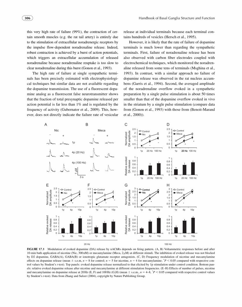
In order to resolve the question of how nicotine both elevates extracellular dopamine and depresses evoked dopa-mine release, the effect of nicotine on the modulation of evoked dopamine release were compared under different firing patterns and found to be dependent on the firing pat-tern of dopamine neurons. While desensitization of nAChRs indeed curbs dopamine released by stimuli emulating tonic firing, it allows a rapid rise in dopamine from stimuli emu-lating the phasic firing patterns associated with incentive/salience paradigms. Nicotine may thus enhance the contrast of dopamine signals associated with behavioral cues ( Rice and Cragg, 2004 ; Zhang and Sulzer, 2004 ) ( Fig. 17.1 ). Interestingly, mu opioid agonists also inhibit dopamine overflow elicited with single-spike stimuli while leaving that produced by burst stimuli unaffected, but these differential effects are mediated by nAChRs and caused by inhibition of cholinergic interneurons ( Britt and McGehee, 2008 ).
Remarkably , most heteroreceptors studied to date have been found to depress dopamine release. Therefore, in addi-tion to D2 autoreceptors, most heteroreceptors in vivo may maintain the dopamine release probability at a low level, which may then provide a marked and rapid increase in dopa-mine concentration during phasic firing. A range of drugs that affect the dopamine system may exert their actions via altering the signal/noise ratio of dopamine by affecting heteroreceptors on dopamine terminals as well as on cell bodies (Zhang and Sulzer, unpublished observations).
2. Failure of Vesicular Release at Individual Dopamine Terminals
In the sympathetic nervous system the release of noradrena-line exhibits a very low probability per release site (about 1%) ( Msghina et al., 1993 ). In other words, although electrically evoked action potentials always reach sympathetic terminals, very often they do not trigger noradrenaline release. Despite
Handbook of Basal Ganglia Structure and Function306
this very high rate of failure (99%), the contraction of cer-tain smooth muscles (e.g. the rat tail artery) is entirely due to the stimulation of extracellular noradrenergic receptors by the impulse flow-dependent noradrenaline release. Indeed, robust contraction is achieved by a burst of action potentials, which triggers an extracellular accumulation of released noradrenaline because noradrenaline reuptake is too slow to clear noradrenaline during this burst ( Gonon et al., 1993 ).
The high rate of failure at single sympathetic termi-nals has been precisely estimated with electrophysiologi-cal techniques but similar data are not available regarding the dopamine transmission. The use of a fluorescent dopa-mine analog as a fluorescent false neurotransmitter shows that the fraction of total presynaptic dopamine released per action potential is far less than 1% and is regulated by the frequency of activity ( Gubernator et al., 2009 ). This, how-ever, does not directly indicate the failure rate of vesicular
release at individual terminals because each terminal con-tains hundreds of vesicles ( Hersch et al., 1995 ).
However , it is likely that the rate of failure of dopamine terminals is much lower than regarding the sympathetic terminals. First, failure of noradrenaline release has been also observed with carbon fiber electrodes coupled with electrochemical techniques, which monitored the noradren-aline released from some tens of terminals ( Msghina et al., 1993 ). In contrast, with a similar approach no failure of dopamine release was observed in the rat nucleus accum-bens ( Garris et al., 1994 ). Second, the averaged amplitude of the noradrenaline overflow evoked in a sympathetic preparation by a single pulse stimulation is about 50 times smaller than that of the dopamine overflow evoked in vivo in the striatum by a single pulse stimulation (compare data from ( Gonon et al., 1993 ) with those from ( Benoit-Marand et al., 2000 )).
A B
1p
4p (20 Hz)
4p (100 Hz)
Nic Meca_
3 s
1 μM
+_
+
Nor
mal
ized
D
A a
mpl
itude
Rel
ativ
e ev
oked
DA
rel
ease
E
**
**
* 0
0.5
1
1.5
2
2.5
1p 4p 10p
ControlMeca
0
0.5
1
1.5
2
2.5
1p 4p 10p
ControlNic
F
**
00.20.40.60.8
1
1p 4p 10p0
0.2
0.40.60.8
1
1p 4p 10p
20 Hz
C
0
0.5
1
1.5
2
2.5
1p 20 Hz 100 Hz
ControlNic
D
*
0
0.5
1
1.5
2
2.5
1p 20 Hz 100 Hz
**
*
20 Hz
ControlMeca
**
**
Nor
mal
ized
DA
am
plitu
de
0
0.2
0.4
0.6
0.8
1
1p 20 Hz 100 Hz0
0.20.40.60.8
1
1p 20 Hz 100 Hz
Rel
ativ
e ev
oked
DA
rel
ease
N
orm
aliz
ed
DA
am
plitu
de
G3
00.5
11.5
22.5
1p 4p 10p
ControlNic
*
ControlNic
*
*
H
00.5
11.5
22.5
3
1p 4p 10p
ControlMeca
**
00.20.40.60.8
1
1p 4p 10p0
0.20.40.60.8
1
1p 4p 10p
Rel
ativ
e ev
oked
DA
rel
ease
100 Hz
FIGURE 17.1 Modulation of evoked dopamine (DA) release by nAChRs depends on firing pattern. (A, B) Voltammetric responses before and after 10-min bath application of nicotine (Nic, 300 nM) or mecamylamine (Meca, 2 μ M) at different stimuli. The inhibition of evoked release was not blocked by D2 dopamine, GABA(A), GABA(B) or ionotropic glutamate receptor antagonists. (C, D) Frequency modulation of nicotine and mecamylamine effects on dopamine release (mean s.e.m, n � 8 for control; n � 5 for nicotine, n � 4 for mecamylamine; * P 0.05 compared with respective con-trol values by Student’s t -test). Top panels: evoked dopamine release normalized to that elicited by 1p stimulation under control condition. Bottom pan-els: relative evoked dopamine release after nicotine and mecamylamine at different stimulation frequencies. (E – H) Effects of number of pulses, nicotine and mecamylamine on dopamine release at 20 Hz (E, F) and 100 Hz (G,H) (mean s.e.m., n � 4 – 8, * P 0.05 compared with respective control values by Student’s t -test). Data from Zhang and Sulzer (2004) , copyright by Nature Publishing Group .

307Chapter | 17 Regulation of Extracellular Dopamine
III. DOPAMINE REUPTAKE
During the 1960s it was shown that dopamine neurons are equipped with a dopamine transporter (DAT) and it was hypothesized that DAT might control the intensity and dura-tion of the dopamine transmission. Here, we focus on this functional role of dopamine reuptake, while other aspects of DAT are reviewed elsewhere ( Uhl, 2003 ).
Microdialysis and voltammetric techniques coupled with carbon fiber electrodes were developed during the 1980s and enabled monitoring the extracellular level of dopamine in vivo . Both technical approaches showed that in resting conditions, this level was low (10 – 20 nM) and that dopamine reuptake strongly contributed to clearing dopamine from the extracellular space. Indeed, pharmacological inhibition of dopamine reuptake potently enhanced the extracellular dopa-mine level ( Gonon and Buda, 1985 ; Church et al., 1987 ) and this enhancement was no longer observed after blocking the impulse flow-dependent dopamine release ( Carboni et al., 1989 ). These approaches, however, were too slow to accu-rately describe the kinetics of dopamine clearance and could not distinguish between an increase in dopamine release and a decrease in dopamine clearance. Both issues were resolved with improvements in voltammetric techniques ( Wightman and Zimmerman, 1990 ; Dugast et al., 1994 ). Moreover, the design of mice lacking DAT ( Giros et al., 1996 ) made it pos-sible to investigate the relative contribution of dopamine reuptake to dopamine clearance versus extracellular degrada-tion, non-neuronal uptake and diffusion.
A. Reuptake Replenishes the Releasable Pool
In dopamine terminals, dopamine is synthesized from l -DOPA by a decarboxylase and l -DOPA is synthesized from tyrosine by tyrosine hydroxylase (TH), an enzyme that is specifically expressed by catecholaminergic neurons. However, pharmacological inhibition of TH activity by alpha-methyl-para-tyrosine (AMPT) only partly and very slowly depresses the dopamine tissue content ( Jones et al., 1998b ) and in vivo dopamine release ( Benoit-Marand et al., 2000 ) in wild-type (WT) mice. In DAT � / � mice, although TH activity is doubled, AMPT induces a profound and rapid decrease of the dopamine tissue content ( Jones et al., 1998b ) and of in vivo dopamine release ( Benoit-Marand et al., 2000 ). These observations demonstrated that in brain struc-tures densely innervated by dopamine terminals, recycling of released dopamine by reuptake plays a major role in replen-ishing the releasable pool of dopamine.
B. Extracellular Elimination of the Released Dopamine is Achieved by Reuptake
Electrical stimulation of the dopamine axons either with a single pulse or with a brief train (e.g. four pulses at 100 Hz) induces a brief dopamine overflow that can be detected with rapid electrochemical techniques either in vitro ( Schmitz et al., 2001 ) or in vivo ( Dugast et al., 1994 ; Garris et al., 1994 ). The decay phase of this evoked dopamine overflow reflects the clearance of released dopamine. Dopamine reuptake inhibitors slow down the clearance kinetics by one order of magnitude ( Garris et al., 1994 ; Suaud-Chagny et al., 1995 ; Schmitz et al., 2001 ). However, in the striatum of DAT � / � mice the decay phase is slowed down by two orders of magnitude both in vitro ( Giros et al., 1996 ; Jones et al., 1998b ) and in vivo ( Benoit-Marand et al., 2000 ). Pharmacological inhibition of dopamine degradation does not further slow the decay phase in DAT � / � mice in vitro. However, in vivo, inhibition of dopamine deg-radation by monoamine oxidase slightly slows the decay phase in the striatum of DAT � / � mice but not in WT mice. Therefore, in brain structures densely innervated by dopamine terminals, dopamine reuptake represents the only mechanism responsible for the clearance of released dopamine ( Jones et al., 1998b ; Benoit-Marand et al., 2000 ). The roles of extracel-lular dopamine degradation and of non-neuronal uptake are negligible compared to dopamine reuptake. In DAT � / � mice, dopamine clearance is mainly due to dopamine diffu-sion ( Jones et al., 1998b ; Benoit-Marand et al., 2000 ).
In brain structures weakly innervated by dopamine termi-nals, such as the amygdala, the prefrontal cortex (PfC) and the cingulated cortex (CgC), the clearance of released dopamine is much slower than in the striatum (Garris and Wightman, 2004). Indeed, the half-life for dopamine uptake is about 2 sec in these structures whereas it was estimated to be 60 ms in the striatum ( Garris and Wightman, 1994 ). Moreover, inhibitors of dopamine reuptake are less efficient at slowing dopamine clearance in these brain structures than in the striatum ( Garris and Wightman, 1995 ; Mundorf et al., 2001 ). At least in PfC and CgC, the released dopamine is partly cleared by the nor-adrenergic transporter ( Mundorf et al., 2001 ).
C. Reuptake Limits Dopamine Diffusion in the Extracellular Fluid
1. Diffusion and Reuptake of Dopamine Quanta in the Extracellular Fluid
In the striatum, most dopamine terminals form small sym-metrical synaptic contacts on the neck of dendritic spines

Handbook of Basal Ganglia Structure and Function308
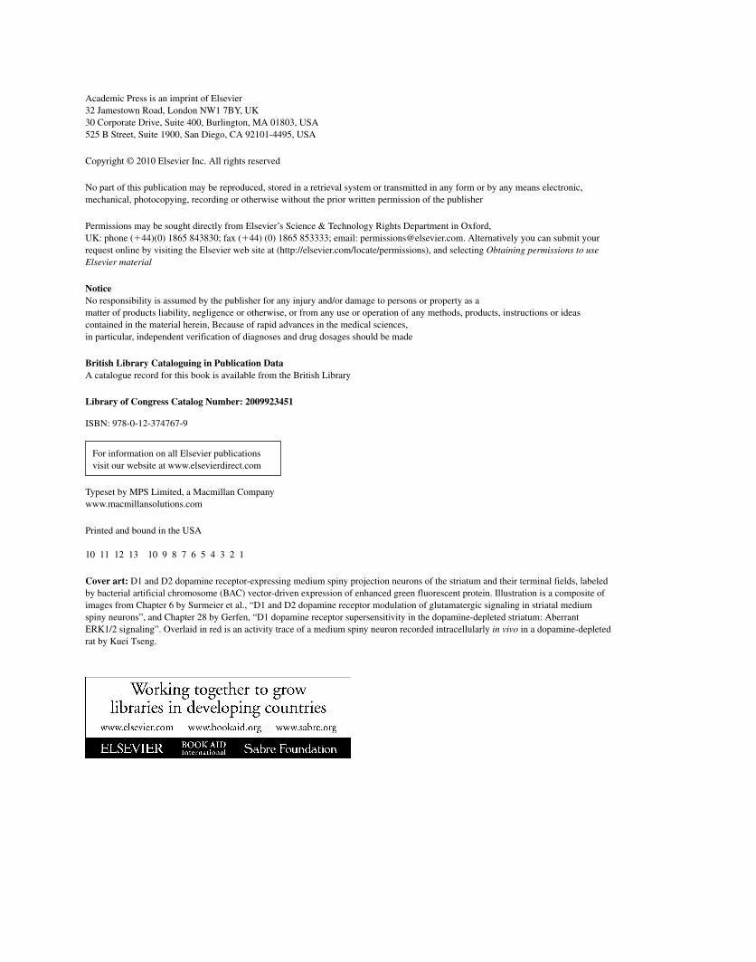
of medium sized spiny neurons ( Freund et al., 1984 ) ( Fig. 17.2A ). Striatal postsynaptic dopamine receptors are either of the D1 or of the D2 type and are not located in front of the dopaminergic contacts ( Hersch et al., 1995 ; Caille et al., 1996 ) but are distributed along the dendritic mem-brane with a higher density in the perisynaptic zone of the asymmetric synapses formed by corticostriatal glutamater-gic terminals on the head of the dendritic spine ( Fig. 17.2A ). Moreover, reuptake sites are evenly distributed on the mem-brane surface of dopamine fibers and are rarely observed on the dopaminergic synaptic membrane ( Nirenberg et al., 1996a ; Pickel et al., 1996 ; Hersch et al., 1997 ). These sub-cellular observations strongly suggest that the dopamine transmission is mainly extrasynaptic ( Pickel et al., 1996 ) and that dopamine diffuses from release sites within the synap-tic cleft to extrasynaptic dopamine receptors. Moreover, all
dopamine receptors belong to the family of G-protein cou-pled receptors. Stimulation of G-protein receptors requires a minimal level of neurotransmitter, which depends on the receptor affinity, present for several tens of milliseconds ( Hille, 1992 ). In the striatum D1 receptors appear to be in a low affinity state (1 μ M) whereas D2 receptors are in the high affinity state (10 nM) ( Richfield et al., 1989 ). Indeed, the basal extracellular dopamine level due to the tonic dis-charge activity of dopamine neurons is in the range of 10 – 20 nM and is high enough to exert a tonic stimulation of presynaptic ( Gonon and Buda, 1985 ) and postsynaptic ( Svenningsson et al., 1999 ) D2 receptors.
Cragg and Rice (2004) have thoroughly investigated the diffusion of dopamine in the extracellular space. Assuming that a quantal release event of dopamine occurs at time t � 0 they calculated the change in extracellular dopamine
DA overflow at carbon fiber electrode
After nomifensine
0.5 μM
r = 1 μm
1 μm
1 μM
2 μm
100 nM 10 nM 1 nM 100 pM
5 μm 10 μm 20 μm
1 pulse
Control
0 200 400 ms
CA
BDA concentration at a distance r from one release site
10 ms 20 ms 50 ms 100 ms 500 ms
Diffusion only: Diffusion + uptake:
DA
GLUDistance : r
DA
Den
drite
D1 receptor
FIGURE 17.2 Kinetics of dopamine diffusion and reuptake in the extracellular space. (A) This drawing summarizes morphological data concerning the dopamine transmission on striatal medium sized spiny neurons. The extracellular volume represents about 15 – 20% of the whole tissue volume. Dopamine terminals form symmetric contact with the neck of dendritic spines, whereas glutamatergic excitatory synapses are characterized by asym-metric contacts on the head of the spines. Postsynaptic dopamine receptors are rarely observed inside the dopaminergic synaptic cleft and are denser in the perisynaptic zone of the glutamatergic synapses. (B) When a quantum of dopamine (Q � 9800 dopamine molecules) is released in the synaptic cleft at time t � 0 (arrows), dopamine diffuses outside the synaptic cleft. At increasing distances from the release site the simulated transient changes in dopamine concentration are increasingly affected by dopamine reuptake. Note progressive changes in concentration and time scales with increasing distances. (C) Effect of reuptake inhibition by nomifensine (20 mg/kg, s.c.) on the dopamine overflow evoked by single pulse stimulation. Single pulse stimulations of the medial forebrain bundle were applied every 15 s. The resulting dopamine overflow was monitored in vivo in the striatum with a car-bon fiber electrode coupled with continuous amperometry. The traces show the averaging of 20 recordings before (control) and 20 min after nomifensine injection. (A and C) data from Gonon (1997) , copyright by Society for Neuroscience. (B) data from Cragg and Rice (2004) , copyright by Elsevier .

309Chapter | 17 Regulation of Extracellular Dopamine
concentration at variable distances from the release site ( Fig. 17.2B ). They showed that at short distance (1 and 2 μ m) diffusion entirely governs the dopamine overflow, whereas at increasing distance from release site (5 to 20 μ m) dopamine reuptake increasingly limits the dopamine over-flow both in term of maximal amplitude and duration. However, in the latter distance range, the maximal ampli-tude of the dopamine overflow is below 100 nM. Therefore, Cragg and Rice suggested that dopamine transmission mediated by D1 receptors occurs at a maximal distance of 2 μ m from the release site and is not affected by dopamine reuptake. In contrast, the dopamine transmission mediated by D2 receptors might be effective at a distance of 7 μ m from release sites. This distance as well as the duration of effective D2 stimulation (i.e. time during which the extra-cellular dopamine concentration exceeds 10 nM) is limited by dopamine reuptake ( Cragg and Rice, 2004 ).
This view that released dopamine escapes the extrasyn-aptic cleft and that its diffusion is not strongly affected by dopamine reuptake in the vicinity of release sites (i.e. at a distance 5 μ m) has been experimentally supported by electrochemical measurements of the electrically evoked dopamine overflow. Indeed, inhibition of dopamine reup-take strongly slows dopamine clearance but only moderately enhances the maximal amplitude of the dopamine overflow evoked by a single pulse ( Fig. 17.2C ) ( Garris et al., 1994 ; Gonon, 1997 ; Schmitz et al., 2001 ). This is due to the fact that diffusion at short distance ( 5 μ m) is faster than dopa-mine reuptake. Indeed, the half-life for dopamine clearance, which has been calculated from in vivo recordings, is about 30 ms ( Garris et al., 1994 ; Gonon et al., 2000 ).
2. Extracellular Summation of Multiple Quanta: Role of Reuptake
In the striatum, the density of dopamine terminals is very high and the average distance between two terminals is about 4 μ m ( Doucet et al., 1986 ). If single pulse stimulation triggers dopamine release in a small fraction of dopamine terminals, every active single release site and its sphere of influence must be considered independently as discussed by Gragg and Rice (2004). Alternatively, if most terminals synchro-nously release single dopamine quanta in response to single pulse stimulation, the extracellular dopamine level largely results from a spatial summation of released quanta. As dis-cussed above under the section on Failure of vesicular release (p. 305) , it is likely that the failure rate of vesicular release is low regarding dopamine terminals in vivo.
This view that the extracellular dopamine level largely results from a spatial summation of quanta from neighboring terminals is further supported by in vivo estimates. Indeed, the maximal amplitude of the dopamine overflow evoked by a single pulse in the rodent striatum and measured in vivo by a carbon fiber electrode, is in the range of 100 – 400 nM ( Fig. 17.2C ) ( Dugast et al., 1994 ; Garris et al., 1994 ; Benoit-Marand et al., 2000 ; Venton et al., 2003 ). However, this observed value represents an underestimate of the genuine change in the intact tissue. Models of electrochemical moni-toring assume that between the intact tissue and the elec-trode surface, there is a dead zone with a thickness of 6 – 8 μ m ( Gonon et al., 2000 ; Schmitz et al., 2001 ; Venton et al., 2003 ). This estimate is consistent with an electron micros-copy study showing the extent of the tissue damage gener-ated by the implantation of a carbon fiber electrode for 4 h in the striatum of anesthetized rats ( Peters et al., 2004 ). This dead zone slows the kinetics of the evoked dopamine over-flow and diminishes their maximal amplitude. Therefore, the dopamine overflow recorded by a carbon fiber electrode is much larger than expected by Cragg and Rice’s calculation unless dopamine overflow summation from several dopamine terminals is taken into account (compare Figs 17.2B and C ). Nevertheless, this apparent spatial summation from multiple release sites is limited by dopamine reuptake ( Gonon, 1997 ).
In summary, dopamine overflow evoked in the striatum by single pulse stimulation exhibits two phases. The rising phase is rapid and mainly reflects dopamine release. The decay phase is slower and reflects dopamine reuptake. The kinetics of release and reuptake can be obtained by simu-lation taking into account the diffusion of dopamine from the intact tissue to the electrode surface through the dead zone. These simulations show that the half-life of released dopamine in the striatum in vivo is about 30 ms ( Garris et al., 1994 ; Gonon et al., 2000 ; Schmitz et al., 2001 ; Venton et al., 2003 ). In this period of time the diffusion distance for dopamine is about 7 μ m. Therefore, in a given point of the extracellular space, release sites at a distance � 7 μ m do not significantly contribute to the dopamine over-flow. Thus, in the striatum and other brain structure densely innervated by dopamine terminals the dopamine transmis-sion is extrasynaptic but, nevertheless, spatially constricted by dopamine reuptake.
In brain structures with a much lower DAT density, such as the PfC and CgC, diffusion plays a larger role in the clearance of released dopamine. However, even in these regions, pharmacological inhibition of DAT strongly slows the clearance of released dopamine ( Mundorf et al., 2001 ).

Handbook of Basal Ganglia Structure and Function310
D. Regulation of Dopamine Reuptake by D2 Autoreceptors
In vivo studies ( Cass and Gerhardt, 1994 ; Rothblat and Schneider, 1997 ) show that D2 antagonists slow the clearance of exogenously applied dopamine. Moreover, D2 antagonists decrease the rate of elimination of endogenous dopamine released in vivo by electrical stimulation ( Benoit-Marand et al., 2001 ; Wu et al., 2002 ). Kinetics analysis of reuptake parameters shows that haloperidol reduces the Vm but does not affect Km ( Wu et al., 2002 ). This inhibition of dopamine reuptake is not due to the direct effect of D2 antagonists on DAT activity because it is not observed in mice lacking D2 receptors ( Dickinson et al., 1999 ).
The stimulation of D2 autoreceptors by the basal extracel-lular dopamine level exerts a tonic inhibition of the impulse flow-dependent dopamine release and, therefore, D2 antago-nists facilitate dopamine release by blocking this D2 inhibi-tion (see Section IIC) . In contrast, the inhibitory effect of D2 antagonists on DAT activity does not seem to be due to the blockade of a tonic stimulation of D2 receptors. Indeed, the rate of elimination of electrically evoked dopamine release is not altered in D2 � / � mice compared to WT mice ( Rouge-Pont et al., 2002 ). Whatever the mechanism, the inhibition of DAT activity acts in synergy with the facilitation of dopa-mine release by D2 antagonists to enhance impulse-flow dependent dopamine overflow. Both mechanisms should be taken into account when considering the therapeutic effects of D2 antagonists as well as their side effects.
IV. RELATIONSHIP BETWEEN THE FIRING OF DOPAMINE NEURONS AND EXTRACELLULAR DOPAMINE
Dopamine neurons exhibit two patterns of discharge activ-ity: a continuous mode with regularly spaced spikes at a frequency between 2 and 5 Hz, and a bursting activity charac-terized by brief bursts of 2 to 6 action potentials ( Grace and Bunney, 1984a,b ) (see also Chapter 16) . Single dopamine neurons switch between the patterns. In resting condition and during sleep most dopamine neurons discharge with the tonic mode, but rewarding or sensorial stimuli predicting a reward trigger in most dopamine neurons a single burst both in rats ( Hyland et al., 2002 ) and monkeys ( Schultz et al., 1993 ; Mirenowicz and Schultz, 1996 ). In rats the intra-burst fre-quency is 15 – 30 Hz ( Grace and Bunney, 1984b ; Hyland et al., 2002 ). Grace and Bunney hypothesized that the bursting mode would be more potent than the tonic pattern to induce
dopamine release ( Grace and Bunney, 1984b ). Accordingly, electrical stimulations mimicking the bursting mode were twice as potent as regularly spaced stimulation having the same whole duration and number of pulse to enhance the extracellular dopamine level ( Gonon, 1988 ). However, these data were erroneously interpreted as a non-linear relationship between dopamine release and the impulse flow frequency.
A. The Tonic Extracellular Dopamine Level
When most dopamine neurons discharge in the tonic mode, the pause between two successive action potentials reach-ing the same release site exceeds 200 ms. Therefore, the dopamine released by one action potential is completely cleared by reuptake before the next action potential. This view has been experimentally demonstrated in vivo with electrical stimulation of the dopamine fiber at 4 Hz ( Chergui et al., 1994 ). Although this point has not been extensively studied to our knowledge, the degree of synchronous activ-ity of dopamine neurons is probably low during tonic activity. Indeed, synchronous activity has been observed between pairs of adjacent neurons recorded with the same electrode ( Wilson et al., 1977 ; Grace and Bunney, 1983 ) but not between pairs of distant neurons recorded with two electrodes ( Wilson et al., 1977 ) and this synchrony has been reported to be more prominent during bursting activ-ity ( Grace and Bunney, 1983 ). Thus, taking into account the very high density of dopamine terminals in the striatum, the tonic activity induces a steady-state extracellular dopamine level, in the range of 10 – 30 nM, which is stable with time and spatially homogenous ( Venton et al., 2003 ). However, this steady-state is almost entirely firing dependent ( Gonon and Buda, 1985 ; Svenningsson et al., 1999 ). A pause in the tonic discharge activity of dopamine neurons induces a rapid and profound ( � 80 %) decrease in the extracellular dopamine level ( Gonon, 1988 ; Suaud-Chagny et al., 1992 ). Dopamine reuptake limits this tonic extracellular level. Indeed, pharmacological inhibition of dopamine reuptake increases this level by � 300% ( Gonon and Buda, 1985 ; Carboni et al., 1989 ; Venton et al., 2003 ) while in DAT � / � mice, the basal extracellular dopamine level is five times as large as in WT mice ( Jones et al., 1998b ).
B. Phasic Changes in Extracellular Dopamine
1. Electrically Evoked Dopamine Overfl ow
Electrical stimuli mimicking the bursting mode are more potent than regularly spaced stimuli in evoking dopamine

311Chapter | 17 Regulation of Extracellular Dopamine
overflow, but this is not due to a facilitation of the release per se. Indeed, in the frequency range of 15 – 100 Hz brief train pulse stimuli (2 to 6 pulses) induce in vivo a larger dopamine overflow than that evoked by a single pulse stimulation, mostly due to extracellular accumulation of the released dopamine with successive pulses ( Chergui et al., 1994 ; Garris et al., 1994 ; Venton et al., 2003 ). Indeed, in DAT � / � mice the maximal amplitude of the dopamine overflow evoked by a four-pulse stimulation is independ-ent of frequency (15 or 4 Hz), whereas in WT mice the ratio S(15 Hz)/S(4 Hz) is 2.4 in the dorsal striatum and 3.0 in the nucleus accumbens ( Fig. 17.3 ) ( Benoit-Marand et al., 2000 ). Burst-induced extracellular accumulation is higher in the nucleus accumbens than in the dorsal striatum because dopamine reuptake is about twice as slow in the former than in the latter region. Therefore, the specificity of the burst-ing mode over the tonic pattern results from the kinetics of dopamine reuptake rather than from changes in the quantal release per pulse. As expected, pharmacological inhibition of DAT activity increases the amplitude and duration of the phasic dopamine overflow evoked by a train pulse stimula-tion ( Garris et al., 1994 ; Suaud-Chagny et al., 1995 ; Venton et al., 2003 ). Indeed, DAT inhibition facilitates the extracel-lular accumulation of dopamine with successive pulses.
2. Dopamine Transients in Behaving Animals
Rewards or stimuli predicting a reward evoke a burst of action potential in most dopamine neurons with a similar delay and
duration both in monkeys ( Mirenowicz and Schultz, 1996 ) and rats ( Pan et al., 2005 ) (see also Chapter 31). Therefore, in behaving rats, brief changes in extracellular dopamine, simi-lar in kinetic and amplitude to dopamine overflow evoked by brief electrical stimulation, can be observed ( Robinson et al., 2001 ; Robinson et al., 2002 ). As expected, DAT inhi-bition increases the maximal amplitude and duration of these dopamine transients to the same extent as dopamine overflow evoked by brief electrical stimulations ( Robinson and Wightman, 2004 ). Moreover, in the nucleus accumbens dopamine transients clearly result from the bursting discharge activity of VTA dopamine neurons. Indeed, NMDA-mediated excitatory inputs to VTA dopamine neurons generate their bursting activity ( Suaud-Chagny et al., 1992 ; Chergui et al., 1993 ; Overton and Clark, 1997 ). Specific manipula-tions of these inputs by local VTA injections of NMDA and of NMDA antagonists in freely moving rats alter the fre-quency of dopamine transients in the expected way ( Sombers et al., 2009 ). Therefore, these dopamine transients represent dopamine overflow evoked by naturally occurring bursts of action potentials.
While dopamine neurons initially respond with a burst to unpredicted reward, after intense training they respond to a cue predicting a reward but no longer to the expected reward, both in monkeys ( Schultz et al., 1993 ; Mirenowicz and Schultz, 1996 ) and rats ( Pan et al., 2005 ). Electrochemical studies show the same relationship between dopamine transients and reward or cue predicting a reward ( Day et al., 2007 ) ( Fig. 17.4 ). Dopamine neurons
FIGURE 17.3 Dopamine overflows evoked in nucleus accumbens (NAc) by stimulations mimicking the spontaneous activity of dopamine neurons. Medial forebrain bundle electrical stimulations consisted of four pulses either at 4 or at 15 Hz. The dopamine overflow was recorded with a carbon fiber electrode coupled with continuous amperometry. Electrodes were calibrated for dopamine concentration after in vivo recordings. The figure shows typi-cal recordings obtained from one DAT � / � and one DAT � / � mouse. Notice that the time scale is not the same with DAT � / � and DAT � / � because dopamine clearance is much slower in DAT � / � mice. Stimulations mimicking a spontaneous burst (four pulses at 15 Hz) were more efficient than stimu-lations mimicking the single spike discharge mode (four pulses at 4 Hz) to evoke dopamine overflow ( Benoit-Marand et al., 2000 ), copyright by Elsevier.
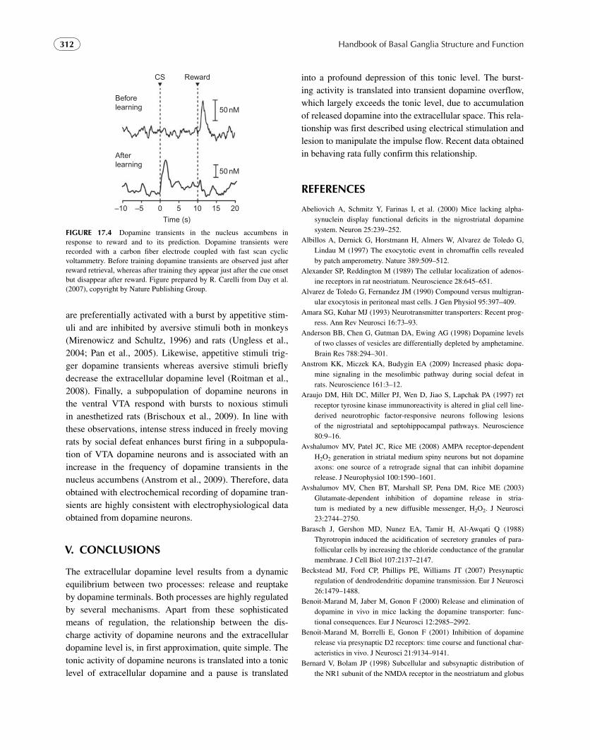
Handbook of Basal Ganglia Structure and Function312
are preferentially activated with a burst by appetitive stim-uli and are inhibited by aversive stimuli both in monkeys ( Mirenowicz and Schultz, 1996 ) and rats ( Ungless et al., 2004 ; Pan et al., 2005 ). Likewise, appetitive stimuli trig-ger dopamine transients whereas aversive stimuli briefly decrease the extracellular dopamine level ( Roitman et al., 2008 ). Finally, a subpopulation of dopamine neurons in the ventral VTA respond with bursts to noxious stimuli in anesthetized rats ( Brischoux et al., 2009 ). In line with these observations, intense stress induced in freely moving rats by social defeat enhances burst firing in a subpopula-tion of VTA dopamine neurons and is associated with an increase in the frequency of dopamine transients in the nucleus accumbens ( Anstrom et al., 2009 ). Therefore, data obtained with electrochemical recording of dopamine tran-sients are highly consistent with electrophysiological data obtained from dopamine neurons.
V. CONCLUSIONS
The extracellular dopamine level results from a dynamic equilibrium between two processes: release and reuptake by dopamine terminals. Both processes are highly regulated by several mechanisms. Apart from these sophisticated means of regulation, the relationship between the dis-charge activity of dopamine neurons and the extracellular dopamine level is, in first approximation, quite simple. The tonic activity of dopamine neurons is translated into a tonic level of extracellular dopamine and a pause is translated
into a profound depression of this tonic level. The burst-ing activity is translated into transient dopamine overflow, which largely exceeds the tonic level, due to accumulation of released dopamine into the extracellular space. This rela-tionship was first described using electrical stimulation and lesion to manipulate the impulse flow. Recent data obtained in behaving rata fully confirm this relationship .
REFERENCES
Abeliovich A , Schmitz Y , Farinas I , et al. ( 2000 ) Mice lacking alpha-synuclein display functional defi cits in the nigrostriatal dopamine system . Neuron 25 : 239 – 252 .
Albillos A , Dernick G , Horstmann H , Almers W , Alvarez de Toledo G , Lindau M ( 1997 ) The exocytotic event in chromaffi n cells revealed by patch amperometry . Nature 389 : 509 – 512 .
Alexander SP , Reddington M ( 1989 ) The cellular localization of adenos-ine receptors in rat neostriatum . Neuroscience 28 : 645 – 651 .
Alvarez de Toledo G , Fernandez JM ( 1990 ) Compound versus multigran-ular exocytosis in peritoneal mast cells . J Gen Physiol 95 : 397 – 409 .
Amara SG , Kuhar MJ ( 1993 ) Neurotransmitter transporters: Recent prog-ress . Ann Rev Neurosci 16 : 73 – 93 .
Anderson BB , Chen G , Gutman DA , Ewing AG ( 1998 ) Dopamine levels of two classes of vesicles are differentially depleted by amphetamine . Brain Res 788 : 294 – 301 .
Anstrom KK , Miczek KA , Budygin EA ( 2009 ) Increased phasic dopa-mine signaling in the mesolimbic pathway during social defeat in rats . Neuroscience 161 : 3 – 12 .
Araujo DM , Hilt DC , Miller PJ , Wen D , Jiao S , Lapchak PA ( 1997 ) ret receptor tyrosine kinase immunoreactivity is altered in glial cell line-derived neurotrophic factor-responsive neurons following lesions of the nigrostriatal and septohippocampal pathways . Neuroscience 80 : 9 – 16 .
Avshalumov MV , Patel JC , Rice ME ( 2008 ) AMPA receptor-dependent H 2 O 2 generation in striatal medium spiny neurons but not dopamine axons: one source of a retrograde signal that can inhibit dopamine release . J Neurophysiol 100 : 1590 – 1601 .
Avshalumov MV , Chen BT , Marshall SP , Pena DM , Rice ME ( 2003 ) Glutamate-dependent inhibition of dopamine release in stria-tum is mediated by a new diffusible messenger, H 2 O 2 . J Neurosci 23 : 2744 – 2750 .
Barasch J , Gershon MD , Nunez EA , Tamir H , Al-Awqati Q ( 1988 ) Thyrotropin induced the acidifi cation of secretory granules of para-follicular cells by increasing the chloride conductance of the granular membrane . J Cell Biol 107 : 2137 – 2147 .
Beckstead MJ , Ford CP , Phillips PE , Williams JT ( 2007 ) Presynaptic regulation of dendrodendritic dopamine transmission . Eur J Neurosci 26 : 1479 – 1488 .
Benoit-Marand M , Jaber M , Gonon F ( 2000 ) Release and elimination of dopamine in vivo in mice lacking the dopamine transporter: func-tional consequences . Eur J Neurosci 12 : 2985 – 2992 .
Benoit-Marand M , Borrelli E , Gonon F ( 2001 ) Inhibition of dopamine release via presynaptic D2 receptors: time course and functional char-acteristics in vivo . J Neurosci 21 : 9134 – 9141 .
Bernard V , Bolam JP ( 1998 ) Subcellular and subsynaptic distribution of the NR1 subunit of the NMDA receptor in the neostriatum and globus
Beforelearning
Afterlearning
CS Reward
50 nM
50 nM
Time (s)
–10 –5 0 5 10 15 20
FIGURE 17.4 Dopamine transients in the nucleus accumbens in response to reward and to its prediction. Dopamine transients were recorded with a carbon fiber electrode coupled with fast scan cyclic voltammetry. Before training dopamine transients are observed just after reward retrieval, whereas after training they appear just after the cue onset but disappear after reward. Figure prepared by R. Carelli from Day et al. (2007) , copyright by Nature Publishing Group.

313Chapter | 17 Regulation of Extracellular Dopamine
pallidus of the rat: co-localization at synapses with the GluR2/3 sub-unit of the AMPA receptor . Eur J Neurosci 10 : 3721 – 3736 .
Blaschko H , Welch AD ( 1953 ) Localization of adrenaline in cytoplasmic particles of the bovine adrenal medulla . Naunyn-Schmiedebergs Arch Exp Pathol Pharmacol 219 : 17 – 22 .
Bonci A , Bernardi G , Grillner P , Mercuri NB ( 2003 ) The dopamine-containing neuron: maestro or simple musician in the orchestra of addiction? Trends Pharmacol Sci 24 : 172 – 177 .
Brischoux F , Chakraborty S , Brierley DI , Ungless MA ( 2009 ) Phasic excitation of dopamine neurons in ventral VTA by noxious stimuli . Proc Natl Acad Sci USA 106 : 4894 – 4899 .
Britt JP , McGehee DS ( 2008 ) Presynaptic opioid and nicotinic receptor modulation of dopamine overfl ow in the nucleus accumbens . J Neurosci 28 : 1672 – 1681 .
Caille I , Dumartin B , Bloch B ( 1996 ) Ultrastructural localization of D1 dopamine receptor immunoreactivity in rat striatonigral neurons and its relation with dopaminergic innervation . Brain Res 730 : 17 – 31 .
Camacho M , Machado JD , Alvarez J , Borges R ( 2008 ) Intravesicular calcium release mediates the motion and exocytosis of secre-tory organelles: a study with adrenal chromaffi n cells . J Biol Chem 283 : 22383 – 22389 .
Camacho M , Machado JD , Montesinos MS , Criado M , Borges R ( 2006 ) Intragranular pH rapidly modulates exocytosis in adrenal chromaffi n cells . J Neurochem 96 : 324 – 334 .
Carboni E , Imperato A , Perezzani L , Di-Chiara G ( 1989 ) Amphetamine, cocaine, phencyclidine and nomifensine increase extracellular dopa-mine concentrations preferentially in the nucleus accumbens of freely moving rats . Neuroscience 28 : 653 – 661 .
Cardozo DL , Bean BP ( 1995 ) Voltage-dependent calcium channels in rat midbrain dopamine neurons: modulation by dopamine and GABAB receptors . J Neurophysiol 74 : 1137 – 1148 .
Carlsson A , Hillarp N-A , Waldeck A ( 1962 ) A Mg � � -ATP dependent storage mechanism in the amine granules of the adrenal medulla . Med Exp 6 : 47 – 53 .
Cass WA , Zahniser NR ( 1991 ) Potassium channel blockers inhibit D2 dopamine, but not A1 adenosine, receptor-mediated inhibition of stri-atal dopamine release . J Neurochem 57 : 147 – 152 .
Cass WA , Gerhardt GA ( 1994 ) Direct in vivo evidence that D2 dopamine receptors can modulate dopamine uptake . Neurosci Lett 176 : 259 – 263 .
Ceccarelli B , Hurlbut WP , Mauro A ( 1972 ) Depletion of vesicles from frog neuromuscular junctions by prolonged tetanic stimulation . J Cell Biol 54 : 30 – 38 .
Ceccarelli B , Hurlbut WP , Mauro A ( 1973 ) Turnover of transmitter and synaptic vesicles at the frog neuromuscular junction . J Cell Biol 57 : 499 – 524 .
Centonze D , Usiello A , Gubellini P , Pisani A , Borrelli E , Bernardi G , Calabresi P ( 2002 ) Dopamine D2 receptor-mediated inhibition of dopaminergic neurons in mice lacking D2L receptors . Neuropsychopharmacology 27 : 723 – 726 .
Chan CS , Guzman JN , Ilijic E , Mercer JN , Rick C , Tkatch T , Meredith GE , Surmeier DJ ( 2007 ) “ Rejuvenation ” protects neurons in mouse models of Parkinson’s disease . Nature 447 : 1081 – 1086 .
Charara A , Heilman C , Levey AI , Smith Y ( 2000 ) Pre- and postsynap-tic localization of GABAB receptors in the basal ganglia in monkeys . Neuroscience 95 : 127 – 140 .
Cheer JF , Wassum KM , Heien ML , Phillips PE , Wightman RM ( 2004 ) Cannabinoids enhance subsecond dopamine release in the nucleus accumbens of awake rats . J Neurosci 24 : 4393 – 4400 .
Cheer JF , Wassum KM , Sombers LA , Heien ML , Ariansen JL , Aragona BJ , Phillips PE , Wightman RM ( 2007 ) Phasic dopamine release
evoked by abused substances requires cannabinoid receptor activa-tion . J Neurosci 27 : 791 – 795 .
Chen BT , Moran KA , Avshalumov MV , Rice ME ( 2006 a ) Limited reg-ulation of somatodendritic dopamine release by voltage-sensitive Ca channels contrasted with strong regulation of axonal dopamine release . J Neurochem 96 : 645 – 655 .
Chen G , Gavin PF , Luo G , Ewing AG ( 1995 ) Observation and quantita-tion of exocytosis from the cell body of a fully developed neuron in Planorbis corneus . J Neurosci 15 : 7747 – 7755 .
Chen Q , Veenman L , Knopp K , Yan Z , Medina L , Song WJ , Surmeier DJ , Reiner A ( 1998 ) Evidence for the preferential localization of gluta-mate receptor-1 subunits of AMPA receptors to the dendritic spines of medium spiny neurons in rat striatum . Neuroscience 83 : 749 – 761 .
Chen Y , Yu FH , Surmeier DJ , Scheuer T , Catterall WA ( 2006 b ) Neuromodulation of Na � channel slow inactivation via cAMP-dependent protein kinase and protein kinase C . Neuron 49 : 409 – 420 .
Chergui K , Suaud-Chagny MF , Gonon F ( 1994 ) Nonlinear relationship between impulse fl ow, dopamine release and dopamine elimination in the rat brain in vivo . Neuroscience 62 : 641 – 645 .
Chergui K , Charlety PJ , Akaoka H , Saunier CF , Brunet JL , Buda M , Svensson TH , Chouvet G ( 1993 ) Tonic activation of NMDA recep-tors causes spontaneous burst discharge of rat midbrain dopamine neurons in vivo . Eur J Neurosci 5 : 137 – 144 .
Chow RH , Von Rueden L , Neher E ( 1992 ) Delay in vesicle fusion revealed by electrochemical monitoring of single secretory events in adrenal chromaffi n cells . Nature 356 : 60 – 63 .
Church WH , Justice JB Jr. , Neill DB ( 1987 ) Detecting behaviorally rel-evant changes in extracellular dopamine with microdialysis . Brain Res 412 : 397 – 399 .
Colliver TL , Pyott SJ , Achalabun M , Ewing AG ( 2000 ) VMAT-mediated changes in quantal size and vesicular volume . J Neurosci 20 : 5276 – 5282 .
Congar P , Bergevin A , Trudeau LE ( 2002 ) D2 receptors inhibit the secre-tory process downstream from calcium infl ux in dopaminergic neu-rons: implication of K( � ) channels . J Neurophysiol 87 : 1046 – 1056 .
Cragg SJ , Greenfi eld SA ( 1997 ) Differential autoreceptor control of somatodendritic and axon terminal dopamine release in substantia nigra, ventral temental area, and striatum . J Neurosci 17 : 1746 – 5738 .
Cragg SJ , Rice ME ( 2004 ) DAncing past the DAT at a DA synapse . Trends Neurosci 27 : 270 – 277 .
Cramer W ( 1918 ) Further observations on the thyroid-adrenal apparatus. A histochemical method for the demonstration of adrenalin granules in the suprarenal gland . J Physiol 52 : 7 – 10 .
Croft BG , Fortin GD , Corera AT , Edwards RH , Beaudet A , Trudeau LE , Fon EA ( 2005 ) Normal biogenesis and cycling of empty synaptic vesicles in dopamine neurons of vesicular monoamine transporter 2 knockout mice . Mol Biol Cell 16 : 306 – 315 .
Cruz HG , Ivanova T , Lunn ML , Stoffel M , Slesinger PA , Luscher C ( 2004 ) Bi-directional effects of GABA(B) receptor agonists on the mesolimbic dopamine system . Nat Neurosci 7 : 153 – 159 .
Cubeddu LX , Hoffmann IS ( 1982 ) Operational characteristics of the inhibitory feedback mechanism for regulation of dopamine release via presynaptic receptors . J Pharmacol Exp Ther 223 : 497 – 501 .
Davila V , Yan Z , Craciun LC , Logothetis D , Sulzer D ( 2003 ) D3 dopa-mine autoreceptors do not activate G-protein-gated inwardly rectify-ing potassium channel currents in substantia nigra dopamine neurons . J Neurosci 23 : 5693 – 5697 .
Day JJ , Roitman MF , Wightman RM , Carelli RM ( 2007 ) Associative learning mediates dynamic shifts in dopamine signaling in the nucleus accumbens . Nat Neurosci 10 : 1020 – 1028 .
Handbook of Basal Ganglia Structure and Function314
De Klippel N , Sarre S , Ebinger G , Michotte Y ( 1993 ) Effect of M1- and M2-muscarinic drugs on striatal dopamine release and metabolism: an in vivo microdialysis study comparing normal and 6-hydroxydo-pamine-lesioned rats . Brain Res 630 : 57 – 64 .
De Robertis ED , Bennett HS ( 1955 ) Some features of the submicroscopic morphology of synapses in frog and earthworm . J Biophys Biochem Cytol 1 : 47 – 58 .
Del Castillo J , Katz B ( 1954 ) Quantal components of the end-plate poten-tial . J Physiol 124 : 560 – 573 .
Dewey SL , Smith GS , Logan J , et al. ( 1993 ) Effects of central cholinergic blockade on striatal dopamine release measured with positron emis-sion tomography in normal human subjects . Proc Natl Acad Sci USA 90 : 11816 – 11820 .
Diaz J , Pilon C , Le Foll B , Gros C , Triller A , Schwartz JC , Sokoloff P ( 2000 ) Dopamine D3 receptors expressed by all mesencephalic dopa-mine neurons . J Neurosci 20 : 8677 – 8684 .
Dickinson SD , Sabeti J , Larson GA , et al. ( 1999 ) Dopamine D2 receptor-defi cient mice exhibit decreased dopamine transporter function but no changes in dopamine release in dorsal striatum . J Neurochem 72 : 148 – 156 .
Doucet G , Descarries L , Garcia S ( 1986 ) Quantifi cation of the dopamine innervation in adult rat neostriatum . Neuroscience 19 : 427 – 445 .
Drory O , Nelson N ( 2006 ) The emerging structure of vacuolar ATPases . Physiology (Bethesda) 21 : 317 – 325 .
Dugast C , Suaud-Chagny MF , Gonon F ( 1994 ) Continuous in vivo moni-toring of evoked dopamine release in the rat nucleus accumbens by amperometry . Neuroscience 62 : 647 – 654 .
Dwoskin LP , Zahniser NR ( 1986 ) Robust modulation of [3H]dopamine release from rat striatal slices by D-2 dopamine receptors . J Pharmacol Exp Ther 239 : 442 – 453 .
Edwards RH ( 2007 ) The neurotransmitter cycle and quantal size . Neuron 55 : 835 – 858 .
Elhamdani A , Palfrey HC , Artalejo CR ( 2001 ) Quantal size is dependent on stimulation frequency and calcium entry in calf chromaffi n cells . Neuron 31 : 819 – 830 .
Fatt P ( 1954 ) Biophysics of junctional transmission . Physiol Rev 34 : 674 – 710 .
Fatt P , Katz B ( 1950 ) Some observations on biological noise . Nature 166 : 597 – 598 .
Fatt P , Katz B ( 1952 ) Spontaneous subthreshold activity at motor nerve endings . J Physiol 117 : 109 – 128 .
Finnegan JM , et al. ( 1996 ) Vesicular quantal size measured by amperometry at chromaffi n, mast, pheochromocytoma, and pancreatic beta-cells . J Neurochem 66 : 1914 – 1923 .
Fortin DL , Troyer MD , Nakamura K , Kubo S , Anthony MD , Edwards RH ( 2004 ) Lipid rafts mediate the synaptic localization of alpha-synuclein . J Neurosci 24 : 6715 – 6723 .
Freund TF , Powell JF , Smith AD ( 1984 ) Tyrosine hydroxylase-immunoreactive boutons in synaptic contact with identifi ed stria-tonigral neurons, with particular reference to dendritic spines . Neuroscience 13 : 1189 – 1215 .
Fusco FR , Martorana A , Giampa C , De March Z , Farini D , D’Angelo V , Sancesario G , Bernardi G ( 2004 ) Immunolocalization of CB1 recep-tor in rat striatal neurons: a confocal microscopy study . Synapse 53 : 159 – 167 .
Gainetdinov RR , Grekhova TV , Sotnikova TD , Rayevsky KS ( 1994 ) Dopamine D2 and D3 receptor preferring antagonists differentially affect striatal dopamine release and metabolism in conscious rats . Eur J Pharmacol 261 : 327 – 331 .
Gandhi SP , Stevens CF ( 2003 ) Three modes of synaptic vesicular recy-cling revealed by single-vesicle imaging . Nature 423 : 607 – 613 .
Garris PA , Wightman RM ( 1994 ) Different kinetics govern dopaminergic transmission in the amygdala, prefrontal cortex, and striatum: an in vivo voltammetric study . J Neurosci 14 : 442 – 450 .
Garris PA , Wightman RM ( 1995 ) Distinct pharmacological regulation of evoked dopamine effl ux in the amygdala and striatum of the rat in vivo . Synapse 20 : 269 – 279 .
Garris PA , Ciolkowski EL , Pastore P , Wightman RM ( 1994 ) Effl ux of dopamine from the synaptic cleft in the nucleus accumbens of the rat brain . J Neurosci 14 : 6084 – 6093 .
Giros B , Jaber M , Jones SR , Wightman RM , Caron MG ( 1996 ) Hyperlocomotion and indifference to cocaine and amphetamine in mice lacking the dopamine transporter . Nature 379 : 606 – 612 .
Glass M , Faull RL , Dragunow M ( 1996 ) Localisation of the adenosine uptake site in the human brain: a comparison with the distribution of adenosine A1 receptors . Brain Res 710 : 79 – 91 .
Golembiowska K , Zylewska A ( 1998 ) Agonists of A1 and A2A adenos-ine receptors attenuate methamphetamine-induced overfl ow of dopa-mine in rat striatum . Brain Res 806 : 202 – 209 .
Gong LW , Hafez I , Alvarez de Toledo G , Lindau M ( 2003 ) Secretory vesicles membrane area is regulated in tandem with quantal size in chromaffi n cells . J Neurosci 23 : 7917 – 7921 .
Gonon F ( 1988 ) Nonlinear relationship between impulse fl ow and dopa-mine release by rat midbrain dopaminergic neurons as studied by in vivo electrochemistry . Neuroscience 24 : 19 – 28 .
Gonon F ( 1997 ) Prolonged and extrasynaptic excitatory action of dopa-mine mediated by D1 receptors in the rat striatum in vivo . J Neurosci 17 : 5972 – 5978 .
Gonon F , Buda M ( 1985 ) Regulation of dopamine release by impulse fl ow and by autoreceptors as studied by in vivo voltammetry in the rat striatum . Neuroscience 14 : 765 – 774 .
Gonon F , Msghina M , Stjarne L ( 1993 ) Kinetics of noradrenaline released by sympathetic nerves . Neuroscience 56 : 535 – 538 .
Gonon F , Burie JB , Jaber M , Benoit-Marand M , Dumartin B , Bloch B ( 2000 ) Geometry and kinetics of dopaminergic transmission in the rat striatum and in mice lacking the dopamine transporter . Prog Brain Res 125 : 291 – 302 .
Gonon F , Cespuglio R , Ponchon JL , Buda M , Jouvet M , Adams RN , Pujol JF ( 1978 ) [In vivo continuous electrochemical determination of dopamine release in rat neostriatum] . C R Acad Sci Hebd Seances Acad Sci D 286 : 1203 – 1206 .
Grabner CP , Price SD , Lysakowski A , Fox AP ( 2005 ) Mouse chromaf-fi n cells have two populations of dense core vesicles . J Neurophysiol 94 : 2093 – 2104 .
Grabner CP , Price SD , Lysakowski A , Cahill AL , Fox AP ( 2006 ) Regulation of large dense-core vesicle volume and neurotransmit-ter content mediated by adaptor protein 3 . Proc Natl Acad Sci USA 103 : 10035 – 10040 .
Grace AA , Bunney BS ( 1983 ) Intracellular and extracellular electrophysi-ology of nigral dopaminergic neurons. 1. Identifi cation and character-ization . Neuroscience 10 : 301 – 315 .
Grace AA , Bunney BS ( 1984 a ) The control of fi ring pattern in nigral dopamine neurons: single spike fi ring . J Neurosci 4 : 2866 – 2876 .
Grace AA , Bunney BS ( 1984 b ) The control of fi ring pattern in nigral dopamine neurons: burst fi ring . J Neurosci 4 : 2877 – 2890 .
Grace AA , Floresco SB , Goto Y , Lodge DJ ( 2007 ) Regulation of fi ring of dopaminergic neurons and control of goal-directed behaviors . Trends Neurosci 30 : 220 – 227 .
315Chapter | 17 Regulation of Extracellular Dopamine
Grenhoff J , Aston-Jones G , Svensson TH ( 1986 ) Nicotinic effects on the fi ring pattern of midbrain dopamine neurons . Acta Physiol Scand 128 : 351 – 358 .
Gubernator NG , Zhang H , Staal RG , Mosharov EV , Pereira DB , Yue M , Balsanek V , Vadola PA , Mukherjee B , Edwards RH , Sulzer D , Sames D ( 2009 ) Fluorescent false neurotransmitters visualize dopamine release from individual presynaptic terminals . Science 324 : 1441 – 1444 .
Han W , Li D , Stout AK , Takimoto K , Levitan ES ( 1999 ) Ca 2 � -induced deprotonation of peptide hormones inside secretory vesicles in prepa-ration for release . J Neurosci 19 : 900 – 905 .
Hersch SM , Yi H , Heilman CJ , Edwards RH , Levey AI ( 1997 ) Subcellular localization and molecular topology of the dopamine transporter in the striatum and substantia nigra . J Comp Neurol 388 : 211 – 227 .
Hersch SM , Ciliax BJ , Gutekunst CA , Rees HD , Heilman CJ , Yung KK , Bolam JP , Ince E , Yi H , Levey AI ( 1995 ) Electron microscopic anal-ysis of D1 and D2 dopamine receptor proteins in the dorsal striatum and their synaptic relationships with motor corticostriatal afferents . J Neurosci 15 : 5222 – 5237 .
Hettinger BD , Lee A , Linden J , Rosin DL ( 2001 ) Ultrastructural localization of adenosine A2A receptors suggests multiple cellular sites for modula-tion of GABAergic neurons in rat striatum . J Comp Neurol 431 : 331 – 346 .
Heuser JE , Reese TS ( 1973 ) Evidence for recycling of synaptic vesicle membrane during transmitter release at the frog neuromuscular junc-tion . J Cell Biol 57 : 315 – 344 .
Hiesinger PR , Fayyazuddin A , Mehta SQ , et al. ( 2005 ) The v-ATPase V0 subunit a1 is required for a late step in synaptic vesicle exocytosis in Drosophila . Cell 121 : 607 – 620 .
Hillarp N-A , Lagerstedt S , Nilson B ( 1953 ) The isolation of a granular fraction from the rurarenal medulla, containing the sympathomimetic catecholamines . Acta Physiol Scand 29 : 251 – 263 .
Hille B ( 1992 ) G-Protein-coupled mechanisms and nervous signaling . Neuron 9 : 187 – 195 .
Holtzman E , Freeman AR , Kashner LA ( 1971 ) Stimulation-dependent alterations in peroxidase uptake at lobster neuromuscular junctions . Science 173 : 733 – 736 .
Hyland BI , Reynolds JN , Hay J , Perk CG , Miller R ( 2002 ) Firing modes of midbrain dopamine cells in the freely moving rat . Neuroscience 114 : 475 – 492 .
Imperato A , Di Chiara G ( 1985 ) Dopamine release and metabolism in awake rats after systemic neuroleptics as studied by trans-striatal dialysis . J Neurosci 5 : 297 – 306 .
Imperato A , Di Chiara G ( 1988 ) Effects of locally applied D-1 and D-2 receptor agonists and antagonists studied with brain dialysis . Eur J Pharmacol 156 : 385 – 393 .
Jaffe EH , Marty A , Schulte A , Chow RH ( 1998 ) Extrasynaptic vesicular transmitter release from the somata of substantia nigra neurons in rat midbrain slices . J Neurosci 18 : 3548 – 3553 .
Jentsch TJ , Poet M , Fuhrmann JC , Zdebik AA ( 2005 ) Physiological func-tions of CLC Cl � channels gleaned from human genetic disease and mouse models . Annu Rev Physiol 67 : 779 – 807 .
Jin S , Johansson B , Fredholm BB ( 1993 ) Effects of adenosine A1 and A2 receptor activation on electrically evoked dopamine and acetylcholine release from rat striatal slices . J Pharmacol Exp Ther 267 : 801 – 808 .
Johnson RG ( 1988 ) Accumulation of biological amines into chromaf-fi n granules: a model for hormone and neurotransmitter transport . Physiol Rev 68 : 232 – 307 .
Jones SR , Gaindetdinov RR , Wightman RM , Caron MG ( 1998 a ) Mechanisms of amphetamine action revealed in mice lacking the dopamine transporter . J Neurosci 18 : 1979 – 1986 .
Jones SR , Gainetdinov RR , Jaber M , Giros B , Wightman RM , Caron MG ( 1998 b ) Profound neuronal plasticity in response to inactivation of the dopamine transporter . Proc Natl Acad Sci USA 95 : 4029 – 4034 .
Joseph JD , Wang YM , Miles PR , Budygin EA , Picetti R , Gainetdinov RR , Caron MG , Wightman RM ( 2002 ) Dopamine autoreceptor regu-lation of release and uptake in mouse brain slices in the absence of D(3) receptors . Neuroscience 112 : 39 – 49 .
Kemel ML , Desban M , Glowinski J , Gauchy C ( 1989 ) Distinct presynap-tic control of dopamine release in striosomal and matrix areas of the cat caudate nucleus . Proc Natl Acad Sci USA 86 : 9006 – 9010 .
Kennedy RT , Jones SR , Wightman RM ( 1992 ) Dynamic observation of dopamine autoreceptor effects in rat striatal slices . J Neurochem 59 : 449 – 455 .
Kim Y , Park MK , Chung S ( 2008 ) Voltage-operated Ca 2 � channels regulate dopamine release from somata of dopamine neurons in the substantia nigra pars compacta . Biochem Biophys Res Commun 373 : 665 – 669 .
Kirshner N ( 1962 ) Uptake of catecholamines by a particulate fraction of the adrenal medulla . J Biol Chem 237: 2311 – 2317 .
Klyachko VA , Jackson MB ( 2002 ) Capacitance steps and fusion pores of small and large-dense-core vesicles in nerve terminals . Nature 418 : 89 – 92 .
Koeltzow TE , Xu M , Cooper DC , Hu XT , Tonegawa S , Wolf ME , White FJ ( 1998 ) Alterations in dopamine release but not dopamine autore-ceptor function in dopamine D3 receptor mutant mice . J Neurosci 18 : 2231 – 2238 .
Kozminski KD , Gutman DA , Davila V , Sulzer D , Ewing AG ( 1998 ) Voltammetric and pharmacological characterization of dopamine released from single quantal events in PC12 cells . Analyt Chem 70 : 3123 – 3130 .
Krapivinsky G , Mochida S , Krapivinsky L , Cibulsky SM , Clapham DE ( 2006 ) The TRPM7 ion channel functions in cholinergic synaptic vesicles and affects transmitter release . Neuron 52 : 485 – 496 .
Kudernatsch M , Sutor B ( 1994 ) Cholinergic modulation of dopamine overfl ow in the rat neostriatum: a fast cyclic voltammetric study in vitro . Neurosci Lett 181 : 107 – 112 .
Kulagina NV , Zigmond MJ , Michael AC ( 2001 ) Glutamate regulates the spontaneous and evoked release of dopamine in the rat striatum . Neuroscience 102 : 121 – 128 .
Kuzhikandathil EV , Oxford GS ( 1999 ) Activation of human D3 dopamine receptor inhibits P/Q-type calcium channels and secretory activity in AtT-20 cells . J Neurosci 19 : 1698 – 1707 .
Kuzhikandathil EV , Oxford GS ( 2000 ) Dominant-negative mutants identify a role for GIRK channels in D3 dopamine receptor-mediated regulation of spontaneous secretory activity . J Gen Physiol 115 : 697 – 706 .
Kuzhikandathil EV , Yu W , Oxford GS ( 1998 ) Human dopamine D3 and D2L receptors couple to inward rectifi er potassium channels in mam-malian cell lines . Mol Cell Neurosci 12 : 390 – 402 .
L’Hirondel M , Cheramy A , Godeheu G , Artaud F , Saiardi A , Borrelli E , Glowinski J ( 1998 ) Lack of autoreceptor-mediated inhibitory control of dopamine release in striatal synaptosomes of D2 receptor-defi cient mice . Brain Res 792 : 253 – 262 .
Larsen KE , Schmitz Y , Troyer M , Mosharov E , Dietrich P , Savalle M , Edwards RH , Stefanis L , Sulzer D ( 2006 ) Alpha-synuclein over-expression in PC12 and chromaffi n cells impairs catecholamine release by interfering with a late step in exocytosis . J Neurosci 26 : 11915 – 11922 .
Lehmann J , Langer SZ ( 1982 ) Muscarinic receptors on dopamine termi-nals in the cat caudate nucleus: neuromodulation of [3H]dopamine release in vitro by endogenous acetylcholine . Brain Res 248: 61 – 69 .

Handbook of Basal Ganglia Structure and Function316
Leszczyszyn DJ , Jankowski JA , Viveros OH , Diliberto EJ Jr. , Near JA , Wightman RM ( 1990 ) Nicotinic receptor-mediated catecholamine secretion from individual chromaffi n cells. Chemical evidence for exocytosis . J Biol Chem 265 : 14736 – 14737 .
Li H , Waites CL , Staal RG , Dobryy Y , Park J , Sulzer DL , Edwards RH ( 2005 ) Sorting of vesicular monoamine transporter 2 to the regulated secretory pathway confers the somatodendritic exocytosis of mono-amines . Neuron 48 : 619 – 633 .
Lupica CR , Riegel AC ( 2005 ) Endocannabinoid release from midbrain dopamine neurons: a potential substrate for cannabinoid receptor antagonist treatment of addiction . Neuropharmacology 48 : 1105 – 1116 .
Machado JD , Morales A , Gomez JF , Borges R ( 2001 ) cAmp modulates exocytotic kinetics and increases quantal size in chromaffi n cells . Mol Pharmacol 60 : 514 – 520 .
Mahan LC , McVittie LD , Smyk-Randall EM , Nakata H , Monsma FJ Jr , Gerfen CR , Sibley DR ( 1991 ) Cloning and expression of an A1 ade-nosine receptor from rat brain . Mol Pharmacol 40 : 1 – 7 .
Mani M , Ryan TA ( 2009 ) Live imaging of synaptic vesicle release and retrieval in dopaminergic neurons . Front Neural Circuits 3 : 3 .
Mansvelder HD , De Rover M , McGehee DS , Brussaard AB ( 2003 ) Cholinergic modulation of dopaminergic reward areas: upstream and downstream targets of nicotine addiction . Eur J Pharmacol 480 : 117 – 123 .
Margolis EB , Hjelmstad GO , Bonci A , Fields HL ( 2003 ) Kappa-opioid agonists directly inhibit midbrain dopaminergic neurons . J Neurosci 23 : 9981 – 9986 .
Markov D , Mosharov EV , Setlik W , Gershon MD , Sulzer D ( 2008 ) Secretory vesicle rebound hyperacidifi cation and increased quantal size resulting from prolonged methamphetamine exposure . J Neurochem 107:1709 – 1721 .
Maron R , Stern Y , Kanner BI , Schuldiner S ( 1983 ) Functional asymme-try of the amine transporter from chromaffi n granules . J Biol Chem 258 : 11476 – 11481 .
Matsuda LA , Bonner TI , Lolait SJ ( 1993 ) Localization of cannabinoid receptor mRNA in rat brain . J Comp Neurol 327 : 535 – 550 .
Matyas F , Urban GM , Watanabe M , Mackie K , Zimmer A , Freund TF , Katona I ( 2008 ) Identifi cation of the sites of 2-arachidonoylglycerol synthesis and action imply retrograde endocannabinoid signaling at both GABAergic and glutamatergic synapses in the ventral tegmental area . Neuropharmacology 54 : 95 – 107 .
May LJ , Wightman RM ( 1989 ) Effects of D-2 antagonists on frequence-dependent stimulated dopamine overfl ow in nucleus accumbens and caudate-putamen . J Neurochem 53 : 898 – 906 .
Mayer A , Limberger N , Starke K ( 1988 ) Transmitter release patterns of noradrenergic, dopaminergic, and cholinergic axons in rabbit brain slices during short pulse trains, and the operation of presynaptic auto-receptors . Naunyn Schmiedegergs Arch Pharmacol 338 : 632 – 643 .
Mercuri NB , Saiardi A , Bonci A , Picetti R , Calabresi P , Bernardi G , Borrelli E ( 1997 ) . Loss of autoreceptor function in dopaminergic neurons from dopamine D2 receptor defi cient mice. Neuroscience 79 : 323 – 327 .
Mirenowicz J , Schultz W ( 1996 ) Preferential activation of midbrain dopamine neurons by appetitive rather than aversive stimuli . Nature 379 : 449 – 451 .
Mitchell SJ , Ryan TA ( 2004 ) Syntaxin-1A is excluded from recycling synaptic vesicles at nerve terminals . J Neurosci 24 : 4884 – 4888 .
Msghina M , Gonon F , Stjarne L ( 1993 ) Intermittent release of noradrena-line by single pulses and release during short trains at high frequencies from sympathetic nerves in rat tail artery . Neuroscience 57 : 887 – 890 .
Mundorf ML , Joseph JD , Austin CM , Caron MG , Wightman RM ( 2001 ) Catecholamine release and uptake in the mouse prefrontal cortex . J Neurochem 79 : 130 – 142 .
Nakanishi-Matsui M , Futai M ( 2006 ) Stochastic proton pumping ATPases: from single molecules to diverse physiological roles . IUBMB Life 58 : 318 – 322 .
Nedergaard S , Flatman JA , Engberg I ( 1993 ) Nifedipine- and omega-conotoxin-sensitive Ca 2 � conductances in guinea-pig substantia nigra pars compacta neurones . J Physiol 466 : 727 – 747 .
Nirenberg MJ , Vaughan RA , Uhl GR , Kuhar MJ , Pickel VM ( 1996 a ) The dopamine transporter is localized to dendritic and axonal plasma mem-branes of nigrostriatal dopaminergic neurons . J Neurosci 15 : 436 – 447 .
Nirenberg MJ , Chan J , Liu Y , Edwards RH , Pickel VM ( 1996 b ) Ultrastructural localization of the vesicular monoamine transporter-2 in midbrain dopaminergic neurons: potential sites for somatodendritic storage and release of dopamine . J Neurosci 16 : 4135 – 4145 .
Njus D , Kelley PM , Harnadek GJ ( 1986 ) Bioenergetics of secretory vesi-cles . Biochimica et Biophysica Acta 853 : 237 – 265 .
O’Neill C , Nolan BJ , Macari A , O’Boyle KM , O’Connor JJ ( 2007 ) Adenosine A1 receptor-mediated inhibition of dopamine release from rat striatal slices is modulated by D1 dopamine receptors . Eur J Neurosci 26 : 3421 – 3428 .
Oliver G , Schafer EA ( 1895 ) The physiological effects of extracts of the suprarenal capsules . J Physiol 18 : 230 – 276 .
Overton PG , Clark D ( 1997 ) Burst fi ring in midbrain dopaminergic neu-rons . Brain Res Brain Res Rev 25 : 312 – 334 .
Palade GE ( 1954 ) Electron microscope observations of intraneuronal and neuromuscular synapses . Anat Rec 118 : 335 – 336 .
Palay SL ( 1956 ) Synapses in the central nervous system . J Biophys Biochem Cytol 2 : 193 – 202 .
Palij P , Bull DR , Sheehan MJ , Millar J , Stamford J , Kruk ZL , Humphrey PP ( 1990 ) Presynaptic regulation of dopamine release in corpus stria-tum monitored in vitro in real time by fast cyclic voltammetry . Brain Res 509 : 172 – 174 .
Pan WX , Schmidt R , Wickens JR , Hyland BI ( 2005 ) Dopamine cells respond to predicted events during classical conditioning: evidence for eligibility traces in the reward-learning network . J Neurosci 25 : 6235 – 6242 .
Paquet M , Smith Y ( 2003 ) Group I metabotropic glutamate receptors in the monkey striatum: subsynaptic association with glutamatergic and dopaminergic afferents . J Neurosci 23 : 7659 – 7669 .
Paquet M , Tremblay M , Soghomonian JJ , Smith Y ( 1997 ) AMPA and NMDA glutamate receptor subunits in midbrain dopaminergic neu-rons in the squirrel monkey: an immunohistochemical and in situ hybridization study . J Neurosci 17 : 1377 – 1396 .
Patel J , Mooslehner KA , Chan PM , Emson PC , Stamford JA ( 2003 ) Presynaptic control of striatal dopamine neurotransmission in adult vesicular monoamine transporter 2 (VMAT2) mutant mice . J Neurochem 85 : 898 – 910 .
Peters C , Bayer MJ , Buhler S , Andersen JS , Mann M , Mayer A ( 2001 ) Trans-complex formation by proteolipid channels in the terminal phase of membrane fusion . Nature 409 : 581 – 588 .
Peters JL , Miner LH , Michael AC , Sesack SR ( 2004 ) Ultrastructure at carbon fi ber microelectrode implantation sites after acute voltam-metric measurements in the striatum of anesthetized rats . J Neurosci Methods 137 : 9 – 23 .
Phillips PE , Stamford JA ( 2000 ) Differential recruitment of N-, P- and Q-type voltage-operated calcium channels in striatal dopamine release evoked by ‘ regular ’ and ‘ burst ’ fi ring . Brain Res 884 : 139 – 146 .

317Chapter | 17 Regulation of Extracellular Dopamine
Phillips PE , Hancock PJ , Stamford JA ( 2002 ) Time window of autore-ceptor-mediated inhibition of limbic and striatal dopamine release . Synapse 44 : 15 – 22 .
Pickel VM , Nirenberg MJ , Milner TA ( 1996 ) Ultrastructural view of cen-tral catecholaminergic transmission: immunocytochemical localiza-tion of synthesizing enzymes, transporters and receptors . J Neurocytol 25 : 843 – 856 .
Pifl C , Drobny H , Reither H , et al. ( 1995 ) Mechanism of the dopamine-releasing actions of amphetamine and cocaine: plasmalemmal dopamine transporter versus vesicular monoamine transporter . Mol Pharmacol 47 : 368 – 373 .
Pontieri FE , Tanda G , Orzi F , Di Chiara G ( 1996 ) Effects of nicotine on the nucleus accumbens and similarity to those of addictive drugs . Nature 382 : 255 – 257 .
Pothos E , Desmond M , Sulzer D ( 1996 ) L-3,4-Dihydroxyphenylalanine increases the quantal size of exocytic dopamine release in vitro . J Neurochem 66 : 629 – 636 .
Pothos EN , Davila V , Sulzer D ( 1998 a ) Presynaptic recording of quanta from midbrain dopamine neurons and modulation of the quantal size . J Neurosci 18 : 4106 – 4118 .
Pothos EN , Przedborski S , Davila V , Schmitz Y , Sulzer D ( 1998 b ) D2-Like dopamine autoreceptor activation reduces quantal size in PC12 cells . J Neurosci 18 : 5575 – 5585 .
Pothos EN , Larsen KE , Krantz DE , Liu Y , Haycock JW , Setlik W , Gershon MD , Edwards RH , Sulzer D ( 2000 ) Synaptic vesicle transporter expression regulates vesicle phenotype and quantal size . J Neurosci 20 : 7297 – 7306 .
Pothos EN , Mosharov E , Liu KP , Setlik W , Haburcak M , Baldini G , Gershon MD , Tamir H , Sulzer D ( 2002 ) Stimulation-dependent regu-lation of the pH, volume and quantal size of bovine and rodent secre-tory vesicles . J Physiol 542 : 453 – 476 .
Puopolo M , Hochstetler SE , Gustincich S , Wightman RM , Raviola E ( 2001 ) Extrasynaptic release of dopamine in a retinal neuron: activity dependence and transmitter modulation . Neuron 30 : 211 – 225 .
Quarta D , Borycz J , Solinas M , et al. ( 2004 ) Adenosine receptor-mediated modulation of dopamine release in the nucleus accumbens depends on glutamate neurotransmission and N-methyl-D-aspartate receptor stimulation . J Neurochem 91 : 873 – 880 .
Raiteri M , Leardi R , Marchi M ( 1984 ) Heterogeneity of presynaptic mus-carinic receptors regulating neurotransmitter release in the rat brain . J Pharmacol Exp Ther 228 : 209 – 214 .
Rice ME , Cragg SJ ( 2004 ) Nicotine amplifi es reward-related dopamine signals in striatum . Nat Neurosci 7 : 583 – 584 .
Richfi eld EK , Penney JB , Young AB ( 1989 ) Anatomical and affi nity state comparisons between dopamine D1 and D2 receptors in the rat cen-tral nervous system . Neuroscience 30 : 767 – 777 .
Riegel AC , Lupica CR ( 2004 ) Independent presynaptic and postsynaptic mechanisms regulate endocannabinoid signaling at multiple synapses in the ventral tegmental area . J Neurosci 24 : 11070 – 11078 .
Rivkees SA , Price SL , Zhou FC ( 1995 ) Immunohistochemical detection of A1 adenosine receptors in rat brain with emphasis on localization in the hippocampal formation, cerebral cortex, cerebellum, and basal ganglia . Brain Res 677 : 193 – 203 .
Robinson DL , Wightman RM ( 2004 ) Nomifensine amplifi es subsec-ond dopamine signals in the ventral striatum of freely-moving rats . J Neurochem 90 : 894 – 903 .
Robinson DL , Heien ML , Wightman RM ( 2002 ) Frequency of dopamine concentration transients increases in dorsal and ventral striatum of male rats during introduction of conspecifi cs . J Neurosci 22 : 10477 – 10486 .
Robinson DL , Phillips PE , Budygin EA , Trafton BJ , Garris PA , Wightman RM ( 2001 ) Sub-second changes in accumbal dopamine during sexual behavior in male rats . Neuroreport 12 : 2549 – 2552 .
Roitman MF , Wheeler RA , Wightman RM , Carelli RM ( 2008 ) Real-time chemical responses in the nucleus accumbens differentiate rewarding and aversive stimuli . Nat Neurosci 11 : 1376 – 1377 .
Romero J , Garcia-Palomero E , Castro JG , Garcia-Gil L , Ramos JA , Fernandez-Ruiz JJ ( 1997 ) Effects of chronic exposure to delta9-tetrahydrocannabinol on cannabinoid receptor binding and mRNA levels in several rat brain regions . Mol Brain Res 46 : 100 – 108 .
Ronken E , Mulder AH , Schoffelmeer AN ( 1993 ) Interacting presynap-tic kappa-opioid and GABAA receptors modulate dopamine release from rat striatal synaptosomes . J Neurochem 61 : 1634 – 1639 .
Rosin DL , Hettinger BD , Lee A , Linden J ( 2003 ) Anatomy of adenosine A2A receptors in brain: morphological substrates for integration of striatal function . Neurology 61 : S12 – S18 .
Rothblat DS , Schneider JS ( 1997 ) Regionally specifi c effects of haloperi-dol and clozapine on dopamine reuptake in the striatum . Neurosci Lett 228 : 119 – 122 .
Rouge-Pont F , Usiello A , Benoit-Marand M , Gonon F , Piazza PV , Borrelli E ( 2002 ) Changes in extracellular dopamine induced by morphine and cocaine: crucial control by D2 receptors . J Neurosci 22 : 3293 – 3301 .
Schlosser B , Kudernatsch MB , Sutor B , ten Bruggencate G ( 1995 ) Delta, mu and kappa opioid receptor agonists inhibit dopamine overfl ow in rat neostriatal slices . Neurosci Lett 191 : 126 – 130 .
Schmitz Y , Schmauss C , Sulzer D ( 2002 ) Altered dopamine release and uptake kinetics in mice lacking D2 receptors . J Neurosci 15 : 8002 – 8009 .
Schmitz Y , Benoit-Marand M , Gonon F , Sulzer D ( 2003 ) Presynaptic regu-lation of dopaminergic neurotransmission . J Neurochem 87 : 273 – 289 .
Schmitz Y , Lee CJ , Schmauss C , Gonon F , Sulzer D ( 2001 ) Amphetamine distorts synaptic dopamine overfl ow: effects on D2 autoreceptors, transporters, and synaptic vesicle stores . J Neurosci 21 : 5916 – 5924 .
Schoffelmeer AN , Van Vliet BJ , Wardeh G , Mulder AH ( 1986 ) Muscarine receptor-mediated modulation of [3H]dopamine and [14C]acetylcholine release from rat neostriatal slices: selective antago-nism by gallamine but not pirenzepine . Eur J Pharmacol 128 : 291 – 294 .
Schultz W , Apicella P , Ljungberg T ( 1993 ) Responses of monkey dopa-mine neurons to reward and conditioned stimuli during successive steps of learning a delayed response task . J Neurosci 13 : 900 – 913 .
Sesack SR , Aoki C , Pickel VM ( 1994 ) Ultrastructural localization of D2 receptor-like immunoreactivity in midbrain dopamine neurons and their striatal targets . J Neurosci 14 : 88 – 106 .
Sidlo Z , Reggio PH , Rice ME ( 2008 ) Inhibition of striatal dopamine release by CB1 receptor activation requires nonsynaptic communication involv-ing GABA, H 2 O 2 , and KATP channels . Neurochem Int 52 : 80 – 88 .
Smolders I , Bogaert L , Ebinger G , Michotte Y ( 1997 ) Muscarinic modu-lation of striatal dopamine, glutamate, and GABA release, as mea-sured with in vivo microdialysis . J Neurochem 68: 1942 – 1948.
Smolders I , De Klippel N , Sarre S , Ebinger G , Michotte Y ( 1995 ) Tonic GABA-ergic modulation of striatal dopamine release studied by in vivo microdialysis in the freely moving rat . Eur J Pharmacol 284 : 83 – 91 .
Sokoloff P , Giros B , Martres M , Bouthene M , Schwartz J ( 1990 ) Molecular cloning and characterization of a novel dopamine receptor (D 3 ) as a target for neuroleptics . Nature 347 : 146 – 151 .
Sombers LA , Beyene M , Carelli RM , Wightman RM ( 2009 ) Synaptic overfl ow of dopamine in the nucleus accumbens arises from neuronal activity in the ventral tegmental area . J Neurosci 29 : 1735 – 1742 .

Handbook of Basal Ganglia Structure and Function318
Staal RG , Mosharov EV , Sulzer D ( 2004 ) Dopamine neurons release transmitter via a fl ickering fusion pore . Nat Neurosci 7 : 341 – 346 .
Staal RG , Hananiya A , Sulzer D ( 2008 ) PKC theta activity maintains nor-mal quantal size in chromaffi n cells . J Neurochem 105 : 1635 – 1641 .
Starke K , Gother M , Kilbinger H ( 1989 ) Modulation of neurotransmitter release by presynaptic autoreceptors . Physiol Rev 69 : 864 – 989 .
Starke K , Reimann W , Zumstein A , Hertting G ( 1978 ) Effect of dopamine receptor agonists and antagonists on release of . Naunyn Schmiedebergs Arch Pharmacol 305 : 27 – 36 .
Suaud-Chagny MF , Ponec J , Gonon F ( 1991 ) Presynaptic autoinhibi-tion of the electrically evoked dopamine release studied in the rat olfactory tubercle by in vivo electrochemistry . Neuroscience 45 : 641 – 652 .
Suaud-Chagny MF , Chergui K , Chouvet G , Gonon F ( 1992 ) Relationship between dopamine release in the rat nucleus accumbens and the dis-charge activity of dopaminergic neurons during local in vivo application of amino acids in the ventral tegmental area . Neuroscience 49 : 63 – 72 .
Suaud-Chagny MF , Dugast C , Chergui K , Msghina M , Gonon F ( 1995 ) Uptake of dopamine released by impulse fl ow in the rat mesolimbic and striatal systems in vivo . J Neurochem 65 : 2603 – 2611 .
Sulzer D , Rayport S ( 1990 ) Amphetamine and other psychostimulants reduce pH gradients in midbrain dopaminergic neurons and chromaf-fi n granules: a mechanism of action . Neuron 5 : 797 – 808 .
Sulzer D , Pothos EN ( 2000 ) Regulation of quantal size by presynaptic mechanisms . Rev Neurosci 11 : 159 – 212 .
Sulzer D , Sonders MS , Poulsen NW , Galli A ( 2005 ) Mechanisms of neu-rotransmitter release by amphetamines: a review . Prog Neurobiol 75 : 406 – 433 .
Sulzer D , Chen TK , Lau YY , Kristensen H , Rayport S , Ewing A ( 1995 ) Amphetamine redistributes dopamine from synaptic vesicles to the cytosol and promotes reverse transport . J Neurosci 15 : 4102 – 4108 .
Sulzer D , Joyce MP , Lin L , Geldwert D , Haber SN , Hattori T , Rayport S ( 1998 ) Dopamine neurons make glutamatergic synapses in vitro . J Neurosci 18 : 4588 – 4602 .
Svenningsson P , Fourreau L , Bloch B , Fredholm BB , Gonon F , Le Moine C ( 1999 ) Opposite tonic modulation of dopamine and adenosine on c-fos gene expression in striatopallidal neurons . Neuroscience 89 : 827 – 837 .
Svingos AL , Clarke CL , Pickel VM ( 1999 ) Localization of the delta-opioid receptor and dopamine transporter in the nucleus accumbens shell: implications for opiate and psychostimulant cross-sensitization . Synapse 34 : 1 – 10 .
Svingos AL , Periasamy S , Pickel VM ( 2000 ) Presynaptic dopamine D(4) receptor localization in the rat nucleus accumbens shell . Synapse 36 : 222 – 232 .
Svingos AL , Chavkin C , Colago EE , Pickel VM ( 2001 ) Major coex-pression of kappa-opioid receptors and the dopamine transporter in nucleus accumbens axonal profi les . Synapse 42 : 185 – 192 .
Szabo B , Muller T , Koch H ( 1999 ) Effects of cannabinoids on dopamine release in the corpus striatum and the nucleus accumbens in vitro . J Neurochem 73 : 1084 – 1089 .
Tamir H , Liu KP , Adlersberg M , Hsuing S , Gershon MD ( 1996 ) Acidifi cation of serotonin-containing secretory vesicles induced by a plasma membrane calcium receptor . J Biol Chem 271 : 6441 – 6450 .
Tang L , Todd RD , O’Malley KL ( 1994 ) Dopamine D2 and D3 receptors inhibit dopamine release . J Pharmacol Exp Ther 270 : 475 – 479 .
Tepper JM , Sun BC , Martin LP , Creese I ( 1997 ) Functional roles of dopa-mine D2 and D3 autoreceptors on nigrostriatal neurons analyzed by antisense knockdown in vivo . J Neurosci 17 : 2519 – 2530 .
Tranzer JP , Thoenen H ( 1967 ) Electron microscopic localization of 5-hydroxy-dopamine (3,4,5-trihydroxy-phenyl-ethylamine) a new ‘ false ’ sympathetic transmitter . Experimentia 23 : 743 – 745 .
Trovero F , Herve D , Desban M , Glowinski J , Tassin JP ( 1990 ) Striatal opiate mu-receptors are not located on dopamine nerve endings in the rat . Neuroscience 39 : 313 – 321 .
Uhl GR ( 2003 ) Dopamine transporter: basic science and human variation of a key molecule for dopaminergic function, locomotion, and par-kinsonism . Mov Disord 18 ( Suppl 7 ) : S71 – S80 .
Ungless MA , Magill PJ , Bolam JP ( 2004 ) Uniform inhibition of dopamine neurons in the ventral tegmental area by aversive stimuli . Science 303 : 2040 – 2042 .
Usiello A , Baik JH , Rouge-Pont F , Picetti R , Dierich A , LeMeur M , Piazza PV , Borrelli E ( 2000 ) Distinct functions of the two isoforms of dopamine D2 receptors . Nature 408 : 199 – 203 .
Valtorta F , Meldolesi J , Fesce R ( 2001 ) Synaptic vesicles: is kissing a matter of competence? Trends Cell Biol 11 : 324 – 328 .
Van der Kloot W ( 1991 ) The regulation of quantal size . Progr Neurobiol 36 : 93 – 130 .
Van der Kloot W , Molgo J ( 1994 ) Quantal acetylcholine release at the vertebrate neuromuscular junction . Physiol Rev 74 : 899 – 991 .
Venton BJ , Zhang H , Garris PA , Phillips PE , Sulzer D , Wightman RM ( 2003 ) Real-time decoding of dopamine concentration changes in the caudate-putamen during tonic and phasic fi ring . J Neurochem 87 : 1284 – 1295 .
Venton BJ , Seipel AT , Phillips PE , Wetsel WC , Gitler D , Greengard P , Augustine GJ , Wightman RM ( 2006 ) Cocaine increases dopamine release by mobilization of a synapsin-dependent reserve pool . J Neurosci 26 : 3206 – 3209 .
Vilaro MT , Palacios JM , Mengod G ( 1990 ) Localization of m5 musca-rinic receptor mRNA in rat brain examined by in situ hybridization histochemistry . Neurosci Lett 114 : 154 – 159 .
Voglmaier SM , Kam K , Yang H , Fortin DL , Hua Z , Nicoll RA , Edwards RH ( 2006 ) Distinct endocytic pathways control the rate and extent of synaptic vesicle protein recycling . Neuron 51 : 71 – 84 .
Wang CT , Lu JC , Bai J , Chang PY , Martin TF , Chapman ER , Jackson MB ( 2003 ) Different domains of synaptotagmin control the choice between kiss-and-run and full fusion . Nature 424 : 943 – 947 .
Wang Y , Xu R , Sasaoka T , Tonegawa S , Kung MP , Sankoorikal EB ( 2000 ) Dopamine D2 long receptor-defi cient mice display alterations in striatum-dependent functions . J Neurosci 20 : 8305 – 8314 .
Weiner DM , Levey AI , Brann MR ( 1990 ) Expression of muscarinic ace-tylcholine and dopamine receptor mRNAs in rat basal ganglia . Proc Natl Acad Sci USA 87 : 7050 – 7054 .
Wess J ( 1996 ) Molecular biology of muscarinic acetylcholine receptors . Crit Rev Neurobiol 10 : 69 – 99 .
Westerink BHC , De Vries JB ( 1989 ) On the mechanism of neuroleptic induced increase in striatal dopamine release: brain dialysis provides direct evidence for mediation by autoreceptors localized on nerve ter-minals . Neurosci Lett 99 : 197 – 202 .
Wightman RM , Zimmerman JB ( 1990 ) Control of dopamine extracellular concentration in rat striatum by impulse fl ow and uptake . Brain Res Rev 15 : 135 – 144 .
Williams RM , Webb WW ( 2000 ) Single granule pH cycling in antigen induced mast cell secretion . J Cell Sci 21 : 3839 – 3850 .
Wilson CJ , Young SJ , Groves PM ( 1977 ) Statistical properties of neuro-nal spike trains in the substantia nigra: cell types and their interac-tions . Brain Res 136 : 243 – 260 .
319Chapter | 17 Regulation of Extracellular Dopamine
Wonnacott S , Kaiser S , Mogg A , Soliakov L , Jones IW ( 2000 ) Presynaptic nicotinic receptors modulating dopamine release in the rat striatum . Eur J Pharmacol 393 : 51 – 58 .
Wood JG ( 1966 ) Electron localization of amines in central nervous tissue . Nature 209 : 1131 – 1133 .
Wu Q , Reith ME , Walker QD , Kuhn CM , Carroll FI , Garris PA ( 2002 ) Concurrent autoreceptor-mediated control of dopamine release and uptake during neurotransmission: an in vivo voltammetric study . J Neurosci 22 : 6272 – 6281 .
Wu Y , Pearl SM , Zigmond MJ , Michael AC ( 2000 ) Inhibitory glutamater-gic regulation of evoked dopamine release in striatum . Neuroscience 96 : 65 – 72 .
Xu J , Tse FW ( 1999 ) Brefeldin A increases the quantal size and alters the kinetics of catecholamine release from rat adrenal chromaffi n cells . J Biol Chem 274 : 19095 – 19102 .
Xu M , Mizobe F , Yamamoto T , Kato T ( 1989 ) Differential effects of M1- and M2-muscarinic drugs on striatal dopamine release and metabolism in freely moving rats . Brain Res 495 : 232 – 242 .
Yavich L , Tanila H , Vepsalainen S , Jakala P ( 2004 ) Role of alpha-synuclein in presynaptic dopamine recruitment . J Neurosci 24 : 11165 – 11170 .
Yavich L , Oksman M , Tanila H , et al. ( 2005 ) Locomotor activity and evoked dopamine release are reduced in mice overexpressing A30P-mutated human alpha-synuclein . Neurobiol Dis 20 : 303 – 313 .
Zapata A , Witkin JM , Shippenberg TS ( 2001 ) Selective D3 receptor ago-nist effects of ( � )-PD 128907 on dialysate dopamine at low doses . Neuropharmacology 41 : 351 – 359 .
Zhang H , Sulzer D ( 2003 ) Glutamate spillover in the striatum depresses dopaminergic transmission by activating group I metabotropic gluta-mate receptors . J Neurosci 23 : 10585 – 10592 .
Zhang H , Sulzer D ( 2004 ) Frequency-dependent modulation of dopamine release by nicotine . Nat Neurosci 7 : 581 – 582 .
Zhang Q , Li Y , Tsien RW ( 2009 a ) The dynamic control of kiss-and-run and vesicular reuse probed with single nanoparticles . Science 323 : 1448 – 1453 .
Zhang T , Zhang L , Liang Y , Siapas AG , Zhou FM , Dani JA ( 2009 b ) Dopamine signaling differences in the nucleus accumbens and dorsal striatum exploited by nicotine . J Neurosci 29 : 4035 – 4043 .
Zhang W , Yamada M , Gomeza J , Basile AS , Wess J ( 2002 ) Multiple mus-carinic acetylcholine receptor subtypes modulate striatal dopamine release, as studied with M1-M5 muscarinic receptor knock-out mice . J Neurosci 22 : 6347 – 6352 .
Zhou FM , Liang Y , Dani JA ( 2001 ) Endogenous nicotinic cholinergic activity regulates dopamine release in the striatum . Nat Neurosci 4 : 1224 – 1229 .
Zhou Z , Misler S , Chow RH ( 1996 ) Rapid fl uctuations in transmit-ter release from single vesicles in bovine adrenal chromaffi n cells . Biophysical Journal 70 : 1543 – 1552 .
Zoli M , Moretti M , Zanardi A , McIntosh JM , Clementi F , Gotti C ( 2002 ) Identifi cation of the nicotinic receptor subtypes expressed on dopa-minergic terminals in the rat striatum . J Neurosci 22 : 8785 – 8789 .