Embed Size (px)
Citation preview

1
ABSTRACTS
IMVIE2 Symposium
Imaging for medical and life sciences
March 1-3 2005 Illkirch / Strasbourg France

2
HONORARY COMMITTEE Gerd BINNIG, Nobel Prize for Physics, 1986 (D) Jean Marie LEHN, Nobel Prize for Chemistry, 1987 (F) Michael PRZYBYLSKI, Uni-konstanz, (D)
SCIENTIFIC COMMITTEE Dario ANSELMETTI, University of Bielefeld (D) Jacques BITTOUN, CIERM Hôpital de Bicêtre (F) Claude BOCCARA, ESPCI, Paris (F) Frank DUBOIS, Université libre, Bruxelles (B) Axel DUCRET, F. Hoffmann-La Roche Ltd, Basel (CH) Michel FAUPEL, Novartis, Basel (CH) Luc FROEHLY, Université de Franche-Comté, Besançon (F) Jean Louis GERSTENMAYER, CEA/DRT, Paris (F) Daniel GRUCKER, IPB, ULP, Strasbourg (F) Stefan HELL, Max Planck Institute, Göttingen (D) Charles HIRLIMANN, IPCMS/CNRS, Strasbourg (F) Olivier HAEBERLE, MIPS/UHA, Mulhouse (F) Jürgen KLENK, Definiens, München (D) Jean-Yves LAVAL, ESPCI, Paris (F) Michel PAINDAVOINE, CNRS, Université de Bourgogne (F) Luc SOLER, IRCAD, Strasbourg (F) Jean-Louis TRIBILLON, DGA, Paris (F) Françoise XAVIER, CNRS, Paris (F)
PLANNING COMMITTEE Jean Pierre GEX (ECRIN, Paris) Jean Louis GERSTENMAYER (ARMIR, Paris) Olivier HAEBERLE (MIPS/UHA, Mulhouse) Patricia HATESUER (ADA, Strasbourg) Roma GRZYMALA (Rhenaphotonics, Illkirch) Jean-Yves LAVAL (ESPCI, SFµ, Paris) Patrick MEYRUEIS (LSP/ULP, Strasbourg) Stéphanie MEYER (ENSPS, Illkirch) Martine RENOULT (Exposium, OPTO 2005, Paris) Paul SMIGIELSKI (ECRIN, Rhenaphotonics Alsace) Françoise XAVIER (CNRS, ECRIN, Paris)

3
IMVIE Project Manager ECRIN 32, Boulevard de Vaugirard, 75015 PARIS (F) Web site: www.ecrin.asso.fr Jean Pierre GEX Phone: + 33 (0)1 42 79 51 10 - Fax: + 33 (0)1 42 79 50 99 Cell Phone: + 33 (0)6 08 73 95 82 e.mail: [email protected] Marie-France PENAZZI Phone : + 33 (0)1 42 79 50 94 - Fax: + 33 (0)1 42 79 50 99 e.mail : [email protected] RHENAPHOTONICS ALSACE (Alsace Optics and Photonics Cluster) Pôle API - IREPA - Laser, Bd Sébastien Brant, 67400 Illkirch (F) Web site: www.rhenaphotonics.com Paul SMIGIELSKI e-mail : [email protected] Roma GRZYMALA Phone: + 33 (0)3 90 24 48 39 - Fax: + 33 (0)3 90 24 46 19 e.mail: [email protected] IMVIE international steering committee Michel FAUPEL and Eliane IMBERT - Formatis, Basel (CH) Phone : + 41 61 324 36 61 - Fax: + 41 61 324 36 43 e.mail: [email protected] Web site : www.formatis.ch Olivier HAEBERLÉ - MIPS/UHA, Mulhouse (F) Phone: + 33 (0)3 89 33 76 60 - Fax:+ 33 (0)3 89 33 76 05 e.mail: [email protected] IMVIE 2 location ENSPS Ecole Nationale Supérieure de Physique de Strasbourg Parc d'Innovation - Bd. Sébastien Brant BP 10413 F - 67412 ILLKIRCH Web site: www.ensps.u-strasbg.f Stéphanie MEYER e.mail: [email protected]
4
SPONSORS Acknowledgments Following societies (including Rhenaphotonics Interreg) and local collectivities have given an effective support to IMVIE 2 on various forms:
5
Conference Background Pharmacology, biomedical analyse, diagnostic, specific surgery, communication between physician and hospital… require adequate images, adapted acquisition and transmission systems as well as elaborated related automations and robotics in order to guarantee the best safety and reliability. A successful conference was given in September 2003 in Strasbourg: IMVIE 1 (Imagerie pour les Sciences du Vivant et la Médecine - Strasbourg september 15/17, 2003) to stimulate interdisciplinary activities and to update the participants (with different backgrounds) to see more and to see better in biology, constructive and reconstructive surgery, tele-surgery, safety and learning (virtual and enhanced reality). This is the second conference, open to the international and assembled a world-class program Committee who created a high-quality program that will appeal to a wide cross-section of industrial and academic scientists alike. The portfolio of topics that will be covered in 2005 include new advanced technologies. These advanced technologies: - take advantage of physical properties of electromagnetic radiation: from terahertz to gamma rays including optics as well as nuclear radiations, ultrasound, magnetic resonance, magnetism, etc. - take advantage of increasingly multi-disciplinary venture, with growing synergies between chemists, biologists, informaticians and molecular biologists. IMVIE mission is foster interaction among these subdisciplines. Lectures during three days, will focus on emergent and applicative imaging techniques applied to biology and medical science. In that context the IMVIE2 congress aims are: 1- bringing together physicians, biologists and European experts of the different image acquisition and processing systems. 2- presenting the latest developments of these innovations, - enhancing and to discuss the transversal innovative aspects of the photonics applications in the life science - easing scientific exchanges between attendees (laboratories, SME, large industries). The program is structured to allow significant times for discussion after each oral presentation.

6
PROGRAM OVERVIEW Chairpersons: M. Faupel, Novartis Institutes for Biomedical Research, Basel (CH) and O. Haeberlé, Lab. MIPS - UHA University, Mulhouse (F) March 1, 2005 08h20 Introduction: A. Beretz, Vice-President of Louis Pasteur University, Strasbourg (F) 8H30-9H00 Opening lecture: G. Mathis(1), J.M. Lehn(2) 1) CIS bio international, Bagnols-sur-Céze (F) 2) ISIS ULP, Strasbourg and Collège de France, Paris (F) Conference chairs: D. Anselmetti, University of Bielefeld (D) and F. Xavier, CNRS, Ecole Centrale, Paris 9H00-9H25 Single Quantum Dot tracking reveals GABAAR membrane ................... page 11 dynamics in nerve growth cones C. Bouzigues(1), S. Levi(2), A. Triller(2), M. Dahan(1) 9H30-9H55 Monitoring brain myelination by diffusion tensor imaging ...................... page 12 L. Harsan, P. Poulet, B. Guignard, J. Steibel, N. Parizel, P. Loureiro de Sousa, D. Grucker, M. Ghandour 10H00-10H30 Coffee break– Exhibition - Posters 10H30-10H55 In vivo and ex vivo analysis of human corneal endothelium ................ page 12 Y. Gavet(1-2), J-C. Pinoli(1), G. Thuret(2), P. Gain(2) 11H00-11H25 Imaging systems and diffuse luminescence imaging.............................. page 12 tomography methods for in vivo detection and localization of bioluminescent reporters O. Coquoz, C. Kuo, D. Stearns Tamara L. Troy, D.A. Zwarg, B.W. Rice 11H30-12H00 Companies snapshots 12H00-13H30 Lunch Conference chairs: Axel Ducret, Roche Center for Medical Genomics, Basel (CH) and S.W. Hell, Max Plank-Institute, Göttingen (D) 13H30-13H55 Opening the nanoscale with focused visible ............................................ page 13 light-concepts and experiments for breaking Abbe’s barrier S.W. Hell 14H00-14H25 Nanobiology: imaging, spectroscopy and manipulation ...................... page 14 of single molecules and cells D. Anselmetti

7
14H30- 14H55 Generation of a 3D photonic nanojet to enhance scattering................. page 15 of light by nanoparticles : interest for microscopy S. Lecler(1),Y. Takakura(2), P. Meyrueis(1) 15H00-15H25 Current applications and development of imaging ............................... page 15 in biomedical proteomics E. Bertrand, S. Hoving, D. Bonenfant, M. Faupel 15H30-16H00 Coffee break – Exhibition - Posters 16H00-16H25 Matrix assisted laser desorption mass spectrometric........................... page 16 imaging applied to biological tissue sections T.C. Rohner, M. Stoeckli , D.Staab 16H30-16H55 Imaging mass spectrometry: a new platform technology .................... page 17 for pharmaceutical discovery A. Ducret(1), H. Meistermann(1), A. Augustin(1), S. Ruepp(2), L. Suter(2), H-R Aerni(3), R.M. Caprioli(3) 17H00-17H25 A spot filtering tool to facilitate image analysis of 2D gels ................. page 17 M. Larbaoui(2), U. Wirth(1), N. Brendlen(1), G. Lambrou(2), J. van Oostrum(1), H. Voshol(1) and E. Bertrand(1-2)
17H30-17H55 Potential of SIMS microscopy in life sciences ....................................... page 19 J-L. Guerquin-Kern , T-D Wu, A. Croisy Institut Curie Recherche, Orsay (F) 18H00-18H25 Biological imaging with nanoSIMS........................................................ page 21 P. Pirrotte, J.-N. Audinot, H.-N. Migeon, F. Lasbennes,C. P. Muller 19H30 Cocktail reception sponsored by Communauté Urbaine de Strasbourg and hosted by Mme Fabienne Keller, Mayor of Strasbourg, at the Hôtel de Ville, Place Broglie, Strasbourg March 2, 2005 Conference chairs: G. Brun, University of Saint-Etienne (F) and A. Constantinesco, CHU Hautepierre, Strasbourg (F) 8H30-8H55 A fluorescence and diffuse optical tomographic system .......................... page 22 for small animal imaging P. Poulet, R. Chabrier et B. Montcel 9H00-9H25 Mouse SPEC cardiac imaging .................................................................... page 22 A.Constantinesco(1), L. Monassier(2), P. Choquet(1), L. El Fertak(3) 9H30-9H55 Detection of motor cortex activation using time-resolved........................ page 23 diffuse optical methods B.Montcel, R. Chabrier, P. Poulet 10H00-10H30 Coffee break - Exhibition - Posters

8
10H30-10H55 Evaluation of hepatobiliary function in mice using............................. page 23 pinhole single photon planar scintigraphy and micro-CT C. Goetz(1), Ph. Choquet(1), L. El Fertak(2), I. Slim(1), M. Claria(1), I.J. Namer(1), J. Auwerxs(2), A. Constantinesco(1) 11H00-11H25 Quantification of global cerebral blood flow......................................... page 24 in rats assessed by pinhole Single Photon Emission Computed Tomography (SPECT). Anatomical registration with micro X-Ray Computed Tomography (microCT) Ph. Choquet(1), P. Bilbault(2), I.J. Namer(1), V. Israël-Jost(1), I. Slim(1), M. Claria(1), F. Schneider(2), A. Constantinesco(1) 11H30-11H55 Terahertz spectroscopy and imaging in biological systems ................. page 25 G. Gallot 12H00-12H20 Companies snapshots 12H20-13H30 Lunch Conference chairs: A. Dubois, ESPCI, Paris and M. Przybylski, University of Konstanz (D) 13H30-13H55 THz spectroscopy and bioapplications .................................................. page 26 J. Demaison 14H00-14H25 Temporal holography used for high resolution, real............................ page 26 time optical tomography G. Brun, M. Jacquot, I. Verrier, D. Reolon, C. Veillas 14H30-14H55 Full-field optical coherence tomography ............................................... page 27 A. Dubois 15H00-15H25 Emerging ultrasound contrast functional ............................................ page 27 imaging techniques S. Lori Bridal, J.M. Correas, O.Lucidarme, A. Ammi, E. Jouanot, P.Laugier 15H30-16H00 Coffee break - Exhibition - Posters 16H00-16H25 Transient states energy by femtosecond laser....................................... page 28 spectroscopy. Innovating advances for life chemistry Y. Gauduel(1), V. Malka(1), T Launay(2), F. Guilloud(2), B. Charles(2) 16H30-16H55 Real-time mapping of intra-protein electric fields ................................ page 28 through absorption spectroscopy of tryptophans S. Haacke 17H00-17H25 Elucidation of antibody-paratopes by combination ............................. page 29 of affinity-proteomics and high resolution Ft-icr mass spectrometry M. Przybylski*, E. Amstalden, A. Marquardt, X. Tian, R. Iacob, R. Stefanescu, E. Damoc

9
17H30-17H55 A state of the art of augmented reality systems ...................................... page 29 for application in surgery: terminology and taxonomy G. Sittler(1) , P. Twardowski(1), T. Blandet(1), J.-B. Fasquel(2), J. Fontaine(1) 18H00-18H30 Oral posters presentation in the conference room 19H30 Reception and Gala dinner with Awards and Animation close to the symposium location at “La croisée des Idées” March 3, 2005 Conference chairs: P. Poulet, Institut de Physique Biologique, Strasbourg (F) and L. Soler, IRCAD, Strasbourg (F) 8H30-8H55 Image processing by infrared thermography............................................ page 31 D. Pajani 9H00_9H25 Improving the 2-D resolution in fluorescence .......................................... page 31 microscopy by non rotationally symmetric apodization and image recombination O. Haeberlé, B. Simon 9H30-9H55 Third harmonic generation microscopy for the velocimetric.................. page 32 analysis of Drosophila embryo development D. Débarre(1), W. Supatto(2), E. Farge(2), B. Moulia(3), M.-C. Schanne-Klein(1), E. Beaurepaire(1) 10H00-10H30 Coffee break - Exhibition - Posters 10H30-10H55 Multiphoton microscopy of unstained living cardiac .......................... page 33 and vascular tissue A.-M. Pena(1), T. Boulesteix(1), N. Pagès(2), K. Senni(3),G. Godeau(3), M.-P. Sauviat(1), E. Beaurepaire(1), M.-C. Schanne-Klein(1) 11H00-11H25 Intra operative computer assisted surgery using low cost .................. page 33 virtual and augmented reality systems L. Soler, S. Nicolau, J. Schmidt, C. Koehl, M. Arenas, D. Mutter, J. Marescaux 11H30-11H55 Real time registration of 2D and 3D images in percutaneous ............ page 35 nephrolithotomies using an Augmented Reality system. A. Osorio, O. Traxer, S. Merran LIMSI – CNRS, Orsay (F) 12H00-12H20 Companies snapshots 12H20-13H30 Lunch Conference chair: E. Bertrand, Novartis Institutes for Biomedical Research, Basel (CH) and J.Y. Laval, ESPCI, Paris (F)

10
13H30-13H55 Mosaicing of video-endoscopic images and evaluation ......................... page 35 of the resulting cartography R. Miranda-Luna, Y. Hernandez-Mier, W.C.P.M. Blondel, Ch. Daul, D. Wolf 14H00-14H25 3D confined ablation and live embryo imaging with............................ page 36 femtosecond laser pulses W. Supatto(1), D. Debarre(2), B. Moulia(3), E. Brouzés(1),J.-L. Martin(2), E. Farge(1), E. Beaurepaire(2) 14H30-14H55 Toward the understanding of the interpretation errors ...................... page 37 in medical imaging J.-L. Gerstenmayer(1), V. Hazebroucq(2) 15H00-15H25 Optimal acquisition protocol definition for 3D modelling .................. page 37 of small animals abdominal tumours and organs from in vivo micro-CT scan L. Soler, A-B. Osswald, M. Bouhadjar, M. Aprahamian, F. Raul, F. Gossé, D. Mutter, J. Marescaux 15H30-16H00 Coffee break - Exhibition - Posters 16H00-16H25 An adaptative statistical method for 4-fluorescence image ................. page 38 sequences denoising with spatio-temporal discontinuities preserving J. Boulanger, C. Kervrann, P. Bouthemy 16H30-16H55 High performance detector design for mono or bi photon ................ page 39 small animal radio-isotopic imaging M. Parmentier(1), N. Tamada(1), A. Bakkali(1), J. Chavanelle(1), M. Paindavoine(2), N. Sultan Salahudin(2), A. Pousse(1), H. Boulahdour(1), B. Kastler(1) 17H00-17H25 Transmission electron tomography: methods....................................... page 39 and applications T. Boudier, C. Messaoudi, S. Marco Institut Curie IMR168, Paris (F) 17H30-17H55 Spline-based approach to orientation assignment ............................... page 40 for three-dimensional electron microscopy S. Jonic(1), C. O. S. Sorzano(2), P. Thévenaz(3), M. Unser(3), N. Boisset(1) 18H00-18H25 Mouse single photon scintigraphy......................................................... page 42 Ph. Choquet(1), L. El Fertak(2), C. Blondet(1), V. Israël-Jost(1), C. Goetz(1), I. Slim(1), M. Claria(1), I.J. Namer(1), A. Constantinesco(1) Posters ............................................................................................................................... page 42 Exhibitors......................................................................................................................... page 48 Résumés (French abstracts presented by colleagues of France)................................. page 51
11
PROGRAM and ABSTRACTS Conferences March 1, 2005 Moderators: M. Faupel, Novartis Institutes for Biomedical Research, Basel (CH) and O. Haeberlé, Lab. MIPS - UHA University, Mulhouse (F) 08h20 Introduction: A. Beretz, Vice-President of Louis Pasteur University, Strasbourg (F) 8H30-9H00 Opening lecture: G. Mathis(1), J.M. Lehn(2) 1) CIS bio international, Bagnols-sur-Céze (F) 2) ISIS ULP, Strasbourg and Collège de France, Paris (F) Conference chairs: D. Anselmetti, University of Bielefeld (D) and F. Xavier, CNRS, Ecole Centrale, Paris 9H00-9H25 Single Quantum Dot tracking reveals GABAAR membrane dynamics in nerve growth cones C. Bouzigues(1), S. Levi(2), A. Triller(2), M. Dahan(1) 1) Lab. Kastler Brossel, Ecole Normale Superieure, Paris (F) 2) Lab. BCS , Ecole Normale Supérieure, Paris (F) For the last decade, there has been a growing interest in imaging biological processes in living systems at the single molecule level. Fluorescent microscopy is one of the most appropriate tools for this purpose but it is often limited by photobleaching of organic probes. Here we present the use of fluorescent semiconductor nanocrystals to track the dynamics of single membrane receptors. These new probes are much more stable, enabling observations of single molecules for unprecedented durations. They however exhibit fluorescence intermittency and thus require new image processing techniques. We applied this tool to track motions of GABA receptors (GABAAR) in spinal growth cones. Dynamics of single receptors in response to axonal guidance cue in growth cone is a question of interest, given that it could be involved in axonal pathfinding regulation. Recorded motions are not purely Brownian and present a speed of drift (<v>=0.29µm.s-1). Using drugs for the cytoskeleton and detailed analysis of the trajectories, we have shown that GABAARs exhibit transient interactions with microtubules and characterized this equilibrium. Recordings over up to one hour also reveal a microtubule dependent diffusion barrier between the axon and the growth cone. These results raise the question of functional roles of these interactions between cytoskeleton and receptors. It led us to focus on experiments combining tracking and local stimulation by guidance cues. This study could eventually provide some explanations for the remarkable accuracy in axonal pathfinding by highlighting coupling between the cytoskeleton and membrane receptors as a potential mean of axon growth regulation.

12
9H30-9H55 Monitoring brain myelination by diffusion tensor imaging L. Harsan, P. Poulet, B. Guignard, J. Steibel, N. Parizel, P. Loureiro de Sousa, D. Grucker, M. Ghandour UMR7004 CNR/ULP, Institut de Physique Biologique, Strasbourg (F) Both axon and myelin abnormalities have impact on directional water diffusion and tissue anisotropy observed in neuropathology. In the present study, transgenic mice that express the HSV1-tk gene in oligodendrocytes were generated. Ganciclovir treatment was used for experimentally induced dysmyelination while diffusion tensor magnetic resonance imaging (DT-MRI) was applied for in vivo quantification of myelin loss. Two mouse phenotypes were created, the first one showed a severe and irreversible dysmyelination in the brain and the other one was characterized by transient loss of myelin followed by remyelination. The severity of dysmyelination was assessed by calculating the radial (D⊥) and axial (D||) diffusion, fractional anisotropy (FA) and averaged principals diffusivities (<D>). Moreover, the remyelination was also detected by DT-MRI in the second model. A significant increase of D⊥, reflecting the increased freedom of motion perpendicular to axons due to the lack of myelin was observed in all selected white matter tracts. This significant elevation of D⊥ was accompanied by a decrease in D||, consistent with histological findings of myelin loss and axonal changes including reduced axonal caliber and overexpression of axonal proteins. Our results showed clearly that myelination does play a role in the degree of diffusion anisotropy since FA was significantly decreased in the white matter of dysmyelinated mice. The remyelination was correlated with the decrease in the magnitude of D⊥ and increase in fractional anisotropy values. 10H00-10H30 Coffee break– Exhibition - Posters 10H30-10H55 In vivo and ex vivo analysis of human corneal endothelium Y. Gavet(1-2), J-C. Pinoli(1), G. Thuret(2), P. Gain(2) 1) École Nationale Supérieure des Mines, Saint-Etienne (F) 2) EA3063 Lab 'Cell survival and adherence', Faculty of Medicine, Saint-Etienne (F) The cornea is the transparent, dome-shaped window covering the front of the eye. It is a powerful refracting surface, providing about 2/3 of the eye's focusing power. The endothelium contains non-regenerative cells tiled in a monolayer and hexagonal mosaic. This layer pumps water from the cornea, keeping it clear. A high cell density and a regular morphometry (polymegethism and pleomorphism) caracterize the good quality of a cornea that can be altered by pathologies or surgery. This proves the importance of the endothelium control. In vivo controls are done by specular microscopy on patients whereas ex vivo controls are done by optical microscopy on corneal button before grafting. Those image acquisition equipments give different types of grayscale images that are then digitally segmented into regions representing cells. The cell skeletons obtained by applying mathematical morphological algorithms are used to compute statistics in order to quantify the cornea quality before medical transplantation. 11H00-11H25 Imaging systems and diffuse luminescence imaging tomography methods for in vivo detection and localization of bioluminescent reporters O. Coquoz, C. Kuo, D. Stearns Tamara L. Troy, D.A. Zwarg, B.W. Rice, Xenogen Corporation, Alameda, CA (USA)
13
The use of bioluminescent reporters in living cells has proven to be a valuable tool for monitoring the expression of targeted genes. High-sensitivity instrumentation optimized for in vivo detection of light emitting reporters in small laboratory animals has been developed at Xenogen Corporation with the commercially available IVIS Imaging Systems. These instruments detect the diffuse projection on the surface of the animal from sources located deeper inside. Therefore, this technique allows the researcher to monitor the relative bioluminescent emission levels inside. Absolute quantification of light sources inside the animal requires the three-dimensional localization of the source distribution. This can be achieved with DLIT (Diffuse Luminescence Imaging Tomography) reconstructions based upon single-view 2D images of the surface topography and photon emission from an animal using the IVIS 200 Series. In addition, resolution is refined when multiple wavelength data are incorporated into the tomographic inversion. Experimental results will be presented, which validate the DLIT method on systems made using a calibrated light source inside a mouse phantom whose optical properties simulate those of living tissues. In vivo measurements obtained from mice injected with a calibrated luminescent bead show that this technique allows the determination of the 3D distribution of light sources, as well as the prediction of their absolute flux in situ, from two or more images acquired at different wavelengths. Finally, a full 3D tomographic imaging system currently under development will be presented. 11H30-12H00 Companies snapshots 12H00-13H30 Lunch Conference chairs: Axel Ducret, Roche Center for Medical Genomics, Basel (CH) and S.W. Hell, Max Plank-Institute, Göttingen (D) 13H30-13H55 Opening the nanoscale with focused visible light-concepts and experiments for breaking Abbe’s barrier S.W. Hell, Max Plank-Institute for Biophysical Chemistry - Department of NanoBiophotonics, Göttingen (D), Invited Since its discovery by Abbe in 1873, the diffraction barrier has received a lot of attention. However, the (nonlinear) subdiffraction microscopy concepts of the mid 20th century remained either too vague or subject to unrealistic physical conditions. Consequently, until recently, all far-field fluorescence microscopes remained conceptually and practically diffraction-limited. We discuss the principle of breaking the diffraction barrier through reversible saturable optical transitions. This principle was first proposed in the mid 1990’s in the form of Stimulated Emission Depletion (STED) [1] and Ground State Depletion (GSD) microscopy [2, 3]. In all cases, the diffraction barrier is broken by a saturated depletion of the ground or the excited state of the fluorophore. The saturation level defines the size of the ultrasharp focal spot and the concomitantly enlarged bandwidth of the optical transfer function (OTF). We show that the resolution can be approximated by ∆x = λ/(πn satII ) = λ/(πn ς ), whereby Isat is the characteristic intensity required for saturating the transition, and I denotes the intensity applied [4]. Hence the quest for nanoscale resolution boils down to maximizing

14
the saturation factor ς = I/Isat, which means increasing I, and if this is not possible, lowering Isat [4-6]. We give first evidence of STED-microscopy displaying PSF of 10-20 nm FWHM, corresponding to a 15-fold enlargement of the OTF over Abbe’s barrier. The success of STED stems from the fact that the saturation of the single-photon transition of stimulated emission provides strong nonlinearities at comparatively low intensities. The reason for that is simple but critical: Unlike in multiphoton events, the nonlinearity produced by saturation does not rely on the joint action of multiple photons, but stems from the population kinetics of the fluorophore states. Hence transitions that are easy to saturate (i.e. with low Isat), allow huge ς at low intensities. Therefore, a further option to STED is the saturation of the triplet state [2], which reduces Isat by ~ 103. Of similar interest is the ‘switching’ between conformational fluorophore states [6-9], which gives even a factor of >106. Suitable candidates for saturable switches are encountered in photochromic compounds [7] and photoswitchable GFP-like proteins [4, 6], which should render nanoscale resolution with the ultralow intensities of a lamp. [1] S. W. Hell and J. Wichmann, Opt. Lett. 19, 780, (1994). [2] S. W. Hell and M. Kroug, Appl. Phys. B 60, 495 (1995). [3] R. Heintzmann, T. M. Jovin, and C. Cremer, J. Opt. Soc. Am. A 19, 1599 (2002). [4] S. W. Hell, Toward fluorescence nanoscopy, Nature Biotech. 21, 1347 (2003). [5] S. W. Hell, in: Topics in Fluor. Spect., Vol. 5 (Lakowicz, ed.), 361, Plenum Press, NY (1997). [6] S. W. Hell, S. Jakobs, and L. Kastrup, Appl. Phys. A 77, 859 (2003). [7] M. Dyba and S. W. Hell, Phys. Rev. Lett. 88, 163901 (2002). [8] S. W. Hell, Phys. Lett. A 326, 140 (2004). [9] S. W. Hell, M. Dyba, and S. Jakobs, Curr. Opin. Neurobiol. 14, in press (2004). 14H00-14H25 Nanobiology: imaging, spectroscopy and manipulation of single molecules and cells D. Anselmetti, Bielefeld University, Dept. of Experimental Biophysics and Applied NanoSciences - Bielefeld Institute for Biophysics and NanoSciences (BINAS), Bielefeld (D), Invited Imaging and visualisation of biological molecules, cells and tissue is of fundamental importance and key for gaining insights into the structure, functional interaction and dynamics of the related cellular processes. Modern microscopy techniques allow imaging of native single molecules, complexes and cells by means of mechanical (atomic force microscopy AFM) and optical concepts (single molecule fluorescence and laser scanning microscopy) under physiological conditions. Beyond imaging, the directed and specific functional interplay between biomolecules (e.g. nucleic acids and proteins) and cells can be investigated by similar concepts in single molecule atomic force spectroscopy and laser tweezers experiments with the precision and sensitivity of single point mutations. This allows insights into the structural and functional mechanisms of specific and selective biomolecular interaction (molecular recognition) with the aim to 1) understand the concepts of nature and to 2) transfer it into new technological concepts (artificial recognition, surface structurization). I will review these concepts of single molecule ´functional imaging´ with recent results of a transcriptional regulating DNA-protein system [1-4], and corresponding artificial systems based on synthetized peptides and supramolecular chemistry [5]. In addition cellular interaction of cells (B-cells) by laser tweezers experiments [6] and label-free imaging of

15
proteins in 2D gels [7] and biological tissue (collagen graft, cartilage tissue) are investigated by UV laser induced fluorescence (UV-LIF) and multifocal 2-photon laser scanning microscopy. [1] F.W. Bartels et al., J. of Struc. Biol., 143, 145-152, (2003). [2] R. Eckel et al., Biophys. J., 85, 1968-1973, (2003). [3] A. Sischka, R. Eckel, K. Toensing, R. Ros, and D. Anselmetti, “Molecular Mechanisms and Kinetics between DNA and DNA-binders“, Biophys. J, in press (2004). [4] R. Ros, et al., J. of Biotech., 112, 5-12 (2004). [5] R. Eckel et al., " Supramolecular Chemistry at the Single Molecule Level", Angew. Chem., in press (2004). [6] A. Sischka et al. Rev. Sci. Instrum., 74, 4827-4831 (2003). [7] J. Roegener et al., Anal. Chem., 75, 157-159, (2003). 14H30-14H55 Generation of a 3D photonic nanojet to enhance scattering of light by nanoparticles : interest for microscopy S. Lecler(1),Y. Takakura(2), P. Meyrueis(1) 1) Lab. des Systèmes Photoniques, Université Louis Pasteur, Strasbourg (F) 2) TRIO-LSIIT UMR 7005, Université Louis Pasteur, Strasbourg (F) Taflove’s team has recently reported the generation of a 2D photonic nanojet simulated by FDTD [1]. We present a new method to create a nanojet of light in three-dimensions, which makes possible to enhance scattering by nanometric particles as proteins. For visible light a micrometric sphere is used to focus an incident monochromatic plane wave. The focus point can be inside or outside the sphere following its refractive index. But when the focus point is just on the boundary of the sphere an interesting phenomenon occurs. The width (FWHM) of the focus point becomes smaller than the wavelength along a distance of few wavelengths. If a biological molecule of a few nanometres (an antibody for example) is put in this nanojet of light its scattering (in particular the forward scattering) is enhanced of several magnitudes and makes possible to detect them, what was not possible before. The analytical calculation of the focalisation and scattering has allowed us to carry out the study in three-dimensions. The particular properties of this near field focalisation point have been studied. Its polarization states have been taken into consideration. The use of a T-matrix algorithm, which is a rigorous extension of the Mie theory makes also possible to predict the scattering diagram of a nanometric spherical particle that would be put on this photonic nanojet. We take multiple scattering and potential resonance effects into account. We will discuss how this new phenomenon can be concretely used to develop a new microscopy technique with a better resolution. Reference [1] Zhigang Chen and Allen Taflove, “Photonic nanojet enhancement of backscattering of light by nanoparticles: a potential novel visible-light ultramicroscopy technique”, Optics Express Vol.12, No.7, April 2004 15H00-15H25 Current applications and development of imaging in biomedical proteomics E. Bertrand, S. Hoving, D. Bonenfant, M. Faupel Functional Genomics, Proteome Sciences, Novartis Institutes for Biomedical Research, Basel (CH) Proteomics is the study of the proteome, that is, the proteins content of a given cell type or tissue. As proteins carry on virtually every process that take places within cells, proteomic

16
studies provides a reliable picture of what's going on in a biological system. Since the early days of proteomics, imaging has accompanied the development of this field and progressed accordingly, for instance, to provide a more accurate analysis of 2D gel experiments. The relation between the two fields is growing tighter as imaging is not simply driven by proteomics but also provides higher-level content as more complex system are being studied. One of the oldest applications of imaging in proteomics has been to provide a quantitative readout of 2D gels in order to perform differential expression studies. As the throughput of the 2D electrophoresis increased, imaging had to deal with more difficult datasets. 2D gel technology has also evolved in order to simplify imaging tasks: with the DIGE technique (Differential In-Gel Electrophoresis) two samples can be compared on the same 2D gel which elegantly solves alignment problems. The next step in proteomics is molecular imaging: looking at spatial expression profiles of multiple proteins in complex organisms by mass spectrometry. In this case, imaging is taking the lead: a software like Cellenger can extract from a complex image the higher level morphological and anatomical features that are necessary to interpret spatial expression profiles in terms of disease progression. This information is essential to support the development of diagnostic tools and drugs. 15H30-16H00 Coffee break – Exhibition - Posters 16H00-16H25 Matrix assisted laser desorption mass spectrometric imaging applied to biological tissue sections T.C. Rohner, M. Stoeckli , D.Staab Analytical and Imaging Sciences, Novartis Institutes for BioMedical Research, Novartis, Basel (CH) In the framework of discovery of new biomarkers and drugs, the biomedical research is increasingly seeking highly sensitive analytical techniques associated with increased throughput. Therefore, imaging techniques are currently developed to unravel biochemical pathways that could lead to new therapies and drugs: they offer the possibility to localize or to follow changes in organisms at the molecular level by tracking component distributions of specific tissues. Already well-established molecular imaging techniques such as MRI and PET need however molecular probes to report the presence of the analytes of interest precludes the simultaneous exploration of different biomolecules. Matrix-assisted laser desorption/ionization mass spectrometric imaging (MALDI MSI) combines the high sensitivity of mass spectrometry instrumentation and the ability of the latter to simultaneously detect a wide range of compounds. To perform MALDI MSI, a UV pulsed laser of the MALDI source is used to raster over a selected area of a biological tissue while acquiring mass spectra of the ablated ions at every image point, i.e. each laser shot position.. Hundreds of analyte specific images can be thus be generated from this array of spectra, by selecting the mass signals of interest. MALDI MSI can be used to track biomarkers such as peptides or proteins but also to map drug/tissue interactions. An overview of the possibilities of MSI will be given, such as biomarker mapping and target. The molecular scanner approach, which gives access to high mass range by combining tissue blotting and digestion in a one-step process, will also be introduced.
17
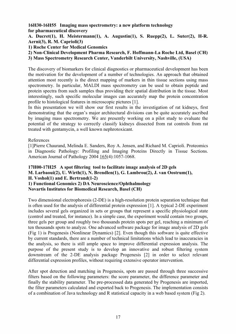
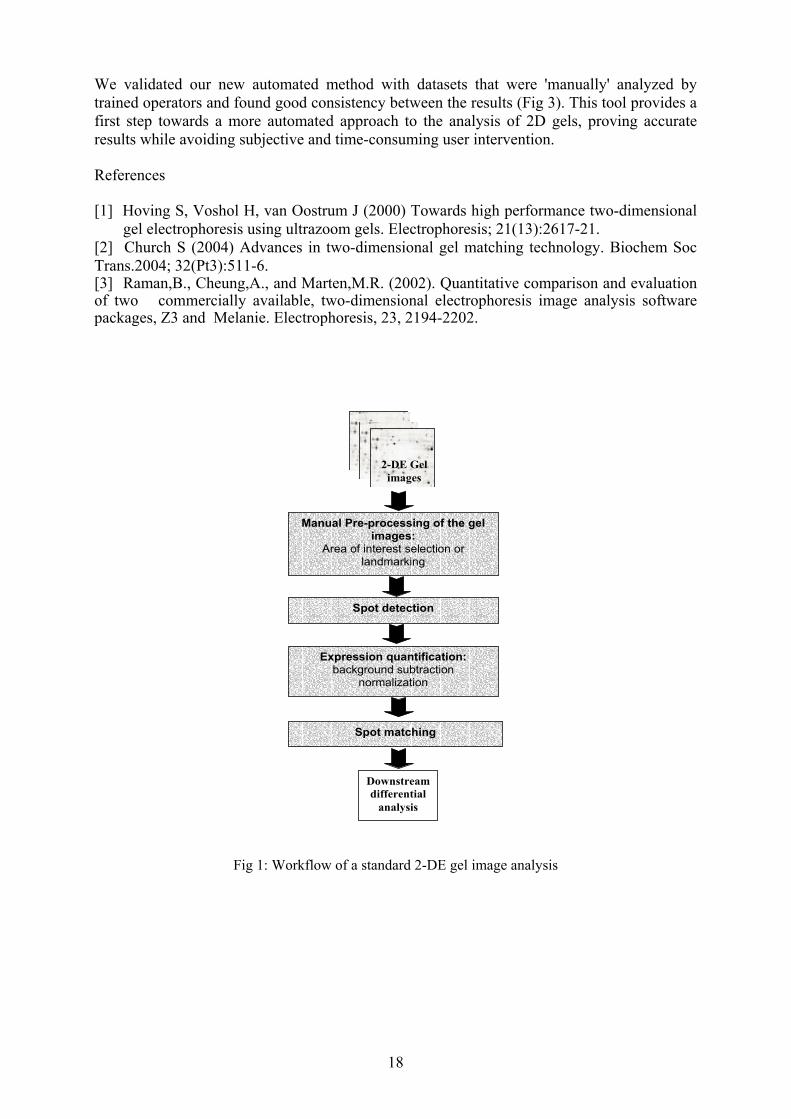
16H30-16H55 Imaging mass spectrometry: a new platform technology for pharmaceutical discovery A. Ducret(1), H. Meistermann(1), A. Augustin(1), S. Ruepp(2), L. Suter(2), H-R. Aerni(3), R. M. Caprioli(3) 1) Roche Center for Medical Genomics 2) Non-Clinical Development Pharma Research, F. Hoffmann-La Roche Ltd, Basel (CH) 3) Mass Spectrometry Research Center, Vanderbilt University, Nashville, (USA) The discovery of biomarkers for clinical diagnostics or pharmaceutical development has been the motivation for the development of a number of technologies. An approach that obtained attention most recently is the direct mapping of markers in thin tissue sections using mass spectrometry. In particular, MALDI mass spectrometry can be used to obtain peptide and protein spectra from such samples thus providing their spatial distribution in the tissue. Most interestingly, such specific molecular images can accurately map the protein concentration profile to histological features in microscopic pictures [1]. In this presentation we will show our first results in the investigation of rat kidneys, first demonstrating that the organ’s major architectural divisions can be quite accurately ascribed by imaging mass spectrometry. We are presently working on a pilot study to evaluate the potential of the strategy to correctly classify kidneys dissected from rat controls from rat treated with gentamycin, a well known nephrotoxicant. References [1]Pierre Chaurand, Melinda E. Sanders, Roy A. Jensen, and Richard M. Caprioli. Proteomics in Diagnostic Pathology: Profiling and Imaging Proteins Directly in Tissue Sections. American Journal of Pathology 2004 165(4):1057-1068. 17H00-17H25 A spot filtering tool to facilitate image analysis of 2D gels M. Larbaoui(2), U. Wirth(1), N. Brendlen(1), G. Lambrou(2), J. van Oostrum(1), H. Voshol(1) and E. Bertrand(1-2) 1) Functional Genomics 2) DA Neuroscience/Ophthalmology Novartis Institutes for Biomedical Research, Basel (CH) Two dimensional electrophoresis (2-DE) is a high-resolution protein separation technique that is often used for the analysis of differential protein expression [1]. A typical 2-DE experiment includes several gels organized in sets or groups that represent a specific physiological state (control and treated, for instance). In a simple case, the experiment would contain two groups, three gels per group and roughly two thousands protein spots per gel, reaching a minimum of ten thousands spots to analyze. One advanced software package for image analysis of 2D gels (Fig 1) is Progenesis (Nonlinear Dynamics) [2]. Even though this software is quite effective by current standards, there are a number of technical limitations which lead to inaccuracies in the analysis, so there is still ample space to improve differential expression analysis. The purpose of the present study is to develop an innovative and robust filtering system downstream of the 2-DE analysis package Progenesis [2] in order to select relevant differential expression profiles, without requiring extensive operator intervention. After spot detection and matching in Progenesis, spots are passed through three successive filters based on the following parameters: the score parameter, the difference parameter and finally the stability parameter. The pre-processed data generated by Progenesis are imported, the filter parameters calculated and exported back to Progenesis. The implementation consists of a combination of Java technology and R statistical capacity in a web based system (Fig 2).
18
Manual Pre-processing of the gel images:
Area of interest selection or landmarking
Spot detection
Expression quantification: background subtraction
normalization
Spot matching
2-DE Gel images
Downstream differential
analysis
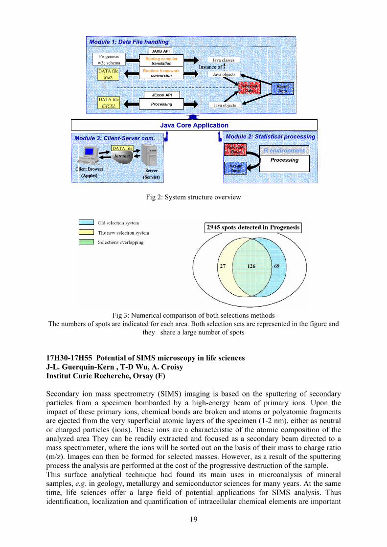
We validated our new automated method with datasets that were 'manually' analyzed by trained operators and found good consistency between the results (Fig 3). This tool provides a first step towards a more automated approach to the analysis of 2D gels, proving accurate results while avoiding subjective and time-consuming user intervention. References [1] Hoving S, Voshol H, van Oostrum J (2000) Towards high performance two-dimensional
gel electrophoresis using ultrazoom gels. Electrophoresis; 21(13):2617-21. [2] Church S (2004) Advances in two-dimensional gel matching technology. Biochem Soc Trans.2004; 32(Pt3):511-6. [3] Raman,B., Cheung,A., and Marten,M.R. (2002). Quantitative comparison and evaluation of two commercially available, two-dimensional electrophoresis image analysis software packages, Z3 and Melanie. Electrophoresis, 23, 2194-2202.
Fig 1: Workflow of a standard 2-DE gel image analysis
19
Fig 2: System structure overview
Fig 3: Numerical comparison of both selections methods The numbers of spots are indicated for each area. Both selection sets are represented in the figure and
they share a large number of spots 17H30-17H55 Potential of SIMS microscopy in life sciences J-L. Guerquin-Kern , T-D Wu, A. Croisy Institut Curie Recherche, Orsay (F) Secondary ion mass spectrometry (SIMS) imaging is based on the sputtering of secondary particles from a specimen bombarded by a high-energy beam of primary ions. Upon the impact of these primary ions, chemical bonds are broken and atoms or polyatomic fragments are ejected from the very superficial atomic layers of the specimen (1-2 nm), either as neutral or charged particles (ions). These ions are a characteristic of the atomic composition of the analyzed area They can be readily extracted and focused as a secondary beam directed to a mass spectrometer, where the ions will be sorted out on the basis of their mass to charge ratio (m/z). Images can then be formed for selected masses. However, as a result of the sputtering process the analysis are performed at the cost of the progressive destruction of the sample. This surface analytical technique had found its main uses in microanalysis of mineral samples, e.g. in geology, metallurgy and semiconductor sciences for many years. At the same time, life sciences offer a large field of potential applications for SIMS analysis. Thus identification, localization and quantification of intracellular chemical elements are important
Module 2: Statistical processingModule 2: Statistical processingModule 2: Statistical processingModule 2: Statistical processing
ProcessingProcessingR environmentR environment
ProcessingProcessingR environmentR environmentRelevant
DataRelevant
Data
ResultData
ResultData
Module 3: Client-Server com.Module 3: Client-Server com.Module 3: Client-Server com.Module 3: Client-Server com.
Client Browser(Applet)
Client Browser(Applet)
Server(Servlet)
Server(Servlet)
IntranetIntranet
DATA fileDATA file
Java Core ApplicationJava Core ApplicationJava Core ApplicationJava Core Application
Module 1: Data File handlingModule 1: Data File handling
Binding compilertranslation
Binding compilertranslation
Runtime frameworkconversion
Runtime frameworkconversion
Progenesisw3c schema Java classes
Instance ofDATA file
XML
DATA fileEXCEL
Java objects
Java objects
ResultData
ResultData
Relevant Data
Relevant Data
JExcel APIJExcel API
ProcessingProcessing
JAXB APIJAXB API
Module 1: Data File handlingModule 1: Data File handlingModule 1: Data File handlingModule 1: Data File handling
Binding compilertranslation
Binding compilertranslation
Runtime frameworkconversion
Runtime frameworkconversion
Progenesisw3c schema Java classes
Instance ofInstance ofDATA file
XML
DATA fileEXCEL
Java objects
Java objects
ResultData
ResultData
Relevant Data
Relevant Data
JExcel APIJExcel API
ProcessingProcessing
JExcel APIJExcel API
ProcessingProcessing
JAXB APIJAXB APIJAXB APIJAXB API

20
questions in many areas of biological research, especially in pharmaco-toxicology to understand mechanisms by which drugs are interfering with living process. Furthermore, this technique is the only micro-analytical method allowing detection of most of the elements and their isotopes, as well as the mapping at the surface of a sample without any specific labeling probe (fluorescence or radioactive). The main characteristics of the last generation of dynamic SIMS microprobe (CAMECA NanoSims 50 [l]) are: i) a high lateral resolution (up to 50nm), ii) ability to measure up to 5 masses (ions) in parallel, issuing from the same micro volume and ensuring perfect isotopic ratio from the same small volume and perfect image superimposition iii) very good transmission even at high mass resolution. These performances enable to analyze sub-cellular structures and chemical heterogeneousness of biological samples at the same time.
At the Institut Curie, the SIMS imaging facility is devoted to research on biological specimens and is opened to the biological scientific community, mainly engaged in cancer research. Thus, several projects have been identified and covered three main areas of application: i) antitumour pharmacology, ii) biomineralization, iii) nuclear medicine and radiotoxicology The principles of SIMS imaging, as well as the sample preparation, that is of a paramount importance in microanalysis to preserve both structural and chemical distribution integrity [2] of the biological samples will be reminded and illustrated by several examples. Experience of correlative imaging using, on the same sample, TEM for the high lateral resolution (Fig. 1) and SIMS microprobe for the microanalysis sensitivity (Fig.2) will be also presented.
Figure 1: TEM image of micro calcification in MDCK cells. Calcifications appear as agglomerates of hydroxyapatite. The same section was analyzed afterwards by Sims imaging (Fig. 2); (image from Dr H.Vali, McGill University, Montreal, Canada).
21
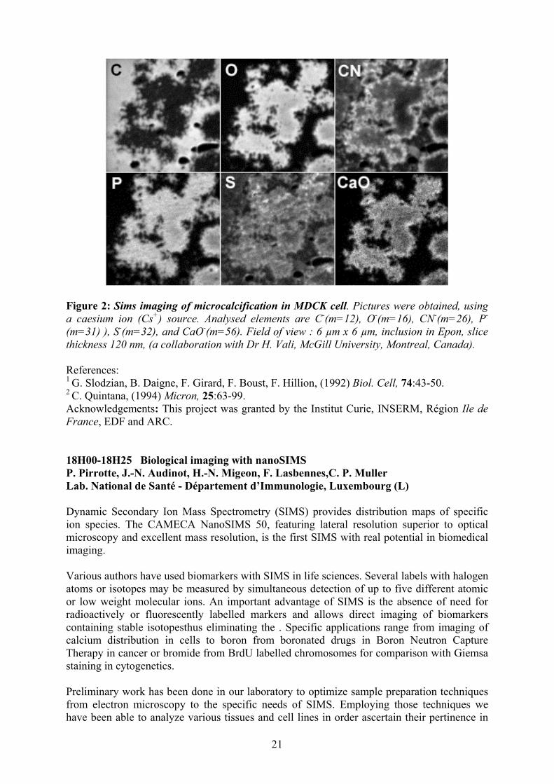
Figure 2: Sims imaging of microcalcification in MDCK cell. Pictures were obtained, using a caesium ion (Cs+) source. Analysed elements are C-(m=12), O-(m=16), CN-(m=26), P-
(m=31) ), S-(m=32), and CaO-(m=56). Field of view : 6 µm x 6 µm, inclusion in Epon, slice thickness 120 nm, (a collaboration with Dr H. Vali, McGill University, Montreal, Canada). References: 1 G. Slodzian, B. Daigne, F. Girard, F. Boust, F. Hillion, (1992) Biol. Cell, 74:43-50. 2 C. Quintana, (1994) Micron, 25:63-99. Acknowledgements: This project was granted by the Institut Curie, INSERM, Région Ile de France, EDF and ARC. 18H00-18H25 Biological imaging with nanoSIMS P. Pirrotte, J.-N. Audinot, H.-N. Migeon, F. Lasbennes,C. P. Muller Lab. National de Santé - Département d’Immunologie, Luxembourg (L) Dynamic Secondary Ion Mass Spectrometry (SIMS) provides distribution maps of specific ion species. The CAMECA NanoSIMS 50, featuring lateral resolution superior to optical microscopy and excellent mass resolution, is the first SIMS with real potential in biomedical imaging. Various authors have used biomarkers with SIMS in life sciences. Several labels with halogen atoms or isotopes may be measured by simultaneous detection of up to five different atomic or low weight molecular ions. An important advantage of SIMS is the absence of need for radioactively or fluorescently labelled markers and allows direct imaging of biomarkers containing stable isotopesthus eliminating the . Specific applications range from imaging of calcium distribution in cells to boron from boronated drugs in Boron Neutron Capture Therapy in cancer or bromide from BrdU labelled chromosomes for comparison with Giemsa staining in cytogenetics. Preliminary work has been done in our laboratory to optimize sample preparation techniques from electron microscopy to the specific needs of SIMS. Employing those techniques we have been able to analyze various tissues and cell lines in order ascertain their pertinence in
22
future studies. Our goal is to establish specific protocols to use NanoSIMS in conjunction with biological probes to investigate cellular processes in immunology. First studies have shown that the matrix effect due to the sample had a variable effect on the ion yield and thus on the distribution maps we obtained and calls for improvement in ionisation yields for biologically relevant ions. 19H30 Cocktail reception sponsored by Communauté Urbaine de Strasbourg and hosted by Mme Fabienne Keller, Mayor of Strasbourg, at the Hôtel de Ville, Place Broglie, Strasbourg March 2 2005 Conference chairs: G. Brun, University of Saint-Etienne (F) and A. Constantinesco, CHU Hautepierre, Strasbourg (F) 8H30-8H55 A fluorescence and diffuse optical tomographic system for small animal imaging P. Poulet, R. Chabrier et B. Montcel UMR 7004 Université Louis Pasteur / CNRS, Institut de Physique Biologique, Strasbourg (F) Various optical approaches have been used for imaging fluorescence in vivo: from confocal and multiphoton microscopy to observe markers close from the surface to photographic macroscopic systems to sense deeper tissues. We developed a tomographic approach that uses diffuse near infrared photons for imaging the optical properties of tissues and the fluorescent probes distribution. This method should improve the spatial resolution and the quantification of fluorescence signals, thanks to multiple projections acquisition and to a reconstruction procedure using the principles of diffuse optical tomography. The scanner assembled uses picosecond laser diodes, an eight-anode photo-multiplier tube (PMT) and time-correlated single photon counting. Two sets of four laser heads, at four wavelengths, are fitted with furcated optical fibers, providing two sequential sources of light. Eight multimode optical fibers were used to detect light. These fibers were connected to the PMT, with an air-gap allowing the insertion of an optical filter to reject the excitation wavelengths. The light sources and detectors can be rotated to increase the number of recorded projections. An interferometry technique using a conoscope and a XY scanning system records the coordinates of the body surface, required for the reconstruction process, before entering the animal in the scanner. Excitation profiles are used for the computation of the absorption and reduced scattering images of the animal. Fluorescence images, free from diffusion and absorption artefacts, are then computed with the knowledge of the optical properties of the animal. The scanner, its performances and previous images of light scattering and fluorescent phantoms will be presented. This work was supported by the Hôpitaux Universitaires de Strasbourg, the Conseil Régional d'Alsace and the Ministère Français de la Recherche (ACI "Technologies pour la santé"). 9H00-9H25 Mouse SPEC cardiac imaging A. Constantinesco(1), L. Monassier(2), P. Choquet(1), L. El Fertak(3) 1) Service de Biophysique et Médecine Nucléaire, CHU Hautepierre, Strasbourg (F) 2) Lab. de Neurobiologie et Pharmacologie Cardiovasculaire, INSERM E333, Faculté de Médecine, Strasbourg (F) 3) Institut Clinique de la Souris, Illkirch (F)

23
Due to breeding consideration and transgenic capabilities, mouse represent a major model of various diseases in biomedical research. Moreover the development of adapted devices allows now for anatomic and functional in vivo molecular imaging studies. In particular, due to impact of cardiovascular ischemic diseases, heart is a target of major interest. To that respect, myocardial tissue perfusion but also left ventricular volumes and motion are key functional data that need to be measured. Using a small animal dedicated micro SPECT (Single Photon Emission Computed Tomography) imager and ECG triggering, we demonstrated, after careful calibration of the imager, that good temporal (10 time bins per cardiac cycle) and sub millimetric reconstructed left ventricular images of the normal anesthetized (isoflurane 2%) mice (CD1) are achievable with 99mTc–Tetrofosmin as perfusion tracer. Quantification of segmented wall perfusion distribution, left ventricular ejection fraction, end systolic and en diastolic volumes as well as wall thickening were obtained in a normal mice series in order to constitute a mandatory reference database before using the method in cardiac diseased mice models. Gated micro SPECT perfusion imaging was then applied to quantitate in vivo myocardial ischemia in mice. 9H30-9H55 Detection of motor cortex activation using time-resolved diffuse optical methods B. Montcel, R. Chabrier, P. Poulet UMR 7004 Université Louis Pasteur / CNRS, Institut de Physique Biologique, Strasbourg (F) It has been demonstrated that local variations in brain perfusion and oxygenation associated with cortical activation can be measured through the skull by near-infrared (NIR) spectroscopy. One of the major drawbacks of continuous NIR spectroscopy is that one cannot differentiate superficial and deeper variations in the optical parameters. Time-resolved methods could overcome this problem and even open the way to a functional imaging of these parameters. The finite element method (FEM) was used to solve the diffusion equation and to compute photon density and transport in a head model, whose optical map was based on the segmentation of a magnetic resonance imaging scan. The simulations were used in order to retrieve information about the depth at which variations in perfusion take place and to improve the detection of cortical activation by means of time-resolved optical techniques. We also performed experimental verifications with our set-up assembled with laser diodes at several wavelengths, a multi-anode micro-channel plate – photo-multiplier tube and time-correlated single photon counting. The adequacy between simulations and experimental data on activation of the motor cortex demonstrates that it is possible to differentiate activation in the motor cortex from a superficial event produced by a Valsalva manoeuvre and to improve the sensitivity of detecting a cortical activation if we take into account only the photons at the adapted detection time. This work was supported by the Hôpitaux Universitaires de Strasbourg, the Conseil Régional d'Alsace and the Ministère Français de la Recherche (ACI "Technologies pour la santé"). 10H00-10H30 Coffee break 10H30-10H55 Evaluation of hepatobiliary function in mice using pinhole single photon planar scintigraphy and micro-CT C. Goetz(1), Ph. Choquet(1), L. El Fertak(2), I. Slim(1), M. Claria(1), I.J. Namer(1), J. Auwerxs(2), A. Constantinesco(1)

24
1) Service de Biophysique et Médecine Nucléaire, Hôpitaux Universitaires de Strasbourg, CHU Hautpierre, Strasbourg (F) 2) Institut Clinique de la Souris, Illkirch (F) Hepatobiliary function could be assessed by scintigraphy, using convenient tracers. Normal data for mice are lacking and the development of trangenic models require the knowledge of these values. Recently new contrast agents have been developped aimed at hepatobiliary function analysis with micro-CT. The aim of this study is to obtain normal data in mice as well as comparing the two modalities eg scintigraphy and micro-CT. A dedicated small animal gamma camera equipped with 1 to 1.5 mm pinhole was used (Gaede Medizinsysteme Gmbh, Freiburg, Germany). The Tc99m labeled trimethylbromoimino-diacetic acid (tBIDA) tracer (Cholecis, CIS-Bio International, Gif/Yvette, France) was injected intravenously using a femoral catheter to normal adult mice under gaseous anesthesia. After administration, sequential anterior abdominal images were obtained at a rate of 1 image/minute during 40 minutes. Micro-CT (eXplore Locus, General Electric Healthcare, London, Canada) images of the same animals were also recorded using a long-lasting iodinated intravenous hepatobiliary imaging agent contained in lipophilic cores of oil-in-water lipid emulsions similar to the naturally-occurring chylomicron remnants (FenestraTM, Alerion Biomedical, Inc.). Acquisition were done at ten to twenty minutes intervals. Normal hepatobiliary transit times were measured using these two techniques. Micro-CT data provide exquisite anatomical visualisation while scintigraphy leads to more robust values. Use of both techniques on pathological hepatobiliary function mice models is to be evaluated from these preliminary results. 11H00-11H25 Quantification of global cerebral blood flow in rats assessed by pinhole Single Photon Emission Computed Tomography (SPECT). Anatomical registration with micro X-Ray Computed Tomography (microCT) Ph. Choquet(1), P. Bilbault(2), I.J. Namer(1), V. Israël-Jost(1), I. Slim(1), M. Claria(1), F. Schneider(2), A. Constantinesco(1) 1) Service de Biophysique et Médecine Nucléaire, Hôpitaux Universitaires de Strasbourg, CHU Hautpierre, Strasbourg (F) 2) Service de Réanimation, Hôpitaux Universitaires de Strasbourg, CHU Hautpierre, Strasbourg (F) Longitudinal follow-up of small animal models of brain diseases suppose the ability of obtaining quantitative values of the data of interest. This work reports the values of gCBF obtained in normal rats measured with [99mTc]-radiolabeled pharmaceuticals using pinhole SPECT. A dedicated small animal SPECT camera (field of view 170 mm x 170 mm, 14 cm focal distance) was used with 1.5 mm pinhole collimator (Gaede Medizinsysteme Gmbh, Freiburg, Germany). 500 to 740 MBq of [99mTc]-hexamethyl propylamine oxime (Amersham Health, Little Chalfont, UK), was administered to Whistar adult rats under general gaseous anesthesia (isoflurane 1.5-2%), through intravenous femoral catheter. During tracer administration, 120 planar images of 0.5s were recorded. Ten minutes after, 48 SPECT projections of 1 min were acquired over a 180° dorsal arc. A specific cone beam algebraic reconstruction algorithm, taking into account distorsion of projection through pinhole, was employed leading to reconstructed voxel’s sizes of 0.34 mm3. Micro-CT (eXplore LOCUS, General Electric Healthcare, London, Canada) images of the same animal were fused with SPECT data for anatomical registration. Using the Patlak formalism, modified by Matsuda, we calculate global CBF values for each cerebral hemisphere.

25
Images contrast reflects normal tracer’s uptake in the different cerebral territories. Signal to noise ratios in human and rat brain perfusion‘s images are comparable. Calculation leads to a gCBF of 110.1 ml/100g/min ± 11.4 value in the range of reference data. This technique could be used to assess CBF in pathological conditions in rats.
11H30-11H55 Terahertz spectroscopy and imaging in biological systems G. Gallot Lab. for Optics and Bioscience, CNRS-Ecole Polytechnique, Palaiseau (F) Terahertz spectroscopy Structural dynamics of proteins are essential to biological functions, since protein flexibility determines enzymatic reaction rates and signal transduction cycles. Seven-helix receptors play key roles in sensory and hormonal transduction processes, as vision (rhodopsin), the adenylate cyclase cascade (serotonin receptors), and olfaction or chemotaxis receptors. There are made of seven-helix transmembrane domains and are most often associated with G proteins. Binding of specific ligand to the seven-helix receptor induces conformational changes that are transmitted to loops on the cytosolic side of the membrane. A given conformational change can be decomposed into collective vibrational modes, which are distinct from the familiar mid-infrared or infrared vibrational modes, which in general involve the motion of small groups of molecules [1]. Conformational modes involve the collective motion of entire subunits of the protein with hundreds of atoms moving in concert. These modes lay in the far-infrared (terahertz range) with frequencies between 1 and 200 cm-1 [2]. Conformational flexibility can then be quantified in terms of the density and spectrum of these low-frequency collective vibrational modes. Terahertz radiation range belongs between two well known spectral areas: microwave and infrared optics. 1 Thz corresponds to 300 µm, 33 cm-1 and 4 meV, then below room temperature thermal noise. The region we are interested in covers the range 1-200 cm-1, corresponding to absorption spectral zones of optical phonons in solids and collective vibrational modes of macromolecules like proteins. Among the terahertz techniques, we focused on short pulses systems based on photoconductive antennas. Applications in biology are becoming more numerous, but are still limited to static spectroscopy [3]. Terahertz imaging Numerous applications are related to terahertz imaging [4], on cancerous tissues [5] or for dental scanning [6]. The group of H. Kurtz [7] works on developing label-free DNA genetic diagnostics. However, spatial resolution of conventional terahertz imaging is limited by diffraction, which leads to best resolution of about 500 µm. It is physically possible to overcome this limitation using near field properties, as demonstrated in Scanning Near-field Optical Microscopy (SNOM). We are working on implementing near field techniques in the terahertz range, where micron resolution has already been demonstrated. We also demonstrated the possibility to work with liquid ionic samples, which opens up the field of biological systems. References [1] B. C. Dian, A. Longarte & T. S. Zwier, Science 296, 2369 (2002) [2] C. L. Brooks, Karplus, M. Pettitt, B. M., A theoretical perspective of dynamics, structure, and thermodynamics. 1988, John Wiley & Sons, New York. [3] A. G. Markelz, A. Roitberg & E. J. Heilweil, Chem. Phys. Lett. 320, 42 (2000) [4] P. Y. Han, G. C. Cho & X.-C. Zhang, Opt. Lett. 25, 242 (2000) [5] R. W. Woodward, V. P. Wallace, R. J. Rye et al., J. Invest. Dermatology 120, 72 (2003) [6] D. Crawley, C. Longbottom, V.P. Wallace et al., J. Biomed. Opt. 8, 303 (2003)
26
[7] M. Brucherseifer, M. Nagel, P. Haring Bolivar, H. Kurz, A. Bosserhoff & R. Büttner, Appl. Phys. Lett. 77, 4049 (2000) 12H00-12H20 Companies snapshots 12H20-13H30 Lunch Conference chairs: A. Dubois, ESPCI, Paris and M. Przybylski, University of Konstanz (D) 13H30-13H55 THz spectroscopy and bioapplications J. Demaison Lab. de Physique des Lasers, Atomes, et Molécules, UMR CNRS 8523, Université des Sciences et Technologies de Lille, Villeneuve d'Ascq (F) Submillimeterwave (or Terahertz) spectroscopy which was born at the end of world war II is the direct consequence of radar research. First, it was a very complicated technique with a very restricted frequency range (mainly microwave). Moreover, the analysis of the spectra was time consuming and required a lot of expertise. That's why it's field of applications remained limited to fundamental research in physical chemistry for a long time. Yet, a lot of progress has be done (larger range, particularly towards high frequencies, better sensitivity, automatization, …) and very performing software make the analysis of the spectra far easier. After the history and the presentation of this spectroscopy, two types of applications will be particularly detailed: 1. Quantitative detection of gases The main use is the detection of gases under difficult conditions. For example, to detect interstellar molecules (it's possible over long distances) or chemical-warfare agents (reliable, automatic and fast). But it can also be easily used for the detection of industrial or domestic pollutants as well as to analyze breath. 2. Study of biomolecules It's the best method to determine the shape of molecules accurately which permits to infer some of their biological properties. It also enables the study of the influence of the solvatation on these properties as well as the intermolecular bonds (hydrogen bond, Van der Waals bond) which play a fundamental part in biology. 14H00-14H25 Temporal holography used for high resolution, real time Optical tomography G. Brun, M. Jacquot, I. Verrier, D. Reolon, C. Veillas LTSI Lab. Traitement du Signal et Instrumentation / UMR CNRS 5516, University Jean Monnet, Saint-Etienne (F) In the field of interaction between photonics sciences and life sciences, non invasive optical methods are interesting to explore and analyze biological tissues. For example, low coherence interferometry offers today an increasing interest to image through turbid media and characterize complex structures. Among various devices, Optical Coherent Tomography has been demonstrated as a good process for imaging tissue morphology through scattering biological media. This technique is based on interferometric device and brings to the inter-correlation between a short reference pulse and the signal issued from the medium. This correlation is obtained by mechanical length modulation of the

27
interferometer reference arm. We propose an original technique using an optical field correlator, which allows to obtain directly, and without length modulation, the inter-correlation signal between the reference and the tests waves. With a large spectral bandwidth light source, the temporal depth of the original pulse is short compared to the signal diffused in the complex medium, and the inter-correlation function may be reduced to the impulse response of the structure to be studied. This temporal analysis could be very interesting to obtain both amplitude and phase parameters on the waves which have been propagated in the medium, and could induce significant data on the medium and its structure. By coupling microscopic device, interferometric correlator and white light source obtained, in a fiber by supercontinuum generation, it is possible to realize directly high resolution optical tomography. We will discuss about efficiency of this method in terms of time measurement, accuracy and of ability to image complex structures and media. References 1. ZEYLIKOVITCH AND R.R.ALFANO, OPT.COMMUN., 135, 217, (1997) 2. R. JONES, M. TZIRAKI, P.M.W. FRENCH, K. KWOLEK, D.D. NOLTE AND M.R. MELLOCH, OPT.EXP., 2, 439-448, (1998) 3. B.E. BOUMA AND G.J. TEARNEY, HANDBOOK OF OPTICAL COHERENCE TOMOGRAPHY, NEW YORK: MARCEL DEKKER, (2002). 4. K. BEN HOUCINE G. BRUN, I. VERRIER, L. FROEHLY AND C. VEILLAS, OPT.LETT., 26, 1969-1971, (2001). 5. K. BEN HOUCINE, G. BRUN, I. VERRIER, M. JACQUOT, C. VEILLAS , OPT.LETT., 26 , 1969 -1971, (2004) 6. G. BRUN, K. BEN HOUCINE, M. JACQUOT, I. VERRIER, C. VEILLAS, EDITIONS FONTIS MEDIA,(2004) 7. K. BEN HOUCINE, G. BRUN, I. VERRIER, C. VEILLAS , EDITIONS HERMES, COLLECTION « SYSTEMES ET MICRO-SYSTEMES POUR LA CARACTERISATION », VOL. 2, P. 75-83, 2001 8. J.K. RANKA, R.S. WINDELER, A.J. STENTZ, OPT. LETT. 25, 25-27 (2000) 9. J.M. DUDLEY, L. PROVINO, N. GROSSARD, H. MAILLOTTE, R.S. WINDELER, B.J. EGGLETON, S. COEN, J. OF OPTICAL SOCIETY OF AMERICA B 19 (2002) 765-771. 14H30-14H55 Full-field optical coherence tomography A. Dubois Lab. d’Optique Physique,Ecole Supérieure de Physique et Chimie Industrielles, CNRS, UPR A0005, Paris (F) Optical coherence tomography (OCT) is an efficient technique for imaging of biological media with micrometer-scale resolution. OCT is based on a fiber Michelson interferometer illuminated by a broad-bandwidth laser. We have developed an alternative OCT technique based on an interference microscope illuminated by a halogen light source using a CCD as detector array. An unprecedented spatial resolution (isotropic) of ~1.0 µm is achieved. Image averaging and pixels binning lead to a detection sensitivity of ~ 90 dB with an acquisition time per image of ~ 1 s. The system is applied to cellular-level imaging of various biological tissues. 15H00-15H25 Emerging ultrasound contrast functional imaging techniques S. Lori Bridal, J.M. Correas, O.Lucidarme, A. Ammi, E. Jouanot, P.Laugier Lab.d’imagerie paramétrique-CNRS - Université de Paris 6, Paris (F)
28
Ultrasound contrast agents (USCAs), consisting of strongly reflecting stabilized gas microbubbles (~ 2 to 7 µm diameter) injected in solution intravenously, improve image quality by increasing the intensity of backscattered echoes from blood filled regions or vascularized tissues. The principle of functional imaging modes using USCA as blood pool tracers will be described and illustrated with clinical examples. Development and optimization of these techniques for clinical application require understanding of microbubble acoustic response and effects due to the destruction of the agent by the ultrasonic field. Experimental results comparing functional contrast imaging to reference techniques and results characterizing destruction thresholds of contrast agent will be presented and discussed in light of their implications for functional contrast ultrasonography. 15H30-16H00 Coffee break - Exhibition - Posters 16H00-16H25 Transient states energy by femtosecond laser spectroscopy. Innovating advances for life chemistry Y. Gauduel(1), V. Malka(1), T Launay(2), F. Guilloud(2), B. Charles(2) 1) L.O.A., CNRS UMR 7639, Ecole Polytechnique - ENS Techniques Avancées, Palaiseau (F) 2) ENSEA, Cergy Pontoise (F) Numerous life science processes involve transient electronic and radical states whose the spatial and temporal characterization represent real challenge for physical chemistry of medical interest. Transient states imaging obtained by femtosecond laser spectroscopy need numerical treatment of experimental data matrix. Those are obtained from cooled CCD detector at –120°C. Transient imaging is developed with advanced graphical methods using OPGS (Open Graphic System). In this lecture, we present advanced numerical 2 and 3D data obtained with amplified femtosecond pulses whose the full widh half magnitude (FWHM) is less than 80 fs (80 x 10-15 s) in the frequency range 27800 – 10400 cm-1. Experimental results concern the real time electron detachment from an atom in aqueous environment and a two-centre-three electron sulphur-sulphur bond making. These works represent a first step for the selective control of biomedical radical events : chlorination of amino acids during inflammatory processes, molecular repairing following an oxidative stress. Concerning ionizing radiations applied to radiobiology, an innovating research concerns the imaging obtained with sub-picosecond relativistic electron bunches in the energy range 3-15 MeV. Numerical treatments of near-infrared signals obtained with a CCD 16 bits camera (Andor technology) open the way to the microdosimetry in the micrometric scale. The parametric analysis of 3D imaging would provide guidance for the investigation of radiation-induced primary radical events in confined environments such as aqueous groove of DNA or sub-cellular media. 16H30-16H55 Real-time mapping of intra-protein electric fields through absorption spectroscopy of tryptophans S. Haacke Institut de Physique et Chimie des Matériaux de Strasbourg (IPCMS), GONLO, Strasbourg (F) Tryptophans (Trp) are known to show strong solvatochromism and evironment dependent fluorescence dynamics. They are therefore ideal, natural probes for resolving intra-protein dynamics on time scales as short as femtoseconds. We have demonstrated this potential using
29
bacteriorhodopsin as model system. Optical excitation of the retinal moiety induces an ultrafast (< 5fs) charge translocation. This leads to an instantaneous bleach signal of specific Trp residues, as we demonstrate by simulating the excitonic coupling of the relevant chromophores. The subsequent dynamics of this signal provides new information about the interplay between the retinal dipole moment change and isomerization dynamics. The results indicate that isomerization is accelerated by electrostatic retinal-protein interactions. We will discuss the applicability of this approach for real-time mapping of photo-induced electric fields in other proteins. 17H00-17H25 Elucidation of antibody- paratopes by combination of affinity proteomics and high resolution Ft-icr mass spectrometry M. Przybylski*, E. Amstalden, A. Marquardt, X. Tian, R. Iacob, R. Stefanescu, E. Damoc Department of Chemistry, Laboratory of Analytical Chemistry, University of Konstanz, Konstanz (D) Invited High resolution Fourier transform ion cyclotron resonance mass spectrometry (FTICR-MS) has been recently developed as a powerful tool in proteomics with unrivalled accuracy. Recent work in our laboratory has focussed on high resolution and high selectivity MS approaches in proteomics, and for identification of antibody recognition structures, as a key pre-requisite for vaccine design and targeting. Selective proteolytic digestion and MS-peptide mapping (epitope excision) has been successfully employed for epitope identification of protein antigens and in proteomics; in addition, "affinity-proteomics" using partial epitope excision is a new approach with unprecedented selectivity for protein identification from biological material. The potential of these methods has been demonstrated by identification of antigen epitopes, development of new diagnostic procedures, and the elucidation of an Aß-plaque-specific epitope recognised by therapeutic antisera from transgenic Alzheimer's disease mice. Using this epitope in an antigen-affinity column and antibody-proteomics by FTICR-MS directly provided the identification of paratope structures within the variable heavy- and light-chain fragments, owing to the high (sub-ppm) mass determination accuracy of FTICR-MS. This direct antibody-proteomics approach and determination of a "molecular paratope signature has broad potential for molecular diagnosis, and for design and evaluation of new vaccine lead structures. References 1. Damoc, E., N. Youhnovski, D. Crettaz, J.D. Tissot, and M. Przybylski (2003) Proteomics 3:1425-1433. 2. Macht, M., A. Marquardt, S.-O. Deininger, E. Damoc, M. Kohlmann, and M. Przybylski (2003) Anal. Bioanal. Chem., 378: 1102-1111. 3. McLaurin, J., R. Cecal, M.E. Kierstead, X. Tian, A.L. Phinney, M. Manea, J.E. French, M.H.L. Lambermon, A.A. Darabie, M.E. Brown, C. Janus, M.A. Chishti, P. Horne, D. Westaway, P.E. Fraser, H.T.J. Mount, M. Przybylski, and P. St.-George-Hyslop (2002) Nature Med. 8: 1263-1269. 4. Przybylski, M., E. Amstalden, E. Damoc, A. Marquardt, R. Iacob, R. Stefanescu, and X. Tian (2004) Nature Meth., submitted. 17H30-17H55 A state of the art of augmented reality systems for application in surgery:terminology and taxonomy G. Sittler(1) , P. Twardowski(1), T. Blandet(1), J.-B. Fasquel(2), J. Fontaine(1) 1) LSP :Lab. des Systèmes Photoniques, Université Louis Pasteur Strasbourg (F) 2) IRCAD :Institut de Recherche contre les Cancers de l’Appareil Digestif, Strasbourg(F)
30
Augmented reality systems take more and more importance in varied contexts. Fields like automotive industry, aeronautic conception, or military applications are actually using the capabilities of such devices. Other fields of researches, like medicine, and more especially surgery, are appropriate for the development of that kind of techniques. In fact, it appears more and more crucial to perform accurate tools for difficult surgical operations and for the formation of surgeons. Based on a state of the art, it appears that augmented reality systems present a real interest, as far as they provide a superimposition of virtual elements onto a real environment. As part of our project, it is relevant to focus on the optical conception of an augmented reality system based on the previous works done at the LSP on virtual reality and 3D display systems. In fact, some works have also been performed at the IRCAD on image matching techniques and 3D body reconstruction. But a system providing a superimposition of these simulations on a surgical environment is still unsatisfactory. This clearly justifies the need of such a device with enhanced features to simplify the work of the surgeon and add some convenience compared to classical endoscopes for laparoscopic surgery. As far as the researches are not starting from scratch, it is important to perform an as complete as possible state of the art on the different augmented reality systems available on the market and those who have been studied by different research teams. We will be considering the systems designed for virtual imaging and those dedicated to human-machine interaction. Then, the different applications will be listed and the research potential of each of these fields will be clarified. A presentation of the different architectures of augmented reality systems will be exposed, thus giving a more precise view of the surgical device that will be developed during the project. This will lead to the presentation of the originalities of the proposed system, as far as, for example, it appears that none of the devices studied proposes a variable focal length unit, which will permit the displaying of the virtual images in the accommodation plane of the surgeon’s sight. After this state of the art, we will present some aspects of terminology and continue with an accurate taxonomy. This will clarify the context of our project and give an idea of the different specifications needed. Some derived notions of the terms of augmented reality, like mediated reality or persuasive computing, will be exposed and explained precisely. The presentation will be concluded by giving a listing of the different functions and some hints on the optical design that will lead to an augmented reality helmet prototype for surgical application. 18H00-18H30 Oral posters presentation in the conference room 19H30 Reception and Gala dinner with Awards and Animation close to the symposium location at “La Croisée des Idées”
31
March 3, 2005 Conference chairs: P. Poulet, Institut de Physique Biologique, Strasbourg (F) and L. Soler, IRCAD, Strasbourg (F) 8H30-8H55 Image processing by infrared thermography D. Pajani Institut de Thermographie, Verrières-le-Buisson (F) This work has as an aim the image processing of an infrared film. This processing is related to the temperature measurement. Many difficulties encountered at the time of measurement by infrared thermography were underlined by Pajani [1] and Balageas and al. [2]. The conversion of the digital levels (DL) provides by the camera in temperature rises from a formalism resulting from the law from Planck. This relation was stated by Pajani [3] and was supplemented by Arconada and al. [4]. Our object is to propose a simpler method for the temperature measurement by taking into account another parameter the distance camera-object. Thus, for a range of temperature or a given configuration of the camera, one presents the response of the apparatus by a law in T4
(where T is the temperature of the object) in the form : bTaDL += 4 . The parameters a and b depend on the distance x and arise in the form : 1axaa o += and 1bb = . The model suggested in this work gives similar results to that proposed in the literature by using a number of lower parameters. The originality of this work is to take into explicit account of manner the distance camera-object for an image processing. References : [1] D. Pajani, Mesures de températures par caméra infrarouge quelques précautions à prendre, Techniques d’applications MESURES, 35-41, (octobre 1984). [2] D. Balageas – A. Deom – D. Boscher, La mesure de température par camera infrarouge, journée d’étude “mesure des températures”, (164), 61-74, Paris, 19-20 (juin 1990). [3] D. Pajani, Thermographie infrarouge quel longueur d’onde choisir 1 ?, Techniques d’applications MESURES, vol 52, 71-76, (mai 1987). [4] A. Arconada - A. Argiriou - F. Papini – R. Pasquetti, la mesure en thermographie infrarouge : calibration et traitement du signal, journal of modern optics, vol 34, (10), 1327-1335, (1987). 9H00-9H25 Improving the 2-D resolution in fluorescence microscopy by non rotationally symmetric apodization and image recombination O. Haeberlé, B. Simon Lab. MIPS, Université de Haute - Alsace, Mulhouse (F) The resolution in optical microscopy is limited by the diffraction effect [1]. In order to get the best resolution, the use of the largest numerical aperture objective is mandatory. To this date, the objective with the highest the available numerical aperture has been developed by Olympus for Total Internal Reflection Microscopy [2]. This special objective with NA=1.65 requires a special immersion oil with noil=1.78 and special coverslips with nglass=1.788. We consider objects, which can be assimilated as 2-D or pseudo 2-D, like for example chromosomes in the metaphase state. These specimens not being living, one can embed them in an appropriate medium with an index of refraction closely matching that of the immersion medium. We first study this special objective in a confocal configuration with the Cascade Blue dye, which has an excitation maximum at λ���=400 nm and emits near λdet=450 nm.
32
To compute the illumination PSFill, we use the vectorial theory of Török and Varga [3], modified by Haeberlé [4]. The detection PSFdet is computed using the vectorial dipole model of Haeberlé et al.[5,6]. The resolution is 103 nm, which is a 25 % improvement compared to a confocal microscope using a NA=1.4 objective imaging in a watery medium (130 nm). In order to further improve the lateral resolution, we propose to use a modified illumination PSFill, by inserting in the illumination arm a phase plate, which reverts the sign of the amplitude of the illumination wave in one-half of the entrance pupil [7]. The obtained illumination PSFill exhibits two lobes, with a Full Width Half Maximum (FWHM) of only 105 nm, much narrower than a standard PSFill from the same objective (135 nm). The two lobes being well separated, it is possible to isolate one of them by confocalization. One obtains a final confocal PSF with a FWHM of only 90 nm. Using one pinhole in fact represents a waste of signal. Because the two lobes are well separated in the imaging plane, it is possible to use two separated pinholes, with two detectors to simultaneously acquire during the scanning process two independent images. Each contains the signal emitted by the same object point, illuminated in one image with the left lobe of the two-lobe PSF, and with the right lobe in the second image, and detected with the left- and the right confocal detector, respectively. These two images can then be recombined by a simple shifting-adding method, which would have for effect to simultaneously improve the signal to noise ratio and to decrease the height of the remaining side-lobe of the confocal PSF relatively to the main peak. References 1. Abbe E., Arch. Mikrosk. Anat. 9, 413-468 (1873) 2. see for example: http://www.olympusamerica.com/seg_section/seg_tirfm.asp 3. P. Török and P. Varga, Appl. Opt. 36, 2305-2312 (1997) 4. O. Haeberlé, Opt. Comm. 216, 55-63 (2003) 5. O. Haeberlé, M. Ammar, H. Furukawa, K. Tenjimbayashi and P. Török, Opt. Exp. 11, 2964-2969 (2003) 6. O. Haeberlé, Opt. Comm. 235, 1-10 (2004) 7. T.A. Klar, E. Engel and S.W. Hell, Phys. Rev. E 64 066613 (2001) 9H30-9H55 Third harmonic generation microscopy for the velocimetric analysis of Drosophila embryo development D. Débarre(1), W. Supatto(2), E. Farge(2), B. Moulia(3), M.-C. Schanne-Klein(1), E. Beaurepaire(1) 1) Lab. for optics and biosciences, CNRS UMR 7645, INSERM U451, Ecole Polytechnique, Palaiseau (F) 2) Mechanics and genetics of developmental embryology, CNRS UMR 168, Curie Institute, Paris (F) 3) Biomechanics group, INRA UMR 547, Clermont-Ferrand (F) During embryo development, cell movements are highly regulated in time and space. Morphogenetic movements involved in Drosophila melanogaster embryo development are of particular interest because Drosophila serves as a major model in developmental genetics, and many mutant strains exhibiting altered movements are available. However, direct imaging of Drosophila development is difficult due to the highly scattering nature of early stage embryos. In addition, fluorescent labelling can introduce unwanted perturbation and might be difficult to obtain in complex mutants. As a consequence, a complete quantitative description of these complex dynamical processes is still lacking in many cases. THG microscopy was recently proposed as a novel technique for obtaining structural images of biological samples with micrometer 3D resolution. By associating this technique with particle image velocimetry (PIV) algorithms adapted from hydrodynamics, we have
33
demonstrated micron-scale quantitative measurements of tissue velocity fields inside unstained opaque embryos. Using a combined 2PEF/THG home-built microscope, we have characterized optical properties of Drosophila embryos. We have provided evidence that sustained THG imaging does not perturb embryo development by following sensitive developmental dynamics and comparing the survival rate of embryos with and without imaging. Finally, we have illustrated velocimetric THG imaging by quantifying morphogenetic movements in live unstained wild-type and mutant embryos. Our results establish third harmonic generation (THG) microscopy as a novel powerful approach for visualizing and quantifying morphogenetic movements in vivo in unstained embryos. Reference Débarre, Supatto and al, Opt. Lett. 29 (2004, in press) 10H00-10H30 Coffee break 10H30-10H55 Multiphoton microscopy of unstained living cardiac and vascular tissue A.-M. Pena(1), T. Boulesteix(1), N. Pagès(2), K. Senni(3),G. Godeau(3), M.-P. Sauviat(1), E. Beaurepaire(1), M.-C. Schanne-Klein(1) 1) Lab. for Optics and Biosciences CNRS-INSERM, Ecole Polytechnique, Palaiseau (F) 2) Louis Pasteur University, Illkirch(F) 3) Paris V University, Montrouge (F) We report novel applications of multiphoton microscopy for the study of intact unlabeled tissue. We used endogenous sources of nonlinear signals and achieved molecular or structural specificity by combining two-photon-excited fluorescence (2PEF) and second harmonic generation (SHG) microscopy using appropriate spectral filters. First, we characterized multiphoton microscopy of intact living vessels: rings were sectioned from rat aorta and carotid arteries and 3D images were recorded ex vivo without any staining. We performed simultaneous detection of 2PEF from elastin laminae and SHG from collagen fibers upon 860 nm excitation. Combined 2PEF/SHG images provide a highly specific, micron scale description of the architecture of these two major components of the vessel wall. We used this methodology to study the effects of lindane (a pesticide) on the artery wall structure and evidenced structural alteration of the vessel morphology. Secondly, we showed that multiphoton microscopy of unstained cardiac myocytes can be used to determine the sarcomere length with sub-resolution accuracy, owing to the remarkable contrast of the SHG signal originating from myosin filaments. We used this technique to measure sarcomere contracture in the presence of saxitoxin, and results were in agreement with mechanical measurements of atrial tissue contracture[1]. These functional applications demonstrate that multiphoton microscopy using intrinsic signals is a promising tool in nanopharmocology of living unlabeled cardiac and vascular tissue. Reference [1] T. Boulesteix, E. Beaurepaire, M.P. Sauviat and M.C. Schanne-Klein, Opt. Lett. 29, 2031 (2004). 11H00-11H25 Intra operative computer assisted surgery using low cost virtual and augmented reality systems L. Soler, S. Nicolau, J. Schmidt, C. Koehl, M. Arenas, D. Mutter, J. Marescaux IRCAD (Research Institute against Digestive Cancer), Strasbourg (F)

34
Medical image processing led to a major improvement of patient care by guiding the surgical gesture. The 3D modelling of patients from their CT-scan or MRI thus allows an improved surgical planning. This preoperative step can be used intra-operatively thanks to the development of Augmented Reality (AR) which consists in superimposing the pre-operative 3D modelling of the patient onto the real intra-operative view of the patient. AR provides surgeons with a view in transparency of their patient and can also guide surgeons thanks to the virtual improvement of their real surgical tools, which are tracked in real-time during the procedure.
We propose here two low cost solutions in order to use intra-operatively the pre-operative 3D modelling of the patient for abdominal surgery. Our first solution consists in using a video mixer providing a merged view between two selected screens. The first screen is the operative live video corresponding either to the scialitic camera, providing an external view of the patient, or to the laparoscopic camera, providing an internal view of the patient. The second screen is a personal computer view allowing to use and to see a live surgical planning using our 3D VSP software (Virtual Surgical Planning), which provides several windows with external and internal views of the patient. This software makes it possible to interactively manipulate and position the virtual patient and virtual surgical instruments in the same position as the real ones. By combining real internal or external views with corresponding virtual ones, the first solution provides an easy interactive and low cost augmented reality view of the surgical procedure. The main limit of this solution being that the registration of virtual and real world is manual, our second solution consists in a real-time fully automatic tracking of the patient and surgical instruments by using specific visual landmarks and two FireWire Cameras connected to a classical Pentium IV personal computer. Thanks to an innovative registration technique and an open source-based tracking system, this second solution provides a real-time Augmented Reality visualisation of the surgical procedure with an accuracy of 2 mm. Moreover, the user interface of this system offers a virtual transparency of any pre-operatively 3D modelled structure from an external view and also from a laparoscopic view. It also offers a new virtual camera allowing to navigate virtually inside the real patient, tracked live during the surgical procedure.
Clinical evaluation of the first system showed clearly that the use of internal and external anatomical landmarks allows to ensure the efficiency of the system even if the registration remains interactive, which is the only real limit. Furthermore, this Interactive Augmented Reality allows to perform a low cost long distance telementoring with augmented reality by requiring the only installation of a simple video-conference system in the OP-room. Pre-clinical evaluation of the second system shows that the limit of the first system interactivity is overcome and that the fully automatic Augmented Reality system provides a 2mm precision for external and laparoscopic views. This second system still non expensive with a global material costs under 10.000 €. Future works will consist in performing the clinical validation of this second system. Then, these results will have to be connected with robotics in order to automate the surgical application, the next step of the surgical revolution of the 21rst century.
The Interactive Augmented Reality system providing a virtual transparency ; external (right) and laparoscopic (left) views of the patien
35
11H30-11H55 Real time registration of 2D and 3D images in percutaneous nephrolithotomies using an Augmented Reality system. A. Osorio, O. Traxer, S. Merran LIMSI – CNRS, Orsay (F) The majority of nephrolithotomies are performed by a percutaneous way, a surgical intervention which has less complications and morbidity. Nowadays the physician quite often has only a standard radiography (KUB or IVP) during the intervention. The main challenge in percutaneous nephrolithotomies is to determine the precise localization of kidneys, renal cavities and calculi in order to identify the best trajectory for the percutaneous puncture. The objective of this presentation is to show the on-line use of a new system of Augmented Reality (AR) merged with a 2D-3D fusion procedure which guides the surgeon in the operating room. We have developed a PC based software for 3D segmentations and instant localization and volume measurement from DICOM images. For this application, renal stones, kidneys and external skin of the body have been reconstructed from CT images. The real-time projection of organs and lesions on the patient in surgical position guides the surgeon towards the target region and helps choosing the best route for percutaneous surgery. The system is improved by the merging in real-time, inside the operating room, of 2D fluoroscopic images with the segmented 3D images. The PCNL surgery is carried out as follows: - off-line reconstruction of the segmented 3D volumes of body skin, iliac crest, 11th-12th ribs, spine, kidney, renal cavities and stones from a standard CT scan examination. - off-line computation of the optimal PCNL trajectory. - projection of the segmented 3D shapes on the patient’s body, in surgery position, applying some geometrical transformations. - interactive aligning of 3D forms with the patient body using control points. The computed trajectory shows in real-time the target region and the best PCNL way. - on-line fusion of the 2D fluoroscopic data with the segmented 3D structures. - comparison of real and computed trajectories and adapting the percutaneous puncture. The real trajectory is then optimized. The device has been validated on phantoms, biological equipment. Later, it has been used in clinical evaluations. Computerized fusion of 2D fluoroscopic images leads to a drastic decrease of radiations for patients and physicians. Furthermore, the overall operating time is decreased by more than 20 %.
12H00-12H20 Companies snapshots 12H20-13H30 Lunch Conference chairs: E. Bertrand, Novartis Institutes for Biomedical Research, Basel (CH) and J.Y. Laval, ESPCI, Paris (F) 13H30-13H55 Mosaicing of video-endoscopic images and evaluationof the resulting cartography R. Miranda-Luna, Y. Hernandez-Mier, W.C.P.M. Blondel, Ch. Daul, D. Wolf CNRS UMR 7039, CRAN (Centre de Recherche en Automatique de Nancy) Vandœuvre-Lès-Nancy (F) This work is related to a mosaicing process of clinical video-endoscopic images. The objective is to construct a 2D-cartography of the internal surface of hollow organs. However,

36
intrinsic characteristics of endoscopic images introduce undesirable information that prevents their direct usage in image processing methods. This paper presents the preprocessing tools employed to adapt the characteristics of endoscopic images to a specifically designed mosaicing algorithm and the methodology implied for a qualitative evaluation. A specific protocol using a 3D-micrometric positioning system was implemented to acquire videoendoscopic images. A high-quality color photography (bladder internal surface) was used as a realistic enough test scene from which, a set of forty images was acquired. Firstly, the non-linear radial distortion is corrected by an algorithm that registers the images of a non-distorted pattern before and after distortion for computing the projective (camera) and polynomial (distortion). Mutual information and stochastic gradient descent were used as similarity measure and optimization method respectively. Secondly, shading correction to avoid lighting heterogeneities and a specific low-pass filter to remove the fiber optic pattern were applied over the set of images. The mutual-information algorithm was then used to obtain the transformation parameters that were applied to build a mosaic (2D-cartography). Finally, the same parameters were applied over a similar sequence of images acquired from a specific reference dot pattern. This approach allowed to compute index parameters for evaluating the quality of the constructed mosaic. Significant results for each process are shown and the evaluation of the algorithm in building such cartography is discussed. 14H00-14H25 3D confined ablation and live embryo imaging with femtosecond laser pulses W. Supatto(1), D. Debarre(2), B. Moulia(3), E. Brouzés(1),J.-L. Martin(2), E. Farge(1), E. Beaurepaire(2) 1) Mechanics and genetics of developmental embryology,CNRS UMR 168, Curie Institute, Paris (F) 2) Lab. for optics and biosciences, CNRS UMR 7645,INSERM U451, Ecole Polytechnique,Palaiseau (F) 3) Biomechanics group, INRA UMR 547, Clermont-Ferrand (F) Embryo development involves a complex choreography of cell movements that are highly regulated both in time and space. The genetic control of morphogenetic movements shaping the embryo continues to be extensively studied, particularly in Drosophila melanogaster, which provides a major model of developmental genetics. On the other hand, we have recently pointed out the influence of mechanical factors in development [1, 2]. We proposed that tissue deformations associated with morphogenetic movements are involved in modulating developmental gene expression during Drosophila gastrulation. Addressing the mechanical regulation of morphogenesis requires to develop original techniques of in vivo investigation. In this context, we show that the combination of femtosecond pulse-induced ablation with nonlinear microcopies appears as a powerful tool for modulating, visualizing and quantifying morphogenetic movements in Drosophila embryos [3]. First, we show that femtosecond laser pulse-induced ablation makes it possible to perform 3D-confined microdissections within developing embryos. Such localized ablations can be used for disrupting the mechanical integrity of tissues inside live embryos and subsequently modulating specific morphogenetic movements during gastrulation. Then, we show that the same laser source can be used to quantitatively analyze native and disrupted morphogenetic movements in vivo in GFP-labelled embryos using two-photon (2PEF) microscopy. Furthermore, this approach can be extended to unlabelled embryo using third harmonic generation (THG) microscopy [4]. This all-optical methodology brings novel insight into the issue of mechano-sensitive gene expression by providing a correlation of cell movements

37
with the pattern of gene expression. More generally, it should lend itself to a wealth of additional applications in developmental biology 14H30-14H55 Toward the understanding of the interpretation errors in medical imaging J.-L. Gerstenmayer(1), V. Hazebroucq(2) 1) CEA, Fontenay aux Roses (F) 2) Assistance Publique-Hôpitaux de Paris and Université Paris-5, Paris (F) Interpreting an image is sometimes a trap. The stakes are of the highest importance when the legal responsibility of the physician, of the operator, or of the manufacturer are examined. An erroneous interpretation, if they are consequences for the human life, is described as error or fault when the human responsibility is clearly concerned, as probability of failure when the instrumentation is in question. In practice, these two causalities are sometimes difficult to disentangle : at the start and at the ending of any experimental measurement, one finds every time the human mind. Thus, it is also of the highest importance to identify the man’s error capability, in order to make progress in prevention, safety and security. In this communication, we do not present the "instrumental filter" (noises, band-width, interfering signals...) which was abundantly treated by the specialists in instrumentation (which we are), nor the question of the ergonomics. On the contrary, our goal is here to identify where are the traps which can mislead the human mind when someone is interpreting an image of a sensor. These errors are due to the vision physiology and to the heuristic treatments of the human brain. This operating mode, which is usually so powerful compared to the sequential and procedural treatment of a computer (the number of blows envisaged in advance by a chess player does not exceed 4 or 5, versus more than 20 for a computer), can mislead even a professional. Now, this question reaches a still increased importance, because the use of neural networks will be generalized for the automatic interpretation of the images. 15H-15H25 Optimal acquisition protocol definition for 3D modelling of small animals abdominal tumours and organs from in vivo micro-CT scan L. Soler, A-B. Osswald, M. Bouhadjar, M. Aprahamian, F. Raul, F. Gossé, D. Mutter, J. Marescaux IRCAD, Strasbourg (F) Medical image processing led to a major improvement of patient care by providing a 3D modelling of patients from their CT-scan or MRI. Such a modelling not only allows to improve diagnosis and therapy planning thanks to a better knowledge of the patient’s anatomy and pathology, but also to improve the follow-up thanks to a 4D vision of tumour evolution. Such an improvement should now be available for fundamental research thanks to the development of micro imaging systems such as micro-CT scan or micro MRI. The main difficulty relies in the fact that an in vivo acquisition protocol on a small animal remains different from the human one. We present here our first results in the optimization of acquisition protocol in order to obtain a 3D modelling of abdominal tumours and organs of small animals from an in vivo micro-CT scanner MICRO Cat II (IMTEK Inc.). This imager provides reconstructed images performed in synchronization with an automated breath tracking allowing to avoid classical breathing
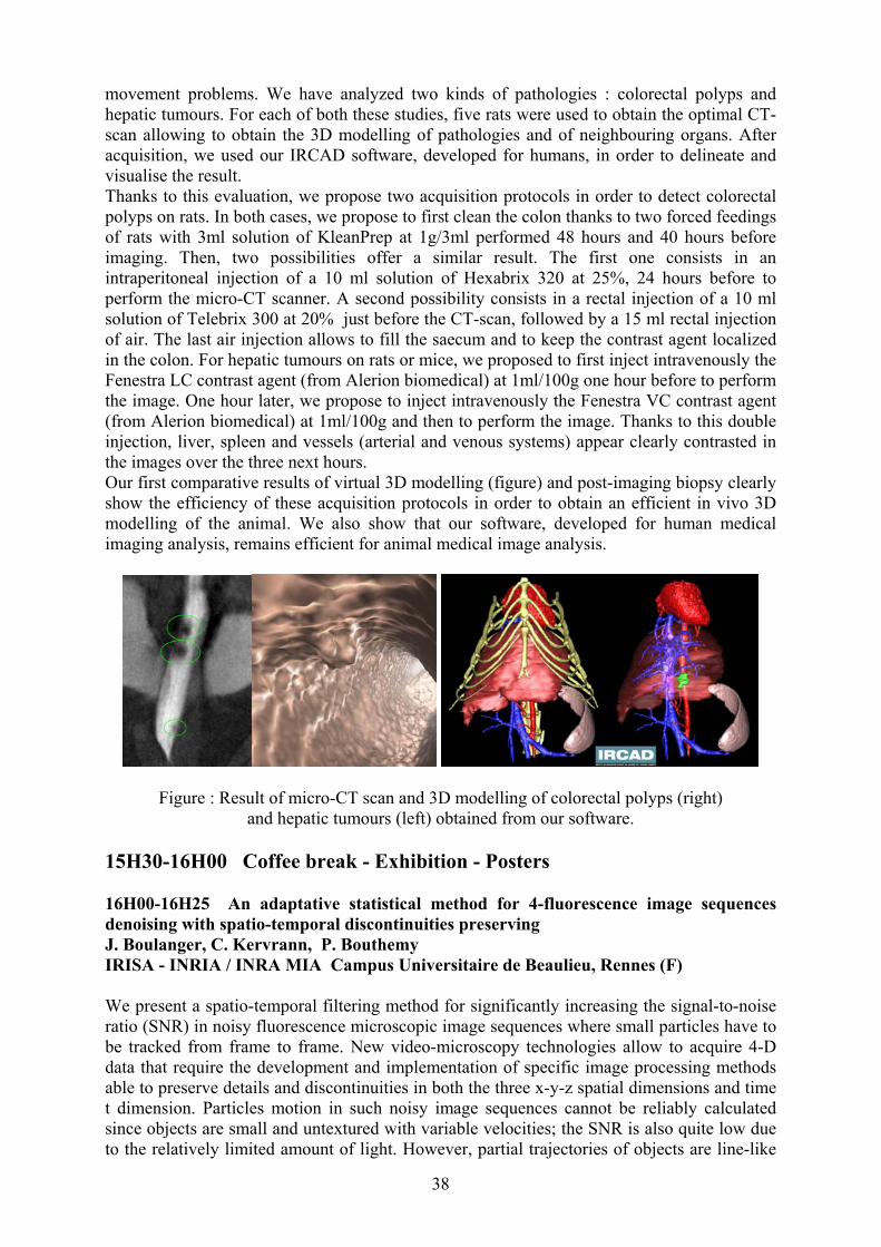
38
movement problems. We have analyzed two kinds of pathologies : colorectal polyps and hepatic tumours. For each of both these studies, five rats were used to obtain the optimal CT-scan allowing to obtain the 3D modelling of pathologies and of neighbouring organs. After acquisition, we used our IRCAD software, developed for humans, in order to delineate and visualise the result. Thanks to this evaluation, we propose two acquisition protocols in order to detect colorectal polyps on rats. In both cases, we propose to first clean the colon thanks to two forced feedings of rats with 3ml solution of KleanPrep at 1g/3ml performed 48 hours and 40 hours before imaging. Then, two possibilities offer a similar result. The first one consists in an intraperitoneal injection of a 10 ml solution of Hexabrix 320 at 25%, 24 hours before to perform the micro-CT scanner. A second possibility consists in a rectal injection of a 10 ml solution of Telebrix 300 at 20% just before the CT-scan, followed by a 15 ml rectal injection of air. The last air injection allows to fill the saecum and to keep the contrast agent localized in the colon. For hepatic tumours on rats or mice, we proposed to first inject intravenously the Fenestra LC contrast agent (from Alerion biomedical) at 1ml/100g one hour before to perform the image. One hour later, we propose to inject intravenously the Fenestra VC contrast agent (from Alerion biomedical) at 1ml/100g and then to perform the image. Thanks to this double injection, liver, spleen and vessels (arterial and venous systems) appear clearly contrasted in the images over the three next hours. Our first comparative results of virtual 3D modelling (figure) and post-imaging biopsy clearly show the efficiency of these acquisition protocols in order to obtain an efficient in vivo 3D modelling of the animal. We also show that our software, developed for human medical imaging analysis, remains efficient for animal medical image analysis.
Figure : Result of micro-CT scan and 3D modelling of colorectal polyps (right) and hepatic tumours (left) obtained from our software.
15H30-16H00 Coffee break - Exhibition - Posters 16H00-16H25 An adaptative statistical method for 4-fluorescence image sequences denoising with spatio-temporal discontinuities preserving J. Boulanger, C. Kervrann, P. Bouthemy IRISA - INRIA / INRA MIA Campus Universitaire de Beaulieu, Rennes (F) We present a spatio-temporal filtering method for significantly increasing the signal-to-noise ratio (SNR) in noisy fluorescence microscopic image sequences where small particles have to be tracked from frame to frame. New video-microscopy technologies allow to acquire 4-D data that require the development and implementation of specific image processing methods able to preserve details and discontinuities in both the three x-y-z spatial dimensions and time t dimension. Particles motion in such noisy image sequences cannot be reliably calculated since objects are small and untextured with variable velocities; the SNR is also quite low due to the relatively limited amount of light. However, partial trajectories of objects are line-like
39
structures in the spatio-temporal x-y-z-t domain. Image restoration can be then achieved by an adaptive window approach which has been already used to efficiently remove noise in still images and to preserve spatial discontinuities. The proposed spatio-temporal technique associates with each voxel the weighted sum of data points within a space-time window. We use statistical 4-D data-driven criteria for automatically choosing the size of the adaptive growing neighborhood. We have applied this method to noisy synthetic and real 4-D images where a large number of small fluorescently labelled vesicles move in regions close to the golgi apparatus. The SNR is shown to be drastically improved resulting enhanced objects which even can be segmented. This novel approach can be further used for biological studies where dynamics have to be analyzed in molecular and subcellular bioimaging. 16H30-16H55 High performance detector design for mono or bi photon small animal radio-isotopic imaging M. Parmentier(1), N. Tamada(1), A. Bakkali(1), J. Chavanelle(1), M. Paindavoine(2), N. Sultan Salahudin(2), A. Pousse(1), H. Boulahdour(1), B. Kastler(1) 1) Lab. Imagerie Ingénierie Santé Université de Franche Compté, Besançon (F) 2) LE2I Lab. Electronique Informatique Image, Dijon (F) Applications for radio-isotopic high resolution in vivo imaging for small animal are very large. They overturn our knowledge in life science domain, by giving quantitative information overcoming data usually obtained by in vitro approach. We have developped at Besançon a single photon gamma imager with a spatial resolution three or four times higher than that one of a classical gamma camera, which is essential for small animal functional explorations. A special rotating collimator device, combined with a powerful deconvolution algorithm leads to a good sensitivity, a high spatial resolution and artifactless images. Significant technological progress in scintillator crystals as well as in photo-detectors and electronic circuitery, allow to develop high performance imagers for single photon emission computed tomography (SPECT), as well as for positron emission tomography (PET). This advances will also benefit to human examinations such as early detection of breast cancer. 17H00-17H25 Transmission electron tomography: methods and applications T. Boudier, C. Messaoudi, S. Marco Institut Curie IMR168, Paris (F) Electron tomography is becoming a powerful tool in the study of sub-cellular biological structures such as cilia [1], mitochondria [2], Golgi apparatus [3,4], basal bodies [5] and primary cilium [6]. This method allows computing three-dimensional (3D) reconstructions from projections of a single object recorded at several tilt angles in the electron microscope [7]. Recently, tomographic reconstruction techniques have been combined with energy filtering transmission electron microscopy in a new structural approach that we have denoted “energy filtering transmission electron tomography” (EFTET). This approach allows analyzing the 3D distribution of single chemical elements [8,9]. Thus, relevant information has been obtained on the analysis of iron distribution in magnetotactic bacteria [10] or in the study of iron oxide nanoparticles distribution in rat lymph nodes [11]. In this context, new methodological approaches to improve the performances of the electron tomography are in development. First, to improve the quality of the recorded images use of cryo methods allow a better preservation of the biological samples. Second, new algorithms for volume alignment make it possible to combine several volumes from one specimen acquired at different orientations and thus increase the final volume resolution. Finally, in the case of EFTET, the development of background subtraction for EFTEM tilted series enables

40
to obtain elemental maps of elements having energy loss values higher than 400eV, such as oxygen and iron. The principles of electron tomography, as well as the new approaches for the samples observation, volume alignment and EFTET will be illustrated by several biological examples. In particular, by: (i) the study of melanosome morphogenesis, these organelles are implied in the synthesis and storage of melanine and are implicated in several skin pigmentation diseases and cancers; (ii) the analysis of the centriolar structure on basal bodies, which plays a substantial role in the cellular motility and are similar to the centrioles of centrosomes which are responsible for the cellular division; and (iii) the study, by EFTET, of granular metal inclusions in bacteria which represents an adaptive mechanism to extreme media.
References 1 McEwen, B.F., Radermacher, M., Rieder, C.L., and Frank, J. 1986 Tomographic three-dimensional reconstruction of cilia ultrastructure from thick sections. Proc Natl Acad Sci U S A 83: 9040-9044. 2 Nicastro, D., Frangakis, A.S., Typke, D., and Baumeister, W. 2000 Cryo-electron tomography of neurospora mitochondria. J Struct Biol 129: 48-56. 3 Ladinsky, M.S., Mastronarde, D.N., McIntosh, J.R., Howell, K.E., and Staehelin, L.A. 1999 Golgi structure in three dimensions: functional insights from the normal rat kidney cell. J Cell Biol 144: 1135-1149. 4 Marsh, B.J., Mastronarde, D.N., McIntosh, J.R., and Howell, K.E. 2001 Structural evidence for multiple transport mechanisms through the Golgi in the pancreatic beta-cell line, HIT-T15. Biochem Soc Trans 29: 461-467. 5 O'Toole, E.T., Giddings, T.H., McIntosh, J.R., and Dutcher, S.K. 2003 Three-dimensional organization of basal bodies from wild-type and delta-tubulin deletion strains of Chlamydomonas reinhardtii. Mol Biol Cell 14: 2999-3012. 6 Jensen C.G., Poole C.A., McGlashan S.R., Marko M., Issa Z.I., Vujcich K.V., Bowser S.S. 2004. Ultrastructural, tomographic and confocal imaging of the chondrocyte primary cilium in situ. Cell Biol Int. 28:101-110. 7 Frank, J. 1992. Electron tomography. Three-dimensional imaging with the transmission electron microscope. Plenum Press. New York 8 Thomas J.M. and Midgley P.A. 2004. High-resolution transmission electron microscopy. The ultimate nanoanalytical technique. Chem Commun (Camb). 7:1253-1267. 9 Thomas J.M., Midgley P.A., Yates T.J., Barnard J.S., Raja R., Arslan I., Weyland M. 2004. The Chemical Application of High-Resolution Electron Tomography: Bright Field or Dark Field? Angew Chem Int Ed Engl. (in press) 10 Weyland, M., and Midgley, P.A. 2003 Extending energy-filtered transmission electron microscopy (EFTEM) into three dimensions using electron tomography. Microsc Microanal 9: 542-555. 11 Bordat, C., Sich, M., Rety, F., Bouet, O., Cournot, G., Cuenod, C.A., and Clement, O. 2000 Distribution of iron oxide nanoparticles in rat lymph nodes studied using electron energy loss spectroscopy (EELS) and electron spectroscopic imaging (ESI). J Magn Reson Imaging 12: 505-509. 17H30-17H55 Spline-based approach to orientation assignment for three-dimensional electron microscopy S. Jonic(1), C. O. S. Sorzano(2), P. Thévenaz(3), M. Unser(3), N. Boisset(1) 1) Lab. de Minéralogie Cristallographie Paris, UPMC Paris 6, Paris (F) 2) Escuela Politécnica Superior, Universidad San Pablo-CEU, Madrid (E) 3) Biomedical Imaging Group, Swiss Federal Institute of Technology Lausanne (EPFL), Lausanne (CH)

41
In our previous work, we have developed a volume-to-image registration algorithm for a continuous a posteriori assignment of the parameters of orientation and position to Electron Cryo-Microscopy (cryo-EM) single particle images for a high-resolution Three-Dimensional (3D) particle reconstruction [1]. To determine these parameters, our algorithm employs a Levenberg-Marquardt gradient-based iterative minimization of a least-squares measure of dissimilarity between the two-dimensional Fourier Transform (FT) of the particle image and the extracted corresponding central slice of the 3D particle model FT relying on the central-slice theorem. The algorithm that is the most similar to ours is FREALIGN [2] which also computes continuous parameters of the particle orientation. While FREALIGN minimizes the phase dissimilarity between the experimental image and its model weighted by the amplitude of the FT of the experimental image, our algorithm minimizes both the amplitude dissimilarity and the phase dissimilarity. Also, contrary to FREALIGN which uses nearest-neighbour interpolation, our algorithm uses cubic B-splines to interpolate the data accurately. Our optimization algorithm is faster than Powell's method of FREALIGN since we have access to the gradient of the cost function. To improve the robustness of the algorithm, we use a frequency-domain weighting of the cost function. As all iterative algorithms, our method is sensitive to the choice of the initial parameters. To improve the robustness to the initial parameters, we have developed a strategy for the assignment based on the minimum final value of the dissimilarity measure for several different initializations. In this paper, we show the performance of our algorithm when using this strategy based on three starting points. A generalization to any number of points is straightforward. We validate the algorithm in a fully controlled simulation environment where the ground-truth solution is known a priori. We assess the assignment accuracy in terms of the warping index that is commonly used in the area of image registration. We synthesize a set of images at known poses using a 3D model of a protein from the PDB (Protein Data Bank). We first show the performance of our algorithm when initialized using the assignment by only one of the three standard quantized-parameter methods [3,4,5]. Then, we present the result of their joint use for initialization. We show that the “mixed” strategy can be used to refine the assignment obtained by the standard algorithms. In these experiments, we achieved the assignment with the warping index smaller than 2 voxel. At the end, we present the performance of our approach in refining a 3D model of a GroEL chaperonin using real cryo-EM data with no ground-truth solution. We observe that this method improves the consistency of the volumes from the previous iteration. Our algorithm is available in the Xmipp package [6]. In the future work, we will apply the techniques described here on other cryo-EM particles and macromolecular assemblies Literature: [1] S. Jonić, Spline Projection-Based Volume-to-Image Registration, Swiss Federal Institute of Technology Lausanne (EPFL), 2003. (Ph.D. Thesis). http://bigwww.epfl.ch/publications/jonic0302.html [2] N. Grigorieff, Three-Dimensional Structure of Bovine NADH: Ubiquinone Oxidoreductase (Complex I) at 22 Å in Ice, J. Mol. Biol., 277, pp. 1033-1046, 1998. [3] P. A. Penczek, R. A. Grassucci, and J. Frank, The Ribosome at Improved Resolution: New Techniques for Merging and Orientation Refinement in 3D Cryo-Electron Microscopy of Biological Particles, Ultramicroscopy, 53, pp. 251-270, 1994. [4] M. Radermacher, Three-Dimensional Reconstruction from Random Projections: Orientational Alignment via Radon Transforms, Ultramicroscopy, 53, pp. 121-136, 1994. [5] C. O. S. Sorzano, S. Jonić, C. El-Bez, J. M. Carazo, S. De Carlo, P. Thévenaz, and M. Unser, A Multiresolution Approach to Orientation Assignment in 3D Electron Microscopy of Single Particles, J. Struct. Biol., 146, pp. 381-392, 2004.
42
[6] R. Marabini, I. M. Masegosa, M. C. SanMartín, S. Marco, J. J. Fernández, L. G. de la Fraga, C. Vaquerizo, and J. M. Carazo, Xmipp: An image processing package for electron microscopy, J. Struct. Biol., 116, pp. 237-240, 1996. 18H00-18H25 Mouse single photon scintigraphy Ph. Choquet(1), L. El Fertak(2), C. Blondet(1), V. Israël-Jost(1), C. Goetz(1), I. Slim(1), M. Claria(1), I.J. Namer(1), A. Constantinesco(1) 1) Service de Biophysique et Médecine Nucléaire, Hôpitaux Universitaires de Strasbourg, CHU Hautpierre, Strasbourg (F) 2) Institut Clinique de la Souris, Illkirch (F) Mouse became the model of choice for number of human pathologies. This is due to several factors including small size, high prolificity and capability of easy production of transgenic animals. The need for longitudinal studies that is to say following disease evolution in the same animal over time leeds to the development of special imaging devices. All modalities are involved in this technological gap : the small size of the animal explain the difficulties of developping adapted systems. Due to the idea that scintigraphy is inherently of poor spatial resolution, attempt to use it for mouse imaging began with a delay relative to other imaging modalities (eg MRI, CT, PET and echography). We used a dedicated Anger type rotating gamma camera with 1 to 1.5 mm pinhole, 170 mm x 170 mm field of view and 25 photomultiplicators (Gaede Medizinsysteme Gmbh, Freiburg, Germany). Various tracers aimed at assessment of different physiological functions were used. One difficulty is that the radioactive tracer should be injected strictly intravenously, through a femoral catheter to avoid extravasation and changes in blood volume of mice under gaseous anesthesia. Depending on the tracers properties, dynamic, planar and tomographic data were acquired. We used medical software designed for analysis of human data to exploit the results and build a normal dtabase for mouse scintigraphy
Posters 1 Monte Carlo simulation of F18 disintegration in biological matter. Application to tumour volume reconstruction in PET C. Leloirec, C. Champion LPMC, Université de Metz, Metz (F) The imaging by positron emission (called PET for Positron Emission Tomography) gives functional information of tumours. Nevertheless, it is still difficult to get accurate anatomical data from this kind of images, essentially due to the fact that a phenomenological seuillage is needed to extract a visualization of the tumor zones from the PET data. That’s why mixed apparatus appeared, especially PET-CT, which combines PET with Computer Tomodensitometry (CT). In this way, functional and anatomical data can be obtained and tumors can be more precisely positioned. However, the spatial resolution of the PET images remains limited (of the order of 5 mm for human) and depends strongly of the radiopharmaceutical injected. In order to solve this problem, we have developed a complete Monte Carlo simulation to study in details the track of the positron and of the post-annihilation photons in biological matter in order to obtain the real energetic cartography induced by all the charged particles created. This energetic image will be compared to the
43
functional image experimentally obtained by PET in order to overcome the problem linked to the phenomenological threshold. The present Monte Carlo code provides all the coordinates of molecular interactions in water, the amount of energy deposited at each event with the corresponding type of interaction, for initial positron energies ranging from 10eV to 1MeV. The 3D energetic cartography also obtained will be compared to PET images and we will point out useful correlations what will permit a better understanding and utilization of the medical images. 2 Two photon confocal microscopy with new optimized chromophores, A. Hayek(1), F. Bolze(1), J.-F. Nicoud(1), K. D. Dorkenoo(2) 1)Organic Materials Group, Institute of Physics and Chemistry of Materials of Strasbourg, UMR CNRS 7504, Strasbourg (F) 2)Nonlinear Optics and Opto-electronics Group, Institute of Physics and Chemistry of Materials of Strasbourg, UMR CNRS 7504,Strasbourg (F) Cellular imaging is a long standing technique for investigation of cell structure and functions. In particular, fluorescence microscopy has been widely adopted since it offers to investigate the details of a variety of physiological behaviors in the cell. More recently two-photon excitation microscopy has broken through the biomedical research, with a variety of cellular and tissue applications. This method take advantages of the non linear optical (NLO) properties of chromophores: the emission occurs only at the focusing point of the laser (quadratic dependence upon intensity) and low bleaching (use IR light instead of the more energetic UV light). A wide effort is therefore being undertaken in order to develop efficient two-photon absorbers with good fluorescence quantum yield, high two-photon cross-section (σ2) and low bleaching at two-photon excitation. The aim of our research subject is the elaboration and characterization of new water soluble chromophores (for biological compatibility), optimized for two-photon induced fluorescence for cell imaging. These molecules derive from chromophores developed by our laboratory for optical limiting. We report here the synthesis of two series of chromophores with different cores and chain’s tall. The determination of the non-linear optical properties of these chromophores demonstrated that their σ2 are much higher than classical water-soluble chromophores (stilbene-3, rhodamine B, fluorescein, etc…), used actually in two-photon microscopy. We also present preliminary results of bio-imaging on epithelial onion’s cells, acquired by an home-made two-photon confocal microscope. 3 Fiber-optic sensors in Magnetic Resonance Imaging A. Pham(1), P. Benoit(2) 1) FISO Technologies Inc., Quebec (C) 2) OPTOPRIM, Vanves (F) Fiber-optic sensors offer inherent advantages for patient monitoring and control during magnetic resonance imaging examination and in other medical applications where electromagnetic fields, radio frequencies and microwaves are used as an adjunct to therapy. Because of the electrically insulating nature of fiber-optic and sensors materials, the sensing probes are totally immune to interference generated by these incident electromagnetic fields. This paper describes the technology involving fiber-optic probes based on Fabry-Perot cavities and low coherence interferometry using a Fizeau interferometer. The fiber-optic probes measure, depending on their types, physical parameters such as temperature, pressure
44
and strain. Principal medical applications where these fiber-optic sensors are used ranges from electromagnetically-induced hyperthermia monitoring to life-supporting intra-aortic balloon pumping. These sensors are also used in intra-cranial pressure monitoring of patient under RMI examination. Furthermore, their extremely compact dimensions (∅ 550 µm) make them very attractive for integration in minimally invasive catheters. The use of low coherence interferometry avoid the use of expensive lasers as input source and eases the integration of small-footprint opto-electronics into multifunction medical monitoring carts. 4 In-line spectroscopy monitors oxygen consumption during bypass operations B. Oderkerk (1), P. Benoit (2) 1) Avantes BV 2) Optoprim, Vanves (F) To study the effect of metabolic changes in the heart muscle it is important to know the oxygen consumption of the heart muscle. The level of oxygen consumption of the heart muscle causes differences in the coronary venous oxygen content. The oxygenation status of coronary sinus blood is determined by the use of VIS/NIR spectroscopy. We have developed a special fiber optic hearth catheter probe that is placed in the coronary sinus of the heart. The probe is connected to a tungsten halogen light source that emits continuous light from 400-2000nm. The other leg of the probe takes the back-reflected light and this light enters the spectrometer, optimized for the 500-1000nm wavelength range. The spectra were online saved and later evaluated, using the comprehensive software package. 5 Experimental test of depth dependence of solutions for time-resolved diffusion equation A. Laidevant, A. da Silva, M. Berger, J-M. Dinten Lab. Imagerie et Systèmes d’Acquisition, Leti – Cea Recherche Technologique, Grenoble (F) The determination of optical properties of a semi-infinite medium such as biological tissue has been widely investigated by many authors. Reflectance formulas can be derived from thediffusion equation for different boundary conditions at the medium-air interface. This quantity can be measured at the medium surface. For realistic objects, such as a mouse, tissue optical properties can only be determined at the object surface. However, near the surface, the diffusion approximation is weak and boundary models have to be considered. In order to investigate the validity of the time resolved reflectance approach at the object boundary, we have estimated optical properties of a liquid semi-infinite medium by this method for different boundary conditions and different positions of the fibers beneath the surface. The time-correlated single photon counting (TCSPC) technique is used to measure the reflectance curve. Our liquid phantoms are made of water, white paint and Ink. Laser light is delivered by a pulsed laser diode. Measurements are then fitted to theoretical solutions expressed as a function of source and detector’s depths and distance. By taking as reference the optical properties obtained from the infinite model with fibers deeply immersed, the influence of the different boundary conditions and bias induced are established for different fibers' depths and a variety of solutions. This influence is analyzed by comparing evolution of the reflectance models, as well as estimations of absorption and reduced scattering coefficients. Keywords: time-resolved reflectance, diffusion approximation, optical imaging, tissue optical properties.

45
6 Infrared imaging for medical and life sciences using synchrotron P. Dumas SOLEIL Synchrotron, L'Orme des Merisiers, Gif-sur-Yvette (F) The application of micro-spectroscopy, and hence imaging, in particular to biological and pathological problems relies on the informations obtained with great details. During the recent years, the use of infrared synchrotron radiation for microscopy has flourished. Thanks to the high brightness of the synchrotron source, enhanced lateral resolution, faster acquisition time and superior spectral quality, have made synchrotron infrared micro-spectroscopy a highly demanded analytical technique in synchrotron facilities. Accordingly, several beamlines are operational, planned or under design around the world (see table I and II in[1]). Spatially resolved infrared micro-spectroscopy produces a 2-dimensional arrays of spectra from which an chemical image, or “maps “ can be generated. Spectroscopic imaging provides diagnostic informations in a visual form, a prospect appealing to physicians and biologists. Image methods can provide potentially far more information to non-specialists than their non-imaging counterparts. However, the analysis and diagnostic potential of IR imaging strongly depends on the quality of the spectra acquired. Clearly, a parameter such as the signal-to-noise, strongly affects the image quality. For biological or biomedical investigations, a comprehensive understanding of all informations contained in a complex sample is rather elusive based on known functional group analysis. Several authors have used the so-called pattern recognition techniques, to enhance the diagnosis capability, and accordingly the imaging capabilities. Computer-based multivariate methologies are available for valid and statistically relevant classification of infrared patterns [2-4]. In this presentation, we illustrate the imaging capabilities of synchrotron infrared micro-spectroscopy for three examples: individual cells and human tissues such as hair and skin. The chemical images (also called univariate image), as well as multivariate images, have been obtained in each example, underlying the high potentiality of this synchrotron-based microanalytical technique. It is clear that synchrotron radiation provides a high brightness source which can be extremely beneficial for biological and biomedical-oriented investigations .With the expected Focal Plane Arrays ( size and numbers) , adapted to the synchrotron source shape, imaging using synchrotron infrared microspectroscopy has a tremendous future, especially with the possibility of combining, on the same sample studies using other synchrotron-based microscopy, such as X-ray microscopy[1]. References: [1] P. Dumas and L. M. Miller, Vibr.Spectr. 32(1) (2003)3. [2] P. Lasch and D. Naumann, Cellular and Molecular Biology 44(1998)189. [3] P. Lasch, W. Haensch, E. N. Lewis , L. H. Kidder, D. Naumann, Applied
Spectroscopy 56(2002,)1. [4] R. A. Shaw, J. R. Mansfield, S. P. Rempel, S. Low-Ying, V. V. Kupriyanov, and H.
H. Mantsch, J. of Mol. Struct. ( Theochem) 500(2000)127. 7 A combination of synchrotron imaging techniques for the study of a reactive matrix: the bone mineral D. Eichert, ID21, X-ray Imaging Group, ESRF, Grenoble (F) Commonly considered as inert, the mineral of hard tissues (bone, teeth) appears in fact as a very reactive and sophisticated material. The composition, the crystal size, the crystal orientation and the surface properties all contribute to adapting the tissue to its biological functions.
46
The various existing types of biological mineralization are based on apatitic calcium compounds and, in the case of vertebrates, on apatitic calcium phosphate compounds (apatite biominerals). For bone, providing mechanical resistance and acting as a primitive ion reservoir are two crucial functions. They are obtained through a tissue organisation involving apatite nanocrystals with a very high specific surface area and a well-developed hydrated layer responsible for ion exchanges with body fluids. Maturation, ion exchange and absorption are involved in the dynamic behaviour of apatitic biominerals. Consequently, bone mineral presents a complex composition. In addition to calcium and phosphate, it contains numerous other ions (hydrogenophosphate, carbonate, magnesium, sodium…), metals and trace elements. All these compositions varies strongly in bone through and with time, but also depending on the diet, turn over rate of the mineral, medical treatments… The aim of this poster is to discuss the processes of maturation of bone mineral, but also about some pathologies, which perturb the natural bone turnover and maturation process, and related proposed medical treatment. New information obtained by using a combination of synchrotron imaging techniques (X-ray microfluorescence, infrared microscopy, micro-diffraction) will be presented. 8 Tissular and cellular chemical microimaging using synchrotron X-ray microprobe S. Bohic (1), B. Fayard (1) (2) 1) X-ray imaging group, ESRF, Grenoble (F) 2) LPS, CNRS, Orsay (F) Normal and pathological functioning of tissues depend on biochemical processes that proceed at the single cell level and which are in most of the cases not yet fully understood. Investigation of single cell level biochemistry may give us key information to understand the functioning of healthy cells/tissues and the processes turning them into ill ones, thus opening the way to find new medicines/treatments to prevent or slow down some harmful intracellular processes. It is clear that bulk analysis of cells/tissues can only provide an idea of the main processes but cannot reflect properly the biochemical changes that take place at the single cell level. The most important requirements for microanalysis of single cells are the high spatial resolution (~ µm) and high analytical sensitivity (~ ppm) for the elements to be investigated. There are several micro-analytical tools available (e.g. PIXE, EDX, LAMMA, SIMS microscopy, IR or Raman microscopy, micro-spectrofluorometry) for micron-scale analysis. Nevertheless most of these techniques have some drawbacks related to difficult sample preparation, small depth of analysis, low sensitivity, destruction of the sample, uncertain quantification, which makes their use in single cell research problematical. In order to obtain more complete information the use of several analytical techniques on the very same sample ROI would be desirable, which is very difficult or not fully possible by the above-mentioned techniques. What’s more, most of these methods do not provide spectroscopic information on the chemistry at the micron scale and others (IR, Raman…), X ray absorption spectroscopy could be a useful complement. Due to the high brilliance of synchrotrons of third generation and to the progress in x-ray optics and in synchrotron instrumentation, synchrotron microbeam techniques have a potential to be an important probing method in cellular chemistry. Recent results on biological and medical application will be shown in the poster and possible perspectives in biology and medical research will be outlined.
47
9 Improvement of lateral resolution in fluorescence microscopy B. Simon, R. Greget, O. Haeberlé Lab. MIPS, Université de Haute - Alsace, Mulhouse (F) We propose a method to improve the lateral resolution in fluorescence microscopy, using laterally interfering excitation beams. The resulting excitation point spread function exhibits several lobes, which Full Width Half Maxima are narrower than for a regular excitation beam. Image processing then permits to remove side-lobes to obtain a better lateral resolution 10 UV imaging with synchrotron radiation at SOLEIL M. Réfrégiers, Synchrotron-SOLEIL, Gif-sur-Yvette (F) 11 Synchrotron radiation for medical imaging, from samples to patients. F. Estève (1), A. Bravin (2) 1) INSERM U647, Grenoble (F) - 2) ID17-ESRF, Grenoble (F)
48
EXHIBITORS Bio-Opto Exposium – 1, rue du Parc F 92593 Levallois-Perret Cedex Phone : +33 (0)1 49 68 51 89 E-Mail : [email protected] Internet: www.optoexpo.com « Photonics for life sciences » BIOVALLEY Parc d’Innovation 9 bld Gonthier d’Andernach F 67400 Illkirch Graffenstaden Phone: +33 (0)3 90 40 30 00 E-Mail : [email protected] Internet: www.biovalley.com “Leading trinational life science cluster” BFi OPTiLAS France ZI la Petite Montagne Sud 4,Allée du Cantal F 91018 Evry Cedex Phone: +33 (0)1 60 79 59 12 / Fax : +33 (0)1 60 79 89 01 E-Mail : [email protected] Internet : www.bfioptilas.avnet.com “BFI OPTILAS's "IMAGING and DETECTION" division is specialized in offering solutions and components in imaging. In the biological domain, BFI OPTILAS offers cameras, softwares and accesories. Cameras : Basler, Pixelink, QImaging and the famous DIAGNOSTIC INSTRUMENTS Stages, shutters and filters wheels : PRIOR SCIENTIFIC. Softwares : Mediacybernetics, Norpix (sequences and time lapse). Optics : THALES OPTEM Accesories : THALES OPTEM & DIAGNOSTIC INSTRUMENTS During IMVIE exhibition, BFI OPTILAS introduces the OPTIGRID system for 3D imaging in microscopy. You are end user or integrator, where ever you are in Europe, you have a BFI OPTILAS Office to help you” ECRIN 32 bd de Vaugirard 75015 Paris Phone: +33 (0)1 42 79 51 10 E-Mail : [email protected] Internet: www.ecrin.asso.fr “Creating collaborations, synergies and partnership between research laboratories and industries, in a multidisciplinary context”

49
HAMAMATSU PHOTONICS France Parc du Moulin de Massy 91300 Massy Phone:+33 (0)1 69 53 71 00 / Fax : +33 (0)1 6953 71 10 E-Mail : [email protected] Internet: www.hamamatsu.fr “Imaging solutions for life sciences: overview of EM-CCD, in vivo imaging AEQUORIA system, FRET and FLIM systems” IMAGINE OPTIC 18, rue Charles de Gaulle 91400 Orsay Phone: +33 (0)1.64.86.15.60 / Fax : +33 (0)1.64.86.15.61. E-Mail : [email protected] Internet : www.imagine-optic.com “Wavefront sensors and wavefront correction by adaptive optic closed loop for confocals and multip hotons mi croscopes. Both wavefront measurement and correction allow multiphotons and confocals microscopes improvements. An adaptive optic close loop is composed by a sensor, a phase shifter (mirror, SLM) and a software makes the close loop. Imagine Optic, leader in this field propose several solutions in function of the microscope type” LEICA Microsystèmes 86, Avenue du 18 Juin 1940 92563 Rueil Malmaison Cedex Phone: +33 (0)1 47 32 85 11 / Fax : +33 (0)1 47 32 85 86 E-Mail : [email protected] Internet : www.leicamicrosystems.com "Discovery at the Speed of Light" MAUNA KEA TECHNOLOGIES 9 rue d’Enghein 75010 Paris Phone: +33 (0)1 48 24 03 45 / Fax : +33 (0)1 48 24 12 18 E-Mail : [email protected] Internet : www.maunakeatech.com “In vivo and in situ molecular imaging system at the cellular level” OPTON Laser International Parc Club Orsay Université 29, rue Jean Rostand 91893 Orsay Cedex Phone: +33 (0)1 69 41 04 05 / Fax : +33 (0)1 69 41 32 90 E-Mail : [email protected] Internet: www.optonlaser.com Opton Laser International is the specialist of lasers and optical products for the scientific, industrial and telecommunication markets. Opton Laser International completes its major offer with instrumentation products for analysis, measurement, modulation, items for detection, visualisation, optics and opto-mechanics, crystals and protective eyewears.
50
OPTOPRIM 21-23 rue Aristide Briand 92170 Vanves Phone: +33 (0)1 41 90 61 80 / Fax : +33 (0)1 41 90 61 89 E-Mail : [email protected] Internet: www.optoprim.com “Optical solutions for bioscience: - DPSS vis & UV Lasers - Fiber sensors for patient monitoring (P & T) while imaging. - Components and detectors for confocal microscopy, FLIM, LIBS - Optical tweezers for cells micromanipulations” PHOTON LINES 55, avenue Auguste Renoir BP 51 78164 Marly-le-Roi Cedex Phone: +33 (0)1 30 08 99 00 / Fax: +33 (0)1 30 08 99 09 E-Mail : [email protected] Internet : www.photonlines.com “Photon Lines offers a broad range of optical products dedicated to life science applications. Besides low light level imaging (cooled and intensified CCD cameras) and imaging processing software associated, Photon Lines distributes lighting systems, precision optics, manual positioners and motorized stages...with a highlight on high performance workstations (optical table top) recommended for use in electro-physiology, cell injection, confocal laser scanning microscopy.. At the IMVIE² exhibition, Photon Lines introduces the LIFA system from Lambert Instruments for FRET and FLIM application (decay time fluorescence measurements...)” RHENAPHOTONICS Pôle API – IREPA-Laser Bd Sébastien Brant 67400 Illkirch Graffenstaden Phone: +33 (0)3 90 24 48 39 / Fax : +33 (0)3 90 24 46 19 E-Mail : [email protected] Internet: www.rhenaphotonics.com “Trinational optics and photonics cluster” XENOGEN CORPORATION 860 Atlantic Avenue Alameda, CA 94501 (USA) 75 avenue Parmentier 75011 PARIS Phone: +33 (0)1 40 21 24 37 E-Mail : [email protected] Internet : www.xenogen.com “Xenogen Bioscience and of the use of Xenogen imaging systems and Bioware ® for bioluminescent and fluorescent in vivo imaging”

51
RÉSUMÉS Conférences March 1, 2005
9H00-9h25 Suivi de nanocristaux individuels : application à la dynamique du récepteur du GABAA dans les cônes de croissance C. Bouzigues1, S. Levi, A. Triller2 et M. Dahan1 1) Lab. Kastler Brossel, Ecole Normale Superieure, Paris (F) 2) Lab. de Biologie Cellulaire de la Synapse , Ecole Normale Supérieure, Paris (F) Au cours de la dernière décennie, l’imagerie de processus biologiques dans des systèmes vivants au niveau de la molécule unique a connu un intérêt croissant. La microscopie de fluorescence est un des outils les plus appropriés dans cette perspective mais a rencontré d’importantes limitations, suite au photoblanchiment des sondes fluorescentes organiques. Nous présenterons ici l’utilisation de nanocristaux semi-conducteurs pour le suivi de protéines membranaires uniques. Ces nouvelles sondes sont stables, permettant l’observation en fluorescence de molécules uniques pour des durées inaccessibles auparavant. L’intermittence de leur fluorescence (ou « blinking ») nécessite cependant l’emploi de nouvelles techniques de traitement d’images. Nous avons appliqué cette méthode au suivi des mouvements des récepteurs du GABA (GABAAR) dans les cônes de croissance de neurones spinaux. La dynamique des récepteurs de molécules de navigation est un problème particulièrement intéressant, dans la mesure où elle pourrait réguler le guidage axonal. Les trajectoires enregistrées ne sont pas purement diffusives et présentent une vitesse d’entraînement (<v>=0.29µm.s-1) due aux interactions avec le cytosquelette. L’utilisation de drogues spécifiques pour le cytosquelette et une analyse fine des trajectoires a permis de caractériser les interactions transitoires entre les microtubules et les récepteurs du GABA. Des enregistrements réalisés à l’échelle de l’heure ont permis de mettre en évidence une barrière de diffusion microtubule dépendante, entre l’axone et le cône de croissance. Ces résultats soulèvent la question du rôle fonctionnel des interactions entre les récepteurs et le cytosquelette. Nous avons donc porté notre attention sur des expériences combinant stimulation locale, à l’aide de substances de guidage, et suivi de particules individuelles. Cette étude pourra fournir des explications à la remarquable fidélité du guidage axonal, en mettant en évidence une éventuelle régulation de celui-ci par le couplage entre le cytosquelette et la dynamique des récepteurs. 11H00-11H25 Systèmes d’imagerie et méthodes d’imagerie tomographique diffuse de luminescence appliquées à la détection et localisation in vivo de reporteurs bioluminescents O. Coquoz, C. Kuo, D. Stearns Tamara L. Troy, D.A. Zwarg, B.W. Rice, Xenogen Corporation, Alameda, CA (USA) L’utilisation de reporteurs bioluminescents dans les cellules vivantes a prouvé être un outil valable pour le suivi de l’expression de gènes ciblés. Une instrumentation de haute sensibilité optimisée pour la détection in vivo de reporteurs émettant de la lumière dans des petits

52
animaux de laboratoire a été développée à Xenogen Corporation, avec la gamme de systèmes d’imagerie IVIS. Ces instruments détectent la projection diffuse à la surface de l’animal provenant de sources situées en profondeur à l’intérieur. De cette façon, cette technique permet au chercheur de suivre les niveaux relatifs d’émission de lumière de bioluminescence. Une quantification absolue des sources de lumière à l’intérieur de l’animal exige la localisation tridimensionnelle de leur distribution spatiale. Ceci peut être réalisé par des reconstructions utilisant DLIT, une technique d’imagerie tomographique diffuse de luminescence de la topographie de la surface et de l’émission de photons provenant d’un animal en utilisant l’IVIS série 200. De plus, la résolution est améliorée quand des données mesurées à plusieurs longueurs d’onde sont incorporées à l’inversion tomographique. Des résultats expérimentaux seront présentés, qui valident la méthode DLIT sur des systèmes comprenant une source de lumière calibrée à l’intérieur d’un fantôme de souris dont les propriétés optiques simulent celles de tissus vivants. Des mesures in vivo obtenues de souris injectées avec une bille luminescente calibrée montrent que cette technique permet la détermination de la distribution 3D de sources de lumière, ainsi que la prédiction de leur flux absolu in situ, à partir de deux ou plusieurs images acquises à différentes longueurs d’onde. Finalement, un système d’imagerie tomographique 3D complète actuellement en développement sera présenté. 17H00-17H25 A spot filtering tool to facilitate image analysis of 2D gels M. Larbaoui(2), U.Wirth(1), N. Brendlen(1), G. Lambrou(2), J. van Oostrum(1), H.Voshol(1) and E. Bertrand(1-2) 1) Functional Genomics 2) DA Neuroscience/Ophthalmology Novartis Institutes for Biomedical Research, Basel (CH) L'électrophorèse 2D est une des principales techniques utilisée en recherche protéomique pour séparer et caractériser les protéines. A l'heure actuelle, plusieurs outils bioinformatiques aident les chercheurs à exploiter l'énorme quantité d'information générée. Malgré leurs bonnes performances dans l'analyse d'image et le pré-traitement des données, ces outils manquent de fiabilité quant à l'analyse de l'expression différentielle. Nous présentons ici une approche innovante en matière d'analyse de l'expression différentielle et son implémentation informatique qui combine la technologie Java avec les capacités statistiques de R au sein d'une application web. L'approche sous-jacente est d'utiliser les données pré-traitées générées par Progenesis (un des outils les plus avancé pour l'analyse d'images de gel 2D) et d'y appliquer un ensemble de filtres afin de ne retenir que les spots qui sont réellement exprimés, puis de détecter de possibles expressions différentielles parmi eux. Cette nouvelle approche présente plusieurs avantages par rapport à l'ancien système d'analyse, elle évite de longues et fastidieuses vérifications par l'utilisateur et fournit des résultats dont nous avons validé la fiabilité. 17H30-17H55 Potentiel de la microscopie par spectrométrie de masse d'ions secondaires dans les sciences du vivant J-L. Guerquin-Kern , T-D Wu, A. Croisy Institut Curie Recherche, Orsay (F) La microscopie ionique ou SIMS (Secondary Ion Mass Spectrometry) est basée sur l'émission de particules secondaires provenant des quelques premières couches d'atomes (1-2 nm) de la surface d'un échantillon solide sous l'effet du bombardement par un faisceau d'ions primaires très énergétiques. Sous l'impact, les liaisons chimiques sont rompues et les atomes ou des combinaisons d'atomes sont libérés, soit sous forme neutre, soit sous forme chargée (ions). Ces ions peuvent être collectés et focalisés sous forme d'un faisceau secondaire jusqu'à

53
l'entrée d'un spectromètre de masse où les ions vont alors être triés en fonction de leur rapport m/z (masse/charge). Des images peuvent ainsi être obtenues pour une masse choisie. Toutefois, cette analyse basée sur l'émission d'ions secondaires se fait au prix d'une destruction progressive de l'échantillon. Cette technique d'analyse de surface est couramment utilisée depuis des années pour la microanalyse de minéraux (géologie, météorite..), en métallurgie et en semi-conducteur. Parallèlement, les sciences du vivant offrent un fort potentiel d'applications pour l'analyse SIMS. Ainsi, l'identification, la localisation et la quantification intracellulaires d'éléments chimiques correspondent à des questions importantes dans beaucoup de domaine de la recherche biologique, notamment pour comprendre les mécanismes par lesquels les drogues peuvent interférer avec les processus biologiques. Par ailleurs, cette technique est la seule méthode de micro-analyse permettant une détection de la plupart des éléments et de leurs isotopes, sans recours à aucune molécule de marquage spécifique (fluorescence ou radioactive). Les caractéristiques principales de la dernière génération de microsonde en SIMS dynamique (CAMECA NanoSims 50 [l]) sont: i) une bonne résolution spatiale (jusqu'à 50nm), ii) une capacité à analyser simultanément jusqu'à 5 masses (ions) issues d'un même micro volume et permettant ainsi une détermination parfaite des rapports isotopiques et, dans le cas d'images de différentes masses, une superposition correcte des images iii) une excellente transmission même avec une résolution en masse élevée. Ces performances offrent pour la première fois la possibilité d'analyser, en même temps, la finesse de structures subcellulaires et la distribution des éléments chimiques dans les échantillons biologiques.
La plateforme de microscopie ionique à l'Institut Curie, est strictement dédiée au domaine des applications biologiques et plus particulièrement en cancérologie. Plusieurs projets ont été ainsi définis et couvrent trois axes principaux d'application: i) la pharmacologie antitumorale, ii) la bio minéralisation, iii) la médicine nucléaire et la radio toxicologie Les principes de la microscopie SIMS, ainsi que ceux de la préparation des échantillons, étape cruciale en microanalyse pour préserver à la fois les caractéristiques morphologiques et chimiques [2] des échantillons biologiques, seront rappelés et illustrés par quelques exemples d'applications. Enfin, l'intérêt d'une imagerie corrélative, effectuée sur le même échantillon, associant la microscopie TEM pour sa très haute résolution (Fig. 1) et la microsonde SIMS pour sa sensibilité en microanalyse (Fig.2), sera présenté.
Figure 1: Image MET de microcalcification dans des cellules MDCK. Les calcifications apparaissent comme des agglomérats d' hydroxyapatite. La même coupe a été analysée ultérieurement par microscopie ionique (Fig2); (image obtenue par Dr H. Vali, McGill University, Montreal, Canada).

54
Figure 2: Analyse ionique de microcalcification dans des cellules MDCK. Les images sont obtenues en utilisant une source Césium (Cs+). Les éléments analysés sont C-(m=12), O-
(m=16), CN-(m=26), P-(m=31) ), S-(m=32), et CaO-(m=56). Champ : 6 µm x 6 µm, inclusion dans l' Epon, épaisseur de coupe 120 nm, (en collaboration avec Dr H Vali, Université McGill, Montréal, Canada). References: 1 G. Slodzian, B. Daigne, F. Girard, F. Boust, F. Hillion, (1992) Biol. Cell, 74:43-50. 2 C. Quintana, (1994) Micron, 25:63-99. REMERCIEMENTS: Ce projet a été soutenu par l'Institut Curie, l'INSERM, la Région Ile de France, l' EDF et l'ARC. March 2, 2005 8H30-8H55 Tomographie optique des photons diffuses et de fluorescence pour le petit animal. P. Poulet, R. Chabrier, B. Montcel UMR 7004 Université Louis Pasteur / CNRS, Institut de Physique Biologique, Strasbourg (F)
L’imagerie tomographique développée utilise la détection résolue en temps des photons multi-diffusés pour établir les cartes des propriétés optiques et la distribution de sondes fluorescentes des corps étudiés. Les images d’absorption, de diffusion réduite et de fluorescence d’objets tests illustrent les potentiels de la méthode et permettent de dégager des perspectives d’application en expérimentation animale. 9H30-9H55 Détection d’activation du cortex moteur par des méthodes d’optique diffuse résolues en temps. B. Montcel, R. Chabrier, P. Poulet UMR 7004 Université Louis Pasteur / CNRS, Institut de Physique Biologique, Strasbourg (F)
55
Les modifications vasculaires induites localement par une activation cérébrale peuvent être mesurées à travers le crâne par spectroscopie PIR. Des simulations, basées sur des modèles issus d’IRM et sur la résolution de l'équation de diffusion par la méthode des éléments finis, suggèrent que les techniques résolues en temps permettent d’améliorer la détection et la localisation en profondeur des activations corticales. Les premières expériences de stimulation du cortex moteur, obtenues avec un appareil à comptage de photons résolu en temps, confirment ces hypothèses 13H30-13H55 Spectroscopie THz et bioapplications Jean Demaison Laboratoire de Physique des Lasers, Atomes, et Molécules, UMR CNRS 8523 Université des Sciences et Technologies de Lille, 59655 Villeneuve d'Ascq La spectroscopie Terahertz (THz), née à l'issue de la deuxième guerre mondiale, est la conséquence directe des recherches sur les radars. Au début, c'était une technique très compliquée avec une gamme de fréquences très réduite (essentiellement microondes). De plus, l'analyse des spectres nécessitait beaucoup de temps et d'expertise. Pour ces raisons, son domaine d'applications s'est longtemps limité à la recherche fondamentale en physico-chimie moléculaire. Cependant, de gros progrès ont été accomplis en technique (plus large gamme en particulier vers les hautes fréquences, meilleure sensibilité, automatisation, …) et des logiciels très performants permettent d'analyser beaucoup plus facilement les spectres. Après un historique et une présentation de cette spectroscopie, deux types d'applications vont être particulièrement détaillés: 1. Détection quantitative des gaz La principale utilisation est la détection des gaz dans des conditions difficiles. Par exemple, la détection des molécules interstellaires (fonctionne à grande distance) ou des gaz de combats (fiable, automatique et rapide). Mais elle peut également être facilement utilisée pour la détection des polluants industriels ou domestiques ainsi que pour analyser l'haleine. 2. Etude des biomolécules C'est la meilleure méthode pour déterminer avec précision la forme des molécules, clef de leurs propriétés biologiques. Elle permet également d'étudier l'influence de la solvatation sur ces propriétés ainsi que les liaisons intermoléculaires (liaison hydrogène, liaison de Van der Waals) qui jouent un rôle fondamental en biologie. 14H30-14H55 Full-field optical coherence tomography A. Dubois Lab. d’Optique Physique, Ecole Supérieure de Physique et Chimie Industrielles, CNRS, UPR A0005, Paris (F) La tomographie par cohérence optique (OCT) est une technique d’imagerie des milieux biologiques mettant en œuvre un interféromètre de Michelson fibré, avec une source lumineuse de spectre large. Nous avons développé une technique alternative basée sur un microscope interférométrique, avec une caméra CCD, et éclairé par une lampe halogène. La résolution spatiale est de 1 µm en 3D. Une sensibilité de détection de 90 dB est atteinte pour un temps d’acquisition par image de 1 s. La technique est appliquée à l’imagerie de divers tissus biologiques à l’échelle cellulaire.

56
15H00-15H25 Techniques émergentes en ultrasonographie de contraste S. Lori Bridal, J-M Correas, O. Lucidarme, A. Ammi, E. Jounnot, P. Laugier Lab.d’imagerie paramétrique-CNRS - Université de Paris 6, Paris (F) Les produits de contraste ultrasonore (PCUS) injectables par voie veineuse périphérique permettent d'augmenter l'intensité du signal échographique du sang circulant. Ces produits sont constitués de microbulles de gaz encapsulées mesurant de 1 à 10 µm de diamètre. Ils possèdent une distribution intravasculaire stricte (absence de passage dans le secteur interstitiel) et sont donc des traceurs potentiels de la perfusion sanguine. Le principe de l’imagerie fonctionnelle de contraste sera décrit et illustré à partir des exemples cliniques. Des résultats expérimentaux seront présentés afin de comparer l’imagerie fonctionnelle de contraste avec des techniques de références et également pour caractériser les seuils acoustiques pour la destruction des microbulles de PCUS.
16H00-16H25 Imagerie d’états transitoires par spectroscopie laser femtoseconde Apports innovants pour la chimie du vivant Y. Gauduel(1), V. Malka(1), T Launay(2), F. Guilloud(2), B. Charles(2) 1) L.O.A., CNRS UMR 7639, Ecole Polytechnique - ENS Techniques Avancées, Palaiseau (F) 2) ENSEA, Cergy Pontoise (F)
De nombreux processus relevant des sciences du vivant mettent en jeu des états
électroniques transitoires dont la caractérisation spatio-temporelle représente un réel défi pour la chimie physique d’intérêt biomédical. L’imagerie d’états transitoires développée par spectroscopie laser femtoseconde implique un traitement numérique de matrices de données expérimentales acquises sur CCD refroidis à -120°C et la mise au point de méthodes graphiques avancées sous OPGS (Open Graphic System).
Cette contribution permettra de présenter des résultats d’images numériques en 2 et 3D obtenues à l’aides d’impulsions laser dont la largeur à mi-hauteur durée n’excède pas 80 fs (80 10-15 s) et dans la gamme de fréquences 27800 – 10400 cm-1. Ces résultats portent sur l’étude en temps réel du départ d’un électron de l’environnement d’un atome ou sur la formation d’une liaison soufre-soufre à deux centres-trois électrons entre deux molécules à nombre limité d’atomes. Ces travaux permettent d’entrevoir le contrôle sélectif de mécanismes radicalaires d’intérêt biomédical (chloration d’acides aminés lors de processus inflammatoires, réparation moléculaire après un stress oxydatif…).
Dans le domaine des rayonnements ionisants appliqués à la radiobiologie, des recherches très innovantes portent sur le développement d’images tomodensitométriques à partir de paquets d’électrons relativistes sub-picosecondes (E ~ 3-15 MeV). Le traitement numérique de signaux optiques mesurés dans le proche IR avec une caméra CCD 16 bits (Andor Technology) devrait ouvrir la voie à la mise en place d’une microdosimétrie à l’échelle micrométrique. L’analyse paramétrique d’images reconstruites en 3D devrait permettre d’aborder des évènements radio-induits primaires dans des environnements confinés tels que les sillions aqueux d’ADN ou les milieux sub-cellulaires.
57
16H30-16H55 Real-time mapping of intra-protein electric fields through absorption spectroscopy of tryptophans S. Haacke Institut de Physique et Chimie des Matériaux de Strasbourg (IPCMS), GONLO, Strasbourg (F) Des expériences de l'absorption transitoire des tryptophanes dans la bactériorhodopsine nous ont permis de suivre les translocations de charge photo-induites en temps réel, avec une résolution de 80 fs. La dynamique du signal de blanchiment met en évidence le lien entre changement de moment dipolaire du rétinal et son isomérisation. Les résultats indiquent que l'isomérisation est accélérée par les interactions électrostatiques rétinal-protéine. Nous allons discuter l'application de cette nouvelle approche expérimentale à d'autres protéines. 17H30-17H55 Un état de l’art sur les systèmes de réalité augmentée appliqués à la chirurgie : Terminologie et taxonomie. G. Sittler(1) , P. Twardowski(1), T. Blandet(1), J.-B. Fasquel(2), J. Fontaine(1) 1) LSP :Lab. des Systèmes Photoniques, Université Louis Pasteur Strasbourg (F) 2) IRCAD :Institut de Recherche contre les Cancers de l’Appareil Digestif, Strasbourg(F) De façon à permettre la construction d’un prototype de casque de réalité augmentée en vue d’applications en chirurgie, il est important d’établir un état de l’art aussi précis que possible sur les systèmes déjà existants, et ce dans différents domaines d’application, ainsi que de justifier l’originalité de la démarche proposée. Le bon déroulement du projet, associant les compétences du LSP et de l’IRCAD, nécessite aussi un approfondissement de la taxonomie et de la terminologie employée, ce qui constituera une partie de l’exposé. March 3, 2005 9H00-9H25 Amélioration de la résolution en 2D en microscopie de fluorescence par utilisation d’une lame de phase et recombinaison d’images. Olivier Haeberlé et Bertrand Simon Laboratoire MIPS - Groupe LabEl, Université de Haute-Alsace IUT Mulhouse, Mulhouse (F) Le microscope de fluorescence est un instrument de choix pour le biologiste. Cependant, comparé à d’autres instruments comme le microscope électronique ou le microscope optique à champ proche, sa résolution est limitée, de l’ordre de 150 nm latéralement. Nous proposons une technique simple utilisant une lame de phase et une recombinaison d’images pour améliorer la résolution latérale d’un microscope confocal à mieux que 100 nm, pour des objets 2D, tels des chromosomes en métaphase, déposés après préparation sur une lame.
58
9H30-9H55 Microscopie par génération de troisième harmonique appliquée à l’analyse vélocimétrique du développement des embryons de drosophile D. Débarre(1), W. Supatto(2), E. Farge(2), B. Moulia(3), M.-C. Schanne-Klein(1), E. Beaurepaire(1) 1) Lab. for optics and biosciences, CNRS UMR 7645, INSERM U451, Ecole Polytechnique, Palaiseau (F) 2) Mechanics and genetics of developmental embryology, CNRS UMR 168, Curie Institute, Paris (F) 3) Biomechanics group, INRA UMR 547, Clermont-Ferrand (F) Durant le développement embryonnaire, les mouvements cellulaires sont hautement régulés dans le temps et l’espace. Les mouvements morphogénétiques impliqués dans le développement embryonnaire de la drosophile présentent un intérêt particulier car la drosophile est un des principaux modèles de la génétique du développement. Cependant, la visualisation de ces mouvements est difficile car l’embryon est très diffusant au début de son développement et l’introduction de colorants peut induire des perturbations indésirables. La microscopie THG est une technique récente permettant d’obtenir des informations structurelles avec une résolution tridimensionnelle micrométrique. En associant cette technique avec des algorithmes d’analyse vélocimétrique adaptés de l’hydrodynamique, nous avons quantifié à l’échelle micrométrique les champs de vitesse des tissus dans des embryons non marqués après avoir vérifié que l’imagerie THG ne perturbait pas le développement embryonnaire. Enfin, nous avons appliqué ces résultats à l’étude d’un embryon mutant pour un gène du développement.
Nos résultats démontrent donc que la génération de troisième harmonique est une approche puissante pour visualiser et quantifier in vivo les mouvements morphogénétiques dans des embryons non marqués. Ref. Débarre, Supatto et al, Opt. Lett. 29, pp. 2881-2883 (2004) 11H00-11H25 Intra operative computer assisted surgery using low cost virtual and augmented reality systems L. Soler, S. Nicolau, J. Schmidt, C. Koehl, M. Arenas, D. Mutter, J. Marescaux IRCAD (Research Institute against Digestive Cancer), Strasbourg (F) Les procédures assistées par ordinateur ont pour objectif l’amélioration des gestes chirurgicaux. Nous présentons ici deux systèmes de réalité augmentée à coût réduit dont le principe repose sur la superposition temps réelle d’images 3D pré-opératoires du patient, reconstruites à partir d’une image TomoDensitoMétrique ou IRM, sur les images vidéos réalisées durant l’intervention. Le premier système, interactif, offre l’avantage de ne nécessiter qu’une unique installation d’un système de visioconférence au bloc opératoire sans modification de la salle opératoire, de la procédure chirurgicale ou des outils chirurgicaux. Il présente également l’avantage d’offrir une possibilité d’aide à distance par la réalité augmentée. Sa limite réside dans l’interactivité du recalage. Le second système, automatique, offre l’avantage d’une automatisation des recalages et du suivi des instruments, permettant de fournir une précision sur le résultat de 2mm. Plus coûteux que le premier, mais ne nécessitant qu’une installation matérielle dont le coût total ne dépasse pas 10.000€, ce second système validé en pré-clinique reste à être validé cliniquement. Cette prochaine étape permettra d’aboutir à terme à une automatisation du geste chirurgical par le couplage de ce système avec un système robotisé, la prochaine révolution chirurgicale du 21ème siècle.

59
11H30-11H55 Real time registration of 2D and 3D images in percutaneous nephrolithotomies using an Augmented Reality system. A. Osorio, O. Traxer, S. Merran LIMSI – CNRS, Orsay (F) Nous présentons les résultats d’un recalage temps réel d’images 2D et 3D dans la planification chirurgicale de néphrolithotomies percutanées combiné à l’utilisation de la réalité augmentée qui sert à guider le chirurgien lors de l’intervention afin qu’il puisse connaître la meilleure trajectoire pour son acte de ponction percutanée. Nos développements reposent sur des logiciels de segmentation 3D à partir d’images scanner standard.
14H30-14H55 Comprendre les erreurs d’interprétation des images médicales J.-L. Gerstenmayer(1), V. Hazebroucq(2) 1) CEA, Fontenay aux Roses (F) 2) Assistance Publique-Hôpitaux de Paris et Université Paris-5, Paris (F) L’interprétation des images pose parfois problème. L’enjeu est de la plus haute importance en ce qui concerne la responsabilité légale, celle du médecin, de l’opérateur ou du fabricant de l’instrumentation : avoir posé une interprétation erronée, généralement lorsqu’il a eu des conséquences sur la vie ou la morbidité du patient, pourra être qualifié d’erreur ou de faute lorsque la responsabilité de l’homme est clairement en jeu, de panne ou d’alea lorsque l’instrument est en cause. En pratique, ces deux causalités sont parfois difficiles à démêler, tant à l’extrémité de toute chaîne de mesure expérimentale, se retrouve à chaque fois le jugement d’un homme de l’art. Il est donc de la plus haute importance d’identifier les causes possibles des erreurs humaines afin de se prémunir contre celles-ci, dans une démarche de prévention. Dans cette communication, nous n’étudions pas le « filtre instrumental » en tant que tel (bruits, bande passante, biais, signaux parasites…), matière déjà abondamment détaillée par les spécialistes en instrumentation que nous sommes, ni la question de l’ergonomie de l’instrument. Dans le prolongement de l’interface homme machine, l’objectif est ici d’identifier quelles sont les pierres d’achoppement qui peuvent occasionner des erreurs lorsque l’être humain est en situation d’interpréter l’image fournie par le capteur. Ces causes sont dues essentiellement au mode de fonctionnement du cerveau, ce fonctionnement heuristique qui le rend habituellement si rapide et si performant en regard du traitement séquentiel et procédural d’un ordinateur (le nombre de coups prévus à l’avance par un joueur d’échec humain ne dépasse pas 4 ou 5, contre plus de 20 pour un ordinateur). Cette question prend une importance encore accrue à l’heure où l’usage des réseaux de neurones est en voie d’être généralisé, en tant que classifieur de formes ou de textures, pour l’interprétation automatique des images.

60
15H00-15H25 Optimal acquisition protocol definition for 3D modelling of small animals abdominal tumours and organs from in vivo micro-CT scan L. Soler, A-B. Osswald, M. Bouhadjar, M. Aprahamian, F. Raul, F. Gossé, D. Mutter, J. Marescaux IRCAD, Strasbourg (F) Nous présentons les résultats d’une étude préliminaire d’optimisation des protocoles d’acquisition in vivo sur micro-scanner X, pour visualiser et modéliser en trois dimensions les cancers de l’appareil digestif sur les souris et les rats. Nous proposons ainsi deux protocoles différents pour les polypes colorectaux et un protocole pour les carcinomes hépatiques réalisés à partir d’un scanner MICRO CAT II (IMTEK) et des logiciels de modélisation 3D développés au sein de l’IRCAD. 17H00-17H25 Applications et principes de la tomographie électronique à transmission T. Boudier, C. Messaoudi, S. Marco Institut Curie IMR168, Paris (F) La tomographie électronique est en train de devenir un outil incontournable dans l'étude des structures biologiques sous cellulaires telles que les cils [1], les mitochondries [2], l’apareil de Golgi [3,4] ou les corpuscules basaux [5]. Cette méthode permet de calculer des reconstructions tridimensionnelles (3D) depuis les projections d'un objet individuel enregistrées à plusieurs angles d'inclinaison dans le microscope électronique [7]. Récemment, des techniques tomographiques de reconstruction ont été combinées à la filtration en perte d'énergie dans une nouvelle approche structurale dénoté “energy filtering transmission electron tomography” (EFTET). Cette approche permet d’analyser la distribution 3D des éléments chimiques [8,9]. Ainsi, des informations concernant la distribution de fer dans les bactéries magnétotactiques [10] ou la distribution de nanoparticules d'oxyde de fer dans les noeuds lymphoïdes de rat [11] peut être analysée. Dans ce contexte, des nouvelles approches méthodologiques pour améliorer les résultats obtenus par la tomographie électronique sont en fort développement. D'abord, pour améliorer la qualité des images enregistrées, les méthodes de cryo-microscopie permettent une meilleure conservation des échantillons biologiques. En second lieu, les nouveaux algorithmes pour l'alignement de volume rendent possible de combiner plusieurs volumes d'un spécimen acquis à différentes orientations et d'augmenter ainsi la résolution finale du volume. Finalement, dans le cas de l'EFTET, le développement d’algorithmes pour la soustraction du bruit de fond en 3D rend possible l’obtention de cartes élémentaires des éléments ayant des valeurs de perte d'énergie supérieures à 400eV, tels que l'oxygène et le fer. Les principes de la tomographie électronique, ainsi que les nouvelles approches pour l'observation d'échantillons, l'alignement des volumes et l'EFTET seront illustrés par plusieurs exemples biologiques. En particulier, par: (i) l'étude de la morphogenèse des mélanosomes, ces organelles sont impliquées dans la synthèse et le stockage de la mélanine et sont impliquées dans plusieurs maladies de pigmentation de la peau et des cancers; (ii) l'analyse de la structure du centriole dans des corps basaux, qui jouent un rôle essentiel dans la motilité cellulaire et qui sont homologues aux centrioles des centrosomes, ces dernières responsables de la division cellulaire ; et (iii) l'étude, par EFTET, des inclusions métalliques granulaires dans des bactéries qui représente un mécanisme adaptatif aux milieux extrêmes.
61
Références 12 McEwen, B.F., Radermacher, M., Rieder, C.L., and Frank, J. 1986 Tomographic three-dimensional reconstruction of cilia ultrastructure from thick sections. Proc Natl Acad Sci U S A 83: 9040-9044. 13 Nicastro, D., Frangakis, A.S., Typke, D., and Baumeister, W. 2000 Cryo-electron tomography of neurospora mitochondria. J Struct Biol 129: 48-56. 14 Ladinsky, M.S., Mastronarde, D.N., McIntosh, J.R., Howell, K.E., and Staehelin, L.A. 1999 Golgi structure in three dimensions: functional insights from the normal rat kidney cell. J Cell Biol 144: 1135-1149. 15 Marsh, B.J., Mastronarde, D.N., McIntosh, J.R., and Howell, K.E. 2001 Structural evidence for multiple transport mechanisms through the Golgi in the pancreatic beta-cell line, HIT-T15. Biochem Soc Trans 29: 461-467. 16 O'Toole, E.T., Giddings, T.H., McIntosh, J.R., and Dutcher, S.K. 2003 Three-dimensional organization of basal bodies from wild-type and delta-tubulin deletion strains of Chlamydomonas reinhardtii. Mol Biol Cell 14: 2999-3012. 17 Jensen C.G., Poole C.A., McGlashan S.R., Marko M., Issa Z.I., Vujcich K.V., Bowser S.S. 2004. Ultrastructural, tomographic and confocal imaging of the chondrocyte primary cilium in situ. Cell Biol Int. 28:101-110. 18 Frank, J. 1992. Electron tomography. Three-dimensional imaging with the transmission electron microscope. Plenum Press. New York 19 Thomas J.M. and Midgley P.A. 2004. High-resolution transmission electron microscopy. The ultimate nanoanalytical technique. Chem Commun (Camb). 7:1253-1267. 20 Thomas J.M., Midgley P.A., Yates T.J., Barnard J.S., Raja R., Arslan I., Weyland M. 2004. The Chemical Application of High-Resolution Electron Tomography: Bright Field or Dark Field? Angew Chem Int Ed Engl. (in press) 21 Weyland, M., and Midgley, P.A. 2003 Extending energy-filtered transmission electron microscopy (EFTEM) into three dimensions using electron tomography. Microsc Microanal 9: 542-555. 22 Bordat, C., Sich, M., Rety, F., Bouet, O., Cournot, G., Cuenod, C.A., and Clement, O. 2000 Distribution of iron oxide nanoparticles in rat lymph nodes studied using electron energy loss spectroscopy (EELS) and electron spectroscopic imaging (ESI). J Magn Reson Imaging 12: 505-509. 17H30-17H55 Spline-based approach to orientation assignment for three-dimensional electron microscopy S. Jonic(1), C. O. S. Sorzano(2), P. Thévenaz(3), M. Unser(3), N. Boisset(1) 1) Lab. de Minéralogie Cristallographie Paris, UPMC Paris 6, Paris (F) 2) Escuela Politécnica Superior, Universidad San Pablo-CEU, Madrid (E) 3) Biomedical Imaging Group, Swiss Federal Institute of Technology Lausanne (EPFL), Lausanne (CH) Nous avons développé un algorithme de mise en correspondance rigide dans le domaine fréquentiel d'un modèle tridimensionnel avec l'ensemble de ses projections bidimensionnelles pour la microscopie électronique tridimensionnelle. Notre algorithme utilise un modèle continu du volume basé sur des splines et le théorème de la coupe centrale afin de simuler les projections des volumes dans le domaine fréquentiel. Ce modèle permet de mieux calculer le gradient de la mesure de dissimilarité entre ces coupes et les projections, condition nécessaire pour une mise en correspondance précise et efficace. La nouveauté de notre approche est de
62
manipuler les paramètres géométriques dans un espace continu, contrairement aux méthodes qui manipulent des paramètres quantifiés. Nous concluons que notre espace continu de paramètres apporte une meilleure reconstruction en 3D des particules. Posters 1 Simulation Monte Carlo d’une désintégration de F18 dans la matière biologique. Application à la reconstruction d’image tumorale en TEP C.Le Loirec et C.Champion LPMC, Université de Metz (F) L’imagerie par émission de positons (ou TEP pour Tomographie par Emission de Positon) donne une information fonctionnelle des tumeurs, dont il est toujours difficile d’obtenir des données anatomiques précises (introduction d’un seuillage phénoménologique pour extraire, à partir des données obtenues par TEP, une visualisation des zones tumorales). C’est essentiellement pour cette raison que des appareils mixtes tel que le PET-CT (qui combine le TEP et le scanner X) ont été développé de manière à coupler les données anatomiques et fonctionnelles, rendant le positionnement des tumeurs plus précis. Cependant la résolution spatiale des images TEP reste limitée (de l’ordre de 5mm) et dépend largement du radio-pharmaceutique injecté. Afin de résoudre ce problème, nous avons développé une simulation Monte Carlo complète pour étudier en détail le parcours des positons et des photons d’annihilation dans la matière biologique. De cette manière, on obtient une réelle cartographie des dépôts d’énergie induits par l’ensemble des particules chargées dans la matière biologique. Cette image énergétique sera comparée à une image fonctionnelle expérimentale obtenue par le TEP afin de remédier au problème du seuillage phénoménologique. 2 Nouveaux chromophores optimisés pour la microscopie confocale à deux photons A. Hayek, F. Bolze, J-F Nicoud Groupe de Matériaux organiques, Institut de Physique et Chimie de Matériaux de Strasbourg, (UMR CNRS 7504), Strasbourg (F) K. D. Dorkenoo Groupe d’Optique Non-Linéaire et Optoélectronique, Institut de Physique et Chimie de Matériaux de Strasbourg, (UMR CNRS 7504), Strasbourg (F) Nous présentons dans ce poster la synthèse et la caractérisation de colorants optimisés pour la microscopie par excitation multi-photonique. Ceux-ci présentent une section efficace d’absorption à deux photons élevée et une fluorescence intense dans le bleu. De plus, ils sont fortement solubles dans l’eau, ce qui est indispensable pour des applications in vivo. Des résultats préliminaires d’imagerie par microscopie bi-photonique sur des cellules épithéliales d’oignons seront aussi exposés. 3 Fiber-optic sensors in Magnetic Resonance Imaging A. Pham(1), P. Benoit(2) 1) FISO Technologies Inc., Quebec (C) 2) OPTOPRIM, Vanves (F) Un capteur à fibre optique miniaturisé grâce à l’utilisation de la technologie MOEMS permet de mesurer le pression sanguine in vivo. Ces sondes s’intègrent aisément dans des cathéters et

63
peuvent s’insérer dans diverse parties du corps humain . Dans le cas des petits animaux, le très faible diamètre de la sonde (550 µm) permet de l’utiliser directement.
L’insensibilité de ces capteurs aux radiations électromagnétiques en font des instruments de choix pour le monitoring de patients en cours d’imagerie ou pour les interventions sous IRM.
4 In-line spectroscopy monitors oxygen consumption during bypass operations B. Oderkerk (1), P. Benoit (2) 1)Avantes BV 2)Optoprim, Vanves (F) En chirurgie cardiaque, 10 à 15 % des opérations sont réalisées à cœur ouvert. Le contrôle du métabolisme du muscle cardiaque est prépondérant au succès de ces interventions, notamment au cours des opérations de pontage cardiaque. La mesure du taux d’oxygénation du sang coronarien par spectrométrie VIS-NIR offre de précieux renseignements sur l’état du muscle en cours d’opération. AVANTES a développé une sonde à fibre optique dans un cathéter que l’on peut insérer dans le sinus coronarien. Un bras de la sonde est connecté à une source tungstène halogène (émission continuede 400 à 200 nm), l’autre bras de la sonde permet d’analyser le spectre de la lumière rétro-réfléchie à l’aide d’un spectromètre AvaSpec optimisé pour le domaine 500-1000 nm. Les spectres recueillis sont sauvegardés en temps réel et seront analysés par les équipes médicales dans un deuxième temps grâce à un logiciel adapté.