Embed Size (px)
Citation preview
ASH 2010 CML report Page 1 of 36 8th December 2010
Abstracts and notes on CML presentations ASH 2010 Orlando
Steve O’Brien
________________________________________________________________________________________
1 Highlights ............................................................................................................................................. 1 2 Biology and Pathophysiology, excluding Therapy: CML Stem Cell/Progenitor Biology [199-204] ...... 4 3 Therapy: Optimizing Treatment Outcome [205-210] ........................................................................... 7 4 Therapy: Optimizing Front-Line Therapy in CML [355-360] .............................................................. 14 5 Novel Molecular Mechanisms and Targets in CML [511-516] ........................................................... 18 6 Therapy: Rethinking Therapy Targets and Prognostic Factors [667-672] ......................................... 22 7 Biology and Pathophysiology, excluding Therapy: Predictors of CML Evolution [883-888] .............. 25 8 Therapy: Management of Imatinib-Resistant CML [889-894] ............................................................ 29 9 Posters: Biology and Pathophysiology, excluding Therapy [1199-1226] .......................................... 33 10 Posters: Therapy I [1127-1241] ......................................................................................................... 33 11 Posters: Therapy II [2265-2301] ........................................................................................................ 34 12 Posters: Biology and Pathophysiology, excluding Therapy II [3375-3406] ....................................... 35 13 Posters: Therapy III [3407-3443] ....................................................................................................... 35
_________________________________________________________________________________________
1 Highlights If you’re pushed for time read the first two or three sides and you’ll get most of the news. I have not aimed to review all the abstracts, rather pick up some key themes. I have focused mainly on the oral presentations (43 of them) and clinically relevant studies. Complete abstracts are included for all oral presentations and for some of them I’ve added notes from the presentations. I’ve taken care to ensure the accuracy of the data but when furiously typing during sessions I can’t always guarantee complete precision! Abstracts are available on line at: http://ash.confex.com/ash/2010/webprogram/start.html Interestingly there wasn’t a ‘CML Educational Session’ as such this year. CML was one third of the MPN educational - a great talk by Jerry Radich from Seattle. Perhaps the ASH organisers think CML is all sorted out. There was no shortage of new data amongst the abstracts however. If I had to highlight three things of most relevance to practising clinicians they would be:
1. The 1st line ‘TKI wars’: the latest on nilotinib, dasatinib and now bosutinib. 2. First data with ponatinib (AP24534). 3. The increasing relevance of early molecular response in predicting longer term outcome.
1. The 1st line ‘TKI wars’: the latest on nilotinib (Novartis), dasatinib (BMS) and (new this year) bosutinib (Wyeth, now Pfizer). Here’s my simplistic, statistically-completely-invalid, comparison of the first line study data. Health warning: a) it’s early days, don’t read too much into the data yet – we don’t know about survival; b) these are four separate studies and you cannot compare across them. It’s still very regrettable in this authors opinion that there are no studies comparing different companies 2nd generation drugs - an opportunity has been missed and regulators and health funders (NICE included) may well wish to see such comparative data in due course. These studies are all industry sponsored/funded although SWOG is investigator-led. It does bother me that rates of molecular response are quite a bit different across the control arms of studies: maybe PCR is not as standardised as we might like to think yet. By the way CMR4.5 is becoming a new currency: it equates to a BCR-ABL/ABL ratio of 0.0032% i.e. virtually undetectable. So I suppose dasatinib and nilotinib look (from early responses) better than imatinib but no different to each other. However look at the number of deaths at this early stage - a simplistic presentation of ‘natural frequencies’ (as Ben Goldacre of ‘Bad Science’ fame might promote) but there’s not much difference: if anything a few more deaths in one of the intervention arms. Although there is much hype and jostling for position to usurp

ASH 2010 CML report Page 2 of 36 8th December 2010
imatinib, long term studies (like, you’ve guessed it, SPIRIT 2) assessing survival and cost effectiveness are crucial. I think bosutinib might struggle. The lack of significanly superior CCR over imatinib (although puzzlingly MMR rate was significantly higher) at one year is disappointing and it has side effects, notably diarrhoea in some patients. Word is that imatinib will be off patent in 2014 or 2015 when presumably the price will come down. That will make the choice of most appropriate first line therapy even more interesting. Expect stringent analyses of cost effectiveness from NICE and others in due course.
n= MMR CMR 4.5 CCR Deaths (% of all patients)
Dasision [206] Dasatinib 100 259 57% (18m) 13% (18m) 78% (18m) 11 (4.2%) Imatinib 400 260 41% (18m) 7% (18m) 70% (18m) 6 (2.3%)
SWOG S0325 [LBA-6] Dasatinib 100 123 59% (12m) 21% (12m) 82% (12m) 3 (2.4%) Imatinib 400 123 43% (12m) 14% (12m) 69% (12m) 4 (3,2%)
ENESTnd [207] Nilotinib 300x2 282 62% (24m) 26% (24m) 85% (24m) 9 (3.2%) Nilotinib 400x2 281 59% (24m) 21% (24m) 82% (24m) 6 (2.1%) Imatinib 400 283 37% (24m) 10% (24m) 74% (24m) 11 (3.9%)
Bosutinib [208] Bosutinib 500 250 39% (12m) ?% (12m) 70% (12m) 3 (1.2%) Imatinib 100 252 26% (12m) ?% (12m) 68% - NS (12m) 8 (3.3%)
Other data on bosutinib [892, 3434], dasatinib [2282 (pleural effusions), 2293, 2295, 3421] and nilotinib [2291] were also presented. There was also a useful presentation looking in more detail at the definition of EFS and PFS across studies [672] and comparing several studies in a meta analysis [3436]. An alternative strategy was presented by the Australian group with their TIDEL II study [209, 2288] in newly-diagnosed patients. A single arm study, they started at 600mg imatinib, if trough level at 28 days <1ng/ml then dose escalated to 800mg; if inadequate response then switched to nilotinib (funded by Novartis). With this approach they achieved an impressive MMR rate of 67% at one year – the highest of any of the studies at this meeting. This is an interesting approach but a) is practically somewhat difficult to implement – plasma drug levels and intensive PCR monitoring; b) it’s going to be difficult to know, with a one arm approach, whether this approach improves comparative survival in due course and c) it’s expensive. Nonetheless the results to date are impressive. 2. First data with ponatinib (AP24534, Ariad). This drug has come out of left field to challenge the main contenders and actually looks pretty good, especially in patients with the dreaded T315I mutation [210]. No mutations that are resistant to this drug have yet been identified (how many tested?). 74 patients have been enrolled in a phase I/II study including 67 patients with CP CML. 95% of patients had failed 2 previous TKIs so quite a difficult group. Overall the CCR rate was 50% and most notably 8 of the 9 patients with T315I achieved a CCR and 7 of those 9 achieved an MMR – really impressive. 35% of patients had discontinued however and side effects, especially rash (22%), were fairly common. A new study (PACE) is starting up and sites in the UK including Newcastle will probably be involved. 3. The increasing relevance of early molecular response in predicting longer term outcome. These reports, based on imatinib-treated patients, have all looked at how one might predict outcome based on early molecular response – a fairly longstanding theme but the data are now becoming more mature and perhaps more reliable. There is an increasing trend towards adopting MMR as the standard way of assessing response although I still don’t think we are quite ready to relinquish bone marrow cytogenetic analysis quite yet. Not far off though. [360] German CML IV study (949 patients analysed). Molecular response (less than 10% BCR-ABL/ABL ratio) at 3 months is predictive of treatment failure and disease progression. Not totally clear whether this predicted differences in survival. [3407, 3426, 3429] also presented similar data. [668 & 669] looked at the predictive value of PCR response at the later time point of 12 months. Similar story. Practically the failure to achieve <10% BCR-ABL/ABL ratio at 3 months is probably the most robust and easiest to adopt in clinical practice.
Other topics of interest
• Lymphocytosis with dasatinib [358, 2275, 1204]. This is a fascinating story I think. In an analysis of the Dasision study, 26% of patients on dasatinib were found to have a lymphocyte count of over 3.6 x10⁹/l and those patients had significantly higher rates of MMR and pleural effusions. A more detailed

ASH 2010 CML report Page 3 of 36 8th December 2010
analysis by Kimmo Porkka’s group has shown that a peak lymphocyte count occurs about 1 hour after taking a dose of the drug so the 26% above may well be an underestimate. Quite why this effect occurs and how it affects MMR/pleural effusions is not known but currently being investigated.
• Experience with TKIs in children. This is increasing all the time with abstracts on imatinib and growth [2277] and dasatinib in children [2265].
• A study from Sweden presented an impressive update of 3,173 patients with CML analysed since 1973 to show just how far treatment has improved [205].
• Poor adherence appears to be one of the commonest causes of loss of response to imatinib in the long term [3414].
• There is increasing experience of adapting ‘standard’ ways of giving imatinib at lower dose, especially in the elderly. [1229] describes giving intermittent imatinib to elderly, [3412] low dose in the elderly, [2293] low dose dasatinib in the elderly and [2285] indicates that smaller doses work pretty well. I think we can probably use less drug, and therefore see less toxicity in our older patients.
• [355] A German study show that additional chromosomal abnormalities at diagnosis generally don't affect prognosis.
• [357]. German CML IV study. CMR4 rates are higher with more imatinib. Survival advantage not established.
• [2276] looked at the effects of imatinib on bone density, [2279] presented a useful overview of mutations and [2281] reported on the occurrence of second malignancies.
• [3398, 3402] A couple of new mutations have been identified: L248R, V304D. • [202] BCL6 appears to be important in BCR-ABL signaling and can be inhibited giving a potential
therapeutic approach. • [514] Inhibiting the ‘Hedgehog’ pathway (e.g. with LDE225) looks interesting and ‘has therapeutic
potential’.
What wasn’t there?
• Stopping imatinb - not much. The French experience was recently published in Lancet Oncology however (Mahon et al.). The message is that at 18 months, 43% of people stopping imatinib (having previously had CMR for at least 2 years) remain PCR negative. The design of a possible ELN ‘Euro SKI’ study was discussed but has no funding at present. A UK study is being considered.
• IL1RAP. The interesting story from EHA this year of this being a specific CML stem cell marker. No update and no corroborating data so far.
• Some other drugs that have fallen by the wayside? There was nothing on PHA739358 (danusertib), XL228, INNO406, LBH589 (panabinostat) but a few abstracts on FTY720. Omacetaxine plods along [2290].

ASH 2010 CML report Page 4 of 36 8th December 2010
2 Biology and Pathophysiology, excluding Therapy: CML Stem Cell/Progenitor Biology [199-204]
2.1. [199] JAK2-Mediated Extrinsic Survival of CML Stem Cells: Exploring the Potential Combination of BCR-ABL and JAK2 Inhibitors In Vivo. Traer. Background: The tyrosine kinase inhibitors (TKIs) imatinib, nilotinib and dasatinib are very effective for the treatment of chronic phase CML. However, the majority of these patients continue to have persistence of CML cells despite continued therapy, suggesting that TKIs fail to target leukemic stem cells (LSCs). There is increasing evidence that the bone marrow microenvironment provides a sanctuary to LSCs, thereby contributing to persistence. Results: We used the human stromal cell lines (HS-5, HS-23, HS-27a) to model the microenvironment. Conditioned media from HS-5, but not HS-23 or HS-27a, reduced apoptosis of CML cell lines treated with TKIs (K562, LAMA-84, KBM-5 and KYO-1), consistent with previous reports. Similarly, CML CD34+ cells were protected from 5 μM imatinib in a 4 day co-culture with HS-5 cells, as assessed by CFU-GM colony survival (23% vs 9% when compared to untreated controls, N=6, p=0.018). We were also able to demonstrate protection from TKIs with transwells over HS-5 and with HS-5 conditioned media, which suggests that factors secreted by HS-5 cells protect CML cells from TKIs. Cytokine analysis of conditioned media revealed relatively higher concentrations of IL6, IL-8, MCP-1, MCP-3, G-CSF and GM-CSF from HS-5 as compared to HS-23 and HS-27a. Since IL-6, G-CSF and GM-CSF are known to signal via JAK2, we tested combinations of imatinib and JAK2 inhibitors (TG101209 or CYT387) using our in vitro assay. Combination treatment with imatinib and CYT387 or TG101209 abrogated the protective effects of HS-5 conditioned media in CML cell lines. Combination treatment of CML CD34+ in HS-5 co-culture assays also abrogated the protective effects of stroma on colony formation. However, we observed that both normal CD34+ and CML CD34+ colony formation was dramatically reduced by JAK2 inhibitors using our HS-5 co-culture system, particularly at higher doses. Thus, it was unclear if a potential therapeutic window existed in vivo. To test the potential of combination therapy in vivo, we infected marrow from Balb/c mice with a retrovirus that simultaneously expresses BCR-ABL and GFP, followed by transplantation into lethally irradiated syngeneic recipients. The mice were separated into five cohorts: vehicle control, TG101209 (200mg/kg/d), nilotinib (75mg/kg/d), nilotinib + low-dose TG101209 (50mg/kg/d) and nilotinib + high-dose TG101209 (200mg/kg/d). The vehicle-treated control group died rapidly of myeloproliferative disease (MPD) with a median survival of 15.5 days. The median survival of mice treated with TG101209 was slightly prolonged at 20.5 days (p=0.06); however, these mice also died of MPD with enlarged spleens/livers and lung hemorrhage. The survival curves of mice treated with nilotinib monotherapy and nilotinib + low-dose TG101209 were similar (median survival not reached at termination of experiment). Mice treated with nilotinib + high-dose TG101209 initially had minimal mortality, however on day 26 the mice began to die without signs of MPD (no definitive cause of death at autopsy) and thus the remaining cohort was sacrificed on day 27 for analysis. In stark contrast to the other cohorts, the spleens of these mice were very small, leukopenic, and largely devoid of normal follicles, with decreased spleen weight compared to mice treated with nilotinib monotherapy (0.025 vs. 0.072 gm, p<0.01). The bone marrow was also profoundly hypocellular, suggesting that myelosuppresion (anemia and leukopenia in particular) may have been a factor in mortality. However, despite toxicity this combination may retain a degree of selectivity for BCR-ABL cells since disease burden, as measured by the percentage of GFP-positive cells, was reduced compared to nilotinib monotherapy (spleen: 4% vs 11.7%, p=0.047; bone marrow: 8.7% vs 13.8%, p=0.22). Conclusions: (1) Factors secreted by human bone marrow stromal cells attenuate the effects of imatinib in CML cell lines and primary CML CD34+ cells in a JAK2-dependent fashion. (2) Simultaneous in vivo inhibition of BCR-ABL and JAK2 dramatically reduces BCR-ABL expressing cells, but at the cost of marrow toxicity. We speculate that this limitation may be overcome by intermittent rather than continuous JAK2 inhibition, a strategy that might avoid toxicity while reducing persistent BCR-ABL disease burden.
2.2. [200] SIRT1 Inhibition Induces Apoptosis In Human CML Progenitors by Enhancing p53 Acetylation and Activation. Li. Imatinib mesylate (IM) treatment is effective in inhibiting CML primitive progenitor growth but induces only modest levels of apoptosis. Improved approaches to enhance elimination of residual CML progenitors in IM-treated patients are required. The NAD+ dependent deacetylase SIRT1 is a stress-response gene that is expressed at higher levels in CML compared to normal CD34+ progenitors. We have shown that inhibition of SIRT1 expression using lentivirus-mediated SIRT1 shRNA expression results in modest induction of apoptosis in CML progenitors and significantly enhanced apoptosis in combination with IM (Blood 2009, 114: 189). SIRT1 inhibition does not induce apoptosis in normal progenitors or increase their sensitivity to IM. SIRT1 can potentially regulate the acetylation of several transcription factors, including the p53 tumor suppressor protein. In contrast to several other cancers, p53 mutations are rare in CP CML, suggesting that p53 may still be subject to activation in CML progenitors. However we have observed that p53 levels are reduced in IM-treated CML CD34+ progenitors. We were therefore interested in investigating whether increased apoptosis of CML progenitors following SIRT1 inhibition was related to enhancement of p53 activity via protein acetylation. We observed that inhibition of SIRT1 using shRNA resulted in increased acetylation of p53 in CML CD34+ cells without increase in total p53 expression on both western blotting and flow cytometry. SIRT1 inhibition also increased p53 acetylation in IM-treated cells. Acetylated p53 was observed to localize to the nuclei of CML CD34+ cells on immunofluorescence microscopy. Q-PCR analysis revealed increased expression of the p53 transcriptional targets, GFI-1 and Necdin, in SIRT1 knockdown CML CD34+ cells (Necdin, Si versus Ctrl, 2.7±0.4 fold, p<0.05, n=3; GFI-1, Si versus Ctrl, 2.4±0.4 fold, p<0.05, n=3). These results suggest that SIRT1 inhibition results in increased p53 acetylation, nuclear localization and transcriptional activity in CML CD34+ cells. To further investigate the role of p53 in mediating the effects of SIRT1 inhibition we concomitantly knocked down both p53 and SIRT1 in CML CD34+ cells. Inhibition of p53 expression by lentivirus mediated delivery of p53 shRNA significantly enhanced growth and reduced apoptosis of SIRT1 knockdown CML CD34+ cells (14±2% apoptosis with SIRT1 knockdown, 7±2% apoptosis with combined SIRT1 and p53 knockdown, p<0.05, n=3). These results confirm an important role for p53 in SIRT1 mediated effects in CML progenitors. SIRT1 inhibition did not inhibit growth or induce apoptosis in CML blast crisis K562 cells, which are p53 null. To further determine the specific role of p53 acetylation in mediating SIRT1 effects, we expressed

ASH 2010 CML report Page 5 of 36 8th December 2010
both wild type and acetylation-deficient p53 constructs in K562 cells. K562 cells ectopically expressing the wild type p53 gene demonstrated significant growth inhibition and apoptosis following SIRT1 knockdown (SIRT1 shRNA, 18±5% versus Ctrl shRNA, 8±3%, p<0.05), increased levels of acetylated p53, and enhanced transactivation of a p53 reporter containing the mdm2 promoter cloned upstream of the luciferase gene (p<0.05). In contrast, K562 cells transfected with an acetylation-defective p53 gene (with all eight acetylation sites mutated) did not demonstrate significant growth inhibition or apoptosis following SIRT1 inhibition. These results indicate that the inhibitory effect of SIRT1 on CML cells is dependent on p53 acetylation. We conclude that inhibition of SIRT1 enhances p53 acetylation and transcriptional activity resulting in enhanced apoptosis of CML progenitors. SIRT1 is a potentially druggable target, and several groups are actively developing SIRT1 inhibitory compounds. Activation of p53 via SIRT1 inhibition represents an attractive approach to eradicate CML stem cells in combination with IM or other treatments.
2.3. [201] The Scd1 Gene Functions as a Tumor Suppressor In Leukemia Stem Cells. Zhang. We have previously shown that the arachidonate 5-lipoxygenase gene (Alox5) functions as a critical regulator of leukemia stem cells (LSCs) in BCR-ABL-induced chronic myeloid leukemia (CML) in mice. The Alox5 pathway appears to represent a major molecular network in LSCs. Taking advantage of our DNA microarray analysis for the identification of critical genes regulated by BCR-ABL in LSCs, we identified a small group of candidate genes that likely play tumor suppressor roles in these stem cells, and among them, a gene called stearoyl-CoA desaturase 1 (Scd1), an endoplasmic reticulum enzyme catalyzing the biosynthesis of monounsaturated fatty acids from saturated fatty acids, was shown to have a strong inhibitory effect on survival of LSCs in CML mice. BCR-ABL transduced bone marrow cells from Scd1-/- mice induced CML much faster than BCR-ABL transduced wild type bone marrow cells, and overexpression of Scd1 dramatically delayed CML development. Therefore, we further investigated whether Scd1 suppresses LSCs. FACS analysis showed that the percentages and total numbers of LSCs (GFP+ Lin-c-Kit+Sca-1+) and long-term (GFP+ Lin-c-Kit+Sca-1+ CD34-) or short-term (GFP+ Lin-c-Kit+Sca-1+ CD34+) LSCs in bone marrow of recipients of BCR-ABL transduced Scd1-/- donor bone marrow cells were significantly higher than those in bone marrow of recipients of BCR-ABL transduced wild type donor bone marrow cells, suggesting that Scd1 suppresses LSCs. Next we did a competitive repopulation assay to examine the function of LSCs. LSCs were sorted by FACS from bone marrow of mice with primary CML induced by transplanting BCR-ABL-transduced Scd1-/- (CD45.2) or wild type (CD45.1) bone marrow cells. The sorted CD45.2 and CD45.1 LSCs were mixed in a 1:1 ratio, followed by transplantation into lethally irradiated recipient mice to induce secondary CML. At 8 weeks after transplantation, only less than 10% of GFP+Gr-1+ cells were CD45.1 leukemia cells derived from wild type mice, whereas more than 75%-80% of GFP+Gr-1+ cells in peripheral blood of the mice were CD45.2 leukemia cells derived from Scd1-/- mice. To determine how Scd1 deficiency affects the maintenance of LSCs, we examined the cell cycle and apoptosis of LSCs. We found the percentages of apoptotic LSCs (Annexin V+ cells) were significantly decreased in bone marrow and spleens from Scd1-/- CML mice compared to the wild type group; however, we did not observe significant differences in the cell cycle status of LSCs from bone marrow and spleen, indicating that Scd1 regulates apoptosis but not cell cycle of LSCs. PPARγ agonist rosiglitazone can increase Scd1 expression, and our real time PCR data showed that rosiglitazone significantly induced Scd1 expression in bone marrow cells from CML mice. Therefore, we used PPARγ agonist rosiglitazone to treat these cells, and observed that rosiglitazone treatment dramatically decreased LSCs and that loss of Scd1 partially rescued the effect of PPARγ agonist on LSCs. Further, we investigated the molecular mechanisms that may contribute to the acceleration of CML development resulting from Scd1 deficiency. The decreased apoptosis in LSCs from Scd1-/- CML mice led us to focus on apoptosis-related genes. Real time PCR analysis showed a significant decrease of p53 in Scd1-/- immature leukemia cells as compared with that in wild type immature leukemia cells; however, loss of Scd1 resulted in dramatically and modest increased expression of Bcl-2 and Mcl-1 respectively in immature leukemia cells. We also found that the tumor suppressor gene Pten was significantly downregulated in Scd1-/- LSCs. Together, our results demonstrate a novel tumor suppressor function of Scd1 in LSCs, and provide a rationale for suppressing LSCs by enhancing Scd1 expression.
2.4. [202] BCL6 Is Required for the Maintenance of Leukemia-Initiating Cells In Chronic Myeloid Leukemia. Hurtz. Background: Chronic myeloid leukemia (CML) can be effectively treated for many years with tyrosine kinase inhibitors (TKI). However, unless CML patients take TKI-treatment life-long, leukemia will eventually recur, which is attributed to the failure of TKI-treatment to eradicate leukemia stem cells in CML. Relapse from leukemia stem cells in CML often results in TKI-resistant blast crisis, which is fatal within months. Approach: Recent work demonstrated that FoxO3A is critical for maintenance of leukemia stem cells in chronic myeloid leukemia (CML). The mechanism of FoxO3A-dependent maintenance of leukemia stem cells remained unclear. Here we identified the BCL6 protooncogene downstream of FoxO3A as a critical effector molecule of self-renewal signaling in CML-initiating cells. BCL6 is known as a proto-oncogene in Diffuse Large B cell Lymphoma (DLBCL), where it functions as transcriptional repressor of p53. BCL6-Null CML cells fail to initiate leukemia: Studying gene expression changes of CML cells in 6 patients before and after treatment with Imatinib, we found that BCL6 mRNA levels were increased by >15-fold in response to Imatinib-treatment. Studying CD34+ CD38- CML cells from leukapheresis samples of two patients and CML cell lines, we found that overnight incubation with Imatinib resulted in a >12-fold increase of BCL6 expression at the mRNA and protein level. Previous studies showed that FoxO factors are required for transcriptional activation of BCL6 and FoxO3A was recently identified as a critical factor of leukemia stem cell maintenance in CML. Here we showed that inducible activation of FoxO3A indeed leads to de novo expression of BCL6 in human CML cells. We next tested the functional significance of BCL6 expression in CML cells in a genetic experiment. To this end, we used a classical mouse model for CML-like leukemia and transformed Lin- Sca-1+ c-kit+ (LSK) cells from BCL6+/+ and BCL6-Null mice with BCR-ABL1. While CML transformation efficiency was similar for BCL6+/+ and BCL6-Null LSK cells, the LSK phenotype was rapidly lost in BCL6-Null CML cells. To elucidate the mechanism of progressive loss of LSK cells in BCL6-Null CML, we performed a systematic analysis of gene expression changes in BCL6+/+ and BCL6-Null CML cells: The ABCG2 transporter, which is required for the side population (SP+) phenotype in LSK cells was reduced by >7-fold in the absence of BCL6. SP+ LSK cells represent a highly drug resistant CML subpopulation with leukemia-initiation capacity. In addition, BCL6-Null CML cells express excessively high levels of p53 at

ASH 2010 CML report Page 6 of 36 8th December 2010
the protein level. A genome-wide mapping approach of BCL6-DNA interactions using ChIP-seq showed that BCL6 strongly binds to and represses the p53 promoter. Importantly, BCL6 expression represents a critical requirement for CML cells to form colonies in semisolid agar. Compared to BCL6+/+ CML cells (>400 colonies/10,000 cells), colony formation by BCL6-Null CML cells was reduced by ~100-fold (<5 colonies). After an initially successful engraftment of BCL6-Null CML cells in NOD/SCID mice (as visualized by luciferase bioimaging), BCL6-Null CML cells failed to initiate leukemia, whereas NOD/SCID mice injected with BCL6+/+ CML cells succumbed to the disease. Targeted inhibition of BCL6 for leukemia stem cell eradication in CML: A retro-inverso BCL6 peptide inhibitor (RI-BPI) was recently developed for targeted therapy of DLBCL. Therefore, we tested the potential therapeutic usefulness of RI-BPI for eradication of BCL6-dependent leukemia-initiating cells. Human CML cells were incubated overnight in the presence of 5 μmol/l RI-BPI or vehicle control. While Hoechst 33342 staining revealed a distinct side population of ~2.8% CML cells, overnight incubation with RI-BPI reduced the frequency of ABCG2+ side population cells by more than 30-fold. Likewise, RI-BPI incubation decreased colony formation by >5-fold in human CML cells. Importantly, RI-BPI treatment of CML cells resulted in significantly prolonged overall survival of xenografted NOD/SCID mice and decreased penetrance of leukemia (10 mice/group; p=0.032), Conclusion: Pharmacological inhibition of BCL6 represents a powerful strategy to eradicate leukemia-initiating cells in CML. Clinical validation of this concept could limit the duration of TKI-treatment in CML patients, which is currently life-long, and substantially decrease the risk of blast crisis transformation.
2.5. [203] Multi-Dimensional Resistance Phenotype Allows Subpopulation of Quiescent Chronic Myeloid Leukemia Stem Cells to Universally Escape From Therapeutic Attack. Jedema. Chronic myeloid leukemia (CML) is well responsive to various therapeutic strategies, including conventional chemotherapy, tyrosine kinase inhibitors (TKI), but also allogeneic stem cell transplantation combined with donor lymphocyte infusions (DLI). Although initial complete responses are frequently achieved, recurrence of the disease is a common phenomenon occurring sometimes years after the therapeutic intervention(s). Although acquired resistance is sometimes observed, the majority of relapses can be controlled with re-introduction of therapeutic pressure. This indicates that an initially undetectable subpopulation of leukemic precursor cells is capable of escaping the therapy and may give rise to a relapse of the disease after discontinuation of the therapeutic pressure. Since the different therapeutic strategies are based on different anti-tumor effector mechanisms, different escape variants of leukemic precursor cells may reside. To test this hypothesis, we investigated in detail the phenotype and apoptosis gene expression fingerprint of the subpopulations of CML precursor cells residing after conventional chemotherapy (Ara-C, daunorubicin, and camptothecin), TKI treatment (imatinib, dasatinib), and immunological interventions with high affinity antigen-specific cytotoxic donor T cells or NK cells. CD34+ CML precursor cells were isolated from patients and labeled with the green or red flurorescent dyes PKH67 or 26 to allow visualization of individual cell divisions. Quantitative flowcytometric analysis applying counterstaining with antibodies for different cell surface molecules allows a detailed analysis of the phenotype of the precursor cells capable of escaping the different treatment modalities. Although as expected the majority of CML precursor cells and their malignant progeny were efficiently targeted, a small subpopulation of non-dividing CD34 bright cells, comprising on average 0.05-0.2% of the initial CD34+ population, resided after the different strategies. In the absence of therapeutic pressure these cells were capable of producing malignant progeny, illustrating their proliferative capacity. In contrast to the quiescent population persisting during therapeutic pressure, the malignant progeny developing from this population after discontinuation of the treatment was again normally responsive to re-introduction of the therapeutic intervention. Next, we performed cross-resistance analysis by combining the different strategies in sequential order. We demonstrated that despite the different anti-tumor effector mechanisms, the population of quiescent leukemic precursor cells residing after each individual therapeutic intervention shows cross-resistance to the other treatment modalities. To investigate the escape mechanisms underlying this general resistance phenotype, we performed detailed analysis of the cell surface expression of molecules involved in the interaction with immune effector cells like T and NK cells. In addition, we isolated the population of quiescent CML precursor cells residing after therapeutic intervention and compared the apoptosis gene expression fingerprint of these cells with the profile of total CD34+ CML precursor cells, their proliferating malignant offspring and CD34+ precursor cells isolated from peripheral blood of healthy stem cell donors after G-CSF induced stem cell mobilization. This fingerprint was made using a quantitative array PCR containing probes for 86 apoptosis-related genes (SABiosciences). The phenotype analysis showed inferior expression of HLA-class I and several adhesion molecules crucial for the formation of a high avidity interaction with immune effector cells, including CD54, CD58 and CD49d. Moreover, the genetic profiling revealed significant downregulation (4 – 182 fold) of crucial players of the different main apoptosis pathways, including caspase-3 and -8, Fas, TNFR, and different association molecules like FADD, TRADD, and TRAFs, and a 4.8-6.2 fold upregulation of the most important inhibitor of the death receptor pathway c-FLIP (CFLAR). Interestingly, we observed a 55 fold downregulation of the TKI-targeted fusion gene partner Abl1. These data show that a subpopulation of CML precursor cells with potential self-renewal capacity harbors a multi-dimensional resistance phenotype allowing them to escape from different therapeutic strategies.
2.6. [204] Stromal Cell-Derived Secreted Factors Contribute to the Innate Imatinib Resistance of Leukemia Stem Cells In a Genetically Defined Murine Model of Chronic Myeloid Leukemia Blast Crisis. Geyer. Treatment of chronic myeloid leukemia (CML) patients with tyrosine kinase inhibitors (TKIs), such as imatinib, nilotinib, and dasatinib, results in a dramatic reduction in proliferating BCR-ABL expressing leukemia cells. However, these agents do not eliminate the CML stem cell population, indicating that inhibiting BCR-ABL kinase activity alone is not sufficient to eradicate the disease. In vitro studies of human CML cell lines and CD34+ cells isolated from CML patients, have shown that bone marrow stromal cell factor (BMSF) conditioned media can maintain important pro-survival and self-renewal activities downstream of BCR-ABL in the presence of TKIs, suggesting a role for secreted BMSFs in innate resistance to BCR-ABL kinase inhibition. However, the ability of BMSFs to maintain the leukemic potential of CML stem cells upon exposure to TKIs has not been reported. We used a standard murine retroviral transduction system to model CML blast crisis (BC-CML) and obtain cells highly enriched for leukemia initiating potential. Purified LIN-, Sca-1+, CD117+ cells (LSKs) were isolated from the bone marrow of C57BL6/J mice and retrovirally-transduced with BCR-ABL-GFP and Nup98/HoxA9-YFP then injected intravenously into

ASH 2010 CML report Page 7 of 36 8th December 2010
recipient C57BL6/J mice. All animals developed leukemia within 21 days characterized by leukocytosis and extensive infiltration of bone marrow and spleen with leukemic blasts. LSKs expressing both BCR-ABL-GFP and Nup98/HoxA9-YFP (GFP+/YFP+ LSKs) were purified from the spleens or bone marrows of leukemic mice and cultured for 72 hrs in BMSF conditioned media across a range of concentrations (0% - 50%) in the presence and absence of imatinib (0 - 1000 nM). BMSF conditioned media reduced the cytotoxic effects of imatinib on GFP+/YFP+ LSKs as assessed by cell counts, trypan blue viability assays, and Annexin V expression by flow cytometry. Furthermore, BMSF conditioned media reduced the inhibitory effects of imatinib on GFP+/YFP+ LSK colony formation in methylcellulose, and beta-catenin expression as assessed by flow cytometry. These observations strongly suggest that signaling by stromal cell-derived soluble factors protects BC-CML stem cells from imatinib therapy by re-activating pro-survival and self-renewal pathways. The ability of BMSFs to reduce the inhibitory effect of imatinib on BC-CML stem cell self-renewal in vivo was assessed by performing secondary transplantation assays. GFP+/YFP+ LSKs were purified from primary CML mice and transplanted into secondary recipients following in vitro exposure to BMSF conditioned media in the presence and absence of 1000 nM imatinib. Survival after transplantation was compared in cohorts of 5 mice per experimental condition: Group 1 (0% BMSF, 0 nM imatinib), Group 2 (50% BMSF, 0 nM imatinib), Group 3 (50% BMSF, 1000 nM imatinib) and Group 4 (0% BMSF, 1000 nM imatinib). Survival was significantly prolonged in Group 4 mice treated with 1000 nM imatinib and this effect was abrogated by treatment with 50% BMSF conditioned media, indicating that cell-derived soluble factors contribute to maintaining BC-CML stem cell function in the presence of imatinib. Our findings strongly suggest that signaling by soluble BMSFs plays an important role in the innate imatinib resistance of CML stem cells, implicating these factors in disease relapse. Genetically defined murine models of CML provide a powerful in vivo system to identify and target soluble factors that contribute to stromal-mediated cytoprotection of CML stem cells from TKIs.
3 Therapy: Optimizing Treatment Outcome [205-210, including late-breaking LBA-6]
3.1. [205] The Success Story of Targeted Therapy In Chronic Myeloid Leukemia: A Population-Based Study of 3,173 Patients Diagnosed In Sweden 1973-2008. Bjorkholm. Background: Little progress in terms of improving survival in patients with chronic myeloid leukemia (CML) was made until the introduction of interferon alpha and allogeneic stem cell transplantation for selected patients in the 1980s. The management changed dramatically with the development of imatinib mesylate, the first tyrosine kinase inhibitor (TKI) that targets the BCR-ABL1 oncoprotein. In Sweden clinical trials started in December 2000 and the drug was approved for clinical use in November 2001. This study evaluates the impact of treatment developments in CML by studying temporal trends in short-term and long-term excess mortality in a population-based setting. Materials and Methods: Using data from the nationwide, population-based Swedish Cancer Registry and Swedish population life-tables stratified by age, sex, and calendar time we characterized trends in relative survival for all patients diagnosed with CML in Sweden 1973-2008 (n=3,173; 1,796 men and 1,377 women; median age 62 years). Patients were categorized into five age groups (<50, 50-59, 60-69, 70-79 and >79 years) and five calendar periods (1973-1979, 1980-1986, 1987-1993, 1994-2000 and 2001-2008). Six hundred and nine stem cell transplants (539 allogeneic and 70 autologous) were reported to the EBMT registry during the study period. Results: Incidence remained stable over time with a consistent male predominance. Relative survival improved with calendar period with the greatest improvement in the last two calendar periods (figure). Five-year cumulative relative survival ratios (RSRs; 95% confidence intervals) were 0.21 (0.17-0.24), 0.23 (0.20-0.27), 0.37 (0.33-0.41), 0.54 (0.50-0.58) and 0.80 (0.75-0.83) in the five calendar periods, respectively. Ten-year RSRs were 0.06 (0.04-0.08) and 0.78 (0.73-0.83) in the first and last calendar periods, respectively. This improvement was confined to age groups up to 79 years of age but most pronounced in patients below 60 years. The 5-year RSRs for patients diagnosed 2001-2008 were 0.91 (0.85-0.94), 0.87 (0.78-0.92), 0.82 (0.72-0.90), 0.75 (0.61-0.86), and 0.25 (0.10-0.47) for the five age groups, respectively. Older age at diagnosis and male sex were associated with significantly higher excess mortality rates in models adjusted for potential confounding factors. Conclusion: In this large population-based study including > 3,000 CML patients survival increased significantly after 2001 (when imatinib mesylate was approved for clinical use in Sweden) for patients up to 79 years of age. Future studies are needed to assess if very old (>79 years) CML patients may benefit from an increased use of TKIs. Also newly introduced, targeted treatment options for CML need to be evaluated in future population-based studies. Figure. Cumulative relative survival by calendar period of diagnosis
3.2. [206] Dasatinib Versus Imatinib In Patients with Newly Diagnosed Chronic Myeloid Leukemia In Chronic Phase (CML-CP) In the DASISION Trial: 18-Month Follow-up. Shah. Background: Dasatinib is 325-fold more potent than imatinib in vitro against unmutated BCR-ABL, and is an established second-line treatment for patients (pts) with CML-CP who are
ASH 2010 CML report Page 8 of 36 8th December 2010
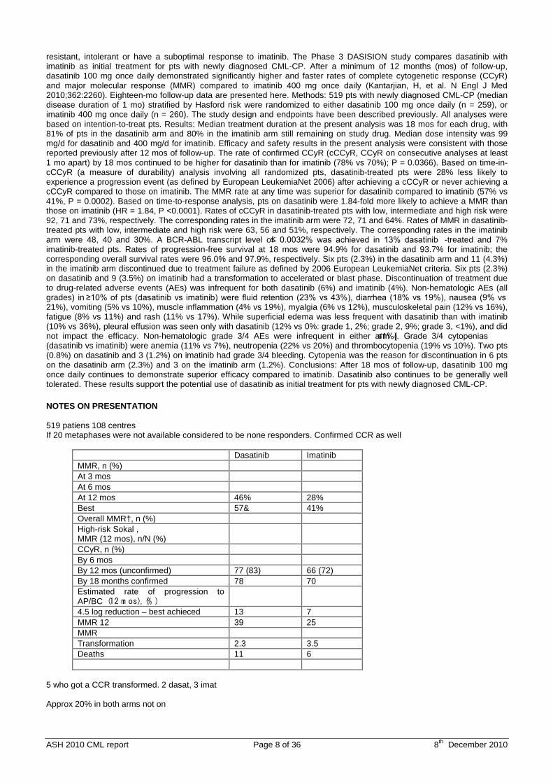
resistant, intolerant or have a suboptimal response to imatinib. The Phase 3 DASISION study compares dasatinib with imatinib as initial treatment for pts with newly diagnosed CML-CP. After a minimum of 12 months (mos) of follow-up, dasatinib 100 mg once daily demonstrated significantly higher and faster rates of complete cytogenetic response (CCyR) and major molecular response (MMR) compared to imatinib 400 mg once daily (Kantarjian, H, et al. N Engl J Med 2010;362:2260). Eighteen-mo follow-up data are presented here. Methods: 519 pts with newly diagnosed CML-CP (median disease duration of 1 mo) stratified by Hasford risk were randomized to either dasatinib 100 mg once daily (n = 259), or imatinib 400 mg once daily (n = 260). The study design and endpoints have been described previously. All analyses were based on intention-to-treat pts. Results: Median treatment duration at the present analysis was 18 mos for each drug, with 81% of pts in the dasatinib arm and 80% in the imatinib arm still remaining on study drug. Median dose intensity was 99 mg/d for dasatinib and 400 mg/d for imatinib. Efficacy and safety results in the present analysis were consistent with those reported previously after 12 mos of follow-up. The rate of confirmed CCyR (cCCyR, CCyR on consecutive analyses at least 1 mo apart) by 18 mos continued to be higher for dasatinib than for imatinib (78% vs 70%); P = 0.0366). Based on time-in-cCCyR (a measure of durability) analysis involving all randomized pts, dasatinib-treated pts were 28% less likely to experience a progression event (as defined by European LeukemiaNet 2006) after achieving a cCCyR or never achieving a cCCyR compared to those on imatinib. The MMR rate at any time was superior for dasatinib compared to imatinib (57% vs 41%, P = 0.0002). Based on time-to-response analysis, pts on dasatinib were 1.84-fold more likely to achieve a MMR than those on imatinib (HR = 1.84, P <0.0001). Rates of cCCyR in dasatinib-treated pts with low, intermediate and high risk were 92, 71 and 73%, respectively. The corresponding rates in the imatinib arm were 72, 71 and 64%. Rates of MMR in dasatinib-treated pts with low, intermediate and high risk were 63, 56 and 51%, respectively. The corresponding rates in the imatinib arm were 48, 40 and 30%. A BCR-ABL transcript level of ≤ 0.0032% was achieved in 13% dasatinib -treated and 7% imatinib-treated pts. Rates of progression-free survival at 18 mos were 94.9% for dasatinib and 93.7% for imatinib; the corresponding overall survival rates were 96.0% and 97.9%, respectively. Six pts (2.3%) in the dasatinib arm and 11 (4.3%) in the imatinib arm discontinued due to treatment failure as defined by 2006 European LeukemiaNet criteria. Six pts (2.3%) on dasatinib and 9 (3.5%) on imatinib had a transformation to accelerated or blast phase. Discontinuation of treatment due to drug-related adverse events (AEs) was infrequent for both dasatinib (6%) and imatinib (4%). Non-hematologic AEs (all grades) in ≥10% of pts (dasatinib vs imatinib) were fluid retention (23% vs 43%), diarrhea (18% vs 19%), nausea (9% vs 21%), vomiting (5% vs 10%), muscle inflammation (4% vs 19%), myalgia (6% vs 12%), musculoskeletal pain (12% vs 16%), fatigue (8% vs 11%) and rash (11% vs 17%). While superficial edema was less frequent with dasatinib than with imatinib (10% vs 36%), pleural effusion was seen only with dasatinib (12% vs 0%: grade 1, 2%; grade 2, 9%; grade 3, <1%), and did not impact the efficacy. Non-hematologic grade 3/4 AEs were infrequent in either arm (≤1%). Grade 3/4 cytopenias (dasatinib vs imatinib) were anemia (11% vs 7%), neutropenia (22% vs 20%) and thrombocytopenia (19% vs 10%). Two pts (0.8%) on dasatinib and 3 (1.2%) on imatinib had grade 3/4 bleeding. Cytopenia was the reason for discontinuation in 6 pts on the dasatinib arm (2.3%) and 3 on the imatinib arm (1.2%). Conclusions: After 18 mos of follow-up, dasatinib 100 mg once daily continues to demonstrate superior efficacy compared to imatinib. Dasatinib also continues to be generally well tolerated. These results support the potential use of dasatinib as initial treatment for pts with newly diagnosed CML-CP. NOTES ON PRESENTATION 519 patiens 108 centres If 20 metaphases were not available considered to be none responders. Confirmed CCR as well
Dasatinib Imatinib MMR, n (%) At 3 mos At 6 mos At 12 mos 46% 28% Best 57& 41% Overall MMR†, n (%) High-risk Sokal , MMR (12 mos), n/N (%)
CCyR, n (%) By 6 mos By 12 mos (unconfirmed) 77 (83) 66 (72) By 18 months confirmed 78 70 Estimated rate of progression to AP/BC(12 m os), (% )
4.5 log reduction – best achieced 13 7 MMR 12 39 25 MMR Transformation 2.3 3.5 Deaths 11 6
5 who got a CCR transformed. 2 dasat, 3 imat Approx 20% in both arms not on

ASH 2010 CML report Page 9 of 36 8th December 2010
6% AE for dasat 11 deaths in dasat , 6 deaths in imatinib. 4 infections deaths on dasat vs nil on imatinib. Don’t think they were neutropenic. Pleural effusions of all grades 12% on dasat 31 of 258 pleural effusions Grade 2 pleural effusions 90% of pts with pleural eff get CCR 3 patients needed a chest tap. Reduces phos Forest plot of AEs
3.3. [LBA-6] A Randomized Phase II Trial of Dasatinib 100 Mg Vs Imatinib 400 Mg In Newly Diagnosed Chronic Myeloid Leukemia In Chronic Phase (CML-CP): The S0325 Intergroup Trial. Radich. Background. The optimal tyrosine kinase inhibitor (TKI) for patients (pts) with newly diagnosed CML-CP is unknown. While dasatinib (DAS) is a more potent TKI in vitro than imatinib (IM), it is unclear if this will translate into improved long-term clinical outcomes for pts with newly diagnosed CML-CP. In this open‑ label phase II trial pts with newly diagnosed CML-CP were randomized to IM 400 mg po qd or DAS 100 mg po qd by four North American cooperative groups (SWOG, ECOG, CALGB, NCIC-CTG). The primary endpoint was >4 log reduction in BCR‑ ABL transcript at 12 months (mos). The study design, with 240 evaluable pts, provided >90% power to detect a difference in this endpoint of >20 percentage points (two-sided alpha=5%). Patients. 253 pts were randomized (12/2006 to 2/2009). Seven were ineligible, primarily due to diagnosis other than CML-CP, or nonevaluable because they received no protocol treatment or withdrew consent. Pretreatment characteristics were balanced between the arms. Treatment outcomes. Outcomes of the 246 included pts (age 18‑ 90, median 49; 60% male; 35% / 30% with Hasford intermediate / high risk) are shown in Table 1. Eighteen DAS (15%) and 13 IM 400mg (11%) pts discontinued study drug because of a variety of toxicities. Eleven pts (3 DAS [2%], 8 IM 400mg [7%]) discontinued due to refusal, and 36 others (12 DAS [10%], 24 IM 400mg [20%]) for other reasons, most often physician or pt concerns about inadequate response, recurrence or progression. Molecular response at 12 months was deeper in the DAS arm (median 3.3 log reduction in BCR-ABL transcript level vs 2.8 with IM 400mg; Wilcoxon P=0.048), although the proportions achieving >4 log or >4.5 log reductions did not differ significantly (molecular response at 12 mos was based on 189 rather than the planned 240 pts, but this provided >80% power to detect a difference of >20 percentage points). The rates of hematologic CR (HemCR) and cytogenetic CR (CCyR) were not significantly higher with DAS, though 11% and 5% of DAS and IM 400mg pts were not adequately assessed for HemCR, and CCyR data were only available for 51% of pts. Overall survival (OS) and progression-free survival (PFS) were similar in the two arms, with very few deaths, relapses or progressions. Among pts with HemCR, 2-year relapse-free survival was 97% in the DAS arm, 95% in the IM 400mg arm. Toxicity. There were no fatal toxicities. The most common grade 3 and 4 toxicities were hematologic, including thrombocytopenia (<50x109/L) in 18% and 8% of DAS and IM 400mg pts, respectively (P=0.024). A variety of grade 4 non-hematologic toxicities were reported for 6% of DAS pts but no IM 400mg pts. An additional 30% and 17% of DAS and IM 400mg pts had a variety of grade 3 non-hematologic toxicities, while another 57% and 79% had non-hematologic grade 1-2 toxicities. Pleural effusion of any grade was reported for 11% and 2% of DAS and IM 400mg pts (P=0.0017); <2% in either arm were grade 3. Deaths. Seven pts have died, all >8 months after entering the study. Three DAS pts died: one at 8 months after progression to blast crisis, one from lung cancer diagnosed 10 months after DAS started, and one in an automobile accident. Two IM 400mg pts died of CML, and two others (ages 70 and 75 at treatment start) of cardiac arrest unrelated to CML or treatment. Conclusions. Both IM 400mg and DAS are highly effective and generally well-tolerated therapies for newly diagnosed CML-CP. DAS induced deeper molecular responses at 12 months, but not significantly higher rates of >4 log or >4.5 log reduction in BCR-ABL, compared to IM 400mg. 12-month PFS and OS were similar between the two arms, with very few events so far. DAS was associated with more grade 3-4 toxicity. Clinical follow-up is continuing to study whether the short-term deeper molecular response seen with DAS will translate into improved long-term outcomes. Table 1. Outcomes on study S0325. OS12: overall survival at 12 mos; PFS12: progression-free survival at 12 mos.
IM 400mg (n=123) DAS (N=123) 2-sided P* HemCR 111 (90%) 104 (86%) 0.25 CCyR 40/58 (69%) 55/67 (82%) 0.097 >3 log 39/90 (43%) 58/99 (59%) 0.042 >4 log 18/90 (20%) 27/99 (27%) 0.31 >4.5 log 13/90 (14%) 21/99 (21%) 0.26 OS12 99% 100% 0.60 PFS12 96% 99% 0.19 * Fisher's exact test for HemCR, CCyR, and molecular response; logrank test for OS, PFS.
3.4. [207] ENESTnd Update: Continued Superiority of Nilotinib Versus Imatinib In Patients with Newly Diagnosed Chronic Myeloid Leukemia In Chronic Phase (CML-CP). Hughes. Background: Results from the phase 3, international, randomized ENESTnd trial have demonstrated the superior efficacy of nilotinib over imatinib with significantly higher rates of major molecular response (MMR), complete cytogenetic response (CCyR), and with significantly lower rates of progression to AP/BC on treatment. Here, we present data with a median follow-up of 18 months. Methods: 846 CML-CP patients were randomized to nilotinib 300 mg twice daily (bid) (n = 282), nilotinib 400 mg bid (n = 281), and imatinib 400 mg once daily (n = 283). Primary endpoint was MMR (≤ 0.1% BCR -ABLIS) rate “at” 12 months, as previously presented. Key secondary

ASH 2010 CML report Page 10 of 36 8th December 2010
endpoint was durable MMR at 24 months. Other endpoints assessed at 24 months include progression to AP/BC (with and without clonal evolution), event-free survival, progression-free survival, and overall survival (OS). Results: With a median follow-up of 18 months, the overall best MMR rate was superior for nilotinib 300 mg bid (66%, P < .0001) and nilotinib 400 mg bid (62%, P < .0001) compared with imatinib (40%). Superior rates of MMR were observed in both nilotinib arms compared with the imatinib arm across all Sokal risk groups (Table). The overall best rate of BCR-ABLIS £ 0.0032% (equivalent to complete molecular response, CMR) was superior for nilotinib 300 mg bid (21%, P < .0001) and nilotinib 400 mg bid (17%, P < .0001) compared with imatinib (6%). The overall best CCyR rate was superior for nilotinib 300 mg bid (85%, P < .001) and nilotinib 400 mg bid (82%, P = .017) compared with imatinib (74%). The superior efficacy of nilotinib was further demonstrated using the 2009 European LeukemiaNet (ELN) 12-month milestone in which fewer patients had suboptimal response or treatment failure on nilotinib 300 mg bid (2%, 3%) and nilotinib 400 mg bid (2%, 2%) vs imatinib (11%, 8%). Rates of progression to AP/BC on treatment were significantly lower for nilotinib 300 mg bid (0.7%, P = .006) and nilotinib 400 mg bid (0.4%, P = .003) compared with imatinib (4.2%). The rate of progression on treatment was also significantly lower for nilotinib when including clonal evolution as a criteria for progression (Table). There were fewer CML-related deaths on nilotinib 300 mg bid (n = 2), and 400 mg bid (n = 1) vs imatinib (n = 8). Estimated OS rate (including data from follow-up after discontinuation) at 18 months was higher for nilotinib 300 mg bid (98.5%, P = .28) and nilotinib 400 mg bid (99.3%, P = .03) vs imatinib (96.9%). Both drugs were well-tolerated. Discontinuations due to adverse events or laboratory abnormalities were lowest for nilotinib 300 mg bid (7%) compared with nilotinib 400 mg bid (12%) and imatinib (9%). With longer follow up there has been minimal change in the occurrence of AEs. Minimum 24-month follow-up data for all patients will be presented. Conclusions: With longer follow-up, nilotinib was associated with a significantly lower rate of progression to AP/BC on treatment and lower rates of suboptimal response or treatment failure vs imatinib. Nilotinib resulted in fewer CML-related deaths and a higher OS rate vs imatinib. Nilotinib induced superior rates of MMR, CMR, and CCyR vs imatinib in patients with newly diagnosed CML-CP. Taken together, these data support nilotinib as a new standard of care for patients with newly diagnosed CML.
Overall Efficacy with Median 18-Month Follow-up
Nilotinib 300 mg bid
(n = 282)
Nilotinib 400 mg bid
(n = 281)
Imatinib 400 mg qd (n = 283)
MMR, % 66 P < .0001*
62 P < .0001*
40
by Sokal, % Low (n = 103, n = 103, n = 104) 70 69 51 Intermediate (n = 101, n = 100, n = 101) 67 63 39 High (n = 78, n = 78, n = 78) 59 51 28 BCR-ABLIS £ 0.0032% , % 21
P < .0001* 17
P < .0001* 6
CCyR, % 85 P < .001*
82 P = .017*
74
Suboptimal response† (at 12 months), % 2 2 11 Treatment failure† (at 12 months), % 3 2 8 Progression to AP/BC Excluding clonal evolution, n (%)
2 (0.7) P = .006**
1 (0.4) P = .003**
12 (4.2)
Including clonal evolution, n (%) 2 (0.7) P <.001**
3 (1.2) P = .002**
17 (6.9)
Total deaths, patients (n) 5 2 9 CML-related deaths, patients (n) 2 1 8 Estimated OS (at 18 months), % 98.5
P = .28** 99.3
P = .03** 96.9
* CMH test stratified by Sokal vs imatinib ** Log-rank test stratified by Sokal vs imatinib for time to AP/BC and OS † According to 2009 ELN criteria at 12 months for suboptimal response (PCyR) and treatment failure (less than PCyR, loss of CHR, loss of CCyR, progression to AP/BC, or clonal evolution)
Treatment Arm Nilotinib,
300 mg BID (N = 282)
Nilotinib, 400 mg BID (N = 281)
Imatinib, 400 mg QD (N = 283)
MMR, n (%) At 3 mos At 6 mos At 12 mos 44 43 22 24 62 59 37 Overall MMR†, n (%) High-risk Sokal , MMR (12 mos), n/N (%)

ASH 2010 CML report Page 11 of 36 8th December 2010
CCyR, n (%) By 6 mos By 12 mos Estimated rate of progression to AP/BC(12 m os), (% )
24 Discon 26 22 33 Receiving planned dose at 24 m 77 74 70 CMR 4.5 best (0.0032%??) 26 21 10 Suboptimal response 18 Treatment failure 18 4 4 16 Progression AP/BC 0.7 1.1 4.2% 24 PFS Deaths 9 6 11 OS re CML deaths 98.9% 98.9 96.7
Elevations of lipase ATL, bili and glucose slightly higher with nilot Rash headache pruritus and alopecia more common in nilot Less than 2% lost MMR between 1 and 2 years Overall discon about 20% Dynamics abs 3431 Hochaus Minimum of 24 months follow up Qns. Quite a few non-CML related deaths in both studies on new intervention
3.5. [208] An Ongoing Phase 3 Study of Bosutinib (SKI-606) Versus Imatinib In Patients with Newly Diagnosed Chronic Phase Chronic Myeloid Leukemia. Gambacorti-Passerini . Bosutinib is an orally bioavailable dual Src/Abl tyrosine kinase inhibitor (TKI), with minimal inhibitory activity against PDGFR or c-kit. In a phase 2 study, bosutinib demonstrated activity in patients with Philadelphia chromosome–positive (Ph+) chronic phase (CP) chronic myeloid leukemia (CML) in the second- and third-line treatment settings (Cortes JE, et al. ASCO 2010, Abstract #6502; Khoury JH, et al. ASCO 2010, Abstract #6514), as well as in patients with advanced Ph+ leukemias (Gambacorti-Passerini C, et al. ASCO 2010, Abstract #6509) following resistance or intolerance to imatinib and other TKIs. The current randomized, open-label, phase 3 study compared the activity and safety of bosutinib with that of imatinib in newly diagnosed patients with CP CML. The study enrolled adults aged ³18 years with cytogenetic diagnosis of Ph+ CP CML within 6 months, adequate hepatic and renal function, and an Eastern Cooperative Oncology Group (ECOG) performance status of 0 or 1. Patients were randomized to daily oral treatment with 500 mg bosutinib or 400 mg imatinib. Adverse events were graded using the National Cancer Institute Common Terminology Criteria, version 3.0. The primary efficacy endpoint was the rate of complete cytogenetic response (CCyR) at 1 year; the rates of hematologic response, molecular response, and progression and transformation to accelerated or blast phase were also evaluated. The study randomized 502 patients: 56.6% male, median age of 48 years (range, 18-91 years), and median time since diagnosis of 0.7 months (range, -0.3-7.9 months; the range minimum is negative due to CML diagnosis during the study screening period, and the range maximum is >6 months because of 1 patient considered a major protocol violator). The median duration of treatment was 11.1 months (range, 0.03-24.8 months). At Week 48 (approximately 11 months), 71.5% and 74.8% of patients (both treatment arms combined) were in CCyR and complete hematologic response (CHR), respectively. During the study, 81.4% of patients achieved a CCyR at or before Week 48, with a median time to CCyR of 24 weeks; 82.6% of patients achieved a CHR, with a median time to CHR of 8 weeks; and 40.6% of patients achieved a major molecular response (MMR), with a median time to MMR of 49 to 61 weeks for the 2 treatment arms. For the combined treatment arms, common treatment-emergent adverse events included diarrhea (43.7%), nausea (32.3%), vomiting (22.0%), rash (16.8%), pyrexia (11.6%), and fatigue (11.0%). The only grade ³3 treatment-emergent adverse event observed in ³2% of patients was diarrhea (5.2%), which was usually limited to the first weeks of treatment. Grade ³3 hematologic laboratory abnormalities included neutropenia (14.2%), thrombocytopenia (12.4%), and anemia (5.8%). Other grade ³3 laboratory abnormalities (³5% of patients) included alanine aminotransferase elevation (11.6%), phosphatemia (7.6%), and aspartate aminotransferase elevation (6.4%). Overall, 22.2% patients discontinued therapy; adverse events led to discontinuation or death in 12.8% of patients, and 4.2% of patients discontinued due to disease progression. The high combined percentage of patients achieving MMR, CCyR, and CHR and the relatively low incidence of generally manageable grade ³3 events observed suggest good efficacy and an overall favorable safety profile. Data for individual treatment arms will be unblinded by the end of August 2010, and will be presented at the meeting.
Bosutinib Imatinib 250 252 ‘Evaluable’ 219 241 MMR, n (%) At 3 mos At 6 mos

ASH 2010 CML report Page 12 of 36 8th December 2010
At 12 mos Best Overall MMR†, n (%) High-risk Sokal , MMR (12 mos), n/N (%)
CCyR, n (%) 70 68 (NS!!) By ‘evaluable’ (dodgy…) Becomes signif By 12 mos (unconfirmed) Estimated rate of progression to AP/BC(12 m os), (% )
4.5 log reduction – best achieced MMR 12 39 26 (Signif) MMR Transformation 2% 4% Deaths 3 8 Discon 29 (19 due to
AE) 20
Treatment failure OS 99.5 96.2 (NS)
No PDGF-R or KIT activity Bos is 500mg daily More LFT elevation in bos. More hypophos in imat More GI tox with bosutinib (68% of all grades) Mostly in first couple of months.
3.6. [209] Selective Escalation of Imatinib Therapy and Early Switching to Nilotinib In De Novo Chronic Phase CML Patients: Interim Results From the TIDEL-II Trial. Yeung. Background: Although the majority of chronic phase (CP) Philadelphia positive (Ph+) chronic myelogenous leukemia (CML) patients (pts) achieve good disease control with imatinib, some pts demonstrate suboptimal responses. Early dose escalation or switching to nilotinib, a more potent BCR-ABL kinase inhibitor, as soon as suboptimal molecular response is recognised may improve response and disease outcome. Aim: To optimise clinical and molecular outcomes in Ph+ CML using imatinib (IM) as frontline therapy with selective IM dose escalation based on pharmacokinetic (PK) results and switching to nilotinib (NIL) in case of suboptimal response, or IM-intolerance. Method: TIDEL-II is a multicentre, single arm prospective ALLG trial in de novo CP-CML pts with 2 separate sequential cohorts. In Cohort I, pts are treated with IM 600mg/d up-front, aiming for BCR-ABL RQ-PCR target values of ≤ 10%, 1%, and 0.1% IS (major molecular response, MMR) at 3, 6, and 12 months respectively. Pts who do not reach these treatment targets are classified as suboptimal responders. Dose escalation to 800mg/d or maximal tolerated dose occurs if trough IM level is <1000ng/mL at day 22, or for suboptimal response. A switch to NIL (400mg bid) is triggered if molecular targets are still not met 3 months after IM escalation, or for loss or response, or for IM intolerance (Grade III/IV or persistent Grade II non-haematological toxicity). Results: 105 pts were assessed with median follow up of 18.9 months (range: 9-33) in cohort I. For pts with a minimum of 12 months follow up (n=80), complete cytogenetic response (CCR), MMR and complete molecular response (CMR)# rates at 12 months were 92%, 66% and 11% respectively. BCR-ABL levels at 3 months were predictive of MMR at 12 months, but not for CMR due to small pt numbers (Table 1). For pts who failed to achieve BCR-ABL of ≤10% at 3 months, the 12 month MMR rate was 25% (vs 5% in TIDEL-I where pts were also started on IM 600mg/d and suboptimal responders were dose escalated to IM 800mg/d). Of the 105 pts, 16 pts dose escalated IM due to a day 22 IM blood level <1000ng/mL, after which 2/16 switched to NIL (1 suboptimal, 1 intolerant); all achieved CCR. Twelve pts dose escalated for suboptimal response, 7 subsequently switched to NIL for again failing treatment targets. In all, 21/105 pts (20%) switched to NIL: 7 for suboptimal response and 14 for intolerance. The median time to switching and the median pre-switch prescribed IM dose were 468 days & 800mg/d for the suboptimal group; and 183 days & 600mg/d for the intolerant group respectively. Of these, 20/21 achieved or remained in CCR. At the time of switching to NIL, 19/21 pts were not in MMR. With a median follow-up of 295 days post switch to NIL, 9/12 intolerant pts (75%) achieved MMR, whereas 1/7 suboptimal IM responders (14%) achieved MMR (median follow up after switching: 286 days). Only 7/105 pts (7%) discontinued treatment: 4 for non-compliance, 1 pt with a T315I mutation and 2 pts with blast crisis (BC). Progression to BC was associated with detectable mutations: 1 pt with 4 different mutations including T315I and 1 pt with H396P mutation. The progression rate to AP/BC was 2%. The overall mutation rate was 5/105 (5%). The 2 pts who progressed and the pt who discontinued when a T315I mutation was detected were among the 28 pts with BCR-ABL values >1.0% at 3 months. In contrast, no resistant mutations were detected or transformations occurred in the 49 pts with BCR-ABL values ≤1.0% at 3 months. Conclusion: A strategy of selective intensification of BCR-ABL inhibitor therapy based on molecular response and PK values resulted in a 66% MMR rate by 12 months. Despite a minority of pts (20%) requiring a switch to NIL, this has enhanced the rate of MMR by 12 months when compared to IM intensification alone as seen in TIDEL-I where the rate of MMR and CMR by 12 months was 47% and 9% respectively. The IM intolerant pts demonstrated excellent response rates after switching to NIL. To date, the results from TIDEL-II compare favourably with other frontline strategies with regards to response and transformation rates. Table 1
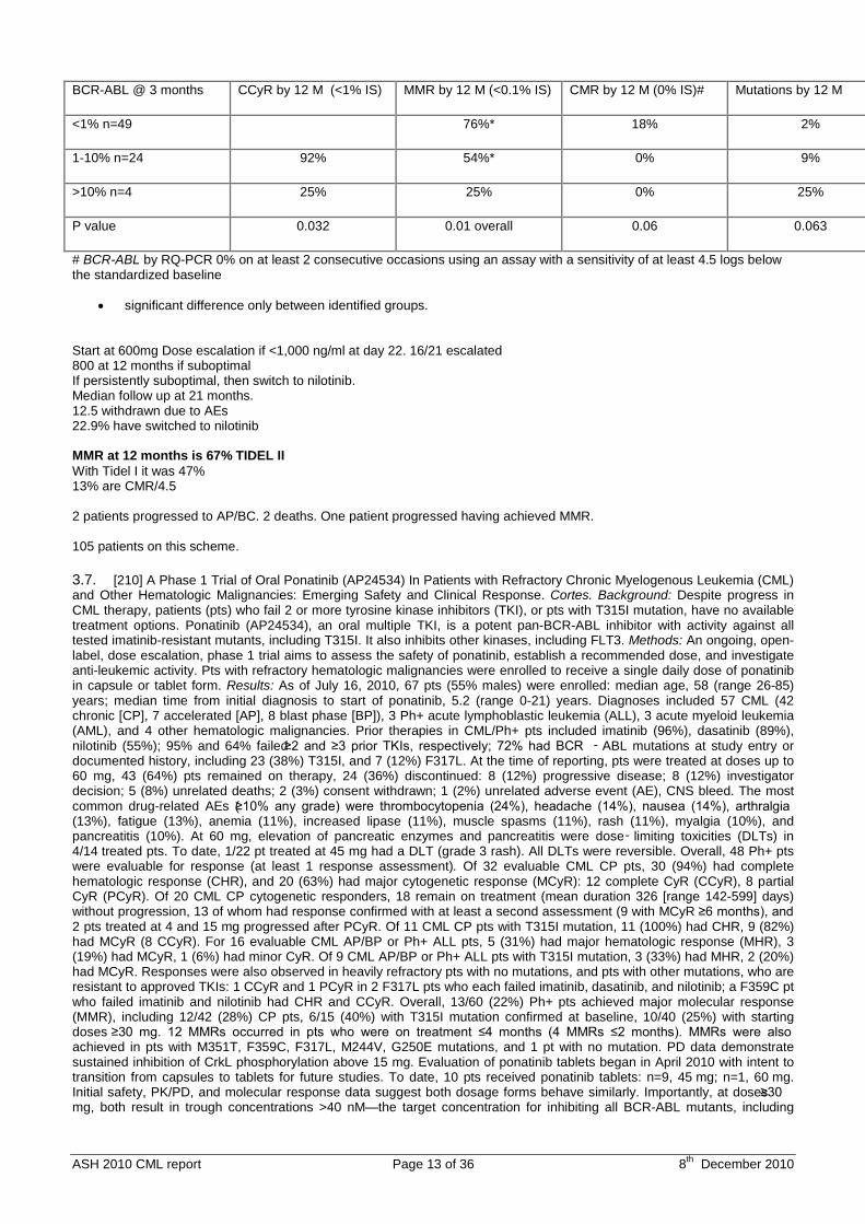
ASH 2010 CML report Page 13 of 36 8th December 2010
BCR-ABL @ 3 months CCyR by 12 M (<1% IS) MMR by 12 M (<0.1% IS) CMR by 12 M (0% IS)# Mutations by 12 M
<1% n=49 76%* 18% 2%
1-10% n=24 92% 54%* 0% 9%
>10% n=4 25% 25% 0% 25%
P value 0.032 0.01 overall 0.06 0.063
# BCR-ABL by RQ-PCR 0% on at least 2 consecutive occasions using an assay with a sensitivity of at least 4.5 logs below the standardized baseline
• significant difference only between identified groups. Start at 600mg Dose escalation if <1,000 ng/ml at day 22. 16/21 escalated 800 at 12 months if suboptimal If persistently suboptimal, then switch to nilotinib. Median follow up at 21 months. 12.5 withdrawn due to AEs 22.9% have switched to nilotinib MMR at 12 months is 67% TIDEL II With Tidel I it was 47% 13% are CMR/4.5 2 patients progressed to AP/BC. 2 deaths. One patient progressed having achieved MMR. 105 patients on this scheme.
3.7. [210] A Phase 1 Trial of Oral Ponatinib (AP24534) In Patients with Refractory Chronic Myelogenous Leukemia (CML) and Other Hematologic Malignancies: Emerging Safety and Clinical Response. Cortes. Background: Despite progress in CML therapy, patients (pts) who fail 2 or more tyrosine kinase inhibitors (TKI), or pts with T315I mutation, have no available treatment options. Ponatinib (AP24534), an oral multiple TKI, is a potent pan-BCR-ABL inhibitor with activity against all tested imatinib-resistant mutants, including T315I. It also inhibits other kinases, including FLT3. Methods: An ongoing, open-label, dose escalation, phase 1 trial aims to assess the safety of ponatinib, establish a recommended dose, and investigate anti-leukemic activity. Pts with refractory hematologic malignancies were enrolled to receive a single daily dose of ponatinib in capsule or tablet form. Results: As of July 16, 2010, 67 pts (55% males) were enrolled: median age, 58 (range 26-85) years; median time from initial diagnosis to start of ponatinib, 5.2 (range 0-21) years. Diagnoses included 57 CML (42 chronic [CP], 7 accelerated [AP], 8 blast phase [BP]), 3 Ph+ acute lymphoblastic leukemia (ALL), 3 acute myeloid leukemia (AML), and 4 other hematologic malignancies. Prior therapies in CML/Ph+ pts included imatinib (96%), dasatinib (89%), nilotinib (55%); 95% and 64% failed ≥2 and ≥3 prior TKIs, respectively; 72% had BCR ‑ ABL mutations at study entry or documented history, including 23 (38%) T315I, and 7 (12%) F317L. At the time of reporting, pts were treated at doses up to 60 mg, 43 (64%) pts remained on therapy, 24 (36%) discontinued: 8 (12%) progressive disease; 8 (12%) investigator decision; 5 (8%) unrelated deaths; 2 (3%) consent withdrawn; 1 (2%) unrelated adverse event (AE), CNS bleed. The most common drug-related AEs (≥10% any grade) were thrombocytopenia (24%), headache (14%), nausea (14%), arthralgia (13%), fatigue (13%), anemia (11%), increased lipase (11%), muscle spasms (11%), rash (11%), myalgia (10%), and pancreatitis (10%). At 60 mg, elevation of pancreatic enzymes and pancreatitis were dose‑ limiting toxicities (DLTs) in 4/14 treated pts. To date, 1/22 pt treated at 45 mg had a DLT (grade 3 rash). All DLTs were reversible. Overall, 48 Ph+ pts were evaluable for response (at least 1 response assessment). Of 32 evaluable CML CP pts, 30 (94%) had complete hematologic response (CHR), and 20 (63%) had major cytogenetic response (MCyR): 12 complete CyR (CCyR), 8 partial CyR (PCyR). Of 20 CML CP cytogenetic responders, 18 remain on treatment (mean duration 326 [range 142-599] days) without progression, 13 of whom had response confirmed with at least a second assessment (9 with MCyR ≥6 months), and 2 pts treated at 4 and 15 mg progressed after PCyR. Of 11 CML CP pts with T315I mutation, 11 (100%) had CHR, 9 (82%) had MCyR (8 CCyR). For 16 evaluable CML AP/BP or Ph+ ALL pts, 5 (31%) had major hematologic response (MHR), 3 (19%) had MCyR, 1 (6%) had minor CyR. Of 9 CML AP/BP or Ph+ ALL pts with T315I mutation, 3 (33%) had MHR, 2 (20%) had MCyR. Responses were also observed in heavily refractory pts with no mutations, and pts with other mutations, who are resistant to approved TKIs: 1 CCyR and 1 PCyR in 2 F317L pts who each failed imatinib, dasatinib, and nilotinib; a F359C pt who failed imatinib and nilotinib had CHR and CCyR. Overall, 13/60 (22%) Ph+ pts achieved major molecular response (MMR), including 12/42 (28%) CP pts, 6/15 (40%) with T315I mutation confirmed at baseline, 10/40 (25%) with starting doses ≥30 mg. 12 MMRs occurred in pts who were on treatment ≤4 months (4 MMRs ≤2 months). MMRs were also achieved in pts with M351T, F359C, F317L, M244V, G250E mutations, and 1 pt with no mutation. PD data demonstrate sustained inhibition of CrkL phosphorylation above 15 mg. Evaluation of ponatinib tablets began in April 2010 with intent to transition from capsules to tablets for future studies. To date, 10 pts received ponatinib tablets: n=9, 45 mg; n=1, 60 mg. Initial safety, PK/PD, and molecular response data suggest both dosage forms behave similarly. Importantly, at doses ≥30 mg, both result in trough concentrations >40 nM—the target concentration for inhibiting all BCR-ABL mutants, including

ASH 2010 CML report Page 14 of 36 8th December 2010
T315I. Conclusion: ponatinib has an acceptable safety profile at clinically effective doses in this refractory population. The 45 mg dose (tablet form) was chosen as the recommended dose for further study. There is strong and continually increasing evidence of anti-leukemic activity in pts with T315I mutations, and pts resistant to second generation TKIs. Emerging MMR data demonstrate early responses in pts refractory to second line agents. Garg. Blood 2009; 114: 4361. Failure free survival of 2nd lin about 20 months. FLT3 FGFR VEGFR PDGFR KIT Not aurora kinase Active against T315I No mutations that are resistant to ponatinib have yet been identified. All sorts Ph-pos disease 63% had a mutation at entry. 28% of the mutations were T315I 35% discontinued. Most common AE is rash – 22%. Arthralgia, headache. Lipase elevation. Some pancreatitis. 50% achieved CCR in CP CML overall 8 of 9 T315I achieved CCR. Pretty good! MMR 42% of CML CP 45 mg is the recommended phase 2 dose. Pancreatic DLT at 60mg. PACE study being set up. 44 of 74 had CML CP. All had received prior TKI. 95% had received >2 TKIs
4 Therapy: Optimizing Front-Line Therapy in CML [355-360]
4.1. [355] Impact of Variant t(9;22) and Additional Cytogenetic Aberrations at Diagnosis on Prognosis of CML. Leitner. Introduction: The prognostic relevance of variant t(9;22) and additional cytogenetic aberrations (ACA) at diagnosis of chronic myeloid leukemia (CML) is conflicting. Patients and Methods: We used baseline and outcome data of 1028 patients (607 male, 421 female, median age 53, range 16-88) with chronic phase CML randomized to the German CML-Study IV (imatinib [IM] 800 mg [n=264] vs IM 400 mg [n=253] vs IM 400 mg + IFN [n=281] vs IM 400 mg after IFN failure [n=108] vs IM 400 mg + AraC [n=122]) to investigate the impact of variant t(9;22) and of clonal ACA at diagnosis on time to complete cytogenetic remission (CCyR) and major molecular response (MMR), accepted markers of prognosis. Cytogenetic analysis was performed after 24- and/or 48 h culture on G-banded metaphases. If appropriate, fluorescent-in-situ-hybridization on metaphases was used in addition. Since lack of the Y chromosome is regarded as a negligible age-related, not leukemia-associated event, those patients were excluded from evaluation. Results: In total, 123/1028 patients (12%) showed additional cytogenetic findings at diagnosis: 52/1028 patients (5.1%) had variants of the t(9;22), 33/1028 patients (3.2%) lacked the Y chromosome, 38/1028 patients (3.7%) had other additional numerical or structural aberrations. 105/1028 patients (10.2%) had only one type of additional cytogenetic finding, while 18/1028 patients (1.8%) showed ≥ 2 types of additional cytogenetic findings. 905/1028 patients (88%) had no variant t(9;22) or ACA. Median age, sex and treatment were similarly distributed (Table 1). In 45/52 patients (86.5%) with variant t(9;22), one further chromosome was involved (three way translocation), whereas in 7/52 patients (13.5%) ≥ 2 chromosomes were involved (complex variant). No involvement of the chromosomes 10, 18, 20, 21, X, or Y has been found. For patients without variant t(9;22) and ACA, with variant t(9;22), with variant t(9;22) and ACA other than –Y, and with ACA other than -Y and variant t(9;22), median time (years) to CCyR was 0.98, 0.84, 1.08 and 1.34, median time (years) to MMR was 1.4, 1.55, 1.8 and 2.17, and probability (%, confidence interval) for 2 years overall survival was 0.97 (0.96-0.98), 0.96 (0.89-0.99), 0.95 (0.90-0.99) and 0.94 (0.85-0.99), respectively. There was no difference regarding time to CCyR, time to major molecular response (MMR) and 2 years overall survival between patients with variant t(9;22) or ACA compared to those without variant t(9;22) or ACA. Conclusion: We conclude that additional chromosomal abnormalities at diagnosis have no negative prognostic impact. This finding is hypothesis generating. For confirmation of this hypothesis longer observation of the course of patients with variant t(9;22) and ACA is needed.
variable no variant t(9;22) or ACA variant t(9;22) Variant t(9;22) and ACA
other than -Y ACA other than –Y and variant t(9;22)
n 905 52 90 38
Median age 52 56 54 53
Sex (% female) 41% 42% 40% 37%
IM after IFN failure 96 4 11 7
IM + Ara C 104 8 13 5
IM 400 mg 224 11 20 9
IM + IFN 253 12 20 8
ASH 2010 CML report Page 15 of 36 8th December 2010
IM 800 mg 228 17 26 9
NOTES ON PRESENTATION: 1028 patients looked at. 905 with standard 9;22. 12% had additional finding. Some benefit in fact for variant 9;22 in terms of early cytogenetic response. But overall no impact of ACAs on survival.
4.2. [356] Early Switching From Imatinib to Nilotinib In CML Patients Failing to Achieve Early Molecular Targets May Not Be An Effective Approach In Patients with Very Low OCT-1 Activity: A TIDEL II Sub-Study. White. We have previously demonstrated that CML patients with very low OCT-1 activity (Quartile 1 (Q1)<4 ng/200,000 cells) have significantly poorer outcomes with respect to response, mutation development, progression, and survival than patients with higher OCT-1 activity(OA). We have also demonstrated that the influx pump OCT-1 is associated with the active uptake of imatinib, but not nilotinib (NIL), into target CML cells. Hence, if the prognostic significance of OA primarily reflects IM transport we predicted that patients with very low OA who had poor initial responses to imatinib, should respond well when switched to NIL. In the Tidel II study CML-CP patients are treated with 600mg/day imatinib (IM) upfront and are dose escalated to 400mg BID or switched to NIL 400mg BID for either intolerance (Grade III/IV or persistent Grade II non-haematological toxicity) or failure to meet pre defined molecular targets. These targets are early markers of sub-optimal response, but not IM failure: failure to achieve <10% BCR-ABL IS by 3 months, <1% by 6 months and <0.1% by 12months. Hence this study is designed to enable early efficacious intervention in the case of poor response, to prevent frank therapeutic failure. In this study we examine the cohort of patients who have switched to NIL and compare their response on NIL to that achieved on IM. To date 63 patients on this trial have greater than 12 month molecular follow up, and OA quantified. 26/63 patients have OA<4ng/200,000 cells. As demonstrated in Table 1, and in keeping with previous findings, patients with low OA have significantly poorer rates of MMR by 12 months, and higher rates of study discontinuation when compared to patients with ≥4.0ng/200,000 (high OA). Importantly the study design of Tidel II has enabled the more careful assessment of therapeutic intervention. Of note 23% of patients with low OA had a dose increase and 42% switched to nilotinib because of failure to meet pre-stated milestones. This compares with only 8% of remaining patients being dose increased and 14% switching to NIL(p=0.007). With a median follow up after switching to NIL of 12 months (Low OA R=6-21 high OA R=9-12 p=0.887) all patients with high OA, not previously in MMR (n=4), achieved MMR within 6 months. In contrast 1/10 low OA patients not previously in MMR achieved MMR on NIL. Interestingly, not all patients with low OA were switched to NIL because of sub-optimal IM response. Four of this cohort were intolerant to 600mg IM (one of these 4 achieved MMR), while all patients in the high OA cohort were switched because of intolerance. This implies that a low OA is not overcome with early switch to NIL, despite NIL transport being unrelated to OCT-1. This may be due to other intrinsic factors, possibly related to leukaemia biology and majority cell type for which the OA assay may be a surrogate measure. Alternatively, the poor response to imatinib in low OA patients has facilitated the activation of other resistance mechanisms, that in turn lead to a poor response after switching implying that patients with low OA may require upfront treatment with a TKI that is OCT-1 independent. Importantly only 1 of the patients on NIL had a mutation prior to switching, with another patient transforming to blast crisis with a T315I mutation while on NIL. Thus the presence of kinase domain mutations does not explain these findings. Conclusions: Low OA is associated with poor response to IM. Here we show that the MMR rate for patients with low OA was not substantially improved by the TIDEL II treatment strategy. This is despite the protocol allowing for early dose escalation or switch for suboptimal response. This preliminary data suggests that patients with low OA may have more generalized insensitivity to kinase inhibitor therapy which is not simply related to drug transport. These are important findings in the setting of multiple TKIs being available, and suggest that in patients with very low OA a regimen of selective TKI intensification may not be the optimal approach.
4.3. [357] Superior CMR-Rates with Tolerability-Adapted Imatinib 800 Mg Vs. 400 Mg Vs. 400 Mg + IFN In CML: The Randomized German CML-Study IV. Hehlmann. Treatment of CML with imatinib of 400 mg can be unsatisfactory. Treatment optimization is warranted. The German CML-Study group has therefore conducted a randomized study comparing imatinib 800 mg vs 400 mg vs 400 mg + IFN. A significantly faster achievement of MMR at 12 months has been observed with imatinib 800 mg in a tolerability adapted manner and MMR by 12 months has been found to translate into better overall survival. Since stable CMR has been associated with durable off-treatment remissions we sought to analyse the impact of tolerability-adapted imatinib 800 mg on CMR and survival. Standardized determinations of molecular response and evaluation of its impact on outcome are goals of CML-Study IV. CMR4 is defined as a BCR-ABL/ABL ratio of <0,01 on the International Scale. From July 2002 – April 30, 2009 1022 newly diagnosed patients with CML in chronic phase were randomized, 1012 were evaluable (338 with imatinib 800 mg, 324 with imatinib 400 mg, 350 with imatinib plus IFN). Median observation time was 40 months. The median average daily imatinib doses were 628 mg in the 800 mg arm and 400 mg in the 400 mg based arms. The actual median daily doses in the 800 mg arm per 3-months periods were: 555 mg, 737 mg, 613 mg, 600 mg, and 600 mg thereafter, reflecting the run–in period with imatinib 400 mg for 6 weeks in the first period and the adaptation to tolerability from the third 3-months period onwards. Median daily imatinib doses in the 400 mg arms were 400 mg throughout. Adaptation of imatinib dose in the 800 mg arm according to tolerability is reflected by similar higher-grade adverse events rates (WHO grades 3 and 4) with all treatments. Significantly higher remission rates were achieved with imatinib 800 mg by 12 months. The cumulative incidences of CCR by 12 months were 63% [95%CI:56.4-67.9] with imatinib 800 mg vs 50% [95%CI:43.0-54.5] with the two 400 mg arms. The cumulative incidences of MMR by 12 months were 54.8% [95%CI:48.7-59.7] with imatinib 800 mg vs 30.8% [95%CI:26.6-36.1] with imatinib 400 mg vs 34.7% [95%CI:29.0-39.2] with imatinib + IFN. The cumulative incidences of CMR4 compared with the MMR incidences over the first 36 months are shown in Table 1. Imatinib 800 mg shows superior CMR4 rates over the entire 36 months period, CMR4 is reached significantly faster with imatinib 800 mg as compared to the 400 mg arms. The CMR4 rates reach 56.8% by 36 months [95%CI:49.4-63.5] as compared to 45.5% with imatinib 400 mg [95%CI:38.7-51.0] and 40.5% with imatinib plus IFN [95%CI:34.6-46.3]. Most patients have stable CMR4 over the entire period. In summary, superior CMR4 rates are achieved with high-dose imatinib adapted to good tolerability, and more patients in the tolerability-adapted 800 mg arm have stable CMR4 qualifying for treatment discontinuation as compared to the 400 mg based arms. With improved application imatinib remains first choice

ASH 2010 CML report Page 16 of 36 8th December 2010
for early CML. Time after start of treat-ment
(months)
Cumulative incidences
MMR(%) CMR4 (%)
IM400 n=306
D IM800 n=328
D IM400 +IFN n=336
IM400 n=306
D IM800 n=328
D IM400 +IFN n=336
6 8.6 9.5 18.1 9.7 8.4 3 0.7 3.7 1.3 2.4
12 30.8 24.0 54.8 20.1 34.7 7.5 12.3 19.8 7.4 12.4
18 50.3 18.1 68.4 14.3 54.1 21.2 12.2 33.4 9.8 23.6
24 63 13.0 76.0 13.2 62.8 30.7 12.3 43 13 30.0
36 79.3 2.3 81.6 10.9 70.7 45.5 11.3 56.8 16.3 40.5
NOTES ON PRESENTATION: 1022 patients randomized. 28-48 month observation periods. Clearly faster response but the curves come together at 3 years. Most 800 patients actually end up on 600. OS is 92%. NO differences betwwen arms. Achievement of MMR by 12 months is associated with improved survival – didn’t quite get this.
4.4. [358] Lymphocytosis Following First-Line Treatment for CML In Chronic Phase with Dasatinib Is Associated with Improved Responses: A Comparison with Imatinib. Schiffer. Background: Persistent expansion of clonal cytotoxic T cells or NK cells has been described in patients with chronic myeloid leukemia (CML) and Ph+ acute lymphoblastic leukemia (ALL) receiving dasatinib. These small studies have suggested a relationship between the development of T/NK lymphocytosis with both toxicity and improved response. A recent analysis of CML patients who were resistant to or intolerant of imatinib indicated that lymphocytosis occurs in ~30% of patients in all stages of CML after treatment with dasatinib (Schiffer 2010 ASCO abstract #6553). In addition, lymphocytosis was associated with improved cytogenetic response and an increased incidence of pleural effusions. The DASISION study (CA180-056) compared dasatinib with imatinib in patients with newly diagnosed, previously untreated chronic phase CML. This subanalysis of the DASISION data was undertaken to determine the safety profile, responses, and outcomes in those patients with sustained lymphocytosis. Methods: Patients enrolled in the DASISION study were retrospectively evaluated for lymphocytosis (N=516). Lymphocytosis was defined as lymphocytes >3.6 x 10⁹/L on ≥2 occasions after 28 days of treatment. Immunophenotyping was not done as part of these studies. Rates of major cytogenetic response (MCyR) and complete cytogenetic response (CCyR), overall survival (OS), progression-free survival (PFS), and adverse events (AEs) were measured in those with and without lymphocytosis. The median follow-up was 14.0 and 14.3 months for dasatinib and imatinib, respectively. Results: Lymphocytosis occurred more frequently and sooner in patients treated with dasatinib compared with imatinib (23.6% vs 5.4% and 3.0 months vs 4.7 months, respectively). Dasatinib-treated patients who developed lymphocytosis had lower baseline Hasford risk scores, whereas imatinib-treated patients who developed lymphocytosis had high Hasford risk scores. In the presence or absence of lymphocytosis, MCyR and CCyR occurred more frequently with dasatinib as compared with imatinib. In dasatinib treated patients, lymphocytosis was associated with a higher MCyR rate (91.8% with vs 83.3% without) and CCyR rate (83.6% with vs 75.1% without). In imatinib-treated patients, lymphocytosis was associated with a lower MCyR rate (50.0% with vs 82.8% without) and CCyR rate (50.0% with vs 69.7% without). Patients with lymphocytosis, compared with those without it, had higher rates of pleural effusion (any grade) on dasatinib (18.0% vs 7.6%, respectively). Only 1 patient treated with imatinib, who did not have lymphocytosis, experienced pleural effusion. Dasatinib-treated patients with lymphocytosis, compared with those without it, had higher rates of fatigue (16.4% vs 9.1%,); this trend was reversed for imatinib-treated patients (7.1% vs 11.9%). Patients with lymphocytosis, compared with those without it had lower rates of myalgias and arthralgias (all grades) on both the dasatinib arm (11.5% vs 18.8%) and the imatinib arm (7.1% vs 24.2%). Conclusions: Lymphocytosis occurred much more frequently and sooner in patients treated with dasatinib than with imatinib. Lymphocytosis was associated with improved responses (MCyR and CCyR) in dasatinib-treated patients. An apparent inverse relationship was observed in imatinib-treated patients, although the number of patients with lymphocytosis was small. It remains to be determined how lymphocytosis, baseline Hasford risk score, and response are correlated. It is possible that some of the antileukemic effects of dasatinib are produced by an immunomodulatory mechanism. Lymphocytosis in dasatinib-treated patients was associated with an increased rate of pleural effusion and a possible decrease in myalgias and arthralgias. Longer follow-up is needed to assess whether lymphocytosis has an effect on PFS.
Lymphocytosis,
n (%)
Cumulative MCyR, n (%)
Cumulative CCyR, n (%)
Patients on treatment with CCyR at 12 months, n (%)
dasatinib (100 mg) Yes 61 (23.6) 56 (91.8) 51 (83.6) 50 (82)

ASH 2010 CML report Page 17 of 36 8th December 2010
(n=258) No 197 (76.4) 164 (83.2) 148 (75.1) 143 (72.6)
P value 0.14 0.22 0.17
imatinib (400 mg) Yes 14 (5.4) 7 (50) 7 (50) 7 (50)
(n=258) No 244 (94.6) 202 (82.8) 170 (69.7) 160 (65.6)
P value 0.007 0.14 0.26
NOTES ON PRESENTATION: Porkka K Cancer 2010 116:377 Kruetzman Blood 2010 116: 772 T/NK cell lymphocytosis. Not present all the time. Possible that incidence is underestimated as the lymphocytosis occurs within 1 hour of taking drug (see Porkka abstract). >3.6 on > 2 occasions after 28 days on treatment. No immunophenotyping done. Cumulative incidence in das arm was 26% in imatinib was 6%. On Das MMR 68 vs 54 for lymphocytosis Landmark analysis @ 12 months. CCR 86 vs 83 (i.e. NO difference). MMR 56 vs 41. Dasatinib +/- lymphocytosis. Pleural effusions: 22 vs 9% (signif)
4.5. [359] Excellent Outcomes at 3 Years with Nilotinib 800 Mg Daily In Early Chronic Phase, Ph+ Chronic Myeloid Leukemia (CML): Results of a Phase 2 GIMEMA CML WP Clinical Trial. Rosti. Background: Nilotinib is a potent and selective inhibitor of BCR-ABL. In the phase 3 ENESTnd trial, nilotinib demonstrated superior efficacy to imatinib with higher and faster molecular responses. With a median follow-up of 18.5 months (ASCO/EHA 2010), the rates of progression to accelerated or blast phase (AP/BC) were 0.7% and 0.4% with nilotinib 300 mg and 400 mg BID, respectively, and significantly lower in comparison to imatinib (4.2% P = .006 and .003, respectively). Based on the results of the ENESTnd trial, nilotinib has been approved (FDA) for the frontline treatment of Ph+ CML. With imatinib 400 mg (IRIS trial), the rate of any event and the rate of progression to AP/BC were higher during the first 3 years on treatment (15.6% and 6.1%, respectively). Consequently, a confirmation of the durability of nilotinib responses at 3 years is extremely important. Aims: To evaluate responses (either cytogenetic and molecular) and to investigate outcomes of patients treated for 3-years with nilotinib 400 mg BID as frontline therapy. Outcomes evaluated include Overall Survival (OS), Progression-Free Survival (PFS), Failure-Free Survival (FFS) and Event-Free Survival (EFS). Methods: A multicentre phase 2 trial (nilotinib 400 mg BID) was conducted by the GIMEMA CML Working Party (ClinicalTrials.gov.NCT00481052). The median follow-up is currently 30 months (3 years by November 2010). Definitions: Major Molecular Response (MMR): BCR-ABL/ABL ratio < 0,1% IS; Complete Molecular Response (CMR): undetectable transcript levels and nested PCR negative; failures: no CHR at 3 months, no CgR at 6 months, no PCgR at 1 year, no CCgR at 18 months, loss CHR or CCgR, progression and death (according to the revised European LeukemiaNet recommendations); events: failures and treatment discontinuation for any reason. All the analysis has been made according to the intention-to-treat principle. Results: 73 patients have been enrolled; median age 51 years (range 18-83); 45% low, 41% intermediate and 14% high Sokal risk. The cumulative CCgR rate (primary endpoint) at 12 months was 100%. CCgR at each milestone: 78% at 3 months, 96% at 6, 12 and 18 months, 92% at 24 months. The cumulative rate of MMR was 96%, while the rates of MMR at 3, 6, 12, 18 and 24 months were 52%, 66%, 85%, 81% and 82%, respectively. The cumulative rate of CMR was 41%, while the rates of CMR at 12 and 24 months were 7% and 12%, respectively. None of the patients who achieved a MMR progressed to AP/BC. Only one patient progressed at 6 months to AP/BC: a 63 years old female with a high Sokal risk disease in CCgR at 3 months, who developed a T315I mutation. During the first 12 months, the mean daily dose was 600-800 mg, 400-599 mg, and less than 400 mg in 74%, 18% and 8% of patients, respectively. The nilotinib last daily dose was as follows: 800 mg in 48 (71%) patients, 400 mg in 19 (28%) patients and 200 mg in 1 (1%) patient. Adverse events (AEs) were mostly grade 1 or 2 and manageable with appropriate dose adaptations. Two patients (3%) showed a prolongation of the QTcF above 450 msec (none above 50 msec). Four events lead to permanent discontinuation of nilotinib: 3 patients discontinued after 9, 15 and 27 months on treatment for recurrent episodes of amylase and/or lipase increase (no pancreatitis) and 1 patient after 25 months due to atrial fibrillation, unrelated to study drug. Three of them are currently on imatinib second-line and 1 on dasatinib third-line. Overall, 5 events have been recorded so far (1 progression to AB/BC and 4 permanent discontinuation of nilotinib due to AEs). At 30 months the OS, PFS and FFS are 99% and the EFS is 92%. Conclusions: The rate of failures was very low during the first 3 years. Responses remain stable. The very high rates of responses achieved during the first 12 months on treatment are being translated into optimal outcome for most of the patients.
4.6. [360] Molecular Response After 3 Months of 1st Line Imatinib Therapy Is Predictive for Treatment Failure and Disease Progression In Patients with Chronic Phase Chronic Myeloid Leukemia - a Follow-up Analysis of the German CML Study IV. Hanfstein. Introduction: The lack of a sufficient response to first line imatinib treatment has been observed in a substantial proportion of CML patients and has been associated with an inferior survival. Therefore, response criteria have been defined to identify patients with treatment failure. A change of drug therapy to 2nd generation tyrosine kinase inhibitors or allogeneic stem cell transplantation is recommended for this group of patients (European LeukemiaNet, ELN, Baccarani

ASH 2010 CML report Page 18 of 36 8th December 2010
et al., JCO 2009). We sought to evaluate the predictive value of early molecular response landmarks for treatment failure and disease progression to identify patients at risk and to provide a guidance for the interpretation of BCR-ABL levels. Patients and methods: 949 patients included into the randomized German CML Study IV and treated with an imatinib based therapy consisting of standard dose imatinib (400 mg/d), high dose imatinib (800 mg/d) and combinations of standard dose imatinib with low dose cytarabine or interferon alpha were evaluable for molecular and cytogenetic analysis. BCR-ABL (IS) was determined by quantitative RT-PCR. The type of BCR-ABL transcript (b2a2, n=424; b3a2, n=464; b2a2 and b3a2, n=148) was defined by multiplex PCR. Patients with atypical BCR-ABL transcripts were excluded from the analysis. Cytogenetic response (CyR) was determined by G-banding metaphase analyses. Treatment failure has been defined according to ELN criteria as a lack of major CyR after 12 months and a lack of complete CyR after 18 months of imatinib treatment, respectively. CyR data were available for 479 pts between 12 and 18 months with a subset of 289 pts evaluable for 3 month molecular response (CyR data after 18 months, n=532; 3 month molecular subset, n=289). Disease progression comprises the incidence of accelerated phase, blast phase and death. Median follow-up for disease progression was 35 months (range 2-85). Fisher's exact test has been performed to evaluate the prognostic significance of 3 month BCR-ABL landmarks for 12 month and 18 month treatment failure. A landmark analysis has been performed for disease progression (logrank test). Results: In 20 of 289 evaluable pts treatment failure has been observed after 12 months, and in 29 of 289 pts after 18 months. 24 of 570 evaluable pts showed a disease progression after a median of 18 months (range 5-71). A stratification into three groups at 3 months reveals a significant difference concerning treatment failure between pts with BCR-ABL levels between 1% and 10% and those with BCR-ABL levels >10%. With regard to disease progression there is a statistical trend. Comparing two groups the 10% BCR-ABL cut-off is highly significant for both, treatment failure and disease progression. Missing the 10% BCR-ABL landmark after 3 months of imatinib treatment defines a poor risk group with a 20.7% risk of treatment failure after 18 months and a 8.1% risk of disease progression (Table). Conclusion: Early assessment of molecular response after 3 months of imatinib therapy allows the identification of a patient cohort with an increased risk of treatment failure and disease progression.
NOTES ON PRESENTATION: 10% threshold at 3 months.
5 Novel Molecular Mechanisms and Targets in CML [511-516]
5.1. [511] Bcr-Abl Directly Activates Stat5 Independent of Jak2. Hantschel. Persistent activation of the transcription factor Stat5 is a signaling hallmark of Chronic Myelogenous Leukemia (CML). In mouse models, Stat5 was required for initial myeloid and lymphoid transformation (by Bcr-Abl p210 or p185 and v-Abl). Most importantly, we and others recently showed that Stat5 was also required for maintenance of Bcr-Abl-dependent leukemia in vivo and for engraftment and reconstitution of Bcr-Abl p210-positive leukemia in secondary recipients. Therefore, Stat5 is of central functional importance in the Bcr-Abl signaling network and represents a possible critical vulnerable node in CML. In contrast to the well-studied functional role of Stat5 in disease models, the molecular mechanism of Bcr-Abl dependent Stat5 activation, including the tyrosine kinase(s) that phosphorylate and activate Stat5, remain only partially understood. In particular, conflicting views on the involvement of the Jak2 kinase and its potential role as a drug target in CML exist. We used conditional Jak2 knock-out mice to study the contribution of Jak2 in Bcr-Abl-dependent transformation and leukemogenicity. Jak2 ablation did not compromise the Bcr-Abl p210-mediated transforming capability in primary murine bone marrow- or fetal liver-derived hematopoietic cells in vitro. In contrast, initial lymphoid transformation by v-abl and Bcr-Abl p185 was abolished in Jak2 knock-out mice. Jak2 deletion did not have an effect on maintenance of lymphoid leukemia cells in vitro, whereas deletion of Stat5 induced a G1 arrest and subsequent apoptosis. In line with this, ablation of Jak2 expression after leukemia induction did not alter disease latency or disease phenotype. Consistently, we did not observe a decrease in Stat5 activation upon siRNA-mediated knock-down of Jak2 alone or all four Jak kinases (Jak1, Jak2, Jak3 and Tyk2) in CML cell lines. Using a panel of pharmacological inhibitors, we found that neither Jak2-selective, nor pan-Jak kinase inhibitors or Src family kinase-selective inhibitors led to a decrease in Stat5 phosphorylation, while the highly selective Bcr-Abl inhibitor nilotinib completely abrogated Stat5 phosphorylation. To study possible contributions of other tyrosine kinases in the Bcr-Abl dependent activation of Stat5, we used Ba/F3 cells expressing the TKI-resistant Bcr-Abl mutant T315I in combination with different broad-specificity tyrosine kinase inhibitors,
ASH 2010 CML report Page 19 of 36 8th December 2010
like dasatinib. At dasatinib concentrations that inhibited several dozens of tyrosine kinases, Stat5 phosphorylation in Ba/F3 Bcr-Abl T315I cells was unaffected, excluding a role for most tyrosine kinases other than Bcr-Abl in Stat5 activation and pointing towards a direct phosphorylation of Stat5 by Bcr-Abl. Together, this data excludes a role of Jak and Src kinases in the activation of Stat5 in Bcr-Abl positive cell lines. Finally, in comprehensive enzyme kinetic analysis experiments using recombinant kinase, Stat5 had a similar KM value for Bcr-Abl as the canonical direct Bcr-Abl substrate CrkL and displayed only mildly lower kinase substrate parameters (vmax, kcat) than CrkL, fully compatible with direct phosphorylation of Stat5 by Bcr-Abl. Together with our earlier data on the pivotal role of Stat5 in the transcriptional and signaling network of Bcr-Abl, we propose a hypersensitive switch-like behavior of the Bcr-Abl-Stat5 kinase substrate pair that mechanistically rationalizes the central functional role of Stat5 in the signaling of CML cells. In summary, we provide compelling evidence that activation of Stat5 by Bcr-Abl is likely to be direct and that targeting of Jak2 in CML may not be of therapeutic benefit, as Jak2 is not required for CML initiation or maintenance.
5.2. [512] p27 Is Mislocalized to the Cytoplasm by BCR-ABL In a Kinase-Independent Manner and Contributes to Leukemogenesis. Agarwal. Background: BCR-ABL promotes cell cycle progression by interfering with the regulatory functions of p27, a cyclin dependent kinase (Cdk) inhibitor and tumor suppressor. We have previously shown that BCR-ABL kinase activity promotes degradation of nuclear p27 (Agarwal, A. et al. Blood 2008). Additionally, in primary CML cells, p27 is mislocalized to the cytoplasm, thereby relieving Cdks from p27 inhibition. Results from studies of solid tumors show that cytoplasmic p27 can actively contribute to oncogenesis, raising the question of whether cytoplasmic p27 in CML cells may actively promote leukemogenesis rather than merely compromise Cdk inhibition. We hypothesize that BCR-ABL disrupts p27 function in a dual manner by reducing nuclear p27, where p27 normally serves as a tumor suppressor, and by increasing cytoplasmic p27, where it might have oncogenic activity. Experimental Approach and Results: Immunoblotting of nuclear and cytoplasmic lysates of CD34+ cells from 11 CML patients revealed that p27 localization is predominantly cytoplasmic in the majority of patients (10/11; 91%) irrespective of disease phase, while p27 was mostly nuclear in normal controls. Similar results were obtained by immunofluorescence microscopy. Imatinib treatment increased nuclear p27 suggesting that nuclear p27 levels are regulated by BCR-ABL kinase activity. However, imatinib does not alter cytoplasmic p27 levels, suggesting that cytoplasmic mislocalization of p27 is a kinase-independent effect of BCR-ABL. Kinase-independent regulation of cytoplasmic p27 localization was also tested by immunofluorescence microscopy of p27-/- MEFs engineered to express active or kinase-dead BCR-ABL in combination with wild-type p27. In these cells cytoplasmic p27 abundance was increased both by kinase-active or kinase-dead BCR-ABL as compared to the vector control. To interrogate the role of p27 in vivo we retrovirally transduced p27+/+ or p27-/- bone marrow with BCR-ABL-GFP retrovirus and sorted Lin-
/c-Kit+/Sca-I+ cells by FACS, allowing for injection of exactly matched numbers of BCR-ABL-expressing GFP+ cells (5000/animal). Median survival was significantly reduced for recipients of p27-/- marrow as compared to p27+/+ controls (34 days vs. 93 days p<0.0001). Recipients of p27-/- marrow also exhibited significantly increased white blood cell (4.5-fold) and platelet counts (3.9-fold) as well as spleen size (6-fold) and liver size (1.6-fold). Accordingly, there was more pronounced leukemic infiltration of myeloid precursors on histopathology as compared to controls. An in vivo competition experiment performed by injecting equal numbers of BCR-ABL-transduced p27-/- and p27+/+ marrow cells in congenic recipients resulted in leukemias in recipient mice (N=8) that were derived exclusively from p27-/- cells. In total, these results suggest that the net function of p27 in CML is tumor suppressive. To functionally dissect the role of nuclear and cytoplasmic p27, we used p27T187A transgenic mice (in which nuclear p27 degradation is reduced) and p27S10A mice (in which p27 export to the cytoplasm is reduced resulting in predominantly nuclear p27). Mice of matched genetic background were used as p27WT controls in CML retroviral transduction/transplantation experiments. In both cases, survival was prolonged compared to controls: 25 vs. 21 days for p27T187A (p=0.05) and 32 vs. 23 days for p27S10A (p=0.01). This suggests that stabilization of nuclear p27 (p27T187A) and more significantly lack of cytoplasmic p27 (p27S10A) attenuate BCR-ABL-mediated leukemogenesis. Consistent with this, autopsy and histopathological analysis revealed reduced hepatosplenomegaly (p27T187A mice) and improved cell differentiation with a relative increase of mature neutophils (p27S10A mice) as compared to wild-type controls. Conclusions: These results provide in vivo evidence that p27 has genetically separable dual roles in CML as both a nuclear tumor suppressor and cytoplasmic oncogene. A kinase-independent activity of BCR-ABL contributes to leukemogenesis through aberrant p27 localization to the cytoplasm. This oncogene activity is independent from the kinase-dependent degradation of nuclear p27. We speculate that the inability of tyrosine kinase inhibitors to reverse cytoplasmic p27 mislocalization may contribute to disease persistence despite effective inhibition of BCR-ABL kinase activity.
5.3. [513] The Tumor Suppressor PTEN Is Required to Prevent Cellular Senescence and Cell Cycle Arrest In B Cell Lineage and Chronic Myeloid Leukemia. Shojaee. Background: The phosphatase and tensin homolog (PTEN) tumor suppressor is a negative regulator of PI3K/AKT signaling and frequently deleted in solid tumors and in T cell lineage acute lymphoblastic leukemia (ALL). Recent work by our group demonstrated that Pten-deletion cooperates with WNT/β-catenin signaling in self-renewal signaling of leukemia stem cells in T cell lineage ALL (Guo et al., Nature 2008). Although PTEN deletions are uncommon in B cell lineage ALL and chronic myeloid leukemia, recent work identified Pten as a tumor suppressor in B cell lineage ALL and chronic myeloid leukemia (CML; Peng et al., Blood 2010). This study showed that Pten-deletion accelerated leukemia in B cell lineage ALL and CML and increased self-renewal capacity of leukemia stem cells. Approach: To investigate the mechanism of Pten-mediated negative regulation of leukemia proliferation and stem cell self-renewal in B cell lineage ALL and CML, we established a conditional mouse model for inducible ablation of Pten in BCR-ABL1-driven B cell lineage ALL and CML. To this end, B cell precursors or Lin- Sca-1+ c-Kit+ (LSK) cells from the bone marrow of Pten-fl/fl mice were transduced with retroviral BCR-ABL1 under B lymphoid or myeloid conditions. When growth-factor-independent B cell lineage or CML-like leukemia formed, Pten-fl/fl leukemia cells were transduced with tamoxifen (4-OHT)-inducible Cre-ERT2 or an ERT2 empty vector control. Results: Deletion of Pten was induced by 4-OHT treatment and near-complete deletion of Pten was observed on day 2 after Cre-ERT2 induction as determined by genomic PCR and Western blot. As expected, deletion of Pten resulted in a strong increase of phospho-AKT, as determined by Western blot. Surprisingly, deletion of Pten in B cell lineage leukemia and CML-like disease did not accelerate leukemia cell growth and

ASH 2010 CML report Page 20 of 36 8th December 2010
had the opposite effect. After three days of Cre-induction, the vast majority of both B cell lineage and CML-like leukemia cells underwent cellular senescence, as measured by staining for senescence-associated β-galactosidase activity (>100-fold increase; p=0.0008). In addition, Pten-deletion induced cell cycle arrest in both G0/G1 and G2/M phase of the cell cycle, which is consistent with cellular senescence. While growth kinetics and viability of Pten-fl/fl leukemia cells carrying the ERT2 empty vector control remained unchanged, Pten-deletion caused a reduction of viability by 80% within 6 days. Compared to B cell lineage leukemia and CML-like leukemia, Pten protein levels were very low in normal B cell precursors and myeloid progenitor cells and the consequences of Pten-deletion were less drastic in normal as compared to leukemia cells. Consistent with cellular senescence, deletion of Pten resulted in dramatic upregulation of p53 and p21 but not p27 cell cycle inhibitors. We recently identified BCL6 as a FoxO-dependent suppressor of p53 in BCR-ABL1-driven leukemias (Duy et al., J Exp Med 2010). Since Pten functions as a positive regulator of FoxO1, FoxO3A and FoxO4, we tested whether excessive upregulation of p53 and cellular senescence were a consequence of loss of BCL6/FoxO function downstream of Pten-deletion. Treatment of BCR-ABL1 B cell lineage and CML-like leukemia cells with Imatinib results in strong upregulation of BCL6 expression downstream of FoxO factors. Upon deletion of Pten, however, Imatinib-treatment failed to upregulate BCL6. In the absence of Pten, BCL6 protein expression was undetectable and p53 protein levels were excessively increased. Conclusion: Pten has been extensively studied as a tumor suppressor in a broad range of malignancies. It is frequently deleted in solid tumors and also in T cell lineage ALL, where Pten-deletion accelerates leukemia cell growth by increased PI3K/AKT survival signaling. In B cell lineage and CML-like leukemia, Pten deletion also increases PI3K/AKT signaling. Unlike T-ALL, however, B cell lineage leukemia and CML cells undergo cellular senescence and cell cycle arrest. In B cell lineage ALL and CML, the BCL6 transcriptional repressor is required to overcome p53-dependent senescence. Here we unexpectedly identify the PTEN phosphatase as a central requirement for FoxO-dependent upregulation of BCL6. Hence, PTEN signaling via FoxO/BCL6 is required for the ability of the BCR-ABL1 ALL and CML cells to evade p53-mediated cellular senescence.
5.4. [514] Inhibition of Chronic Myeloid Leukemia Stem Cells by the Combination of the Hedgehog Pathway Inhibitor LDE225 with Nilotinib. Zhang. Background: Tyrosine kinase inhibitors (TKI), although effective in inducing remissions and improving survival in CML patients, fail to eliminate leukemia stem cells (LSC), which remain a potential source of relapse on stopping treatment. Additional strategies to enhance elimination of LSC in TKI-treated CML patients are required. The Hedgehog (Hh) pathway, important for developmental hematopoiesis, has been shown to be activated in BCR-ABL-expressing LSC, in association with upregulation of Smoothened (SMO), and contributes to maintenance of BCR-ABL+ LSC. However the role of Hh signaling in chronic phase (CP) CML LSC is not clear. LDE225 (LDE, Novartis Pharma) is a small molecule SMO antagonist which is being clinically evaluated in patients with solid tumors. We have reported that LDE does not significantly affect proliferation and apoptosis of primary CP CML CD34+ cells, or reduce colony growth in CFC assays, but results in significant reduction in CML CFC replating efficiency and secondary colony formation. Treatment with LDE + Nilotinib resulted in significant reduction in colony formation from CD34+ CML cells in LTCIC assays compared to Nilotinib alone or untreated controls. These observations suggest that LDE may preferentially inhibit growth of primitive CML progenitors and progenitor self-renewal. We therefore further investigated the effect of LDE on growth of primitive CML LSC in vivo. Methods and Results: 1) CP CML CD34+ cells were treated with LDE (10nM), Nilotinib (5μM) or LDE + Nilotinib for 72 hours followed by transplantation into NOD-SCID γ-chain- (NSG) mice. Treatment with LDE + Nilotinib resulted in reduced engraftment of CML CD45+ cells (p=0.06) and CD34+ cells (p=0.02) compared with controls, and significantly reduced engraftment of CML cells with CFC capacity (p=0.005). In contrast LDE or Nilotinib alone did not reduce CML cell engraftment in the bone marrow (BM) compared with untreated controls. LDE, Nilotinib, or LDE + Nilotinib treatment did not significantly inhibit engraftment of normal human CD34+ cells in NSG mice compared to controls. 2) We also used the transgenic Scl-tTa-BCR-ABL mouse model of CP CML to investigate the effect of in vivo treatment with LDE on CML LSC. BM cells from GFP-SCL-tTA/BCR-ABL mice were transplanted into wild type congenic recipients to establish a cohort of mice with CML-like disease. Recipient mice developed CML-like disease 3-4 weeks after transplantation. Transplanted CML cells were identifiable through GFP expression. Mice were treated with LDE225 (80mg/kg/d by gavage), Nilotinib (50 mg/kg/d by gavage), LDE + Nilotinib, or vehicle alone (control) for 3 weeks. Treatment with Nilotinib, LDE, and LDE + Nilotinib resulted in normalization of WBC and neutrophil counts in peripheral blood. LDE + Nilotinib treatment significantly reduced the number of splenic long term hematopoietic stem cells (LT-HSC, Lin-Sca-1+Kit+Flt3-CD150+CD48-, p<0.01) and granulocyte-macrophage progenitors (GMP) compared to controls, but did not significantly alter LT-HSC numbers in the BM. LDE alone reduced splenic LT-HSC but not GMP, whereas Nilotinib alone did not reduce LT-HSC numbers in spleen or BM but significantly reduced splenic GMP numbers. The mechanisms underlying enhanced targeting of LSC in the spleen compared to the BM are not clear but could reflect greater dependence on Hh signaling in the context of the splenic microenvironment and/or relocalization of LDE treated LT-HSC to BM. Experiments in which BM and spleen cells from treated mice were transplanted into secondary recipients to determine functional stem cell capacity of remaining LT-HSC are ongoing. Importantly mice treated with LDE + Nilotinib demonstrated enhanced survival on follow up after discontinuation of treatment compared with control mice or mice treated with LDE or Nilotinib alone. Conclusions: We conclude that LDE225 can target LSC from CP CML patients and in a transgenic BCR-ABL model of CP CML, and that LDE + Nilotinib treatment may represent a promising strategy to enhance elimination of residual LSC in TKI-treated CML patients.
5.5. [515] BCR-ABL1 Kinase Activity but Not Its Expression Is Dispensable for Ph+ Quiescent Stem Cell Survival Which Depends on the PP2A-Controlled Jak2 Activation and Is Sensitive to FTY720 Treatment. Neviani. The success of tyrosine kinase inhibitors as first line therapy for t(9;22) Chronic Myelogenous Leukemia (CML) depends on the addiction that Philadelphia-positive (Ph+) hematopoietic progenitors, but not quiescent Ph+ stem cells, have for BCR-ABL1 tyrosine kinase activity. We reported that the activity of the tumor suppressor PP2A is inhibited in a SET-dependent manner in CML progenitors and CD34+/CD38- stem cells from chronic phase (CP) and, to a greater extent, blast crisis (BC) CML patients (Neviani P. et al.: Cancer Cell 2005, J. Clin. Invest 2007, and ASH 2008). Restoration of PP2A activity by the immunosuppressive sphingosine analogue FTY720 markedly decreases the number of Ph+ but not normal long-term
ASH 2010 CML report Page 21 of 36 8th December 2010
culture-initiating cells (LTC-IC) and quiescent stem cells (CFSEMAX/CD34+) by suppressing the BCR/ABL kinase-independent enhancement of b-catenin expression/transcriptional activity (Oaks JJ., et al., ASH 2009). Here we report that FTY720 induces apoptosis of Ph+ CD34+/CD38- cells independent from its phosphorylation as treatment with a phosphorylated FTY720 did not alter the number of Ph+ CFSEMAX/CD34+ cells. By contrast, two non phosphorylatable and non immunosuppressive FTY720 derivatives did significantly affect survival of Ph+ stem/progenitor cells. Interestingly, we also noted that the activity but not expression of BCR-ABL1 is considerably lower in quiescent CFSEMAX/CD34+ than CFSE+/CD34+ cells that underwent at least one division (~80% decrease; n=3). Conversely, BCR-ABL1 expression is significantly higher in quiescent than proliferating CFSE+/CD34+ cells, suggesting that BCR-ABL1 might serve as a scaffold for other kinase(s) able to sustain survival and quiescence of Ph+ stem cells. Indeed, we found that expression of the K1172R kinase-deficient BCR-ABL1 mutant enhances expression and activity of Jak2, a kinase that is not only associated with BCR-ABL1 but is also capable of inactivating and being inactivated by PP2A. Accordingly, lentiviral shRNA-mediated BCR-ABL1 downregulation in Ph+ CD34+/CD38- stem cells resulted in marked (≥70% inhibition, P<0.05) reduction of Jak2 activity (measured by Jak2pY1007/8 intracellular flow cytometry) and of the PP2A inhibitor SET (≥20% inhibition, P<0.05). Consistent with a role of Jak2 as regulator of Ph+ stem cell survival, pharmacologic Jak2 inhibition (Jak2 inhibitor 1, 1uM; TG101209, 2.5uM; or TG101348, 1uM and 0.5uM) significantly reduces the number of Ph+ CFSEMAX/CD34+ (47% reduction, n=4), impairs LTC-IC Ph+ colony output (60% reduction, n=4), and induces apoptosis of Ph+ CD34+/CD38- cells. Inhibition of Jak2 also resulted in impaired b-catenin dependent transcriptional activity (61% reduction by LET-TCF luciferase assay, n=2), suggesting the existence of an active Jak2-PP2A interplay likely controlling survival and self-renewal of Ph+ stem cells through the Wnt pathway. In this regard, in vivo administration of FTY720 (4 weeks; 10mg/Kg/day) to FVB/N recipient mice transplanted with either GFP-sorted 2x10⁶ whole bone marrow or 3x10³ Lin-/Sca-1+/c-Kit+ (LSK) stem cells from leukemic scl-tTA/BCR-ABL/GFP mice not only significantly decreased the number of leukemic common and granulocyte-macrophage (CMP and GMP) progenitors but, more importantly, it resulted in 70-80% reduction in the number of the long-term hematopoietic stem cells (Lin-/Sca-1+/c-Kit+/CD48+/CD150-/Flt3-) compared to untreated leukemic mice. Furthermore, severely impaired engraftment (~90% untreated vs. ~37% FTY720-treated) was observed in secondary recipients transplanted with 2x10⁶ bone marrow of FTY720-treated leukemic mice, suggesting that FTY720-induced PP2A activation inhibits the ability of BCR-ABL1-expressing LSK cells to undergo extensive self renewal in primary recipients. Thus, expression but not activity of the oncogenic product of the t(9;22) translocation is important for recruiting and allowing SET-mediated inhibition of PP2A and activation of Jak2; two events important for Ph+ stem cell survival and self renewal. Moreover, the ability of FTY720 and of its non-immunosuppressive derivatives to induce apoptosis of Ph+ progenitors and Ph+ but not normal quiescent stem cells emphasizes the notion that FTY720 and its derivatives represent strong and non-toxic anti-leukemic agents potentially useful not only for the treatment but, perhaps, for eradicating Ph+ leukemias.
5.6. [516] The Pan-Bcl-2 Family Inhibitor 97C1 Targets Blast Crisis Chronic Myeloid Leukemia Stem Cells but Spares Normal Cord Blood Progenitor Cells. Goff. Introduction: Several studies have demonstrated the role of leukemia stem cells (LSC) in the development and maintenance of human chronic myeloid leukemia (CML). These cells, which first develop in chronic phase CML (CP CML) with acquisition of the BCR-ABL fusion protein, are often quiescent and can be highly resistant to apoptosis induced by drugs and radiotherapy that target rapidly dividing cells. Data has also shown that CML LSC become increasingly resistant to BCR-ABL inhibition with progression to blast crisis CML (BC CML). Bcl-2 family proteins are key regulators of apoptosis and have been shown by numerous studies to regulate cancer resistance to chemotherapy. This family of proteins has also been implicated in the development of BC CML, however most studies have focused on CML cell lines and their expression of Bcl-2 family proteins in vitro. Thus, there is relatively little data on expression of Bcl-2 family proteins in primary CML LSC and on the role of these proteins in regulating chemotherapy resistance in CML LSC in vivo. As Bcl-2 family proteins are known regulators of chemotherapy resistance we hypothesized that human BC CML LSC may overexpress these proteins compared to normal hematopoietic stem cells. We analyzed Bcl-2 family mRNA and protein expression in CP CML and BC CML LSC and compared this expression to normal cord blood stem and progenitor cells. We also analyzed whether these cells were sensitive to chemotherapy treatment in vitro. Finally, we tested whether a high potency pan-Bcl-2 inhibitor, 97C1, could effectively kill CML LSC in vitro and in vivo. Methods: Bcl-2 and Mcl-1 protein expression was measured in primary CP CML, BC CML, and normal cord blood cells using intracellular FACS. We also measured Bcl-2, Mcl-1, Bcl-X, and Bfl-1 mRNA expression in FACS sorted CD34+CD38+lin- cells (LSC) from these samples. For all drug studies we used either serially transplanted CD34+ cells derived from primary BC CML patient samples or primary CD34+ normal cord blood cells. In vitro drug responses were tested by culturing CD34+ cells either alone or in co-culture with a mouse bone marrow stromal cell line (SL/M2). Effects on colony formation and replating were also tested by culturing sorted CD34+CD38+lin- cells in methylcellulose in the presence and absence of drug. For in vivo testing of 97C1 we transplanted neonatal RAG2-/-yc
-/- mice with CD34+ cells from 3 different BC CML and cord blood samples. Transplanted mice were screened for peripheral blood engraftment at 6-8 weeks post-transplant and engrafted mice were then treated for 2 weeks with 97C1 by IP injection. Following the treatment period the mice were sacrificed and hemotapoietic organs were analyzed for human engraftment by FACS. Results: BC CML progenitors expressed higher levels of Bcl-2 and Mcl-1 protein compared to normal cord blood and chronic phase CML cells. mRNA expression of Mcl-1, Bcl-X, and Bfl-1 was also increased in BC CML progenitors compared to CP CML progenitors. While BC CML LSC cultured in vitro were resistant to etoposide and dasatinib-induced cell death, 97C1 treatment led to a dose-dependent increase in cell death along with a dose-dependent decrease in the frequency of CD34+CD38+lin- cells compared to vehicle treated controls. While cord blood progenitor cells were also sensitive to 97C1 treatment they had an IC50 around 10 times higher than that for the BC CML cells (100nM versus 10nM). Importantly, 97C1 treatment did not inhibit cord blood colony formation or colony replating in vitro. Mice transplanted with BC CML LSC developed CML in 6-8 weeks post-transplant with diffuse myeloid sarcomas and engraftment of human CD34+CD38+lin- cells in the peripheral blood, liver, spleen, and bone marrow. In vivo treatment with 97C1 led to a significant reduction in both total human engraftment and engraftment of CD34+CD38+lin- cells in all hematopoietic organs analyzed. Conclusions: Our results demonstrate that BC CML LSC are resistant to conventional chemotherapy but are sensitive to 97C1 in vitro and in vivo. Broad-spectrum
ASH 2010 CML report Page 22 of 36 8th December 2010
inhibition of Bcl-2 family proteins may help to eliminate CML LSC while sparing normal hematopoietic stem and progenitor cells.
6 Therapy: Rethinking Therapy Targets and Prognostic Factors [667-672]
6.1. [667] The Proportion of Ph+ CD34+CD38neg Leukemic Stem Cells In the Bone Marrow of Newly Diagnosed Patients with Chronic Myeloid Leukemia (CML) In Chronic Phase (CP) Is Variable and Correlates with High Sokal Risk, High Leukocyte Count, Low Hemoglobin Concentration, Splenomegaly and Increased Hematological Toxicity During Initial TKI-Therapy. Data From a Randomized Phase II NordCML006 Study. Mustjoki. Background: Targeted tyrosine kinase inhibitor (TKI) therapy efficiently induces rapid hematologic and cytogenetic responses in most CML patients. In vitro studies have suggested that CML stem cells are resistant to TKIs and therefore the treatment may need to be life-long. However, the in vivo effects of TKIs on the leukemic stem cell pool in a patient population have not been prospectively assessed. In addition, the biological impact and prognostic value of leukemic stem cell burden at diagnosis is unknown. Aim: To analyze the proportions of Ph+ cells in the stem cell compartment in newly diagnosed CP CML patients at diagnosis, and correlate the initial leukemic stem cell burden to biological variables and hematological toxicity during first 3 months of TKI therapy. Patients and methods: 42 newly diagnosed CP CML patients within the Nordic countries were randomized to receive either dasatinib 100 mg (n=21) or imatinib 400 mg (n=21) once daily. Stem cell assays were performed at diagnosis and at 1, 3, and 6 months from start of TKI therapy. After pre-selection of CD34+ cells from large volume bone marrow (BM) aspirates with paramagnetic beads, the CD34+ cells were fractionated into CD38 positive and negative pools using a sorting flow cytometer. The proportion of Ph+ cells in the stem cell fractions was assayed by counting 1000 cells with interphase FISH for BCR-ABL1. Results: Measurement of Ph+ stem cells was feasible in most patients at diagnosis and results from 36 evaluable patients will be presented. The median volume of BM aspirate was 28, 37, 40 and 36 ml at diagnosis, 1, 3, and 6 months after therapy start, respectively. The median yield of BM mononuclear cells was 1000, 110, 93 and 79 x106, respectively. The median proportion of Ph+ cells was significantly lower in the more primitive CD34+CD38neg fraction when compared to the CD34+CD38+ fraction or to unfractionated BM (79%, range 0.6-100%; 96%, 50-100%; and 96%, 57-100%, respectively, p=0.0001). The proportion of Ph+ cells in the CD34+CD38neg fraction at diagnosis correlated with high leukocyte count (r=0.59, p<0.001), enlarged spleen size (r=0.46, p=0.005), low hemoglobin concentration (r=-0.46, p=0.006) and high blast percentage in peripheral blood (r=0.62, p<0.001). No correlation was found to age, gender, platelets, eosinophils or basophils. The proportion of Ph+ cells in the CD34+CD38+ fraction correlated only to the leukocyte count (r=0.33, p=0.049). Patients with high Sokal risk had a significantly higher proportion of Ph+ CD34+CD38neg stem cells at diagnosis as compared to low and intermediate risk patients (94% vs. 75%, respectively, p=0.036). Patients who had a higher leukemic stem cell burden (more than the median value of 79% of Ph+ CD34+CD38neg cells, Table 1) at diagnosis experienced more grade ≥2 hematological toxicity (neutropenia in particular) during first 3 months of TKI therapy as compared to patients with a lower (<79%) stem cell burden (62% vs. 19%, respectively, p=0.027). Conclusions: The proportion of Ph+ stem cells at the time of diagnosis varied from 1 to 100% between individual CML patients. It was correlated with hemoglobin concentration, leukocyte count, blast percentage and spleen size at diagnosis and with hematological toxicity during early course of treatment, mirroring paucity of healthy hematopoietic stem cell reservoir. The size of the leukemic stem cell pool at diagnosis may be a powerful prognostic marker and a major biological determinant for the high Sokal risk group. The effect of TKI therapy on the malignant stem cell pool size and correlation to therapy responses will be evaluated when all patients have reached the primary study endpoint of 6 months.
6.2. [668] Evaluating the Response to Imatinib In Philadelphia-Positive Chronic Myeloid Leukemia (Ph+ CML): The Value of Major Molecular Response (MMolR) at 12 Months. Baccarani. The cytogenetic response is a confirmed early surrogate marker of the outcome of Ph+ CML patients treated with imatinib (Baccarani et al, JCO 2009;27:6041-51). Many reports and reviews highlight the value of MMolR, defined as a BCR–ABL value equal/less than 0.1% on the International Scale (Hughes et al, Blood 2006;108:20-37). The European LeukemiaNet recommendations use the BCR-ABL level for the definition of optimal response at 12 months (achievement of MMolR) and suboptimal response at 18 months (less than MMolR), but not for the definition of failure (Baccarani et al, JCO 2009; 27: 6041-51). As the prognostic value of achieving a MMolR may increase with the introduction of 2nd generation tyrosine kinase inhibitors (TKIs) in the frontline treatment of Ph+ CML, we have reviewed and compared the cytogenetic and molecular data of 4 company–sponsored studies and 4 independent investigator–sponsored studies, for a total number of 2466 patients treated frontline with imatinib (Table 1). The CCgR rates at 12 months ranged between 49% and 88% (median 66%). The MMolR rates at 12 months ranged over a wider range, between 15% and 65% (median 33%). The ratio CCgR/MMolR ranged between 1.31 and 1.90 (median 1.60) in 5 out of 8 studies, while in the other 3 studies (Hammersmith, ENESTnd, and DASISION) the ratio was much higher, ranging between 2.35 and 4.00. Comparing Hammersmith, ENESTnd and DASISION studies to the other 5 studies (IRIS, TOPS, ELN, GIMEMA, and TIDEL), the CCgR rates were similar (median 64% vs. 67%, range 45-73% vs. 58-88%), while the MMolR rates were lower (median 22% vs. 40%, range 15-31% vs. 26-65%). These data cannot be used to argue that a lab, a dose of imatinib, or a study, is “better or worse” than the others, but higlight the consistency of CCgR worldwide and warns from overemphasizing the MMolR rate at 12 months for response evaluation and for treatment adaptation or modification.
STUDY RISK GROUP (Sokal)
IMATINIB DOSE
No. of PTS CCgR at 12 mo.
MMolR at 12 mo
RATIO CCgR/MMol
R IRIS(1,2) All 400 553 74% 39% 1.90
HAMMERSMITH(3) All 400 204 59% 15% 3.93 TOPS(4) All 400 157 66% 38% 1.74
ENESTnd(5) All 400 283 65% 22% 2.95 DASISION(6) All 400 260 71% 28% 2.53

ASH 2010 CML report Page 23 of 36 8th December 2010
TIDEL(7) All 6-800 103 88% 47% 1.87 TOPS(4) All 800 319 70% 45% 1.55 TOPS(4) Low + Int 400 115 67% 43% 1.56
GIMEMA(8) Low + Int 400 371 85% 65% 1.31 ENESTnd(5) Low + Int 400 205 71% 24% 2.96 DASISION(6) Low + Int* 400 210 73% 31% 2.35
ELN(9) High 400 108 58% 33% 1.76 TOPS(4) High 400 42 62% 26% 2.38
ENESTnd(5) High 400 78 49% 17% 2.88 DASISION(6) High* 400 50 64% 16% 4.00
ELN(9) High 800 108 64% 40% 1.60 TOPS(4) High 800 73 63% 40% 1.57
* Euro Score; (1) O’Brien et al, NEJM 2003;348:994-1004; (2) Hughes et al, NEJM 2003; 349: 1423-32; (3) De Lavallade et al, JCO 2008; 26:3358-63; (4) Cortes et al, JCO 2009; 28:424-430; (5) Saglio et al, NEJM 2010;362:2251-9; (6) Kantarjian et al, NEJM 2010; 362:2260-70; (7) Hughes et al, Blood 2008;112:3965-73; (8) Castagnetti et al, JCO 2010;28: 2748-54; (9) Baccarani et al, Blood 2009; 113:4497-4504 Molecular studies sere performed: IRIS at 3 labs (Hammersmith, Adelaide; Seattle), Hammersmith at 1 lab, TIDEL at 1 lab (Adelaide), ELN at Bologna and many other labs, TOPS at 3 labs (Naples, Adelaide and Seattle), GIMEMA at 1 lab (Bologna), ENESTnd and DASISION at 1 lab (Portland)
6.3. [669] Molecular Response <1% BCR-ABL IS at 12 Months Is Associated with Improved Overall and Progression-Free Survival. The Randomized German CML-Study IV. Müller. Introduction: The prognostic relevance of major molecular remission (MMR, <0.1% BCR-ABL according International Scale, IS) for survival has remained uncertain. Gold standard for the evaluation of treatment response is the achievement of complete cytogenetic remission in spite of its limited sensitivity and the requirement of bone marrow puncture. The standardization of PCR methods and the introduction of conversion factors to account for differences among European laboratories, has resulted in a uniform reporting system allowing comparable BCR-ABL expression levels derived from peripheral blood samples. We sought to evaluate an association of the degree of molecular response and survival. Patients and methods: We have analyzed 848 patients within the CML-Study IV (randomized comparison of imatinib 800 mg vs 400 mg vs 400 mg + IFN). BCR-ABL (IS) was determined by quantitative RT-PCR. Patients with atypical BCR-ABL transcripts were excluded from the analysis. Median observation time was 40 months (minimum 12). Landmark analyses have been performed at 12 months for overall and progression-free survival using 3 groups of response (<0.1%, 0.1%-1%, >1% BCR-ABL IS). Results: 341 patients achieved a BCR-ABL expression <0.1% (MMR), 240 patients between 0.1% and 1% and 267 patients >1% by 12 months. Independent of treatment approach, the groups of patients achieving MMR and 0.1%-<1% at 12 months showed significantly higher progression free survival (PFS) (p=0.0023; 99% [95% CI: 97-100%] vs 97% [95% CI: 94-99%] vs 94% [95% CI: 90-97%] at 3 years) and better overall survival (p=0.0011; 99% [95% CI: 97-100%] vs 98% [95% CI: 95-100%] vs 93% [95% CI: 90-96%] at 3 years) compared to the group with >1% BCR-ABL by 12 months (Figure). Conclusion: Faster and deeper response to imatinib-based treatment by 12 months revealed to be associated with improved PFS and overall survival. The critical cutoff level seems to be 1% BCR-ABL IS which has been shown to closely correlate with complete cytogenetic remission.
6.4. [670] Specific Drug Transporter Genotypes Are Significantly Associated with Increased Rates of Major and Complete Molecular Responses In Newly Diagnosed Chronic Myeloid Leukemia Patients Treated with Imatinib – A TOPS Correlative Substudy. Soverini. The availability of multiple options for chronic myeloid leukemia (CML) treatment is not paralleled by the availability of biological predictors of outcome allowing to identify patients (pts) who are more likely to benefit from dasatinib or nilotinib rather than imatinib (IM). Pharmacogenetics has proven a potential source of biomarkers given the known influence of polymorphisms in key genes encoding drug transporters and metabolizing enzymes on drug delivery – hence effectiveness. In CML, only two studies had so far explored this field, but both were conducted in heterogeneous populations including pts at different stages of disease, not all receiving IM first-line. We thus aimed to investigate a panel of 20 single nucleotide polymorphisms (SNPs) in ABCB1, ABCG2, SLC22A1, OATP1A2, OCTN1, CYP3A4 and CYP3A5 genes that can be hypothesized to influence IM transport and metabolism in 189 newly diagnosed CML pts enrolled in the TOPS phase III trial (Cortes et al, J Clin Oncol 2010). Pts selection was exclusively based on availability of written informed consent and sufficient amount of archived material. Median age was 46 years; male to female ratio was 103 to 86; 156 (83%) pts were Caucasian and 23 (12%) were Asian; low, intermediate and high Sokal risk pts were 84 (44.4%), 65 (34.4%) and 40 (21.2%), respectively. Baseline demographic/clinical features did not differ significantly from those of the overall population. Treatment outcomes (complete cytogenetic response [CCyR]; major molecular response [MMR] and complete molecular response [CMR]) were compared

ASH 2010 CML report Page 24 of 36 8th December 2010
according to i) each candidate genotype ii) summary measures based on combinations of SNPs in the same gene and iii) summary measures based on combinations of SNPs in functionally related genes (uptake; efflux). CC genotype in OCTN1 had a favorable impact on the achievement of MMR at 12 months (MMR@12m; P = 0.03). With respect to the summary measures, combination of SNPs in the SLC22A1 gene was significantly correlated with MMR@12m (P = 0.03). When considering summary measures of uptake and efflux, the former was found to be associated with both MMR@12m and CMR@12m (P = 0.003 and P = 0.01, respectively). A separate analysis limited to Caucasian pts (n=156) yielded similar results (Table 1). In addition, the analysis in the Caucasian subgroup evidenced a significant association between the CC genotype in ABCB1 rs60023214 and MMR@12m (P = 0.005) (Table 1). Cumulative incidence plots based on the Kaplan-Meier method were also analyzed in the overall population and in Caucasians, with comparable results. Representative plots are shown in Figure 1. There was evidence for difference among MMR cumulative incidence curves for 2 single SNPs and 2 score measures. Presence of the major allele in OCTN1 (CC) and of the minor allele in CYP3A4 rs2740574 (GG) were associated with increased MMR rate (P = 0.028 and P = 0.042, respectively, in the overall population and P = 0.027 and P = 0.038, respectively, in Caucasians). Similarly, an increase in the number of favorable alleles in the SLC22A1 gene was associated with increased MMR rate (P = 0.030 and P = 0.043 in the overall population and in Caucasians, respectively). In addition, the combination of favorable alleles in the genes involved in IM uptake was associated with increased rates of both MMR and CMR (P = 0.004 and P = 0.015, respectively, in the overall population and P = 0.005 and P = 0.009, respectively, in Caucasians). Our results suggest that SNP genotyping might be helpful in selecting pts who are more likely to benefit from first-line use of more potent inhibitors. Further assessment of the SNPs here identified in larger series of pts is warranted.
6.5. [671] Pretreatment Expression Levels of MDR1 and OCT-1 Predict Response to First Line Imatinib Treatment In Chronic Phase CML Patients. Erben. Introduction: The goal of imatinib therapy in newly diagnosed CML patients is the rapid achievement of cytogenetic and molecular responses. However, suboptimal response or resistance occur in a significant proportion of patients. BCR-ABL kinase domain mutations and various BCR-ABL independent mechanisms, such as clonal evolution are considered as leading causes of resistance and progression. It has been shown that imatinib efficacy depends on intracellular drug levels which are influenced by the activity of the human organic cation transporter 1 (OCT-1) influx protein and the multidrug resistance 1 (MDR1) efflux transporter protein. We therefore assessed the predictive significance of MDR1 and OCT-1 expression levels for molecular and cytogenetic response of chronic phase CML patients on first line imatinib treatment. Methods: A cohort of 170 newly diagnosed chronic phase CML patients (68 female, median age 53 years, range 19-79) treated with imatinib 400 mg/day in the German CML-Study IV were investigated. Multidrug resistance 1 efflux transporter protein (MDR1) and human organic cation transporter 1 (OCT-1) mRNA expression levels were determined by quantitative reverse transcription PCR using LightCycler™ technology and normalized against beta-glucuronidase (GUS) expression. Cytogenetic response was determined by G-banding metaphase analyses. Cut-off levels were defined by minimizing p-values. The Log-rank test (LR) and Gehan-Breslow-Wilcoxon test (GB) were performed to compare the time to major molecular remission (MMR) and complete cytogenetic remission (CCyR). Results: CCyR was achieved after a median of 6 months (range 3-42), MMR (BCR-ABL levels ≤0.1% on the International Scale) was achieved after a median of 13 months (range 3-43). Further, a significantly higher MMR and CCyR rates were observed in patients with high MDR1 and OCT-1 expression compared to patients with low MDR1 and OCT-1 expression. In addition, a prognostic score resulting in a three group stratification: Good risk, high MDR1 and high OCT-1 expression; intermediate risk, high MDR1 or high OCT-1 expression; poor risk, low MDR1 and low OCT-1 expression was also determined. MMR and CCyR rates were significantly higher in good risk compared to poor risk patients (Table). Conclusions: Pretreatment expression levels of MDR1 and OCT-1 appear to predict the achievement of MMR and CCyR under imatinib therapy in chronic phase CML patients over a period of 2 years of therapy. These findings might allow risk stratification in order to tailor the individualized first line therapy in CML.
6.6. [672] Different Definitions of Progression-Free Survival (PFS) and Event-Free Survival (EFS) May Result In Perceived but Not Real Differences In Long-Term Outcome When Comparing Trials In Chronic Myeloid Leukemia (CML). Kantarjian. Background. The favorable results of second generation tyrosine kinase inhibitors (TKIs; nilotinib, dasatinib) in frontline therapy of Philadelphia chromosome (Ph) positive-CML may establish them as new standards in frontline therapy. This depends on the maturing data with the long-term endpoints of PFS and EFS. Different definitions are used to define “progression” and “event “ in different studies. This may result in perceived but not real differences in outcomes with various TKIs when comparing trials. In addition, multi-institutional sponsored trial designs may compound the variable definitions: 1) patients taken off study for occurrences other than the defined “progression” or “event” (eg toxicity, intolerance, other) are censored at the time they are off therapy and not coded for progression/event/death once they are off TKI; and 2) such studies have limited capacities to follow up patients for progression/event after they are off drug therapy for more than 30-60 days. Single institutional studies have a potential advantage of continuing to monitor patients for progression/events after they are off the particular protocol TKI. Study Aim. To analyze the impact of differences in the definitions of PFS and EFS used in the IRIS, ENEST-nd, DASISION, and M.D. Anderson (MDACC) trials on outcome, when these definitions are applied to patients with newly diagnosed CML treated with TKIs on MDACC studies. Patients and Methods. 435 patients (July 2000-April 2010) with early chronic phase Ph-positive CML treated with imatinib (n=281), nilotinib (n=78), and dasatinib (n=76)were analyzed for outcome using different definitions. Definitions were: 1) PFS- ENEST: progression = accelerated or blastic phase (AP-BP) on nilotinib/imatinib therapy + CML related death on nilotinib/imatinib therapy or within 30 days off therapy; 2) EFS-IRIS: event = progression to AP-BP on imatinib + death of any cause on imatinib + loss of CHR or major CG response; 3) PFS-DASISION: progression = EFS-IRIS definition + WBC increase to more than 20; deaths are coded on dasatinib and within 60 days off dasatinib; 4) EFS MDACC: event = progression to AP-BP + loss of major CG response +resistance/ loss of CHR/lack of achievement of response by ELN criteria + off for toxicity + death from any cause on or off therapy (if not counted prior to death as progression/event). Results. The median follow-up is 67 months (2-116). Of the 435 patients treated, 312 (72%) remain on TKI therapy; 123 (28%) were taken off for the following reasons: resistance / loss of response n = 33; blastic phase on TKI therapy n=6 ; intolerance/toxicity n= 29; other causes n = 55. Reasons off for the

ASH 2010 CML report Page 25 of 36 8th December 2010
latter 55 patients are: lost to follow – up (n=14); non-compliance (n=11); financial issues (n=8): intercurrent illness (n=7); patient choice (n=5); referral to SCT in chronic phase (n=2); and death from non-CML cause (n=8 : 1 complications of surgery, 2 old age, 1 CHF, 1 pneumonia, 2 car accident/suicide, 1 cardiac infarction). So far, 33 patients (7.6%) have died; 8 while on TKI therapy (none from CML; detailed earlier); 2 within 60 days off TKIs (1 AML, 1 renal cancer); and 23 off TKIs for > 60 days. Deaths in the latter 23 were from: 10 post resistance/relapse/BP (accounted for as event/resistance at time off TKI); 10 taken off for toxicity/ intolerance (censored at time off; 8 deaths later from CML, 1 post SCT, 1 unknown); 4 off for other illness/non-compliance/lost to FU/pt choice (3 deaths later from CML; 1 from other). Thus, of the 33 deaths, only 19 (8 deaths on TKI + 2 deaths within 60 days + 9 off for resistance/relapse/BP) would be counted as progression/events on the IRIS/ENEST/DASISION studies while 14 would be censored at time off TKI. Based on these 4 definitions, the number of progression/events were : PFS-ENEST 26 progressions; EFS-IRIS 40 events; PFS-DASISION 43 progression/events; EFS-MDACC 76 events. The corresponding 5-year PFS EFS rates were 93%, 90%, 89%, and 81%. Conclusions. With the importance of EFS and PFS in determining whether new TKIs are better than imatinib in frontline therapy, precise and common definitions of these endpoints across randomized clinical trials and single institutional trials are needed. Randomized multi-institutional trials may not collect accurately all events after patients are off TKI therapy for 30-60 days. (Figure)
7 Biology and Pathophysiology, excluding Therapy: Predictors of CML Evolution [883-888]
7.1. [883] Persistence of BCR-ABL-Expressing LEUKEMIC STEM CELLS IN Chronic MYELOID LEUKEMIA (CML) PATIENTS IN COMPLETE REMISSION with Undetectable MOLECULAR Disease. Turhan. Currently Imatinib Mesylate (IM) represent the first line therapy for chronic myeloid leukemia (CML). Recent data suggest that despite unprecedented rates of complete cytogenetic responses (CCR) and major molecular responses (MMR) obtained, leukemic stem cells (LSC) persist in the majority of patients (pts). LSC have been shown to be resistant to in vitro treatments with tyrosine kinase inhibitors (TKI). Consequently, discontinuation of IM in pts with undetectable molecular residual leukemia (UMRL) attested by RQ-PCR, leads to molecular relapses in the majority of the cases. Although the persistence of CD34+ CD38- leukemic stem cells has been demonstrated in pts with complete cytogenetic remission (CCR), the persistence of BCR-ABL+ leukemic stem cells in UMRL pts with has not been studied so far. For this purpose, we have performed an extensive analysis of bone-marrow-derived clonogenic and primitive hematopoietic stem cells in 6 pts with RQ-PCR constantly negative in their blood samples. Concerning the treatments; 5 out of 6 pts were off therapy, 3 pts (UPN1, 2, 3) had been treated with interferon-a only (IFN) for 6-13 years and their therapy was discontinued for 11, 16 and 8 years, respectively and 2 pts (UPN4 and 5) had been treated successively with IFN and IM and their IM therapy was discontinued for 2 years. One patient (UPN6) had been treated with IM followed by dasatinib and was on dasatinib at the time of the study. UPN7 was previously treated with first IFN then IM (which induced a stable UMRL) and then she switched to dasatinib because of side effect with IM. Bone marrow cells were collected and CD34+ cells purified using immunomagnetic columns. After performing a clonogenic assay, CD34+ cells were used in long-term culture initiating cell (LTC-IC) assays with weekly half medium changes. At week+5, clonogenic assays were performed and LTC-IC-derived clonogenic cells activity was calculated. For each patient 20 individual and 20 pools of 10 clonogenic cells and 20 individual and 20 pools of 10 LTC-IC derived CFU-C were plucked in order to obtain information in at least 220 CFU-C. After RNA extraction, BCR-ABL was quantified by RQ-PCR and in each positive CFU-C a nested PCR was performed to confirm the results. In one patient (UPN7) a NOD/SCID mouse assay was performed. All 3 pts treated with IFN alone, had BCR-ABL+ clonogenic cells varying from 0.5% (UPN1, 2) to 45 % (UPN3). All 3 had

ASH 2010 CML report Page 26 of 36 8th December 2010
LTC-IC derived CFU-C positive for BCR-ABL (UPN1: 20%; UPN2 5%; UPN3 3%). In two pts previously treated with IFN and IM, clonogenic CFU-C BCR-ABL positivity was 10% and 5% whereas LTC-IC-derived CFU-C was 5% in UPN4) and undetected on UPN5. In UPN6 treated with IM then dasatinib, 5% of CFU-C was BCR-ABL+ whereas 100% of LTC-IC derived CFU-C was positive. The analysis of SCID-NOD assays performed in CD34+ cells from patient UPN7 is ongoing. Overall, these data show, for the first time to our knowledge, that in pts in IFN and IFN/IM-induced long-term remissions, there is persistent clonogenic BCR-ABL+ output maintained by BCR-ABL-expressing stem cells in the absence of relapse. In the only patient with successively treated with IM and dasatinib, 100 % of primitive hematopoietic stem cells are BCR-ABL+, despite PCR-negativity in peripheral blood, suggesting their possible quiescence in vivo and highlighting a theoretical risk of relapse. It remains to be determined if in pts with TKI-induced remissions, the analysis of stem cell compartments could be of use for clinical decisions to discontinue therapy.
7.2. [884] A Comprehensive Deep-Sequencing Study of Blast Crisis Chronic Myeloid Leukemia (CML) Reveals New Insights Into Molecular Heterogeneity and Detects Mutations In 12 Different Genes In 82.5% of Cases. Grossmann. Blast crisis is the terminal phase of chronic myeloid leukemia (CML) with a short median survival of approximately six months. At present, little is known about molecular mechanisms underlying disease progression. We hypothesized that mutations occurring in other myeloid and lymphatic malignancies are acquired during disease progression from chronic phase to blast crisis. Here, in total 40 blast crisis CML cases (n=25 myeloid, n=10 lymphoid, n=5 not specified) were analyzed, all diagnosed between 9/2005 and 7/2009. First, all cases were investigated for IKZF1 deletions by PCR using specific primer pairs for the common intragenic deletions spanning from exon 2-7, or exon 4-7 as published by Iacobucci et al. (Blood, 114:2159-67, 2009). In total, in 17.5% (7/40) of cases intragenic IKZF1 deletions were detected. Secondly, next-generation deep-sequencing (454 Life Sciences, Branford, CT) was used to investigate 11 candidate genes in all 40 patients for a broad molecular screening. Known hotspot regions were sequenced for CBL (exons 8 and 9), NRAS (exons 2 and 3), KRAS (exons 2 and 3), IDH1 (exon 4), IDH2 (exon 4), and NPM1 (exon 12). Complete coding regions were analyzed for RUNX1, TET2, WT1, and TP53. To perform this comprehensive study, amplicon-based deep-sequencing was applied using the small volume Titanium chemistry assay. To cope with the great number of amplicons, in total 59, 48.48 Access Arrays were applied (Fluidigm, South San Francisco, CA), amplifying and barcode-tagging 48 amplicons across 48 samples in one single array (2,304 reactions). In median, 430 reads per amplicon were obtained, thus yielding sufficient coverage for detection of mutations with high sensitivity. Further, ASXL1 exon 12 aberrations were investigated by Sanger sequencing. In summary, after excluding known polymorphisms and silent mutations in 33/40 patients 53 mutations were identified: RUNX1 (16/40; 40.0%), ASXL1 (12/40; 30.0%), WT1 (6/40; 15.0%), NRAS (2/40; 5.0%), KRAS (2/40; 5.0%), TET2 (3/40; 7.5%), CBL (1/40; 2.5%), TP53 (1/40; 2.5%), IDH1 (3/40; 7.5%), IDH2 (0/40), and NPM1 (0/40). Thus, 82.5% of blast crisis CML patients harbored at least one molecular aberration. In median, one affected gene per patient was observed (range 1-5). In detail, RUNX1 was associated with additional mutations in other genes, i.e. 9/16 cases were harboring additional mutations in combination with RUNX1. Similarly, in 8/12 patients with ASXL1 mutations additional aberrations were detected. With respect to myeloid or lymphoid features ASXL1 mutations (n=11) were exclusively observed in patients with myeloid blast crisis (n=1 not specified), in contrast 5/7 IKZF1 cases were detected in cases with lymphoid features (n=1 myeloid, n=1 not specified). Interestingly, besides IKZF1 (n=5) and RUNX1 (n=3) alterations there was no other mutated gene occurring in lymphoid blast crisis CML. In addition, no aberration was detected in NPM1, and in contrast to published data, in our cohort only one patient harbored a mutation in TP53. Moreover, for 8 patients with mutations in IKZF1 (n=3), RUNX1 (n=3), ASXL1 (n=1), WT1 (n=2), and IDH1 (n=2), matched DNA from the initial diagnosis at chronic state was available. In these specimens respective IKZF1 deletions, RUNX1, and ASXL1 mutations were not detectable indicating that IKZF1, RUNX1, and ASXL1 mutations had been developed during disease progression and act as driver mutations in these cases. WT1 and IDH1 mutations occurred at first diagnosis in one case each, indicating these genes would constitute passenger mutations. In conclusion, this comprehensive study on 12 molecular markers enabled to characterize for the first time that 82.5% of blast crisis CML cases harbor specific molecular mutations. IKZF1 and RUNX1 alterations were identified as important markers of disease progression from chronic state to blast crisis. Moreover, technically, a novel combination of a high-throughput sample preparation assay for targeted PCR-based next-generation deep-sequencing was developed and allowed to broaden our molecular understanding in blast crisis CML.
7.3. [885] Whole-Transcriptome Sequencing In Chronic Myeloid Leukemia Reveals Novel Gene Mutations That May Be Associated with Disease Pathogenesis and Progression. Soverini. Philadelphia-positive (Ph+) chronic myeloid leukemia (CML) has always been regarded as a genetically homogeneous disease. However, the fact that a proportion of patients (pts), especially in the high Sokal risk setting, fail tyrosine kinase inhibitor therapy and progress to blast crisis (BC) suggests that a certain degree of heterogeneity exists. It can be hypothesized that genetic factors additional to the Ph+ chromosome may be present in these pts. To address this issue, we are currently using massively parallel sequencing to perform a qualitative and quantitative survey of the whole transcriptome of Ph+ CML cells at diagnosis and at progression to BC. Results are being integrated with genome-wide search for copy number alterations by Affymetrix SNP 6.0 arrays. We used a Solexa Illumina Genome Analyzer to scan the transcriptome of a CML patient at the time of diagnosis, at the time of remission (major molecular response) and at the time of progression from chronic phase (CP) to lymphoid blast crisis (BC). Both custom scripts and published algorithms were used for read alignment against the human reference genome, for single nucleotide variant (SNV) calling, for identification of alternative splicings and fusion transcripts, and for digital gene expression profiling. Comparison of the SNVs identified in the diagnosis and relapse samples with the SNVs detected in the remission sample – representing inherited sequence variants not specific for the Ph+ clone – allowed the identification of eight missense mutations at diagnosis affecting the coding sequences of AMPD3 (encoding adenosine monophosphate deaminase 3), SUCNR1 (succinate receptor 1), FANCD2 (Fanconi anemia, complementation group D2), INCENP (inner centromere protein), BSPRY (B-box and SPRY domain containing), HEXDC (hexosaminidase containing), NUDT9 (ADP-ribose diphosphatase) and KIAA2018 (encoding a protein with predicted DNA binding and transcriptional regulation activity) genes. Six of these mutations (FANCD2, INCENP, BSPRY, HEXDC, NUDT9) were also detected in the Ph+ clone re-

ASH 2010 CML report Page 27 of 36 8th December 2010
emerged at the time of disease progression, together with seven additional missense mutations affecting the coding sequences of IDH2 (isocitrate dehydrogenase isoform 2), DECR1 (2,4-dienoyl CoA reductase 1), C4Orf14 (mitochondrial nitric oxide synthase), MRM1 (mitochondrial rRNA methyltransferase 1), PRKD2 (protein kinase D2), TCHP (mitostatin) and ABL1 genes. Digital gene expression analysis showed downregulation of SUCNR1, that might be a consequence of the P292A mutation we detected. IDH2, MRM1, AMPD3, and KIAA2018 mutations were found in additional pts. The IDH2 R140Q mutation was detected in 3/75 (4%) myeloid BC, 1/31 (3.2%) lymphoid BC, 0/34 Ph+ ALL and 0/23 Philadelphia-negative (Ph-) ALL pts. The MRM1 C120S mutation was found in 6/70 (9%) additional BC pts (2 lymphoid and 4 myeloid). AMPD3 and KIAA2018 genes were found to harbour the same point mutations (N334S and S1818G, respectively) in 1 out of 20 additional CP patients analyzed. Massively parallel sequencing of the sample collected at diagnosis also revealed that the Bcr-Abl kinase domain was already harbouring point mutations at low levels (E308D, A344G, R386S) but not the T315I that was selected at the time of disease progression. Point mutations in untraslated regions where miRNAs are known to bind were also detected, and are currently under validation. Digital gene expression profiling comparing progression to diagnosis showed significant expression changes including upregulation of 134 genes and downregulation of 88 genes. In particular, we observed an upregulation of the B-cell developmental factor PAX5, its interactor Lef-1 and its targets IRF4, BLNK, Bik, EBF1, CD79A, CD79B, CD19, VpreB1, VpreB3, BOB1, RAG1 and RAG2; upregulation of PAX9; upregulation of WNT3A, WNT9A, GLI3 and downregulation of SFRP1, resulting in aberrant activation of the Wnt signalling pathway. In summary, our preliminary data highlighted putative key genes whose deregulation may be recurrent in a subset of CML patients and may be linked to disease pathogenesis or progression. Their actual role in CML is currently being exlored. Massively parallel sequencing of additional patients is ongoing.
7.4. [886] Leukemic Hierarchy and Systemic Factors Dictating Disease Evolution In Chronic Myelogenous Leukemia. Reynaud. Chronic myelogenous leukemia (CML) is a clonal myeloproliferative neoplasm (MPN) characterized by the t(9;22)(q34;q11) reciprocal translocation, which leads to the expression of the BCR/ABL fusion protein. CML is clinically characterized by the abnormal expansion of the myeloid lineage leading to progressive peripheral neutrophilia. This indolent disease has the propensity to evolve into an acute leukemia-like blastic phase involving either myeloid or B lymphoid cells. As such, CML constitutes a paradigm for understanding cellular and molecular events controlling chronic diseases and their evolution toward frank leukemia. CML arise from a small pool of leukemic stem cells (LSC) that can be operationally defined by their ability to sustain tumor growth over time and to transplant leukemia to recipient mice. Multiple lines of evidence indicate that LSCs originate from abnormally behaving hematopoietic stem cells (HSCs), although it is still largely unknown how BCR/ABL affects the biology of HSCs and the fate of downstream progenitor cells. To study the impact of BCR/ABL expression on these compartments, we used a transgenic mouse model that allows for inducible expression of BCR/ABL in HSCs and downstream progeny (Blood 105: 324, 2005). Induction of BCR/ABL expression in this model recapitulated many features of human CML such as myeloid bone marrow (BM) hyperplasia, myelofibrosis, splenomegaly and myeloid cell infiltration in non-hematopoietic organs. Disease development also correlated with a dramatic reorganization of the stem and progenitor compartments in the BM and their relocation/expansion in the spleen. In the BM, we observed a marked reduction in the number of Lin– Sca1+ c-Kit+ Flk2– CD48– CD150+ long-term (LT) and Lin– Sca1+ c-Kit+ Flk2– CD48– CD150– short-term (ST) HSCs associated with an expansion of several types of non-self-renewing multipotent progenitors (MPP) and myeloid committed progenitors. We confirmed by limited dilution transplantation experiments that BCR/ABL+ LT-HSCs were true LSCs as they were the only cells capable of transferring sustained CML disease in recipient mice with as few as 50 cells injected. We also found that BCR/ABL expression impacts on the biology of the ST-HSC and Lin– Sca1+ c-Kit+ Flk2+ MPP compartments. Transplantation of both populations induced dramatic but transient hyperplasia, which could eventually mimic a leukemic phenotype with high doses of cell injected (4,000 cells per mouse). However, while transplantation of BCR/ABL+ ST-HSCs led to the expected myeloid hyperplasia, transplantation of BCR/ABL+ MPPs led to a massive accumulation of B-cell progenitors in the BM that resembles lymphoid blast crisis. Strikingly, co-transplantation of 4,000 BCR/ABL+ LT- or ST-HSCs with 4,000 BCR/ABL+ MPPs almost always resulted in myeloid hyperplasia suggesting an active inhibition of MPP-derived lymphoid progeny by the leukemic myeloid compartment. We reasoned that the molecular effectors for this lymphoid inhibitory effect could be extracellular signaling molecules that will be detectable in the serum of CML-developing BCR/ABL mice. Using antibody arrays and enzyme-linked immunosorbent assays (ELISA), we found that the serum concentration of the proinflammatory cytokine interleukine-6 (IL-6) correlates with CML progression both in primary and transplanted mice. Moreover, we showed in vitro that IL-6 controls lineage fate decision of leukemic progenitors by promoting myeloid differentiation from MPPs at the expense of the B lymphoid lineage differentiation. As such, IL-6 targets both normal and malignant MPPs thereby providing a positive feedback loops that promote CML development. In summary, our results identify and functionally characterize a pathological hierarchy in CML that includes LSC and immature leukemic progenitors. They uncover a novel fate-regulatory mechanism at the systemic level that controls the differentiation outcome of the leukemic progenitors and can have key implication for disease progression. Taken together, they demonstrate that CML evolution is the result of a balance between BCR/ABL cell intrinsic effects and environmental cues and provide a rational for the paradoxical myeloid-lymphoid conversion that can be observed during lymphoid blast crisis.
7.5. [887] Peptides Derived From Mutated BCR-ABL Elicit T Cell Immunity In CML Patients. Cai. Over 20 BCR-ABL mutations have been identified that result in imatinib resistance and relapse of chronic myelogenous leukemia (CML). CML is highly responsive to immunological manipulations and we therefore hypothesized that mutated BCR-ABL-derived peptides could serve as immunogenic tumor-specific targets. Herein, we present a multi-step strategy for identifying tumor-specific T cell epitopes generated from gene mutation. We first investigated whether peptides derived from 24 frequent BCR-ABL mutations could potentially bind 8 common class I MHC molecules by applying the well-validated prediction servers IEDB and NetMHC to tiled 9- and 10-mers around each mutation. More than 60 peptides were predicted to bind to one or more of the following alleles with IC50<1000: A*0201, A*0301, A*1101, B*0702, B*0801, B*1501, A*0101 or A*2402. From NetMHC, 24 of 84 (29%) were predicted as high (IC50<50), 42 (50%) as intermediate (IC50=50-500), and 18 (21%) as weak binders (1000> IC50>500). From IEDB, 9 of 61 (15%) were predicted as high, 35 (57%) as intermediate and 17 (38%) as weak

ASH 2010 CML report Page 28 of 36 8th December 2010
binders. 24 of 84 mutated peptides (29%) and 24 of 61 mutated peptides (39%) were predicted as binding with at least two-fold higher affinity compared to parental peptides, using NetMHC and IEDB, respectively. These predictions indicated that cells from 7 of 9 imatinib-resistant CML patients had the potential to present at least one mutated BCR-ABL derived peptide by binding autologous HLA alleles (with IC50<1000). CML cells from 3 of the 5 patients had an E255K mutation and expressed HLA-A3, and were predicted to generate two promising candidate peptides: E255K-A247-255 (KLGGGQYGK, IEDB IC50=113; NetMHC IC50=192) and E255K-B255-263 (KVYEGVWKK, IEDB IC50=29; NetMHC IC50=28). Both peptides were predicted to bind HLA-A*0301 at least ten-fold more tightly than parental peptides. Using a competitive MHC binding assay, we confirmed that E255K-A and –B were high binders with IC50 scores of 208nM and 17nM, respectively and that they both had at least ten-fold fold greater affinity than parental peptides. In addition, E255K-B also bound to the other HLA-A3 superfamily members HLA-A*1101, HLA-A*3001, HLA-A*3101, HLA-A*6801 (IC50: 39-603nM). We next successfully generated T cell lines against E255K-B but not E255K–A from a normal HLA-A3+ donor that demonstrated greater specificity against the mutated peptide (2330±325 SFC/million cells, by IFNγ-ELISPOT) than the parental peptide (1270±42 SFC/million cells). E255K-B is endogenously processed and presented since E255K-B reactive T cells also responded to HLA-A3+ antigen-presenting cells (APCs) that were transfected with a minigene encompassing 227 base pairs surrounding the E255K mutation (1900±85 SFC/million cells). Finally, we assessed the potential for E255K-B to stimulate T cell responses in CML patients. E255K-B elicits T cell immunity in vivo in that we could detect antigen-specific CD8+ T cell reactivity from two HLA-A3+ CML patients bearing the E255K mutation by IFNγ-ELISPOT and by antigen-specific tetramer staining. T cell responses could be abrogated in the presence of class I blocking antibody w6/32. For both patients, reactive T cells were polyfunctional, expressing GM-CSF, IP10 and TNFα in response to APCs pulsed with mutated peptide or expressing the E255K minigene. For Patient 2, E255K reactivity developed only in the setting of donor-derived engraftment following curative allogeneic stem cell transplantation. Further analysis to explore the kinetics of mutated peptide-specific immunity in relationship to burden of mutation-expressing leukemia cells is in progress. Our studies have demonstrated that leukemia-driven genetic alterations can provide immunogenic tumor-specific antigen targets associated with clinical response in vivo. Importantly, even though BCR-ABL mutations generate resistance to imatinib, they also create novel epitopes that can be effectively recognized by cytotoxic CD8+ T cells. Our studies support the development of specific, nontoxic and personalized vaccination strategies for eradication of drug-resistant CML.
7.6. [888] KIR2DS1 Genotype Predicts for Cytogenetic Response, Progression-Free Survival and Overall Survival In Patients with Chronic Phase CML on Imatinib. Gabriel. Natural killer (NK) cells are expanded in patients with chronic myeloid leukemia (CML) on tyrosine kinase inhibitors (TKI) and exert cytotoxicity against CML cells. NK cytotoxicity is determined by the balance between inhibitory and activating signals from cell surface killer immunoglobulin-like receptors (KIRs). The inherited repertoire of KIR genes may therefore influence susceptibility to treatment and prognosis in CML patients treated with TKI. To determine if innate immunity is associated with CML susceptibility and response to TKI, we investigated the impact of KIR genotype on the outcome of 166 consecutive patients with Philadelphia+ CML in chronic phase (CP) who received imatinib as first line treatment in our institution. The median follow up was 36 months and 75% of the patients were followed for a minimum of 24 months. One hundred and nineteen patients (71.7%) achieved CCyR, 10 (6.0%) progressed to advanced phase disease and 7 (4.2%) died. The 2-year cumulative incidence of CCyR was 72.2% and the 2 year probability of PFS and OS were 95.7% and 97.3% respectively. The presence of 2 KIR genes, namely K2DL5A and KIR2DS1, was associated with a significantly lower 2 year probability of CCyR (78.8% vs 63.2%, p=0.04 and 82.3% and 65.6%, p=0.03) respectively, PFS (98.9% vs 91.5%, p=0.02 and 98.4% vs 91.0%, p=0.01 respectively) and OS (100% vs 93.9%, p=0.02 and 100% vs 92.6% p=0.03 respectively). On multivariate analysis, the presence of KIR2DS1 (RR=0.66, p=0.03) and Sokal risk score (low risk RR=1, intermediate risk RR= 0.65, p= 0.04 and high risk RR= 0.59, p=0.034) were the only independent predictors for the achievement of CCyR. Furthermore, on multivariate analysis, the presence of KIR2DS1 was the only independent predictor for both PFS (P=0.02) and OS (P=0.03). In order to validate our results we explored the influence of the KIR genotype on achievement of CCyR and PFS and OS in a second independent patient group: KIR genotype was determined in 174 patients with CML-CP treated with first line imatinib within the multi-center STI571 Prospective International Randomised Trial (SPIRIT). The 65 patients who were KIR2DL5A positive had a significantly lower 2 year probability of achieving CCyR, PFS and OS than the 106 patients who were negative for this gene, namely 80.4% vs 86.8 (p=0.04), 85.7% vs 98.1% (p=0.01) and 94.3% vs 100% (p=0.02) respectively. Similarly, the 66 patients who were KIR2DS1 positive had a lower probability of achieving CCyR and lower PFS and OS than the 106 patients who did not have the gene (76.9% vs 87.9%, p=0.004, 85.3% vs 98.1% p=0.01 and 94.4% vs 100%, p=0.02 respectively). On multivariate analysis of this patient cohort KIR2DS1 remained the only independent predictor for the three outcomes. When patients were categorized based on their Sokal risk score and KIR2DS1 genotype, the most significant difference in CCyR, PFS and OS was observed in the 77 patients who had a high Sokal risk score and those with an intermediate Sokal risk score who were KIR2DS1 positive compared to the 89 patients who had a low Sokal risk score and those with an intermediate Sokal risk score who were KIR2DS1 negative; namely, 82.4% vs 59.3% (p=0.002), 92.1% vs 98.8% (p=0.03) and 94.2% vs 100% (p=0.03) and respectively (Figure 1). The mechanism by which KIR2DS1 predicts for a sub-optimal response to therapy is unknown. KIR2DS1+ve NK cells have been shown to secrete transforming growth factor-beta (TGF-beta) upon interaction with their ligand. TGF-b was recently shown to inhibit Akt signalling, a suppressor of the forkhead O transcription factor, FOXO3a, in the CML leukaemia-initiating cells (LICs) and may represent an important mechanism for the CML LIC to survive imatinib. Alternatively, KIR2DS1 may be simply a surrogate marker for another neighboring gene that is directly involved in pathogenesis of CML. In conclusion, our data demonstrate that KIR immunogenetics represent a novel prognostic tool for patients with CML-CP on TKI, and that KIR2DS1 positivity may predict response to imatinib and identify patients at greater risk of treatment failure. Functional and phenotypic studies to determine the expression of KIR2DS1 on the surface of NK cells and to assess the role of the cytokine milieu and the NK phenotype on outcome are currently underway.

ASH 2010 CML report Page 29 of 36 8th December 2010
8 Therapy: Management of Imatinib-Resistant CML [889-894]
8.1. [889] A Review of Mutation Analysis In the TOPS Trial of Standard Dose Versus High Dose IM In CML Suggests That Refinements to the ELN Recommendations for Mutation Screening May Be Appropriate. Branford. Background. BCR-ABL mutations are a major mechanism of acquired imatinib (IM) resistance in CML. Consequently, mutation analysis is recommended at milestones indicative of IM failure or suboptimal response (Baccarani JCO 2009: ELN recommendations). These criteria include lack of specified treatment responses up to 18 months of therapy, loss of response at any time, or progression to accelerated phase (AP) or blast crisis (BC). Mutation analysis was performed for patients (pts) enrolled in the TOPS trial (400 mg vs 800 mg of IM) for failure to achieve a major cytogenetic response (MCyR) by 6 months, failure to achieve a major molecular response (MMR) at 12 months, clinical evidence of resistance (including loss of any response and progression to AP/BC) and a significant rise in BCR-ABL. We determined whether the ELN recommendations for mutation screening were appropriate to optimally identify pts who acquired mutations and to limit the number of samples requiring mutation screening. Methods. 462 pts were on study for at least 3 months and were included in the analysis (median 39 months, range 3.7-54). Of the 462 pts, 280 had mutation analysis performed at least once by HPLC (high performance liquid chromatography) and/or direct sequencing. If mutation analysis was performed at a particular timepoint and no mutation was detected, then for the purposes of this analysis it was assumed the patient did not have a mutation at prior timepoints. When a mutation was detected, prior samples were tested to determine when the mutation first emerged. Results. Twenty-six mutations were detected in 20 pts (median 13 months, range 6-36). Multiple mutations were detected in 5/20 pts (25%). The mutations were confirmed at several timepoints in 17/20 pts, whereas mutations in the remaining 3 pts became detectable at the last sample collected before discontinuation. Sixteen of the 20 pts with mutations discontinued treatment. The frequency of mutations for the treatment arms was similar; 400 mg 6/155 (3.9%), 800 mg 14/307 (4.6%). However, the median month of mutation detection was earlier for pts treated on the 800 mg arm compared to the 400 mg arm, 10 versus 27 months, respectively. Pts treated on the 800 mg arm also had a higher frequency of the T315I mutation compared to the 400 mg arm; 6/14 pts (43%) on 800 mg compared to 1/6 pts (17%) on 400mg. In the total patient cohort, mutations were more commonly detected in those with high Sokal score (10/111 pts, 9%) compared to intermediate (6/160 pts, 3.8%) or low (4/191 pts, 2.1%). Furthermore, highly IM-resistant mutations (T315I, Y253H/F, E255V/K, L248V, G250E, F486S; Baccarani JCO 2009) were common in pts with a high or intermediate Sokal score, 9/10 (90%) and 4/6 pts (67%) respectively, compared to 0/4 with low Sokal score. Early indicators of IM failure, as recommended by the ELN, are no complete hematologic response (CHR) by 3 months and >95% Philadelphia chromosome (Ph) at 6 months. No mutations were detected in the total cohort of pts who met these failure criteria and who were tested for mutations (Table). This is consistent with previous studies indicating a low frequency of mutations in pts with primary IM resistance. However, lack of some milestone responses and loss of response were associated with mutations (Table, some pts lost more than 1 type of response). Failure to achieve an MMR by 18 months is an ELN criterion to perform mutation analysis. However, only 5% of pts in this category had a mutation and the majority of these pts also failed to achieve a CCyR. Lack of an MMR by 18 months when CCyR is achieved does not appear to be an indication for mutation analysis. Conclusion. The data suggest that the ELN recommendations for performing mutation analysis that would most frequently detect mutations in IM-treated pts are failure to achieve MCyR/CCyR at 12 months, loss of any response and AP/BC. IM doses of 800 mg were associated with the earlier acquisition of mutations, including those that are highly resistant. Table: Response/loss of response and the acquisition of mutations
Response Month of Response/Loss of response
Number of Pts with Mutations/Number of Pts tested
with the response (%)
No CHR 3 0/22 (0) >95% Ph 6 0/15 (0) No MCyR 12 6/25 (24) MCyR, no CCyR 12 3/31 (10) No MMR 18 5/101 (5) AP/BC any time 3/14 (21) Loss of CHR any time 7/17 (41) Loss of MCyR any time 4/11 (36) Loss of CCyR any time 1/8 (13) Loss of MMR any time 6/45 (13)
8.2. [890] Nilotinib Exposure-Response Analysis In Patients with Imatinib-Resistant or -Intolerant Chronic Myeloid Leukemia (CML). Giles. Purpose: Nilotinib is a highly selective and potent BCR-ABL inhibitor, approved for the treatment of patients with newly diagnosed Ph+ CML in chronic phase (CP) and CML patients who fail imatinib. The long-term clinical outcome of nilotinib therapy in patients with imatinib-resistant or -intolerant CML has been reported recently in a 24-month follow-up. In contrast to imatinib, the cellular uptake of nilotinib is independent of active transporter expression, so that systemic exposure is likely to be more closely related to patient response. The purpose of this analysis is to evaluate the population pharmacokinetics (PK) of nilotinib and its potential relationship to efficacy and safety in patients with imatinib-resistant or -intolerant CML. Methods: A non-linear mixed effects modeling analysis was performed to determine nilotinib PK. Serum-concentration data from 495 CML patients in CP (n=235), accelerated phase (AP, n=135) or blast crisis (BC, n=125) from Ph I and II studies was used. An exposure-efficacy analysis was performed in patients with CML-CP in Ph II, where the steady-state trough concentrations (Cmin) of nilotinib were computed for individual patients, and then correlated to efficacy
ASH 2010 CML report Page 30 of 36 8th December 2010
measures, including CCyR at 12 months, MMR at 12 and 24 months, time to CCyR, time to MMR, and time to progression (TTP). Patients with baseline T315I mutations or those who had dose escalation to nilotinib 600 mg twice daily prior to the efficacy endpoints were excluded from the analysis. Baseline prognostic factors and mutation status that were previously suggested to affect the clinical benefit of nilotinib therapy were also investigated as potential covariates in the exposure-efficacy analysis, according to Akaike Information Criteria (Kantarjian et al, ASH abstract 2009). The prognostic score was defined as follows: 0 when the patient had baseline HGB >120 g/L, baseline basophils < 4% and no insensitive mutation; 1 if the patient did not satisfy one of the criteria; and 2 if the patient did not satisfy 2 of the criteria. The relationship between nilotinib Cmin, UGT genotype, and total bilirubin levels over 24 months was assessed in all patients from Ph I and II studies. Results: Nilotinib PK was found to be similar in patients with CML-CP, -AP or -BC. Patient age, body weight, ethnicity, and racial group did not significantly affect nilotinib PK. Overall, patients with lower nilotinib Cmin (quartile Q1) tended to have lower CCyR at 12 months, lower MMR at 12 and 24 months, longer time to achieve CCyR and MMR, and shorter TTP compared with patients with higher nilotinib Cmin (quartiles Q2-Q4, Table). Logistic regression of CCyR at 12 months and Cox proportional hazard analysis of TTP demonstrated that in addition to nilotinib Cmin, baseline prognostic scores also significantly affected CCyR (57% and 21% for patients with prognostic scores of 0-1 and 2, respectively) and TTP (27.9 and 18.7 months for patients with prognostic scores of 0-1 and 2, respectively). Both nilotinib Cmin and UGT genotype were significantly associated with the occurrence of total bilirubin abnormality (both p<0.1). Patients with nilotinib Cmin in Q1 (<422 ng/ml, n = 122) had a lower incidence of grade 3/4 bilirubin abnormalities (5.7%) than patients with nilotinib Cmin in Q2 ([422,610) ng/mL, n=120, 9.2%), Q3 ([610,842) ng/mL, n=121, 10.9%), and Q4 (>=842 ng/mL, n = 121, 14.1%), respectively. The occurrence of grade 3/4 bilirubin abnormalities was 6%, 12% and 48% for patients with TA(6)/TA(6), TA(6)/TA(7), and TA(7)/TA(7) UGT genotypes, respectively. Conclusion: Patients with lower nilotinib Cmin and higher baseline prognostic risk score showed a higher risk of progression as well as a trend of poorer response. These data suggest that adherence to nilotinib dose in order to maintain sufficient Cmin is important in maximizing the clinical efficacy of nilotinib therapy.
8.3. [891] Detection of Low Level Nilotinib or Dasatinib Resistant BCR-ABL Mutations by Mass Spectrometry In CML Patients Who Fail Imatinib Is Highly Predictive of Their Subsequent Clonal Expansion When Treated with the Drug for Which Their Mutation Confers Resistance. Parker. Imatinib (IM) resistance is commonly associated with the acquisition of BCR-ABL kinase domain (KD) mutations. Nilotinib (NIL) and dasatinib (DAS) are active against the majority of IM resistant mutations, however a small number confer clinical resistance to NIL (Y253H, E255K/V and F359V/C) or DAS (V299L, T315A and F317L/I), or both (T315I). These mutations are associated with low response rates and their detection after IM failure aids selection of the most appropriate therapy; however, mutations may be present below the detection limit of conventional direct sequencing (dir-seq, sensitivity 10-20%). We aimed to determine whether more sensitive detection of NIL/DAS resistant mutations prior to commencing NIL or DAS therapy in patients (pts) who failed IM could predict their subsequent clonal expansion in the presence of the inhibitor for which they confer resistance, and whether this affects response. These mutations will be referred to as ‘inhibitor resistant’ when detected in a patient subsequently treated with the inhibitor for which the mutation confers resistance, and conversely, ‘inhibitor sensitive’ when detected in a pt treated with the inhibitor which retains activity against the mutation. We developed 4 multiplexed genotyping assays using high throughput chip-based mass spectrometry (Sequenom MassARRAY; M-A) to detect 27 mutations that account for approx. 88% of all mutations, including all of the NIL/DAS resistant mutations (sensitivity 0.05-0.5%). Samples of 210 CML pts treated with NIL (n=85) or DAS (n=125) after IM failure (CP n=102, AP n=64, BC n=44) were retrospectively analyzed by M-A at baseline (before commencing NIL or DAS), and by dir-seq at baseline and during follow up (FU) (median FU 12 mo, r 1-36). When a mutation was detected by dir-seq, the concordance of detection by M-A was 99.4%. Dir-seq detected 26 inhibitor resistant mutations at baseline in 24 pts. However, M-A detected 22 additional inhibitor resistant mutations in 19 pts (CP 7/102 7%, AP 8/64 13%, MBC 4/44 9%). Twelve of the 22 mutations were T315I and 11 (92%) subsequently became a dominant clone detectable by dir-seq (median 3 mo, r 1-12). Of the remaining 10 inhibitor resistant mutations, 7 (70%) became dominant (median 7.5 mo, r 1-24). The exceptions were F317L in 2 pts and E255K in 1. In these 3 pts, T315I was also present at baseline (1) or became dominant at 3 or 9 mo (2), suggesting a hierarchy of emergent resistant mutations according to mutant drug sensitivity. A complete cytogenetic response (CCyR, or its BCR-ABL equivalent of <1% IS) was achieved by 0/25 pts with inhibitor resistant mutations detectable at baseline by dir-seq, and by 2/19 (10%; CP n=1, AP n=1) pts with inhibitor resistant mutations detected only by M-A (3 pts overlapped both groups). In contrast, 170 pts had no inhibitor resistant mutations detected at baseline by dir-seq or M-A and 64 (38%) achieved CCyR (P<.001; 87/170 pts were CP and 44% achieved CCyR). In addition to the mutations already discussed, 22 NIL/DAS resistant mutations were detected only by M-A in 17/210 pts at baseline who were not subsequently treated with the inhibitor for which the mutation confers resistance (inhibitor sensitive mutations), e.g. F317L in a pt subsequently treated with NIL. During FU, 3/22 inhibitor sensitive mutations subsequently emerged in 2 pts, but 19 never emerged (86%; median FU 9 mo, r 1-30). Importantly, none of the 27 mutations in the M-A assay were detected in the ABL KD of 30 normal donors or 20 replicates of a BCR-ABL-negative cell line. In conclusion, our multiplex M-A assay, with sensitivity to 0.05%, could simultaneously detect the bulk of IM resistant mutations and predict the subsequent clonal expansion of resistant mutations during NIL/DAS therapy with high confidence. Overall, 82% of the inhibitor resistant mutations detected only by M-A at baseline became a dominant mutation. In contrast, only 14% of the inhibitor sensitive mutations detected only by M-A at baseline became detectable by dir-seq, P<.0001 (table). Sensitive detection of NIL/DAS resistant mutations after IM failure offers vital information when considering therapeutic options. Table. NIL/DAS resistant mutations detectable only by M-A at baseline and their subsequent detection by dir-seq during NIL or DAS therapy
T315I (12) NIL Resistant (23)* DAS Resistant (9)*

ASH 2010 CML report Page 31 of 36 8th December 2010
Mutation Emerged on NIL
3/3 (100%) 6/7 (85%) 1/6 (17%)
Mutation Emerged on DAS
8/9 (89%) 2/16 (13%) 1/3 (33%)
*not T315I
8.4. [892] Bosutinib as Third-Line Treatment for Chronic Phase Chronic Myeloid Leukemia Following Failure of Second-Line Therapy with Dasatinib or Nilotinib. Khoury. Bosutinib (SKI-606) is an orally available, dual Src/Abl tyrosine kinase inhibitor (TKI) with minimal inhibitory activity against PDGFR or c-kit. This open-label, phase 1/2 study evaluated the safety and efficacy of bosutinib as third-line therapy in patients with Philadelphia chromosome–positive (Ph+) chronic phase (CP) chronic myeloid leukemia (CML). Adults (aged 18 ye a rs ) who ha d fa ile d prior ima tinib (IM) the ra py a nd we re re s is ta nt or intolerant to dasatinib (DAS; n = 35 and n = 51, respectively) or resistant to nilotinib (NIL; n = 28) received oral daily treatment with a starting dose of 500 mg bosutinib. Of the 118 patients enrolled, 46% were male, the median age was 56 years (range, 20-79 years), and the median time from CML diagnosis to start of bosutinib was 6.7 years (range, 0.6-19.2 years). The median daily dose of bosutinib was 446 mg (range, 140-563 mg). At week 24, 26% of patients achieved a major cytogenetic response (MCyR), including 13% with a complete cytogenetic response (CCyR; see Table). Cumulative response rates were 34% for MCyR and 22% for CCyR. The majority (81%) of patients who achieved a MCyR still retained their response as of the data snapshot date (median follow-up duration of 23 months). Comparable rates of response were observed across Bcr-Abl kinase domain mutations, except for the T315I mutation. The most frequently reported treatment-emergent adverse events (TEAEs; 20% of pa tie nts , a ll gra de s ) we re dia rrhe a (83%), naus e a (45%), vomiting (36%), ra s h (26%), headache (25%), and fatigue (22%). The incidence of TEAEs was generally similar for DAS-resistant, DAS-intolerant, and NIL-resistant patients. Gastrointestinal events were predominantly grade 1/2, had an early onset, and usually subsided within the first 4 weeks of treatment. The only grade 3/4 TEAE reported in 5% of pa tie nts wa s dia rrhe a (8%). One grade 3 pleural effusion was observed in a patient with concomitant pneumonia and a history of recurrent pleural effusions on DAS. Grade 3/4 hematologic laboratory abnormalities included thrombocytopenia (23%), neutropenia (16%), and anemia (8%); all were usually transient. Other grade 3/4 laboratory abnormalities (5% of pa tie nts ) included e le va tions of magnesium (12%), alanine transaminase (ALT; 7%), and lipase (5%). Grade 3/4 transaminase elevations were observed more frequently in NIL-resistant patients (ALT, 18% [grade 4, n = 1]; aspartate transaminase [AST], 11% [grade 4, n = 1]) compared with DAS-resistant/intolerant patients (ALT, 3-4%; AST, 0-3%). On-treatment QTcF interval prolongation was observed in 13 patients (11%), but was only grade 1/2 (500 mse c) with a re porte d ra te of a rrhythmia of <1%. Adverse events led to treatment discontinuation in 27% of DAS-intolerant patients, 14% of DAS-resistant patients, and 11% of NIL-resistant patients; thrombocytopenia (4%), neutropenia (3%), and increased ALT (3%) were the only events resulting in discontinuation of >2 patients. In conclusion, bosutinib has an acceptable safety profile in patients with CP CML following failure of IM and DAS or NIL, with primarily low-grade and transient gastrointestinal TEAEs. Bosutinib also demonstrated clinical activity as a third-line therapy, with over one-third of patients achieving a MCyR. These results emphasize the therapeutic potential of bosutinib for CP CML patients with resistance or, particularly, intolerance to other second-generation TKI therapies. Table.
IM failure +D A S resistant
IM failure +D A S intolerant
IM failure +N IL resistant Total
Cytogenetic response at week 24, n (%)
Evaluablea 26 29 15 70
MCyR 5 (19) 9 (31) 4 (27) 18 (26)
CCyR 2 (8) 5 (17) 2 (13) 9 (13)
Cytogenetic response (cumulative), n (%)
Evaluableb 32 38 21 91
MCyR 10 (31) 14 (37) 7 (33) 31 (34)
CCyR 3 (9) 13 (34) 4 (19) 20 (22)
Patients retaining MCyR at time of latest data snapshot, n (%) 7 (70) 11 (79) 7 (100) 25 (81)
ASH 2010 CML report Page 32 of 36 8th December 2010
Median follow-up time (range), months 12 (3-47) 27 (0.3-43) 7 (1-42) 23 (0.3-47)
Median duration of MCyR (range), weeks 24 (6-64) 68 (7-124) 18 (6-185) 37 (6-185)
aEvaluable patients had baseline and week 24 cytogenetic assessments. Patients who had experienced early progression or death before having a post-baseline assessment were also evaluable.
bEvaluable patients had baseline and post-baseline cytogenetic assessments. Patients who had experienced early progression or death before having a post-baseline assessment were also evaluable.
8.5. [893] Stability of Conversion Factors for BCR-ABL Monitoring -– Implications for the Frequency of Validation Rounds. Müller. Introduction: A European collaborative harmonization study involving 61 laboratories is being conducted under the auspices of the European Treatment and Outcome Study (EUTOS) for CML that aims to facilitate reporting of molecular BCR-ABL quantification results according to the International Scale (IS). The aim of this analysis was to investigate the effectiveness of this process and specifically the stability of conversion factors (CF) over time. Methods: The currently accepted way of adopting the IS is to establish and validate a laboratory-specific CF which is then used to convert local results to the IS. For round 1, preliminary CFs were calculated by centrally distributing standard samples containing 10-20 million WBC approximating to 10%, 1%, 0.1%, and 0.01% BCR-ABL IS. Rounds 2 and 3 were employed to refine the CF calculations using 25–30 CML patient samples from each participating laboratories covering a range of BCR-ABL levels between 0.01% and 10%. Log BCR-ABL values for the same samples were compared between reference and local laboratories applying the Bland-Altman bias plot. In order to judge the stability of each laboratory´s methodology, a CF index (ratio of round 3 CF divided by round 2 CF) was calculated and evaluated according to its capability to achieve optimum concordance of results. Results: Of the 61 laboratories participating in round 1, evaluable patient samples have been provided to date by 56 and 30 laboratories in rounds 2 and 3, respectively. Of the 30 laboratories with complete data, 12 had stable CFs (defined as a CF index within 0.75-1.33) whereas 18 laboratories were outside this range. Comparison of the CFs derived from round 2 with those derived from round 3 revealed better and more consistent concordance between laboratories with stable CFs compared to those with unstable CFs. For the 12 stable laboratories, 79% (round 3 CF) vs 79% (round 2 CF) of the samples were within a 2-fold range (0.5–2.0) and 93% vs 89% were within a 3-fold range (0.33–3.0). For the 18 unstable laboratories, 74% vs 55% of the samples were within a 2-fold range (0.5–2.0), p=0.0005 and 92% vs 77% were within a 3-fold range (0.33–3.0), p=0.0005. 2 of 12 laboratories with stable CFs and 8 of 18 laboratories with unstable CFs indicated changes in either one or more components of their procedures (cDNA synthesis, PCR platform, RQ-PCR protocol) that may have impacted on their CFs. Conclusion: These data indicate that CFs may be unstable in some laboratories even in the absence of significant changes to laboratory protocols. Further, it supports the need for continuous revalidation of CFs. In laboratories with unstable CFs we suggest revalidation within 3 to 6 months whereas those with stable CFs should be assessed on a yearly basis. We also suggest that laboratories with unstable CFs need to rigorously examine their internal processes to identify potential sources of variation.
8.6. [894] Harmonized Testing for BCR-ABL Kinase Domain Mutations In CML: Results of a Survey and First Control Round within 28 National Reference Laboratories In Europe. Ernst. Various techniques have been employed to detect BCR-ABL kinase domain mutations in patients with chronic myeloid leukemia (CML) resistant to imatinib or second generation tyrosine kinase inhibitors. This variation may at least partially explain the different frequencies of mutations that have been reported. Furthermore, the pattern of individual mutations reported seems to depend on the specific method used for mutation detection. Standardized techniques and protocols for the detection of BCR-ABL mutations will be necessary to obtain comparable mutation results within clinical studies. The first objective of this study conducted within the EUTOS (European Treatment and Outcome Study) for CML program was to record the mutation analysis techniques and protocols that are used for routine diagnostics by 28 national reference laboratories in 23 European countries, 9 of whom perform regular mutation analyses as a central laboratory for national or international clinical trials. The second objective was to evaluate the techniques by analysis of blinded samples containing various BCR-ABL kinase domain mutations. Initially a web-based survey was conducted with a total of 39 technical and PCR-specific questions. Most laboratories (n=17; 61%) perform 1-10 mutation tests per month (range <1 to >100) and use published protocols for nested RT-PCR from peripheral blood or bone marrow leukocytes. Failure to amplify BCR-ABL mRNA at low BCR-ABL transcript levels was reported to be the most common PCR related difficulty in 16 (57%) laboratories. Sanger sequencing is applied for routine BCR-ABL mutation analysis in 27 (96%) laboratories. Additional screening techniques (e.g. denaturing high-performance liquid chromatography, high-resolution melting) or more sensitive detection methods (e.g. allele-specific oligonucleotide PCR, pyrosequencing) are used routinely by eight laboratories each, respectively. The application of positive or negative mutation control samples was reported by 13 (46%) or 17 (61%) laboratories, respectively. Quantitative mutation analysis is performed routinely in nine (32%) laboratories. To evaluate laboratory performance we prepared a series of control samples that were distributed in a blinded fashion to testing laboratories. Seventeen Ba/F3BCR-ABL cell lines harboring various BCR-ABL kinase domain mutations were mixed with non-mutated Ba/F3BCR-ABL to produce dilutions ranging from 1% to 100% of mutant alleles. Three samples were non-mutated Ba/F3BCR-ABL only and were used as negative controls. Mutated and non-mutated Ba/F3BCR-ABL cell lines were diluted into HL60 cells to simulate a BCR-ABL level of 10% on the International Scale. Twenty blinded cDNA samples were sent out on dry ice to each participating laboratory (total of 560 samples). Twenty-three labs have reported their results so far (460 samples). The three non-mutated samples were identified correctly in 68/69 (99%) tests. For the 20 mutated samples we distinguished those with high (≥20%; n=11) and low (≤10%; n=9) levels of mutation. For the high level samples 218/253 (86%) tests were reported correctly. Of the incorrect results, 22 were scored

ASH 2010 CML report Page 33 of 36 8th December 2010
as negative and 13 were false positives (T315I, n=7; D276G, K357E, D363Y, N374I, S386S, E505K, n=1 each). For the low level samples only 15/138 (11%) tests correctly identified the mutation. Most (122/138; 88%) were reported as undetectable and a false positive result (L323H) was reported in one case. We conclude that Sanger sequencing is the most frequently applied technique for routine analysis of BCR-ABL kinase domain mutations in CML in Europe. In general it reliably identifies mutations when the proportion of mutant alleles comprise 20% or more. Nevertheless, false negative and false positive results were reported in a substantial proportion of samples with ≥20% mutation level (35/253, 14%). For mutations that are present at 10% or less mutant alleles, routine methods mainly failed to identify mutations. This study provides a basis for further comparisons and standardization efforts comparable with the introduction of the international scale for quantification of BCR-ABL transcripts.
9 Posters: Biology and Pathophysiology, excluding Therapy [1199-1226] If you click on the abstract, you will be taken to the full details on the ASH abstract web site. http://ash.confex.com/ash/2010/webprogram/Session2779.html 1199. Induction of CABLES1 by the TKI Inhibits the Cell Cycle Progression and Induces Apoptosis In CML Cell 1200. Arsenic Sulfide Binds c-CBL to Promote BCR-ABL Ubiquitination and Degradation In Chronic Myelogenous Leukemia 1201. Interferon Alpha Treated Patients with Chronic Myeloid Leukemia (CML) In Prolonged Complete Remission Have Increased Numbers of NK-Cells and Clonal Gamma-Delta T-Cells, and a Distinct Plasma Cytokine Profile 1202. Identification of Basophils as Source of Hepatocyte Growth Factor (HGF) In CML: a Potential Trigger of Disease Acceleration 1203. An RNA Interference Screen Reveals a Critical Role for Mcl-1 In Survival of CML Progenitor Cells 1204. Dasatinib Induces a Rapid, Dose-Controllable Mobilization of Cytotoxic Lymphocytes: A Novel Immunomodulatory Effect Associated with Prolonged Therapy Responses In Advanced Leukemia 1205. In Contrast to Imatinib, Dasatinib Intracellular Concentration In CML-CD34+ Progenitors Is Not Significantly Different Than That Observed In CD34- Mature Cells 1206. Generation of Induced Pluripotent Stem Cells From Primary Chronic Myelogenous Leukemia Patient Sample 1207. Disturbed NK Cell Compartment In Human CML and Bcr-Abl Positive Mice 1208. Enhanced SH3:Linker Interaction Suppresses Activating Mutations of the c-Abl Protein-Tyrosine Kinase 1209. The Transcription Factor ELF4 Promotes Survival of Myeloid Leukemic Stem Cells 1210. CML-CP Mouse Model of Genomic Instability 1211. Mitochondrial Respiratory Chain Complex III Causes Genomic Instability In CML-CP 1212. Altered Niche Interactions of Leukemia Stem Cells In a Chronic Phase Chronic Myelogenous Leukemia (CML) Transgenic Mouse Model 1213. MicroRNA-150 Is Down-Regulated In Chronic Myeloid Leukemia and MicroRNA-150 Expression Promotes Myeloid Differentiation 1214. Distinct Functions of Stat5A and Stat5B in Chronic Myeloid Leukemia (CML): Stat5B Is Implicated in Survival and Self-Renewal and Stat5A in Imatinib Resistance 1215. Hedgehog Signaling Is Useful as a Novel Molecular Marker for Predicting Relapse and Resistance During Chronic Myeloid Leukemia Treatment 1216. Deregulated Activity and Localization of Glycogen Synthase Kinase 3β In Chronic Myeloid Leukemia Progenitors: Role In Leukemia Maintenance and Targeted Therapy 1217. Loss of Stress Sensor GADD45a and GADD45b Accelerates BCR-ABL-Driven Leukemogenesis Via Distinct Signaling and Cellular Pathways 1218. The Gamma Catenin/CBP Complex Maintains Survivin Transcription In β-Catenin Deficient/Depleted Cancer Cells 1219. Microenvironmental Changes In An In Vivo Model of Myeloid Leukemia Negatively Regulate Osteoblastic Cells 1220. Jak2 Regulates Bcr-Abl In CD34+ Cells From Imatinib Mesylate-Resistant CML Patients 1221. Involvement of a B1 Progenitor In a Murine Model of BCR-FGFR1 Induced Leukemogenesis 1222. The BCR-ABL1-Regulated hnRNP A1, hnRNP E2, and hnRNP K Are Differentially Expressed Between CD34+ and CD34+/CD38- Ph+ Cells, and After Blastic Transformation of CML 1223. Human Blast Crisis Leukemia Stem Cell Inhibition with a Novel Smoothened Antagonist 1224. Hematopathologic Evaluation of Myelodysplastic Features of Patients with CP-CML on Therapy with Clonal Cytogenetic Abnormalities In Ph Negative Clones 1225. Distinct Roles for the NF-κB Pathway In Myeloid and Lymphoid Transformation and Leukemogenesis by BCR-ABL 1226. Characteristics and Outcome of Chronic Myeloid Leukemia (CML) Patients with E255K and E255V BCR-ABL Kinase Domain Mutations
10 Posters: Therapy I [1127-1241] http://ash.confex.com/ash/2010/webprogram/Session2782.html 1127. Frequency of Complete Molecular Response In Chronic Myeloid Leukemia Patients In Real Life Practice 1228. Potential Pitfalls In Quantitative Real-Time PCR for Molecular Monitoring of BCR-ABL In the Management of CML 1229. Imatinib In Very Elderly CML Patients: What Can We Achieve? 1230. BCR-ABL Fusion Transcript Do Not Significantly Influence the Outcome of Chronic Myeloid Leukemia Patients In Early Chronic Phase Treated with Imatinib Mesylate: a GIMEMA CML WP Analysis 1231. The Effect of HO-1 on AMN107 In Promoting K562/A02 Apoptosis 1232. Chromosomal Abnormalities In Philadelphia Chromosome (Ph)-Negative Metaphases Appearing During Second Generation Tyrosine Kinase Inhibitors (2nd TKI) Therapy In Patients (pts) with Chronic Myeloid Leukemia (CML) 1233. Infectious Events In Patients with Chronic Myeloid Leukemia Treated with Nilotinib as a Front Line Therapy and After Imatinib Failure 1234. Analysis of Outcomes In Adolescents and Young Adults (AYA) with Chronic Myeloid Leukemia (CML) Treated with Upfront Tyrosine Kinase Inhibitors (TKI) 1235. Adherence to Treatment In Patients with Chronic Myelogenous Leukemia During a 10-Year Time Period: A Medical Record Review 1236. Risk Score at Diagnosis and the Dynamics of Response to TKI Therapy In Chronic Myeloid Leukemia 1237. Survival and Response Outcomes to Different Treatment Schedules in CML Patients Starting Therapy with Imatinib. Results from the CML Spanish Registry (RELMC) 1238. Outcome of Patients (pts) with Chronic Myeloid Leukemia (CML) After Failure to 2nd and 3rd Tyrosine Kinase Inhibitors (TKIs)

ASH 2010 CML report Page 34 of 36 8th December 2010
1239. The Outcome of Unselected Patients with Chronic Myeloid Leukemia (CML) In Chronic Phase (CP) Treated with Imatinib In the First Line and the Prognostic Value of ELN Defined Responses – Population Based Analysis of 458 Patients Treated Between 2003-2009 1240. Impact of Baseline BCR-ABL Transcript Levels on the Response to Imatinib In CP CML Patients 1241. Is There a Clinical Benefit for Less Than a Complete Cytogenetic Response (CCyR) for Patients (Pts) with Chornic Myeloid Leukemia (CML) Receiving Tyrosine Kinase Inhibitors (TKI) as Second or Third Line Therapy?
11 Posters: Therapy II [2265-2301] If you click on the abstract, you will be taken to the full details on the ASH abstract web site. 2265. Dasatinib In Children and Adolescents with Relapsed or Refractory Leukemia: Results of the CA180018 Phase 1 Dose-Escalation Study of the Innovative Therapies for Children with Cancer (ITCC) Consortium 2266. Allogeneic Stem Cell Transplantation for Blast Crisis (BC) Chronic Myelogenous Leukemia (CML) In the Tyrosine Kinase Inhibitors (TKIs) Era. Analysis of Pre-Transplant Variables on Transplant Outcome. On Behalf of the Societe Française De Greffe De Moelle Et De Therapie Cellulaire and the French Group of CML 2267. Impact of Prior Second-Generation Tyrosine Kinase Inhibitors on the Outcome of Hematopoietic Stem Cell Transplantation for Chronic Myeloid Leukemia 2268. Ultra-Deep Next-Generation Sequencing (NGS) Detects BCR-ABL1 Kinase Domain Mutations with High Sensitivity and Allows to Monitor the Composition of Distinct Subclones During Tyrosine Kinase Inhibitor Treatment 2269. Proliferation Kinetics of Subclones Carrying Point Mutations In the BCR-ABL TKD During TKI Treatment In CML Patients: Quantitative Monitoring by LD-PCR 2270. Retrospective Application of European LeukemiaNet Provisional Criteria for Second-Generation TKI Chronic Myeloid Leukemia 2271. High Dose Imatinib Induction Therapy (800 mg/day, 6 Months) In Pre-Treated Chronic Phase CML Patients Improves Cytogenetic and Molecular Responses but Does Not Improve Overall and Progression Free Survival – Final Results of the CELSG Phase III CML11 “ISTAHIT” Trial 2272. Trough Plasma Imatinib Levels and ABCG2 Polymorphisms Are Correlated with Optimal Cytogenetic Responses at 6 Months After Treatment with Standard Dose of Imatinib In Newly Diagnosed CML 2273. Health-Related Quality of Life In Patients with Chronic Myeloid Leukemia Undergoing First Line Treatment with Imatinib for at Least Three Years Compared with the General Population. A Multicenter Study Including 448 Patients 2274. Efficacy of Tyrosine Kinase Inhibitors (TKIs) as Third Line Therapy In Patients with Chronic Myeloid Leukaemia In Chronic Phase Who Have Failed Two Prior TKIs 2275. Multicenter Retrospective Study on the Development of Peripheral Lymphocytosis During Second-Line Dasatinib Therapy for Chronic Myeloid Leukemia 2276. Effects of Long Term Imatinib on Bone Mineral Density In Patients with Chronic Myelogenous Leukemia (CML) or Gastrointestinal Stromal Tumor (GIST) 2277. Distinct Impact of Imatinib on Growth In Prepubertal and Pubertal Children with Chronic Myeloid Leukemia 2278. Presence of Prior-to-Treatment BCR-ABL Mutations In CD34+CD38- Stem Cells of Newly Diagnosed Chronic Phase CML Patients and Their Correlation with Imatinib Resistance: Implications of Cancer Pharmacogenomics and Pre-Therapeutic Genetic Testing In Personalized Treatment of BCR-ABL+ Leukemia 2279. Bcr-Abl Kinase Domain Mutations in Imatinib and in Second-Generation Tyrosine Kinase Inhibitor Eras: Seven Years of Mutation Analysis, a Report by the GIMEMA CML Working Party 2280. Nilotinib and Dasatinib Produce Synergistic Growth-Inhibitory Effects In Imatinib-Resistant CML Cells, Including Subclones Bearing the Multi-Resistant BCR/ABL Mutant T315I 2281. Incidence and Mortality of Second Malignancies In 559 Patients with Chronic Myeloid Leukemia (CML) Treated with Imatinib Frontline: Data From the GIMEMA CML Working Party 2282. Occurrence, Management, and Outcomes In Patients with Pleural Effusion During Dasatinib Treatment for Chronic-Phase Chronic Myeloid Leukemia (CML-CP) In the First-Line Setting: Analysis of the DASISION Trial 2283. Molecular Predictors for the Response to Nilotinib Treatment After Acquired Imatinib Failure In Ph+ Chronic Myeloid Leukemia 2284. Towards DNA-Based Monitoring of Therapy In Chronic Myeloid Leukemia 2285. Non-Standard Doses of Dasatinib Sustain Cytogenetic Response In Chronic Myeloid Leukemia (CML) Patients 2286. Safety and Efficacy of Dasatinib Versus Imatinib by Baseline Cardiovascular Comorbidity In Patients with Chronic Myeloid Leukemia In Chronic Phase (CML-CP): Analysis of the DASISION Trial 2287 Seven-Year Follow-up of Patients Receiving Imatinib for the Treatment of Chronic Myelogenous Leukemia by the TARGET System 2288. Imatinib PK: Observations From the TIDEL II Study 2289. The Achievement of a 3-Month Complete Cytogenetic Response (3-mo CCyR) to Second Generation (2nd) Tyrosine Kinase Inhibitors (TKI) Post Imatinib Failure Is the Only Predictive Factor for Event-Free (EFS) and Overall Survival (OS) 2290. Subcutaneous Omacetaxine (OM) Treatment of Chronic Phase (CP) Chronic Myeloid Leukemia (CML) Patients Following Multiple Tyrosine Kinase Inhibitor (TKI) Failure 2291. Cardiac Safety Profile of Imatinib and Nilotinib In Patients (pts) with Newly Diagnosed Chronic Myeloid Leukemia In Chronic Phase (CML-CP): Results From ENESTnd 2292. A Worldwide Observational Registry Collecting Longitudinal Data on Management of Chronic Myeloid Leukemia Patients (The WORLD CML Registry) – 2nd Annual Interim Analysis 2293. Low-Dose Dasatinib as Front-Line Therapy for Elderly (> 60 Years) Patients with CML 2294. Outcome of Patients with CML Treated with Dasatinib or Nilotinib after Failure of Second Prior TKIs 2295. Dasatinib (Versus Imatinib) In Patients (Pts) with Newly Diagnosed Chronic Myeloid Leukemia In Chronic Phase (CML-CP): Analysis of Safety and Efficacy by Use of Baseline Medications In the DASISION Trial 2296. Impact of Comorbidity In Event-Free Survival, Toxicity and Adherence to Treatment In Chronic Myeloid Leukemia Patients Treated with Imatinib 2297. Patients with Chronic Myeloid Leukemia In Chronic Phase Carrying More Than One BCR-ABL Kinase Domain Mutation Exhibit Poorer Response Rates and Outcomes to Second-Line Dasatinib Compared to Those with No or Only One BCR-ABL Mutation 2298. A More Sensitive RQ-PCR to Assess Complete Molecular Remission Does Not Allow the Prediction of Relapse After Discontinuation of Imatinib In Chronic Myeloid Leukemia 2299. Interferon Alpha Alone Is Able to Cure Chronic Myeloid Leukemia In a Small Subset of Patients Despite the Persistence of Leukemic Cells: Experience of Long Follow up After Treatment Discontinuation 2300. Important Role of Early Therapy and Maintaining Uninterrupted Treatment In the Outcomes of Chronic Myelogenous Leukemia Patients Treated with Imatinib 2301. Patients (Pts) with Ph+ Chronic Myeloid Leukemia In Chronic Phase (CML-CP) with a Suboptimal Molecular Response to Imatinib

ASH 2010 CML report Page 35 of 36 8th December 2010
(IM) Can Achieve Deeper Responses When Switched to Nilotinib
12 Posters: Biology and Pathophysiology, excluding Therapy II [3375-3406] If you click on the abstract, you will be taken to the full details on the ASH abstract web site. 3375. Severe Telomeric Erosion In Ph-Negative Hematopoiesis After Successful CML Treatment: Association with Acquired Cytogenetic Lesions and Hematological Toxicity 3376. Quantitative Phosphoproteomics Identified a New Syk-Lyn-Axl Signalling Pathway Involved In Resistance to Nilotinib In Chronic Myeloid Leukemia Cells 3377. Mutations of TET2, IDH1, IDH2 and ASXL1 In Chronic Myeloid Leukemia 3378. Clinical Phenotype and Response to Imatinib of Chronic Myelogenous Leukemia Patients Harbouring Atypical BCR-ABL Transcripts. A Retrospective Analysis From the French Group of CML (Fi-LMC) and the French Group of Molecular Biologists for Hematological Malignancies (GBMHM) 3379. Chronic Myeloid Leukemia Patients Sensitive and Resistant to Imatinib Treatment Show Different Metabolic Responses 3380. Hormone-Conditional Activation of Bcr-Abl Kinase Highlights Stat5 Anti-Apoptotic Pathway Prior to Promoting Cell Growth. 3381. Bcr-Abl Mediated-Depletion of Dual-Specificity MAP Kinase Phosphatase, DUSP1, Expression Promotes the Cell Proliferation 3382. BCR/ABL+ CML Stem Cells (CD34+/CD38-) Express High Levels of CD33 and Are Responsive to a CD33-Targeting Drug: a New Potential Concept for Eradication of CML Stem Cells 3383. Modelling of TKI Resistance In CML Cell Lines: Kinase Domain Mutations Usually Arise In the Setting of BCR-ABL Overexpression 3384. Role of the Spleen In CML: Lessons from an Inducible Transgenic Mouse Model 3385. Mechanisms of Heme Oxygenase 1-Induced Resistance to Imatinib In CML Cells 3386. Effects of the Second Generation Tyrosin Kinase Inhibitors on Osteogenic Differentiation 3387. Genome-Wide Single-Nucleotide Polymorphism-Array Based Karyotyping Detects Clonal Aberrations, and Predicts the Risk of Imatinib Failure In Chronic Myeloid Leukemia 3388. In Silico modelling of Sensitivity to Novel Tyrosine Kinase Inhibitors of Highly Resistant Single and Polymutant BCR-ABL1 3389. Effects of Plerixafor (AMD3100) In Combination with Tyrosine Kinase Inhibition In a Murine Model of CML 3390. Role of Stromal Microenvironment In Non-Pharmacological Resistance of CML to Tyrosine Kinase Inhibitors through Lyn/CXCR4 Interactions In Lipid Rafts 3391. Molecular Mechanism Regulating Foxo In Leukemia Initiating Cells of Chronic Myeloid Leukemia 3392. A Single Retroviral Vector Design for the Simultaneous Expression of a Mir30 Based Shrna with An Oncogene – Identification of Raf-1 but Not BRAF as a Crucial Mediator for BCR-ABL Mediated Leukemogenesis 3393. Interferon Gamma (IFNγ) Interferes with the Effects of Tyrosine Kinase Inhibitors (TKI) In CML Cells 3394. The Multi-Kinase/ABL Inhibitor R763/AS703569 Induces DNA Endoreduplication and Apoptosis In Imatinib-Resistant CML Cells and Synergizes with Nilotinib, Dasatinib, and the Plk-1 Inhibitor BI 2536, In Producing Growth Inhibition 3395. Killing of Quiescent and Cycling Chronic Myeloid Leukemia CD34+ Cells by Autologous Ex-Vivo Expanded Natural Killer Cells Is Enhanced by Bortezomib Treatment 3396. CBL, CBLB, TET2, ASXL1, and IDH1/2 Mutations as Well as Additional Chromosomal Aberrations Constitute Molecular Events Contributing to Malignant Progression In Advanced Philadelphia Chromosome-Positive Disorders 3397. Analysis of BCR-ABL1 Tyrosine Kinase Domain Mutations In Primitive Chronic Myeloid Leukemia Cells Identifies a Unique Mutator Phenotype 3398. A New BCR-ABL1 Mutation (L248R) Is Highly Resistant to Imatinib, Bosutinib, Nilotinib and Dasatinib, but Can Be Inhibited by AP-24534 and DCC-2036 3399. The Elevated Expression of FBP1, a Key-Enzyme of Gluconeogenesis Gene, Is Associated to High Sokal Risk In Chronic Myeloid Leukemia Patients 3400. Potentiation of the Antileukemic Effects of Imatinib through the Modulation of BCRP/ABCG2 Activity 3401. The Dark Side of NRF2: Upregulation of NRF2 as a Mechanism for Resistance to Imatinib In CML 3402. The Novel BCR-ABL1 V304D Mutation Induces Pan-Tyrosine Kinase Inhibitor Resistance by a Unique Kinase Lateral Escape Mechanism and Is Associated with Very Poor Prognosis In Patients (PTS) with Chronic Myeloid Leukemia (CML) 3403. IL3R Directed Agents, SL-401 and SL-501, Inhibit the Growth of Leukemia Stem Cells In CML 3404. Combined Targeting of BCR-ABL and JAK2 with ABL and JAK2 Inhibitors Is Effective Against CML Patients’ Leukemic Stem/Progenitor Cells 3405. Sensitivity to Imatinib In BCR-ABL1-Positive Chronic Myeloid Leukemia Cells Depends on the Presence of Normal ABL1 3406. Adaptor Protein Lnk Negatively Regulates Bcr-Abl-Induced Cell Proliferation through Inhibition of the Stat5 Signaling Pathway
13 Posters: Therapy III [3407-3443] If you click on the abstract, you will be taken to the full details on the ASH abstract web site. 3407. The Achievement of Early Major Molecular Response In Early Chronic Phase Chronic Myeloid Leukemia on Imatinib 400 Mg/Day Is a Major Prognostic Factor for Failure-Free and Progression-Free Survival 3408. Clinical Outcomes In Patients with Chronic Myelogenous Leukemia (CML) and the BCR-ABL 35 Base Pair (bp) Intron 8 Insertion Mutation 3409. Hammersmith Score Is Able to Identify Chronic Myeloid Leukemia Patients with Poor Prognosis Before Treatment with Second-Generation TKIs 3410. The Presence of the BCR-ABL T315I Mutation In Chronic Phase Chronic Myelogenous Leukemia Resistant to Tyrosine Kinase Inhibitors Profoundly Compromises Overall Survival and Progression Free Survival. Preliminary Results of a Matched Pair Analysis 3411. Therapy with Imatinib In Elderly CML Patients (>65years): Results of the German CML-Study IV 3412. One Year of Intermittent Imatinib (IM) Treatment (InterIM) Maintains the Complete Cytogenetic Response (CCgR) Previously Achieved with Standard IM Therapy In Elderly (≥ 65 years) Ph+ CML Patients – EudraCT Number 2007-005102-42, ClinicalTrials.Gov NCT 00858806 3413. Evaluation of Residual CD34+/Ph+ Stem Cells In Chronic Myeloid Leukemia Patients In Complete Cytogenetic Response during First Line Nilotinib Therapy 3414. Poor Adherence Is the Main Reason for Loss of CCyR and Imatinib Failure for CML Patients On Long Term Imatinib Therapy 3415. Targeting XIAP and ARC (apoptosis repressor with caspase recruitment domain) Overcomes Imatinib Resitance In Blast Crisis CML Cells 3416. Development and Evaluation of a Quantitative Multiplex Assay for the Harmonization of BCR-ABL1 Measurement on the International

ASH 2010 CML report Page 36 of 36 8th December 2010
Reporting Scale 3417. Sustained Superior Long-Term Outcomes of Imatinib Therapy In Japanese Patients with Newly Diagnosed Chronic Myelogenous Leukemia In Chronic Phase: Sub-Analysis According to the Mean Daily Dose of Imatinib and the Plasma Trough Levels In JALSG CML202 Study After 66 Months Follow-up 3418. Long-Term Follow-up of Philadelphia Chromosome-Positive Chronic Myeloid Leukemia Patients After Stem Cell Mobilisation Under Imatinib 3419. Minimal Residual Resistance In CML: Stable BCR-ABL Gene Expression at Levels 0.01-0.1% (IS) In the Consecutive Samples May Be Associated with BCR-ABL Mutation Development and MMoR Lost In a Small Number of Tyrosine Kinase Inhibitor Responders 3420. Response to Imatinib Mesylate In CML Patients as Assessed by Quantitative Real Time PCR Can Predict the Probability for Resistance Mutations In the BCR-ABL1 Kinase Domain 3421. Safety and Efficacy of Dasatinib (DAS) Vs. Imatinib (IM) by Baseline Comorbidity In Patients with Chronic Myeloid Leukemia In Chronic Phase (CML-CP): Analysis of the DASISION Trial 3422. Evaluation of BCR-ABL/ABL Ratio Increase That Corresponds to BCR-ABL Mutation In Chronic Myeloid Leukemia Patients Treated by Imatinib 3423. Health-Related Quality of Life In Patients with Chronic Myeloid Leukemia: What Have We Learned Over the Last Twenty Years? 3424. Imatinib Mesylate Directly Impairs Class Switch Recombination through Downregulation of AID: Its Potential Efficacy as An AID Suppressor 3425. Application of Kinase Activity Profiles to Predict Upcoming TKI Resistance In CML-Patients 3426. BCR-ABLIS Expression at Diagnosis and After 3 or 6 Months of Treatment Predicts CML Response to IMATINIB Therapy 3427. Nilotinib 300 Mg Twice Daily as First Line Treatment of Ph-Positive Chronic Myeloid Leukemia In Chronic Phase: Updated Results of the ICORG 0802 Phase 2 Study with Analysis of the GeneXpert System Versus IS BCR-ABL RQ PCR 3428. Characteristics and Outcome of Patients (pts) with V299L BCR-ABL Kinase Domain (KD) Mutation After Therapy with Tyrosine Kinase Inhibitors (TKIs) 3429. The Achievement of An Early Complete Cytogenetic Response (CCyR) Is A Major Determinant for Outcome In Patients (pts) with Early Chronic Phase (CP) Chronic Myeloid Leukemia (CML) Treated with Tyrosine Kinase Inhibitors (TKIs) 3430. Efficacy and Safety of Nilotinib In Chronic Phase (CP) Chronic Myeloid Leukemia (CML) Patients (Pts) with Type 2 Diabetes In the ENESTnd Trial 3431. Nilotinib Lowers the Incidence of BCR-ABL Mutations and Improves the Molecular Response Kinetics Compared with Imatinib in Patients (Pts) with Newly Diagnosed Chronic Myeloid Leukemia (CML) 3432. Pharmacokinetics of Dasatinib as a First Line Therapy In Newly Diagnosed CML Patients (OPTIM dasatinib trial): Correlation with Safety and Response 3433. Cytogenetic and SNP Array Study of CML Patients In Complete Cytogenetic Remission 3434. Clinical Activity of Bosutinib by Mutational Status In Patients with Previously Treated Philadelphia Chromosome–positive Leukemias 3435. Prognostic Significance of the Lost of a Major Molecular Response In Philadelphia Chromosome-Positive Chronic Myelogenous Leukemia (Ph+CML) 3436. Comparative Efficacy of First Line Treatment of Chronic Myeloid Leukaemia (CML): A Systematic Review and Meta-Analysis 3437. Comparison of Adherence Between Nilotinib and Dasatinib as Second-Line Therapies In Chronic Myeloid Leukemia 3438. Seven-Year Follow-up Data on Sequential Prospective Trials of Imatinib 400mg Vs 800mg Daily Schedule for Front-Line Treatment of Chronic Myeloid Leukemia (CML) 3439. Significance of ELN Provisional Response Definitions In Predicting Long-Term Outcomes of Patients with CP-CML Treated with Dasatinib After Imatinib Failure 3440. Comparasion Between Nilotinib and Dasatinib as Second-Line Therapy for Patients with Chronic Myeloid Leukemia: A Single Center Retrospective Study 3441. Mutation Analysis of BCR-ABL Tyrosine Kinase Domain In New Chronic Phase-Chronic Myeloid Leukemia Patients with Suboptimal Response or Treatment Failure From Imatinib Treatment 3442. Outcome After Failure to Second Generation Thyrosine Kinase Inhibitors(TKI) Treatment as Frontline Therapy for Patients with Chronic Myeloid Leukemia (CML) In Chronic Phase(CP) 3443. Dynamics and Characteristics of BCR-ABL Multiple Mutations In Tyrosine Kinase Inhibitor Resistant Chronic Myeloid Leukemia