Embed Size (px)
Citation preview
AuguSt 2012 s9 Volume 11 • Issue 8 (supplement)
CopyrIght © 2012 CASE VIGNETTE Journal of Drugs In Dermatology
Fillers and the “Three Curves of Youth”
Rebecca Fitzgerald MD Private Practice, Los Angeles, CA
David Geffen School of Medicine, University of California Los Angeles, Los Angeles, CA
A 40-year-old Asian female presented complaining of looking tired. She had no significant medical history and was in good health. She had received botulinum toxin injection in the glabellar area routinely over the last several years but had no history of injectable fillers.
J Drugs Dermatol. 2012;11(suppl 8):s9-s11.
ABSTRACT
INTRODUCTION
Americans spent nearly $10.7 billion on cosmetic procedures in 2011. Of that total almost $6.6 billion was spent on surgical procedures; $1.9 billion on inject-
able procedures; $1.8 billion on skin rejuvenation procedures. Recent data imply that patients are addressing aging changes ear-lier than in the past. Although baby boomers have traditionally “led the pack,” recent statistics from the American Society of Aes-thetic Plastic Surgeons show that “GenX”ers (age 35–50) had the most procedures—more than 4 million and 44% of the total while baby boomers (age 51–64) accounted for 28%.1 Therefore, the pa-tient presented here is in that younger demographic.
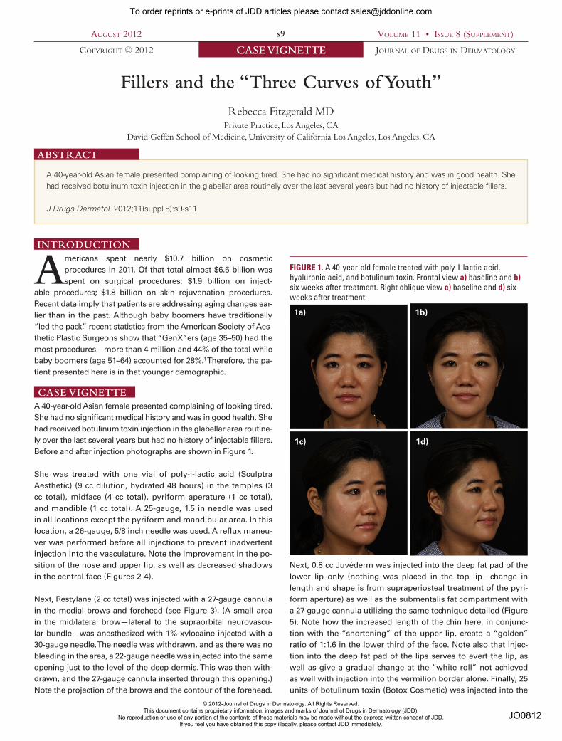
CASE VIGNETTEA 40-year-old Asian female presented complaining of looking tired. She had no significant medical history and was in good health. She had received botulinum toxin injection in the glabellar area routine-ly over the last several years but had no history of injectable fillers. Before and after injection photographs are shown in Figure 1.
She was treated with one vial of poly-l-lactic acid (Sculptra Aesthetic) (9 cc dilution, hydrated 48 hours) in the temples (3 cc total), midface (4 cc total), pyriform aperature (1 cc total), and mandible (1 cc total). A 25-gauge, 1.5 in needle was used in all locations except the pyriform and mandibular area. In this location, a 26-gauge, 5/8 inch needle was used. A reflux maneu-ver was performed before all injections to prevent inadvertent injection into the vasculature. Note the improvement in the po-sition of the nose and upper lip, as well as decreased shadows in the central face (Figures 2-4).
Next, Restylane (2 cc total) was injected with a 27-gauge cannula in the medial brows and forehead (see Figure 3). (A small area in the mid/lateral brow—lateral to the supraorbital neurovascu-lar bundle—was anesthesized with 1% xylocaine injected with a 30-gauge needle. The needle was withdrawn, and as there was no bleeding in the area, a 22-gauge needle was injected into the same opening just to the level of the deep dermis. This was then with-drawn, and the 27-gauge cannula inserted through this opening.) Note the projection of the brows and the contour of the forehead.
Next, 0.8 cc Juvéderm was injected into the deep fat pad of the lower lip only (nothing was placed in the top lip—change in length and shape is from supraperiosteal treatment of the pyri-form aperture) as well as the submentalis fat compartment with a 27-gauge cannula utilizing the same technique detailed (Figure 5). Note how the increased length of the chin here, in conjunc-tion with the “shortening” of the upper lip, create a “golden” ratio of 1:1.6 in the lower third of the face. Note also that injec-tion into the deep fat pad of the lips serves to evert the lip, as well as give a gradual change at the “white roll” not achieved as well with injection into the vermilion border alone. Finally, 25 units of botulinum toxin (Botox Cosmetic) was injected into the
FIGURE 1. A 40-year-old female treated with poly-I-lactic acid, hyaluronic acid, and botulinum toxin. Frontal view a) baseline and b) six weeks after treatment. Right oblique view c) baseline and d) six weeks after treatment.
1b)1a)
1d)1c)
© 2012-Journal of Drugs in Dermatology. All Rights Reserved. This document contains proprietary information, images and marks of Journal of Drugs in Dermatology (JDD).
No reproduction or use of any portion of the contents of these materials may be made without the express written consent of JDD. If you feel you have obtained this copy illegally, please contact JDD immediately.
JO0812
To order reprints or e-prints of JDD articles please contact [email protected]
Do Not CopyPenalties Apply
s10
Journal of Drugs In Dermatology
august 2012 • Volume 11 • Issue 8 (supplement)R. Fitzgerald
1d)1c)
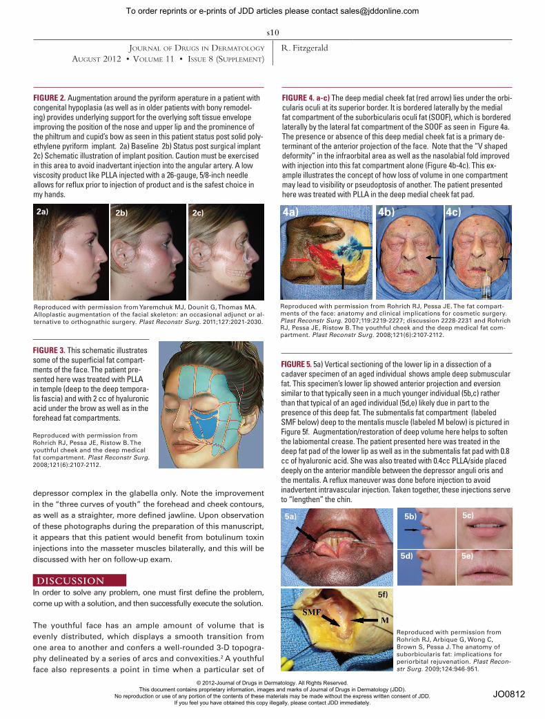
FIGURE 2. Augmentation around the pyriform aperature in a patient with congenital hypoplasia (as well as in older patients with bony remodel-ing) provides underlying support for the overlying soft tissue envelope improving the position of the nose and upper lip and the prominence of the philtrum and cupid’s bow as seen in this patient status post solid poly-ethylene pyriform implant. 2a) Baseline 2b) Status post surgical implant 2c) Schematic illustration of implant position. Caution must be exercised in this area to avoid inadvertant injection into the angular artery. A low viscosity product like PLLA injected with a 26-gauge, 5/8-inch needle allows for reflux prior to injection of product and is the safest choice in my hands.
Reproduced with permission from Yaremchuk MJ, Dounit G, Thomas MA. Alloplastic augmentation of the facial skeleton: an occasional adjunct or al-ternative to orthognathic surgery. Plast Reconstr Surg. 2011;127:2021-2030.
FIGURE 3. This schematic illustrates some of the superficial fat compart-ments of the face. The patient pre-sented here was treated with PLLA in temple (deep to the deep tempora-lis fascia) and with 2 cc of hyaluronic acid under the brow as well as in the forehead fat compartments.
Reproduced with permission from Rohrich RJ, Pessa JE, Ristow B. The youthful cheek and the deep medical fat compartment. Plast Reconstr Surg. 2008;121(6):2107-2112.
2a) 2c)2b)
FIGURE 4. a-c) The deep medial cheek fat (red arrow) lies under the orbi-cularis oculi at its superior border. It is bordered laterally by the medial fat compartment of the suborbicularis oculi fat (SOOF), which is bordered laterally by the lateral fat compartment of the SOOF as seen in Figure 4a. The presence or absence of this deep medial cheek fat is a primary de-terminant of the anterior projection of the face. Note that the “V shaped deformity” in the infraorbital area as well as the nasolabial fold improved with injection into this fat compartment alone (Figure 4b-4c). This ex-ample illustrates the concept of how loss of volume in one compartment may lead to visibility or pseudoptosis of another. The patient presented here was treated with PLLA in the deep medial cheek fat pad.
4a) 4c)4b)
Reproduced with permission from Rohrich RJ, Pessa JE. The fat compart-ments of the face: anatomy and clinical implications for cosmetic surgery. Plast Reconstr Surg. 2007;119:2219-2227; discussion 2228-2231 and Rohrich RJ, Pessa JE, Ristow B. The youthful cheek and the deep medical fat com-partment. Plast Reconstr Surg. 2008;121(6):2107-2112.
FIGURE 5. 5a) Vertical sectioning of the lower lip in a dissection of a cadaver specimen of an aged individual shows ample deep submuscular fat. This specimen’s lower lip showed anterior projection and eversion similar to that typically seen in a much younger individual (5b,c) rather than that typical of an aged individual (5d,e) likely due in part to the presence of this deep fat. The submentalis fat compartment (labeled SMF below) deep to the mentalis muscle (labeled M below) is pictured in Figure 5f. Augmentation/restoration of deep volume here helps to soften the labiomental crease. The patient presented here was treated in the deep fat pad of the lower lip as well as in the submentalis fat pad with 0.8 cc of hyaluronic acid. She was also treated with 0.4cc PLLA/side placed deeply on the anterior mandible between the depressor anguli oris and the mentalis. A reflux maneuver was done before injection to avoid inadvertent intravascular injection. Taken together, these injections serve to “lengthen” the chin.
Reproduced with permission from Rohrich RJ, Arbique G, Wong C, Brown S, Pessa J. The anatomy of suborbicularis fat: implications for periorbital rejuvenation. Plast Recon-str Surg. 2009;124:946-951.
5a)
5f)
5b) 5c)
5d) 5e)
depressor complex in the glabella only. Note the improvement in the “three curves of youth” the forehead and cheek contours, as well as a straighter, more defined jawline. Upon observation of these photographs during the preparation of this manuscript, it appears that this patient would benefit from botulinum toxin injections into the masseter muscles bilaterally, and this will be discussed with her on follow-up exam.
DISCUSSION In order to solve any problem, one must first define the problem, come up with a solution, and then successfully execute the solution.
The youthful face has an ample amount of volume that is evenly distributed, which displays a smooth transition from one area to another and confers a well-rounded 3-D topogra-phy delineated by a series of arcs and convexities.2 A youthful face also represents a point in time when a particular set of
© 2012-Journal of Drugs in Dermatology. All Rights Reserved. This document contains proprietary information, images and marks of Journal of Drugs in Dermatology (JDD).
No reproduction or use of any portion of the contents of these materials may be made without the express written consent of JDD. If you feel you have obtained this copy illegally, please contact JDD immediately.
JO0812
To order reprints or e-prints of JDD articles please contact [email protected]
Do Not CopyPenalties Apply

s11
Journal of Drugs In Dermatology
august 2012 • Volume 11 • Issue 8 (supplement)R. Fitzgerald
skeletal proportions is ideal for their overlying soft tissue en-velope—a place we likely grow into from infancy and away from with age.3,4
In addition to gains in technical insights that have improved our understanding of how to use the currently available products to best advantage, where to use these products to best advantage in facial filling has also improved enormously with ever-evolving insights into the changes observed in the aging face. Current lit-erature reveals that these changes are occurring in all tissue structures of the face and that these changes are interdependent (i.e., a change in one area may lead to a cascade of predictable, secondary events).5 The central role of volume loss and deflation in the aging face has been eloquently illustrated by Lambros in a longitudinal photographic analysis of more than 100 patients spanning an average period of 25 years.6 This work, in conjunc-tion with the work on changes seen with age related skeletal remodeling postulated by Pessa et al4 and now supported by nu-merous studies7,8 as well as the landmark studies carried out at University of Texas Southwestern by Rohrich, Pessa et al in the anatomy of facial fat and its contribution to the changes ob-served in the aging face9-12 are truly “game changers.” The value of this work lies in its implications for treatment. Although the sequence of events as we age is predictable, it’s pace is not. This holds true not just between individuals, but between different structural layers in one individual as well. Recognition of where volume has been lost (or sometimes lacking in the first place) in each individual will greatly enhance our ability to achieve opti-mal and natural-looking results, by enabling us to treat the specific morphology of a particular individual at a particular point in time with site-specific corrections. In my experience, this anatomically based approach to individual facial morphology seems to almost effortlessly improve the shape, contours, to-pography, and proportions of the face treated in this manner.
REFERENCES1. Statistics. American Society for Aesthetic Plastic Surgery online. Accessed
February 29, 2012 at http://www.surgery.org/media/statistics.2. Donofrio LM. Fat distribution: a morphologic study of the
aging face. Dermatol Surg. 2000;26:1107-1112.3. Stuzin JM. Restoring facial shape in face lifting: The role of skeletal sup-
port in facial analysis and midface soft-tissue repositioning. Plast Reconstr Surg. 2007;119(1):362-376.
4. Pessa JE, Zadoo VP, Yuan C, et al. Concertina effect and facial aging: non-linear aspects of youthfulness and skeletal remodeling, and why, perhaps, infants have jowls. Plast Reconstr Surg. 1999;103:635-644.
5. Fitzgerald RL, Vleggaar D. Facial volume restoration of the aging face with poly-l-lactic acid. Dermatol Therapy. 2011;(24):2-27.
6. Lambros V. Observations on periorbital and midfacial aging. Plast Recon-struct Surg. 2007;120:1367-1376.
7. Sharabi SE, Hatef DA, Koshy JC, Hollier LH Jr, Yaremchuk MJ. Mechano-transduction: the missing link in the facial aging puzzle? Aesthetic Plast Surg. 2010;34(5):603-611.
8. Rohrich R, Pessa J. Discussion. Aging of the facial skeleton: aes-thetic implications and rejuvenation strategies. Plast Reconstr Surg. 2011;127(1):384-385.
9. Rohrich RJ, Pessa JE. The fat compartments of the face: anatomy and clini-cal implications for cosmetic surgery. Plast Reconstr Surg. 2007;119:2219-2227; discussion 2228-2231.
10. Rohrich RJ, Pessa JE, Ristow B. The youthful cheek and the deep medical fat compartment. Plast Reconstr Surg. 2008;121(6):2107-2112.
COMMENTARY
Dr. Fitzgerald has eloquently portrayed the next era of cos-metic medicine with the use of non-invasive regimens us-ing neuromodulators and the highly selective use of current U.S. FDA-approved fillers. This is based upon her tremen-dous knowledge of how the human face ages anatomically. Her astute analysis of this patient’s early aging process and how she’s approached it is undeniably the next era of how to maximize the use of FDA fillers and neuromodulators.
The selective use of poly-l-lactic acid in the midface pyriform aperture, mandible, and temporal areas (the earliest areas to show aging, as shown by anatomic studies done at the University of Texas Southwestern) portray how subtle im-provements in these key anatomic areas will rejuvenate a patient’s appearance in a natural manner. The ability to as-tutely perform a combination of injecting Juvéderm to the deep fat compartment of the lip to enhance overall upper lip shape as well as in submentalis fat to give more proportion-ality to the lower face is remarkable. Dr. Fitzgerald has set a high bar for this new era of non-surgical facial rejuvenation with her astute facial analysis and choice of optimal fillers.
The key to future aesthetic non-surgical rejuvenation with neuromodulators and fillers so eloquently demonstrated by Dr. Fitzgerald’s work is a precise analysis of the early aging process and the capability to restore the deep facial anatomy, specifically, the deep facial fat compartments. Dr. Fitzgerald is to be congratulated for her incredible analysis and expertise in the unique use of multiple different fillers and neuromodula-tors to achieve a natural-looking, youthful face.
Rod J. Rohrich MDUniversity of Texas Southwestern Medical Center, Dallas, TX
11. Rohrich RJ, Arbique G, Wong C, Brown S, Pessa J. The anatomy of subor-bicularis fat: implications for periorbital rejuvenation. Plast Reconstr Surg. 2009;124:946-951.
12. Rohrich RJ, Pessa JE. The anatomy and clinical implications of perioral sub-muscular fat. Plast Reconstr Surg. 2009:124(1):266-271.
ADDRESS FOR CORRESPONDENCE
Rebecca Fitzgerald MD321 North Larchmont Boulevard #906Los Angeles, CA 90004Phone:..............................................................(323) 464-8046Email:.....................................................fitzmd@earthlink.net
© 2012-Journal of Drugs in Dermatology. All Rights Reserved. This document contains proprietary information, images and marks of Journal of Drugs in Dermatology (JDD).
No reproduction or use of any portion of the contents of these materials may be made without the express written consent of JDD. If you feel you have obtained this copy illegally, please contact JDD immediately.
JO0812
To order reprints or e-prints of JDD articles please contact [email protected]
Do Not CopyPenalties Apply