Embed Size (px)
Citation preview
IKKα regulation of canonical NF-κB activation downstream
of Nod1-mediated peptidoglycan recognition
and
Endocytosis-independent function of clathrin heavy chain in
the control of basal NF-κB activation
Inauguraldissertation
zur
Erlangung der Würde eines Doktors der Philosophie
vorgelegt der
Philosophisch-Naturwissenschaftlichen Fakultät
der Universität Basel
Von
Man Lyang Kim aus South Korea
Basel, 2010
Genehmigt von der Philosophisch-Naturwissenschaftlichen Fakultät
auf Antrag von
Prof. Dr. Guy Cornelis (Referaat)
Prof. Dr. Christoph Dehio (Korreferaat)
Basel, den 16 November 2010
Prof. Dr. Martin Spiess
Dekan
ABSTRACT
ABSTRACT
NF-κB is a transcription factor involved in the regulation of inflammation and
innate immunity. The IκB kinase (IKK) complex contains two catalytic subunits, IKKα
and IKKβ, and plays an essential role in the activation of NF-κB through the
phosphorylation and degradation of the NF-κB inhibitor IκBα, thereby allowing
translocation of NF-κB into the nucleus. Numerous evidences indicate that IKKβ
mediates NF-κB activation in response to pro-inflammatory cytokines and microbial
products, but the role of IKKα in inflammation and innate immunity is unknown.
In the first part of dissertation, we focus on understanding the previously
unknown function of IKKα in the canonical NF-κB pathway, associated with
inflammation and innate immunity. We show that silencing of IKKα by RNA
interference (RNAi) significantly reduced phosphorylation and degradation of IκBα,
and nuclear translocation of NF-κB, and secretion of the pro-inflammatory chemokine
interleukin-8 (IL-8) during Shigella flexneri infection of human epithelial HeLa cells.
This suggests that IKKα like IKKβ plays a pivotal role in inflammation and innate
immunity by mediating NF-κB activation in response to microbial infection.
Proper control of NF-κB activation is essential for inflammation and innate
immunity triggered by microbial infection, but the dysregulation of NF-κB is associated
with various diseases such as chronic inflammatory diseases and cancers. Thus, the NF-
κB pathway has been a target of therapeutic drug development. Although constitutive
and excessive NF-κB activation has been detected in many inflammation-related
diseases, the cause of the constitutive NF-κB activation in non-stimulated cells is
largely unknown.
i
ABSTRACT
In the second part of dissertation, we focus on clathrin heavy chain (CHC), a
well-known regulator of endocytosis that plays a novel endocytosis-independent
function as an inhibitor of basal NF-κB activation. We show that silencing of CHC
induced constitutive NF-κB nuclear translocation and high level of IL-8 secretion in
resting cells. We revealed that constitutive NF-κB nuclear translocation was mediated
through the constant IκBα degradation in an IKKα-dependent mechanism. We further
showed that CHC depletion-induced constitutive IκBα degradation and high level of IL-
8 secretion in resting cells was independent of the inhibition of clathrin-mediated
endocytosis (CME) as silencing of μ2 subunit of AP2 complex (AP2M1), an adaptor
protein essential for CME failed to induce the constitutive IκBα degradation and high
level of IL-8 secretion. Therefore, the results presented may suggest a potential link
between a defect in CHC expression and chronic inflammatory disorders and cancers.
ii
TABLE OF CONTENTS
TABLE OF CONTENTS
ABSTRACT.....................................................................................................................i
LIST OF ABBREVIATIONS...........................................................................................v
CHAPTER 1: General Introduction..................................................................................1
1.1 The transcription factor NF-κB............................................................................1 1.1.1 NF-κB and IκB proteins..........................................................................................1 1.1.2 Canonical and non-canonical NF-κB signaling pathways......................................3 1.1.3 Mechanisms of IKK complex activation and inhibition.........................................6 1.1.4 Activation of NF-κB pathway by ubiquitin signaling...........................................9 1.1.5 Inhibition of NF-κB pathway by deubiquitination..................................................13
1.2 NF-κB signalling pathways during pathogen infection......................................14 1.2.1 Pathogen recognition by PRRs..............................................................................14 1.2.2 NF-κB signalling transduction during Shigella infection.....................................18
1.3 Function and transcriptional regulation of IL-8..................................................21 1.3.1 IL-8-mediated inflammatory response to Shigella infection...................................21 1.3.2 Transcriptional regulation of IL-8 by NF-κB........................................................21 1.3.3 Epigenetic regulation of IL-8 gene .......................................................................22 1.3.4 Subversion of host inflammatory signalling pathways by Shigella effectors.......23
1.4 NF-κB and diseases............................................................................................25 1.4.1 Mutations in the NF-κB signalling pathways........................................................25 1.4.2 NF-κB as a therapeutic target................................................................................25
1.5 Cellular functions of clathrin.............................................................................27 1.5.1 Clathrin-mediated endocytosis..............................................................................27 1.5.2 Endocytosis-independent functions of clathrin.....................................................29 1.5.3 Regulation of NF-κB signalling pathway by clathrin-binding endocytic proteins.29
1.6 Aim of the study.................................................................................................32
CHAPTER 2: IKKα regulation of canonical NF-κB activation downstream of Nod1- mediated peptidoglycan recognition...............................................................................33
2.1 Abstract...............................................................................................................35 2.2 Introduction.........................................................................................................37 2.3 Materials and Methods........................................................................................41 2.4 Results.................................................................................................................47
iii
TABLE OF CONTENTS
2.4.1 RNAi screen identifies the role of IKKα in the nuclear translocation of NF-κB p65 during infection of epithelial cells by S. flexneri.....................................................47
2.4.2 Depletion of IKKα inhibits S. flexneri-induced p65 nuclear translocation and IL-8 expression without affecting bacterial invasion............................................50
2.4.3 Both IKKα and IKKβ contribute to the phosphorylation and the degradation of IκBα during S. flexneri infection.........................................................................53
2.4.4 IKKα is involved in Nod1-mediated peptidoglycan recognition...........................55 2.5 Discussion...........................................................................................................57 2.6 Acknowledgements.............................................................................................60 2.7 References...........................................................................................................61
CHAPTER 3: Endocytosis-independent function of clathrin heavy chain in the control of basal NF-κB activation...............................................................................................63
3.1 Abstract...............................................................................................................65 3.2 Introduction.........................................................................................................67 3.3 Materials and Methods.......................................................................................70 3.4 Results.................................................................................................................74 3.4.1 CHC prevents constitutive IKK-mediated phosphorylation and degradation of
IκBα in unstimulated epithelial cells......................................................................74 3.4.2 CHC prevents constitutive IKK-mediated phosphorylation and degradation of
IκBα in unstimulated epithelial cells.......................................................................76 3.4.3 CHC prevents constitutive IL-8 secretion in unstimulated epithelial cells.............78 3.4.4 CHC controls basal NF-κB activation independently of endocytosis and clathrin
light chains.......................................................................................................80 3.5 Discussion...........................................................................................................84 3.6 Acknowledgements.............................................................................................88 3.7 References...........................................................................................................89
CONCLUSIONS AND OUTLOOK...............................................................................91
REFERENCES................................................................................................................98
ACKNOWLEDGMENTS.............................................................................................105
APPENDICES...............................................................................................................107
CURRICULUM VITAE...............................................................................................125
iv
ABBREVIATIONS
LIST OF ABBREVIATIONS
ABIN A20 binding inhibitor of NF-κB ANK ankyrin-repeat AP2M1 μ2 subunit of AP2 complex ATF cyclic AMP-dependent transcription factor BCL-3 B-cell lymphoma 3 BIR baculovirus inhibitor of apoptosis repeat CARD caspase recruitment domain CC coiled-coil cDNA complementary DNA CHC clathrin heavy chain CHUK conserved helix-loop-helix ubiquitous kinase CKII casein kinase II CLC clathrin light chain CLR C-type lectin receptor CME clathrin-mediated endocytosis C-terminal carboxyterminal CYLD cylindromatosis tumor suppressor protein DAP diaminopimelic acid DD death domain DNA dioxyribonucleic acid Dub deubiquitination enzyme ELISA enzyme-linked immunosorbent assay GPCR G-protein-coupled receptor GRR glycin-rich region H.pylori Helicobacter pylori HAT histone actetyltransferase HDAC histone deacetylase HLH helix-loop-helix HRP horseradish peroxidase HSP heat shock protein iE-DAP D-g-Glu-meso-diaminopimelic acid IKK inhibitor of NF-κB (IκB) kinase IL interleukin IL-1R IL-1 receptor IRAK interleukin-1 receptor-associated kinase IκB inhibitor of NF-κB JNK c-Jun N-terminal protein kinase K48 lysine 48 K63 lysine 63 L.monocytogenes Listeria monocytogenes LLO listeriolysin O LPS lipopolysaccharide
v
ABBREVIATIONS
LT lympotoxin LZ leucine zipper MAP3K mitogen-activated protein kinase kinase kinase MAPK mitogen-activated protein kinase MDP muramyl dipeptide MMP matrix metalloproteinase MOI multiplicity of infection mRNA messenger RNA MSK1 mitogen- and stress-activated protein kinase1 MyD88 myeloid differentiation primary response gene 88 NAG N-acetylglucosamine NAM N-acetyl muramic acid NBD NEMO binding domain NEMO NF-κB essential modulator NF-κB nuclear factor kappa-B NLR NOD-like receptor NLS nuclear localization signal NOD nucleotide binding oligomerization domain NRR leucine-rich repeat N-terminal amionterminal OspB outer Shigella protein B OspF outer Shigella protein F OspG outer Shigella protein G OUT ovarian tumor PAMP pathogen-associated molecular patterns PBS phosphoate buffered saline PCR polymerase chain reaction PEST proline-, glutamic acid-, serine- and threonine-rich PFA paraformaldehyde PGN peptidoglycan PI3K phosphoinositide-3-kinase PKA protein kinase A PKB protein kinase B PMN polymorphonuclear leukocyte PP protein phosphatase PRR pattern recognition receptor RANK receptor activator of NF-κB Rb retinoblastoma protein Rel reticuloendotheliosis oncogene RHD Rel homology domain RIG retinoic acid-inducible gene RIP receptor interacting protein RLR RIG-I-like receptor RNA ribonucleic acid RNAi RNA interference
vi
ABBREVIATIONS
S.flexneri Shigella flexneri SCF Skp1-Cdc53/Cullin1-F-box siRNA small interfering RNA SODD silencer of death domain T3SS type III secretion system TAB TAK1 binding protein TAD transactivation domain TAK TGF-beta activated kinase TANK TRAF family member-associated NF-κB activator TBK1 TANK-binding kinase 1 TD transactivation domain TfR transferrin receptor TGF transforming growth factor TLR toll-like receptor TNF tumor necrosis factor TNFR TNF receptor Tollip Toll-interacting protein Tom1 target of Myb1 TRAF TNF receptor-associated factor TrCP transducin repeat-containing protein Tri-DAP L-Ala-D-γ-Glu-meso-diaminopimelic acid TSB tryptic soy broth UBC ubiquitin-conjugating enzyme UV ultraviolet ZF zinc finger
vii
CHAPTER 1 GENERAL INTRODUCTION
CHAPTER 1
GENERAL INTRODUCTION
1.1 The transcription factor NF-κB
1.1.1 NF-κκB and IκB proteins
Nuclear factor kappa-B (NF-κB) was originally discovered by Sen and Baltimore in 1986
as a transcription factor present in activated B-cells that strongly activates the
immunoglobulin κB light chain gene expression (Sen and Baltimore 1986). 25 years of
research on NF-κB has revealed that this transcription factor plays important roles in
diverse physiological responses including inflammation, adaptive immunity, cell adhesion,
cell growth, differentiation, oxidative stress responses and apoptosis (Gilmore 2006). Not
surprisingly, dysregulation of NF-κB has been implicated in an ever-expanding list of
diseases such as Crohn’s disease, arthritis, diabetes and cancers (Lawrence 2009).
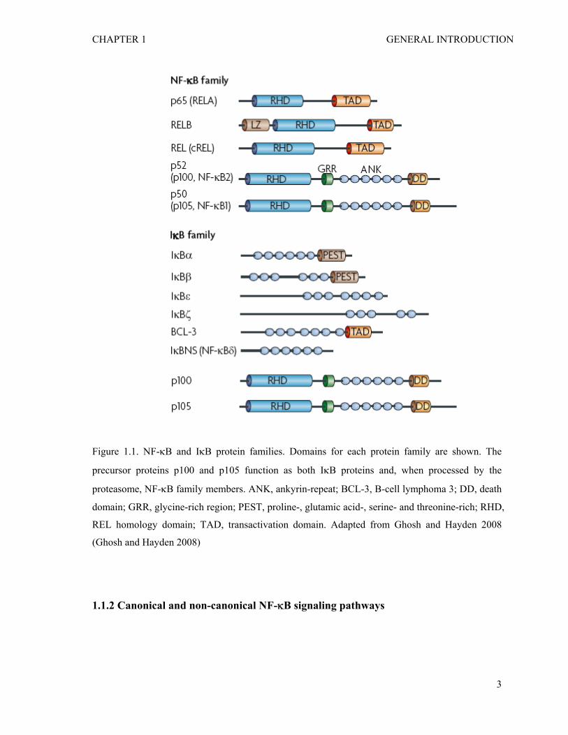
There are five known members of the mammalian NF-κB/Rel family forming
various homo- and hetero-dimers: RelA (p65), RelB, c-Rel, NF-κB1 (p105/p50), and NF-
κB2 (p100/p52) (Figure 1.1) (Ghosh and Hayden 2008). All the NF-κB proteins share a
highly conserved Rel-homology domain (RHD) for dimerization, nuclear localization, and
DNA binding. The Rel proteins (RelA, RelB and c-Rel) contain C-terminal transactivation
domains. NF-κB1 (p105/p50) and NF-κB2 (p100/p52) are distinguished by their long C-
terminal domains that contain multiple copies of ankyrin repeats, which act to inhibit these
proteins. NF-κB1 (p105/p50) and NF-κB2 (p100/p52) become shorter and active DNA-
binding proteins (p105 to p50 and p100 to p52) by limited proteolysis (Fan and Maniatis
1991; Betts and Nabel 1996). The processing of p105 and p100 is mediated by the
1
CHAPTER 1 GENERAL INTRODUCTION
ubiquitin-proteasome pathway and involves selective degradation of their C-terminal
region containing ankyrin repeats (Fan and Maniatis 1991; Betts and Nabel 1996). Glycine-
rich region (GRR) provides the stop signal for processing of p105 and p100 (Lin and
Ghosh 1996; Heusch, Lin et al. 1999). The most abundant form of NF-κB is the
heterodimer of RelA (p65) and p50, retained in the cytoplasm through interaction with IκB
proteins, which masks nuclear localization signal (NLS) of NF-κB proteins (Jacobs and
Harrison 1998).
The IκB (NF-κB inhibitor) proteins include IκBα, IκBβ, IκBε, IκBζ, Bcl-3, IκBNS
(NF-κBδ), and the NF-κB precursors p100 and p105 (Figure 1.1) (Ghosh and Hayden
2008). All IκBs contain five to seven ankyrin-repeats mediating the binding to the RHD
masking the nuclear localization signal (NLS) of NF-κB. The best-characterized IκB
proteins is IκBα, composed of three regions: an N-terminal region, which regulates signal-
dependent degradation; an ankyrin repeat domain; and a C-terminal PEST region
regulating basal degradation. Nearly all of the NF-κB is bound to IκBα, resulting in near-
complete inhibition of nuclear localization and transcriptional activation (Ferreiro and
Komives).
2
CHAPTER 1 GENERAL INTRODUCTION
Figure 1.1. NF-κB and IκB protein families. Domains for each protein family are shown. The
precursor proteins p100 and p105 function as both IκB proteins and, when processed by the
proteasome, NF-κB family members. ANK, ankyrin-repeat; BCL-3, B-cell lymphoma 3; DD, death
domain; GRR, glycine-rich region; PEST, proline-, glutamic acid-, serine- and threonine-rich; RHD,
REL homology domain; TAD, transactivation domain. Adapted from Ghosh and Hayden 2008
(Ghosh and Hayden 2008)
1.1.2 Canonical and non-canonical NF-κB signaling pathways
3
CHAPTER 1 GENERAL INTRODUCTION
With the exception of mature B cells where they are constitutively nuclear, in all other cell
types NF-κB dimers are present in the cytoplasm through association with the IκBs as
inactive forms (Liou and Baltimore 1993). Activation of NF-κB (usually assessed by the
presence of nuclear NF-κB) is induced by diverse extracellular stimuli including
inflammatory cytokines such as tumor necrosis factor alpha (TNFα) and interleukin-1 (IL-
1), receptor ligands such as CD40-ligand, physical stress such as ultraviolet (UV)
irradiation, and many bacteria and viruses (Pahl 1999). The active NF-κB promotes the
expression of hundreds of target genes including cytokines, chemokines, cell adhesion
molecules, stress response genes, and the regulators of apoptosis [nf-kb.org]. A key step for
controlling NF-κB activity is the regulation of the NF-κB- IκBα interaction.
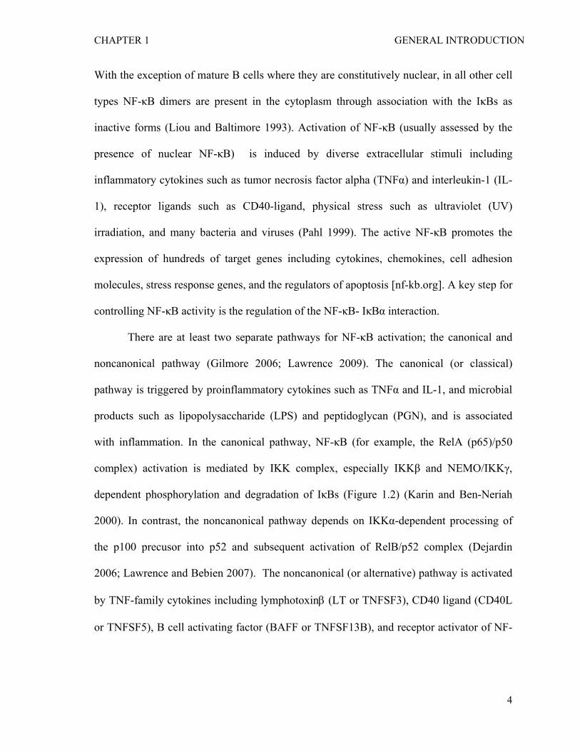
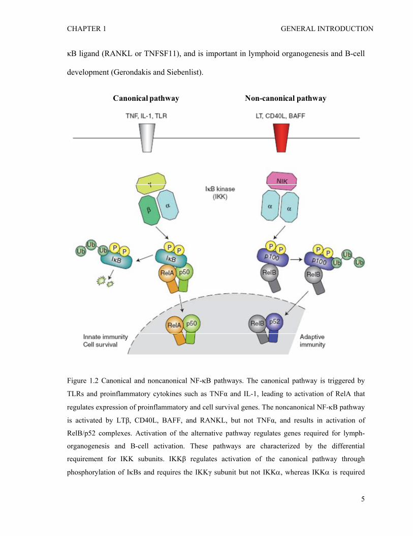
There are at least two separate pathways for NF-κB activation; the canonical and
noncanonical pathway (Gilmore 2006; Lawrence 2009). The canonical (or classical)
pathway is triggered by proinflammatory cytokines such as TNFα and IL-1, and microbial
products such as lipopolysaccharide (LPS) and peptidoglycan (PGN), and is associated
with inflammation. In the canonical pathway, NF-κB (for example, the RelA (p65)/p50
complex) activation is mediated by IKK complex, especially IKKβ and NEMO/IKKγ,
dependent phosphorylation and degradation of IκBs (Figure 1.2) (Karin and Ben-Neriah
2000). In contrast, the noncanonical pathway depends on IKKα-dependent processing of
the p100 precusor into p52 and subsequent activation of RelB/p52 complex (Dejardin
2006; Lawrence and Bebien 2007). The noncanonical (or alternative) pathway is activated
by TNF-family cytokines including lymphotoxinβ (LT or TNFSF3), CD40 ligand (CD40L
or TNFSF5), B cell activating factor (BAFF or TNFSF13B), and receptor activator of NF-
4
CHAPTER 1 GENERAL INTRODUCTION
κB ligand (RANKL or TNFSF11), and is important in lymphoid organogenesis and B-cell
development (Gerondakis and Siebenlist).
Canonical pathway Non-canonical pathway
Figure 1.2 Canonical and noncanonical NF-κB pathways. The canonical pathway is triggered by
TLRs and proinflammatory cytokines such as TNFα and IL-1, leading to activation of RelA that
regulates expression of proinflammatory and cell survival genes. The noncanonical NF-κB pathway
is activated by LTβ, CD40L, BAFF, and RANKL, but not TNFα, and results in activation of
RelB/p52 complexes. Activation of the alternative pathway regulates genes required for lymph-
organogenesis and B-cell activation. These pathways are characterized by the differential
requirement for IKK subunits. IKKβ regulates activation of the canonical pathway through
phosphorylation of IκBs and requires the IKKγ subunit but not IKKα, whereas IKKα is required
5
CHAPTER 1 GENERAL INTRODUCTION
for activation of the non-canonical pathway through the phosphorylation and processing of p100,
the precursor for p52, but this is independent of both IKKβ and IKKγ. Adapted from Lawrence T.
Cold Spring Harb Perspect Biol 2009 (Lawrence 2009).
1.1.3 Mechanisms of IKK complex activation and inhibition
The IKK complex is composed of at least two highly homologous kinase subunits, IKKα
/CHUK and IKKβ, and a regulatory subunit IKKγ/NEMO (NF-κB essential modulator)
(Figure 1.3) (Hacker and Karin 2006). Based on mutational analyses, it is generally
believed that IKKβ and NEMO are essential for IκBα phosphorylation and degradation in
most canonical NF-κB signalling pathways, whereas IKKα is dispensable in the canonical
pathway, but is essential for p100 phosphorylation and processing to p52 in the non-
canonical pathway. In addition to the core IKK components IKKα, IKKβ, and NEMO,
additional subunits are reported to associate with the IKK complex. HSP-90/Cdc37
functions as a chaperone during assembly of the IKK complex upon stimulation (Hinz,
Broemer et al. 2007). The HSP-90 inhibitor geldanamycin has been shown to inhibit
activation of IKK by TNFα (Lewis, Devin et al. 2000). ELKS is reported to be associated
with IKK complex as a regulatory component like NEMO (Ducut Sigala, Bottero et al.
2004).
IKKα and IKKβ dimerize through the leucine zipper domain, which is also required
for kinase activity (Mercurio, Zhu et al. 1997; Woronicz, Gao et al. 1997; Zandi, Rothwarf
et al. 1997). IKKα and IKKβ bind NEMO through the C-terminal NEMO-binding domain
(NBD) (Figure 1.4) (Hayden and Ghosh 2008). The N-terminal coiled-coil motif of NEMO
is responsible for the interaction with IKKα and IKKβ (Drew 2007). Activation of IKK
6
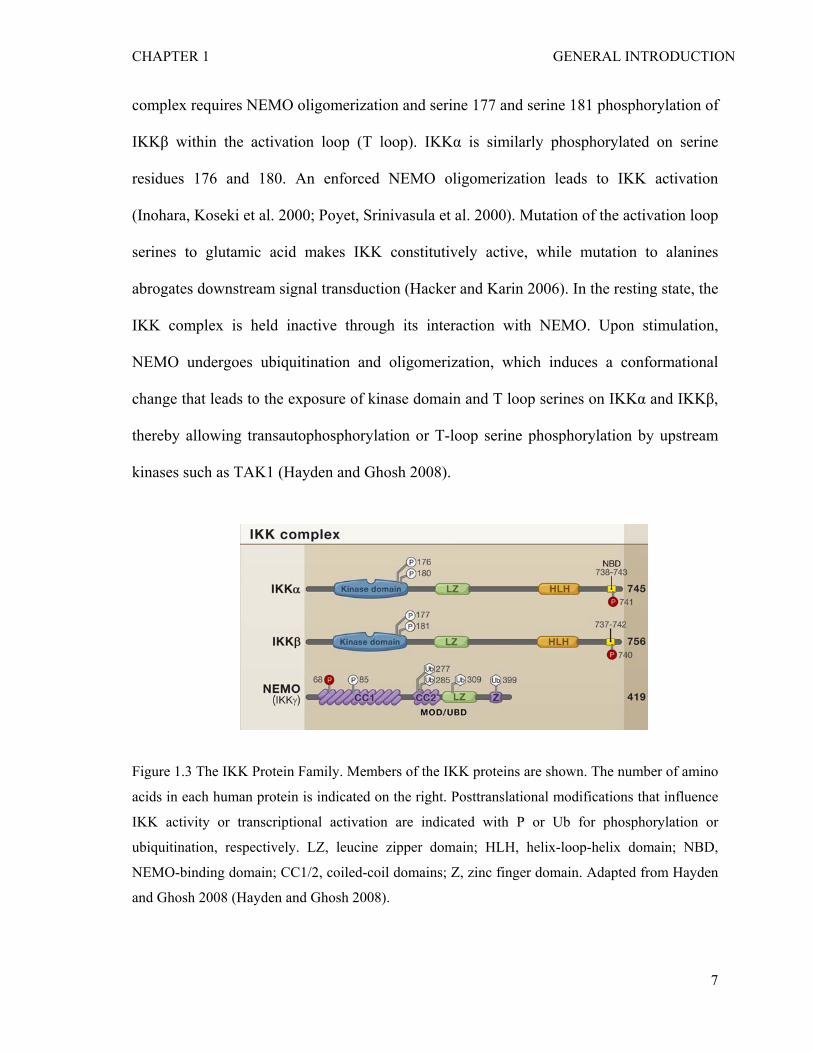
CHAPTER 1 GENERAL INTRODUCTION
complex requires NEMO oligomerization and serine 177 and serine 181 phosphorylation of
IKKβ within the activation loop (T loop). IKKα is similarly phosphorylated on serine
residues 176 and 180. An enforced NEMO oligomerization leads to IKK activation
(Inohara, Koseki et al. 2000; Poyet, Srinivasula et al. 2000). Mutation of the activation loop
serines to glutamic acid makes IKK constitutively active, while mutation to alanines
abrogates downstream signal transduction (Hacker and Karin 2006). In the resting state, the
IKK complex is held inactive through its interaction with NEMO. Upon stimulation,
NEMO undergoes ubiquitination and oligomerization, which induces a conformational
change that leads to the exposure of kinase domain and T loop serines on IKKα and IKKβ,
thereby allowing transautophosphorylation or T-loop serine phosphorylation by upstream
kinases such as TAK1 (Hayden and Ghosh 2008).
Figure 1.3 The IKK Protein Family. Members of the IKK proteins are shown. The number of amino
acids in each human protein is indicated on the right. Posttranslational modifications that influence
IKK activity or transcriptional activation are indicated with P or Ub for phosphorylation or
ubiquitination, respectively. LZ, leucine zipper domain; HLH, helix-loop-helix domain; NBD,
NEMO-binding domain; CC1/2, coiled-coil domains; Z, zinc finger domain. Adapted from Hayden
and Ghosh 2008 (Hayden and Ghosh 2008).
7
CHAPTER 1 GENERAL INTRODUCTION
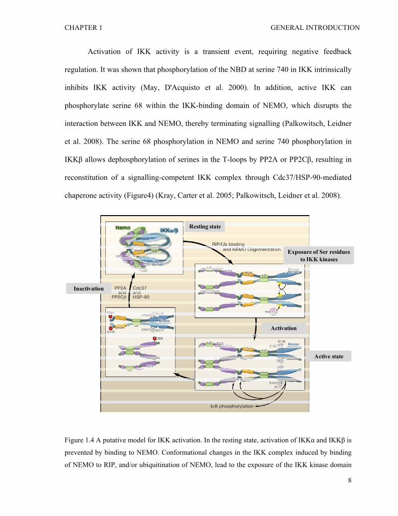
Activation of IKK activity is a transient event, requiring negative feedback
regulation. It was shown that phosphorylation of the NBD at serine 740 in IKK intrinsically
inhibits IKK activity (May, D'Acquisto et al. 2000). In addition, active IKK can
phosphorylate serine 68 within the IKK-binding domain of NEMO, which disrupts the
interaction between IKK and NEMO, thereby terminating signalling (Palkowitsch, Leidner
et al. 2008). The serine 68 phosphorylation in NEMO and serine 740 phosphorylation in
IKKβ allows dephosphorylation of serines in the T-loops by PP2A or PP2Cβ, resulting in
reconstitution of a signalling-competent IKK complex through Cdc37/HSP-90-mediated
chaperone activity (Figure4) (Kray, Carter et al. 2005; Palkowitsch, Leidner et al. 2008).
Resting state
Exposure of Ser residues to IKK kinases
Active state
Inactivation
Activation
Figure 1.4 A putative model for IKK activation. In the resting state, activation of IKKα and IKKβ is
prevented by binding to NEMO. Conformational changes in the IKK complex induced by binding
of NEMO to RIP, and/or ubiquitination of NEMO, lead to the exposure of the IKK kinase domain
8
CHAPTER 1 GENERAL INTRODUCTION
and T loop serines and consequent transautophosphorylation or phosphorylation by an IKK-K such
as TAK1. The active IKK then phosphorylates downstream substrates, including serine 740 within
the IKK NBD and serine 68 in NEMO. NEMO phosphorylation results in the separation of stable
NEMO dimers and NEMO binding to IKK. Dephosphorylation of the IKK T loop results in kinase
inactivation, whereas phosphorylation of the IKK NBD and NEMO serine 68 prevents reactivation
of the kinase. Cdc37/ HSP-90-mediated chaperone activity and PP2A and PP2Cβ phosphatase
activity may then mediate regeneration of the IKK complex. Adapted from Hayden and Ghosh
2008 (Hayden and Ghosh 2008).
1.1.4 Activation of the NF-κB pathway by ubiquitin signaling
Ubiquitination is a reversible covalent modification by which ubiquitin is attached to a
target protein through an isopeptide bond between the C-terminus of ubiquitin and the ε-
amino group of a lysine residue in the target protein (Pickart and Eddins 2004). This
process is catalyzed by three enzymatic steps via an ubiquitin-activating enzyme (E1), an
ubiquitin-conjugating enzyme (E2 or UBC), and an ubiquitin-protein ligase (E3). Ubiquitin
contains seven lysine residues that can be attached to other ubiquitins to form a
polyubiquitin chain (Pickart and Eddins 2004).
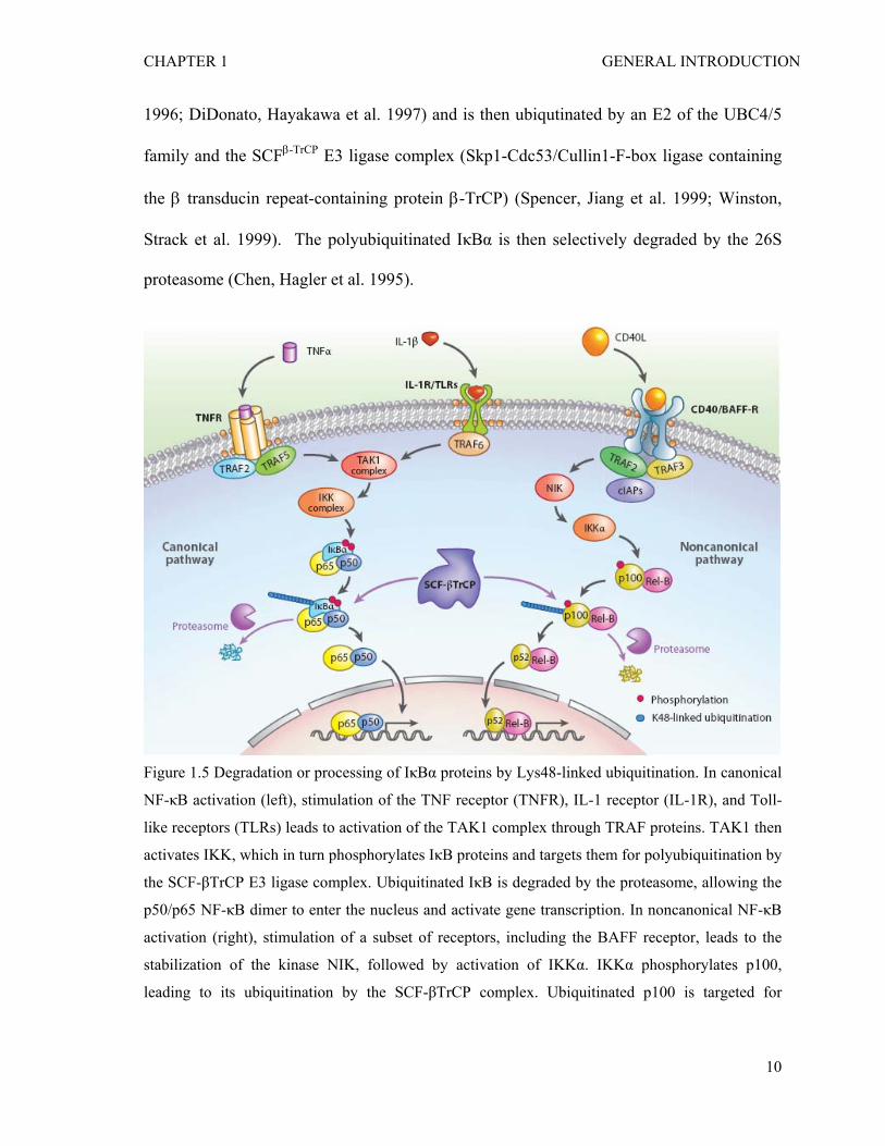
A polyubiquitin chain linked through lysine 48 (Lys-48) of ubiquitin targets a
protein for degradation by the proteasome (Pickart and Eddins 2004). This ubiquitin-
proteasome pathway is responsible for the degradation of the NF-κB inhibitor IκBα in the
canonical NF-κB signaling pathway or the processing of p100 in the non-canonical
pathway (Figure 1.5) (Alkalay, Yaron et al. 1995; Chen, Hagler et al. 1995; Skaug, Jiang et
al. 2009). In response to cytokines such as TNFα, IκBα is phosphorylated by IKKα/β at
two serine residues near the N-terminus (Ser-32 and Ser-36) (DiDonato, Mercurio et al.
9
CHAPTER 1 GENERAL INTRODUCTION
1996; DiDonato, Hayakawa et al. 1997) and is then ubiqutinated by an E2 of the UBC4/5
family and the SCFβ-TrCP E3 ligase complex (Skp1-Cdc53/Cullin1-F-box ligase containing
the β transducin repeat-containing protein β-TrCP) (Spencer, Jiang et al. 1999; Winston,
Strack et al. 1999). The polyubiquitinated IκBα is then selectively degraded by the 26S
proteasome (Chen, Hagler et al. 1995).
Figure 1.5 Degradation or processing of IκBα proteins by Lys48-linked ubiquitination. In canonical
NF-κB activation (left), stimulation of the TNF receptor (TNFR), IL-1 receptor (IL-1R), and Toll-
like receptors (TLRs) leads to activation of the TAK1 complex through TRAF proteins. TAK1 then
activates IKK, which in turn phosphorylates IκB proteins and targets them for polyubiquitination by
the SCF-βTrCP E3 ligase complex. Ubiquitinated IκB is degraded by the proteasome, allowing the
p50/p65 NF-κB dimer to enter the nucleus and activate gene transcription. In noncanonical NF-κB
activation (right), stimulation of a subset of receptors, including the BAFF receptor, leads to the
stabilization of the kinase NIK, followed by activation of IKKα. IKKα phosphorylates p100,
leading to its ubiquitination by the SCF-βTrCP complex. Ubiquitinated p100 is targeted for
10
CHAPTER 1 GENERAL INTRODUCTION
proteasomal processing to p52. The p52/REL-B dimer then translocates into the nucleus to activate
gene transcription.
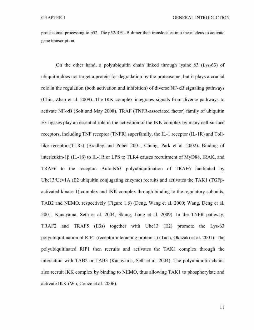
On the other hand, a polyubiquitin chain linked through lysine 63 (Lys-63) of
ubiquitin does not target a protein for degradation by the proteasome, but it plays a crucial
role in the regulation (both activation and inhibition) of diverse NF-κB signaling pathways
(Chiu, Zhao et al. 2009). The IKK complex integrates signals from diverse pathways to
activate NF-κB (Solt and May 2008). TRAF (TNFR-associated factor) family of ubiquitin
E3 ligases play an essential role in the activation of the IKK complex by many cell-surface
receptors, including TNF receptor (TNFR) superfamily, the IL-1 receptor (IL-1R) and Toll-
like receptors(TLRs) (Bradley and Pober 2001; Chung, Park et al. 2002). Binding of
interleukin-1β (IL-1β) to IL-1R or LPS to TLR4 causes recruitment of MyD88, IRAK, and
TRAF6 to the receptor. Auto-K63 polyubiquitination of TRAF6 facilitated by
Ubc13/Uev1A (E2 ubiquitin conjugating enzyme) recruits and activates the TAK1 (TGFβ-
activated kinase 1) complex and IKK complex through binding to the regulatory subunits,
TAB2 and NEMO, respectively (Figure 1.6) (Deng, Wang et al. 2000; Wang, Deng et al.
2001; Kanayama, Seth et al. 2004; Skaug, Jiang et al. 2009). In the TNFR pathway,
TRAF2 and TRAF5 (E3s) together with Ubc13 (E2) promote the Lys-63
polyubiquitination of RIP1 (receptor interacting protein 1) (Tada, Okazaki et al. 2001). The
polyubiquitinated RIP1 then recruits and activates the TAK1 complex through the
interaction with TAB2 or TAB3 (Kanayama, Seth et al. 2004). The polyubiquitin chains
also recruit IKK complex by binding to NEMO, thus allowing TAK1 to phosphorylate and
activate IKK (Wu, Conze et al. 2006).
11
CHAPTER 1 GENERAL INTRODUCTION
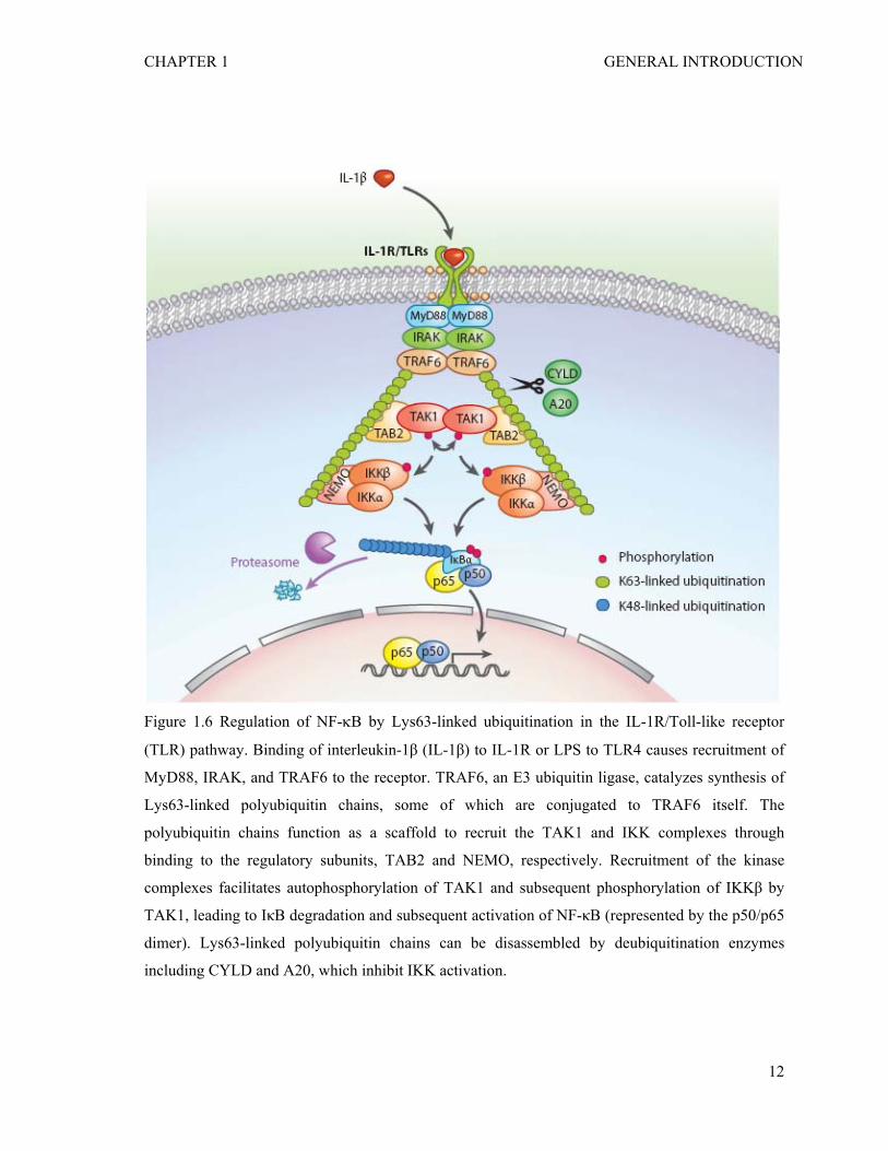
Figure 1.6 Regulation of NF-κB by Lys63-linked ubiquitination in the IL-1R/Toll-like receptor
(TLR) pathway. Binding of interleukin-1β (IL-1β) to IL-1R or LPS to TLR4 causes recruitment of
MyD88, IRAK, and TRAF6 to the receptor. TRAF6, an E3 ubiquitin ligase, catalyzes synthesis of
Lys63-linked polyubiquitin chains, some of which are conjugated to TRAF6 itself. The
polyubiquitin chains function as a scaffold to recruit the TAK1 and IKK complexes through
binding to the regulatory subunits, TAB2 and NEMO, respectively. Recruitment of the kinase
complexes facilitates autophosphorylation of TAK1 and subsequent phosphorylation of IKKβ by
TAK1, leading to IκB degradation and subsequent activation of NF-κB (represented by the p50/p65
dimer). Lys63-linked polyubiquitin chains can be disassembled by deubiquitination enzymes
including CYLD and A20, which inhibit IKK activation.
12
CHAPTER 1 GENERAL INTRODUCTION
1.1.5 Inhibition of the NF-κB pathway by deubiquitination
Polyubiquitination is subject to disassembly by deubiquitination, which is carried out by
members of deubiquitination enzymes (Dubs) (Amerik and Hochstrasser 2004). Two Dubs
are best-characterized in inhibiting NF-κB activation to prevent uncontrolled NF-κB
activities. CYLD (cylindromatosis tumor suppressor protein) inhibits IKK activation by
cleaving K63-linked polyubiquitin chains on target proteins, including TRAF2, TRAF6 and
NEMO following stimulation with TNFα or IL-1β (Figure 1.6) (Brummelkamp, Nijman et
al. 2003; Kovalenko, Chable-Bessia et al. 2003; Trompouki, Hatzivassiliou et al. 2003;
Skaug, Jiang et al. 2009). Overexpression of CYLD inhibits IKK and NF-κB activation,
whereas RNAi of CYLD enhances IKK and NF-κB activation. However, little is known
about how CYLD activity is regulated in resting cells and during stimulation.
A20 is an NF-κB induced Dub protein containing a ovarian tumor (OTU)-type Dub
domain that inhibits NF-κB in a negative-feedback loop by cleaving K63-linked
polyubiqutin chains on RIP and TRAF6 following stimulation with TNFα or IL-1β (Boone,
Turer et al. 2004; Evans, Ovaa et al. 2004; Wertz, O'Rourke et al. 2004; Skaug, Jiang et al.
2009). Interestingly, A20 contains several zinc-finger domains through which it functions
as an ubiquitin ligase to assemble K48-linked polyubiquitin chains on RIP after the K63
chains are cleaved by the OTU domain (Wertz, O'Rourke et al. 2004). K48
polyubiquitination targets RIP for degradation by the proteasome, further diminishing IKK
activaton.
13
CHAPTER 1 GENERAL INTRODUCTION
1.2 NF-κB signalling pathways during pathogen infection
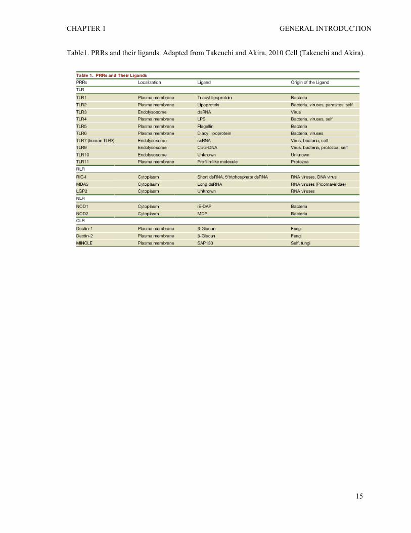
1.2.1 Pathogen recognition by PRRs
The innate immune system is the first line of host defense against pathogens and recognizes
microorganism such as bacteria via germline-encoded pattern recognition receptors (PRRs)
(Takeuchi and Akira ; Medzhitov 2007). Different PRRs react with specific microbial
components, known as pathogen-associated molecular patterns (PAMPs). Currently, four
different classes of PRR families have been identified (Proell, Riedl et al. 2008). These
families include transmembrane proteins such as the Toll-like receptors (TLRs) and C-type
lectin receptors (CLRs), as well as cytoplasmic proteins such as the Retinoic acid-inducible
gene (RIG)-I-like receptors (RLRs) and NOD-like receptors (NLRs) (see table 1).
TLRs are evolutionary conserved from Caenorhabditis elegans to mammals (Kawai
and Akira). To date, 12 members of the TLR family have been identified in mammals.
TLR2 in combination with TLR1 or TLR6 recognize lipoproteins (triacyl and diacyl
lipoproteins, respectively), whereas TLR3, TLR7/TLR8, and TLR9 recognize nucleic acids
(dsRNA, ssRNA and CpG-DNA, respectively). TLR4 and TLR5 recognize
lipopolysaccharide (LPS) and flagellin, respectively (Akira, Uematsu et al. 2006).
Recognition of bacterial components by TLRs takes place at either the cell surface or
endolysosome compartments.
14
CHAPTER 1 GENERAL INTRODUCTION
Table1. PRRs and their ligands. Adapted from Takeuchi and Akira, 2010 Cell (Takeuchi and Akira).
15
CHAPTER 1 GENERAL INTRODUCTION
CLRs recognize carbohydrates from viruses, bacteria and fungi via a carbohydrate-
binding domain (Geijtenbeek and Gringhuis 2009). For example, Dectin-1 and Dectin-2 are
responsible for sensing β-glucans from fungi (Goodridge, Wolf et al. 2009; Robinson,
Osorio et al. 2009). MINCLE, a CLR from macrophage, can sense not only fungal
infection but also an endogenous protein, spliceosome-associated protein 130 (SAP130)
from necrotic host cells (Yamasaki, Ishikawa et al. 2008).
RLRs are localized in the cytoplasm and primarily sense viral double stranded RNA
(dsRNA) (Takeuchi and Akira 2009). RLRs are composed of two N-terminal caspase
recruitment domains (CARDs), a central DEAD box helicase/ATPase domain, and a C-
terminal regulatory domain that mediates the binding to dsRNAs. RIG-1 and MDA5
recognize relatively short (up to 1kb) and long (more than 2kb) double stranded RNA
(dsRNA), respectively, and are essential for stimulating type I interferon (IFN) production
in response to RNA viruses (Kato, Takeuchi et al. 2008; Loo, Fornek et al. 2008).
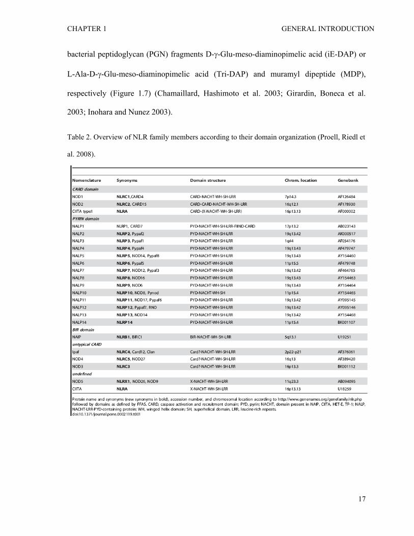
NLRs detect pathogens that have invaded the cytosol of host cells. The NLR family
of proteins is defined by a tripartite structure consisting of a C-terminal leucine-rich repeat
(LRR) that mediates ligand (pathogen) sensing; a central nucleotide binding
oligomerization domain (NOD); and a N-terminal effector domain, such as CARDs,
PYRIN, or baculovirus inhibitor of apoptosis repeat (BIR) domains (Inohara, Chamaillard
et al. 2005; Martinon and Tschopp 2005) . In human, the NLR family is composed of 22
proteins (see the Table 2 for the list) (Proell, Riedl et al. 2008). Although primarily
expressed in immune cells, including antigen-presenting cells such as macrophages and
dendritic cells, NLRs can also be expressed in nonimmune cells, including epithelial cells
(Chen, Shaw et al. 2009). Nod1 and Nod2 are the best-characterized NLRs and recognize
16
CHAPTER 1 GENERAL INTRODUCTION
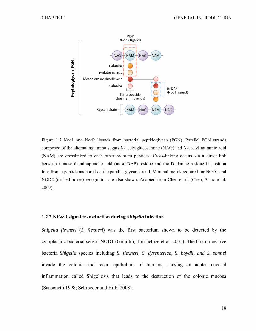
bacterial peptidoglycan (PGN) fragments D-γ-Glu-meso-diaminopimelic acid (iE-DAP) or
L-Ala-D-γ-Glu-meso-diaminopimelic acid (Tri-DAP) and muramyl dipeptide (MDP),
respectively (Figure 1.7) (Chamaillard, Hashimoto et al. 2003; Girardin, Boneca et al.
2003; Inohara and Nunez 2003).
Table 2. Overview of NLR family members according to their domain organization (Proell, Riedl et
al. 2008).
17
CHAPTER 1 GENERAL INTRODUCTION
Figure 1.7 Nod1 and Nod2 ligands from bacterial peptidoglycan (PGN). Parallel PGN strands
composed of the alternating amino sugars N-acetylglucosamine (NAG) and N-acetyl muramic acid
(NAM) are crosslinked to each other by stem peptides. Cross-linking occurs via a direct link
between a meso-diaminopimelic acid (meso-DAP) residue and the D-alanine residue in position
four from a peptide anchored on the parallel glycan strand. Minimal motifs required for NOD1 and
NOD2 (dashed boxes) recognition are also shown. Adapted from Chen et al. (Chen, Shaw et al.
2009).
1.2.2 NF-κB signal transduction during Shigella infection
Shigella flexneri (S. flexneri) was the first bacterium shown to be detected by the
cytoplasmic bacterial sensor NOD1 (Girardin, Tournebize et al. 2001). The Gram-negative
bacteria Shigella species including S. flexneri, S. dysenteriae, S. boydii, and S. sonnei
invade the colonic and rectal epithelium of humans, causing an acute mucosal
inflammation called Shigellosis that leads to the destruction of the colonic mucosa
(Sansonetti 1998; Schroeder and Hilbi 2008).
18
CHAPTER 1 GENERAL INTRODUCTION
S. flexneri invades intestinal epithelial cells by inducing cytoskeletal rearrangement
localized at the site of infection (Bourdet-Sicard, Egile et al. 2000; Tran Van Nhieu,
Bourdet-Sicard et al. 2000). This entry process depends on the activities of several
effectors including IpaA, IpaB, IpaC and IpaD secreted from the Shigella type III secretion
system (T3SS) (High, Mounier et al. 1992; Menard, Prevost et al. 1996; Tran Van Nhieu,
Ben-Ze'ev et al. 1997; Tran Van Nhieu, Caron et al. 1999). The Shigella effector IpaC
triggers actin polymerization and the formation of filopodial and lamellipodial extensions,
which are dependent on the Rho small GTPases Cdc42 and Rac (Mounier, Laurent et al.
1999), and the protein tyrosine kinase Src (Dehio, Prevost et al. 1995). On the other hand,
IpaA binds to the focal adhesion protein vinculin and induces depolymerization of actin
filaments, allowing the transformation of the IpaC-induced extensions into a structure that
is productive for bacterial entry (Tran Van Nhieu, Ben-Ze'ev et al. 1997).
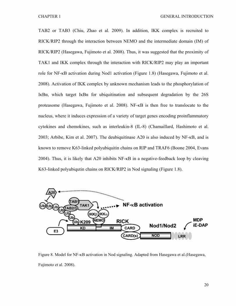
Upon entry of Shigella into host cell, a peptidoglycan (PGN)-derived small peptide
iE-DAP (γ-D-glutamyl-meso-diaminopimelic acid) is released into the cytosol by carboxy-
peptidases and hydrolases activity (Boneca 2005), and sensed by NOD1 (Chamaillard,
Hashimoto et al. 2003). Recognition of iE-DAP through leucine-rich repeat (LRR) domain
induces self-oligomerization of NOD1 (Inohara, Koseki et al. 2000). Oligomerization of
NOD1 allows binding to a downstream effector molecule RICK/RIP2 through CARD-
CARD interaction (Park, Kim et al. 2007). RICK/RIP2 is then conjugated with lysine-63-
linked polyubiquitin chains at lysine 209 (K209) located in its kinase domain by an
unknown E3 ubiquitin ligase (Hasegawa, Fujimoto et al. 2008). Unlike K48-linked
polyubiquitin chains, which target a protein for proteasomal degradation, the
nondegradative K63-linked polyubiquitinated chains further recruit the TAK1 complex via
19
CHAPTER 1 GENERAL INTRODUCTION
TAB2 or TAB3 (Chiu, Zhao et al. 2009). In addition, IKK complex is recruited to
RICK/RIP2 through the interaction between NEMO and the intermediate domain (IM) of
RICK/RIP2 (Hasegawa, Fujimoto et al. 2008). Thus, it was suggested that the proximity of
TAK1 and IKK complex through the interaction with RICK/RIP2 may play an important
role for NF-κB activation during Nod1 activation (Figure 1.8) (Hasegawa, Fujimoto et al.
2008). Activation of IKK complex by unknown mechanism leads to the phosphorylation of
IκBα, which target IκBα for ubiquitination and subsequent degradation by the 26S
proteasome (Hasegawa, Fujimoto et al. 2008). NF-κB is then free to translocate to the
nucleus, where it induces expression of a variety of target genes encoding proinflammatory
cytokines and chemokines, such as interleukin-8 (IL-8) (Chamaillard, Hashimoto et al.
2003; Arbibe, Kim et al. 2007). The deubiquitinase A20 is also induced by NF-κB, and is
known to remove K63-linked polyubiquitin chains on RIP and TRAF6 (Boone 2004, Evans
2004). Thus, it is likely that A20 inhibits NF-κB in a negative-feedback loop by cleaving
K63-linked polyubiqutin chains on RICK/RIP2 in Nod signaling (Figure 1.8).
Figure 8. Model for NF-κB activation in Nod signaling. Adapted from Hasegawa et al.(Hasegawa,
Fujimoto et al. 2008).
20
CHAPTER 1 GENERAL INTRODUCTION
1.3 Function and transcriptional regulation of IL-8
1.3.1 IL-8-mediated inflammatory response to Shigella infection
When tissues get injured or infected by pathogens like S.flexneri, generally macro
symptoms of redness, swelling, heat and pain appear. This process is called inflammation
or inflammatory response. At the cellular level, inflammation is caused by chemical
mediators called chemokines. Interleukin-8 (IL-8) is one of the major epithelial cells-
secreted chemokines associated with inflammation (Jung, Eckmann et al. 1995). Secreted
IL-8 then recruits phagocytes, in particular, neutrophils from the blood stream to the site of
infection (Perdomo, Cavaillon et al. 1994; Kobayashi 2008). The function of IL-8 in
inflammation during S.flexneri infection was shown in the rabbit model of shigellosis
where intense IL-8 expression in the infected epithelial layer and neutrophil infiltration in
the infected tissue was observed (Sansonetti, Tran Van Nhieu et al. 1999). Several recent
studies have demonstrated that NF-κB activation is required but not sufficient to induce IL-
8 expression upon S.flexneri infection (see below).
1.3.2 Transcriptional regulation of IL-8 by NF-κB
IL-8 promoter contains a NF-κB binding site known as kappa B (κB) element that is
essential for transcriptional regulation of the gene (Mukaida, Mahe et al. 1990; Harant, de
Martin et al. 1996). In line with the cytoplasmic retention of transcription factor NF-κB by
its binding to IκB proteins, IL-8 expression is very low in unstimulated cells. Thus, nuclear
translocation of NF-κB is critical for IL-8 production in response to a wide range of stimuli
including proinflammatory cytokines such as TNFα, and bacterial products such as LPS or
PGN (Philpott, Yamaoka et al. 2000). However, binding of the NF-κB element to the IL-8
21
CHAPTER 1 GENERAL INTRODUCTION
gene promoter is not sufficient. The NF-κB components, especially p65 subunit needs to be
phosphorylated in its transactivation domain (TD) to be fully active (Hoffmann, Natoli et al.
2006). Serine 276 phosphorylation in the TD of p65 by protein kinase A (PKA) (Zhong,
Voll et al. 1998), casein kinase II (CKII) or protein kinase B (PKB or Akt) (Bird, Schooley
et al. 1997), and serine 536 phosphorylation by IKKα and IKKβ (Sakurai, Chiba et al.
1999), PI3K/Akt (Sizemore, Leung et al. 1999) and IKKε/TBK1 (Buss, Dorrie et al. 2004;
Adli and Baldwin 2006) are suggested to be required for its transactivation function. In
addition to phosphorylation of NF-κB subunits, acetylation and methylation can modulate
NF-κB transcriptional activity (Huang, Yang et al. ; Perkins 2006).
1.3.3 Epigenetic regulation of IL-8 gene
Epigenetic events such as histone acetylation and phosphorylation are known to play an
important role in regulating gene expression (Munshi, Shafi et al. 2009). While repression
of transcriptional activity is commonly correlated with histone hypoacteylation due to
histone deacetylase (HDAC) activity, histone acetylation mediated by histone
actetyltransferase (HAT) activity generally promotes transcriptional activation of genes
after conformational changes within the chromatin (Kuo and Allis 1998; Wilson 2008).
When it comes to epigenetic regulation of IL-8 gene, Wen et al. have demonstrated that the
HDAC activity tightly controls the transcription of the IL-8 gene in Caco-2 intestinal
epithelial cells (Wen and Wu 2001). Muegge et al. have also shown that histone H3
phosphorylation at serine 10 and acetylation at lysine 14 facilitates NF-κB binding to IL-8
promoter (Muegge 2002). Histone H3 phosphorylation at serine 10 is induced by mitogen-
and stress-activated protein kinase 1 (MSK1) downstream of p38 or ERK signaling
pathways (Thomson, Clayton et al. 1999). In addition, JNK contributes to IL-8 expression
22
CHAPTER 1 GENERAL INTRODUCTION
through the activation of the transcriptional regulator AP-1 composed of c-JUN, ATF, c-
FOS, and JDP families, which binds to AP-1-binding site present in the core IL-8 promoter
(Hess, Angel et al. 2004; Bogoyevitch, Ngoei et al. 2010).
1.3.4 Subversion of host inflammatory signaling pathways by Shigella effectors
Subversion of host inflammatory signaling pathways is an important mechanism used by
multiple bacteria (Bhavsar, Guttman et al. 2007). Especially, histone modifications
induced by bacterial toxins are shared by multiple bacteria including S.flexneri, Listeria
monocytogenes and Helicobacter pylori (see below). For example, the Shigella type III
effectors OspF (Outer Shigella protein F) induces dephosphorylation of p38 and ERK in
the nucleus, which subsequently prevents histone H3 Ser10 phosphorylation (Arbibe, Kim
et al. 2007; Li, Xu et al. 2007). L.monocytogenes secretes listeriolysin O (LLO), which
induces a dramatic dephosphorylation of histone H3 at serine 10 (H3 Ser10) and
deacetylation of histone H4 (Hamon, Batsche et al. 2007). Similarly, H. pylori induces
cagPAI-dependent dephosphorylation of histone H3 at serine 10 and deacetylation of H3
lysine at lysine 23 (Ding, Fischer et al. 2010).
Besides OspF, Shigella use other effectors to downregulate the host inflammatory
response. For example, OspB targets the nucleus to downregulate the host cytokine
production via interactions with retinoblastoma protein (Rb) (Zurawski, Mumy et al. 2009).
OspG binds to the ubiquitin-conjugating enzyme UbcH5b and inhibits the degradation of
IκBα, blocking the NF-κB activation (Kim, Lenzen et al. 2005). IpaH9.8 is an E3 ubiquitin
ligase that promotes the ubiquitin-binding adaptor protein ABIN-1 (A20 binding inhibitor
of NF-κB)-dependent polyubiquitination and proteasome-dependent degradation of NEMO,
23
CHAPTER 1 GENERAL INTRODUCTION
modulating the NF-κB activation and reducing NF-κB-mediated inflammatory response
(Rohde, Breitkreutz et al. 2007; Ashida, Kim et al. 2010).
24
CHAPTER 1 GENERAL INTRODUCTION
1.4 NF-κB and diseases
Given the fact that NF-κB controls hundreds of target genes involved in diverse cellular
functions, it is not surprising that dysregulation of NF-κB has been implicated in an ever-
expanding list of diseases such as immune deficiency, arthritis, diabetes and cancers (Karin
and Greten 2005; Okamoto 2006). Epidemiological studies have shown that about 15% of
human deaths from cancer are associated with chronic viral or bacterial infections (Karin
and Greten 2005). It is thought that there are 1.2 million cases of infection-related
maliganancies per year (Kuper, Adami et al. 2000; Bogoyevitch, Ngoei et al. 2010).
1.4.1 Mutations in NF-κB signaling pathways
Mutations in NF-κB signaling pathways have been associated with human diseases such as
chronic inflammatory diseases and immune diseases by affecting expression of target genes
(Courtois and Gilmore 2006). For example, multiple variants of mutations in Nod2 are
closely linked to Crohn’s disease, an inflammatory bowel disease, causing inflammation of
intestine (Hugot, Chamaillard et al. 2001; Bonen, Ogura et al. 2003), whereas certain
mutations in Nod1 are associated with an increased risk of developing asthma (Hysi,
Kabesch et al. 2005). A point mutation at serine 32 residue of IκBα is associated with an
impaired innate immune response and a severe immune deficiency as IκBα
phosphorylation and degradation (and subsequent NF-κB activation) is impaired in cells
with this mutation (Courtois, Smahi et al. 2003; Janssen, van Wengen et al. 2004).
1.4.2 NF-κB as a therapeutic target
Given the implication in many human diseases, the NF-κB pathway is a good therapeutic
target. Over 785 inhibitors of the NF-κB pathway have been identified and the number is
25
CHAPTER 1 GENERAL INTRODUCTION
keep growing (Gilmore 2006). Among those, a number of small chemical compound
targeting IKK complex are under pre-clinical trials as a therapeutic intervention of cancers
(Lee and Hung 2008). IKKβ specific inhibitors include PS-1145 (Hideshima, Chauhan et al.
2002), SPC-839 (Palanki 2002), ML120B (Wen, Nong et al. 2006) and SC-514A (Kishore,
Sommers et al. 2003). One of the prominent features of cancer cells is resistance to
apoptosis via NF-κB dependent anti-apoptotic gene expression. Thus, it is likely that NF-
κB inhibitors can be used to sensitize cancer cells in response to apoptosis-inducing agents.
A study with sodium salicylate and asprin known as NF-κB inhibitors has shown that these
agents decreased NF-κB activation and high levels of the anti-apoptotic protein cFLIP
expression in leukemic cells, allowing TNFα-induced apoptoiss (Kopp and Ghosh 1994).
26
CHAPTER 1 GENERAL INTRODUCTION
1.5 Cellular functions of clathrin
No function of clathrin in NF-κB signalling pathways has been shown so far, but the
second part of my thesis reveals a novel clathrin function in NF-κB signalling pathway.
Thus, it is worth mentioning general information on clathrin in this section.
1.5.1 Clathrin-mediated endocytosis
Well-chracterized functions of clathrin include endocytosis of many receptors, channels,
transporters as well as various soluble macromolecules and viruses (“cargo”) (Conner and
Schmid 2003). Several motifs for clathrin-dependent internalization are known including
the tyrosine-based motif (YXXΦ), di-leucine-based motif, NPXY and mono-/multi-
ubiquitination (Mousavi, Malerod et al. 2004). During internalization, adaptor proteins
recognize trafficking motifs of cargo proteins, link them to clathrin, and concentrate them
in clathrin-coated pits. The clathrin-coated pits invaginate into the cytoplasm, and
eventually pinch off from the plasma membrane to form clathrin-coated vesicles in a
GTPase dynamin-dependent manner (Figure 10) (Conner and Schmid 2003). The main
component of clathrin-coated pits and vesicles is the clathrin triskelion, consisting of three
heavy chain (CHC) and three light chain (CLC) (Kirchhausen 2000). Interactions between
clathrin and adaptor proteins are mediated through the N-terminal domain of CHC and the
clathrin boxes (LLpL[-] where L typically denotes a leucine, and p and [-] denote a polar
and a negatively charged residue, respectively) in adaptor proteins. For example, AP-2 is a
heterotetramer composed of α and β2 adaptins and μ2 and σ2 subunits. β2 adaptin contains
the clathrin box LLNLD. Among others, the transferrin receptor (TfR) is well estabilished
to be specifically internalized via the clathrin-dependent pathway, and therefore can be
employed as a marker for clathrin-dependent endocytotic compartments. The μ2 subunit of
27
CHAPTER 1 GENERAL INTRODUCTION
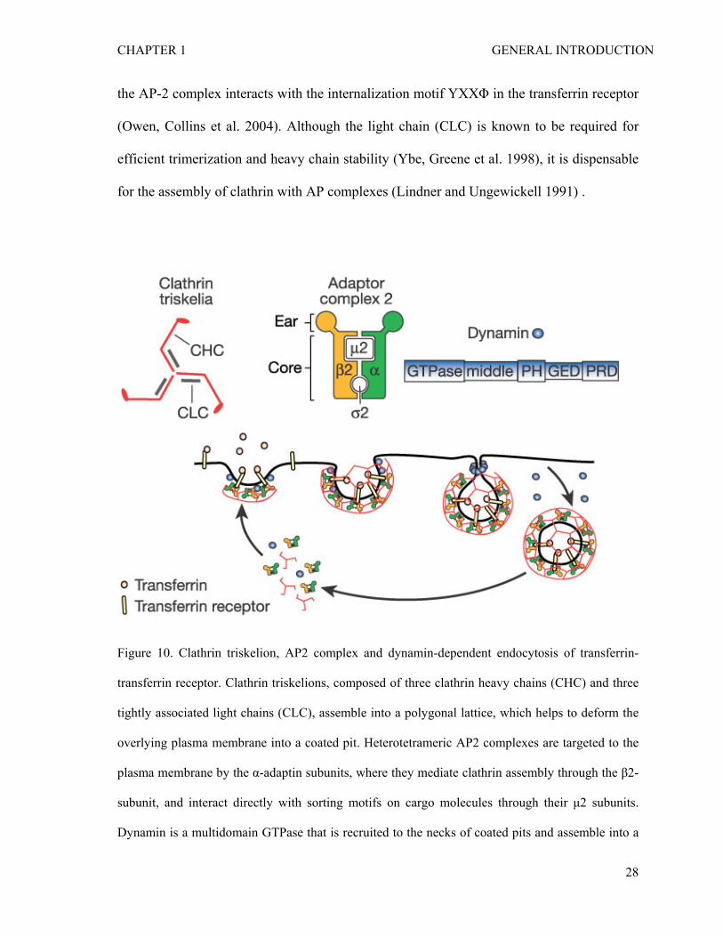
the AP-2 complex interacts with the internalization motif YXXΦ in the transferrin receptor
(Owen, Collins et al. 2004). Although the light chain (CLC) is known to be required for
efficient trimerization and heavy chain stability (Ybe, Greene et al. 1998), it is dispensable
for the assembly of clathrin with AP complexes (Lindner and Ungewickell 1991) .
Figure 10. Clathrin triskelion, AP2 complex and dynamin-dependent endocytosis of transferrin-
transferrin receptor. Clathrin triskelions, composed of three clathrin heavy chains (CHC) and three
tightly associated light chains (CLC), assemble into a polygonal lattice, which helps to deform the
overlying plasma membrane into a coated pit. Heterotetrameric AP2 complexes are targeted to the
plasma membrane by the α-adaptin subunits, where they mediate clathrin assembly through the β2-
subunit, and interact directly with sorting motifs on cargo molecules through their μ2 subunits.
Dynamin is a multidomain GTPase that is recruited to the necks of coated pits and assemble into a
28
CHAPTER 1 GENERAL INTRODUCTION
spiral, resulting in the scission and release of CCVs. A subsequent uncoating reaction recycles the
coat constituents for reuse. Adapted from Conner and Schmid 2003.
1.5.2 Endocytosis-independent functions of clathrin
Many proteins possess multiple and sometimes unexpected functions. This is also the case
of clathrin, especially its heavy chain (CHC). In addition to the well-characterized function
in endocytosis, moonlighting functions of CHC in the nucleus have been reported. Ten
years ago, Okamoto et al. have found that clathrin associates with mitotic spindle during
mitosis as formation of clathrin-coated vesicles is shut-down in cells undergoing mitosis
(Okamoto 2006). In 2005, Royle et al. have demonstrated for the first time that clathrin
stabilizes fibers of the mitotic spindle to assist congression of chromosomes (Royle, Bright
et al. 2005). They observed that mitosis is prolonged due to destabilized kinetochore fibers
and defective congression of chromosomes when CHC is depleted by RNAi (Royle, Bright
et al. 2005). In addition, Enari et al have shown that nuclear CHC binds to the tumor
suppressor p53 to enhance p53-dependent transactivation, which promotes p53 target gene
expression (Enari, Ohmori et al. 2006).
1.5.3 Regulation of NF-κB signalling pathway by clathrin-binding proteins of
endocytic pathway
Activation of NF-κB is mediated by sequential phosphorylation and activation of signalling
proteins involved in the NF-κB pathways upon receptor stimulation (e.g.TNFR-RIP-
TAK1-IKK, TLR/IL-1R-IRAK-TAK1-IKK, and Nod1-RIP2-TAK1-IKK). However, how
29
CHAPTER 1 GENERAL INTRODUCTION
those kinases remain inactive in resting cells is largely unknown. Protein-protein
interaction studies have revealed that several endocytic proteins play additional function as
inhibitors of NF-κB signalling pathways. Tom1 (target of Myb1) has been shown to
interact with Tollip (Toll-interacting protein), forming a complex to regulate endosomal
trafficking of ubiquitinated proteins such as IL-1R (Brissoni, Agostini et al. 2006). In
addition, Tollip forms a complex with IRAK (IL-1R-associated kinase) and blocks
phosphorylation of IRAK, which prevents IKK and NF-κB activation upon stimulation of
IL-1R but not TNFR (Burns, Clatworthy et al. 2000). Tom1 is proposed to be a common
negative regulator of signalling pathways induced by IL-1β and TNFα (Yamakami and
Yokosawa 2004). Recently, it was shown that Tom1 also inhibits NF-κB activation upon
TLR2/4 stimulation (Oglesby, Bray et al. 2010). Tom1 can also bind to CHC via a typical
clathrin binding motif (DLIDMG) and ubiquitin chains (Yamamoto, Verma et al. 2003).
However, the connection between Tom1/Tollip and CHC in the regulation of NF-κB has
not been addressed.
β-arrestins (β-arrestin 1 and β-arrestin 2) were initially known as negative regulators
of G-protein-coupled receptors (GPCRs)-mediated signalling (Reiter and Lefkowitz 2006).
Activation of GPCRs such as β2-adrenergic receptor promotes the recruitment of cytosolic
β-arrestins to the phosphorylated (activated) receptor. This uncouples the receptor from G
proteins and promotes the receptor internalization, thus causing desensitization (Claing,
Laporte et al. 2002). However, new roles of β-arrestins in MAPK signaling, and NF-κB and
p53-mediated transcriptional regulation have been discovered (Gao, Sun et al. 2004;
Lefkowitz and Whalen 2004; Shenoy, Drake et al. 2006). β-arrestins-IκBα interaction was
identified in yeast two-hybrid assays (Witherow, Garrison et al. 2004). Moreover,
30
CHAPTER 1 GENERAL INTRODUCTION
stimulation of β2-adrenergic receptor in HEK 293, HeLa and COS-7 cells significantly
increases the amount of β-arrestin 2, which is then associated with IκBα (Gao, Sun et al.
2004). The interaction with β-arrestin 2 prevents phosphorylation and degradation of IκBα
and thus attenuates activation of NF-κB and transcription of NF-κB target genes (Gao, Sun
et al. 2004; Witherow, Garrison et al. 2004). Functional relevance of β-arrestins regulation
of NF-κB was further confirmed in the NF-κB activating signalling pathways. Luan et al.
demonstrated that β-arrestin 2 can function as a suppressor of ultraviolet-induced NF-κB
activation through a direct interaction with IκBα (Luan, Zhang et al. 2005). In addition,
Wang et al. discovered that β-arrestins modulate TLR/IL-1R-mediated NF-κB signalling
through their interaction with TRAF6, preventing autoubiquitination of TRAF6 (Wang,
Tang et al. 2006). However, different from the β-arrestins- IκBα interaction, stimulation of
β2-adrenergic receptor has no effect on the interaction of β-arrestins and TRAF6,
suggesting that β-arrestins-TRAF6 interaction is regulated by IL1R independently of
GPCR stimulation (Wang, Tang et al. 2006). β-arrestins can bind to clathrin through the C-
terminal clathin binding domain (Krupnick, Goodman et al. 1997).
31

CHAPTER 1 GENERAL INTRODUCTION
32
1.6 Aim of the study
Nuclear factor-kappaB (NF-κB) is a cytosolic transcription factor in resting cells and
translocates into the nucleus and becomes active in response to pro-inflammatory stimuli
and bacterial infection. The IKK complex contains two catalytic subunits IKKα and IKKβ
and is essential for the activation of NF-κB through phosphorylation and degradation of the
inhibitor of NF-κB (IκBα). IKKα and IKKβ are structurally similar but functionally distinct
each other, with IKKα being important for lymphocyte organogenesis but IKKβ being
critical for inflammation and innate immunity.
The primary goal of my Ph.D. thesis research was to identify signaling proteins
important for NF-κB regulation in resting cells and in response to Shigella flexneri. Initial
efforts have been focused on an RNAi screen. Following the screen, special aims of the
follow-up studies were:
1) To reveal an unknown function of IKKα in inflammation and innate immunity
2) To investigate an endocytosis-independent function of clathrin heavy chain
(CHC) in the regulation of basal NF-κB activation.
CHAPTER 2 IKKα regulation of canonical NF-κB activation
CHAPTER 2
IKKα contributes to canonical NF-κB activation downstream of Nod1-
mediated peptidoglycan recognition
(MANUSCRIPT IN PRESS)
33
CHAPTER 2 IKKα regulation of canonical NF-κB activation
IKKα contributes to canonical NF-κB activation downstream of Nod1-mediated
peptidoglycan recognition
Man Lyang Kim, Hyun Gyeong Jeong, Christoph Alexander Kasper and Cécile
Arrieumerlou*
Biozentrum, University of Basel, Klingelbergstrasse 50-70, 4056 Basel, Switzerland
Running title: IKKα in NF-κB activation
*Correspondence: Prof. Cécile Arrieumerlou Focal Area Infection Biology, Biozentrum, University of Basel, Klingelbergstrasse 50-70, CH-4056 Basel, Switzerland. Tel.: +41 61 267 21 20 Fax: +41 61 267 21 18 E-mail: [email protected]
34
CHAPTER 2 IKKα regulation of canonical NF-κB activation
2.1 ABSTRACT
Background: During pathogen infection, innate immunity is initiated via the recognition of
microbial products by pattern recognition receptors and the subsequent activation of
transcription factors that upregulate proinflammatory genes. By controlling the expression
of cytokines, chemokines, anti-bacterial peptides and adhesion molecules, the transcription
factor nuclear factor-kappa B (NF-κB) has a central function in this process. In a typical
model of NF-κB activation, the recognition of pathogen associated molecules triggers the
canonical NF-κB pathway that depends on the phosphorylation of Inhibitor of NF-κB (IκB)
by the catalytic subunit IκB kinase β (IKKβ), its degradation and the nuclear translocation
of NF-κB dimers.
Methodology: Here, we performed an RNA interference (RNAi) screen on Shigella
flexneri-induced NF-κB activation to identify new factors involved in the regulation of NF-
κB following infection of epithelial cells by invasive bacteria. By targeting a subset of the
human signaling proteome, we found that the catalytic subunit IKKα is also required for
complete NF-κB activation during infection. Depletion of IKKα by RNAi strongly reduces
the nuclear translocation of NF-κB p65 during S. flexneri infection as well as the
expression of the proinflammatory chemokine interleukin-8. Similar to IKKβ, IKKα
contributes to the phosphorylation of IκBα on serines 32 and 36, and to its degradation.
Experiments performed with the synthetic Nod1 ligand L-Ala-D-γ-Glu-meso-
diaminopimelic acid confirmed that IKKα is involved in NF-κB activation triggered
downstream of Nod1-mediated peptidoglycan recognition.
Conclusions: Taken together, these results demonstrate the unexpected role of IKKα in the
canonical NF-κB pathway triggered by peptidoglycan recognition during bacterial infection.
35
CHAPTER 2 IKKα regulation of canonical NF-κB activation
In addition, they suggest that IKKα may be an important drug target for the development of
treatments that aim at limiting inflammation in bacterial infection.
36
CHAPTER 2 IKKα regulation of canonical NF-κB activation
2.2 INTRODUCTION
During pathogen infection, structurally conserved microbial molecules are recognized by
germline-encoded pathogen recognition receptors (PRRs) that function as sensors for non-
self detection and initiate innate immunity (Takeuchi and Akira ; Medzhitov 2007). PRRs
include transmembrane proteins such as Toll-like receptors and C-type lectin receptors, as
well as cytoplasmic proteins such as retinoic acid-inducible gene (RIG)-I-like receptors and
NOD-like receptors (Blasius and Beutler ; Franchi, Park et al. 2008; Kawai and Akira
2008). They are expressed in macrophages and dendritic cells but also in various non-
professional immune cells including epithelial and endothelial cells. PRRs recognize a
large variety of pathogen associated molecular patterns (PAMPs) from both extracellular
and intracellular pathogens including lipopolysaccharide, peptidoglycan, lipoproteins,
dsRNA, ssRNA, CpG-DNA and flagellin (Rasmussen, Reinert et al. 2009). Signaling
pathways of PAMP recognition converge into the activation of the pleiotropic transcription
factor nuclear factor-kappa B (NF-κB) that, in the context of innate immunity, regulates the
expression of proinflammatory genes encoding cytokines, chemokines, anti-bacterial
peptides and adhesion molecules (Beutler 2009). The mammalian NF-κB family consists of
the members RelA/p65, RelB, c-Rel, p50 (NF-κB1) and p52 (NF-κB2) (Hayden and Ghosh
2004). All five proteins share a Rel homology domain and form homo- and heterodimers
that regulate transcription by binding to κB sites in promoters or enhancers of target genes.
In unstimulated cells, most of the NF-κB dimers are sequestrated in the cytoplasm by the
proteins of the Inhibitor of NF-κB (IκB) family whose prototype is IκBα. In the canonical
pathway of NF-κB activation triggered by most stimuli including bacterial and viral
infection, cytokines and stress-induced responses, phosphorylation of IκBα on Serine 32
37
CHAPTER 2 IKKα regulation of canonical NF-κB activation
and Serine 36 residues by the IκB kinase (IKK) complex is a decisive regulatory step (Solt
and May 2008). The IKK complex is comprised of three subunits: two catalytic subunits,
IKKα and IKKβ, and the regulatory scaffold component NF-κB essential modulator
(NEMO). The respective contribution of IKKα and IKKβ in the phosphorylation of IκBα is
unclear. Although it is generally accepted that IKKβ is critical for IκBα phosphorylation
through the canonical pathway, two recent reports demonstrate the equal importance of
IKKα for the activation of NF-κB by the inflammatory cytokines interleukin-1 (IL-1) in
mouse embryonic fibroblasts and tumor necrosis factor alpha (TNFα) in HeLa cells (Adli,
Merkhofer et al. ; Solt, Madge et al. 2007). The phosphorylation of IκBα is followed by its
rapid polyubiquitination and subsequent degradation by the 26S proteasome complex
(Gilmore 2006). The release of NF-κB with unmasked nuclear localization sequence leads
then to the translocation of the transcription factor to the nucleus where it regulates gene
expression (Hayden and Ghosh 2008).
Although the role of NF-κB is central to many pathways triggered by
pathogen recognition, the molecular processes that govern its activation are only partially
elucidated. In particular, the mechanisms triggered by the detection of invasive bacteria
such as the pathogen Shigella flexneri remain largely uncharacterized. S. flexneri makes use
of a type III secretion (T3S) apparatus to locally rearrange the host actin cytoskeleton and
penetrate into intestinal epithelial cells (Schroeder and Hilbi 2008). Once internalized,
bacteria multiply in the host cytoplasm and use actin-based motility to spread to adjacent
epithelial cells. During infection, massive inflammation is observed in colonic mucosal
tissues (Islam, Veress et al. 1997). In infected epithelial cells, intracellular bacteria release
peptidoglycan-derived peptides that are specifically recognized by Nod1 (Girardin, Boneca
38
CHAPTER 2 IKKα regulation of canonical NF-κB activation
et al. 2003). Upon ligand binding, Nod1 homo-dimerizes and recruits the downstream
kinase RICK/RIPK2 through heterologous caspase-recruitment domain interactions
(Inohara, Koseki et al. 2000). This converges to the sequential recruitment and activation of
the TAK1/TAB1/TAB2 and IKKα/IKKβ/IKKγ complexes, the nuclear translocation of NF-
κB and the upregulation of proinflammatory genes encoding for cytokines and chemokines,
including interleukin-8 (IL-8) and TNFα (Sansonetti, Arondel et al. 1999). The chemokine
IL-8 recruits polymorphonuclear cells to the site of infection and therefore contributes to
contain the dissemination of bacteria within the intestinal tissue. Interestingly, S. flexneri
uses the T3S apparatus to secrete several effectors that alter multiple signaling pathways in
infected cells and reduce the expression of proinflammatory genes (Ogawa, Handa et al.
2008). Among others, the effector OspF suppresses the expression of IL-8 by
dephosphorylating the MAP kinases p38 and ERK in the nucleus of infected cells (Arbibe,
Kim et al. 2007; Li, Xu et al. 2007), thereby impairing the phosphorylation of Histone H3,
a process that regulates the access of chromatin to transcription factors.
Here, we performed an RNA interference (RNAi) screen on S. flexneri-
induced NF-κB activation to identify new factors involved in the regulation of NF-κB
following infection of epithelial cells by invasive bacteria. By targeting a subset of the
human signaling proteome, we identified IKKα as a protein required for S. flexneri-induced
NF-κB nuclear translocation and IL-8 secretion in HeLa cells. This result was unexpected
because, except for IL-1 and TNFα (Adli, Merkhofer et al. ; Solt, Madge et al. 2007), it is
generally accepted that IKKβ is the component of the IKK complex involved in the
canonical pathway of NF-κB activation. Depletion of IKKα or IKKβ indicated that S.
flexneri-induced NF-κB activation in HeLa cells requires indeed both catalytic subunits.
39
CHAPTER 2 IKKα regulation of canonical NF-κB activation
We further characterized the role of IKKα and found that, during S. flexneri infection,
IKKα was required for the phosphorylation of IκBα on serines 32 and 36, and for its
degradation. Experiments performed with the synthetic Nod1 ligand L-Ala-D-γ-Glu-meso-
diaminopimelic acid (Tri-DAP) indicated that IKKα was involved in Nod1-mediated
signaling pathway of NF-κB activation. Taken together, these results show that, although
Nod1 signaling triggers the canonical pathway of NF-κB activation, both IKKα and IKKβ
are required for full NF-κB activation.
40
CHAPTER 2 IKKα regulation of canonical NF-κB activation
2.3 MATERIALS AND METHODS
Antibodies and reagents
Antibodies against NF-κB p65, IκBα and IKKα were obtained from Santa Cruz
Biotechnology (Santa Cruz, USA) whereas the anti-actin was from Chemicon (Billerica,
USA) and the anti-phospho-IκBα was from Cell signaling technology (Beverly, USA). The
anti-mouse IgG-Cy5 was obtained from Zymed (San Francisco, USA) and the anti-rabbit
IgG-HRP and anti-mouse IgG-HRP from GE Healthcare (Pittsburgh, USA). Hoechst and
FITC-phalloidin were from Invitrogen (Carlsbad, USA), TNFα from R & D systems
(Minneapolis, USA).
Cell culture and transfection
HeLa cells were maintained in Dulbecco’s modified Eagle’s medium (high glucose)
supplemented with 10% fetal bovine serum, 100 units/ml penicillin, and 100 μg/ml
streptomycin at 37 °C in 10% CO2. HeLa cells were transfected with siRNAs and DNA
plasmids using Lipofectamine 2000 (Invitrogen, Carlsbad, USA) and jetPEI (Poly plus
transfection, Illkirch, France), respectively. siRNAs ON-TARGETplus SMARTpool
targeting IKKα (#L-003473-00-005) and ON-TARGETplus siCONTROL (Dharmacon,
Dallas, USA) were used in all our study except for the experiments where IKKα, IKKβ and
NEMO were silenced in parallel. In this case, all siRNAs were from Qiagen (Valencia, CA,
USA).
In vitro diced siRNA library
41
CHAPTER 2 IKKα regulation of canonical NF-κB activation
An in vitro diced siRNA library targeting 132 genes coding for a subset of the signaling
proteome was generated as previously described (Liou, Kim et al. 2005; Brandman, Liou et
al. 2007; Galvez, Teruel et al. 2007). Briefly, for each gene, a 600 base pair cDNA was
generated by PCR from a total cDNA library. An additional set of nested primers was used
to add T7 promoters at both ends of the final cDNA fragment. Nested PCR products were
subject to in vitro transcription, dicing, and purification to produce gene specific siRNA
pools. Dicing was performed with the turbo dicer siRNA generation kit from Genlantis
(San Diego, USA). The concentration of all siRNA pools was normalized.
Bacterial strains
The S. flexneri strains M90T wild-type and the icsA (virG) deletion mutant (ΔvirG) were
generously provided by Dr. P. Sansonetti (Institut Pasteur, Paris, France). All strains were
transformed with the pMW211 plasmid to express the DsRed protein under control of a
constitutive promoter. The pMW211 plasmid was a generous gift from Dr. D. Bumann
(Biozentrum, University of Basel, Switzerland). The ΔospF deletion mutant used in IL-8
expression experiments, was generated from the ΔvirG mutant by allelic exchange using a
modification of the lambda red-mediated gene deletion (Datsenko and Wanner 2000).
Briefly, the genes for lambda red recombination were expressed from the pKM208 plasmid
(Murphy and Campellone 2003). The chloramphenicol resistance cassette (cat) of the
pKD3 plasmid was amplified using the primers listed in Table 1. After DpnI digestion, the
PCR product was electroporated into the ΔvirG mutant. Recombinants were selected on
TSB plates containing 5 or 10 μg ml-1 chloramphenicol. The cat cassette was removed by
42
CHAPTER 2 IKKα regulation of canonical NF-κB activation
-1transformation of pCP20 and incubation at 30°C on TSB plates containing 100 μg ml
ampicillin (Datsenko and Wanner 2000). Single colonies were screened by PCR.
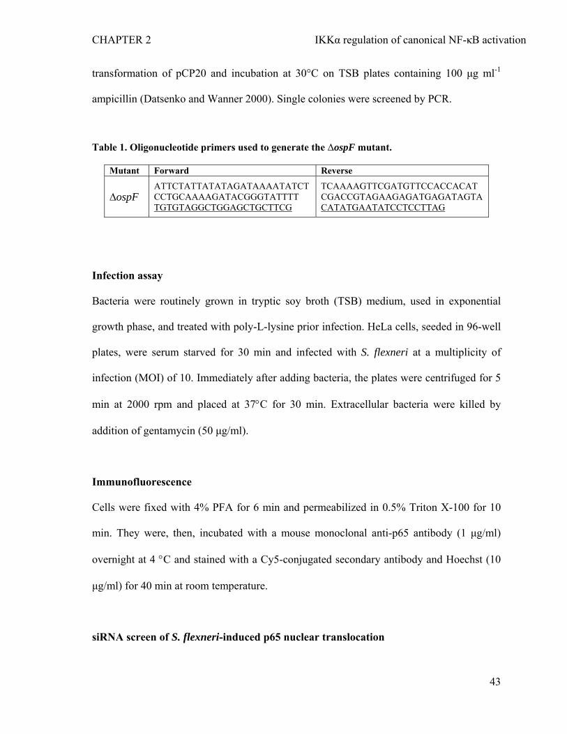
Table 1. Oligonucleotide primers used to generate the ΔospF mutant.
Mutant Forward Reverse
ATTCTATTATATAGATAAAATATCTCCTGCAAAAGATACGGGTATTTT
TCAAAAGTTCGATGTTCCACCACATCGACCGTAGAAGAGATGAGATAGTA∆ospF
TGTGTAGGCTGGAGCTGCTTCG CATATGAATATCCTCCTTAG
Infection assay
Bacteria were routinely grown in tryptic soy broth (TSB) medium, used in exponential
growth phase, and treated with poly-L-lysine prior infection. HeLa cells, seeded in 96-well
plates, were serum starved for 30 min and infected with S. flexneri at a multiplicity of
infection (MOI) of 10. Immediately after adding bacteria, the plates were centrifuged for 5
min at 2000 rpm and placed at 37°C for 30 min. Extracellular bacteria were killed by
addition of gentamycin (50 μg/ml).
Immunofluorescence
Cells were fixed with 4% PFA for 6 min and permeabilized in 0.5% Triton X-100 for 10
min. They were, then, incubated with a mouse monoclonal anti-p65 antibody (1 μg/ml)
overnight at 4 °C and stained with a Cy5-conjugated secondary antibody and Hoechst (10
μg/ml) for 40 min at room temperature.
siRNA screen of S. flexneri-induced p65 nuclear translocation
43
CHAPTER 2 IKKα regulation of canonical NF-κB activation
The in vitro diced siRNA library was screened on S. flexneri-induced p65 translocation
assay in a 96-well format. The firefly luciferase (GL3) siRNA was used as a non-silencing
negative control as described previously (Liou, Kim et al. 2005; Brandman, Liou et al.
2007; Galvez, Teruel et al. 2007). siRNA pools against Nod1, RIPK2 and Src were used as
positive controls. The screen was performed three times in duplicate as follows. Three
thousand HeLa cells per well were transfected by reverse transfection with the individual
132 siRNA pools in 96-well plates. After 48 hours, cells were infected with DsRed S.
flexneri at MOI of 10 for 90 min and then fixed, permeabilized, and stained for p65, F-
actin, and DNA. Images were acquired at 12 random sites of each well using the automated
ImageXpress microscope (Molecular devices, Sunnyvale, USA). At each site, images at
360 nm, 480 nm, 594 nm, 640 nm were acquired to visualize Hoechst, Phalloidin, DsRed S.
flexneri and p65, respectively. The nuclear localization of p65 was automatically quantified
by using the Enhanced-Translocation module of MetaXpress (Molecular devices,
Sunnyvale, USA). Briefly, the Hoechst staining was used as a mask to automatically
identify nuclei in the p65 staining image. The cytoplasmic area was defined by a ring
around each nucleus. For each cell, the ratio of p65 intensity in the nucleus and in the
cytoplasmic ring defined as the Nuc/Cyt p65 ratio was calculated and averaged over
several thousands of cells per well. The results of the screen were expressed as individual
scores. The score of a particular gene represents the fold standard deviation from the mean
of the GL3 control wells. A negative or a positive sign was assigned to the score when the
Nuc/Cyt p65 ratio was lower or higher than the GL3 control ratio, respectively.
Enzyme- linked Immunosorbent Assay (ELISA)
44
CHAPTER 2 IKKα regulation of canonical NF-κB activation
IL-8 secretion was measured by ELISA in the supernatant of HeLa cells 6 hours post
infection. Cell-free supernatants from triplicate wells were analyzed for their IL-8 content
using a commercial ELISA kit (BD Pharmingen, San Jose, USA).
Western Blot Analysis
HeLa cells were transfected with siRNAs in a 6-well plate. 72 hours post transfection, cells
were lysed in Phosphosafe Extraction Buffer (Novagen, Darmstadt, Germany)
supplemented with 1x protease inhibitor cocktail (Calbiochem, Darmstadt, Germany).
Protein concentration was measured using the bicinchonic acid (BCA) kit (Pierce,
Rockford, USA). Equal amounts of proteins were resolved by SDS-PAGE and transferred
to Hybond C-Extra membrane (Amersham Bioscience, Pittsburgh, USA) for
immunoblotting with individual antibodies. Primary antibodies were detected using
horseradish peroxidase-conjugated anti-rabbit or anti-mouse IgG antibodies, and visualized
with the ECL system (Pierce). Quantification of the blots was performed using the
densitometry feature of Photoshop.
Tri-DAP treatment
Cells were serum starved 30 min before treatment with L-Ala-γ-D-Glu-mesoDAP (Tri-
DAP). Tri-DAP treatment was performed by calcium phosphate transfection using 0.4
μg/ml Tri-DAP final concentration for 90 min. Then cells were fixed with 4% PFA and
analyzed by immunofluorescence as described above.
45
CHAPTER 2 IKKα regulation of canonical NF-κB activation
Statistical analysis
Data are expressed as mean ± standard deviation calculated from the number of replicates
specified in figure legends. p values were calculated with a two-tailed two-sample equal
variance t-test.
46
CHAPTER 2 IKKα regulation of canonical NF-κB activation
2.4 RESULTS
2.4.1 RNAi screen identifies the role of IKKα in the nuclear translocation of NF-κB
p65 during infection of epithelial cells by S. flexneri
Over the past 10 years, RNAi has been an essential tool to study gene function in
mammalian cells (Liou, Kim et al. 2005; Pelkmans, Fava et al. 2005; Zhang, Liu et al.
2009). Using S. flexneri-induced NF-κB activation in HeLa cells as a model system, we
performed an image-based RNAi screen to identify new proteins involved in the molecular
mechanisms that control NF-κB activation following pathogen recognition. The nuclear
localization of NF-κB p65 visualized by anti-p65 immunofluorescence microscopy was
used as readout for NF-κB activation. In uninfected cells, p65 was mainly localized in the
cytoplasm (Figure 2.1A, left panel). In contrast, following infection with dsRed-expressing
S. flexneri, a strong nuclear translocation of p65 was observed (Figure 2.1A, right panel).
To validate the RNAi approach, silencing of the intracellular pattern recognition receptor
Nod1 involved in S. flexneri recognition was tested. HeLa cells were transfected with pools
of in vitro diced small interference RNA (siRNA) targeting Nod1 or the firefly Luciferase
used as control (GL3). After 48 hours, cells were infected with S. flexneri, stained for p65
and DNA with an anti-p65 antibody and Hoechst, respectively. Visual inspection of images
showed that S. flexneri-induced p65 nuclear translocation was suppressed in Nod1-depleted
cells (Figure 2.1B). This observation was quantified by measuring for each cell the ratio of
p65 intensity in the nucleus and in the cytoplasm by automated image processing (Figure
2.1C). The Hoechst staining was used to automatically identify nuclei whereas the
cytoplasmic area was defined by a ring around each nucleus. Quantification of the
Nucleus/Cytoplasm p65 intensity ratio (Nuc/Cyt p65 ratio) confirmed that the depletion of
47
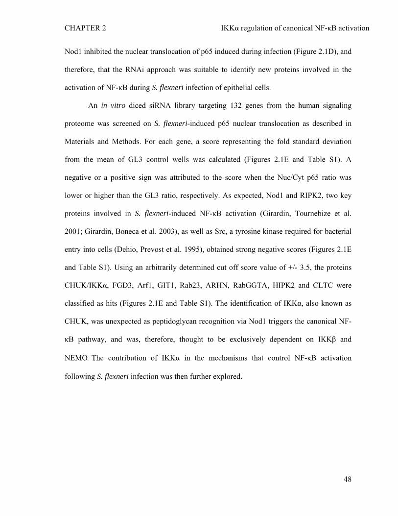
CHAPTER 2 IKKα regulation of canonical NF-κB activation
Nod1 inhibited the nuclear translocation of p65 induced during infection (Figure 2.1D), and
therefore, that the RNAi approach was suitable to identify new proteins involved in the
activation of NF-κB during S. flexneri infection of epithelial cells.
An in vitro diced siRNA library targeting 132 genes from the human signaling
proteome was screened on S. flexneri-induced p65 nuclear translocation as described in
Materials and Methods. For each gene, a score representing the fold standard deviation
from the mean of GL3 control wells was calculated (Figures 2.1E and Table S1). A
negative or a positive sign was attributed to the score when the Nuc/Cyt p65 ratio was
lower or higher than the GL3 ratio, respectively. As expected, Nod1 and RIPK2, two key
proteins involved in S. flexneri-induced NF-κB activation (Girardin, Tournebize et al.
2001; Girardin, Boneca et al. 2003), as well as Src, a tyrosine kinase required for bacterial
entry into cells (Dehio, Prevost et al. 1995), obtained strong negative scores (Figures 2.1E
and Table S1). Using an arbitrarily determined cut off score value of +/- 3.5, the proteins
CHUK/IKKα, FGD3, Arf1, GIT1, Rab23, ARHN, RabGGTA, HIPK2 and CLTC were
classified as hits (Figures 2.1E and Table S1). The identification of IKKα, also known as
CHUK, was unexpected as peptidoglycan recognition via Nod1 triggers the canonical NF-
κB pathway, and was, therefore, thought to be exclusively dependent on IKKβ and
NEMO. The contribution of IKKα in the mechanisms that control NF-κB activation
following S. flexneri infection was then further explored.
48
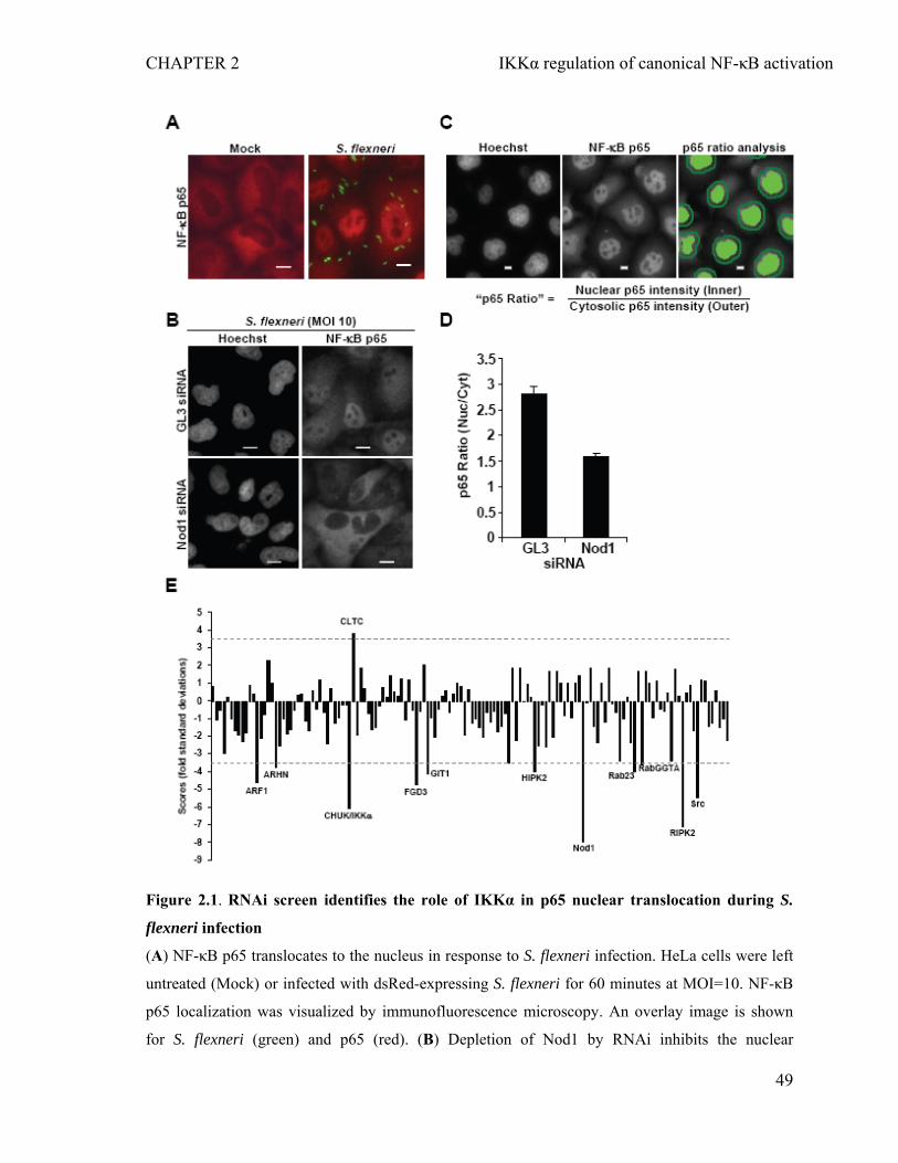
CHAPTER 2 IKKα regulation of canonical NF-κB activation
Figure 2.1. RNAi screen identifies the role of IKKα in p65 nuclear translocation during S.
flexneri infection
(A) NF-κB p65 translocates to the nucleus in response to S. flexneri infection. HeLa cells were left
untreated (Mock) or infected with dsRed-expressing S. flexneri for 60 minutes at MOI=10. NF-κB
p65 localization was visualized by immunofluorescence microscopy. An overlay image is shown
for S. flexneri (green) and p65 (red). (B) Depletion of Nod1 by RNAi inhibits the nuclear
49
CHAPTER 2 IKKα regulation of canonical NF-κB activation
translocation of NF-κB p65 in response to S. flexneri infection. HeLa cells were transfected with
GL3 or Nod1 siRNA and infected with S. flexneri for 60 minutes at MOI=10. p65 and DNA were
visualized by anti-p65 and Hoechst staining, respectively. (C) Images illustrating the quantification
of the p65 ratio. The Hoechst staining was used as a mask to automatically identify the nuclei in the
p65 staining image. The cytoplasmic area was defined by a ring surrounding each nucleus. Scale
bars, 10 μm (D) Quantification of the p65 ratio in control and Nod1-depleted cells following
infection by S. flexneri. Results represent the mean ± SD of 12 images; graph representative of 3
independent experiments. (E) RNAi screen of S. flexneri-induced p65 nuclear translocation in HeLa
cells. Scores are fold standard deviations from the mean of GL3 control wells. Dashed lines
represent scores of ± 3.5 (See Materials and Methods for details).
2.4.2 Depletion of IKKα inhibits S. flexneri-induced p65 nuclear translocation and
IL-8 expression without affecting bacterial invasion
First, to validate the role of IKKα in S. flexneri-induced p65 nuclear translocation, we
tested a pool of four synthetic IKKα siRNAs that had no overlapping sequences with the in
vitro diced siRNAs used in the screen. In conditions where around 90% of endogenous
protein was depleted (Figure 2.2A), a massive reduction of p65 nuclear translocation was
observed (Figures 2.2B and 2.2C). This result was not explained by a reduction of bacterial
uptake as the number of internalized bacteria was similar in control and in IKKα-depleted
cells (Figure 2.2B). Taken together, these results indicated that IKKα is required for the
activation of NF-κB during infection of epithelial cells by S. flexneri.
During infection, NF-κB positively regulates the expression of multiple
proinflammatory genes (Pedron, Thibault et al. 2003). In particular, it induces the
expression of the chemokine IL-8, which recruits PMNs on site of infection, and thereby
limits the spread of bacterial invasion within the intestinal tissue (Sansonetti, Arondel et al.
50
CHAPTER 2 IKKα regulation of canonical NF-κB activation
1999). To test whether IKKα contributed to the upregulation of IL-8 expression during
infection, we measured by ELISA the secretion of IL-8 in the supernatant of control and
IKKα-depleted HeLa cells six hours post infection. To increase the amount of IL-8
produced in response to infection, cells were infected with a mutant of S. flexneri deleted
for the type III effector OspF (ΔospF) that dampens inflammation signaling by
dephosphorylating p38 in the nucleus of infected cells (Arbibe, Kim et al. 2007).
Consistent with NF-κB data, a reduction of IL-8 secretion was observed in response to S.
flexneri ΔospF infection when cells were depleted for IKKα (Figure 2.2D), showing that
IKKα is involved in the signaling pathways that control the expression of a critical
inflammatory chemokine during bacterial infection.
51
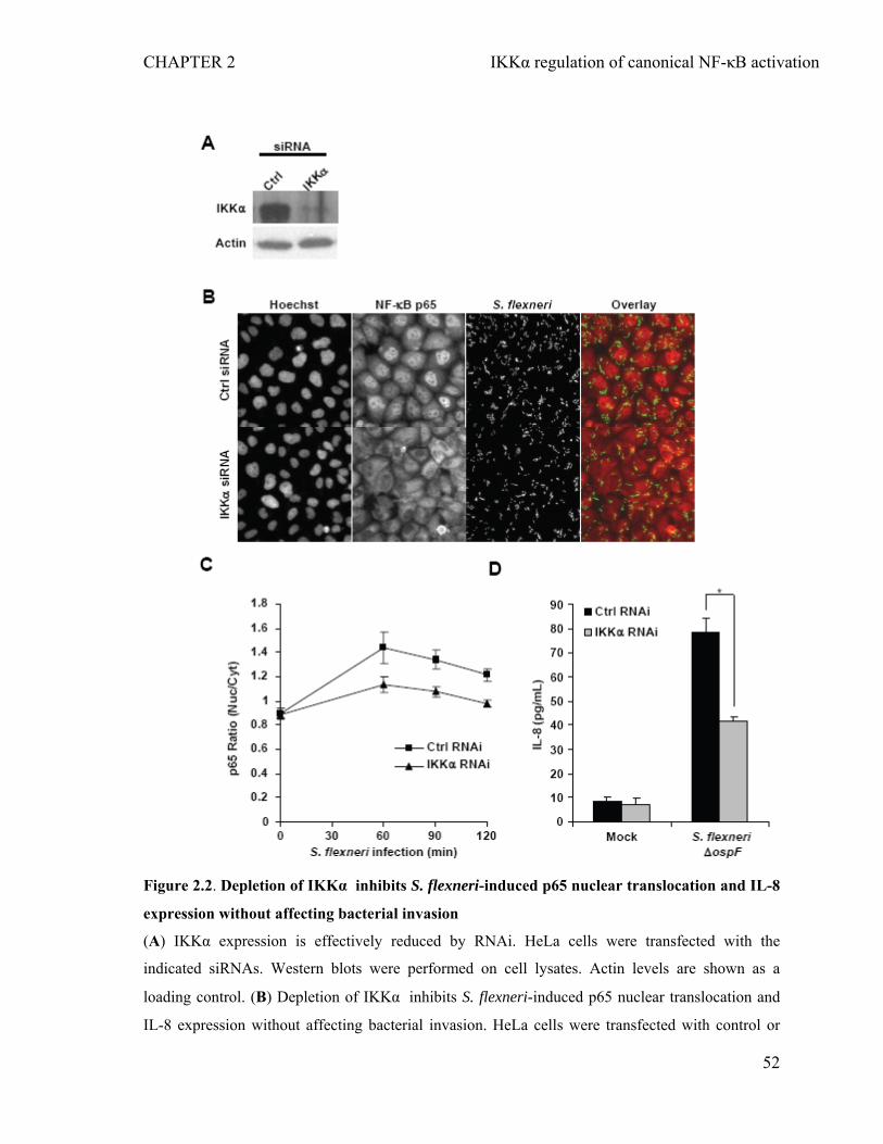
CHAPTER 2 IKKα regulation of canonical NF-κB activation
Figure 2.2. Depletion of IKKα inhibits S. flexneri-induced p65 nuclear translocation and IL-8
expression without affecting bacterial invasion
(A) IKKα expression is effectively reduced by RNAi. HeLa cells were transfected with the
indicated siRNAs. Western blots were performed on cell lysates. Actin levels are shown as a
loading control. (B) Depletion of IKKα inhibits S. flexneri-induced p65 nuclear translocation and
IL-8 expression without affecting bacterial invasion. HeLa cells were transfected with control or
52
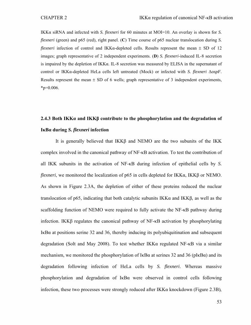
CHAPTER 2 IKKα regulation of canonical NF-κB activation
IKKα siRNA and infected with S. flexneri for 60 minutes at MOI=10. An overlay is shown for S.
flexneri (green) and p65 (red), right panel. (C) Time course of p65 nuclear translocation during S.
flexneri infection of control and IKKα-depleted cells. Results represent the mean ± SD of 12
images; graph representative of 2 independent experiments. (D) S. flexneri-induced IL-8 secretion
is impaired by the depletion of IKKα. IL-8 secretion was measured by ELISA in the supernatant of
control or IKKα-depleted HeLa cells left untreated (Mock) or infected with S. flexneri ΔospF.
Results represent the mean ± SD of 6 wells; graph representative of 3 independent experiments,
*p=0.006.
2.4.3 Both IKKα and IKKβ contribute to the phosphorylation and the degradation of
IκBα during S. flexneri infection
It is generally believed that IKKβ and NEMO are the two subunits of the IKK
complex involved in the canonical pathway of NF-κB activation. To test the contribution of
all IKK subunits in the activation of NF-κB during infection of epithelial cells by S.
flexneri, we monitored the localization of p65 in cells depleted for IKKα, IKKβ or NEMO.
As shown in Figure 2.3A, the depletion of either of these proteins reduced the nuclear
translocation of p65, indicating that both catalytic subunits IKKα and IKKβ, as well as the
scaffolding function of NEMO were required to fully activate the NF-κB pathway during
infection. IKKβ regulates the canonical pathway of NF-κB activation by phosphorylating
IκBα at positions serine 32 and 36, thereby inducing its polyubiquitination and subsequent
degradation (Solt and May 2008). To test whether IKKα regulated NF-κB via a similar
mechanism, we monitored the phosphorylation of IκBα at serines 32 and 36 (pIκBα) and its
degradation following infection of HeLa cells by S. flexneri. Whereas massive
phosphorylation and degradation of IκBα were observed in control cells following
infection, these two processes were strongly reduced after IKKα knockdown (Figure 2.3B),
53
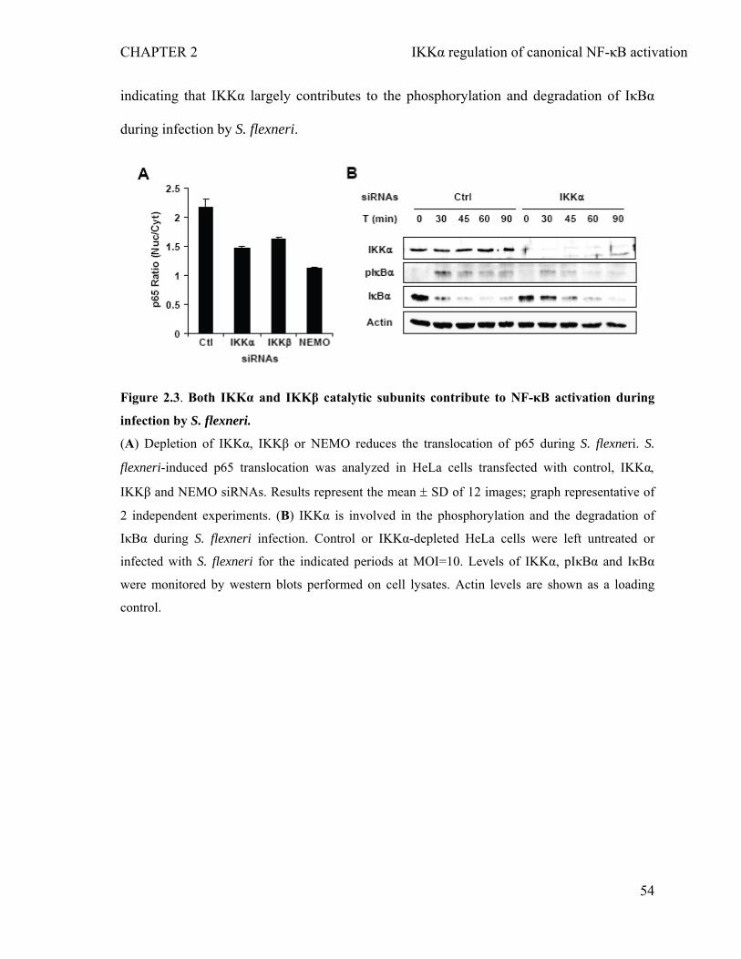
CHAPTER 2 IKKα regulation of canonical NF-κB activation
indicating that IKKα largely contributes to the phosphorylation and degradation of IκBα
during infection by S. flexneri.
Figure 2.3. Both IKKα and IKKβ catalytic subunits contribute to NF-κB activation during
infection by S. flexneri.
(A) Depletion of IKKα, IKKβ or NEMO reduces the translocation of p65 during S. flexneri. S.
flexneri-induced p65 translocation was analyzed in HeLa cells transfected with control, IKKα,
IKKβ and NEMO siRNAs. Results represent the mean ± SD of 12 images; graph representative of
2 independent experiments. (B) IKKα is involved in the phosphorylation and the degradation of
IκBα during S. flexneri infection. Control or IKKα-depleted HeLa cells were left untreated or
infected with S. flexneri for the indicated periods at MOI=10. Levels of IKKα, pIκBα and IκBα
were monitored by western blots performed on cell lysates. Actin levels are shown as a loading
control.
54
CHAPTER 2 IKKα regulation of canonical NF-κB activation
2.4.4 IKKα is involved in Nod1-mediated peptidoglycan recognition
During S. flexneri infection, the activation of NF-κB is initiated by the recognition of
peptidoglycan fragments via the intracellular receptor Nod1 (Girardin, Boneca et al. 2003).
To specifically examine the implication of IKKα in Nod1-mediated signaling, the effect of
IKKα depletion was directly tested in cells exposed to the Nod1 ligand Tri-DAP. Whereas
Tri-DAP treatment induced a clear nuclear translocation of p65 in control cells, this
translocation was severely impaired in IKKα-depleted cells (Figures 2.4A and 2.4B),
demonstrating that IKKα is required for the activation of NF-κB following Nod1-mediated
peptidoglycan recognition.
Figure 2.4 IKKα is required for the activation of NF-κB induced by Nod1-mediated
peptidoglycan recognition
(A) IKKα depletion reduces the activation of NF-κB induced by Nod1-mediated peptidoglycan
recognition. Control or IKKα-depleted HeLa cells were treated with Tri-DAP as described in
Materials and Methods. After 1 hour, cells were fixed, stained for p65 and DNA with an anti-p65
antibody and Hoechst, respectively. (B) Quantification of the effect of IKKα depletion on Tri-DAP-
induced p65 nuclear translocation. Results represent the mean ± SD of 12 images; graph
representative of 3 independent experiments, **p ≤ 8.6E-4, ns: non-significant.
55
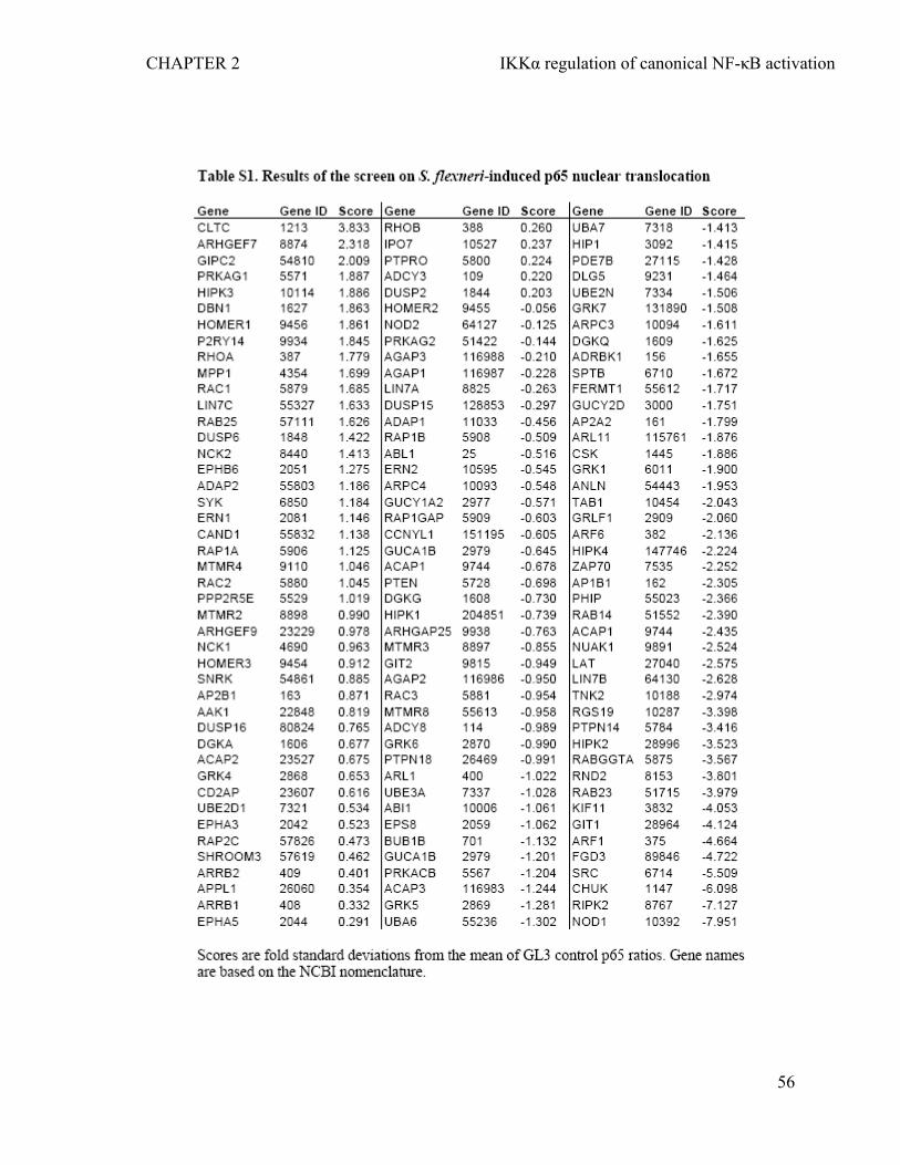
CHAPTER 2 IKKα regulation of canonical NF-κB activation
56
CHAPTER 2 IKKα regulation of canonical NF-κB activation
2.5 DISCUSSION
Although NF-κB is a transcription factor that has been the subject of intensive
research by academic laboratories and the pharmaceutical industry, the complex molecular
mechanisms controlling its activation are only partially elucidated. Here we performed an
RNAi screen to identify new proteins involved in the mechanisms that control NF-κB
activation following pathogen recognition using infection of epithelial cells by the invasive
bacterium S. flexneri as a model system. The presence of S. flexneri in the cytoplasm is
recognized via the detection of peptidoglycan by the receptor Nod1 (Girardin, Boneca et al.
2003). This recognition leads to NF-κB activation and the upregulation of proinflammatory
genes that orchestrate the host inflammatory response (Pedron, Thibault et al. 2003). The
protein IKKα was identified in the screen as a protein required for the activation of NF-κB
in response to S. flexneri infection. Since bacterial uptake was not affected by the depletion
of IKKα, this result indicated that IKKα is involved in the activation of NF-κB in response
to infection. The depletion of IKKβ or NEMO also induced a reduction of S. flexneri-
induced-p65 nuclear translocation, confirming that all subunits were required for the full
activation of NF-κB during infection. As the translocation of p65 depends on the
degradation of IκBα primed by its phosphorylation on serines 32 and 36, the role of IKKα
in these two processes was analyzed. A reduction of IκBα phosphorylation and delayed
degradation were observed in cells depleted for IKKα , indicating that similar to IKKβ,
IKKα triggers NF-κB activation by inducing the phosphorylation and the degradation of
IκBα. Taken together, these results provided new evidence for a role of IKKα in the
canonical pathway of NF-κB activation, and were in line with few reports indicating that
the dominant model of canonical NF-κB activation based on IKKβ and NEMO is
57
CHAPTER 2 IKKα regulation of canonical NF-κB activation
incomplete. In particular, it has been reported that IKKα is the key subunit responsible for
the Receptor activator of NF-κB (RANK)-induced classical NF-κB activation in mammary
epithelial cells (Cao, Bonizzi et al. 2001). In addition, Solt et al. showed that IL-1-induced
NF-κB activation requires the interaction of IKKα with NEMO and occurs in the absence
of IKKβ (Solt, Madge et al. 2007) . Finally, it has been shown recently that both IKKα and
IKKβ contribute to IκBα phosphorylation and NF-κB activation in response to TNFα
stimulation in HeLa cells (Adli, Merkhofer et al.). For the first time, our results
demonstrate that IKKα is also implicated in the canonical pathway of NF-κB activation
triggered by bacterial infection. To further demonstrate that pathogen recognition induced
NF-κB activation in an IKKα-dependent manner during S. flexneri infection, we directly
tested whether IKKα was involved in the activation of NF-κB induced by Nod1-mediated
peptidoglycan recognition. For this purpose, controls or IKKα-depleted cells were directly
stimulated with the purified Nod1 ligand Tri-DAP. The analysis of the p65 nuclear
translocation showed that IKKα was required for the full activation of NF-κB in response
to Tri-DAP treatment, indicating that IKKα is involved in the molecular mechanism
signaling the recognition of peptidoglycan-derived peptides by Nod1.
Following infection of epithelial cells by S. flexneri, the activation of NF-κB leads
to the upregulation of genes encoding for inflammatory cytokines including IL-8 and TNFα
(Pedron, Thibault et al. 2003). To confirm the implication of IKKα in the expression of
genes induced by the canonical NF-κB pathway, the deletion of IKKα was tested on S.
flexneri-induced IL-8 expression. Consistent with NF-κB data, the secretion of IL-8 was
reduced when IKKα was depleted, showing that the contribution of IKKα to NF-κB
activation has a functional impact on the amplitude of the inflammatory response mounted
58
CHAPTER 2 IKKα regulation of canonical NF-κB activation
in response to bacterial infection. These results suggest that inhibition of IKKα activity
may be critical to control inflammation upon bacterial infection. Inhibitors of IKKβ have
demonstrated therapeutic benefits in various animal models of inflammatory diseases and
are currently in early clinical trials (Ogawa, Azuma et al. ; Ziegelbauer, Gantner et al.
2005; Izmailova, Paz et al. 2007). The data presented in this manuscript suggest that IKKα
inhibitors should also be developed and used in combination with IKKβ inhibitors to limit
inflammation during bacterial infection or in inflammatory disorders that may involve
Nod1 signaling, including asthma, eczema and inflammatory bowel diseases (Reijmerink,
Bottema et al. ; Weidinger, Klopp et al. 2005; Le Bourhis, Benko et al. 2007).
59
CHAPTER 2 IKKα regulation of canonical NF-κB activation
2.6 ACKNOWLEDGMENTS
We thank Prof. Dirk Bumann for the pMW211 plasmid and Dr. Isabel Sorg for critical
reading and helpful suggestions. This work was supported by Swiss National Science
Foundation (grant 3100A0-113561), the University of Basel and the InfectX grant from
SystemsX.ch. C. Kasper was supported by the Werner-Siemens Foundation.
60
CHAPTER 2 IKKα regulation of canonical NF-κB activation
2.7 REFERENCES Adli, M., E. Merkhofer, et al. "IKKalpha and IKKbeta each function to regulate NF-
kappaB activation in the TNF-induced/canonical pathway." PLoS One 5(2): e9428. Arbibe, L., D. W. Kim, et al. (2007). "An injected bacterial effector targets chromatin
access for transcription factor NF-kappaB to alter transcription of host genes involved in immune responses." Nat Immunol 8(1): 47-56.
Beutler, B. (2009). "Microbe sensing, positive feedback loops, and the pathogenesis of inflammatory diseases." Immunol Rev 227(1): 248-263.
Blasius, A. L. and B. Beutler "Intracellular toll-like receptors." Immunity 32(3): 305-315. Brandman, O., J. Liou, et al. (2007). "STIM2 is a feedback regulator that stabilizes basal
cytosolic and endoplasmic reticulum Ca2+ levels." Cell 131(7): 1327-1339. Cao, Y., G. Bonizzi, et al. (2001). "IKKalpha provides an essential link between RANK
signaling and cyclin D1 expression during mammary gland development." Cell 107(6): 763-775.
Datsenko, K. A. and B. L. Wanner (2000). "One-step inactivation of chromosomal genes in Escherichia coli K-12 using PCR products." Proc Natl Acad Sci U S A 97(12): 6640-6645.
Dehio, C., M. C. Prevost, et al. (1995). "Invasion of epithelial cells by Shigella flexneri induces tyrosine phosphorylation of cortactin by a pp60c-src-mediated signalling pathway." Embo J 14(11): 2471-2482.
Franchi, L., J. H. Park, et al. (2008). "Intracellular NOD-like receptors in innate immunity, infection and disease." Cell Microbiol 10(1): 1-8.
Galvez, T., M. N. Teruel, et al. (2007). "siRNA screen of the human signaling proteome identifies the PtdIns(3,4,5)P3-mTOR signaling pathway as a primary regulator of transferrin uptake." Genome Biol 8(7): R142.
Gilmore, T. D. (2006). "Introduction to NF-kappaB: players, pathways, perspectives." Oncogene 25(51): 6680-6684.
Girardin, S. E., I. G. Boneca, et al. (2003). "Nod1 detects a unique muropeptide from gram-negative bacterial peptidoglycan." Science 300(5625): 1584-1587.
Girardin, S. E., R. Tournebize, et al. (2001). "CARD4/Nod1 mediates NF-kappaB and JNK activation by invasive Shigella flexneri." EMBO Rep 2(8): 736-742.
Hayden, M. S. and S. Ghosh (2004). "Signaling to NF-kappaB." Genes Dev 18(18): 2195-2224.
Hayden, M. S. and S. Ghosh (2008). "Shared principles in NF-kappaB signaling." Cell 132(3): 344-362.
Inohara, N., T. Koseki, et al. (2000). "An induced proximity model for NF-kappa B activation in the Nod1/RICK and RIP signaling pathways." J Biol Chem 275(36): 27823-27831.
Islam, D., B. Veress, et al. (1997). "In situ characterization of inflammatory responses in the rectal mucosae of patients with shigellosis." Infect Immun 65(2): 739-749.
Izmailova, E. S., N. Paz, et al. (2007). "Use of molecular imaging to quantify response to IKK-2 inhibitor treatment in murine arthritis." Arthritis Rheum 56(1): 117-128.
Kawai, T. and S. Akira (2008). "Toll-like receptor and RIG-I-like receptor signaling." Ann N Y Acad Sci 1143: 1-20.
Le Bourhis, L., S. Benko, et al. (2007). "Nod1 and Nod2 in innate immunity and human inflammatory disorders." Biochem Soc Trans 35(Pt 6): 1479-1484.
61
CHAPTER 2 IKKα regulation of canonical NF-κB activation
Li, H., H. Xu, et al. (2007). "The phosphothreonine lyase activity of a bacterial type III effector family." Science 315(5814): 1000-1003.
Liou, J., M. L. Kim, et al. (2005). "STIM is a Ca2+ sensor essential for Ca2+-store-depletion-triggered Ca2+ influx." Curr Biol 15(13): 1235-1241.
Medzhitov, R. (2007). "Recognition of microorganisms and activation of the immune response." Nature 449(7164): 819-826.
Murphy, K. C. and K. G. Campellone (2003). "Lambda Red-mediated recombinogenic engineering of enterohemorrhagic and enteropathogenic E. coli." BMC Mol Biol 4: 11.
Ogawa, H., M. Azuma, et al. "IkappaB kinase beta inhibitor IMD-0354 suppresses airway remodelling in a Dermatophagoides pteronyssinus-sensitized mouse model of chronic asthma." Clin Exp Allergy.
Ogawa, M., Y. Handa, et al. (2008). "The versatility of Shigella effectors." Nat Rev Microbiol 6(1): 11-16.
Pedron, T., C. Thibault, et al. (2003). "The invasive phenotype of Shigella flexneri directs a distinct gene expression pattern in the human intestinal epithelial cell line Caco-2." J Biol Chem 278(36): 33878-33886.
Pelkmans, L., E. Fava, et al. (2005). "Genome-wide analysis of human kinases in clathrin- and caveolae/raft-mediated endocytosis." Nature 436(7047): 78-86.
Rasmussen, S. B., L. S. Reinert, et al. (2009). "Innate recognition of intracellular pathogens: detection and activation of the first line of defense." Apmis 117(5-6): 323-337.
Reijmerink, N. E., R. W. Bottema, et al. "TLR-related pathway analysis: novel gene-gene interactions in the development of asthma and atopy." Allergy 65(2): 199-207.
Sansonetti, P. J., J. Arondel, et al. (1999). "Interleukin-8 controls bacterial transepithelial translocation at the cost of epithelial destruction in experimental shigellosis." Infect Immun 67(3): 1471-1480.
Schroeder, G. N. and H. Hilbi (2008). "Molecular pathogenesis of Shigella spp.: controlling host cell signaling, invasion, and death by type III secretion." Clin Microbiol Rev 21(1): 134-156.
Solt, L. A., L. A. Madge, et al. (2007). "Interleukin-1-induced NF-kappaB activation is NEMO-dependent but does not require IKKbeta." J Biol Chem 282(12): 8724-8733.
Solt, L. A. and M. J. May (2008). "The IkappaB kinase complex: master regulator of NF-kappaB signaling." Immunol Res 42(1-3): 3-18.
Takeuchi, O. and S. Akira "Pattern recognition receptors and inflammation." Cell 140(6): 805-820.
Weidinger, S., N. Klopp, et al. (2005). "Association of NOD1 polymorphisms with atopic eczema and related phenotypes." J Allergy Clin Immunol 116(1): 177-184.
Zhang, E. E., A. C. Liu, et al. (2009). "A genome-wide RNAi screen for modifiers of the circadian clock in human cells." Cell 139(1): 199-210.
Ziegelbauer, K., F. Gantner, et al. (2005). "A selective novel low-molecular-weight inhibitor of IkappaB kinase-beta (IKK-beta) prevents pulmonary inflammation and shows broad anti-inflammatory activity." Br J Pharmacol 145(2): 178-192.
62

CHAPTER 3 CHC regulation of basal NF-κB activation
CHAPTER 3
Endocytosis-independent function of clathrin heavy chain in the control
of basal NF-κB activation
(MANUSCRIPT SUBMITTED)
63
CHAPTER 3 CHC regulation of basal NF-κB activation
Endocytosis-independent function of clathrin heavy chain in the control of basal NF-
κB activation
Man Lyang Kim, Isabel Sorg and Cécile Arrieumerlou*
Biozentrum, University of Basel, Klingelbergstrasse 50/70, 4056 Basel, Switzerland
Running title: CHC in NF-κB signaling
*Correspondence: Prof. Cécile Arrieumerlou Focal Area Infection Biology, Biozentrum, University of Basel, Klingelbergstrasse 50/70, CH-4056 Basel, Switzerland. Tel.: +41 61 267 21 20; Fax: +41 61 267 21 18; E-mail: [email protected]
64
CHAPTER 3 CHC regulation of basal NF-κB activation
3.1 ABSTRACT
Background: Nuclear factor-κB (NF-κB) is a transcription factor that regulates the
transcription of genes involved in a variety of biological processes, including innate and
adaptive immunity, stress responses and cell proliferation. Constitutive or excessive NF-κB
activity has been associated with inflammatory disorders and higher risk of cancer. In
contrast to the mechanisms controlling inducible activation, the regulation of basal NF-κB
activation is not well understood. Here we test whether clathrin heavy chain (CHC)
contributes to the regulation of basal NF-κB activity in epithelial cells.
Methodology: Using RNA interference to reduce endogenous CHC expression, we found
that CHC is required to prevent constitutive activation of NF-κB and gene expression.
Immunofluorescence staining showed constitutive nuclear localization of the NF-κB
subunit p65 in absence of stimulation after CHC knockdown. Elevated basal p65 nuclear
localization is caused by constitutive phosphorylation and degradation of inhibitor of NF-
κB (IκBα) through an IκBα kinase α (IKKα)-dependent mechanism. The role of CHC in
NF-κB signaling is functionally relevant as constitutive expression of the proinflammatory
chemokine interleukin-8 (IL-8), whose expression is regulated by NF-κB, was found after
CHC knockdown. Disruption of clathrin-mediated endocytosis by depletion of the μ2-
subunit of the endocytosis adaptor protein AP-2 and knockdown of clathrin light chain a
(CHLa), failed to induce constitutive NF-κB activation and IL-8 expression, showing that
CHC acts on NF-κB independently of endocytosis and CLCa.
65

CHAPTER 3 CHC regulation of basal NF-κB activation
Conclusions: We conclude that CHC functions as a built-in molecular brake that ensures a
tight control of basal NF-κB activation and gene expression in unstimulated cells.
Furthermore, our data suggest a potential link between a defect in CHC expression and
chronic inflammation disorder and cancer.
66
CHAPTER 3 CHC regulation of basal NF-κB activation
3.2 INTRODUCTION
Nuclear factor-kappa B (NF-κB) transcription factors control the expression of genes
involved in a large spectrum of biological processes, including inflammation, adaptive
immunity, stress responses, angiogenesis, cell proliferation and invasion (Basseres and
Baldwin 2006; Hayden, West et al. 2006). Aberrant regulation of NF-κB activity has been
associated with immune disorders and numerous cancers (Prasad, Ravindran et al.).
Although NF-κB has been the subject of intensive investigation, the molecular mechanisms
underlying its regulation are not fully understood. There are five NF-κB isoforms in
mammalian cells: p65/RelA, RelB, c-Rel, p50 (NF-κB1) and p52 (NF-κB2). All these
proteins share a Rel homology domain responsible for homo- and heterodimerization as
well as for sequence-specific DNA binding. Among the various hetero-and homodimers
formed by NF-κB proteins, the p50/p65 heterodimer is predominant in many cell types
(Baldwin 1996). Dimers of NF-κB proteins bind κB sites in promoters or enhancers of
target genes and regulate transcription via the recruitment of transcriptional co-activators
and co-repressors. A number of posttranslational modifications of the NF-κB proteins,
including phosphorylations and acetylations, further modulate DNA binding and, therefore,
transcriptional activity (Perkins 2006). In absence of stimulation, most of the NF-κB
dimers are retained in the cytoplasm by the Inhibitor of NF-κB (IκB) family members
whose prototype is the protein IκBα (Baeuerle and Baltimore 1988; Beg and Baldwin
1993; Baldwin 1996). IκBα contains several ankyrin repeats that mediate the binding to
NF-κB dimers and mask the nuclear localization signal (NLS) of p65. Following cell
stimulation by proinflammatory cytokines, such as tumor necrosis factor α (TNFα) and
67

CHAPTER 3 CHC regulation of basal NF-κB activation
interleukin-1, IκBα is rapidly phosphorylated on serine 32 and serine 36 residues by the
IκB kinase (IKK) complex composed of three subunits: two catalytic subunits, IKKα and
IKKβ, and the regulatory scaffold component NF-κB essential modulator (NEMO). IκBα
phosphorylation is then followed by rapid polyubiquitination and degradation via the 26S
proteasome. Released NF-κB dimers translocate into the nucleus where they drive gene
expression (Gilmore 2006; Hayden and Ghosh 2008). As the gene encoding IκBα is
rapidly upregulated following NF-κB activation, IκBα is promptly resynthesized
(Hoffmann, Levchenko et al. 2002). Newly synthesized IκBα proteins bind to nuclear NF-
κB dimers and dissociate them from DNA. This mechanism terminates the transcriptional
activity of NF-κB and resets gene expression to basal level.
Although constitutive NF-κB activation has been associated with
inflammatory disorders and numerous cancers (Prasad, Ravindran et al. ; Yamaguchi, Ito et
al. 2009), the mechanisms leading to elevated basal NF-κB activation remain unclear.
Proposed mechanisms include activation of kinases, overexpression of cytokines,
dysregulation of cell surface receptors and activation of oncoproteins. We recently
performed an RNA interference (RNAi) screen targeting host signaling proteins that could
potentially be involved in the inflammatory response following infection by Shigella
flexneri (Kim et al., submitted). From that screen, we identified clathrin heavy chain (CHC)
as one of the proteins that, when knocked down, strongly enhanced activation of NF-κB. In
this study, we examine the role of CHC in the control of basal NF-κB activation.
CHC is mainly known as a structural component of clathrin and for its role in
clathrin-mediated endocytosis (CME) (Royle 2006; Miaczynska and Stenmark 2008). The
association of three CHCs and up to three clathrin light chains (CLCs) forms a clathrin
68
CHAPTER 3 CHC regulation of basal NF-κB activation
triskelion structure that self-polymerizes to form a curved lattice around invaginated pits.
Through this mechanism, CHC is involved in the uptake of nutrients, the internalization of
pathogens, the downregulation of certain ligand-induced receptors and in protein sorting at
the trans-Golgi network (TGN) during protein secretion (Conner and Schmid 2003; Veiga
and Cossart 2005; Miaczynska and Stenmark 2008). However, similar to other endocytic
proteins (Pilecka, Banach-Orlowska et al. 2007), CHC appears to perform multiple
functions in cells. It has been reported that CHC is involved in chromosome segregation
during mitosis (Royle, Bright et al. 2005). In addition, a fraction of CHC proteins that
localize to the nucleus bind to the p53-responsive promoter and favor p53-mediated
transcription (Enari, Ohmori et al. 2006).
Here we have used RNAi to effectively knock down CHC in epithelial cells.
Surprisingly, we found that the depletion of CHC induces constitutive nuclear localization
of the NF-κB subunit p65 in absence of stimulation. Elevated basal p65 nuclear
localization was associated with constitutive phosphorylation and degradation of IκBα via
an IKKα dependent mechanism and constitutive expression of the proinflammatory
chemokine interleukin-8 (IL-8), whose expression is regulated by NF-κB. Interestingly,
CHC acted on NF-κB independently from its roles in CME and from CLCs. Taken together,
our data reveal a new function of CHC in the control of basal NF-κB activity and gene
expression in epithelial cells.
69
CHAPTER 3 CHC regulation of basal NF-κB activation
3.3 MATERIALS AND METHODS
Antibodies and reagents
Antibodies against NF-κB p65, IκBα, CLCa and IKKα were obtained from Santa Cruz
Biotechnology (Santa Cruz, USA) while the anti-CHC antibody was from BD Transduction
Laboratories (San Jose, USA). The anti-actin antibody was from Chemicon (Billerica,
USA) and the anti-phospho-IκBα was from Cell signaling technology (Beverly, USA). The
anti-mouse IgG-Cy5 was obtained from Zymed (San Francisco, USA) and the anti-rabbit
IgG-HRP and anti-mouse IgG-HRP from GE Healthcare (Pittsburgh, USA). Hoeschst and
FITC-phalloidin were from Invitrogen (Carlsbad, USA).
Cell culture and siRNA transfection
HeLa Kyoto (Neumann, Held et al. 2006) and MCF-7 cells (ATCC, Manassas, USA) were
maintained in Dulbecco’s modified Eagle’s medium (high glucose) supplemented with
10% fetal bovine serum, 100 units/ml penicillin, and 100 μg/ml streptomycin at 37 °C in
10% CO2. HeLa and MCF-7 cells were transfected with different siRNAs at 10 nM using
Lipofectamine 2000 (Invitrogen, Carlsbad, USA). ON-TARGETplus SMARTpool siRNAs
for clathrin heavy chain (CHC/CLTC, #L-004001-00-005), clathrin light chain a
(CLCa/CLTA, #L-004002-00-005), AP2M1 (#L-008170-00-005), IKKα (#L-003473-00-
005) and ON-TARGETplus siCONTROL were obtained from Dharmacon (Dallas, USA).
Immunofluorescence and microscopy
70
CHAPTER 3 CHC regulation of basal NF-κB activation
Cells were fixed with 4% PFA for 6 min and permeabilized in 0.5% Triton X-100 for 10
min. They were, then, incubated with a mouse monoclonal anti-p65 antibody (1 μg/ml)
overnight at 4 °C and stained with a Cy5-conjugated secondary antibody and Hoechst (10
μg/ml) for 40 min at room temperature. Images were acquired at 12 random sites of each
well using the automated ImageXpress microscope (Molecular devices, Sunnyvale, USA).
The nuclear localization of p65 was automatically quantified by using the Enhanced-
Translocation module of MetaXpress (Molecular devices, Sunnyvale, USA). Briefly, the
Hoechst staining was used as a mask to automatically identify nuclei in the p65 staining
image. The cytoplasmic area of each cell was defined by a ring around the nucleus. For
each cell, the ratio of p65 intensity in the nucleus and in the cytoplasmic ring defined as the
Nuc/Cyt p65 NF-κB ratio was calculated and averaged over several thousands of cells per
well.
Transferrin uptake assay
Transferrin uptake was measured as described by Galvez T. et al (Galvez, Teruel et al.
2007). Briefly, HeLa cells were treated with Alexa 594-conjugated transferrin (Invitrogen)
for 10 min followed by a quick acid wash to cleave off the receptor-bound transferrin from
the plasma membrane. Cells were then fixed with 4% PFA and stained with Hoechst.
Transferrin uptake was automatically quantified by using the Multi-wave Length Cell
Scoring module of MetaXpress (Molecular devices, Sunnyvale, USA).
Enzyme- linked Immunosorbent Assay (ELISA)
71
CHAPTER 3 CHC regulation of basal NF-κB activation
IL-8 secretion was measured by ELISA in the supernatant of siRNA-transfected HeLa and
MCF-7 cells, 72 hours post transfection. Cell-free supernatants from triplicate wells were
analyzed for their IL-8 content using a commercial ELISA kit (BD Pharmingen, San Jose,
USA). In parallel, cells from the plate were stained with Hoechst to quantify cell numbers.
IL-8 measurements were normalized to the number of cells for each condition.
Western Blot Analysis
HeLa or MCF-7 cells were transfected with siRNAs in a 6-well plate. 72 hours post
transfection, cells were lysed in Phosphosafe Extraction Buffer (Novagen, Darmstadt,
Germany) supplemented with 1x protease inhibitor cocktail (Calbiochem, Darmstadt,
Germany). Protein concentration was measured using the bicinchoninic acid (BCA) kit
(Pierce, Rockford, USA). Equal amounts of proteins were resolved by SDS-PAGE and
transferred to Hybond C-Extra membrane (Amersham Bioscience, Pittsburgh, USA) for
immunoblotting with indicated antibodies. Primary antibodies were detected using
horseradish peroxidase-conjugated anti-rabbit or anti-mouse IgG antibodies, and visualized
with the ECL system (Pierce). Quantification of the blots was performed using the
densitometry feature of Photoshop.
Quantitative real-time PCR
Total RNA was isolated from control or CHC siRNA transfected cells in a 6-well plate
using the total RNA purification system (Invitrogen, Carlsbad, USA). cDNAs were
generated by using the Superscript III 1st Strand Synthesis Kit (Invitrogen, Carlsbad,
USA). Real-time PCR was performed on an ABI Prism 7700 system (Applied Biosystems,
72
CHAPTER 3 CHC regulation of basal NF-κB activation
Foster city, USA) using the SYBR green PCR Master Mix (Applied Biosystems, Foster
city, USA) to measure relative IκBα mRNA level in CHC-depleted and control cells.
GAPDH was used as an internal control to normalize mRNA expression. Each sample was
analyzed in triplicate. The primer sequences used are as follows. IκBα-forward: 5’-
GACCTGGTGTCACTCCTGTTG; IκBα-reverse: 5’-
CTCTCCTCATCCTCACTCTCTGG; GAPDH-forward: 5’-GAAGGTGAAGGTCG
GAGTC; GAPDH-reverse: 5’-GAAGATGGTGATGGGATTTC.
Statistical analysis
Results are expressed as the mean ± SD as specified in figure legends. p values were
calculated with a two-tailed two-sample equal variance t-test. p values of less than 0.05
were considered statistically significant.
73
CHAPTER 3 CHC regulation of basal NF-κB activation
3.4 RESULTS
3.4.1 CHC prevents constitutive NF-κB p65 nuclear localization in unstimulated
epithelial cells
To investigate the implication of CHC in the regulation of basal NF-κB activation, we
tested whether CHC interfered with the localization of the NF-κB subunit p65 in absence
of stimulation. For this purpose, HeLa cells were depleted of CHC by transfection with a
pool of four siRNAs targeting CHC. A pool of four none-targeting siRNAs was used as
control in parallel. The efficiency of knockdown after 72 hours was controlled by
measuring the expression of CHC by western immunoblotting (Figure 3.1A). The
localization of p65 was visualized by immunofluorescence microscopy with an anti-p65
antibody. As expected, p65 was mostly present in the cytoplasm of control cells (Figure
3.1B, left panel). Surprisingly, both cytoplasmic and nuclear localization of p65 was
observed after CHC knockdown (Figure 3.1B, right panel). This observation was
confirmed by quantification with automated image processing of the nuclear/cytoplasmic
p65 intensity ratio (Figure 3.1C). These results showed that CHC expression is required to
prevent constitutive nuclear localization of p65 in unstimulated HeLa cells.
74

CHAPTER 3 CHC regulation of basal NF-κB activation
Figure 3.1 CHC prevents constitutive NF-κB p65 nuclear localization in unstimulated
epithelial cells.
(A) Effective knockdown of CHC after siRNA transfection. Lysates from HeLa cells transfected
with control (Ctrl) or CHC siRNAs for 72 hours were analyzed by western immunoblotting using
the indicated antibodies. Actin is shown as a loading control. (B) Constitutive nuclear localization
of p65 after CHC knockdown. HeLa cells were transfected with either control or CHC siRNA and
p65 localization was visualized by immunofluorescence microscopy. White arrows indicate cells
showing a clear nuclear localization of p65. Scale bars, 10 μm. (C) Quantification of the
nuclear/cytosolic p65 intensity ratio in control and CHC siRNA transfected HeLa cells (results are
expressed as the mean ± SD of 12 images; *p=3.14E-07, graph representative of 3 independent
experiments).
75
CHAPTER 3 CHC regulation of basal NF-κB activation
3.4.2 CHC prevents constitutive IKK-mediated phosphorylation and degradation of
IκBα in unstimulated epithelial cells
The localization of p65 results from a complex equilibrium between cytoplasm to nucleus
translocation and nuclear export (Hoffmann, Levchenko et al. 2002). We analyzed, most
specifically, the implication of CHC in the mechanisms that control the nuclear
translocation of p65. In the canonical NF-κB activation pathway triggered by most stimuli,
this process is tightly controlled by IKK complex-dependent phosphorylation and
proteolytic degradation of IκB proteins. As we observed more p65 in the nuclei of CHC-
depleted cells, we hypothesized that basal IκB degradation was elevated in these cells. To
directly test this assumption, the level of IκBα in CHC and control siRNA transfected cells
was analyzed by western immunoblotting. As shown in Figure 3.2A, a strong reduction in
the level of IκBα was found after CHC knockdown. To exclude the hypothesis that this
diminution resulted from reduced IκBα gene transcription, the level of IκBα mRNA was
analyzed by quantitative real-time PCR. A two-fold increase in IκBα mRNA was measured
after knockdown compared to control (Figure 3.2B), indicating that the reduction of IκBα
level was not caused by an inhibition of transcription but, most likely, by constitutive
degradation of IκBα proteins. Because the catalytic subunit IKKα largely contributes to
IκBα phosphorylation and degradation in HeLa cells (Adli, Merkhofer et al.), we examined
the effect of IKKα knockdown on constitutive IκBα degradation. For this purpose, HeLa
cells were transfected with combinations of CHC and IKKα siRNAs for single or co-
depletion experiments as described in Figure 3.2C. When IKKα was depleted, the
knockdown of CHC had no effect on the level of IκBα (Figure 3.2C), showing that CHC
76

CHAPTER 3 CHC regulation of basal NF-κB activation
controls basal IκBα degradation by a mechanism dependent on IKKα. ΙκΒα proteins are
subject to phosphorylation by the IKK complex prior to degradation. Therefore we tested if
constitutive IκBα degradation resulted from an increase of IκBα phosphorylation (p-IκBα).
The phosphorylation at position serine 32 was analyzed by western immunoblotting using a
phospho-specific antibody. Interestingly, a two-fold increase in the level of p-IκBα was
observed in CHC-depleted cells (Figures 3.2D and 3.2E) after normalization to the level of
IκBα, suggesting that constitutive degradation of IκBα was caused by constitutive IκBα
phosphorylation. Taken together, these results showed that CHC prevents constitutive NF-
κB activation in unstimulated HeLa cells by blocking the spontaneous phosphorylation and
degradation of IκBα by the IKK complex.
77
CHAPTER 3 CHC regulation of basal NF-κB activation
Figure 3.2 CHC prevents constitutive degradation and phosphorylation of IκBα by an IKKα-
dependent mechanism.
(A) Reduced level of IκBα after CHC knockdown in HeLa cells. Cell lysates from control or CHC
siRNA transfected cells were analyzed by western immunoblotting using the indicated antibodies.
Actin is shown as a loading control (representative of 3 independent experiments). (B)
Quantification of the level of IκBα mRNA by quantitative RT-PCR in control or CHC-depleted
HeLa cells. GAPDH mRNA was used as an internal control for normalization (results are expressed
as the mean ± SD of 3 independent experiments). (C) IKKα-depletion abolishes the constitutive
degradation of IκBα induced by CHC knockdown. Lysates from cells transfected with different
combinations of IKKα and CHC siRNAs were analyzed by immunoblotting using the indicated
antibodies. Total siRNA concentration was kept constant by adding appropriate amounts of control
siRNAs. Actin is shown as a loading control (data representative of 2 independent experiments).
(D) CHC prevents enhanced basal phosphorylation of IκBα at position serine 32. Lysates from
control or CHC-depleted HeLa cells were analyzed by immunoblotting using the indicated
antibodies. Actin is shown as a loading control. (E) Densitometric quantification of the p-
IκBα/IκBα ratio (results are expressed as the mean ± SD of 3 independent experiments).
3.4.3 CHC prevents constitutive IL-8 secretion in unstimulated epithelial cells
Previous results indicated that CHC was required to prevent constitutive p65
nuclear translocation. Because this process directly contributes to the regulation of gene
expression, we tested whether the presence of CHC was also necessary to prevent
constitutive expression of genes regulated by NF-κB. In particular, we investigated the
expression of the proinflammatory chemokine IL-8. IL-8 secretion was measured by
ELISA in the supernatant of CHC and control siRNA transfected HeLa cells. In line with
the results obtained on NF-κB activation, knocking down CHC strongly enhanced basal IL-
8 secretion (Figure 3.3A), showing that, indeed, the expression of CHC was critical to
prevent constitutive IL-8 expression in HeLa cells. The same result was obtained in the
78
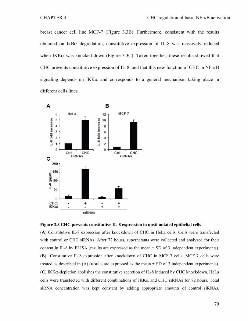
CHAPTER 3 CHC regulation of basal NF-κB activation
breast cancer cell line MCF-7 (Figure 3.3B). Furthermore, consistent with the results
obtained on IκBα degradation, constitutive expression of IL-8 was massively reduced
when IKKα was knocked down (Figure 3.3C). Taken together, these results showed that
CHC prevents constitutive expression of IL-8, and that this new function of CHC in NF-κB
signaling depends on IKKα and corresponds to a general mechanism taking place in
different cells lines.
Figure 3.3 CHC prevents constitutive IL-8 expression in unstimulated epithelial cells
(A) Constitutive IL-8 expression after knockdown of CHC in HeLa cells. Cells were transfected
with control or CHC siRNAs. After 72 hours, supernatants were collected and analyzed for their
content in IL-8 by ELISA (results are expressed as the mean ± SD of 3 independent experiments).
(B) Constitutive IL-8 expression after knockdown of CHC in MCF-7 cells. MCF-7 cells were
treated as described in (A) (results are expressed as the mean ± SD of 3 independent experiments).
(C) IKKα-depletion abolishes the constitutive secretion of IL-8 induced by CHC knockdown. HeLa
cells were transfected with different combinations of IKKα and CHC siRNAs for 72 hours. Total
siRNA concentration was kept constant by adding appropriate amounts of control siRNAs.
79
CHAPTER 3 CHC regulation of basal NF-κB activation
Supernatants were collected to measure the concentration of IL-8 by ELISA (results are expressed
as the mean ± SD of 3 independent experiments).
3.4.4 CHC controls basal NF-κB activation independently of endocytosis and clathrin
light chains
Through its activity in CME, CHC is involved in the internalization of nutrients,
pathogens, antigens, growth factors and receptors (Conner and Schmid 2003; Veiga and
Cossart 2005; Miaczynska and Stenmark 2008). To test whether CHC regulated indirectly
the NF-κB pathway via its function in CME, we measured p65 nuclear translocation and
IL-8 secretion in cells where CME was disrupted by RNAi-mediated depletion of the μ2-
subunit of the main CME adaptor protein AP-2 (AP2M1). The recruitment of AP-2 at the
plasma membrane is critical for the initiation of CME (Motley, Bright et al. 2003). AP-2
interacts with sorting signals present in the cytoplasmic domains of membrane proteins
destined to become cargo in the coated vesicles. In addition, AP-2 recruits clathrin onto the
membrane, where it functions as a scaffold for vesicle budding. First, in order to
demonstrate that CME was impaired in AP2M1 and CHC-depleted cells, the CME-
dependent mechanism of transferrin uptake was monitored in HeLa cells. As previously
reported (Motley, Bright et al. 2003), depletion of both CHC and AP2M1 impaired the
uptake of fluorescently labeled transferrin (Figures 3.4A and 3.4B). However, although the
depletion of AP2M1 blocked transferrin uptake to the same extent as CHC knockdown, it
failed to increase basal IκBα degradation (Figures 3.4C and 3.4D) and IL-8 secretion
(Figure 3.4E), indicating that CHC controls basal NF-κB activation and gene expression
independently of its activity in CME.
80
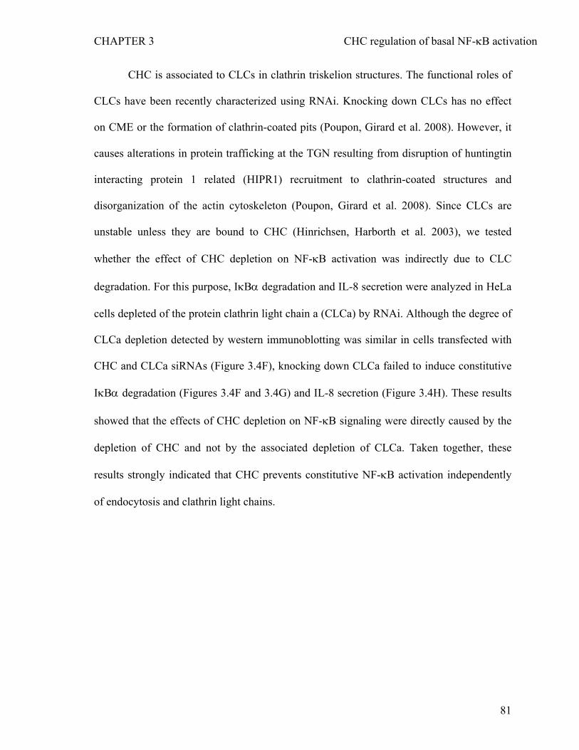
CHAPTER 3 CHC regulation of basal NF-κB activation
CHC is associated to CLCs in clathrin triskelion structures. The functional roles of
CLCs have been recently characterized using RNAi. Knocking down CLCs has no effect
on CME or the formation of clathrin-coated pits (Poupon, Girard et al. 2008). However, it
causes alterations in protein trafficking at the TGN resulting from disruption of huntingtin
interacting protein 1 related (HIPR1) recruitment to clathrin-coated structures and
disorganization of the actin cytoskeleton (Poupon, Girard et al. 2008). Since CLCs are
unstable unless they are bound to CHC (Hinrichsen, Harborth et al. 2003), we tested
whether the effect of CHC depletion on NF-κB activation was indirectly due to CLC
degradation. For this purpose, IκBα degradation and IL-8 secretion were analyzed in HeLa
cells depleted of the protein clathrin light chain a (CLCa) by RNAi. Although the degree of
CLCa depletion detected by western immunoblotting was similar in cells transfected with
CHC and CLCa siRNAs (Figure 3.4F), knocking down CLCa failed to induce constitutive
IκBα degradation (Figures 3.4F and 3.4G) and IL-8 secretion (Figure 3.4H). These results
showed that the effects of CHC depletion on NF-κB signaling were directly caused by the
depletion of CHC and not by the associated depletion of CLCa. Taken together, these
results strongly indicated that CHC prevents constitutive NF-κB activation independently
of endocytosis and clathrin light chains.
81
CHAPTER 3 CHC regulation of basal NF-κB activation
Figure 3.4 CHC regulates NF-κB activation independently of endocytosis and CLCa.
(A) Uptake of Alexa 594-transferrin (Alexa 594-Tf) in cells transfected with control (left panels),
AP2M1 (middle panels) or CHC (right panels) siRNAs. Scale bars, 10 μm (B) Quantification of
transferrin uptake by automated image analysis (results are expressed as the mean ± SD of 12
images; graph representative of 2 independent experiments). (C) AP2M1 knockdown fails to
enhance IκBα degradation. Cell lysates from control, AP2M1 or CHC siRNA-transfected cells
were analyzed by immunoblotting using indicated antibodies. Actin is shown as a loading control.
(D) Densitometric quantification of the levels of IκBα shown in Figure 3.4C (graph representative
82
CHAPTER 3 CHC regulation of basal NF-κB activation
of 2 independent experiments). (E) AP2M1 knockdown fails to induce constitutive IL-8 expression.
HeLa cells were transfected with control, AP2M1 or CHC siRNAs for 72 hours. Supernatants were
collected to measure the concentration of IL-8 by ELISA (results are expressed as the mean ± SD of
3 independent experiments). (F) CLCa knockdown fails to enhance IκBα degradation. Cell lysates
from control, CLCa or CHC siRNA transfected cells were analyzed by immunoblotting using
indicated antibodies. Actin is shown as loading control. (G) Densitometric quantification of IκBα
levels shown in Figure 3.4F (Graph representative of 2 independent experiments). (H) CLCa
knockdown fails to induce constitutive IL-8 expression. HeLa cells were transfected with control,
CLCa or CHC siRNAs for 72 hours and supernatants were collected to measure the concentration
of IL-8 by ELISA (results are expressed as the mean ± SD of 3 independent experiments).
83
CHAPTER 3 CHC regulation of basal NF-κB activation
3.5 DISCUSSION
Most research groups investigating the regulation of NF-κB activation have focused
their studies on the mechanisms induced after cell exposure to various stimuli, including
inflammatory cytokines and microbial products. In particular, the canonical pathway of
NF-κB activation that depends on the phosphorylation and degradation of IκB proteins
downstream of the activation of the IKK complex has been well characterized. In contrast,
although constitutive NF-κB activation has been associated with inflammatory disorders
and numerous cancers (Prasad, Ravindran et al. ; Yamaguchi, Ito et al. 2009), the
mechanisms that lead to elevated basal NF-κB activation remain unclear.
Here we show that CHC functions as a built-in molecular brake that ensures a tight
control of basal NF-κB activation and gene expression by preventing constitutive nuclear
localization of p65 in absence of stimulation. Using RNAi to reduce cellular levels of CHC,
we found that CHC is required for the proper spatial regulation of p65 in unstimulated
epithelial cells. Whereas p65 was almost exclusively localized in the cytoplasm of control
cells, both cytoplasmic and nuclear localization was observed in cells depleted of CHC.
The localization of p65 is largely dependent on IκB proteins that sequestrate the
transcription factor in the cytoplasm. Genetic deletion or mutations of IκBα lead to
constitutive nuclear localization and NF-κB activation (Beg, Sha et al. 1995; Yamaguchi,
Ito et al. 2009). In line with these studies, we found that the effect of CHC depletion on p65
was associated with a strong reduction in the level of IκBα, suggesting that constitutive
p65 nuclear localization was likely due to reduced IκBα level. Quantification of IκBα
mRNA by quantitative real time PCR revealed that the level of IκBα mRNA was slightly
84
CHAPTER 3 CHC regulation of basal NF-κB activation
elevated in CHC depleted cells. This result, which can be explained by the fact that IκBα is
a target gene of NF-κB that is upregulated by constitutive NF-κB activation, indicated that
the reduction of IκBα found in CHC depleted cells, was not due to transcriptional
inhibition but to elevated basal IκBα degradation. This hypothesis was further supported
by data showing that the knockdown of CHC induced constitutive phosphorylation of IκBα
at position 32, a phosphorylation event critical to target IκBα for rapid degradation via the
ubiquitin proteasome pathway. The role of CHC in the control of basal NF-κB activation
was functionally relevant. Indeed a strong induction of IL-8, whose expression is controlled
by NF-κB, was observed in cells depleted of CHC showing that, via its activity on NF-κB,
CHC participates to gene regulation. Interestingly, a role of CHC in p53-mediated
transcription has been recently reported. Enari et al. showed that a fraction of CHC proteins
that localize to the nucleus bind to p53-responsive promoters and favor transcription by
stabilizing p53 interactions with proteins such as the histone acetyltransferase p300 (Enari,
Ohmori et al. 2006). Although CHC contributes to p53 and NF-κB-regulated gene
expression by different mechanisms, our data provide a second set of evidence for a role of
CHC in gene regulation.
Via its role in CME, CHC is involved in many cellular processes including
intracellular trafficking of receptors and nutrient uptake (Miaczynska and Stenmark 2008).
To investigate whether CHC indirectly affected basal NF-κB activation via its implication
in CME, we analyzed the level of IκBα and IL-8 expression in conditions where CME was
disrupted by the depletion of the μ2-subunit of the main CME adaptor AP-2. Whereas the
CME-dependent process of transferrin uptake was almost completely abolished, AP2M1
depletion had no effect on the NF-κB signaling pathway, suggesting that CHC was
85
CHAPTER 3 CHC regulation of basal NF-κB activation
involved in the NF-κB pathway independently of endocytosis. Although we cannot
completely rule out the contribution of a CME mechanism independent of AP-2, these
results provide strong evidence for an endocytosis-independent function of CHC in NF-κB-
mediated gene regulation. As CLCs are unstable unless bound to CHC, the knockdown of
CHC is associated with cellular depletion of CLCs. Interestingly, we showed that the
depletion of CLCa has no effect on IκBα degradation and IL-8 expression, indicating that
CHC alone contributes to the regulation of the NF-κB pathway. It also suggested that CHC
functions in this pathway independently of the roles that CHC and CLCa share in protein
trafficking at the TGN (Poupon, Girard et al. 2008).
A raising number of proteins involved at different levels of the signaling pathway
contribute to the tight control of basal NF-κB activation in resting cells (Prasad, Ravindran
et al.). For instance, expression of the tumor suppressor Gprc5a prevents constitutive p65
nuclear localization and NF-κB activation (Deng, Fujimoto et al.). Although the
mechanism remains unclear, the authors propose that the presence of Gprc5a may promote
the stabilizing interaction between β-arrestin and IκBα. The role of silencer of death
domain (SODD) has also been reported (Jiang, Woronicz et al. 1999). SODD deficiency
leads to an increase of NF-κB activation and cytokine expression in absence of stimulation.
This protein functions as a gatekeeper that constitutively associates with the cytoplasmic
death domain of TNF receptors (TNFR) and blocks TNFR signaling in the absence of
ligand. Constitutive activity of NF-κB was also described in c-Abl null fibroblasts
(Liberatore, Goff et al. 2009). In contrast to previous mechanisms, unstimulated fibroblasts
did not exhibit an increase in IκBα degradation or p65 nuclear translocation but reduced
levels of the negative regulator histone deacetylase HDAC1. The mechanism by which
86
CHAPTER 3 CHC regulation of basal NF-κB activation
CHC functions in NF-κB signaling remains to be elucidated. Based on our data, we
propose that this protein acts as a built-in molecular break that prevents the spontaneous
activation of the IKK complex. This hypothesis is supported by the observation that
constitutive IκBα degradation and IL-8 secretion were almost completely abolished when
IKKα was depleted, and that IκBα was enhanced after CHC knockdown. Because the
localization of IKKs is critical for their activity (Weil, Schwamborn et al. 2003), the
implication of CHC in the sub-cellular distribution of the IKK complex should be further
investigated.
We also report here that CHC impedes constitutive IL-8 secretion by unstimulated
HeLa and MCF-7 cells. It is well established that constitutive IL-8 secretion by epithelial
cells can lead to chronic recruitment of macrophages that produce and secrete into the
microenvironement a variety of cytokines, chemokines and growth factors involved in
inflammation-related diseases such as inflammatory bowel disease. In addition, some of
these factors promote angiogenesis and act directly on epithelial cells to favor adenoma
formation and progression to adenocarcinomas (Basseres and Baldwin 2006). Therefore by
showing that CHC prevents constitutive expression of the proinflammatory and
tumorigenic factor IL-8, our data suggest that alterations in CHC expression may be
associated with chronic inflammation disorder or cancer. As a consequence, CHC
expression, that can be regulated by external stimuli such as androgens (Prescott and
Tindall 1998), should be systematically investigated in tumors and inflamed tissues.
87
CHAPTER 3 CHC regulation of basal NF-κB activation
3.6 ACKNOWLEDGMENTS
We thank Maxime Quebatte and Philippe Demougin for technical help with real-time PCR
analysis and Dr. Pascal Crottet for sharing antibodies. We thank C. Kasper for critical
reading and helpful suggestions. This work was funded by the Swiss National Science
Foundation (grant 3100A0-113561 to C.A.) and the University of Basel.
88
CHAPTER 3 CHC regulation of basal NF-κB activation
3.7 REFERENCES
Adli, M., E. Merkhofer, et al. "IKKalpha and IKKbeta each function to regulate NF-
kappaB activation in the TNF-induced/canonical pathway." PLoS One 5(2): e9428. Baeuerle, P. A. and D. Baltimore (1988). "I kappa B: a specific inhibitor of the NF-kappa B
transcription factor." Science 242(4878): 540-546. Baldwin, A. S., Jr. (1996). "The NF-kappa B and I kappa B proteins: new discoveries and
insights." Annu Rev Immunol 14: 649-683. Basseres, D. S. and A. S. Baldwin (2006). "Nuclear factor-kappaB and inhibitor of kappaB
kinase pathways in oncogenic initiation and progression." Oncogene 25(51): 6817-6830.
Beg, A. A. and A. S. Baldwin, Jr. (1993). "The I kappa B proteins: multifunctional regulators of Rel/NF-kappa B transcription factors." Genes Dev 7(11): 2064-2070.
Beg, A. A., W. C. Sha, et al. (1995). "Constitutive NF-kappa B activation, enhanced granulopoiesis, and neonatal lethality in I kappa B alpha-deficient mice." Genes Dev 9(22): 2736-2746.
Conner, S. D. and S. L. Schmid (2003). "Regulated portals of entry into the cell." Nature 422(6927): 37-44.
Deng, J., J. Fujimoto, et al. "Knockout of the tumor suppressor gene Gprc5a in mice leads to NF-kappaB activation in airway epithelium and promotes lung inflammation and tumorigenesis." Cancer Prev Res (Phila Pa) 3(4): 424-437.
Enari, M., K. Ohmori, et al. (2006). "Requirement of clathrin heavy chain for p53-mediated transcription." Genes Dev 20(9): 1087-1099.
Galvez, T., M. N. Teruel, et al. (2007). "siRNA screen of the human signaling proteome identifies the PtdIns(3,4,5)P3-mTOR signaling pathway as a primary regulator of transferrin uptake." Genome Biol 8(7): R142.
Gilmore, T. D. (2006). "Introduction to NF-kappaB: players, pathways, perspectives." Oncogene 25(51): 6680-6684.
Hayden, M. S. and S. Ghosh (2008). "Shared principles in NF-kappaB signaling." Cell 132(3): 344-362.
Hayden, M. S., A. P. West, et al. (2006). "SnapShot: NF-kappaB signaling pathways." Cell 127(6): 1286-1287.
Hinrichsen, L., J. Harborth, et al. (2003). "Effect of clathrin heavy chain- and alpha-adaptin-specific small inhibitory RNAs on endocytic accessory proteins and receptor trafficking in HeLa cells." J Biol Chem 278(46): 45160-45170.
Hoffmann, A., A. Levchenko, et al. (2002). "The IkappaB-NF-kappaB signaling module: temporal control and selective gene activation." Science 298(5596): 1241-1245.
Jiang, Y., J. D. Woronicz, et al. (1999). "Prevention of constitutive TNF receptor 1 signaling by silencer of death domains." Science 283(5401): 543-546.
Liberatore, R. A., S. P. Goff, et al. (2009). "NF-kappaB activity is constitutively elevated in c-Abl null fibroblasts." Proc Natl Acad Sci U S A 106(42): 17823-17828.
Miaczynska, M. and H. Stenmark (2008). "Mechanisms and functions of endocytosis." J Cell Biol 180(1): 7-11.
Motley, A., N. A. Bright, et al. (2003). "Clathrin-mediated endocytosis in AP-2-depleted cells." J Cell Biol 162(5): 909-918.
89
CHAPTER 3 CHC regulation of basal NF-κB activation
Neumann, B., M. Held, et al. (2006). "High-throughput RNAi screening by time-lapse imaging of live human cells." Nat Meth 3(5): 385-390.
Perkins, N. D. (2006). "Post-translational modifications regulating the activity and function of the nuclear factor kappa B pathway." Oncogene 25(51): 6717-6730.
Pilecka, I., M. Banach-Orlowska, et al. (2007). "Nuclear functions of endocytic proteins." Eur J Cell Biol 86(9): 533-547.
Poupon, V., M. Girard, et al. (2008). "Clathrin light chains function in mannose phosphate receptor trafficking via regulation of actin assembly." Proc Natl Acad Sci U S A 105(1): 168-173.
Prasad, S., J. Ravindran, et al. "NF-kappaB and cancer: how intimate is this relationship." Mol Cell Biochem 336(1-2): 25-37.
Prescott, J. L. and D. J. Tindall (1998). "Clathrin gene expression is androgen regulated in the prostate." Endocrinology 139(4): 2111-2119.
Royle, S. J. (2006). "The cellular functions of clathrin." Cell Mol Life Sci 63(16): 1823-1832.
Royle, S. J., N. A. Bright, et al. (2005). "Clathrin is required for the function of the mitotic spindle." Nature 434(7037): 1152-1157.
Veiga, E. and P. Cossart (2005). "Listeria hijacks the clathrin-dependent endocytic machinery to invade mammalian cells." Nat Cell Biol 7(9): 894-900.
Weil, R., K. Schwamborn, et al. (2003). "Induction of the NF-kappaB cascade by recruitment of the scaffold molecule NEMO to the T cell receptor." Immunity 18(1): 13-26.
Yamaguchi, N., T. Ito, et al. (2009). "Constitutive activation of nuclear factor-kappaB is preferentially involved in the proliferation of basal-like subtype breast cancer cell lines." Cancer Sci 100(9): 1668-1674.
90
CONCLUSIONS AND OUTLOOK
CONCLUSIONS AND OUTLOOK
Role of IKKα in inflammation and innate immunity
Pro-inflammatory cytokines such as TNFα and IL-1, and many pathogens
including S. flexneri activate the transcription factor NF-κB via the so-called canonical
pathways, which results in the expression of chemokines such as IL-8, one of the key
mediators in inflammation and innate immunity (Akira, Uematsu et al. 2006; Lawrence
2009). Ample evidence indicates that in the canonical NF-κB pathways IKKβ subunit
of the IKK complex is essential for phosphorylation and degradation of IκBα, causing it
to dissociate from NF-κB and allowing NF-κB to translocate to the nucleus, whereas
IKKα subunit regulates the non-canonical or alternative pathway important for B-cell
maturation and lymphoid organogenesis rather than inflammation and innate immunity
(Karin and Ben-Neriah 2000; Bonizzi and Karin 2004; Dejardin 2006; Lawrence and
Bebien 2007). In contrast to this dominant model of IKKβ-dependent NF-κB activation
in the canonical pathways, we have shown here for the first time that IKKα also
regulates the canonical NF-κB activation and subsequent IL-8 expression downstream
of Nod1-mediated peptidoglycan recognition during S. flexneri infection of HeLa cells.
A very recent study also shows that both IKKα and IKKβ contribute to IκBα
phosphorylation and NF-κB activation in response to TNFα stimulation in HeLa cells
(Adli, Merkhofer et al. 2010), supporting the role of IKKα in the canonical NF-κB
activation. Therefore, it is conceivable that IKKα as well as IKKβ plays an essential
role in inflammation and innate immunity. However, it still remains an open question as
to how IKKα and IKKβ stay as an inactive complex in resting cells and gets rapidly
activated upon stimulation.
Mechanism of IKKα activation 91
CONCLUSIONS AND OUTLOOK
A putative model for the activation of IKKα and IKKβ is their activation loop
(T-loop) phosphorylation in the N-terminal kinase domain by upstream kinases such as
TAK1 (Hayden and Ghosh 2008). Thus, mutation of the serine residues (S176/S180 for
IKKα and S177/S181 for IKKβ) in the T-loop to alanines abrogates NF-κB activation in
response to TNFα stimulation (Hacker and Karin 2006). However, different from the
TNFR signaling, phosphorylation at the two serine residues was barely detectable in
response to S.flexneri infection or Tri-DAP treatment up to 60 minutes when IκBα
degradation occurs maximally (unpublished data), suggesting that Nod1-mediated NF-
κB activation might be independent of the T-loop phosphorylation of IKKα and IKKβ.
This may further indicate that the serine residues in the T-loop of IKKα and IKKβ may
be structurally required for NF-κB activation but the serines phosphorylation may not
be functionally responsible for IκBα phosphorylation since IκBα phosphorylation and
degradation occurs in the absence of IKK phosphorylation upon S.flexneri infection or
Nod1 stimulation. A study showed that in addition to phosphorylation of two serine
residues in the T-loop, phosphorylation of two tyrosine residues (Y187/Y198 for IKKα
and Y188/Y199 for IKKβ) is induced by protein kinase C-dependent c-Src activation
and is important for TNFα-induced NF-κB activation and subsequent target gene
expression (Huang, Chen et al. 2003; Huang, Chen et al. 2003). Tyrosine
phosphorylation of IKKα and IKKβ in the Nod1 signaling pathways is yet unknown. In
an independent study, we observed that S.flexneri infection or Tri-DAP treatment of
HeLa cells induced strong tyrosine phosphorylation of multiple undefined proteins,
which is detected by both immnofluorescence and western blot analysis using an anti-
phospho tyrosine (pY) antibody that specifically binds to tyrosine phosphorylated
proteins (unpublished data). Given that Src is a tyrosine kinase known to be activated
during Shigella entry process (Dehio, Prevost et al. 1995), it would be worth
92
CONCLUSIONS AND OUTLOOK
investigating whether or not Src can induce tyrosine phosphorylation of IKKα and
IKKβ during S.flexneri infection, and tyrosine phosphorylation of IKKα and IKKβ are
responsible for NF-κB activation.
IKKα and histone phosphorylation
In addition to its activity toward IκBα phosphorylation, IKKα is involved in
histone H3 phosphorylation, which facilitates NF-κB binding to IL-8 promoter
(Yamamoto, Verma et al. 2003). Previous studies showed that IL-8 is poorly secreted
during S.flexneri infection in part due to the downregulation of host signalling by the
Shigella type-III effectors (Arbibe, Kim et al. 2007; Zurawski, Mumy et al. 2009).
Especially, the nucleus-targeted OspF dephosphorylates p38 and ERK, which are
responsible for histone H3 Ser10 phosphorylation (Arbibe, Kim et al. 2007; Li, Xu et al.
2007). Interestingly, I have recently found that IKKα and IKKβ phosphorylation at the
two serine residues in the T-loop is induced not by wild type S.flexneri but by the
OspF-deficient one (unpublished data). It will be of particular interest to study first of
all, the previously unknown implication of S176/S180 phosphorylation of IKKα in the
regulation of histone phosphorylation rather than IκBα phosphorylation and secondly,
the unknown but possible connection between IKK and p38 (and ERK) as both are
involved in histone H3 phosphorylation.
In conclusion, this part of my thesis on IKKα not only shows the previously
unknown function of IKKα in inflammation and innate immunity during bacterial
infection but also may draw further attention for the IKKα-dependent NF-κB activation
in many other canonical pathways. Furthermore, this finding may present new
therapeutic opportunities for IKKα inhibitors in the treatment of infectious diseases.
93
CONCLUSIONS AND OUTLOOK
Inhibition of basal NF-κB activation by clathrin heavy chain
Constitutive NF-κB activation has been associated with many chronic
inflammatory diseases such as cancer (Karin and Greten 2005), but the underlying
molecular mechanism by which basal NF-κB is elevated remains unclear. I showed in
the second part of my dissertation that depletion of clathrin heavy chain (CHC) resulted
in constitutive IκBα degradation, NF-κB activation and high level of IL-8 secretion in
resting cells. Normally, NF-κB is retained in the cytoplasm of resting cells through an
interaction with IκBα, thereby preventing undesired activation of NF-κB (Liou and
Baltimore 1993; Hayden and Ghosh 2008). Thus, genetic deletion of IκBα leads to
constitutive NF-κB activation (Beg, Sha et al. 1995). However, unlike the constitutive
NF-κB activation by genetic deletion of IκBα, which is simply due to the absence of
IκBα expression that can bind to and retain NF-κB in the cytoplasm, CHC depletion
induced higher mRNA expression of IκBα, indicating a constant turnover of IκBα at the
post-translational level through an active event upstream of IκBα such as an activation
of IKK complex.
Inhibition of basal IKK activation and IκBα turnover by clathrin heavy chain
Degradation of IκBα is induced by cytokines such as TNFα and bacterial
products such Tri-DAP and is mediated by IKKα- and IKKβ-dependent
phosphorylation and ubiquitin-proteasome pathway (Karin and Ben-Neriah 2000). A
recent study shows that NLRC5, a member of NLR family inhibits the NF-κB
signalling pathway by interacting with IKKα and IKKβ and blocking their
phosphorylation after LPS stimulation (Cui, Zhu et al. 2010). Accordingly, depletion of
94
CONCLUSIONS AND OUTLOOK
NLRC5 enhanced the activation of NF-κB and its responsive genes, TNFα and IL-6.
Similarly, depletion of CHC enhanced the activation of NF-κB and the secretion of IL-8
after S.flexneri infection or TNFα stimulation (data not shown). Although it is unclear
whether CHC can also interact with and sequestrate the IKK complex in resting cells,
depletion of IKKα drastically reduced CHC silencing-induced IκBα degradation and IL-
8 secretion, suggesting CHC depletion-induced NF-κB activation is also IKKα
dependent. Thus, it is plausible that CHC might inhibit basal IKK activation via the
interaction with the IKK complex or an upstream NF-κB regulator in resting cells.
Inhibition of IKK activation by CHC-binding proteins
Protein interaction studies have revealed several proteins of endocytic pathway
that play additional functions as inhibitors of NF-κB signalling pathways. Tom1 (target
of Myb1) interacts with CHC and Tollip (Toll-interacting protein), forming a complex
regulating endocytosis of IL-1 receptor (IL-1R) (Yamakami, Yoshimori et al. 2003;
Brissoni, Agostini et al. 2006). In addition, Tom1 inhibits NF-κB activation in the IL-
1R, TNFR, and TLR2/4 signalling pathways (Yamakami and Yokosawa 2004; Oglesby,
Bray et al. 2010). Tollip forms a complex with IRAK (IL-1R-associated kinase) to
block phosphorylation of IRAK, which prevents IKK and NF-κB activation upon
stimulation of IL-1R (Burns, Clatworthy et al. 2000). However, functions of Tom1 and
Tollip as inhibitors of NF-κB in resting cells were not clearly addressed in their studies.
CHC interaction with and inhibition of IRAK in resting cells is unknown, but might be
worth testing. A proteomics analysis designed to identify binding partners of CHC may
help to understand the molecular mechanism by which CHC inhibits NF-κB activation
in resting cells. In a proteomics analysis, IKAP (IκB kinase complex-associated protein)
was identified as a component in the clathrin-containing vesicles although the physical
95
CONCLUSIONS AND OUTLOOK
interaction between IKAP and clathrin was not tested (Borner, Harbour et al. 2006). An
independent study showed that IKAP interacts with NIK and IKK complex and is a
scaffold protein of the IKK complex (Cohen, Henzel et al. 1998). Whether IKAP and
CHC interacts with each other and, if so, whether the interaction contributes to the
regulation of NF-κB await further investigation.
Endocytosis-independent functions of clathrin heavy chain
Although NF-κB activation can be induced by many different mechanisms, little
is known about the interconnection between endocytosis and regulation of NF-κB.
Whether an inhibition of normal clathrin-mediated endocytosis (CME) can induce NF-
κB activation was examined. Our data showed that depletion of μ2 subunit of AP2
complex (AP2M1) blocked transferrin-transferrin receptor internalization, a typical
CME process, to a degree similar to CHC depletion, but failed to induce basal IκBα
degradation and IL-8 secretion. This suggests that basal NF-κB activation by depletion
of CHC was not merely attributed to the inhibition of endocytosis, and CHC might play
an endocytosis-independent function as a negative regulator of NF-κB. However, we
cannot rule out the possibility that the inhibition of CHC-dependent but AP2 complex-
independent endocytosis might be implicated in NF-κB activation. Some endocytosis-
independent functions were reported. A study showed that a small fraction of clathrin
heavy chain (CHC) in the nucleus can contribute to the tumor suppressor p53-mediated
gene expression by interacting with p53 and facilitating recruitment of transcriptional
cofactors (Enari, Ohmori et al. 2006). Another study showed that CHC stabilizes
spindle fibers to assist congression of chromosome during mitosis when endocytosis is
in halt (Royle, Bright et al. 2005).
96
CONCLUSIONS AND OUTLOOK
Although the exact action of CHC in the regulation of basal NF-κB activity
remains to be further characterized, results presented in this dissertation suggest that
CHC may play a new role as an inhibitor of NF-κB in resting cells, independently of
endocytosis. Given the constitutive NF-κB activation and high level of IL-8 secretion
by the depletion of CHC, CHC protein concentration might be an important factor in
the regulation of basal IKK activity toward phosphorylation and degradation of IκB,
further suggesting that a defect in CHC expression may be associated with chronic
inflammatory disorders or cancers.
Collectively, results presented in this dissertation highlight the complexity of
NF-κB regulation far from being elucidated. Nevertheless, the RNAi technique must be
appreciated in helping us reveal previously unknown functions of the well-characterized
proteins, IKKα and CHC. A large scale functional genomics-approach like a genome-
wide RNAi screen will eventually help us better understand the complex NF-κB
signalling network.
97
REFERENCES
REFERENCES
Adli, M. and A. S. Baldwin (2006). "IKK-i/IKKepsilon controls constitutive, cancer cell-associated NF-kappaB activity via regulation of Ser-536 p65/RelA phosphorylation." J Biol Chem 281(37): 26976-26984.
Adli, M., E. Merkhofer, et al. (2010). "IKKalpha and IKKbeta each function to regulate NF-kappaB activation in the TNF-induced/canonical pathway." PLoS One 5(2): e9428.
Akira, S., S. Uematsu, et al. (2006). "Pathogen recognition and innate immunity." Cell 124(4): 783-801.
Alkalay, I., A. Yaron, et al. (1995). "Stimulation-dependent I kappa B alpha phosphorylation marks the NF-kappa B inhibitor for degradation via the ubiquitin-proteasome pathway." Proc Natl Acad Sci U S A 92(23): 10599-10603.
Amerik, A. Y. and M. Hochstrasser (2004). "Mechanism and function of deubiquitinating enzymes." Biochim Biophys Acta 1695(1-3): 189-207.
Arbibe, L., D. W. Kim, et al. (2007). "An injected bacterial effector targets chromatin access for transcription factor NF-kappaB to alter transcription of host genes involved in immune responses." Nat Immunol 8(1): 47-56.
Ashida, H., M. Kim, et al. (2010). "A bacterial E3 ubiquitin ligase IpaH9.8 targets NEMO/IKKgamma to dampen the host NF-kappaB-mediated inflammatory response." Nat Cell Biol 12(1): 66-73; sup pp 61-69.
Beg, A. A., W. C. Sha, et al. (1995). "Constitutive NF-kappa B activation, enhanced granulopoiesis, and neonatal lethality in I kappa B alpha-deficient mice." Genes Dev 9(22): 2736-2746.
Betts, J. C. and G. J. Nabel (1996). "Differential regulation of NF-kappaB2(p100) processing and control by amino-terminal sequences." Mol Cell Biol 16(11): 6363-6371.
Bhavsar, A. P., J. A. Guttman, et al. (2007). "Manipulation of host-cell pathways by bacterial pathogens." Nature 449(7164): 827-834.
Bird, T. A., K. Schooley, et al. (1997). "Activation of nuclear transcription factor NF-kappaB by interleukin-1 is accompanied by casein kinase II-mediated phosphorylation of the p65 subunit." J Biol Chem 272(51): 32606-32612.
Bogoyevitch, M. A., K. R. Ngoei, et al. (2010). "c-Jun N-terminal kinase (JNK) signaling: recent advances and challenges." Biochim Biophys Acta 1804(3): 463-475.
Boneca, I. G. (2005). "The role of peptidoglycan in pathogenesis." Curr Opin Microbiol 8(1): 46-53.
Bonen, D. K., Y. Ogura, et al. (2003). "Crohn's disease-associated NOD2 variants share a signaling defect in response to lipopolysaccharide and peptidoglycan." Gastroenterology 124(1): 140-146.
Bonizzi, G. and M. Karin (2004). "The two NF-kappaB activation pathways and their role in innate and adaptive immunity." Trends Immunol 25(6): 280-288.
Boone, D. L., E. E. Turer, et al. (2004). "The ubiquitin-modifying enzyme A20 is required for termination of Toll-like receptor responses." Nat Immunol 5(10): 1052-1060.
Borner, G. H., M. Harbour, et al. (2006). "Comparative proteomics of clathrin-coated vesicles." J Cell Biol 175(4): 571-578.
Bourdet-Sicard, R., C. Egile, et al. (2000). "Diversion of cytoskeletal processes by Shigella during invasion of epithelial cells." Microbes Infect 2(7): 813-819.
Bradley, J. R. and J. S. Pober (2001). "Tumor necrosis factor receptor-associated factors (TRAFs)." Oncogene 20(44): 6482-6491.
Brissoni, B., L. Agostini, et al. (2006). "Intracellular trafficking of interleukin-1 receptor I requires Tollip." Curr Biol 16(22): 2265-2270.
Brummelkamp, T. R., S. M. Nijman, et al. (2003). "Loss of the cylindromatosis tumour suppressor inhibits apoptosis by activating NF-kappaB." Nature 424(6950): 797-801.
Burns, K., J. Clatworthy, et al. (2000). "Tollip, a new component of the IL-1RI pathway, links IRAK to the IL-1 receptor." Nat Cell Biol 2(6): 346-351.
98
REFERENCES
Buss, H., A. Dorrie, et al. (2004). "Constitutive and interleukin-1-inducible phosphorylation of p65 NF-{kappa}B at serine 536 is mediated by multiple protein kinases including I{kappa}B kinase (IKK)-{alpha}, IKK{beta}, IKK{epsilon}, TRAF family member-associated (TANK)-binding kinase 1 (TBK1), and an unknown kinase and couples p65 to TATA-binding protein-associated factor II31-mediated interleukin-8 transcription." J Biol Chem 279(53): 55633-55643.
Chamaillard, M., M. Hashimoto, et al. (2003). "An essential role for NOD1 in host recognition of bacterial peptidoglycan containing diaminopimelic acid." Nat Immunol 4(7): 702-707.
Chen, G., M. H. Shaw, et al. (2009). "NOD-like receptors: role in innate immunity and inflammatory disease." Annu Rev Pathol 4: 365-398.
Chen, Z., J. Hagler, et al. (1995). "Signal-induced site-specific phosphorylation targets I kappa B alpha to the ubiquitin-proteasome pathway." Genes Dev 9(13): 1586-1597.
Chiu, Y. H., M. Zhao, et al. (2009). "Ubiquitin in NF-kappaB signaling." Chem Rev 109(4): 1549-1560.
Chung, J. Y., Y. C. Park, et al. (2002). "All TRAFs are not created equal: common and distinct molecular mechanisms of TRAF-mediated signal transduction." J Cell Sci 115(Pt 4): 679-688.
Claing, A., S. A. Laporte, et al. (2002). "Endocytosis of G protein-coupled receptors: roles of G protein-coupled receptor kinases and beta-arrestin proteins." Prog Neurobiol 66(2): 61-79.
Cohen, L., W. J. Henzel, et al. (1998). "IKAP is a scaffold protein of the IkappaB kinase complex." Nature 395(6699): 292-296.
Conner, S. D. and S. L. Schmid (2003). "Regulated portals of entry into the cell." Nature 422(6927): 37-44.
Courtois, G. and T. D. Gilmore (2006). "Mutations in the NF-kappaB signaling pathway: implications for human disease." Oncogene 25(51): 6831-6843.
Courtois, G., A. Smahi, et al. (2003). "A hypermorphic IkappaBalpha mutation is associated with autosomal dominant anhidrotic ectodermal dysplasia and T cell immunodeficiency." J Clin Invest 112(7): 1108-1115.
Cui, J., L. Zhu, et al. (2010). "NLRC5 negatively regulates the NF-kappaB and type I interferon signaling pathways." Cell 141(3): 483-496.
Dehio, C., M. C. Prevost, et al. (1995). "Invasion of epithelial cells by Shigella flexneri induces tyrosine phosphorylation of cortactin by a pp60c-src-mediated signalling pathway." EMBO J 14(11): 2471-2482.
Dejardin, E. (2006). "The alternative NF-kappaB pathway from biochemistry to biology: pitfalls and promises for future drug development." Biochem Pharmacol 72(9): 1161-1179.
Deng, L., C. Wang, et al. (2000). "Activation of the IkappaB kinase complex by TRAF6 requires a dimeric ubiquitin-conjugating enzyme complex and a unique polyubiquitin chain." Cell 103(2): 351-361.
DiDonato, J., F. Mercurio, et al. (1996). "Mapping of the inducible IkappaB phosphorylation sites that signal its ubiquitination and degradation." Mol Cell Biol 16(4): 1295-1304.
DiDonato, J. A., M. Hayakawa, et al. (1997). "A cytokine-responsive IkappaB kinase that activates the transcription factor NF-kappaB." Nature 388(6642): 548-554.
Ding, S. Z., W. Fischer, et al. (2010). "Helicobacter pylori-induced histone modification, associated gene expression in gastric epithelial cells, and its implication in pathogenesis." PLoS One 5(4): e9875.
Ducut Sigala, J. L., V. Bottero, et al. (2004). "Activation of transcription factor NF-kappaB requires ELKS, an IkappaB kinase regulatory subunit." Science 304(5679): 1963-1967.
Enari, M., K. Ohmori, et al. (2006). "Requirement of clathrin heavy chain for p53-mediated transcription." Genes Dev 20(9): 1087-1099.
Evans, P. C., H. Ovaa, et al. (2004). "Zinc-finger protein A20, a regulator of inflammation and cell survival, has de-ubiquitinating activity." Biochem J 378(Pt 3): 727-734.
99
REFERENCES
Fan, C. M. and T. Maniatis (1991). "Generation of p50 subunit of NF-kappa B by processing of p105 through an ATP-dependent pathway." Nature 354(6352): 395-398.
Ferreiro, D. U. and E. A. Komives "Molecular mechanisms of system control of NF-kappaB signaling by IkappaBalpha." Biochemistry 49(8): 1560-1567.
Gao, H., Y. Sun, et al. (2004). "Identification of beta-arrestin2 as a G protein-coupled receptor-stimulated regulator of NF-kappaB pathways." Mol Cell 14(3): 303-317.
Geijtenbeek, T. B. and S. I. Gringhuis (2009). "Signalling through C-type lectin receptors: shaping immune responses." Nat Rev Immunol 9(7): 465-479.
Gerondakis, S. and U. Siebenlist "Roles of the NF-kappaB pathway in lymphocyte development and function." Cold Spring Harb Perspect Biol 2(5): a000182.
Ghosh, S. and M. S. Hayden (2008). "New regulators of NF-kappaB in inflammation." Nat Rev Immunol 8(11): 837-848.
Gilmore, T. D. (2006). "Introduction to NF-kappaB: players, pathways, perspectives." Oncogene 25(51): 6680-6684.
Girardin, S. E., I. G. Boneca, et al. (2003). "Nod2 is a general sensor of peptidoglycan through muramyl dipeptide (MDP) detection." J Biol Chem 278(11): 8869-8872.
Girardin, S. E., R. Tournebize, et al. (2001). "CARD4/Nod1 mediates NF-kappaB and JNK activation by invasive Shigella flexneri." EMBO Rep 2(8): 736-742.
Goodridge, H. S., A. J. Wolf, et al. (2009). "Beta-glucan recognition by the innate immune system." Immunol Rev 230(1): 38-50.
Hacker, H. and M. Karin (2006). "Regulation and function of IKK and IKK-related kinases." Sci STKE 2006(357): re13.
Hamon, M. A., E. Batsche, et al. (2007). "Histone modifications induced by a family of bacterial toxins." Proc Natl Acad Sci U S A 104(33): 13467-13472.
Harant, H., R. de Martin, et al. (1996). "Synergistic activation of interleukin-8 gene transcription by all-trans-retinoic acid and tumor necrosis factor-alpha involves the transcription factor NF-kappaB." J Biol Chem 271(43): 26954-26961.
Hasegawa, M., Y. Fujimoto, et al. (2008). "A critical role of RICK/RIP2 polyubiquitination in Nod-induced NF-kappaB activation." Embo J 27(2): 373-383.
Hayden, M. S. and S. Ghosh (2008). "Shared principles in NF-kappaB signaling." Cell 132(3): 344-362.
Hess, J., P. Angel, et al. (2004). "AP-1 subunits: quarrel and harmony among siblings." J Cell Sci 117(Pt 25): 5965-5973.
Heusch, M., L. Lin, et al. (1999). "The generation of nfkb2 p52: mechanism and efficiency." Oncogene 18(46): 6201-6208.
Hideshima, T., D. Chauhan, et al. (2002). "NF-kappa B as a therapeutic target in multiple myeloma." J Biol Chem 277(19): 16639-16647.
High, N., J. Mounier, et al. (1992). "IpaB of Shigella flexneri causes entry into epithelial cells and escape from the phagocytic vacuole." Embo J 11(5): 1991-1999.
Hinz, M., M. Broemer, et al. (2007). "Signal responsiveness of IkappaB kinases is determined by Cdc37-assisted transient interaction with Hsp90." J Biol Chem 282(44): 32311-32319.
Hoffmann, A., G. Natoli, et al. (2006). "Transcriptional regulation via the NF-kappaB signaling module." Oncogene 25(51): 6706-6716.
Huang, B., X. D. Yang, et al. "Posttranslational modifications of NF-kappaB: another layer of regulation for NF-kappaB signaling pathway." Cell Signal 22(9): 1282-1290.
Huang, W. C., J. J. Chen, et al. (2003). "c-Src-dependent tyrosine phosphorylation of IKKbeta is involved in tumor necrosis factor-alpha-induced intercellular adhesion molecule-1 expression." J Biol Chem 278(11): 9944-9952.
Huang, W. C., J. J. Chen, et al. (2003). "Tyrosine phosphorylation of I-kappa B kinase alpha/beta by protein kinase C-dependent c-Src activation is involved in TNF-alpha-induced cyclooxygenase-2 expression." J Immunol 170(9): 4767-4775.
Hugot, J. P., M. Chamaillard, et al. (2001). "Association of NOD2 leucine-rich repeat variants with susceptibility to Crohn's disease." Nature 411(6837): 599-603.
100
REFERENCES
Hysi, P., M. Kabesch, et al. (2005). "NOD1 variation, immunoglobulin E and asthma." Hum Mol Genet 14(7): 935-941.
Inohara, Chamaillard, et al. (2005). "NOD-LRR proteins: role in host-microbial interactions and inflammatory disease." Annu Rev Biochem 74: 355-383.
Inohara, N., T. Koseki, et al. (2000). "An induced proximity model for NF-kappa B activation in the Nod1/RICK and RIP signaling pathways." J Biol Chem 275(36): 27823-27831.
Inohara, N. and G. Nunez (2003). "NODs: intracellular proteins involved in inflammation and apoptosis." Nat Rev Immunol 3(5): 371-382.
Jacobs, M. D. and S. C. Harrison (1998). "Structure of an IkappaBalpha/NF-kappaB complex." Cell 95(6): 749-758.
Janssen, R., A. van Wengen, et al. (2004). "The same IkappaBalpha mutation in two related individuals leads to completely different clinical syndromes." J Exp Med 200(5): 559-568.
Jung, H. C., L. Eckmann, et al. (1995). "A distinct array of proinflammatory cytokines is expressed in human colon epithelial cells in response to bacterial invasion." J Clin Invest 95(1): 55-65.
Kanayama, A., R. B. Seth, et al. (2004). "TAB2 and TAB3 activate the NF-kappaB pathway through binding to polyubiquitin chains." Mol Cell 15(4): 535-548.
Karin, M. and Y. Ben-Neriah (2000). "Phosphorylation meets ubiquitination: the control of NF-[kappa]B activity." Annu Rev Immunol 18: 621-663.
Karin, M. and F. R. Greten (2005). "NF-kappaB: linking inflammation and immunity to cancer development and progression." Nat Rev Immunol 5(10): 749-759.
Kato, H., O. Takeuchi, et al. (2008). "Length-dependent recognition of double-stranded ribonucleic acids by retinoic acid-inducible gene-I and melanoma differentiation-associated gene 5." J Exp Med 205(7): 1601-1610.
Kawai, T. and S. Akira "The role of pattern-recognition receptors in innate immunity: update on Toll-like receptors." Nat Immunol 11(5): 373-384.
Kim, D. W., G. Lenzen, et al. (2005). "The Shigella flexneri effector OspG interferes with innate immune responses by targeting ubiquitin-conjugating enzymes." Proc Natl Acad Sci U S A 102(39): 14046-14051.
Kirchhausen, T. (2000). "Clathrin." Annu Rev Biochem 69: 699-727. Kishore, N., C. Sommers, et al. (2003). "A selective IKK-2 inhibitor blocks NF-kappa B-
dependent gene expression in interleukin-1 beta-stimulated synovial fibroblasts." J Biol Chem 278(35): 32861-32871.
Kobayashi, Y. (2008). "The role of chemokines in neutrophil biology." Front Biosci 13: 2400-2407.
Kopp, E. and S. Ghosh (1994). "Inhibition of NF-kappa B by sodium salicylate and aspirin." Science 265(5174): 956-959.
Kovalenko, A., C. Chable-Bessia, et al. (2003). "The tumour suppressor CYLD negatively regulates NF-kappaB signalling by deubiquitination." Nature 424(6950): 801-805.
Kray, A. E., R. S. Carter, et al. (2005). "Positive regulation of IkappaB kinase signaling by protein serine/threonine phosphatase 2A." J Biol Chem 280(43): 35974-35982.
Krupnick, J. G., O. B. Goodman, Jr., et al. (1997). "Arrestin/clathrin interaction. Localization of the clathrin binding domain of nonvisual arrestins to the carboxy terminus." J Biol Chem 272(23): 15011-15016.
Kuo, M. H. and C. D. Allis (1998). "Roles of histone acetyltransferases and deacetylases in gene regulation." Bioessays 20(8): 615-626.
Kuper, H., H. O. Adami, et al. (2000). "Infections as a major preventable cause of human cancer." J Intern Med 248(3): 171-183.
Lawrence, T. (2009). "The nuclear factor NF-kappaB pathway in inflammation." Cold Spring Harb Perspect Biol 1(6): a001651.
Lawrence, T. and M. Bebien (2007). "IKKalpha in the regulation of inflammation and adaptive immunity." Biochem Soc Trans 35(Pt 2): 270-272.
Lee, D. F. and M. C. Hung (2008). "Advances in targeting IKK and IKK-related kinases for cancer therapy." Clin Cancer Res 14(18): 5656-5662.
101
REFERENCES
Lefkowitz, R. J. and E. J. Whalen (2004). "beta-arrestins: traffic cops of cell signaling." Curr Opin Cell Biol 16(2): 162-168.
Lewis, J., A. Devin, et al. (2000). "Disruption of hsp90 function results in degradation of the death domain kinase, receptor-interacting protein (RIP), and blockage of tumor necrosis factor-induced nuclear factor-kappaB activation." J Biol Chem 275(14): 10519-10526.
Li, H., H. Xu, et al. (2007). "The phosphothreonine lyase activity of a bacterial type III effector family." Science 315(5814): 1000-1003.
Lin, L. and S. Ghosh (1996). "A glycine-rich region in NF-kappaB p105 functions as a processing signal for the generation of the p50 subunit." Mol Cell Biol 16(5): 2248-2254.
Lindner, R. and E. Ungewickell (1991). "Light-chain-independent binding of adaptors, AP180, and auxilin to clathrin." Biochemistry 30(37): 9097-9101.
Liou, H. C. and D. Baltimore (1993). "Regulation of the NF-kappa B/rel transcription factor and I kappa B inhibitor system." Curr Opin Cell Biol 5(3): 477-487.
Loo, Y. M., J. Fornek, et al. (2008). "Distinct RIG-I and MDA5 signaling by RNA viruses in innate immunity." J Virol 82(1): 335-345.
Luan, B., Z. Zhang, et al. (2005). "Beta-arrestin2 functions as a phosphorylation-regulated suppressor of UV-induced NF-kappaB activation." EMBO J 24(24): 4237-4246.
Martinon, F. and J. Tschopp (2005). "NLRs join TLRs as innate sensors of pathogens." Trends Immunol 26(8): 447-454.
May, M. J., F. D'Acquisto, et al. (2000). "Selective inhibition of NF-kappaB activation by a peptide that blocks the interaction of NEMO with the IkappaB kinase complex." Science 289(5484): 1550-1554.
Medzhitov, R. (2007). "Recognition of microorganisms and activation of the immune response." Nature 449(7164): 819-826.
Menard, R., M. C. Prevost, et al. (1996). "The secreted Ipa complex of Shigella flexneri promotes entry into mammalian cells." Proc Natl Acad Sci U S A 93(3): 1254-1258.
Mercurio, F., H. Zhu, et al. (1997). "IKK-1 and IKK-2: cytokine-activated IkappaB kinases essential for NF-kappaB activation." Science 278(5339): 860-866.
Mounier, J., V. Laurent, et al. (1999). "Rho family GTPases control entry of Shigella flexneri into epithelial cells but not intracellular motility." J Cell Sci 112 ( Pt 13): 2069-2080.
Mousavi, S. A., L. Malerod, et al. (2004). "Clathrin-dependent endocytosis." Biochem J 377(Pt 1): 1-16.
Muegge, K. (2002). "Preparing the target for the bullet." Nat Immunol 3(1): 16-17. Mukaida, N., Y. Mahe, et al. (1990). "Cooperative interaction of nuclear factor-kappa B- and
cis-regulatory enhancer binding protein-like factor binding elements in activating the interleukin-8 gene by pro-inflammatory cytokines." J Biol Chem 265(34): 21128-21133.
Munshi, A., G. Shafi, et al. (2009). "Histone modifications dictate specific biological readouts." J Genet Genomics 36(2): 75-88.
Oglesby, I. K., I. M. Bray, et al. (2010). "miR-126 is downregulated in cystic fibrosis airway epithelial cells and regulates TOM1 expression." J Immunol 184(4): 1702-1709.
Okamoto, T. (2006). "NF-kappaB and rheumatic diseases." Endocr Metab Immune Disord Drug Targets 6(4): 359-372.
Owen, D. J., B. M. Collins, et al. (2004). "Adaptors for clathrin coats: structure and function." Annu Rev Cell Dev Biol 20: 153-191.
Pahl, H. L. (1999). "Activators and target genes of Rel/NF-kappaB transcription factors." Oncogene 18(49): 6853-6866.
Palanki, M. S. (2002). "Inhibitors of AP-1 and NF-kappa B mediated transcriptional activation: therapeutic potential in autoimmune diseases and structural diversity." Curr Med Chem 9(2): 219-227.
Palkowitsch, L., J. Leidner, et al. (2008). "Phosphorylation of serine 68 in the IkappaB kinase (IKK)-binding domain of NEMO interferes with the structure of the IKK complex and tumor necrosis factor-alpha-induced NF-kappaB activity." J Biol Chem 283(1): 76-86.
Park, J. H., Y. G. Kim, et al. (2007). "RICK/RIP2 mediates innate immune responses induced through Nod1 and Nod2 but not TLRs." J Immunol 178(4): 2380-2386.
102
REFERENCES
Perdomo, O. J., J. M. Cavaillon, et al. (1994). "Acute inflammation causes epithelial invasion and mucosal destruction in experimental shigellosis." J Exp Med 180(4): 1307-1319.
Perkins, N. D. (2006). "Post-translational modifications regulating the activity and function of the nuclear factor kappa B pathway." Oncogene 25(51): 6717-6730.
Philpott, D. J., S. Yamaoka, et al. (2000). "Invasive Shigella flexneri activates NF-kappa B through a lipopolysaccharide-dependent innate intracellular response and leads to IL-8 expression in epithelial cells." J Immunol 165(2): 903-914.
Pickart, C. M. and M. J. Eddins (2004). "Ubiquitin: structures, functions, mechanisms." Biochim Biophys Acta 1695(1-3): 55-72.
Poyet, J. L., S. M. Srinivasula, et al. (2000). "Activation of the Ikappa B kinases by RIP via IKKgamma /NEMO-mediated oligomerization." J Biol Chem 275(48): 37966-37977.
Proell, M., S. J. Riedl, et al. (2008). "The Nod-like receptor (NLR) family: a tale of similarities and differences." PLoS One 3(4): e2119.
Reiter, E. and R. J. Lefkowitz (2006). "GRKs and beta-arrestins: roles in receptor silencing, trafficking and signaling." Trends Endocrinol Metab 17(4): 159-165.
Robinson, M. J., F. Osorio, et al. (2009). "Dectin-2 is a Syk-coupled pattern recognition receptor crucial for Th17 responses to fungal infection." J Exp Med 206(9): 2037-2051.
Rohde, J. R., A. Breitkreutz, et al. (2007). "Type III secretion effectors of the IpaH family are E3 ubiquitin ligases." Cell Host Microbe 1(1): 77-83.
Royle, S. J., N. A. Bright, et al. (2005). "Clathrin is required for the function of the mitotic spindle." Nature 434(7037): 1152-1157.
Sakurai, H., H. Chiba, et al. (1999). "IkappaB kinases phosphorylate NF-kappaB p65 subunit on serine 536 in the transactivation domain." J Biol Chem 274(43): 30353-30356.
Sansonetti, P. J. (1998). "Pathogenesis of shigellosis: from molecular and cellular biology of epithelial cell invasion to tissue inflammation and vaccine development." Jpn J Med Sci Biol 51 Suppl: S69-80.
Sansonetti, P. J., G. Tran Van Nhieu, et al. (1999). "Rupture of the intestinal epithelial barrier and mucosal invasion by Shigella flexneri." Clin Infect Dis 28(3): 466-475.
Schroeder, G. N. and H. Hilbi (2008). "Molecular pathogenesis of Shigella spp.: controlling host cell signaling, invasion, and death by type III secretion." Clin Microbiol Rev 21(1): 134-156.
Sen, R. and D. Baltimore (1986). "Inducibility of kappa immunoglobulin enhancer-binding protein Nf-kappa B by a posttranslational mechanism." Cell 47(6): 921-928.
Shenoy, S. K., M. T. Drake, et al. (2006). "beta-arrestin-dependent, G protein-independent ERK1/2 activation by the beta2 adrenergic receptor." J Biol Chem 281(2): 1261-1273.
Sizemore, N., S. Leung, et al. (1999). "Activation of phosphatidylinositol 3-kinase in response to interleukin-1 leads to phosphorylation and activation of the NF-kappaB p65/RelA subunit." Mol Cell Biol 19(7): 4798-4805.
Skaug, B., X. Jiang, et al. (2009). "The role of ubiquitin in NF-kappaB regulatory pathways." Annu Rev Biochem 78: 769-796.
Solt, L. A. and M. J. May (2008). "The IkappaB kinase complex: master regulator of NF-kappaB signaling." Immunol Res 42(1-3): 3-18.
Spencer, E., J. Jiang, et al. (1999). "Signal-induced ubiquitination of IkappaBalpha by the F-box protein Slimb/beta-TrCP." Genes Dev 13(3): 284-294.
Tada, K., T. Okazaki, et al. (2001). "Critical roles of TRAF2 and TRAF5 in tumor necrosis factor-induced NF-kappa B activation and protection from cell death." J Biol Chem 276(39): 36530-36534.
Takeuchi, O. and S. Akira "Pattern recognition receptors and inflammation." Cell 140(6): 805-820.
Takeuchi, O. and S. Akira (2009). "Innate immunity to virus infection." Immunol Rev 227(1): 75-86.
Thomson, S., A. L. Clayton, et al. (1999). "The nucleosomal response associated with immediate-early gene induction is mediated via alternative MAP kinase cascades: MSK1 as a potential histone H3/HMG-14 kinase." EMBO J 18(17): 4779-4793.
103
REFERENCES
Tran Van Nhieu, G., A. Ben-Ze'ev, et al. (1997). "Modulation of bacterial entry into epithelial cells by association between vinculin and the Shigella IpaA invasin." Embo J 16(10): 2717-2729.
Tran Van Nhieu, G., R. Bourdet-Sicard, et al. (2000). "Bacterial signals and cell responses during Shigella entry into epithelial cells." Cell Microbiol 2(3): 187-193.
Tran Van Nhieu, G., E. Caron, et al. (1999). "IpaC induces actin polymerization and filopodia formation during Shigella entry into epithelial cells." Embo J 18(12): 3249-3262.
Trompouki, E., E. Hatzivassiliou, et al. (2003). "CYLD is a deubiquitinating enzyme that negatively regulates NF-kappaB activation by TNFR family members." Nature 424(6950): 793-796.
Wang, C., L. Deng, et al. (2001). "TAK1 is a ubiquitin-dependent kinase of MKK and IKK." Nature 412(6844): 346-351.
Wang, Y., Y. Tang, et al. (2006). "Association of beta-arrestin and TRAF6 negatively regulates Toll-like receptor-interleukin 1 receptor signaling." Nat Immunol 7(2): 139-147.
Wen, D., Y. Nong, et al. (2006). "A selective small molecule IkappaB Kinase beta inhibitor blocks nuclear factor kappaB-mediated inflammatory responses in human fibroblast-like synoviocytes, chondrocytes, and mast cells." J Pharmacol Exp Ther 317(3): 989-1001.
Wen, X. and G. D. Wu (2001). "Evidence for epigenetic mechanisms that silence both basal and immune-stimulated transcription of the IL-8 gene." J Immunol 166(12): 7290-7299.
Wertz, I. E., K. M. O'Rourke, et al. (2004). "De-ubiquitination and ubiquitin ligase domains of A20 downregulate NF-kappaB signalling." Nature 430(7000): 694-699.
Wilson, A. G. (2008). "Epigenetic regulation of gene expression in the inflammatory response and relevance to common diseases." J Periodontol 79(8 Suppl): 1514-1519.
Winston, J. T., P. Strack, et al. (1999). "The SCFbeta-TRCP-ubiquitin ligase complex associates specifically with phosphorylated destruction motifs in IkappaBalpha and beta-catenin and stimulates IkappaBalpha ubiquitination in vitro." Genes Dev 13(3): 270-283.
Witherow, D. S., T. R. Garrison, et al. (2004). "beta-Arrestin inhibits NF-kappaB activity by means of its interaction with the NF-kappaB inhibitor IkappaBalpha." Proc Natl Acad Sci U S A 101(23): 8603-8607.
Woronicz, J. D., X. Gao, et al. (1997). "IkappaB kinase-beta: NF-kappaB activation and complex formation with IkappaB kinase-alpha and NIK." Science 278(5339): 866-869.
Wu, C. J., D. B. Conze, et al. (2006). "Sensing of Lys 63-linked polyubiquitination by NEMO is a key event in NF-kappaB activation [corrected]." Nat Cell Biol 8(4): 398-406.
Yamakami, M. and H. Yokosawa (2004). "Tom1 (target of Myb 1) is a novel negative regulator of interleukin-1- and tumor necrosis factor-induced signaling pathways." Biol Pharm Bull 27(4): 564-566.
Yamakami, M., T. Yoshimori, et al. (2003). "Tom1, a VHS domain-containing protein, interacts with tollip, ubiquitin, and clathrin." J Biol Chem 278(52): 52865-52872.
Yamamoto, Y., U. N. Verma, et al. (2003). "Histone H3 phosphorylation by IKK-alpha is critical for cytokine-induced gene expression." Nature 423(6940): 655-659.
Yamasaki, S., E. Ishikawa, et al. (2008). "Mincle is an ITAM-coupled activating receptor that senses damaged cells." Nat Immunol 9(10): 1179-1188.
Ybe, J. A., B. Greene, et al. (1998). "Clathrin self-assembly is regulated by three light-chain residues controlling the formation of critical salt bridges." EMBO J 17(5): 1297-1303.
Zandi, E., D. M. Rothwarf, et al. (1997). "The IkappaB kinase complex (IKK) contains two kinase subunits, IKKalpha and IKKbeta, necessary for IkappaB phosphorylation and NF-kappaB activation." Cell 91(2): 243-252.
Zhong, H., R. E. Voll, et al. (1998). "Phosphorylation of NF-kappa B p65 by PKA stimulates transcriptional activity by promoting a novel bivalent interaction with the coactivator CBP/p300." Mol Cell 1(5): 661-671.
Zurawski, D. V., K. L. Mumy, et al. (2009). "Shigella flexneri type III secretion system effectors OspB and OspF target the nucleus to downregulate the host inflammatory response via interactions with retinoblastoma protein." Mol Microbiol 71(2): 350-368.
104
ACKNOWLEDGEMENTS
ACKNOWLEDGEMENTS
For the performance of my Ph.D. thesis, unconditional helps from many people
were essential in and outside of the laboratory from the start until this moment.
Therefore, I would like to thank them all from the bottom of my heart.
First of all, I am grateful to Professor Cécile Arrieumerlou for giving me the
opportunity not only to perform my Ph.D. thesis in her laboratory but also to meet great
scientists (students, postdoc, professors, etc) in Biozentrum, in Basel and in Switzerland.
I am also grateful to Professors Christoph Dehio and Michael Hall not only for being
member of my thesis committee but also for helpful scientific discussions and
suggestions. Special thanks to Professor Guy Cornelis for his encouraging support and
big effort to take over the role of referee for my thesis.
Many thanks to past and present laboratory members, namely, Harry
Wischnewski, Christoph Kasper, Hyun Gyeong Jeong, Gregory Melroe, Sonja
Weichsel, Christoph Schmutz, Isabel Sorg, Therese Tschon, Lars Grossniklaus and
Veronika Reiterer for the helps, and all the discussions, chats, laughs, and good and bad
news we shared, and the delicious home-made birthday cakes every year. Special
thanks to Greg and Isa for their critical reading and helpful suggestions on the
manuscripts. Many thanks also to Maxime Quebatte and Philippe Demougin for their
generous technical help with real-time PCR analysis, and Pascal Crottet for sharing
antibodies. Many thanks to people in the group of Prof. G. Cornelis for sharing reagents
and equipments, and helpful suggestions and discussions.
I would like to specially thank Michaela Hanisch and Andrea Thoma for all the
administrative supports and works for me in any circumstances and language. Thanks to
Elvira Amstutz and Claudia Erbel for the helpful advices and encouraging suggestions.
Thanks to Andreas Hefti for his constant IT support whenever asked. Thanks to Roger
105

ACKNOWLEDGEMENTS
Sauder and Marina Kuhn for their seamless ordering of reagents and lab supplies as
well as their excellent floor management.
Many thanks to Hwain Shin for the helps and supports in and outside of the
workplace. Special thanks to Kathrin Thedieck for helpful discussions and suggestions
on research, and all the supports and valuable tips on raising a baby/child. The
collaborating works with you was a real fun. I would like to specially thank my friends
Sung Sik Lee and Sung Hye Park for their helps and sincere prays for me whenever
needed, and all the good times since 2007.
Thanks to Samuel Steiner, Tina Jaeger, Jacob Malone, Neil Burton, Andreas
Diepold, Salome Meyer, Matthias Kaiser, and Henri Saenz for the good times at the
VITIS sportscenter in Allschwil, playing badminton regularly as well as chatting
friendly over a drink after the matches.
Finally, I cannot but thank my parents for supporting me in every possible way.
Finally, I would like to express my thankfulness to my wife Hyun Gyeong for her
constant support, motivation and great effort to take good care of two daughters Dana
and Taehee to whom I am grateful for bringing big smiles and joys since birth. I LOVE
YOU ALL!
Man Lyang Kim
November 2010, Basel
106
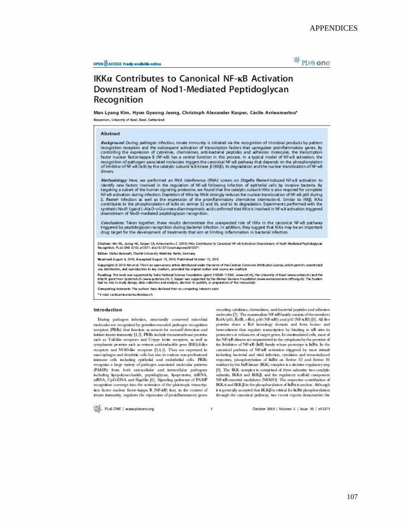
APPENDICES
107
APPENDICES
108
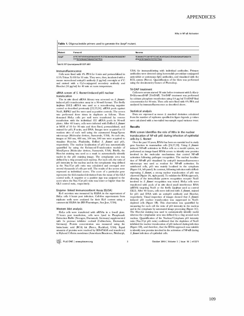
APPENDICES
109
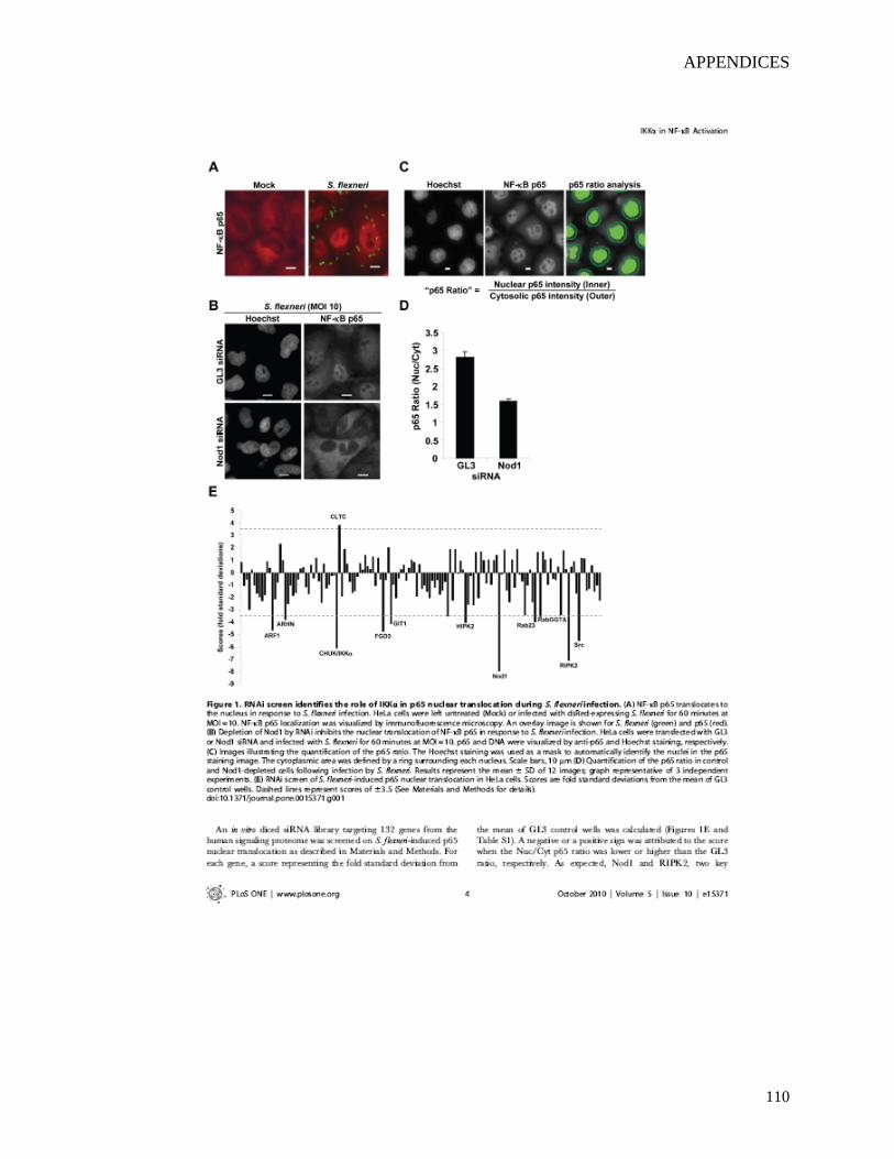
APPENDICES
110
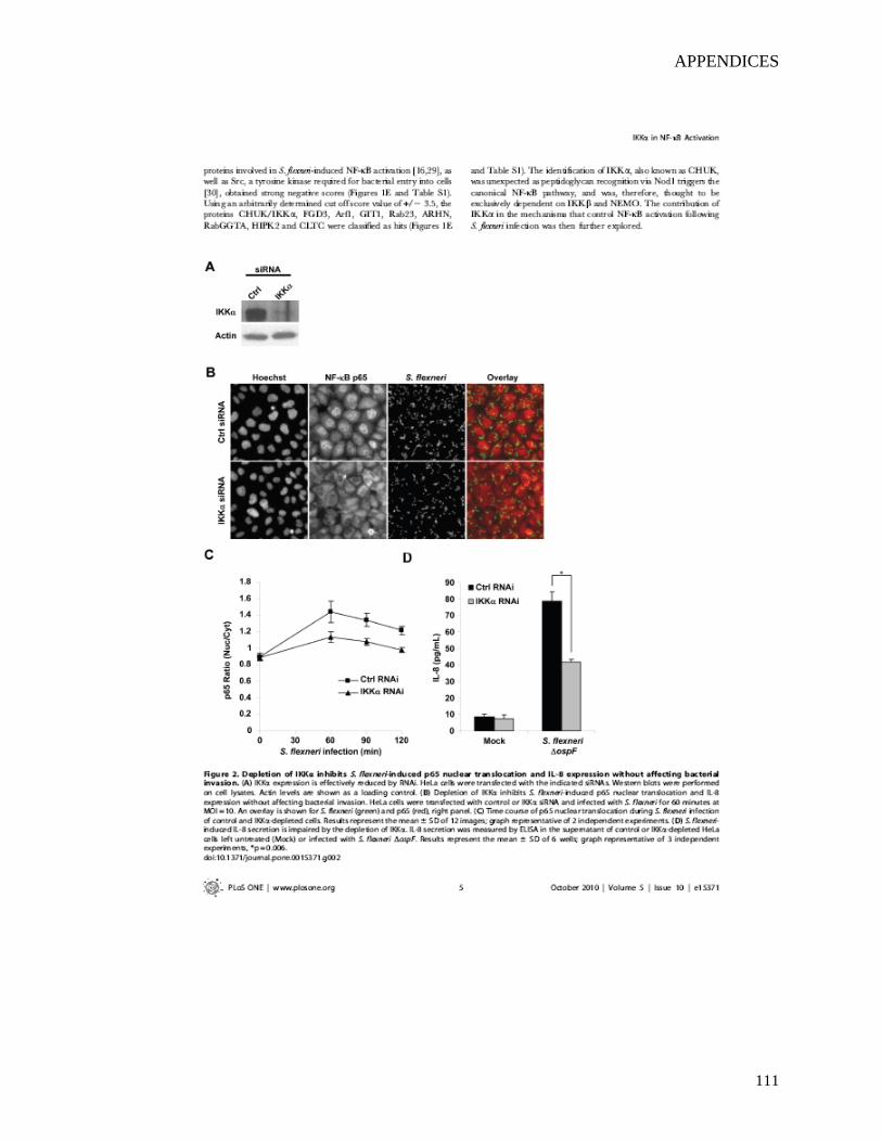
APPENDICES
111
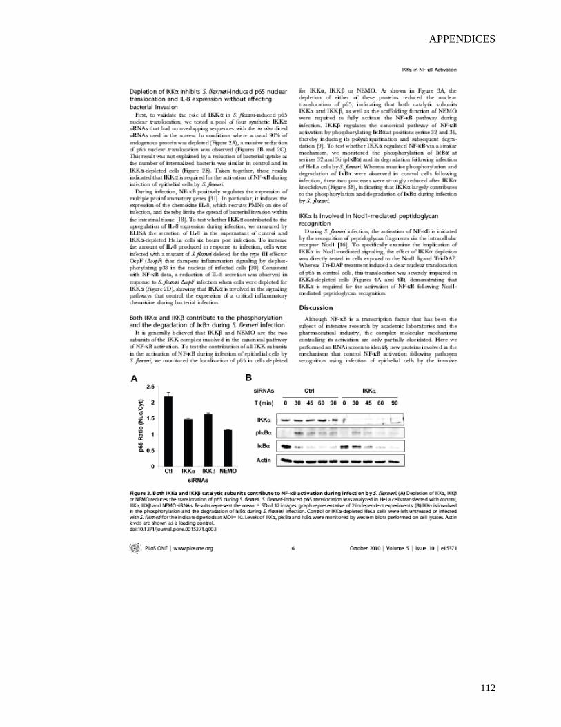
APPENDICES
112
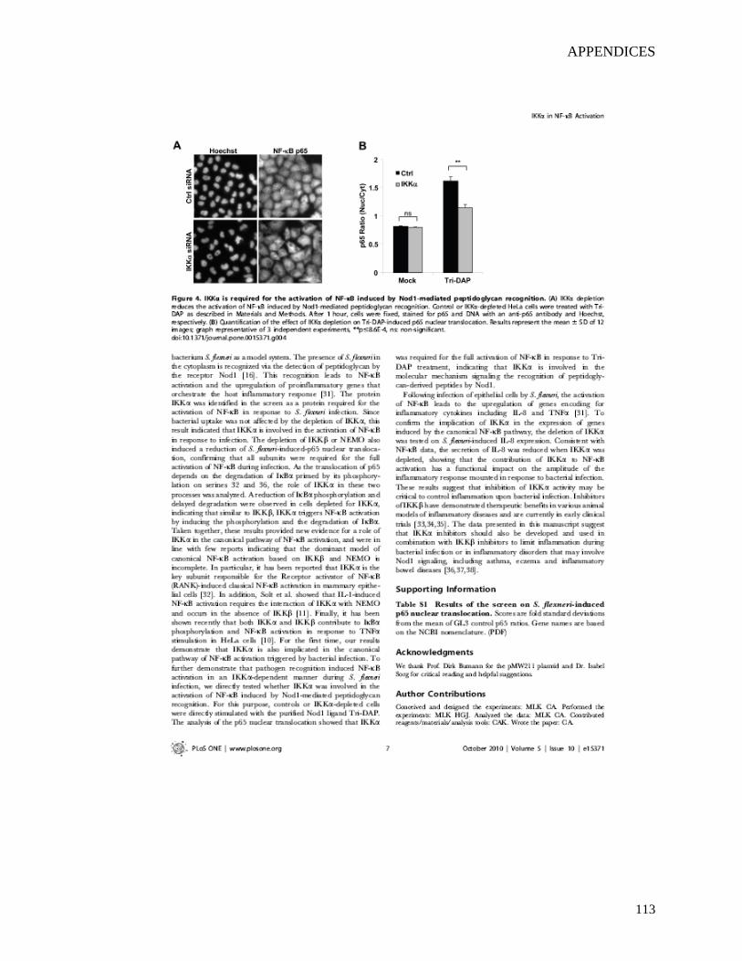
APPENDICES
113
APPENDICES
114
APPENDICES
115
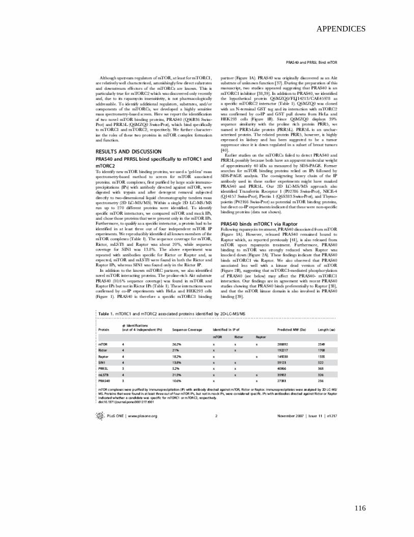
APPENDICES
116
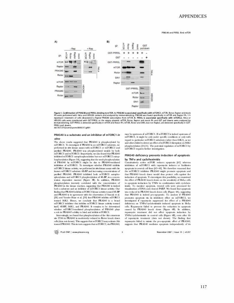
APPENDICES
117
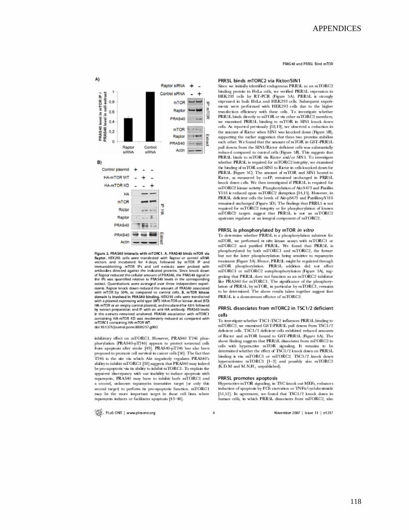
APPENDICES
118
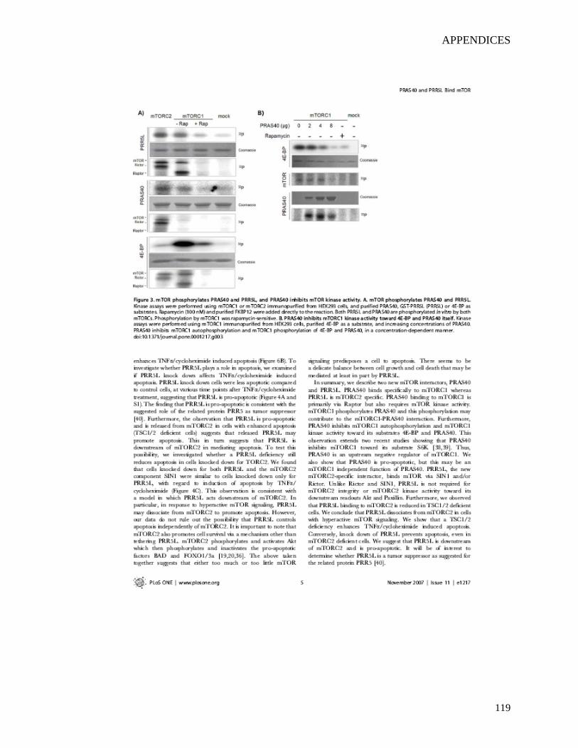
APPENDICES
119
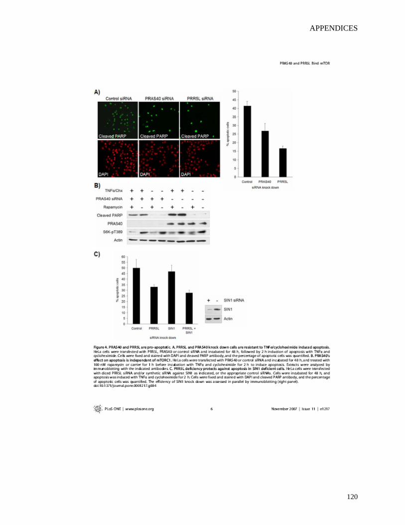
APPENDICES
120
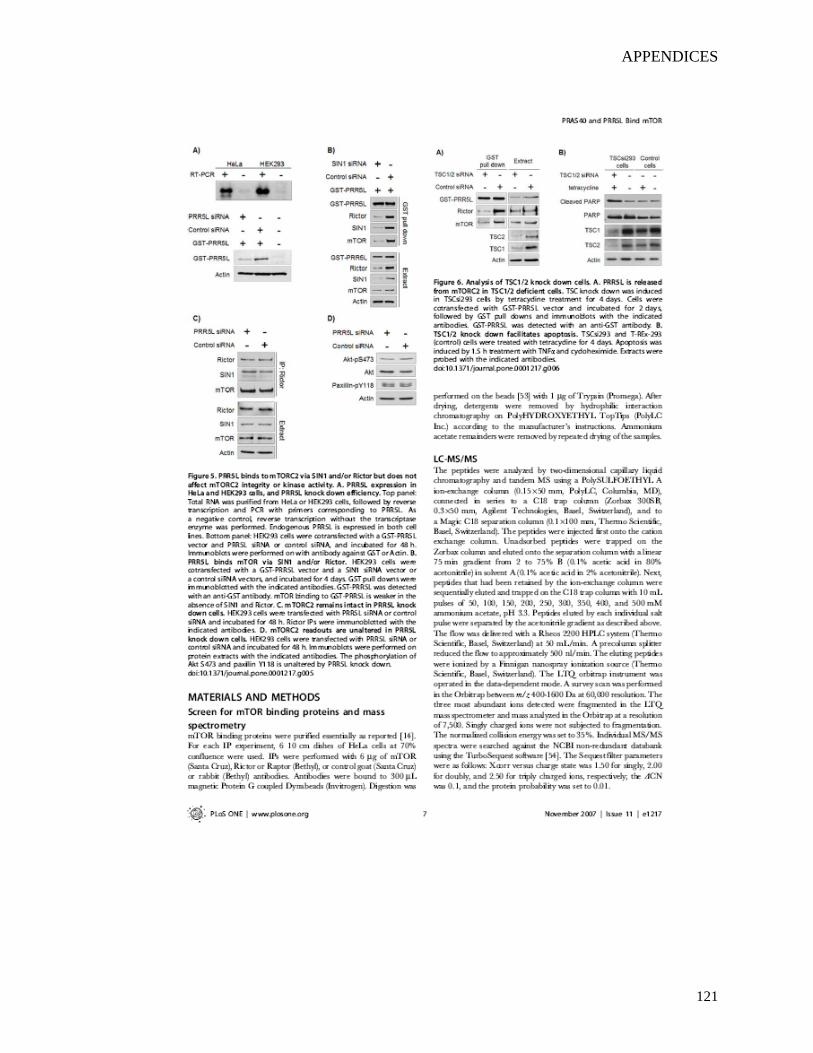
APPENDICES
121
APPENDICES
122
APPENDICES
123
APPENDICES
124
CURRICULUM VITAE
Curriculum Vitae Personal Information First name: Man Lyang Last name: Kim Birthdate: 17. 06. 1975 Nationality: South Korea Address: Metzerstrasse 38, 4056 Basel Telephone: 061 267 2124 E-mail: [email protected] Education March 1994 - February 2001 (*) B.S. in Biochemistry, Gyeonsang National University, South Korea March 2001 - February 2003 M.S. in Molecular Biology, Gyeonsang National University, South Korea September 2006 - Present PhD candidate, Focal Area Infection Biology, Biozentrum, University of Basel, Switzerland * November 1995 - January 1998 Military Service Work Experience February 2003 - September 2006 Life Science Research Assistant II,
Department of Chemicals and Systems Biology (Prof. Tobias Meyer),
Stanford University, USA Publications
Kasper CA, Sorg I, Wischnewski H, Tschon T, Schmutz C, Kim ML and Arrieumerlou C. Cell-cell propagation of NF-κB transcription factor and MAP kinase activation amplifies innate immunity against bacterial infection. Immunity. 2010 Nov24;33(5):804-16. Kim ML, Jeong HG, Kasper CA, Arrieumerlou C. IKKα contributes to canonical NF-κB activation downstream of Nod1-mediated peptidoglycan recognition. PLoS One. 2010 Oct 15;5(10):e15371. Park HC, Kim ML, Kim HS, Park JH, Jung MS, Shen M, Kang CH, Kim MC, Lee SY, Cho MJ, Chung WS, Yun DJ. Specificity of DNA sequences recognized by the zinc-finger homeodomain protein, GmZF-HD1 in soybean. Phytochemistry. 2010 Nov;71(16):1832-8.
125
CURRICULUM VITAE
Park HC, Kim ML, Kang YH, Jeong JC, Cheong MS, Choi W, Lee SY, Cho MJ, Kim MC, Chung WS, Yun DJ. Functional analysis of the stress-inducible soybean calmodulin isoform-4 (GmCaM-4) promoter in transgenic tobacco plants. Mol Cells. 2009;27(4):475-80.
Thedieck K, Polak P, Kim ML, Molle KD, Cohen A, Jenö P, Arrieumerlou C, Hall MN. PRAS40 and PRR5 Like Protein Are New mTOR Interactors that Regulate Apoptosis. PLoS One. 2007; 2(11):e1217
Galvez T, Teruel MN, Heo WD, Jones JT, Kim ML, Liou J, Myers JW, Meyer T. siRNA screen of the human signaling proteome identifies the PtdIns(3,4,5)P3-mTOR signaling pathway as a primary regulator of transferrin uptake. Genome Biol. 2007;8(7):R142.
Park HC*, Kim ML*, Lee SM, Bahk JD, Yun DJ, Lim CO, Hong JC, Lee SY, Cho MJ, Chung WS. Pathogen-induced binding of the soybean zinc finger homeodomain proteins GmZF-HD1 and GmZF-HD2 to two repeats of ATTA homeodomain binding site in the calmodulin isoform 4 (GmCaM4) promoter. Nucleic Acids Res. 2007;35(11):3612-23. (* These authors contributed equally to this work)
Heo WD, Inoue T, Park WS, Kim ML, Park BO, Wandless TJ, Meyer T. PI(3,4,5)P3 and PI(4,5)P2 lipids target proteins with polybasic clusters to the plasma membrane. Science. 2006; 314(5804):1458-61.
Liou J, Kim ML, Heo WD, Jones JT, Myers JW, Ferrell JE Jr, Meyer T. STIM is a Ca2+-sensor protein essential for Ca2+-store depletion triggered Ca2+-influx: Curr Biol. 2005;15(13):1235-1241
Park HC, Kim ML, Kang YH, Jeon JM, Yoo JH, Kim MC, Park CY, Jeong JC, Moon BC, Lee JH, Yoon HW, Lee SH, Chung WS, Lim CO, Lee SY, Hong JC, Cho MJ. Pathogen- and NaCl-induced expression of the SCaM-4 promoter is mediated in part by a GT-1 box that interacts with a GT-1-like transcription factor. Plant Physiol. 2004;135(4):2150-61.
Kim CY, Koo YD, Jin JB, Moon BC, Kang CH, Kim ST, Park BO, Lee SY, Kim ML, Hwang I, Kang KY, Bahk JD, Lee SY, Cho MJ. Rice C2-domain proteins are induced and translocated to the plasma membrane in response to a fungal elicitor. Biochemistry. 2003;42(40):11625-33.
126