Embed Size (px)
Citation preview
Abscisic acid maintains S-type anion channel activity in ATP-depletedVicia faba guard cells
Martin Schwarz1, Julian I. Schroeder*Department of Biology and Center for Molecular Genetics, University of California, San Diego, La Jolla, CA 92093-0116, USA
Received 6 April 1998
Abstract The plant hormone abscisic acid (ABA) regulatesimportant developmental and stress responses. Recent data showthat ABA activates phosphorylation events, but whether dephos-phorylation events are post-translationally regulated by ABA orwhether these are constitutive remains unknown. Slow anionchannels in the plasma membrane of guard cells have beenproposed to play an important role during ABA-induced stomatalclosing. Anion channels are deactivated by removal of cytosolicATP. However, when guard cells were treated with ABA anddepleted of ATP, anion currents remained active. Subsequentremoval of extracellular ABA caused deactivation of currents.Deactivation of currents was reversed by reintroduction ofcytosolic MgATP. These data show that anion channels areregulated by ABA even in the absence of cytosolic ATP requiredfor kinase-induced phosphorylation events and that anion channelactivity is maintained by ABA under conditions that favordephosphorylation-induced deactivation. Furthermore, channelactivation proceeded at high ATP concentrations with nanomolarcytosolic Ca2+ showing a Ca2+-independent final step in anionchannel activation.z 1998 Federation of European Biochemical Societies.
Key words: Abscisic acid; Clÿ channel; Stomate;Protein kinase; Protein phosphatase; Abscisic acid-insensitive
1. Introduction
The plant hormone abscisic acid (ABA) plays importantroles in the regulation of diverse plant growth and develop-mental responses [1,2]. ABA is synthesized during droughtstress [3] and induces closing of stomatal pores in leaves [4].Stomatal closing reduces transpirational water loss in plants.ABA-induced stomatal closing is mediated by e¥ux of K�
and anions across the plasma membrane of guard cells andby organic osmolyte metabolism. Anion channels have beenproposed to provide an important control mechanism for theregulation of ion e¥ux from guard cells during stomatal clos-ing [5^8]. Biophysical studies have shown that the slow andsustained (`S-type') anion channels in guard cells can producesustained anion e¥ux, which would result in a long-lastingdepolarization of the plasma membrane [5,7,8]. Depolariza-tions, can activate outward-rectifying K� channels proposedto mediate K� release during stomatal closing [9,10]. Recentstudies in Arabidopsis and tobacco guard cells have shownthat ABA regulates S-type anion channels [11,12], while re-vealing new regulation mechanisms in Arabidopsis, which act
in addition to those studied here (see Section 4). However, nodirect evidence for ABA regulation of S-type anion channelshas been shown in Vicia faba, despite many correlative studiesin this species [5,7,8,13^15].
Anion channel regulation has been proposed to play impor-tant roles in physiological processes of other plant cells, in-cluding hypocotyl growth, root transport, and blue light andred light signaling [16^19]. In Vicia faba guard cells, S-typeanion channels were shown to be activated by phosphoryla-tion events: depletion of the intracellular ATP pool leads to astrong down-regulation of S-type anion currents, which can-not be restored by even 4 mM of non-hydrolyzable ATP ana-logues or GTP analogues [13]. The protein kinase inhibitorsK252a and H7 abolished slow anion channel activity in thepresence of excess ATP and also abolished ABA-induced sto-matal closing, supporting central roles of phosphorylationevents for ABA signaling [13,15]. Biochemical studies haverecently directly shown a rapid enhancement of a 48 kDaprotein kinase activity by ABA in Vicia faba guard cell ex-tracts [20,21] and ABA activation of additional types of pro-tein kinases has been found in other tissues [22,23].
The protein phosphatase inhibitor okadaic acid (OA) inhib-its the activity of inward-rectifying K� channels in Vicia fabaguard cells, providing evidence for modulation by OA-sensi-tive protein phosphatases in guard cells [24,25]. Furthermore,OA maintained slow anion current activity even in the ab-sence of intracellular ATP [13]. In correlation to this ¢nding,ABA-induced stomatal closing is enhanced by OA in Viciafaba and Commelina communis [13,15]. ABA-regulated S-type anion channels in tobacco guard cells show stimulationby OA [12]. Together, these data suggest that ABA signalingand S-type anion channel regulation in Vicia faba are medi-ated by protein kinase activation and that OA-sensitive de-phosphorylation events are negative regulators of this path-way [13].
The recent identi¢cations of the Arabidopsis ABI1 and ABI2(ABA-insensitive) genes as protein phosphatases type 2C(PP2C) show that at least two additional OA-insensitive pro-tein phosphatases a¡ect ABA signaling [26^29]. PP2Cs areinsensitive to OA, as was demonstrated in vitro for theABI1 and ABI2 proteins [28,30]. Although several distinctprotein phosphatase activities appear to a¡ect ABA signaling(abi1, abi2 and two opposing OA-sensitive phosphatases, seeSection 4), no information exists whether ABA post-transla-tionally up- or down-regulates any of these phosphatases orwhether their constitutive activity a¡ects the ABA signalingpathway, as is proposed for yeast kinase signaling cascades[31]. For example, because both stomatal closing and activa-tion of slow anion channels in the presence of ABA werefurther stimulated by simultaneous addition of OA in Viciafaba, Commelina communis and tobacco guard cells [12,13,15],
FEBS 20291 29-5-98
0014-5793/98/$19.00 ß 1998 Federation of European Biochemical Societies. All rights reserved.PII S 0 0 1 4 - 5 7 9 3 ( 9 8 ) 0 0 5 2 6 - 2
*Corresponding author. Fax: (1) (619) 534-7108.E-mail: [email protected]
1Present address: Zentrum fuër Molekulare Neurobiologie,Falkenried 94, D-20251 Hamburg, Germany.
FEBS 20291 FEBS Letters 428 (1998) 177^182
the question whether ABA down-regulates OA-sensitive phos-phatases, in addition to kinase activation, remains unknown.
The ¢ndings of ABA signaling and anion channel regula-tion in guard cells without resolving changes in cytosolicCa2�, has led to models that include important Ca2�-inde-pendent steps and pathways in stomatal closing [12,32^34].Elevation in the cytosolic free Ca2� concentration ([Ca2�]cyt)can cause enhancement of guard cell slow and rapid anioncurrents [5,6]. This Ca2� e¡ect on S-type channels was hy-pothesized to be indirect, based on `wash-out' [5]. The strongactivation of S-type anion channels by phosphorylation eventsat resting [Ca2�]cyt, led to suggestions that important steps inthis regulation may be Ca2�-independent [11^13]. However,the question whether directly bu¡ering of [Ca2�]cyt to nano-molar levels, under strongly phosphorylating conditions, canactivate S-type anion channels remains unknown and wasanalyzed here.
The present study has speci¢cally focused on the followingpertinent questions relating to S-type anion channel regulationand ABA signaling:
1. Does ABA regulate S-type anion channels in Vicia fabaguard cells?
2. Is the strong deactivation of S-type anion channels bydepletion of cytosolic ATP reversible?
3. Can ABA maintain anion channel activity under ATP-free conditions in Vicia faba, which abolish e¡ects ofABA-activated kinases [20^23]? ABA regulation underATP-free conditions would allow initial cell biologicalinsight into the question whether ABA post-translation-ally regulates dephosphorylation events or whether theseactivities are constitutive.
4. Can activation of anion channels under strongly phos-phorylating conditions occur with cytosolic Ca2� bu¡-ered to nanomolar levels?
The present study demonstrates that ABA maintains S-typeanion channel activation in Vicia faba guard cells under ATP-free dephosphorylating conditions, that this regulation is re-versible and that a Ca2�-independent ¢nal step exists in anionchannel activation.
2. Materials and methods
Guard cell protoplasts were isolated from leaves of Vicia faba byenzymatic digestion as described previously [7]. Pipette solutions con-tained 150 mM CsCl, 2 mM MgCl2, 6.7 mM EGTA-(Tris)2, 3.35 mMCaCl2, 10 mM HEPES adjusted to pH 7.1 with Tris and 5 mMMgATP unless otherwise indicated. Chloride activities were 114 mMafter correction for ionic activities, which lies in the upper physiolog-ical range measured in guard cells [35,36]. The osmolality was ad-justed to 510 mosmol/kg by adding D-mannitol. MgATP was omittedfrom the pipette solution, when indicated in the text. Guard cells werebathed either in a `Ca2� bath' medium of 40 mM CaCl2, 2 mMMgCl2 and 10 mM MES-Tris, pH 5.6 or in a `Cs� bath' of 30 mMCsCl, 1 mM CaCl2, 2 mM MgCl2 and 10 mM MES-Tris, pH 5.6,adjusted to 490 mosmol/kg using D-mannitol. ABA ([ þ ]cis-trans iso-mer, Sigma) was prepared as a 50 mM stock solution in 100% ethanoland added at the indicated concentrations to the bath and/or pipettesolution. The ¢nal concentration of ethanol in all experiments did notexceed 0.2% (v/v) and ethanol control experiments did not show ef-fects.
Whole-cell recordings were performed, with an Axopatch-1D am-pli¢er as previously described [7]. For time course analyses, 8-s volt-
age pulses were applied over a period of 30 min. For current-voltageanalyses a voltage protocol as in the top inset of Fig. 1A was used.Peak currents at 385 mV of each individual guard cell at the indi-cated times were normalized relative to the maximum current ampli-tude measured during the whole-cell recording and expressed in per-cent of maximum current. Peak currents were measured 100 ms afterthe onset of each individual voltage pulse.
3. Results
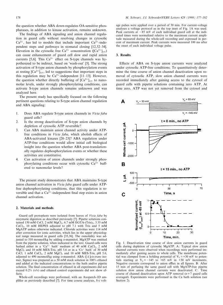
E¡ects of ABA on S-type anion currents were analyzedunder cytosolic ATP-free conditions. To quantitatively deter-mine the time course of anion channel deactivation upon re-moval of cytosolic ATP, slow anion channel currents wererecorded immediately after gaining access to the cytosol ofguard cells with pipette solutions containing zero ATP. Attime zero, ATP was not yet removed from the cytosol and
FEBS 20291 29-5-98
Fig. 1. Deactivation time course of slow anion currents in guardcells during depletion of cytosolic MgATP. A: Typical slow anionchannel currents were observed when recordings were performed im-mediately after gaining access to whole cells. The membrane poten-tial was clamped from a holding potential of Vh = +30 mV to poten-tials starting at Vp =3145 to +65 mV in +30 mV increments.Negative currents correspond to anion e¥ux in all ¢gures. B: After15 min of perfusing the same guard cell with MgATP-free pipettesolution slow anion channel currents were deactivated. C: Timecourse of channel deactivation upon ATP removal (n = 7 guard cellsaveraged). Experiments were performed in the Ca bath solution (seeSection 2).
M. Schwarz, J.I. Schroeder/FEBS Letters 428 (1998) 177^182178
large S-type anion currents were fully activated by depolari-zation of guard cells followed by hyperpolarizing voltagepulses (Fig. 1A) as reported previously [5]. However, as thecytosol of guard cells equilibrated with the ATP-free pipettesolution, anion channel current activities declined over timeuntil s 98% of the current disappeared (Fig. 1B). This strongdeactivation had an average half-maximal deactivation timeof 9.4 min (Fig. 1C). Controls in the presence of ATP showedreduction of anion currents by only 19 þ 8% after 15 min inthis set of experiments.
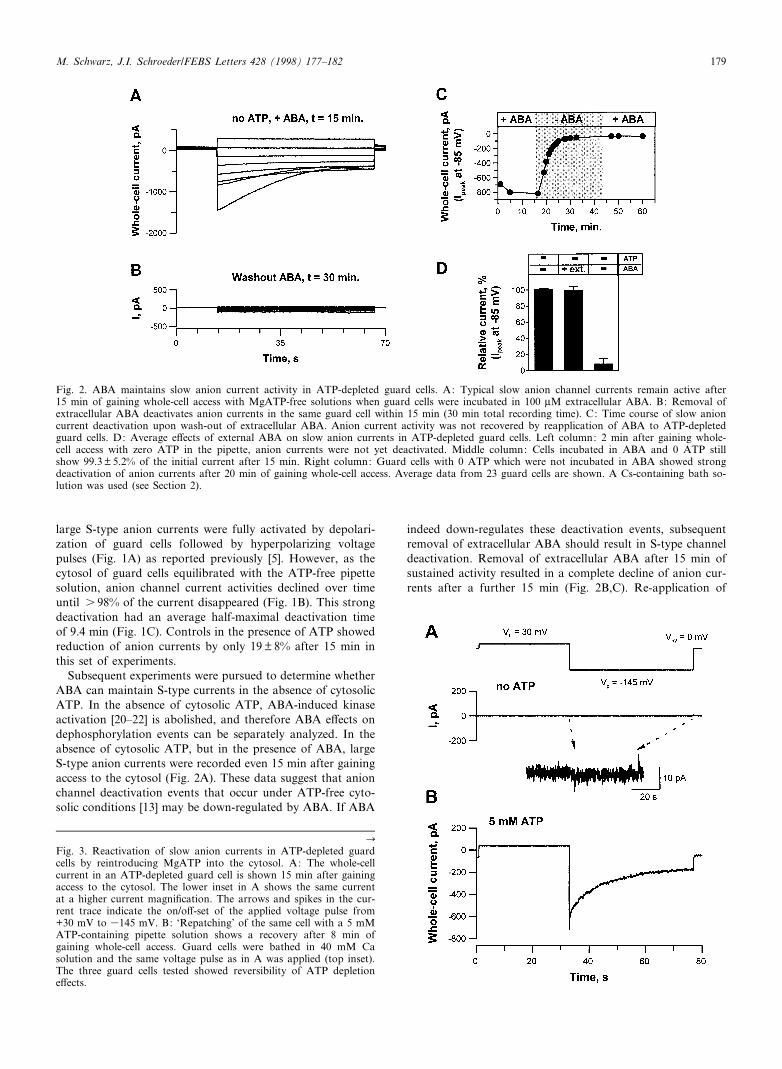
Subsequent experiments were pursued to determine whetherABA can maintain S-type currents in the absence of cytosolicATP. In the absence of cytosolic ATP, ABA-induced kinaseactivation [20^22] is abolished, and therefore ABA e¡ects ondephosphorylation events can be separately analyzed. In theabsence of cytosolic ATP, but in the presence of ABA, largeS-type anion currents were recorded even 15 min after gainingaccess to the cytosol (Fig. 2A). These data suggest that anionchannel deactivation events that occur under ATP-free cyto-solic conditions [13] may be down-regulated by ABA. If ABA
indeed down-regulates these deactivation events, subsequentremoval of extracellular ABA should result in S-type channeldeactivation. Removal of extracellular ABA after 15 min ofsustained activity resulted in a complete decline of anion cur-rents after a further 15 min (Fig. 2B,C). Re-application of
FEBS 20291 29-5-98
Fig. 2. ABA maintains slow anion current activity in ATP-depleted guard cells. A: Typical slow anion channel currents remain active after15 min of gaining whole-cell access with MgATP-free solutions when guard cells were incubated in 100 WM extracellular ABA. B: Removal ofextracellular ABA deactivates anion currents in the same guard cell within 15 min (30 min total recording time). C: Time course of slow anioncurrent deactivation upon wash-out of extracellular ABA. Anion current activity was not recovered by reapplication of ABA to ATP-depletedguard cells. D: Average e¡ects of external ABA on slow anion currents in ATP-depleted guard cells. Left column: 2 min after gaining whole-cell access with zero ATP in the pipette, anion currents were not yet deactivated. Middle column: Cells incubated in ABA and 0 ATP stillshow 99.3 þ 5.2% of the initial current after 15 min. Right column: Guard cells with 0 ATP which were not incubated in ABA showed strongdeactivation of anion currents after 20 min of gaining whole-cell access. Average data from 23 guard cells are shown. A Cs-containing bath so-lution was used (see Section 2).
CFig. 3. Reactivation of slow anion currents in ATP-depleted guardcells by reintroducing MgATP into the cytosol. A: The whole-cellcurrent in an ATP-depleted guard cell is shown 15 min after gainingaccess to the cytosol. The lower inset in A shows the same currentat a higher current magni¢cation. The arrows and spikes in the cur-rent trace indicate the on/o¡-set of the applied voltage pulse from+30 mV to 3145 mV. B: `Repatching' of the same cell with a 5 mMATP-containing pipette solution shows a recovery after 8 min ofgaining whole-cell access. Guard cells were bathed in 40 mM Casolution and the same voltage pulse as in A was applied (top inset).The three guard cells tested showed reversibility of ATP depletione¡ects.
M. Schwarz, J.I. Schroeder/FEBS Letters 428 (1998) 177^182 179
extracellular ABA to the bath solution did not lead to a re-newed activation of slow anion currents (Fig. 2C), as can beexpected under cytosolic ATP-free conditions, because ATPacts as the substrate for phosphorylation [13]. On average,ATP-free cells, exposed to extracellular ABA in the Cs�
bath solution (see Section 2) showed 99.3 þ 5.2% of the initialcurrent after 15 min of whole-cell recording (Fig. 2D, middlecolumn). However, cells not exposed to ABA showed a strongdeactivation of slow anion currents (Fig. 2D, right column).After only 2 min of whole-cell recording deactivation had notyet occurred (Fig. 2D, left column; Fig. 1C).
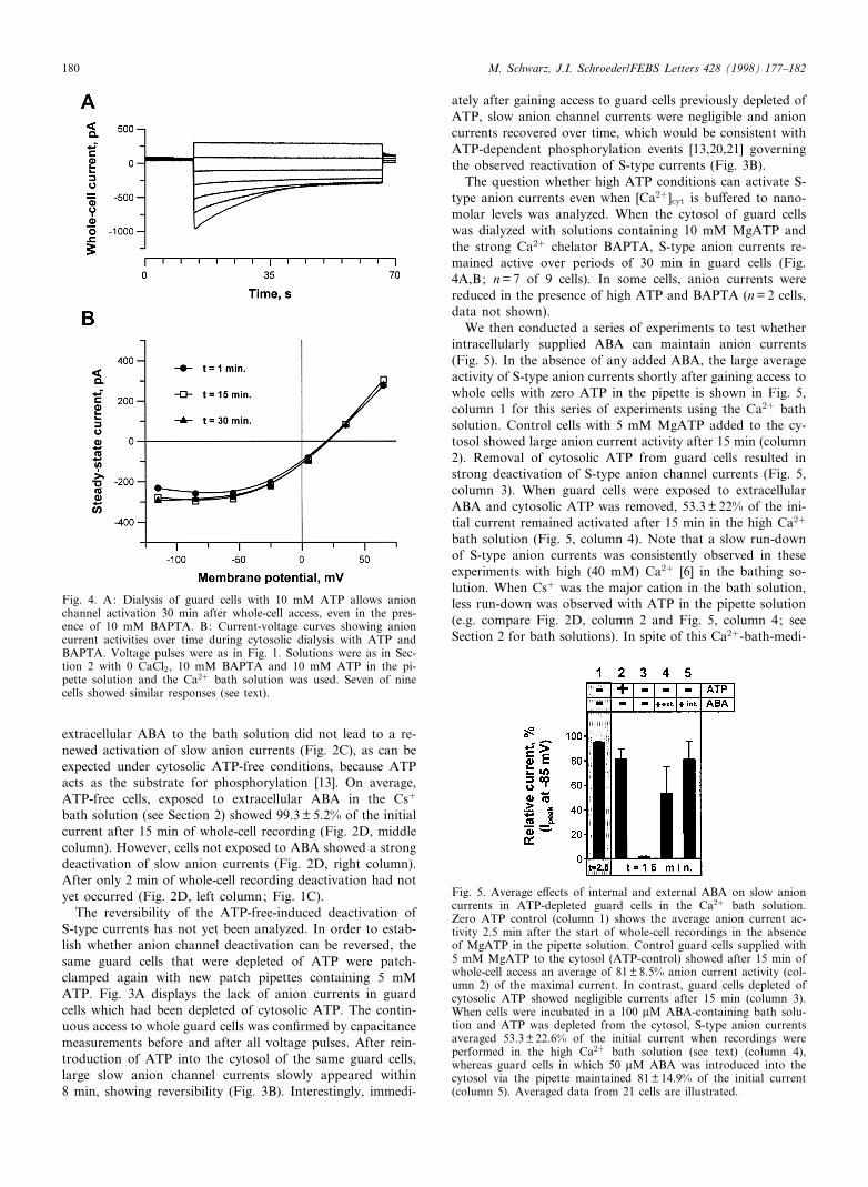
The reversibility of the ATP-free-induced deactivation ofS-type currents has not yet been analyzed. In order to estab-lish whether anion channel deactivation can be reversed, thesame guard cells that were depleted of ATP were patch-clamped again with new patch pipettes containing 5 mMATP. Fig. 3A displays the lack of anion currents in guardcells which had been depleted of cytosolic ATP. The contin-uous access to whole guard cells was con¢rmed by capacitancemeasurements before and after all voltage pulses. After rein-troduction of ATP into the cytosol of the same guard cells,large slow anion channel currents slowly appeared within8 min, showing reversibility (Fig. 3B). Interestingly, immedi-
ately after gaining access to guard cells previously depleted ofATP, slow anion channel currents were negligible and anioncurrents recovered over time, which would be consistent withATP-dependent phosphorylation events [13,20,21] governingthe observed reactivation of S-type currents (Fig. 3B).
The question whether high ATP conditions can activate S-type anion currents even when [Ca2�]cyt is bu¡ered to nano-molar levels was analyzed. When the cytosol of guard cellswas dialyzed with solutions containing 10 mM MgATP andthe strong Ca2� chelator BAPTA, S-type anion currents re-mained active over periods of 30 min in guard cells (Fig.4A,B; n = 7 of 9 cells). In some cells, anion currents werereduced in the presence of high ATP and BAPTA (n = 2 cells,data not shown).
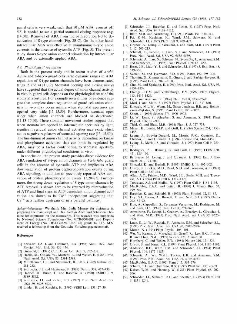
We then conducted a series of experiments to test whetherintracellularly supplied ABA can maintain anion currents(Fig. 5). In the absence of any added ABA, the large averageactivity of S-type anion currents shortly after gaining access towhole cells with zero ATP in the pipette is shown in Fig. 5,column 1 for this series of experiments using the Ca2� bathsolution. Control cells with 5 mM MgATP added to the cy-tosol showed large anion current activity after 15 min (column2). Removal of cytosolic ATP from guard cells resulted instrong deactivation of S-type anion channel currents (Fig. 5,column 3). When guard cells were exposed to extracellularABA and cytosolic ATP was removed, 53.3 þ 22% of the ini-tial current remained activated after 15 min in the high Ca2�
bath solution (Fig. 5, column 4). Note that a slow run-downof S-type anion currents was consistently observed in theseexperiments with high (40 mM) Ca2� [6] in the bathing so-lution. When Cs� was the major cation in the bath solution,less run-down was observed with ATP in the pipette solution(e.g. compare Fig. 2D, column 2 and Fig. 5, column 4; seeSection 2 for bath solutions). In spite of this Ca2�-bath-medi-
FEBS 20291 29-5-98
Fig. 4. A: Dialysis of guard cells with 10 mM ATP allows anionchannel activation 30 min after whole-cell access, even in the pres-ence of 10 mM BAPTA. B: Current-voltage curves showing anioncurrent activities over time during cytosolic dialysis with ATP andBAPTA. Voltage pulses were as in Fig. 1. Solutions were as in Sec-tion 2 with 0 CaCl2, 10 mM BAPTA and 10 mM ATP in the pi-pette solution and the Ca2� bath solution was used. Seven of ninecells showed similar responses (see text).
Fig. 5. Average e¡ects of internal and external ABA on slow anioncurrents in ATP-depleted guard cells in the Ca2� bath solution.Zero ATP control (column 1) shows the average anion current ac-tivity 2.5 min after the start of whole-cell recordings in the absenceof MgATP in the pipette solution. Control guard cells supplied with5 mM MgATP to the cytosol (ATP-control) showed after 15 min ofwhole-cell access an average of 81 þ 8.5% anion current activity (col-umn 2) of the maximal current. In contrast, guard cells depleted ofcytosolic ATP showed negligible currents after 15 min (column 3).When cells were incubated in a 100 WM ABA-containing bath solu-tion and ATP was depleted from the cytosol, S-type anion currentsaveraged 53.3 þ 22.6% of the initial current when recordings wereperformed in the high Ca2� bath solution (see text) (column 4),whereas guard cells in which 50 WM ABA was introduced into thecytosol via the pipette maintained 81 þ 14.9% of the initial current(column 5). Averaged data from 21 cells are illustrated.
M. Schwarz, J.I. Schroeder/FEBS Letters 428 (1998) 177^182180
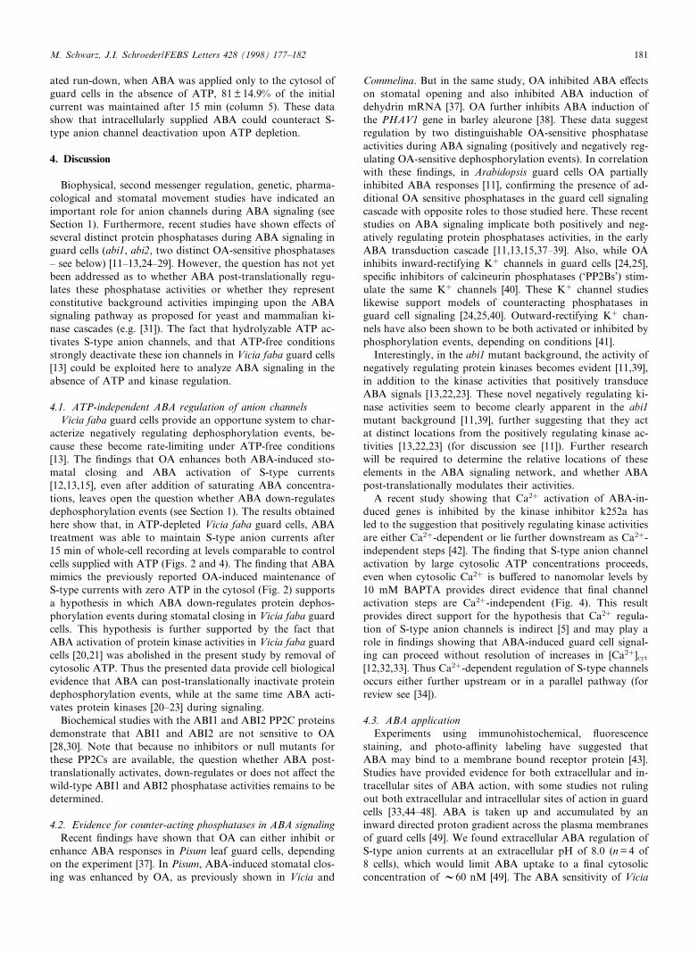
ated run-down, when ABA was applied only to the cytosol ofguard cells in the absence of ATP, 81 þ 14.9% of the initialcurrent was maintained after 15 min (column 5). These datashow that intracellularly supplied ABA could counteract S-type anion channel deactivation upon ATP depletion.
4. Discussion
Biophysical, second messenger regulation, genetic, pharma-cological and stomatal movement studies have indicated animportant role for anion channels during ABA signaling (seeSection 1). Furthermore, recent studies have shown e¡ects ofseveral distinct protein phosphatases during ABA signaling inguard cells (abi1, abi2, two distinct OA-sensitive phosphatases^ see below) [11^13,24^29]. However, the question has not yetbeen addressed as to whether ABA post-translationally regu-lates these phosphatase activities or whether they representconstitutive background activities impinging upon the ABAsignaling pathway as proposed for yeast and mammalian ki-nase cascades (e.g. [31]). The fact that hydrolyzable ATP ac-tivates S-type anion channels, and that ATP-free conditionsstrongly deactivate these ion channels in Vicia faba guard cells[13] could be exploited here to analyze ABA signaling in theabsence of ATP and kinase regulation.
4.1. ATP-independent ABA regulation of anion channelsVicia faba guard cells provide an opportune system to char-
acterize negatively regulating dephosphorylation events, be-cause these become rate-limiting under ATP-free conditions[13]. The ¢ndings that OA enhances both ABA-induced sto-matal closing and ABA activation of S-type currents[12,13,15], even after addition of saturating ABA concentra-tions, leaves open the question whether ABA down-regulatesdephosphorylation events (see Section 1). The results obtainedhere show that, in ATP-depleted Vicia faba guard cells, ABAtreatment was able to maintain S-type anion currents after15 min of whole-cell recording at levels comparable to controlcells supplied with ATP (Figs. 2 and 4). The ¢nding that ABAmimics the previously reported OA-induced maintenance ofS-type currents with zero ATP in the cytosol (Fig. 2) supportsa hypothesis in which ABA down-regulates protein dephos-phorylation events during stomatal closing in Vicia faba guardcells. This hypothesis is further supported by the fact thatABA activation of protein kinase activities in Vicia faba guardcells [20,21] was abolished in the present study by removal ofcytosolic ATP. Thus the presented data provide cell biologicalevidence that ABA can post-translationally inactivate proteindephosphorylation events, while at the same time ABA acti-vates protein kinases [20^23] during signaling.
Biochemical studies with the ABI1 and ABI2 PP2C proteinsdemonstrate that ABI1 and ABI2 are not sensitive to OA[28,30]. Note that because no inhibitors or null mutants forthese PP2Cs are available, the question whether ABA post-translationally activates, down-regulates or does not a¡ect thewild-type ABI1 and ABI2 phosphatase activities remains to bedetermined.
4.2. Evidence for counter-acting phosphatases in ABA signalingRecent ¢ndings have shown that OA can either inhibit or
enhance ABA responses in Pisum leaf guard cells, dependingon the experiment [37]. In Pisum, ABA-induced stomatal clos-ing was enhanced by OA, as previously shown in Vicia and
Commelina. But in the same study, OA inhibited ABA e¡ectson stomatal opening and also inhibited ABA induction ofdehydrin mRNA [37]. OA further inhibits ABA induction ofthe PHAV1 gene in barley aleurone [38]. These data suggestregulation by two distinguishable OA-sensitive phosphataseactivities during ABA signaling (positively and negatively reg-ulating OA-sensitive dephosphorylation events). In correlationwith these ¢ndings, in Arabidopsis guard cells OA partiallyinhibited ABA responses [11], con¢rming the presence of ad-ditional OA sensitive phosphatases in the guard cell signalingcascade with opposite roles to those studied here. These recentstudies on ABA signaling implicate both positively and neg-atively regulating protein phosphatases activities, in the earlyABA transduction cascade [11,13,15,37^39]. Also, while OAinhibits inward-rectifying K� channels in guard cells [24,25],speci¢c inhibitors of calcineurin phosphatases (`PP2Bs') stim-ulate the same K� channels [40]. These K� channel studieslikewise support models of counteracting phosphatases inguard cell signaling [24,25,40]. Outward-rectifying K� chan-nels have also been shown to be both activated or inhibited byphosphorylation events, depending on conditions [41].
Interestingly, in the abi1 mutant background, the activity ofnegatively regulating protein kinases becomes evident [11,39],in addition to the kinase activities that positively transduceABA signals [13,22,23]. These novel negatively regulating ki-nase activities seem to become clearly apparent in the abi1mutant background [11,39], further suggesting that they actat distinct locations from the positively regulating kinase ac-tivities [13,22,23] (for discussion see [11]). Further researchwill be required to determine the relative locations of theseelements in the ABA signaling network, and whether ABApost-translationally modulates their activities.
A recent study showing that Ca2� activation of ABA-in-duced genes is inhibited by the kinase inhibitor k252a hasled to the suggestion that positively regulating kinase activitiesare either Ca2�-dependent or lie further downstream as Ca2�-independent steps [42]. The ¢nding that S-type anion channelactivation by large cytosolic ATP concentrations proceeds,even when cytosolic Ca2� is bu¡ered to nanomolar levels by10 mM BAPTA provides direct evidence that ¢nal channelactivation steps are Ca2�-independent (Fig. 4). This resultprovides direct support for the hypothesis that Ca2� regula-tion of S-type anion channels is indirect [5] and may play arole in ¢ndings showing that ABA-induced guard cell signal-ing can proceed without resolution of increases in [Ca2�]cyt
[12,32,33]. Thus Ca2�-dependent regulation of S-type channelsoccurs either further upstream or in a parallel pathway (forreview see [34]).
4.3. ABA applicationExperiments using immunohistochemical, £uorescence
staining, and photo-a¤nity labeling have suggested thatABA may bind to a membrane bound receptor protein [43].Studies have provided evidence for both extracellular and in-tracellular sites of ABA action, with some studies not rulingout both extracellular and intracellular sites of action in guardcells [33,44^48]. ABA is taken up and accumulated by aninward directed proton gradient across the plasma membranesof guard cells [49]. We found extracellular ABA regulation ofS-type anion currents at an extracellular pH of 8.0 (n = 4 of8 cells), which would limit ABA uptake to a ¢nal cytosolicconcentration of V60 nM [49]. The ABA sensitivity of Vicia
FEBS 20291 29-5-98
M. Schwarz, J.I. Schroeder/FEBS Letters 428 (1998) 177^182 181
guard cells is very weak, such that 50 WM ABA, even at pH5.5, is needed to see a partial stomatal closing response (e.g.[14,50]). Removal of ABA from the bath solution led to de-activation of S-type channels (Fig. 2B,C). On the other hand,intracellular ABA was e¡ective at maintaining S-type anioncurrents in the absence of cytosolic ATP (Fig. 5). The presentstudy shows S-type anion channel stimulation by intracellularABA and by externally applied ABA.
4.4. Physiological regulationBoth in the present study and in recent studies of Arabi-
dopsis and tobacco guard cells large dynamic ranges in ABAregulation of S-type anion channels have been demonstrated(Figs. 2 and 4) [11,12]. Stomatal opening and closing assayshave suggested that the actual degree of anion channel activityin vivo in guard cells depends on the physiological state of thestomatal apertures. For example several lines of evidence sug-gest that complete down-regulation of guard cell anion chan-nels in vivo may occur mainly when stomatal apertures areopened very wide [13^15,50]. Furthermore, stomata openwider when anion channels are blocked or deactivated[11,13^15,50]. These stomatal movement studies suggest thatwhen stomata are opened to intermediate apertures by light,signi¢cant residual anion channel activities may exist, whichact as negative regulators of stomatal opening (see [13^15,50]).The ¢ne-tuning of anion channel activity depending on kinaseand phosphatase activities, that can both be regulated byABA, may be a factor contributing to stomatal aperturesunder di¡erent physiological conditions in vivo.
In conclusion, the present study provides direct evidence forABA regulation of S-type anion channels in Vicia faba guardcells in the absence of cytosolic ATP and implicates ABAdown-regulation of protein dephosphorylation events duringABA signaling, in addition to previously reported ABA acti-vation of protein phosphorylation events [13,20^23]. Further-more, the strong down-regulation of S-type anion channels byATP removal is shown here to be reversed by reintroductionof ATP and ¢nal steps in ATP-dependent anion channel acti-vation are shown to be Ca2� independent suggesting thatCa2� acts further upstream or in a parallel pathway.
Acknowledgements: We thank Mrs. Judie Murray for assistance inpreparing the manuscript and Drs. Gethyn Allen and Sebastien Tho-mine for comments on the manuscript. This research was supportedby National Science Foundation (No. MCB-9506191) and Depart-ment of Energy (No. DE-FG03-94-ER20148) grants to J.I.S. M.S.received a fellowship from the Deutsche Forschungsgemeinschaft.
References
[1] Zeevaart, J.A.D. and Creelman, R.A. (1988) Annu. Rev. PlantPhysiol. Mol. Biol. 39, 439^474.
[2] Giraudat, J. (1995) Curr. Opin. Cell Biol. 7, 232^238.[3] Harris, M., Outlaw, W., Martens, R. and Weiler, E. (1988) Proc.
Natl. Acad. Sci. USA 85, 2584^2588.[4] Mittelheuser, C.J. and Steveninck, R.F.M.v. (1969) Nature 221,
281^282.[5] Schroeder, J.I. and Hagiwara, S. (1989) Nature 338, 427^430.[6] Hedrich, R., Busch, H. and Raschke, K. (1990) EMBO J. 9,
3889^3892.[7] Schroeder, J.I. and Keller, B.U. (1992) Proc. Natl. Acad. Sci.
USA 89, 5025^5029.[8] Linder, B. and Raschke, K. (1992) FEBS Lett. 131, 27^30.
[9] Schroeder, J.I., Raschke, K. and Neher, E. (1987) Proc. Natl.Acad. Sci. USA 84, 4108^4112.
[10] Blatt, M.R. and Armstrong, F. (1993) Planta 191, 330^341.[11] Pei, Z.-M., Kuchitsu, K., Ward, J.M., Schwarz, M. and
Schroeder, J.I. (1997) Plant Cell 9, 409^423.[12] Grabov, A., Leung, J., Giraudat, J. and Blatt, M.R. (1997) Plant
J. 12, 203^213.[13] Schmidt, C., Schelle, I., Liao, Y.J. and Schroeder, J.I. (1995)
Proc. Natl. Acad. Sci. USA 92, 9535^9539.[14] Schwartz, A., Ilan, N., Schwarz, N., Schea¡er, J., Assmann, S.M.
and Schroeder, J.I. (1995) Plant Physiol. 109, 651^658.[15] Esser, J.E., Liao, Y.-J. and Schroeder, J.I. (1997) J. Exp. Bot. 48,
539^550.[16] Skerett, M. and Tyermann, S.D. (1994) Planta 192, 295^305.[17] Thomine, S., Zimmermann, S., Guern, J. and Barbier-Brygoo, H.
(1995) Plant Cell 7, 2091^2100.[18] Cho, M. and Spalding, E. (1996) Proc. Natl. Acad. Sci. USA 93,
8134^8138.[19] Elzenga, J.T.M. and Volkenburgh, E.V. (1997) Plant Physiol.
113, 1419^1426.[20] Li, J. and Assmann, S. (1996) Plant Cell 8, 2359^2368.[21] Mori, I. and Muto, S. (1997) Plant Physiol. 113, 833^840.[22] Knetsch, M.L.W., Wang, M., Snaar-Jagalska, B.E. and Heimo-
vaara-Dijkstra, S. (1996) Plant Cell 8, 1061^1067.[23] Sheen, J. (1996) Science 274, 1900^1902.[24] Li, W., Luan, S., Schreiber, S. and Assmann, S. (1994) Plant
Physiol. 106, 963^970.[25] Thiel, G. and Blatt, M.R. (1994) Plant J. 5, 727^733.[26] Meyer, K., Leube, M.P. and Grill, E. (1994) Science 264, 1452^
1455.[27] Leung, J., Bouvier-Durand, M., Morris, P.-C., Guerrier, D.,
Chefdor, F. and Giraudat, J. (1994) Science 264, 1448^1452.[28] Leung, J., Merlot, S. and Giraudat, J. (1997) Plant Cell 9, 759^
771.[29] Rodriguez, P.L., Benning, G. and Grill, E. (1998) FEBS Lett.
421, 185^190.[30] Bertauche, N., Leung, J. and Giraudat, J. (1996) Eur. J. Bio-
chem. 241, 193^200.[31] Shiozaki, K. and Russell, P. (1995) EMBO J. 14, 492^502.[32] Gilroy, S., Fricker, M.D., Read, N.D. and Trewavas, A.J. (1991)
Plant Cell 3, 333^344.[33] Allan, A.C., Fricker, M.D., Ward, J.L., Beale, M.H. and Trewa-
vas, A.J. (1994) Plant Cell 6, 1319^1328.[34] Trewavas, A.J. and Malho, R. (1997) Plant Cell 9, 1181^1195.[35] MacRobbie, E.A.C. and Lettau, B. (1980) J. Memb. Biol. 53,
199^207.[36] Raschke, K. and Schnabl, H. (1978) Plant Physiol. 62, 84^87.[37] Hey, S.J., Bacon, A., Burnett, E. and Neill, S.J. (1997) Planta
202, 85^92.[38] Kuo, A., Cappelluti, S., Cervantes-Vervantes, M., Rodriguez, M.
and Bush, D.S. (1996) Plant Cell 8, 259^269.[39] Armstrong, F., Leung, J., Grabov, A., Brearley, J., Giraudat, J.
and Blatt, M.R. (1995) Proc. Natl. Acad. Sci. USA 92, 9520^9524.
[40] Luan, S., Li, W., Rusnak, F., Assmann, S.M. and Schreiber, S.L.(1993) Proc. Natl. Acad. Sci. USA 90, 2202^2206.
[41] Moran, N. (1994) Plant Physiol. 105, 101.[42] Wu, Y., Kuzma, J., Marechal, E., Grae¡, R., Lee, H.C., Foster,
R. and Chua, N.-H. (1997) Science 278, 2126^2130.[43] Hornberg, C. and Weiler, E.W. (1984) Nature 310, 321^324.[44] Gilroy, S. and Jones, R.L. (1994) Plant Physiol. 104, 1185^1192.[45] Anderson, B.E., Ward, J.M. and Schroeder, J.I. (1994) Plant
Physiol. 104, 1177^1183.[46] Schwartz, A., Wu, W.-H., Tucker, E.B. and Assmann, S.M.
(1994) Proc. Natl. Acad. Sci. USA 91, 4019^4023.[47] MacRobbie, E.A.C. (1995) Plant J. 7, 565^576.[48] Schultz, T.F. and Quatrano, R.S. (1997) Plant Sci. 130, 63^71.[49] Kaiser, W.M. and Hartung, W. (1981) Plant Physiol. 68, 202^
206.[50] Schroeder, J.I., Schmidt, R.C. and Shea¡er, J. (1993) Plant Cell
5, 1831^1841.
FEBS 20291 29-5-98
M. Schwarz, J.I. Schroeder/FEBS Letters 428 (1998) 177^182182