Embed Size (px)
Citation preview
ABO/Rh Blood Group System
A . Amirzargar
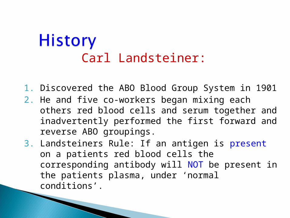
Carl Landsteiner:
1. Discovered the ABO Blood Group System in 19012. He and five co-workers began mixing each others
red blood cells and serum together and inadvertently performed the first forward and reverse ABO groupings.
3. Landsteiners Rule: If an antigen is present on a patients red blood cells the corresponding antibody will NOT be present in the patients plasma, under ‘normal conditions’.

Blood group systems
Each system represents either a single gene or a cluster of two or three closely linked homologous genes
ABO Blood Group SystemABO Blood Group System
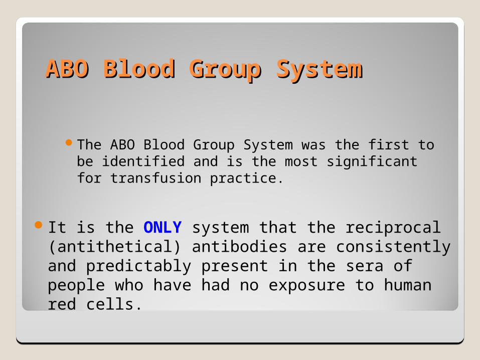
The ABO Blood Group System was the first to be identified and is the most significant for transfusion practice.
It is the ONLY system that the reciprocal (antithetical) antibodies are consistently and predictably present in the sera of people who have had no exposure to human red cells.
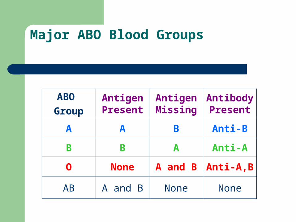
Major ABO Blood Groups
ABO
GroupAntigen Present
Antigen Missing
Antibody Present
A A B Anti-B
B B A Anti-A
O None A and B Anti-A,B
AB A and B None None
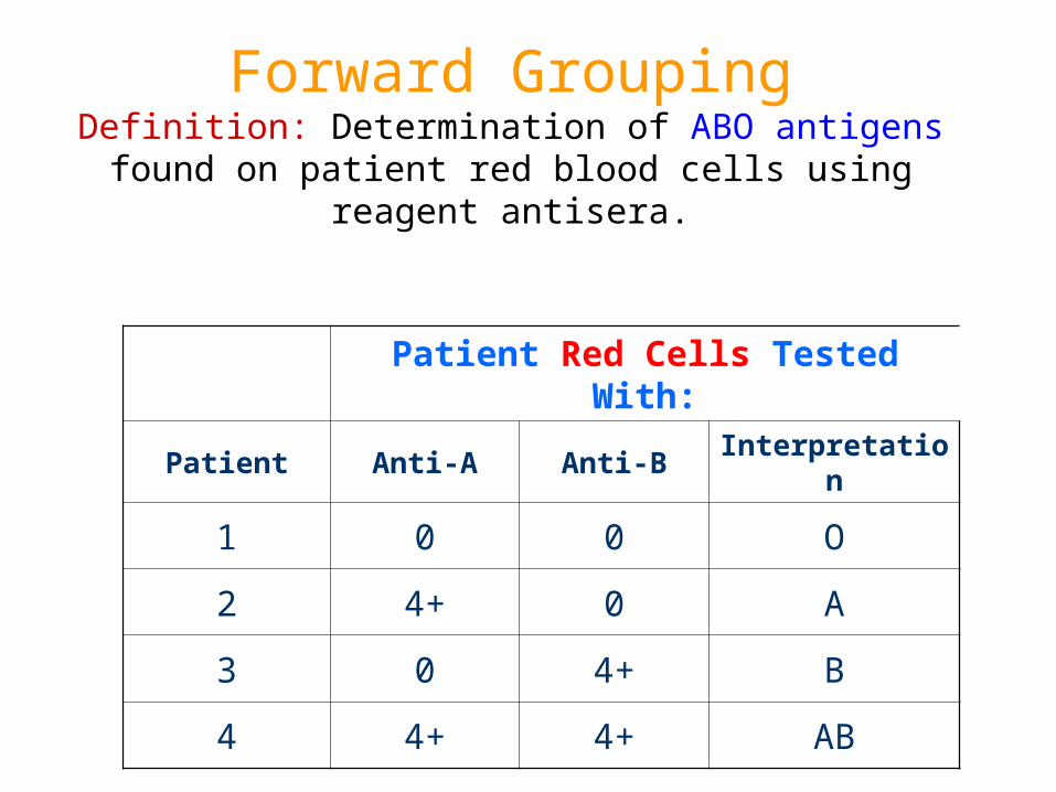
Forward GroupingDefinition: Determination of ABO antigens found on patient red
blood cells using reagent antisera.
Patient Red Cells Tested With:
Patient Anti-A Anti-B Interpretation
1 0 0 O
2 4+ 0 A
3 0 4+ B
4 4+ 4+ AB
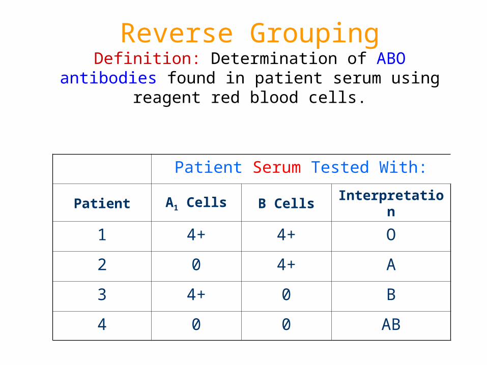
Reverse GroupingDefinition: Determination of ABO antibodies found in patient
serum using reagent red blood cells.
Patient Serum Tested With:
Patient A1 Cells B Cells Interpretation
1 4+ 4+ O
2 0 4+ A
3 4+ 0 B
4 0 0 AB
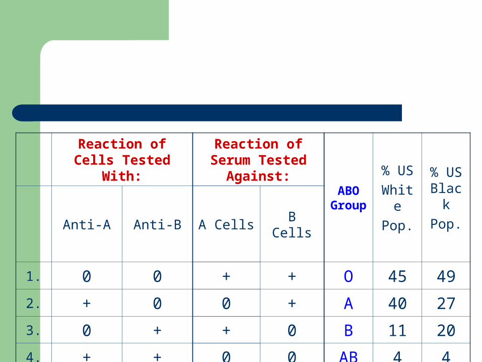
Reaction of Cells Tested With:
Reaction of Serum Tested Against:
ABO Group
% US
White
Pop.
% US Black
Pop.Anti-A Anti-B A Cells B Cells
1. 0 0 + + O 45 49
2. + 0 0 + A 40 27
3. 0 + + 0 B 11 20
4. + + 0 0 AB 4 4

H gene acts on a Precursor substance(PS)* by adding Fucose
H Antigen
*PS = oligosaccharide chain attached to either glycosphingo-lipid, Type 2 chain (on RBC) or glycoprotein, Type 1 chain (in secretions)
The H gene on ch. 19 near the Se gene, codes for an enzyme (fucosylytranferase) that adds a Fucose to the terminal sugar of a Precursor Substance (PS*). The
biochemical structure below constitutes the H Antigen. (h gene is an amorph.)
H antigen is the foundation upon which A and B antigens are built.
A and B genes code for enzymes that add an
immunodominant sugar to theH antigen.

Formation of the A Antigen
The A gene codes for an enzyme that
adds GalNAc (N-Acetyl-D
galactosamine) to the terminal
sugar of theH Antigen. This biochemical structure
constitutes the A antigen.
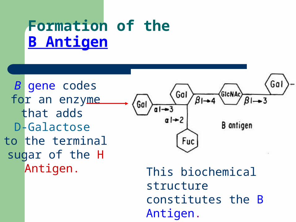
Formation of the B Antigen
B gene codes for an enzyme that adds
D-Galactose to the terminal sugar
of the H Antigen.
This biochemical structure constitutes the B Antigen.
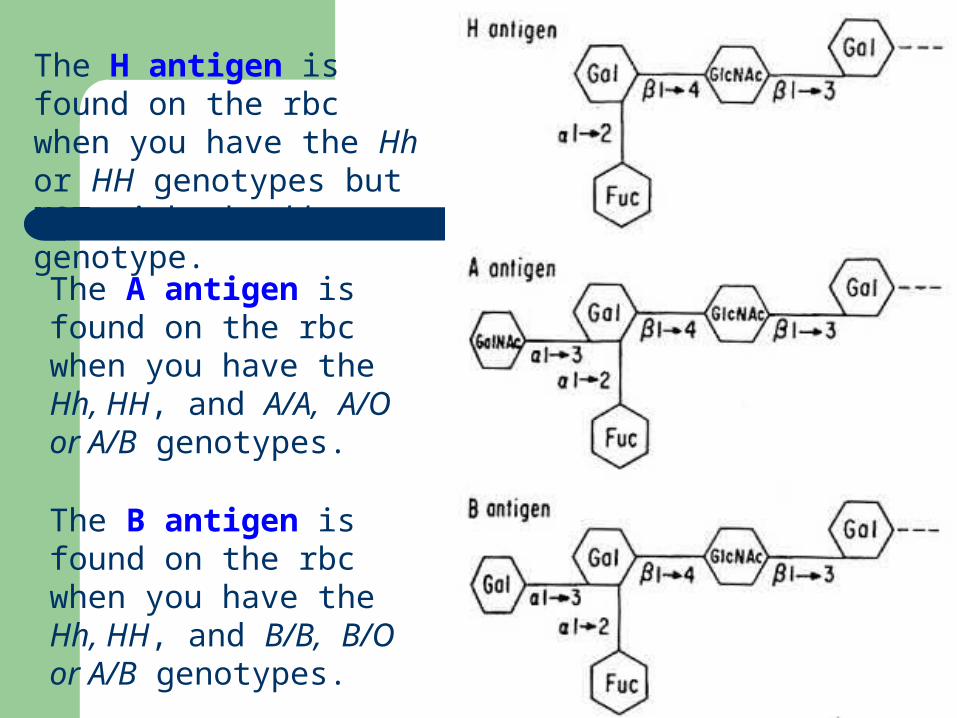
The H antigen is found on the rbc when you have the Hh or HH genotypes but NOT with the hh genotype.
The A antigen is found on the rbc when you have the Hh, HH, and A/A, A/O or A/B genotypes.
The B antigen is found on the rbc when you have the Hh, HH, and B/B, B/O or A/B genotypes.
ABO Genetics
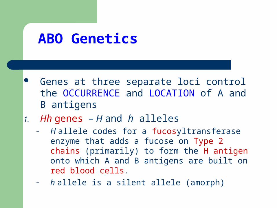
Genes at three separate loci control the OCCURRENCE and LOCATION of A and B antigens
1. Hh genes – H and h alleles– H allele codes for a fucosyltransferase enzyme
that adds a fucose on Type 2 chains (primarily) to form the H antigen onto which A and B antigens are built on red blood cells.
– h allele is a silent allele (amorph)
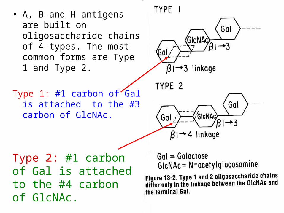
• A, B and H antigens are built on oligosaccharide chains of 4 types. The most common forms are Type 1 and Type 2.
Type 1: #1 carbon of Gal is attached to the #3 carbon of GlcNAc.
Type 2: #1 carbon of Gal is attached to the #4 carbon of GlcNAc.

ABO Genetics
2. Se genes – Se and se alleles– Se allele codes for a fucosyltransferase enzyme that
adds fuscos onto Type 1 chains (primarily) in secretory glands. Controls expression of H antigens in secretions (i.e. saliva, body fluids, etc.)
– se allele is an amorph
3. ABO genes – A, B and O alleles– A and B alleles code for glycosyltransferase enzymes
that add a sugar onto H antigens to produce A and B antigens
– O allele does not code a functional enzyme
H Ag concentration in ABO
17
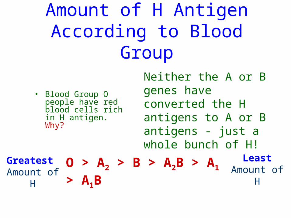
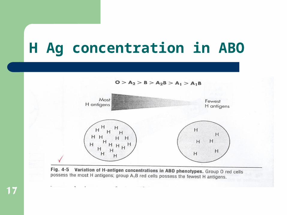
Amount of H Antigen According to Blood Group
• Blood Group O people have red blood cells rich in H antigen. Why?
Neither the A or B genes have converted the H antigens to A or B antigens - just a whole bunch of H!
Greatest Amount of H
LeastAmount of H
O > A2 > B > A2B > A1 > A1B
Bombay (Oh) Phenotype
Homozygous inheritance of the h gene (hh) results in the inability to form the H antigen and subsequently the A or B antigens.
This is referred as the Bombay or Oh phenotype due to the location of its discovery.
This phenotype has no H, A or B antigens on the red blood cell membrane, only an abundant amount of precursor substance.
They also have anti-H, anti-A and Anti-B. What blood type can we safely transfuse?
ABO Antigens in Secretions
Secretions: – Body fluids including plasma, saliva, synovial fluid, etc.
Blood Group Substance: Soluble antigen– Soluble antigen found in the secretions not bound to a
membrane such as a rbc or epithelial cell.
Soluble blood group substances (A, B and H) can be found in the secretions. This is controlled by the H and
Se genes.
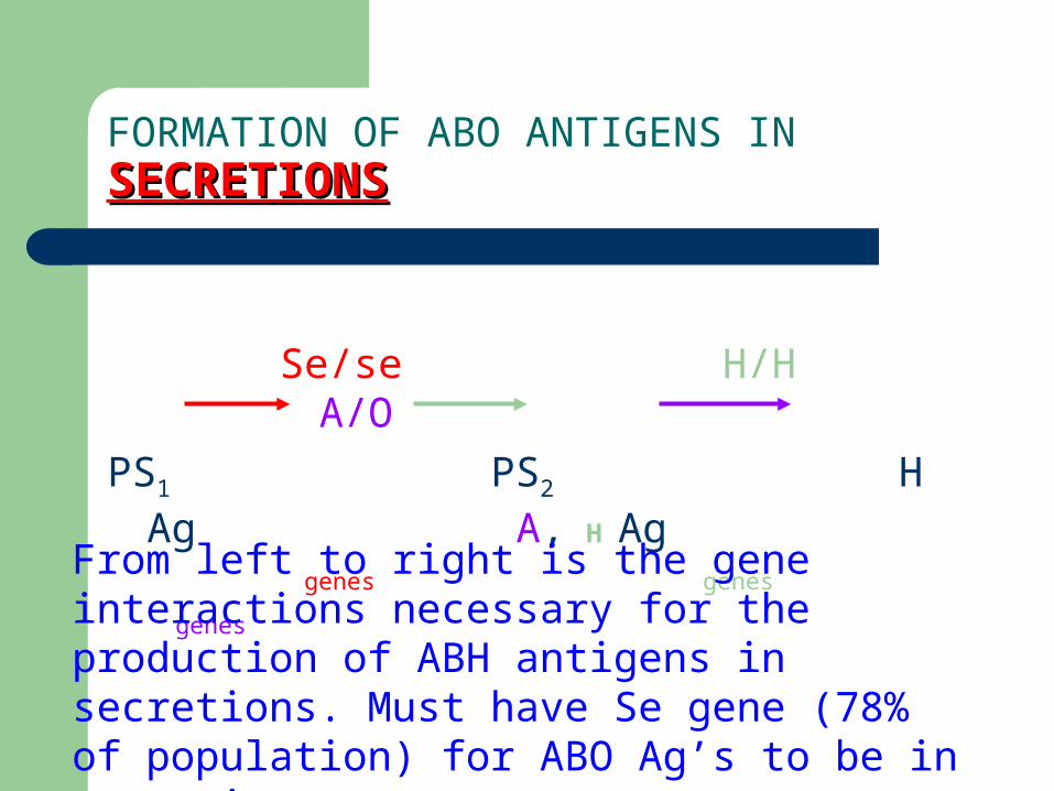
FORMATION OF ABO ANTIGENS IN SECRETIONSSECRETIONS
Se/se H/H A/O
PS1 PS2 H Ag A, H Ag
genes genes genes
From left to right is the gene interactions necessary for the production of ABH antigens in secretions. Must have Se gene (78% of population) for ABO Ag’s to be in secretions.
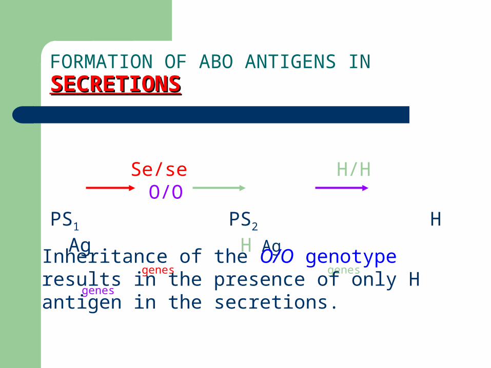
FORMATION OF ABO ANTIGENS IN SECRETIONSSECRETIONS
Se/se H/H O/O
PS1 PS2 H Ag H Ag
genes genes genes
Inheritance of the O/O genotype results in the presence of only H antigen in the secretions.
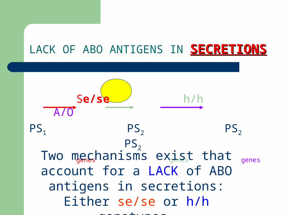
LACK OF ABO ANTIGENS IN SECRETIONSSECRETIONS
Se/se h/h A/O
PS1 PS2 PS2 PS2
genes genes genes
Two mechanisms exist that account for a LACK of ABO antigens in secretions:
Either se/se or h/h genotypes.
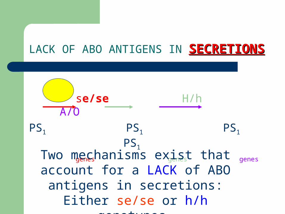
LACK OF ABO ANTIGENS IN SECRETIONSSECRETIONS
se/se H/h A/O
PS1 PS1 PS1 PS1
genes genes genes
Two mechanisms exist that account for a LACK of ABO antigens in secretions:
Either se/se or h/h genotypes.

ABO Antibodies
Generally IgM class antibodies. ABO Antibody Development: Hypothesis
– Immune response following exposure to environmental antigens (such as bacterial cell walls) similar to A and B antigens during infancy results in production of ABO antibodies. Remember, babies have a tendency to put EVERYTHING into their mouths…
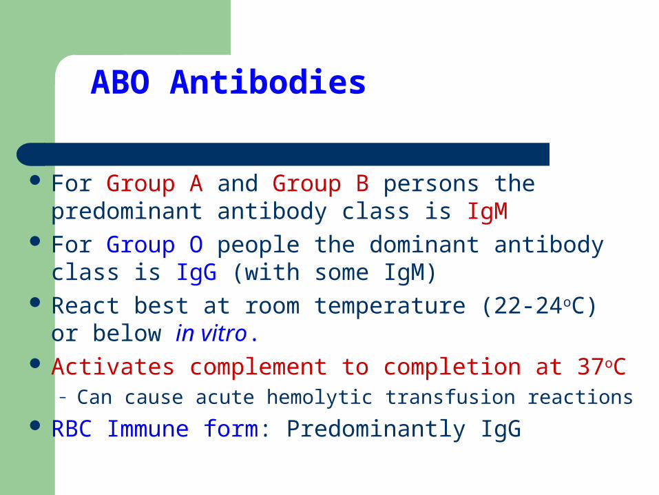
ABO Antibodies
For Group A and Group B persons the predominant antibody class is IgM
For Group O people the dominant antibody class is IgG (with some IgM)
React best at room temperature (22-24oC) or below in vitro.
Activates complement to completion at 37oC– Can cause acute hemolytic transfusion reactions
RBC Immune form: Predominantly IgG
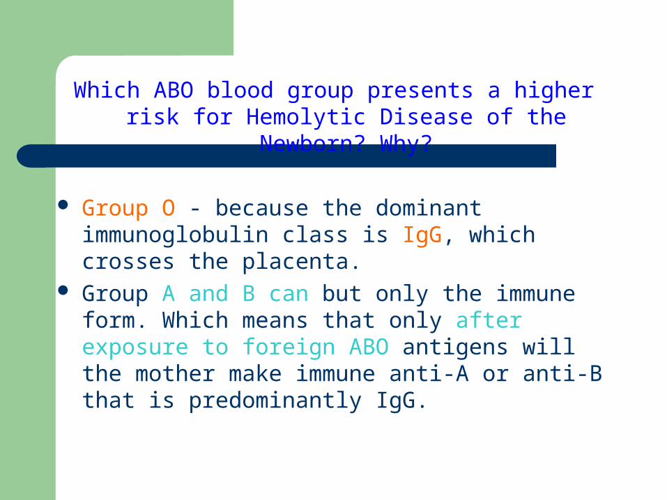
Which ABO blood group presents a higher risk for Hemolytic Disease of the Newborn? Why?
Group O - because the dominant immunoglobulin class is IgG, which crosses the placenta.
Group A and B can but only the immune form. Which means that only after exposure to foreign ABO antigens will the mother make immune anti-A or anti-B that is predominantly IgG.
ABO Antibodies
Time of appearance: Generally present within first 4-6 months of life
– Do we perform a reverse grouping on newborns (<4-6 months of age) and cord blood?
– If there are anti-A or anti-B antibodies in newborn serum where did they most likely originate? What source?
ABO antibody titers with age:– Reach adult level at 5-10 years of age– Level off through adult life– Begin to decrease in later years: >65 years of age
ABO AntibodiesGroup O Phenotype
Anti-A,B Antibody– Inseparable anti-A and anti-B antibody. If we add A
cells to anti-A,B serum all of the antibody activity is removed, not just anti-A!!
RBC immune Anti-A,B– When exposed to Group A or B antigens (or both)
Group O persons will have an immune response that results in the production of separate immune anti-A and/or anti-B antibodies. This could be seen in a fetomaternal bleed of a Group O mom with a Group A baby. (Hemolytic Disease of the Newborn)
ABO Antibodies Group B or O phenotype
Have both anti-A and Anti-A1 antibodies
Anti-A Reacts with both A1 and A2 red blood cell antigens
Anti-A1
Reacts only with A1 antigens on red blood cells A2 and A2B phenotypes can make anti-A1 antibodies.
What is clinical significance? Thermal range is up to 25oC - not usually clinically significant. Can cause an ABO discrepancy.
31

ABO Antibodies
Is there a reagent anti-A1 antisera? NO!!
But there is Dolichos biflorus, a plant lectin that has anti-A1 activity when diluted properly.
This is not an antibody, but a chemical that acts like an antibody in that it specifically agglutinates A1 red blood cells.
ABO Subgroups
ABO Phenotypes that differ in the amount of antigen carried on red cell and saliva, for secretors: There are fewer Ag sites!
Subgroups are the results of less effective glycosyltransferase enzymes – just not as good at attaching the immunodominant sugar to the H antigen.
Subgroups of A are more common than Subgroups of B.
ABO Subgroups
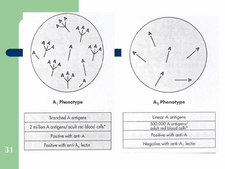
80% of all Group A’s are A1 and about 19% are A2.– A1’s have 4-6 times the # of antigen sites on the
RBC surface than A2’s.
– Both react strongly with reagent Anti-A but…– Only A1 cells are agglutinated with Dolichos biflorus
plant lectin and not A2 cells.
The remainder of the Subgroups of A have even weaker expression of A antigen.
Rh Blood Group System
Currently – 5 common antigen– three nomenclatures – two theories of inheritance
Rh antigen
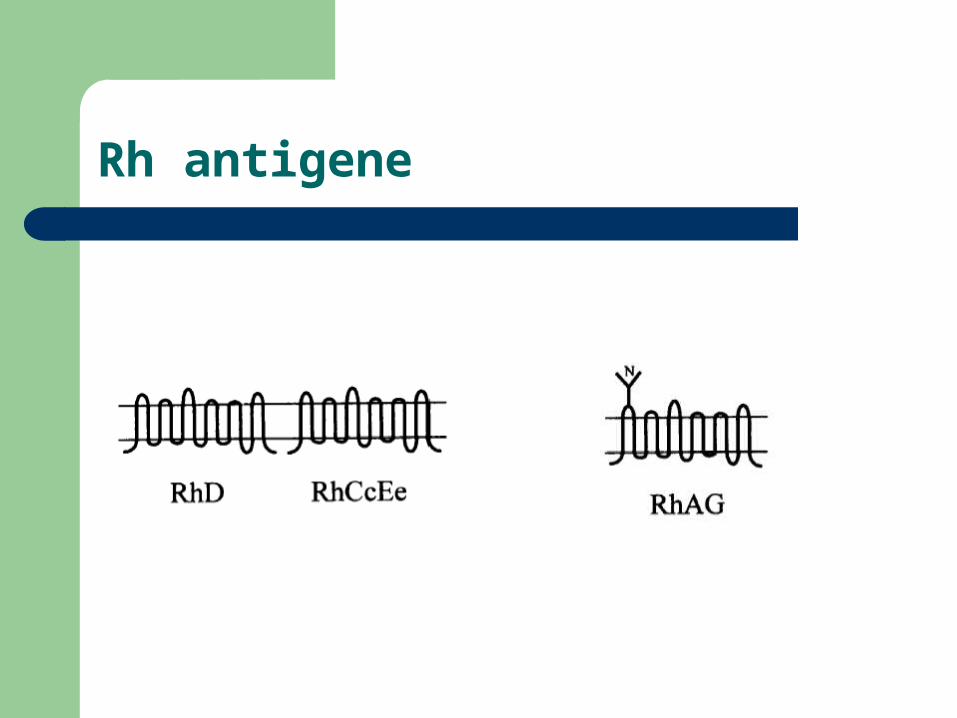
Rh antigene
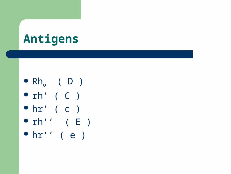
Antigens
Rho ( D ) rh’ ( C ) hr’ ( c ) rh’’ ( E ) hr’’ ( e )
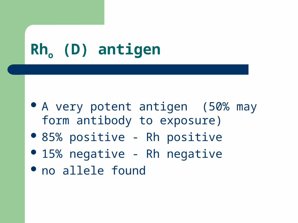
Rho (D) antigen
A very potent antigen (50% may form antibody to exposure)
85% positive - Rh positive 15% negative - Rh negative no allele found
Inheritance
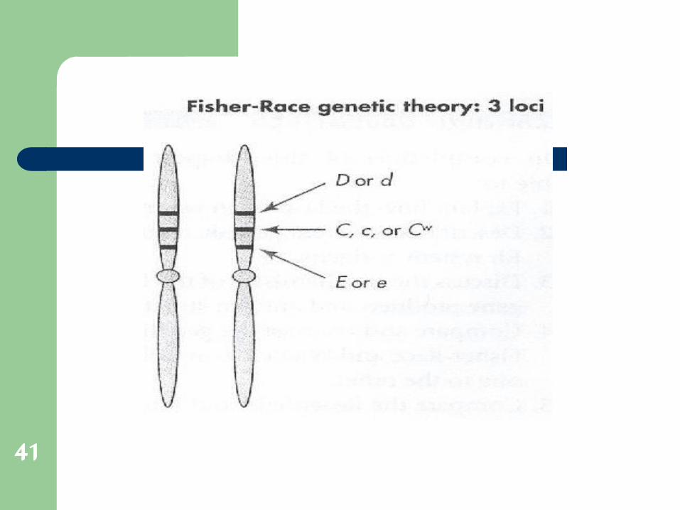
Fisher - Race– Rh antigens produced under the control of three
sets of allelic genes at closely linked locus– Nomenclature is C, D, E, c, e– Certain combinations of the antigens that are
inherited more often than others
41
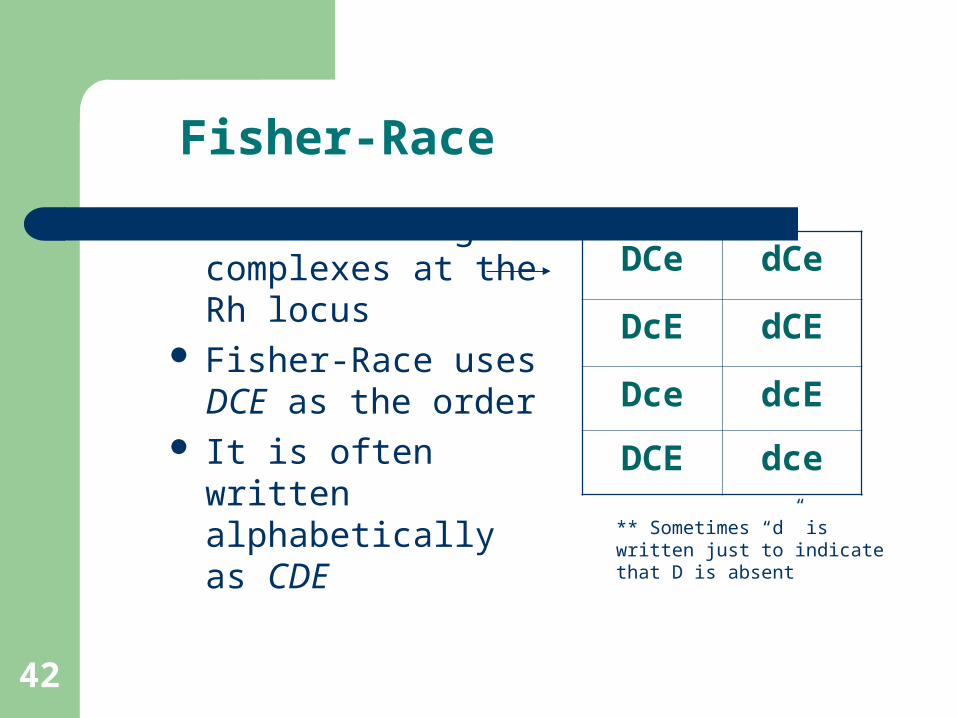
Fisher-Race
There are 8 gene complexes at the Rh locus
Fisher-Race uses DCE as the order
It is often written alphabetically as CDE
DCe dCe
DcE dCE
Dce dcE
DCE dce
** Sometimes “d” is written just to indicate that D is absent
42

Weak D Phenotype
Most D positive rbc’s react macroscopically with Reagent anti-D at immediate spin
– These patients are referred to as Rh positive– Reacting from 1+ to 3+ or greater
HOWEVER, some D-positive rbc’s DO NOT react (do NOT agglutinate) at Immediate Spin using Reagent Anti-D. These require further testing (37oC and/or AHG) to determine the D status of the patient.
Weakened Antigens
The Rh-Hr system has a number of antigens that are suppressed by other antigens or only a weakened form of the antigens are present
Weakened D antigen – often does not react with initial spin– may require 37o incubation or antiglobulin test to
detect sensitization– two forms - inherited or suppression by C antigen
in the trans position
Weak D Mechanism’s
There are three mechanisms that account for the Weak D antigen.
1. Genetically Transmissible
2. Position Effect
3. Partial D (D Mosaic)
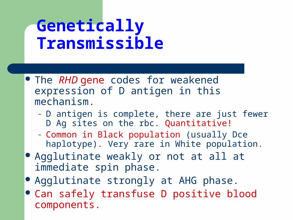
Genetically Transmissible
The RHD gene codes for weakened expression of D antigen in this mechanism.– D antigen is complete, there are just fewer D Ag
sites on the rbc. Quantitative!– Common in Black population (usually Dce
haplotype). Very rare in White population. Agglutinate weakly or not at all at immediate
spin phase. Agglutinate strongly at AHG phase. Can safely transfuse D positive blood
components.

Position Effect (Gene interaction effect)
C allele in trans position to D allele– Example: Dce/dCe, DcE/dCE
In both of these cases the C allele is in the trans position in relation to the D allele.
D antigen is normal, C antigen appears to be crowding the D antigen. (Steric hindrance)
Does NOT happen when C is in cis position– Example: DCe/dce
Can safely transfuse D positive blood components.
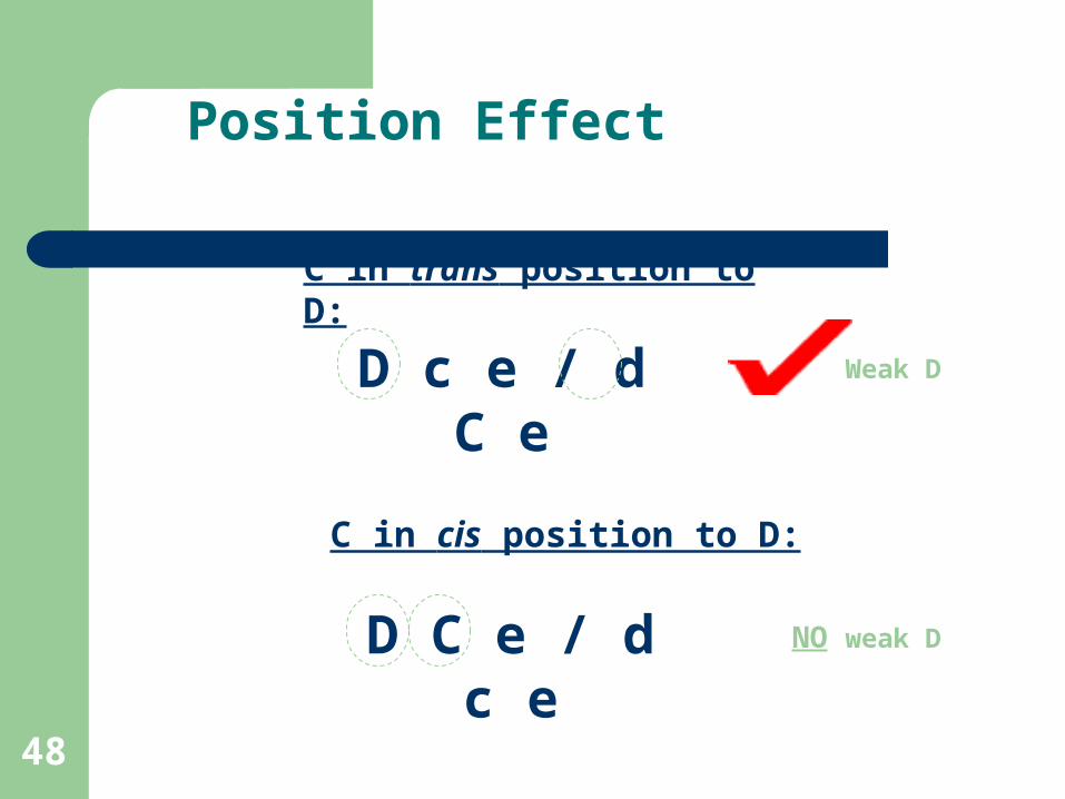
Position Effect
C in trans position to D:
D c e / d C e
C in cis position to D:
D C e / d c e
Weak D
NO weak D
48
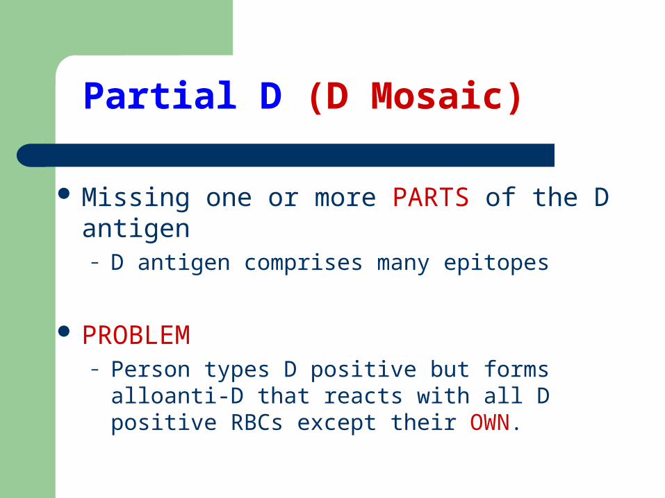
Partial D (D Mosaic)
Missing one or more PARTS of the D antigen– D antigen comprises many epitopes
PROBLEM– Person types D positive but forms alloanti-D that
reacts with all D positive RBCs except their OWN.
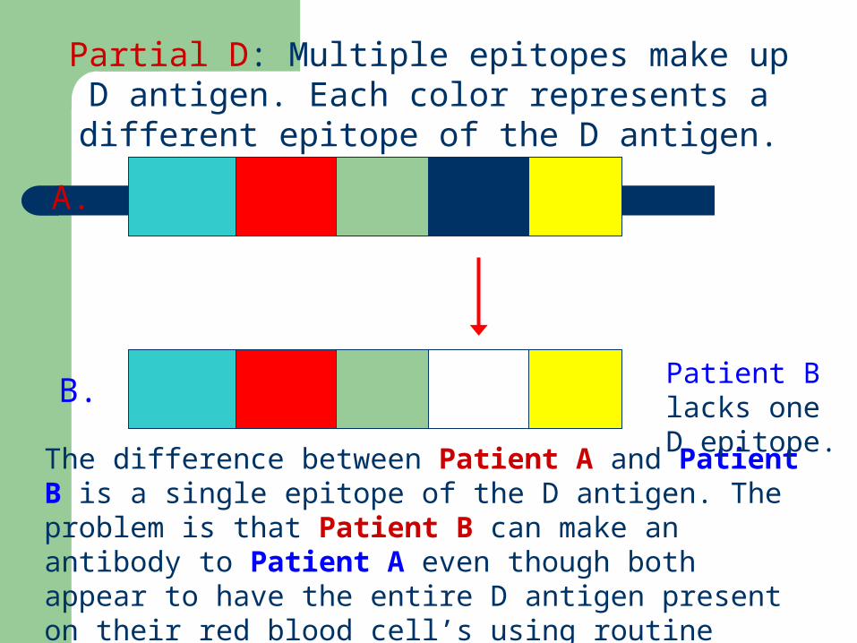
Partial D: Multiple epitopes make up D antigen. Each color represents a different epitope of the D antigen.
The difference between Patient A and Patient B is a single epitope of the D antigen. The problem is that Patient B can make an antibody to Patient A even though both appear to have the entire D antigen present on their red blood cell’s using routine anti-D typing reagents..
A.
B.Patient B lacks one D epitope.
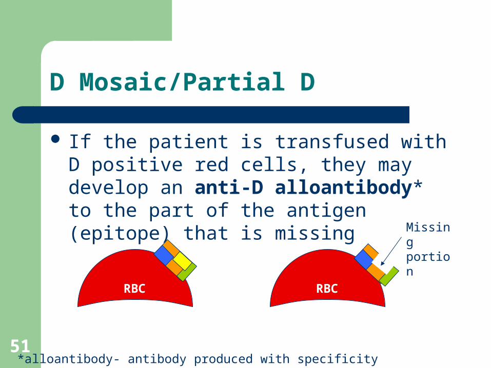
D Mosaic/Partial D
If the patient is transfused with D positive red cells, they may develop an anti-D alloantibody* to the part of the antigen (epitope) that is missing
*alloantibody- antibody produced with specificity other than self
Missing portion
RBC RBC
51
Weak-D Determination: Donor Blood
When testing Donor Blood for the D antigen, testing is required through all phases.– Weak-D testing is REQUIRED
We need to know the D Status of all Donor Blood. Why? – Main problem is Rh Negative women of child bearing
age and pediatric patients. Donor RBCs are labelled Rh positive if any
part of the D antigen is present on the red blood cell membrane.
Unusual Phenotypes
D-Deletion
Rh null
53
D-Deletion No reaction when RBCs are tested with anti-E,
anti-e, anti-C or anti-c
Requires transfusion of other D-deletion red cells, because these individuals may produce antibodies with single or separate specificities( anti-Hr0 or anti-Rh17)
Red cells that lack C/c or E/e antigens may demonstrate stronger D antigen activity
Written as D- - or -D-
54

Rh Null
Lack all Rh antigens
The lack of antigens causes the red cell membrane abnormalities
Immunized idividuals have anti-Rh29( “Total Rh” or Rh29)
2 Rh null phenotypes:– Regulatory type– gene inherited(Xºr) (X¹r is a normal regulator gene) the Rh gene are inherited
but not expressed.
– Amorph type –Result from the r amorph gene RHD gene is absent, lack of expression of the RHCE gene
55
Rh Antibodies
Most antibodies react at 37o and require a coombs procedure to demonstrate the reaction.
Some react at saline and room temperature Most are IgG None fix Complement All are important in HDN and HTR

Rh System Antibodies
1. React optimally
2. RBC Immune
3. Clinically Significant
1. 37oC and AHG Phases
2. Transfusion or pregnancy, IgG, HDN, HTR, etc.
3. Will result in shortened red cell survival - need to transfuse antigen negative blood
Rh typing
Normal typing for Rh antigens only includes typing for Rho (D).
The result of this typing determines the Rh status of the cells (Rh - positive or Rh - negative). Other antigens are identified for genotyping
Some Rh typing sera is diluted in high protein solutions and may require a negative control.
HEMOLYTIC DISEASE OF THE NEWBORN
HDN: THE DISEASE
Caused by blood group system or HLA maternal/fetal incompatibility (mother has IgG1 or IgG3 Abs to Ag on baby RBCs)
With Rh HDN, previous matherno-fetal bleeds usually the stimulus for Ab production; with other HDNs, stimulus may be unclear
Begins in utero Range of severity from asymptomatic --> mild
anemia --> kernicterus --> stillborn ABO, Rh, and Kell groups most commonly involved
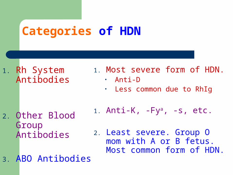
Categories of HDN
1. Rh System Antibodies
2. Other Blood Group Antibodies
3. ABO Antibodies
1. Most severe form of HDN. • Anti-D • Less common due to RhIg
1. Anti-K, -Fya, -s, etc.
2. Least severe. Group O mom with A or B fetus. Most common form of HDN.
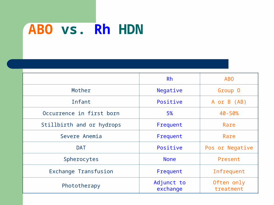
ABO vs. Rh HDN
Rh ABO
Mother Negative Group O
Infant Positive A or B (AB)
Occurrence in first born 5% 40-50%
Stillbirth and or hydrops Frequent Rare
Severe Anemia Frequent Rare
DAT Positive Pos or Negative
Spherocytes None Present
Exchange Transfusion Frequent Infrequent
Phototherapy Adjunct to exchange Often only treatment

Risk is also increased in pregnancies complicated by : placental abruption spontaneous or therapeutic abortion Toxemia after cesarean delivery ectopic pregnancy Amniocentesis chorionic villus sampling cordocentesis
After sensitization, maternal anti-D antibodies cross the placenta into fetal circulation and attach to Rh antigen on fetal RBCs, which form rosettes on macrophages in the reticuloendothelial system, especially in the spleen.
These antibody-coated RBCs are lysed by lysosomal enzymes released by macrophages and natural killer lymphocytes, and they are independent of the activation of the complement system
Rh HDN
Most severe form of HDN (Usually*) affects only 2nd or subsequent
pregnancies (mother allo-immunized at delivery of 1st pregnancy; has pre-formed Abs during subsequent pregnancies)
Usually Ab directed to D Ag but Abs to C, c, and E also seen
Antibodies cause destruction of the red cells Anemia heart failure fetal death
BEFORE BIRTH
Metabolism of bilirubin:
Before delivery: Fetal bilirubin produced by the breakdown of
sensitized RBCs in fetal spleen is sefely metabolized by the maternal liver.
After delivery: Newborn’s liver doesn’t produce glucuronyl
transferase and cannot convert bilirubin to an excretable form.as a result,it collects in tissues and causes brain damage.
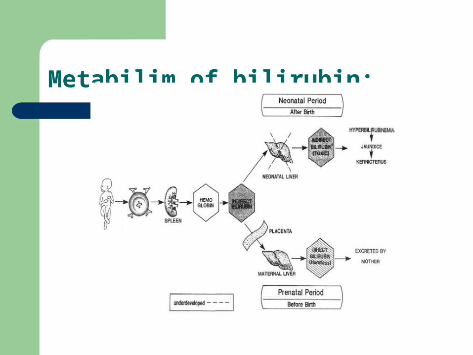
Metabilim of bilirubin:
HDN: SIGNS & SYMPTOMS
Anemia (Hb < 16 mg/dL) - begins in utero
Increased bilirubin - begins after birth– Baby’s liver does not conjugate bilirubin efficiently;
unconjugated (toxic) bilirubin increases as RBCs continue to be destroyed
– If > 18 mg/dL, may have to exchange transfuse
Jaundice Hepato- and splenomegaly
Antibodies cause destruction of the red cells Anemia Heart failure Build up of bilirubin Kernicterus Severe retardation
AFTER BIRTH
Anemia – heart failure– erythroblastosis
General edema -Called hydrops fetalis and erythroblastosis fetalis Kernicterus ( a condition with severe neural symptoms,
associated with high level of bilirubin in the blood) – severe retardation
PROBLEMS FOR BABY
Bilirubin has been postulated to cause neurotoxicity via 4 distinct mechanisms:
(1) interruption of normal neurotransmission (inhibits phosphorylation of enzymes critical in release of neurotransmitters)
(2) mitochondrial dysfunction
(3) cellular and intracellular membrane impairment (bilirubin acid affects membrane ion channels and precipitates on phospholipid membranes of mitochondria
(4) interference with enzyme activity (binds to specific bilirubin receptor sites on enzymes).
PREVENTION
Before birth– Work up mother for risk and evaluation of complications
After birth– Rh immune globulin - IgG anti-D given to prevent primary
immunization
Before birth workup
– Identify women at risk– ABO - Rh -(Du) - Antibody screen
based on results continue testing (Handout)
– IgM antibodies are insignificant – IgG antibodies - titer - freeze and store -
retiter with a second sample - looking for a 1:32 rise or change in titer
Before birth workup
– titer identifies mothers who need amniocentesis
– titer every 4 week until 24th week - then every 2 weeks
– amniocentesis is performed after 21st week on high titer - high mortality
PRENATAL TESTING
Test father to see if he is Ag positive (if he is negative, not a concern)
If Ab is significant and father is positive for Ag, perform Ab titers (> 32 significant) on mother’s serum
If serum Ab titer high, test amniocytes for presence of Ag
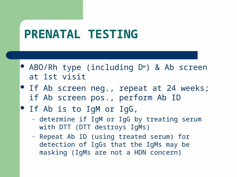
PRENATAL TESTING
ABO/Rh type (including Dw) & Ab screen at 1st visit If Ab screen neg., repeat at 24 weeks; if Ab screen
pos., perform Ab ID If Ab is to IgM or IgG,
– determine if IgM or IgG by treating serum with DTT (DTT destroys IgMs)
– Repeat Ab ID (using treated serum) for detection of IgGs that the IgMs may be masking (IgMs are not a HDN concern)
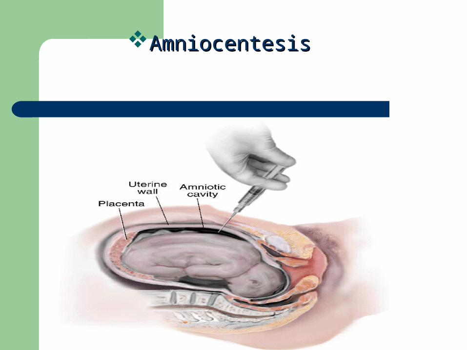
AmniocentesisAmniocentesis
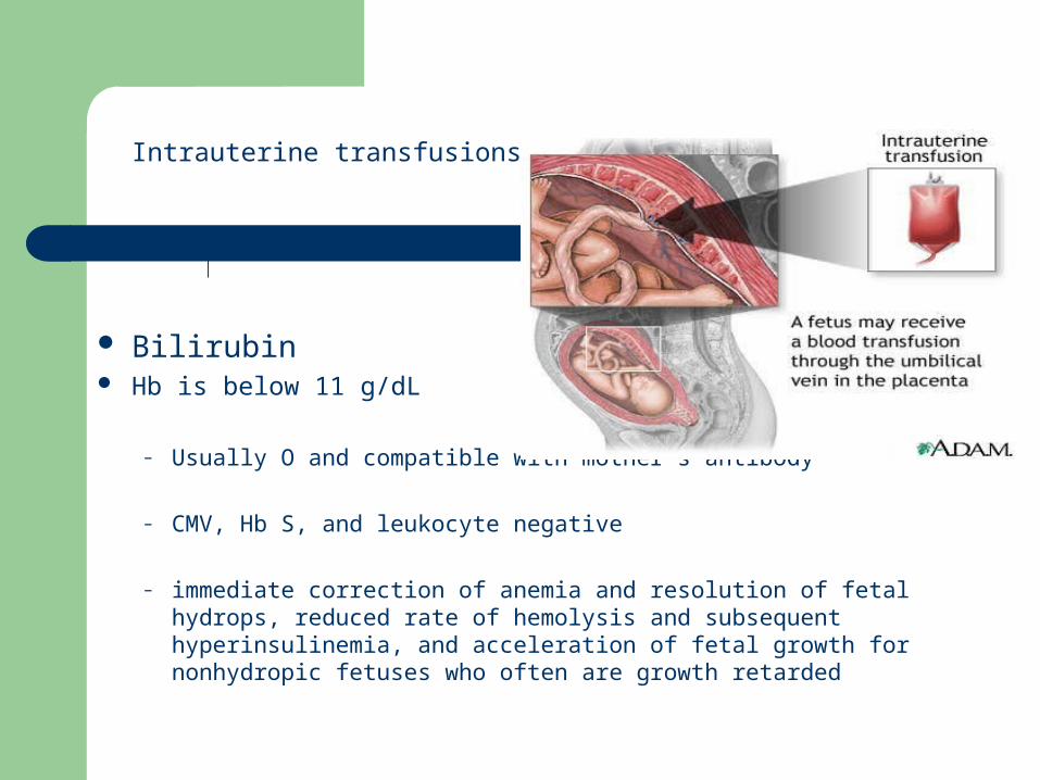
Bilirubin Hb is below 11 g/dL
– Usually O and compatible with mother’s antibody
– CMV, Hb S, and leukocyte negative
– immediate correction of anemia and resolution of fetal hydrops, reduced rate of hemolysis and subsequent hyperinsulinemia, and acceleration of fetal growth for nonhydropic fetuses who often are growth retarded
Intrauterine transfusions
Post Natal Laboratory Studies
Mother– ABO - Rh - Du (micro) - Antibody screen -
Antibody identification if necessary Baby
– ABO - Rh - Du - DAT for IgG antibodies - elute DAT positive and identify antibody
– CBC– Imaging studies

TREATMENT
Exchange transfusion
Phototherapy
After birth
Rh Immune Globulin
– Give antenatal 28 -32 weeks– also after amniocentesis - IUT - abortion - ectopic
pregnancy - miscarriage– Each vial contains 300 ugm and will prevent
sensitization by 15 ml RBC or 30 ml whole blood
Rh (D) HDN: PREVENTION
Rh Immune Globulin is a potent solution of Anti-D Anti-D covers D epitopes in baby RBCs in maternal
circulation Coated cells removed by splenic macrophages D-bearing RBCs destroyed before mother can mount
immune response Very effective preventative treatment
Rh (D) HDN: PREVENTION
Rh Immune Globulin given to mother at 28 weeks gestation and within 72 hours of delivery of infant if:– Baby is Rh positive– Rh type of fetus is unknown, and – Mother is known to be negative for Anti-D
Dosage calculated using results of the Kleihauer-Betke test
1 Rh Immune Globulin dose protects against 30 mL fetal whole blood
Kleihauer-Betke test– sample from mother treated with acid then stained;
fetal cells resistant to acid, maternal cells become ghost cells
– determine # fetal cells in first 2000 maternal cells counted
– % fetal x 50 = Whole blood bleed
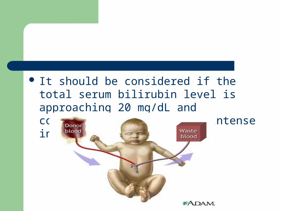
It should be considered if the total serum bilirubin level is approaching 20 mg/dL and continues to rise despite intense in-hospital phototherapy.
Selection of blood for exchange transfusion:
Group o(or ABO-compatible) Fresh(less than 7 days old)RBCs resuspended
in fresh frozen plasma CMV negative Irradiate blood HbS negative Blood lack of Ag corresponding to maternal Ab Compatible crossmatch with maternal serum
Exchange Transfusions Objectives
– decrease serum bilirubin and prevent kernicterus
– provide compatible red cells to provide oxygen carrying capacity
– decrease amount of incompatible antibody
– remove fetal antibody coated red cells
The following are requirements for exchange transfusion :
Severe anemia (Hb <10 g/dL)
Rate of bilirubin rises more than 0.5 mg/dL despite optimal phototherapy
Hyperbilirubinemia
DAT
Potential complications of exchange transfusion include the following:
– Cardiac - Arrhythmia, volume overload, congestive failure, and arrest
– Hematologic - Overheparinization, neutropenia, thrombocytopenia, and graft versus host disease
– Infectious - Bacterial, viral (CMV, HIV, hepatitis), and malarial
– Metabolic - Acidosis, hypocalcemia, hypoglycemia, hyperkalemia, and hypernatremia
– Vascular - Embolization, thrombosis, necrotizing enterocolitis, and perforation of umbilical vessel
– Systemic - Hypothermia
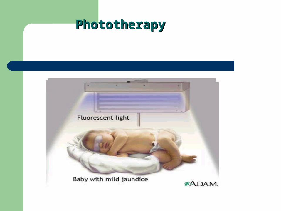
PhototherapyPhototherapy
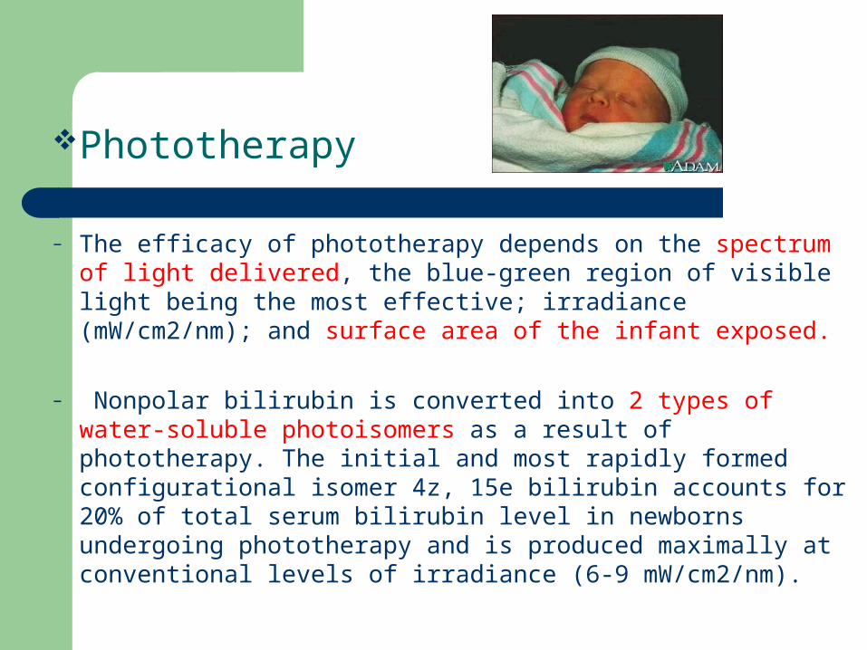
Phototherapy
– The efficacy of phototherapy depends on the spectrum of light delivered, the blue-green region of visible light being the most effective; irradiance (mW/cm2/nm); and surface area of the infant exposed.
– Nonpolar bilirubin is converted into 2 types of water-soluble photoisomers as a result of phototherapy. The initial and most rapidly formed configurational isomer 4z, 15e bilirubin accounts for 20% of total serum bilirubin level in newborns undergoing phototherapy and is produced maximally at conventional levels of irradiance (6-9 mW/cm2/nm).
Phototherapy
The structural isomer lumirubin is formed slowly, and its formation is irreversible and is directly proportional to the irradiance of phototherapy on skin.
Lumirubin is the predominant isomer formed during high-intensity phototherapy. Decrease in bilirubin is mainly the result of excretion of these photoproducts in bile and removal via stool.
In the absence of conjugation, these photoisomers can be reabsorbed by way of the enterohepatic circulation and diminish the effectiveness of phototherapy
ABO HDN
Most common form of HDN Mother is “O” with IgG form of Anti-A,B Baby is “A” or “B” May occur with 1st or subsequent pregnancies Usually less severe than Rh HDN (babies’ A and B
Ags not fully developed) Most cases treated only with phototherapy
ABO incompatibility
ABO incompatibility is limited to type O mothers with fetuses who have type A or B blood
in type O mothers, the antibodies are predominantly IgG in nature
Because A and B antigens are widely expressed in a variety of tissues besides RBCs, only small portion of antibodies crossing the placenta is available to bind to fetal RBCs. In addition, fetal RBCs appear to have less surface expression of A or B antigen, resulting in few reactive sites—hence the low incidence of significant hemolysis in affected neonates.
ABO HDN: PREVENTION AND TREATMENT
Not preventable as with Rh (D) HDN Usually treatable using only phototherapy If exchange transfusion required, use type
“O” cells and “AB” plasma ABO HDN sometimes protects babies from
the more severe forms of Rh HDN