Embed Size (px)
Citation preview
Revised 2020
ACR Appropriateness Criteria® 1 Abnormal Uterine Bleeding
American College of Radiology ACR Appropriateness Criteria®
Abnormal Uterine Bleeding
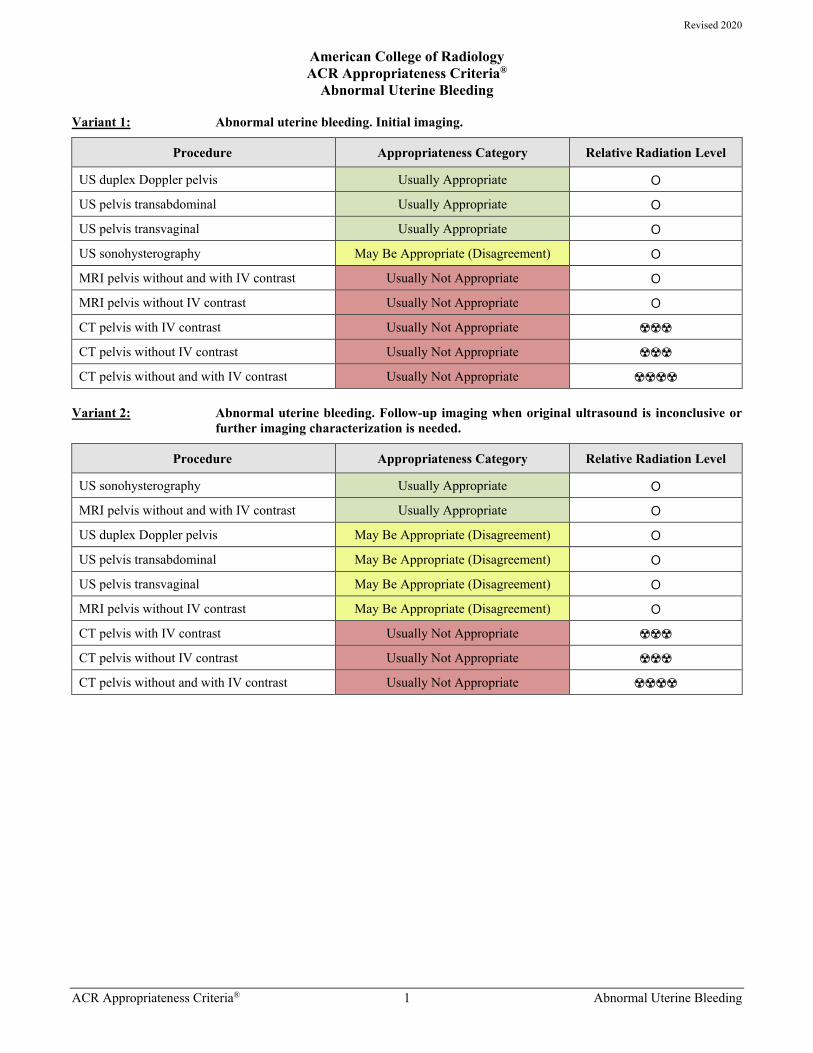
Variant 1: Abnormal uterine bleeding. Initial imaging.
Procedure Appropriateness Category Relative Radiation Level
US duplex Doppler pelvis Usually Appropriate O
US pelvis transabdominal Usually Appropriate O
US pelvis transvaginal Usually Appropriate O
US sonohysterography May Be Appropriate (Disagreement) O
MRI pelvis without and with IV contrast Usually Not Appropriate O
MRI pelvis without IV contrast Usually Not Appropriate O
CT pelvis with IV contrast Usually Not Appropriate ☢☢☢
CT pelvis without IV contrast Usually Not Appropriate ☢☢☢
CT pelvis without and with IV contrast Usually Not Appropriate ☢☢☢☢
Variant 2: Abnormal uterine bleeding. Follow-up imaging when original ultrasound is inconclusive or further imaging characterization is needed.
Procedure Appropriateness Category Relative Radiation Level
US sonohysterography Usually Appropriate O
MRI pelvis without and with IV contrast Usually Appropriate O
US duplex Doppler pelvis May Be Appropriate (Disagreement) O
US pelvis transabdominal May Be Appropriate (Disagreement) O
US pelvis transvaginal May Be Appropriate (Disagreement) O
MRI pelvis without IV contrast May Be Appropriate (Disagreement) O
CT pelvis with IV contrast Usually Not Appropriate ☢☢☢
CT pelvis without IV contrast Usually Not Appropriate ☢☢☢
CT pelvis without and with IV contrast Usually Not Appropriate ☢☢☢☢
ACR Appropriateness Criteria® 2 Abnormal Uterine Bleeding
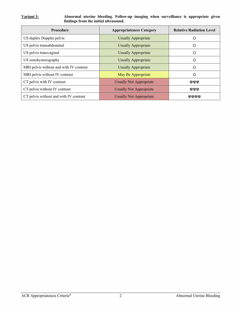
Variant 3: Abnormal uterine bleeding. Follow-up imaging when surveillance is appropriate given findings from the initial ultrasound.
Procedure Appropriateness Category Relative Radiation Level
US duplex Doppler pelvis Usually Appropriate O
US pelvis transabdominal Usually Appropriate O
US pelvis transvaginal Usually Appropriate O
US sonohysterography Usually Appropriate O
MRI pelvis without and with IV contrast Usually Appropriate O
MRI pelvis without IV contrast May Be Appropriate O
CT pelvis with IV contrast Usually Not Appropriate ☢☢☢
CT pelvis without IV contrast Usually Not Appropriate ☢☢☢
CT pelvis without and with IV contrast Usually Not Appropriate ☢☢☢☢
ACR Appropriateness Criteria® 3 Abnormal Uterine Bleeding
ABNORMAL UTERINE BLEEDING
Expert Panel on GYN and OB Imaging: Jessica B. Robbins, MDa; Elizabeth A. Sadowski, MDb; Katherine E. Maturen, MD, MSc; Esma A. Akin, MDd; Susan M. Ascher, MDe; Olga R. Brook, MDf; Courtney R. Cassella, MDg; Mark Dassel, MDh; Tara L. Henrichsen, MDi; Lee A. Learman, MD, PhDj; Michael N. Patlas, MDk; Carl Saphier, MDl; Ashish P. Wasnik, MDm; Phyllis Glanc, MD.n
Summary of Literature Review
Introduction/Background At least one-third of women can be expected to experience abnormal uterine bleeding (AUB) at some time in their lives [1]. AUB is defined as bleeding from the uterus that is abnormal in regularity, volume, frequency, or duration and occurs in the absence of pregnancy [2]. Causes of AUB can be partitioned into structural and nonstructural etiologies. Structural causes of AUB include endometrial polyps, adenomyosis, leiomyoma, malignancy (endometrial or myometrial), and endometrial hyperplasia. Nonstructural causes include coagulopathies, ovulatory dysfunction, primary endometrial disorders (such as molecular deficiencies in the regulation of endometrial hemostasis), iatrogenic etiologies (including exogenous gonadal steroids and intrauterine devices), and other causes not otherwise classified [1,2]. Because the nonstructural causes of AUB cannot be assessed with imaging, these will not be further discussed in this document. On the other hand, the structural causes can be diagnosed with imaging. In premenopausal women, polyps, adenomyosis, and leiomyoma are the common structural sources of AUB [3]. Although postmenopausal bleeding may be due to structural causes such as polyps or endometrial hyperplasia, endometrial cancer is the most serious etiology and is thus the main focus of the evaluation of the workup of AUB in the postmenopausal population [3-5]. The workup of a woman with abnormal bleeding begins with a thorough history, physical examination, and appropriate laboratory tests and may include imaging to primarily assess for structural abnormalities and help triage and manage both reproductive age and postmenopausal women with AUB [3,5-7]. Endometrial sampling may be considered in patients with AUB who have an increased risk for endometrial cancer due to obesity, chronic anovulation, family history, or age irrespective of imaging findings. AUB during pregnancy is beyond the scope of this document and is addressed in the ACR Appropriateness Criteria® topic on “First Trimester Vaginal Bleeding” [8] and the ACR Appropriateness Criteria® topic on “Second and Third Trimester Bleeding” [9].
Special Imaging Considerations There are limitations to transvaginal ultrasound (TVUS), and it is not possible to assess the uterus in all women with AUB [10,11]. The position of the uterus, patient body habitus, and presence of uterine pathology, such as adenomyosis and leiomyomas, can all cause incomplete visualization of portions of the uterus and endometrium. In women with AUB, the main differential includes both endometrial causes (eg, endometrial cancer, hyperplasia, polyp, or endometrial atrophy) and myometrial causes (eg, leiomyomas, leiomyosarcomas, or adenomyosis). If the endometrium cannot be completely evaluated by ultrasound (US), endometrial sampling should be considered based upon each patient’s risk factors for endometrial cancer [10]. MRI can also be considered for the assessment of the endometrium. Even in the presence of leiomyomas and adenomyosis, MRI can visualize the endometrium because of its multiplanar capability and excellent tissue contrast resolution [12,13] when US cannot.
Although pelvic MRI protocols may vary somewhat between institutions, we specifically note that the addition of a diffusion-weighted imaging sequence is important in the MRI examination of the uterus in women with AUB. Diffusion-weighted imaging has been shown to improve the sensitivity and specificity of MRI for the accurate diagnosis of uterine pathology [14-16]. This has been most extensively studied in endometrial cancer; however, there is ongoing research in using diffusion-weighted imaging for differentiating leiomyosarcomas from benign leiomyomas [16-23]. MR angiography may be incorporated into the MRI protocol in the preprocedural workup of aPanel Vice-Chair, University of Wisconsin, Madison, Wisconsin. bUniversity of Wisconsin, Madison, Wisconsin. cPanel Chair, University of Michigan, Ann Arbor, Michigan. dGeorge Washington University Hospital, Washington, District of Columbia. eGeorgetown University Hospital, Washington, District of Columbia. fBeth Israel Deaconess Medical Center, Boston, Massachusetts. gReading Hospital, Reading, Pennsylvania; American College of Emergency Physicians. hCleveland Clinic, Cleveland, Ohio; American College of Obstetricians and Gynecologists. iMayo Clinic, Rochester, Minnesota. jVirginia Tech Carilion School of Medicine, Roanoke, Virginia; American College of Obstetricians and Gynecologists. kMcMaster University, Hamilton, Ontario, Canada. lWomen’s Ultrasound, LLC, Englewood, New Jersey; American College of Obstetricians and Gynecologists. mUniversity of Michigan, Ann Arbor, Michigan. nSpecialty Chair, University of Toronto and Sunnybrook Health Sciences Centre, Toronto, Ontario, Canada. The American College of Radiology seeks and encourages collaboration with other organizations on the development of the ACR Appropriateness Criteria through society representation on expert panels. Participation by representatives from collaborating societies on the expert panel does not necessarily imply individual or society endorsement of the final document. Reprint requests to: [email protected]
ACR Appropriateness Criteria® 4 Abnormal Uterine Bleeding
leiomyomas prior to uterine artery embolization because the vascular anatomy serves as a road map for the interventional radiologist [24,25].
In the initial assessment of leiomyomas and other uterine pathologies, 3-D US may also be considered, because this technique has the potential to help with spatial assessment and treatment planning [26]. Research is ongoing to determine the clinical accuracy of 3-D US compared with 2-D US alone [27-29]. Three-dimensional US may increase the confidence of the diagnosis and be helpful when communicating with the referring physician on the location and size of uterine pathology [30,31].
US elastography measures tissue stiffness. Small studies have shown that, in combination with routine 2-D US, strain, elastography may increase the diagnostic accuracy of US in differentiating endometrial polyps from submucosal leiomyomas [32] and differentiating leiomyomas from adenomyosis [33].
Initial Imaging Definition Imaging at the beginning of the care episode for the medical condition defined by the variant. More than one procedure can be considered usually appropriate in the initial imaging evaluation when
• There are procedures that are equivalent alternatives (ie, only one procedure will be ordered to provide the clinical information to effectively manage the patient’s care)
OR
• There are complementary procedures (ie, more than one procedure is ordered as a set or simultaneously in which each procedure provides unique clinical information to effectively manage the patient’s care).
Discussion of Procedures by Variant Variant 1: Abnormal uterine bleeding. Initial imaging. CT Pelvis To our knowledge, there is no relevant literature to support the use of CT pelvis in the initial imaging evaluation of AUB.
MRI Pelvis To our knowledge, there is no relevant literature to support the use of MRI pelvis in the initial imaging evaluation of AUB.
US Duplex Doppler Pelvis Although it is rated as a separate imaging procedure per ACR methodology, this document considers Doppler imaging to be a standard component of pelvic US. Color and spectral Doppler are routinely used in pelvic US examinations to evaluate internal vascularity of pelvic findings and distinguish fluid from vascular soft-tissue.
US duplex Doppler evaluation of the vascularity of the endometrium can help identify vessels within endometrial polyps or cancer, or the lack of vascularity in the normal endometrium [30]. Visualization of a vascular pedicle during transvaginal color Doppler imaging has a specificity of 62% to 98% and a negative predictive value of 50% to 94% for the detection of endometrial polyps [34-36].
Currently, investigations are ongoing to evaluate whether color Doppler patterns can differentiate endometrial polyps from endometrial cancer [4,34,37]. The addition of Doppler may improve the sensitivity, specificity, and positive predictive value of US for the diagnosis of adenomyosis [38]. To our knowledge, there is no relevant literature that has assessed vascularity of leiomyomas and leiomyosarcomas in order to differentiate these two entities.
US Pelvis Transabdominal A combined transabdominal and transvaginal approach is typically used for pelvic US imaging. Transabdominal US is most helpful in the case of a significantly enlarged uterus or uterine tumor, in which the limited field-of-view of TVUS cannot image all portions of the uterus or uterine tumor.
US Pelvis Transvaginal TVUS should be combined with transabdominal US whenever possible in order to fully assess the pelvic structures. Combining the anatomic overview provided by the transabdominal approach with the greater spatial and contrast resolution of transvaginal imaging will allow for more complete assessment of the pelvis. TVUS is able to detect
ACR Appropriateness Criteria® 5 Abnormal Uterine Bleeding
both benign endometrial or myometrial pathologies such as endometrial hyperplasia, polyps, adenomyosis, or leiomyomas [39-41]. In the setting of postmenopausal bleeding, TVUS is considered the first-line screening test for endometrial cancer [5,42]. An endometrial thickness of ≤4 mm in a postmenopausal woman conveys a negative predictive value for cancer of nearly 100% [5,10,43-45]. Although TVUS is sensitive for the evaluation of endometrial thickness, it cannot reliably determine the etiology of endometrial thickening [4]. Therefore, particularly in postmenopausal women, a thickened endometrium (≥5 mm) generally prompts evaluation by endometrial tissue sampling [10]. In premenopausal women, normal endometrial thickness varies with the phase of the menstrual cycle. Many studies have shown that the thickness of the endometrium in premenopausal women is not an indicator of endometrial pathology, and even if the thickness is <5 mm, endometrial polyps or other endometrial pathology may be present [43]. There is no validated absolute upper limit cutoff for endometrial thickness in premenopausal women [46]. For both postmenopausal and premenopausal women, abnormal echogenicity and texture of the endometrium has been correlated with significant underlying uterine pathology [5,44,45].
Leiomyomas and adenomyosis are other structural causes of AUB. In a meta-analysis of 14 studies including 1,898 women who had US for uterine pathology, the pooled sensitivity and specificity for TVUS for the diagnosis of adenomyosis were 82.5% and 84.6%, respectively [41]. However, detection of adenomyosis at TVUS may be limited if there is coexisting uterine pathology, such as leiomyomas. In one study, the sensitivity and specificity of TVUS for diagnosing adenomyosis in patients with and without leiomyomas were 33.3% and 78% and 97.8% and 97.1%, respectively [47].
US Sonohysterography US sonohysterography, also referred to as hysterosonography, can be used in the setting of AUB, particularly if the initial TVUS demonstrates a focal endometrial abnormality [48]. The technique involves transcervical injection of sterile fluid, such as saline, in combination with routine TVUS [10,49]. Although some authors describe transcervical injection of gel [37], sterile saline is currently the accepted standard endometrial contrast agent [49]. The literature supports the use of US sonohysterography as an examination to further characterize endometrial observations on TVUS [48-51].
Variant 2: Abnormal uterine bleeding. Follow-up imaging when original ultrasound is inconclusive or further imaging characterization is needed. CT Pelvis To our knowledge, there is no relevant literature to support the use of CT pelvis for the reassessment or follow up imaging of AUB.
MRI Pelvis When MRI is performed, the use of a gadolinium-based intravenous (IV) contrast agent is preferred. Please refer to the ACR Manual on Contrast Media for additional information [52]. The inclusion of diffusion-weighted sequences should also be strongly considered.
When the uterus is incompletely visualized by US or findings are indeterminate, MRI can be considered because of its multiplanar capabilities and excellent tissue contrast and resolution in the pelvis. MRI can display the endometrium, even in the setting of leiomyomas and adenomyosis, features which may obscure the endometrium on US. MRI can identify malignant uterine pathology with sensitivity and specificity up to 79% and 89%, respectively, for endometrial cancer and 100% and 100%, respectively, for leiomyosarcomas [17,53] and can differentiate benign endometrial pathologies (hyperplasia and polyps) from endometrial cancer [12-14,53]. An abnormal signal on diffusion-weighted images and irregularity of the endometrial-myometrial interface were the most helpful MRI features in differentiating benign from malignant pathologies, with area under the curve of 0.89 [14]. Please refer to the ACR Appropriateness Criteria® topic on “Pretreatment Evaluation and Follow-Up of Endometrial Cancer” [54] for additional information.
In the case of assessing leiomyomas prior to treatment, MRI has been shown to have increased sensitivity and specificity for location and size, in addition to helping exclude the coexistence of a leiomyosarcoma [12,16,17,20,21]. MRI has a sensitivity of approximately 78% and specificity of nearly 93% for the diagnosis of adenomyosis, and it can be used in the reassessment of women with AUB to exclude adenomyosis [13,55].
ACR Appropriateness Criteria® 6 Abnormal Uterine Bleeding
US Duplex Doppler Pelvis Although it is rated as a separate imaging procedure per ACR methodology, this document considers Doppler imaging to be a standard component of pelvic US. US duplex Doppler evaluation of the vascularity of the endometrium can help identify vessels within endometrial polyps or cancer [30,56]. Currently, there are no definitive studies demonstrating whether Doppler can differentiate between benign and malignant endometrial lesions; however, research is ongoing [37,56,57].
US duplex Doppler evaluation of leiomyomas and adenomyosis can be performed; however, there are no definitive studies demonstrating Doppler can differentiate between these two entities [58].
US Pelvis Transabdominal A combined transabdominal and transvaginal approach is most appropriate for pelvic US imaging. Transabdominal US is most helpful in the case of an enlarged uterus or uterine tumor, in which the limited field-of-view of TVUS cannot image all portions of the uterus or uterine tumor.
US Pelvis Transvaginal If the initial imaging and clinical evaluation (eg, endometrial sampling) of women with AUB are negative, endometrial cancer in these women is extremely unlikely [10]. Nonetheless, repeat TVUS can be performed to reassess the endometrium, because endometrial cancers may be missed on initial imaging or endometrial sampling [59]. If on repeat imaging, the endometrium remains <4 mm in a postmenopausal woman, the negative predictive value for cancer is nearly 100% [10].
US Sonohysterography US sonohysterography, also referred to as hysterosonography, can be used in the setting of AUB, particularly if the initial TVUS demonstrates a focal endometrial abnormality [48]. The technique involves transcervical injection of sterile fluid, such as saline, in combination with routine TVUS [10,49]. Although some authors describe transcervical injection of gel [37], sterile saline is currently the accepted standard endometrial contrast agent [49].
The literature supports the use of US sonohysterography as an examination to further characterize endometrial observations on TVUS [48-51]. US sonohysterography can help distinguish between leiomyomas and endometrial polyps with pooled accuracies of 97% [51]. With the sonohysterographic features of an intact myometrial-endometrial interface, a single vessel, an acute angle with the endometrium, and a homogenous echogenicity, the likelihood ratio of an endometrial polyp is optimized, whereas the combined features of absent endometrial-myometrial interface, arborized vascular pattern, obtuse angle with the endometrium, and a heterogeneous echogenicity maximizes the likelihood ratio of an intracavitary or submucosal leiomyoma [50].
Despite its ability to differentiate polyps and leiomyomata, sonohysterography cannot distinguish between benign endometrial pathology and endometrial cancer with a high degree of certainty, and endometrial sampling or direct visualization with hysteroscopy is recommended in women with suspected endometrial pathology [10,50,51]. In the setting of focal endometrial pathology, an accurate description of the location of the abnormality may direct hysteroscopic resection.
US sonohysterography is helpful to distinguish between focal or diffuse pathology in the setting of a postmenopausal woman with AUB and a thickened endometrium on TVUS [58]. However, US sonohysterography cannot distinguish between benign endometrial pathology and endometrial cancer with a high degree of certainty, and endometrial sampling or direct visualization with hysteroscopy is recommended in women with suspected endometrial pathology [10]. Additionally, US sonohysterography can confirm the diagnosis of endometrial atrophy. Although sonohysterography could be considered in the setting of a previously inconclusive US, there is no current evidence confirming this approach is helpful if the endometrium could not be visualized on conventional TVUS [28].
Variant 3: Abnormal uterine bleeding. Follow-up imaging when surveillance is appropriate given findings from the initial ultrasound. CT Pelvis To our knowledge, there is no relevant literature to support the use of CT pelvis for the reassessment or follow up imaging of AUB.
ACR Appropriateness Criteria® 7 Abnormal Uterine Bleeding
MRI Pelvis When MRI is performed, the use of a gadolinium-based IV contrast agent is preferred. Please refer to the ACR Manual on Contrast Media for additional information [52]. The inclusion of diffusion-weighted sequences should also be strongly considered.
MRI can identify malignant uterine pathology with sensitivity and specificity up to 79% and 89%, respectively, for endometrial cancer and 100% and 100%, respectively, for leiomyosarcomas [17,53] and can differentiate benign endometrial pathologies (hyperplasia and polyps) from endometrial cancer [12-14,53]. An abnormal signal on diffusion-weighted images and irregularity of the endometrial-myometrial interface were the most helpful MRI features in differentiating benign from malignant pathologies, with area under the curve of 0.89 [14]. Please refer to the ACR Appropriateness Criteria® topic on “Pretreatment Evaluation and Follow-Up of Endometrial Cancer” [54] for additional information.
In the case of assessing leiomyomas prior to treatment or for potential growth, MRI has been shown to have increased sensitivity and specificity for location and size, in addition to helping exclude the coexistence of a leiomyosarcoma [12,16,17,20,21]. MRI has a sensitivity of approximately 78% and specificity of nearly 93% for the diagnosis of adenomyosis, and it can be used in the reassessment of women with AUB to exclude adenomyosis [13,55].
US Duplex Doppler Pelvis Although it is rated as a separate imaging procedure per ACR methodology, this document considers Doppler imaging to be a standard component of pelvic US. US duplex Doppler evaluation of the vascularity of the endometrium can help identify vessels within endometrial polyps or cancer [30,56]. Currently, there are no definitive studies demonstrating whether Doppler can differentiate between benign and malignant endometrial lesions; however, research is ongoing [37,56,57].
US duplex Doppler evaluation of leiomyomas and adenomyosis can be performed; however, there are no definitive studies demonstrating Doppler can differentiate between these two entities [58].
US Pelvis Transabdominal A combined transabdominal and transvaginal approach is most appropriate for pelvic US imaging. Transabdominal US is most helpful in the case of an enlarged uterus or uterine tumor, in which the limited field-of-view of TVUS cannot image all portions of the uterus or uterine tumor.
US Pelvis Transvaginal If the initial imaging and clinical evaluation (eg, endometrial sampling) of women with AUB are negative, endometrial cancer in these women is extremely unlikely [10]. Nonetheless, repeat TVUS can be performed to reassess the endometrium, because endometrial cancers may be missed on initial imaging or endometrial sampling [59]. If on repeat imaging, the endometrium remains <4 mm in a postmenopausal woman, the negative predictive value for cancer is nearly 100% [10].
US Sonohysterography US sonohysterography, also referred to as hysterosonography, can be used in the setting of AUB, particularly if the initial TVUS demonstrates a focal endometrial abnormality [48]. The technique involves transcervical injection of sterile fluid, such as saline, in combination with routine TVUS [10,49]. Although some authors describe transcervical injection of gel [37], sterile saline is currently the accepted standard endometrial contrast agent [49].
The literature supports the use of US sonohysterography as an examination to further characterize endometrial observations on TVUS [48-51]. US sonohysterography can help distinguish between leiomyomas and endometrial polyps with pooled accuracies of 97% [51]. With the sonohysterographic features of an intact myometrial-endometrial interface, a single vessel, an acute angle with the endometrium, and a homogenous echogenicity, the likelihood ratio of an endometrial polyp is optimized, whereas the combined features of absent endometrial-myometrial interface, arborized vascular pattern, obtuse angle with the endometrium, and a heterogeneous echogenicity maximizes the likelihood ratio of an intracavitary or submucosal leiomyoma [50].
Despite its ability to differentiate polyps and leiomyomata, sonohysterography cannot distinguish between benign endometrial pathology and endometrial cancer with a high degree of certainty, and endometrial sampling or direct visualization with hysteroscopy is recommended in women with suspected endometrial pathology [10,50,51]. In the setting of focal endometrial pathology, an accurate description of the location of the abnormality may direct hysteroscopic resection.
ACR Appropriateness Criteria® 8 Abnormal Uterine Bleeding
US sonohysterography is highly sensitive for endometrial lesions and can be used in the follow-up setting to assess for the presence and growth of endometrial pathology [60]. Additionally, US sonohysterography is helpful to distinguish between focal or diffuse pathology in the setting of a postmenopausal woman with AUB and a thickened endometrium on TVUS [58]. However, US sonohysterography cannot distinguish between benign endometrial pathology and endometrial cancer with a high degree of certainty, and endometrial sampling or direct visualization with hysteroscopy is recommended in women with suspected endometrial pathology [10].
Summary of Recommendations • Variant 1: US duplex Doppler pelvis, US pelvis transvaginal, and US pelvis transabdominal are usually
appropriate for the initial imaging of AUB. These procedures are complementary (ie, more than one procedure is ordered as a set or simultaneously where each procedure provides unique clinical information to effectively manage the patient’s care.) The panel did not agree on recommending US sonohysterography for patients in this clinical scenario. There is insufficient medical literature to conclude whether or not these patients would benefit from this procedure. Imaging with US sonohysterography is controversial but may be appropriate.
• Variant 2: US sonohysterography and MRI pelvis without and with IV contrast are usually appropriate for the follow-up imaging of AUB when the original US is inconclusive or further imaging characterization is needed. These procedures are complementary (ie, more than one procedure is ordered as a set or simultaneously where each procedure provides unique clinical information to effectively manage the patient’s care.) The panel did not agree on recommending MRI pelvis without IV contrast, US duplex Doppler pelvis, US pelvis transabdominal, and US pelvis transvaginal for patients in this clinical scenario. There is insufficient medical literature to conclude whether or not these patients would benefit from these procedures. Imaging with these procedures is controversial but may be appropriate.
• Variant 3: US pelvis transvaginal, US duplex Doppler pelvis, US pelvis transabdominal, US sonohysterography, and MRI pelvis without and with IV contrast are usually appropriate for the follow-up imaging of AUB when surveillance is appropriate given findings from the initial US. These procedures are complementary (ie, more than one procedure is ordered as a set or simultaneously where each procedure provides unique clinical information to effectively manage the patient’s care.)
Supporting Documents The evidence table, literature search, and appendix for this topic are available at https://acsearch.acr.org/list. The appendix includes the strength of evidence assessment and rating round tabulations for each recommendation.
For additional information on the Appropriateness Criteria methodology and other supporting documents go to www.acr.org/ac.
ACR Appropriateness Criteria® 9 Abnormal Uterine Bleeding
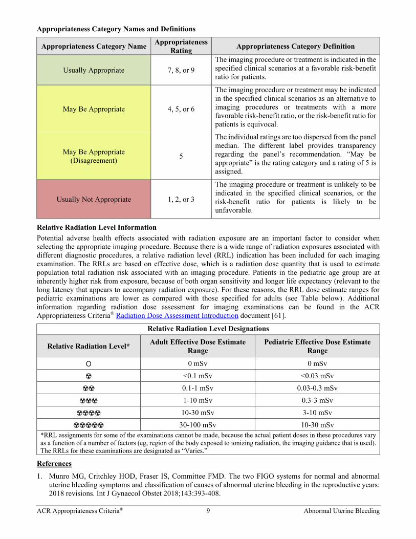
Appropriateness Category Names and Definitions
Appropriateness Category Name Appropriateness Rating Appropriateness Category Definition
Usually Appropriate 7, 8, or 9 The imaging procedure or treatment is indicated in the specified clinical scenarios at a favorable risk-benefit ratio for patients.
May Be Appropriate 4, 5, or 6
The imaging procedure or treatment may be indicated in the specified clinical scenarios as an alternative to imaging procedures or treatments with a more favorable risk-benefit ratio, or the risk-benefit ratio for patients is equivocal.
May Be Appropriate (Disagreement) 5
The individual ratings are too dispersed from the panel median. The different label provides transparency regarding the panel’s recommendation. “May be appropriate” is the rating category and a rating of 5 is assigned.
Usually Not Appropriate 1, 2, or 3
The imaging procedure or treatment is unlikely to be indicated in the specified clinical scenarios, or the risk-benefit ratio for patients is likely to be unfavorable.
Relative Radiation Level Information Potential adverse health effects associated with radiation exposure are an important factor to consider when selecting the appropriate imaging procedure. Because there is a wide range of radiation exposures associated with different diagnostic procedures, a relative radiation level (RRL) indication has been included for each imaging examination. The RRLs are based on effective dose, which is a radiation dose quantity that is used to estimate population total radiation risk associated with an imaging procedure. Patients in the pediatric age group are at inherently higher risk from exposure, because of both organ sensitivity and longer life expectancy (relevant to the long latency that appears to accompany radiation exposure). For these reasons, the RRL dose estimate ranges for pediatric examinations are lower as compared with those specified for adults (see Table below). Additional information regarding radiation dose assessment for imaging examinations can be found in the ACR Appropriateness Criteria® Radiation Dose Assessment Introduction document [61].
Relative Radiation Level Designations
Relative Radiation Level* Adult Effective Dose Estimate Range
Pediatric Effective Dose Estimate Range
O 0 mSv 0 mSv
☢ <0.1 mSv <0.03 mSv
☢☢ 0.1-1 mSv 0.03-0.3 mSv
☢☢☢ 1-10 mSv 0.3-3 mSv
☢☢☢☢ 10-30 mSv 3-10 mSv
☢☢☢☢☢ 30-100 mSv 10-30 mSv *RRL assignments for some of the examinations cannot be made, because the actual patient doses in these procedures vary as a function of a number of factors (eg, region of the body exposed to ionizing radiation, the imaging guidance that is used). The RRLs for these examinations are designated as “Varies.”
References
1. Munro MG, Critchley HOD, Fraser IS, Committee FMD. The two FIGO systems for normal and abnormal uterine bleeding symptoms and classification of causes of abnormal uterine bleeding in the reproductive years: 2018 revisions. Int J Gynaecol Obstet 2018;143:393-408.
ACR Appropriateness Criteria® 10 Abnormal Uterine Bleeding
2. Munro MG, Critchley HO, Broder MS, Fraser IS, Disorders FWGoM. FIGO classification system (PALM-COEIN) for causes of abnormal uterine bleeding in nongravid women of reproductive age. Int J Gynaecol Obstet 2011;113:3-13.
3. Valentin L. Imaging techniques in the management of abnormal vaginal bleeding in non-pregnant women before and after menopause. Best Pract Res Clin Obstet Gynaecol 2014;28:637-54.
4. Dias DS, Bueloni-Dias FN, Dias R, et al. Usefulness of clinical, ultrasonographic, hysteroscopic, and immunohistochemical parameters in differentiating endometrial polyps from endometrial cancer. Journal of minimally invasive gynecology 2014;21:296-302.
5. Goldstein RB, Bree RL, Benson CB, et al. Evaluation of the woman with postmenopausal bleeding: Society of Radiologists in Ultrasound-Sponsored Consensus Conference statement. J Ultrasound Med 2001;20:1025-36.
6. Bayer SR, DeCherney AH. Clinical manifestations and treatment of dysfunctional uterine bleeding. JAMA 1993;269:1823-8.
7. Sweet MG, Schmidt-Dalton TA, Weiss PM, Madsen KP. Evaluation and management of abnormal uterine bleeding in premenopausal women. Am Fam Physician 2012;85:35-43.
8. Brown DL, Packard A, Maturen KE, et al. ACR Appropriateness Criteria® First Trimester Vaginal Bleeding. J Am Coll Radiol 2018;15:S69-S77.
9. American College of Radiology. ACR Appropriateness Criteria®: Second and Third Trimester Bleeding. Available at: https://acsearch.acr.org/docs/69465/Narrative/. Accessed March 27, 2020.
10. ACOG Committee Opinion No. 734: The Role of Transvaginal Ultrasonography in Evaluating the Endometrium of Women With Postmenopausal Bleeding. Obstet Gynecol 2018;131:e124-e29.
11. Ragupathy K, Cawley N, Ridout A, Iqbal P, Alloub M. Non-assessable endometrium in women with post-menopausal bleeding: to investigate or ignore. Arch Gynecol Obstet 2013;288:375-8.
12. Kim YJ, Kim KG, Lee SR, Lee SH, Kang BC. Preoperative 3-dimensional Magnetic Resonance Imaging of Uterine Myoma and Endometrium Before Myomectomy. Journal of minimally invasive gynecology 2017;24:309-14.
13. Abbott JA. Adenomyosis and Abnormal Uterine Bleeding (AUB-A)-Pathogenesis, diagnosis, and management. Best Pract Res Clin Obstet Gynaecol 2017;40:68-81.
14. Kierans AS, Bennett GL, Haghighi M, Rosenkrantz AB. Utility of conventional and diffusion-weighted MRI features in distinguishing benign from malignant endometrial lesions. European journal of radiology 2014;83:726-32.
15. Jha RC, Zanello PA, Ascher SM, Rajan S. Diffusion-weighted imaging (DWI) of adenomyosis and fibroids of the uterus. Abdom Imaging 2014;39:562-9.
16. Lin G, Yang LY, Huang YT, et al. Comparison of the diagnostic accuracy of contrast-enhanced MRI and diffusion-weighted MRI in the differentiation between uterine leiomyosarcoma / smooth muscle tumor with uncertain malignant potential and benign leiomyoma. J Magn Reson Imaging 2016;43:333-42.
17. Lakhman Y, Veeraraghavan H, Chaim J, et al. Differentiation of Uterine Leiomyosarcoma from Atypical Leiomyoma: Diagnostic Accuracy of Qualitative MR Imaging Features and Feasibility of Texture Analysis. Eur Radiol 2017;27:2903-15.
18. Tanaka T, Terai Y, Ono YJ, et al. Preoperative MRI and intraoperative frozen section diagnosis of myometrial invasion in patients with endometrial cancer. Int J Gynecol Cancer 2015;25:879-83.
19. Mitamura T, Watari H, Todo Y, et al. Lymphadenectomy can be omitted for low-risk endometrial cancer based on preoperative assessments. J Gynecol Oncol 2014;25:301-5.
20. Li HM, Liu J, Qiang JW, Zhang H, Zhang GF, Ma F. Diffusion-Weighted Imaging for Differentiating Uterine Leiomyosarcoma From Degenerated Leiomyoma. J Comput Assist Tomogr 2017;41:599-606.
21. Sato K, Yuasa N, Fujita M, Fukushima Y. Clinical application of diffusion-weighted imaging for preoperative differentiation between uterine leiomyoma and leiomyosarcoma. Am J Obstet Gynecol 2014;210:368 e1-68 e8.
22. Gaetke-Udager K, McLean K, Sciallis AP, et al. Diagnostic Accuracy of Ultrasound, Contrast-enhanced CT, and Conventional MRI for Differentiating Leiomyoma From Leiomyosarcoma. Acad Radiol 2016;23:1290-7.
23. Thomassin-Naggara I, Dechoux S, Bonneau C, et al. How to differentiate benign from malignant myometrial tumours using MR imaging. Eur Radiol 2013;23:2306-14.
24. Gupta A, Grunhagen T. Live MR angiographic roadmapping for uterine artery embolization: a feasibility study. J Vasc Interv Radiol 2013;24:1690-7.
25. Kubik-Huch RA, Weston M, Nougaret S, et al. European Society of Urogenital Radiology (ESUR) Guidelines: MR Imaging of Leiomyomas. Eur Radiol 2018;28:3125-37.
ACR Appropriateness Criteria® 11 Abnormal Uterine Bleeding
26. Canverenler E, Buke B, Canverneler S. The Up to Date Status of Three-Dimensional Ultrasonography in Postmenopausal Bleeding. J Gynecol Neonatal 2017;1:101.
27. El-Sherbiny W, El-Mazny A, Abou-Salem N, Mostafa WS. The diagnostic accuracy of two- vs three-dimensional sonohysterography for evaluation of the uterine cavity in the reproductive age. Journal of minimally invasive gynecology 2015;22:127-31.
28. Nergiz S, Demircan-Sezer S, Kucuk M, Yuksel H, Odabasi AR, Altinkaya SO. Comparison of diagnostic methods for evaluation of postmenopausal bleeding. European journal of gynaecological oncology 2014;35:292-7.
29. Nieuwenhuis LL, Bij de Vaate MA, Hehenkamp WJ, et al. Reproducibility of three-dimensional gel installation sonohysterography in the assessment and classification of intrauterine abnormalities. European journal of obstetrics, gynecology, and reproductive biology 2014;179:141-6.
30. Fang L, Su Y, Guo Y, Sun Y. Value of 3-dimensional and power Doppler sonography for diagnosis of endometrial polyps. J Ultrasound Med 2013;32:247-55.
31. Inoue T, Kitajima M, Taniguchi K, Masuzaki H. Three-dimensional saline-infusion sonohysterography is useful for the identification of endometrial polyp. The journal of obstetrics and gynaecology research 2016;42:855-9.
32. Czuczwar P, Wozniak S, Szkodziak P, Kudla MJ, Pyra K, Paszkowski T. Elastography Improves the Diagnostic Accuracy of Sonography in Differentiating Endometrial Polyps and Submucosal Fibroids. J Ultrasound Med 2016;35:2389-95.
33. Stoelinga B, Hehenkamp WJ, Brolmann HA, Huirne JA. Real-time elastography for assessment of uterine disorders. Ultrasound Obstet Gynecol 2014;43:218-26.
34. Kabil Kucur S, Temizkan O, Atis A, et al. Role of endometrial power Doppler ultrasound using the international endometrial tumor analysis group classification in predicting intrauterine pathology. Arch Gynecol Obstet 2013;288:649-54.
35. Timmerman D, Verguts J, Konstantinovic ML, et al. The pedicle artery sign based on sonography with color Doppler imaging can replace second-stage tests in women with abnormal vaginal bleeding. Ultrasound Obstet Gynecol 2003;22:166-71.
36. Kamaya A, Yu PC, Lloyd CR, Chen BH, Desser TS, Maturen KE. Sonographic Evaluation for Endometrial Polyps: The Interrupted Mucosa Sign. J Ultrasound Med 2016;35:2381-87.
37. Dueholm M, Christensen JW, Rydbjerg S, Hansen ES, Ortoft G. Two- and three-dimensional transvaginal ultrasound with power Doppler angiography and gel infusion sonography for diagnosis of endometrial malignancy. Ultrasound Obstet Gynecol 2015;45:734-43.
38. Kara Bozkurt D, Bozkurt M, Cil AS, Barut MU, Ersahin AA, Caliskan E. Concomitant use of transvaginal sonography and Doppler indices improve diagnosis of adenomyosis. J Obstet Gynaecol 2017;37:888-95.
39. Dubinsky TJ. Value of sonography in the diagnosis of abnormal vaginal bleeding. J Clin Ultrasound 2004;32:348-53.
40. Smith-Bindman R, Kerlikowske K, Feldstein VA, et al. Endovaginal ultrasound to exclude endometrial cancer and other endometrial abnormalities. JAMA 1998;280:1510-7.
41. Meredith SM, Sanchez-Ramos L, Kaunitz AM. Diagnostic accuracy of transvaginal sonography for the diagnosis of adenomyosis: systematic review and metaanalysis. Am J Obstet Gynecol 2009;201:107 e1-6.
42. Wong AS, Lao TT, Cheung CW, et al. Reappraisal of endometrial thickness for the detection of endometrial cancer in postmenopausal bleeding: a retrospective cohort study. BJOG 2016;123:439-46.
43. Breitkopf DM, Frederickson RA, Snyder RR. Detection of benign endometrial masses by endometrial stripe measurement in premenopausal women. Obstet Gynecol 2004;104:120-5.
44. Farquhar C, Ekeroma A, Furness S, Arroll B. A systematic review of transvaginal ultrasonography, sonohysterography and hysteroscopy for the investigation of abnormal uterine bleeding in premenopausal women. Acta Obstet Gynecol Scand 2003;82:493-504.
45. Ozdemir S, Celik C, Gezginc K, Kiresi D, Esen H. Evaluation of endometrial thickness with transvaginal ultrasonography and histopathology in premenopausal women with abnormal vaginal bleeding. Arch Gynecol Obstet 2010;282:395-9.
46. Hulka CA, Hall DA, McCarthy K, Simeone JF. Endometrial polyps, hyperplasia, and carcinoma in postmenopausal women: differentiation with endovaginal sonography. Radiology 1994;191:755-8.
47. Bazot M, Cortez A, Darai E, et al. Ultrasonography compared with magnetic resonance imaging for the diagnosis of adenomyosis: correlation with histopathology. Hum Reprod 2001;16:2427-33.
ACR Appropriateness Criteria® 12 Abnormal Uterine Bleeding
48. Guideline developed in collaboration with the American College of R, American College of O, Gynecologists, Society of Radiologists in U. AIUM Practice Guideline for the Performance of Sonohysterography. J Ultrasound Med 2015;34:1-6.
49. American College of O, Gynecologists’ Committee on Gynecologic P. Technology Assessment No. 12: Sonohysterography. Obstet Gynecol 2016;128:e38-42.
50. Bhaduri M, Tomlinson G, Glanc P. Likelihood ratio of sonohysterographic findings for discriminating endometrial polyps from submucosal fibroids. J Ultrasound Med 2014;33:149-54.
51. Bittencourt CA, Dos Santos Simoes R, Bernardo WM, et al. Accuracy of saline contrast sonohysterography in detection of endometrial polyps and submucosal leiomyomas in women of reproductive age with abnormal uterine bleeding: systematic review and meta-analysis. Ultrasound Obstet Gynecol 2017;50:32-39.
52. American College of Radiology. Manual on Contrast Media. Available at: https://www.acr.org/Clinical-Resources/Contrast-Manual. Accessed March 27, 2020.
53. McComiskey MH, McCluggage WG, Grey A, Harley I, Dobbs S, Nagar HA. Diagnostic accuracy of magnetic resonance imaging in endometrial cancer. Int J Gynecol Cancer 2012;22:1020-5.
54. American College of Radiology. ACR Appropriateness Criteria®: Pretreatment Evaluation and Follow-Up of Endometrial Cancer. Available at: https://acsearch.acr.org/docs/69459/Narrative/. Accessed March 27, 2020.
55. Hoyos LR, Benacerraf B, Puscheck EE. Imaging in Endometriosis and Adenomyosis. Clin Obstet Gynecol 2017;60:27-37.
56. Makled AK, Elmekkawi SF, El-Refaie TA, El-Sherbiny MA. Three-dimensional power Doppler and endometrial volume as predictors of malignancy in patients with postmenopausal bleeding. The journal of obstetrics and gynaecology research 2013;39:1045-51.
57. El-Sharkawy M, El-Mazny A, Ramadan W, et al. Three-dimensional ultrasonography and power Doppler for discrimination between benign and malignant endometrium in premenopausal women with abnormal uterine bleeding. BMC Womens Health 2016;16:18.
58. Cogendez E, Eken MK, Bakal N, Gun I, Kaygusuz EI, Karateke A. The role of transvaginal power Doppler ultrasound in the differential diagnosis of benign intrauterine focal lesions. J Med Ultrason (2001) 2015;42:533-40.
59. Chandavarkar U, Kuperman JM, Muderspach LI, Opper N, Felix JC, Roman L. Endometrial echo complex thickness in postmenopausal endometrial cancer. Gynecol Oncol 2013;131:109-12.
60. Erdem M, Bilgin U, Bozkurt N, Erdem A. Comparison of transvaginal ultrasonography and saline infusion sonohysterography in evaluating the endometrial cavity in pre- and postmenopausal women with abnormal uterine bleeding. Menopause 2007;14:846-52.
61. American College of Radiology. ACR Appropriateness Criteria® Radiation Dose Assessment Introduction. Available at: https://www.acr.org/-/media/ACR/Files/Appropriateness-Criteria/RadiationDoseAssessmentIntro.pdf. Accessed March 27, 2020.
The ACR Committee on Appropriateness Criteria and its expert panels have developed criteria for determining appropriate imaging examinations for diagnosis and treatment of specified medical condition(s). These criteria are intended to guide radiologists, radiation oncologists and referring physicians in making decisions regarding radiologic imaging and treatment. Generally, the complexity and severity of a patient’s clinical condition should dictate the selection of appropriate imaging procedures or treatments. Only those examinations generally used for evaluation of the patient’s condition are ranked. Other imaging studies necessary to evaluate other co-existent diseases or other medical consequences of this condition are not considered in this document. The availability of equipment or personnel may influence the selection of appropriate imaging procedures or treatments. Imaging techniques classified as investigational by the FDA have not been considered in developing these criteria; however, study of new equipment and applications should be encouraged. The ultimate decision regarding the appropriateness of any specific radiologic examination or treatment must be made by the referring physician and radiologist in light of all the circumstances presented in an individual examination.


![Clinicopathological analysis of abnormal uterine bleeding in … · 2020. 2. 21. · abnormal uterine bleeding [AUB].3,4 Abnormal uterine bleeding is a common clinical complaints](https://img.pdfslide.us/doc/110x75/60c479f05ea55521530b1040/clinicopathological-analysis-of-abnormal-uterine-bleeding-in-2020-2-21-abnormal.jpg)