Embed Size (px)
Citation preview

Abnormal respiratory cilia in non-syndromic Leber
congenital amaurosis with CEP290 mutations
Jean Francois Papon, Isabelle Perrault, Andre Coste, Bruno Louis, Xavier
Gerard, Sylvain Hanein, Lucas Fares-Taie, Sylvie Gerber, Sabine
Defoort-Dhellemmes, Anne Marie Vojtek, et al.
To cite this version:
Jean Francois Papon, Isabelle Perrault, Andre Coste, Bruno Louis, Xavier Gerard, et al..Abnormal respiratory cilia in non-syndromic Leber congenital amaurosis with CEP290 mu-tations. Journal of Medical Genetics, BMJ Publishing Group, 2010, 47 (12), pp.829.<10.1136/jmg.2010.077883>. <hal-00560778>
HAL Id: hal-00560778
https://hal.archives-ouvertes.fr/hal-00560778
Submitted on 30 Jan 2011
HAL is a multi-disciplinary open accessarchive for the deposit and dissemination of sci-entific research documents, whether they are pub-lished or not. The documents may come fromteaching and research institutions in France orabroad, or from public or private research centers.
L’archive ouverte pluridisciplinaire HAL, estdestinee au depot et a la diffusion de documentsscientifiques de niveau recherche, publies ou non,emanant des etablissements d’enseignement et derecherche francais ou etrangers, des laboratoirespublics ou prives.
brought to you by COREView metadata, citation and similar papers at core.ac.uk
provided by HAL Descartes


1
Abnormal respiratory cilia in non-syndromic Leber congenital amaurosis with
CEP290 mutations
JF Papon 1,2,3,4,5*, I Perrault 6*, A Coste 1,2,3,4, B Louis 1, X Gérard 7,S Hanein 6, L
Fares-Taie 6, S Gerber 6, S Defoort-Dhellemmes 8, Anne Marie Vojtek 9, J Kaplan 6,
JM Rozet 6, Escudier E 1,9,10,11
1. INSERM, Unit U955, Creteil, F-94010, France;
2. Universite Paris-Est Creteil Val de Marne, Faculte de Medecine, UMR_S955, Creteil, F-94010,
France;
3. AP-HP, Groupe Henri-Mondor – Albert Chenevier, service d’ORL et de chirurgie cervico-faciale,
Creteil, F-94010, France;
4. Hopital intercommunal, service d’ORL et de chirurgie cervico-faciale, Creteil, F-94010, France;
5. INSERM, Unit U933 Paris, F-75012, France;
6. Unité de Recherches en Génétique et Epigénétique des Maladies Métaboliques, Neurosensorielles
et du Développement, INSERM U781 and Université Paris Descartes, CHU Necker Enfants Malades,
Paris, France ;
7. Genethon Evry, France ;
8. Service d’Ophtalmogie, CHRU Roger Salengro, Lille, France ;
9. Hopital intercommunal, service d’anatomo-pathologie (laboratoire de microscopie electronique),
Creteil, F-94010, France;
10. Universite Paris 6, Faculte de Medecine, Paris, F-75013, France;
11. AP-HP, Hopital Armand-Trousseau, service de genetique et d’embryologie medicales, Paris, F-
75012, France.
*JF Papon and I Perrault contributed equally to this work
Correspondance to Jean-Michel Rozet :
Postal address Hopital Necker-Enfants Malades
149 rue de Sèvres

2
75743 Paris CEDEX 15
E-mail : [email protected]
Telephone number : + 33 1 44 49 51 56
Fax number : + 33 1 44 49 51 50
Key words : LCA, CEP290, ciliopathies, axonemal ultrastructure, ciliary movement
Word count: 3632/4000
Competing Interest: None declared.
3
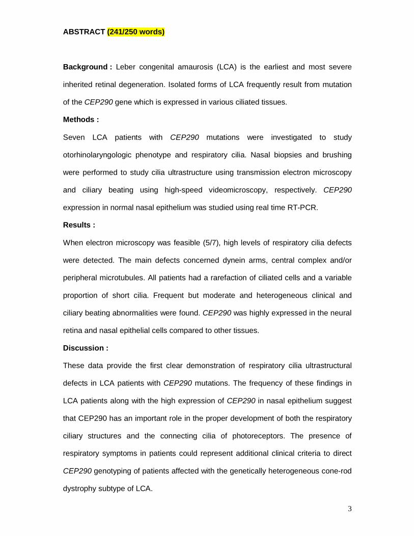
ABSTRACT (241/250 words)
Background : Leber congenital amaurosis (LCA) is the earliest and most severe
inherited retinal degeneration. Isolated forms of LCA frequently result from mutation
of the CEP290 gene which is expressed in various ciliated tissues.
Methods :
Seven LCA patients with CEP290 mutations were investigated to study
otorhinolaryngologic phenotype and respiratory cilia. Nasal biopsies and brushing
were performed to study cilia ultrastructure using transmission electron microscopy
and ciliary beating using high-speed videomicroscopy, respectively. CEP290
expression in normal nasal epithelium was studied using real time RT-PCR.
Results :
When electron microscopy was feasible (5/7), high levels of respiratory cilia defects
were detected. The main defects concerned dynein arms, central complex and/or
peripheral microtubules. All patients had a rarefaction of ciliated cells and a variable
proportion of short cilia. Frequent but moderate and heterogeneous clinical and
ciliary beating abnormalities were found. CEP290 was highly expressed in the neural
retina and nasal epithelial cells compared to other tissues.
Discussion :
These data provide the first clear demonstration of respiratory cilia ultrastructural
defects in LCA patients with CEP290 mutations. The frequency of these findings in
LCA patients along with the high expression of CEP290 in nasal epithelium suggest
that CEP290 has an important role in the proper development of both the respiratory
ciliary structures and the connecting cilia of photoreceptors. The presence of
respiratory symptoms in patients could represent additional clinical criteria to direct
CEP290 genotyping of patients affected with the genetically heterogeneous cone-rod
dystrophy subtype of LCA.
4
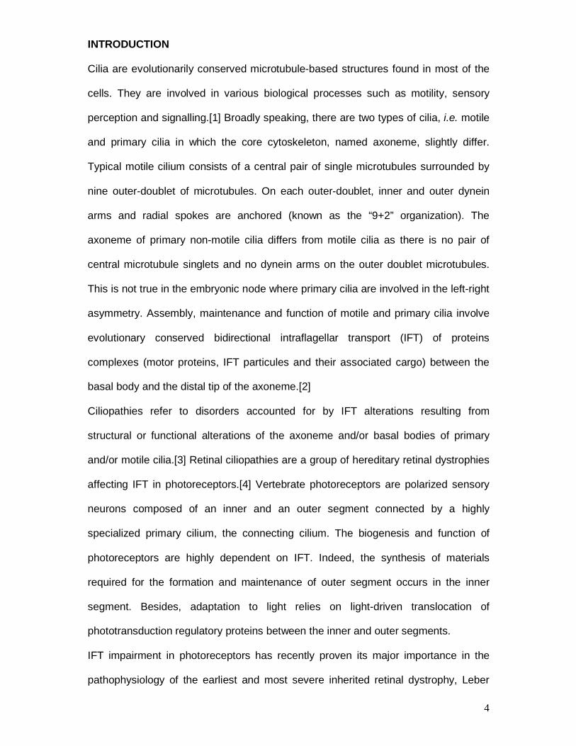
INTRODUCTION
Cilia are evolutionarily conserved microtubule-based structures found in most of the
cells. They are involved in various biological processes such as motility, sensory
perception and signalling.[1] Broadly speaking, there are two types of cilia, i.e. motile
and primary cilia in which the core cytoskeleton, named axoneme, slightly differ.
Typical motile cilium consists of a central pair of single microtubules surrounded by
nine outer-doublet of microtubules. On each outer-doublet, inner and outer dynein
arms and radial spokes are anchored (known as the “9+2” organization). The
axoneme of primary non-motile cilia differs from motile cilia as there is no pair of
central microtubule singlets and no dynein arms on the outer doublet microtubules.
This is not true in the embryonic node where primary cilia are involved in the left-right
asymmetry. Assembly, maintenance and function of motile and primary cilia involve
evolutionary conserved bidirectional intraflagellar transport (IFT) of proteins
complexes (motor proteins, IFT particules and their associated cargo) between the
basal body and the distal tip of the axoneme.[2]
Ciliopathies refer to disorders accounted for by IFT alterations resulting from
structural or functional alterations of the axoneme and/or basal bodies of primary
and/or motile cilia.[3] Retinal ciliopathies are a group of hereditary retinal dystrophies
affecting IFT in photoreceptors.[4] Vertebrate photoreceptors are polarized sensory
neurons composed of an inner and an outer segment connected by a highly
specialized primary cilium, the connecting cilium. The biogenesis and function of
photoreceptors are highly dependent on IFT. Indeed, the synthesis of materials
required for the formation and maintenance of outer segment occurs in the inner
segment. Besides, adaptation to light relies on light-driven translocation of
phototransduction regulatory proteins between the inner and outer segments.
IFT impairment in photoreceptors has recently proven its major importance in the
pathophysiology of the earliest and most severe inherited retinal dystrophy, Leber

5
congenital amaurosis (LCA) (MIM204000). As a matter of fact, 4 genes out of 13 so
far identified LCA genes encode proteins located in photoreceptor connecting cilia or
basal bodies: RPGRIP1,[5] TULP1,[6] CEP290,[7] and LCA5.[8] Conversely to
RPGRIP1 and TULP1 which are specifically or preferentially expressed in the
retina,[5, 6] LCA5 and CEP290 are expressed in various ciliated tissues.[7, 8] To our
knowledge, LCA5 mutations were reported in few patients only affected with non-
syndromic LCA. [8, 9] Yet, CEP290 mutations were shown to be a frequent cause of
non-syndromic LCA as well as syndromic LCA including Joubert syndrome, (JBTS5,
MIM610188) Senior-Loken syndrome, (SLSN6, MIM610189) Meckel syndrome
(MKS4, MIM611134) and possibly Bardet-Bield syndrome (BBS14, MIM610142).
MKS4, JBTS5 and SLSN6 were consistently ascribed to loss-of-function CEP290
mutations.[10-12] Most patients affected with non-syndromic forms of LCA carry a
intronic mutation of CEP 290 regarded by some authors as hypomorphic
(c.2991+1655A>G;[7, 13]). However, some other patients were reported to harbour
homozygous or compound heterozygous loss-of-function alleles.[13]
The pleiotropic phenotype accounted for by CEP290 mutations is consistent with the
location of the protein in the primary cilia of photoreceptor cells, in the kidney and
cerebellum. In the retina, CEP290 associates with several microtubule-based
transport proteins including retinitis pimentosa GTPase-regulator (RPGR) which
mutations are responsible for ca. 10% of X-linked retinitis pigmentosa (RP)
(MIM312610). Interestingly, some syndromic RP phenotypes consistent with motile
and primary cilia abnormalities were associated with RPGR mutations.[14-17] Here,
we report that LCA patients with CEP290 mutations, including the intronic
c.2991+1655A>G change, exhibit high levels of airway motile cilia defects and
frequent respiratory symptoms. The frequency of these findings in LCA patients
along with the high expression levels of CEP290 in the neural retina and nasal

6
ciliated cells compared to other tissues suggest that the respiratory symptoms in LCA
patients may not be coincidental.
PATIENTS AND METHODS
LCA patients with CEP290 mutations
Seven patients (2 adults, 5 children; age range 9-44 yrs; mean age 17.5 yrs; Table 1)
from six unrelated French families were ascertained from the Genetic and
Ophthalmologic Departments of the University Hospitals of Paris Necker – Enfants
Malades and Lille. All patients fulfilled the minimal criteria for the diagnosis of LCA
described elsewhere.[18]
No patient had sign of known ciliopathy (visceral malposition, cystic formation in
kidney, liver or pancreas…) but one patient presented with hypofertility due to
asthenospermia (Patient 5, Table 1). The CEP290 genotype had been determined
prior to the present study by direct sequencing of the 3’ and 5’UTR, the coding region
and exon-intron boundaries of the gene as described elsewhere (Table 1).[13] One
out the patients was homozygous for the intronic CEP290 c.2991+1655A>G mutation
while four out of them were compound heterozygous for this intronic change and a
mutation presumably resulting in loss-of-function. Finally, two patients were
homozygous for the p.Lys1575X mutation which may be regarded as a loss-of
function allele according to i) the absence of splice change predicted by the Splice
Site Prediction Program available at http://fruitfly.org/cgi-bin/seq_tools/splice.pl and ii)
the amplification of a unique transcript identical to the wild-type transcript (primers
designed in exons 33 and 37, respectively; data not shown available on request).
Rhino- and Otologic phenotype characterization
The patients were examined by an otorhinolaryngologist who firstly recorded clinical
history of airway diseases by interviewing the patients and/or their parents. Five
different nasal symptoms (nasal obstruction, hypo-anosmia, rhinorrhea, nasal
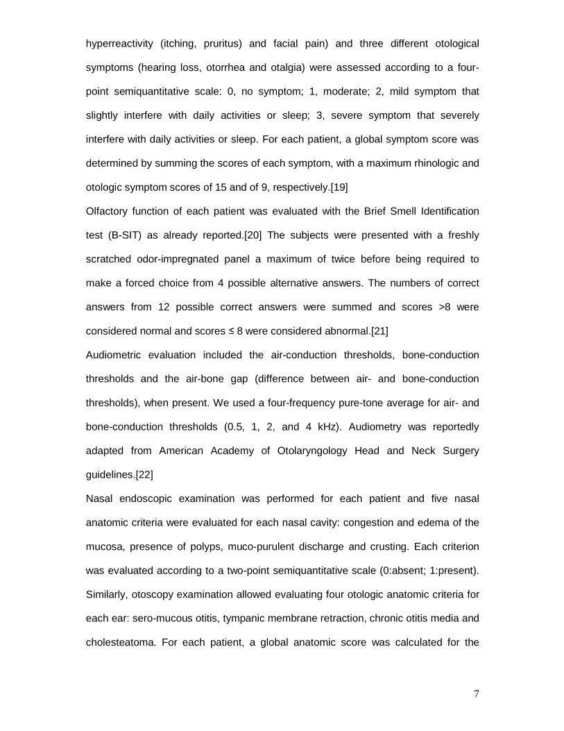
7
hyperreactivity (itching, pruritus) and facial pain) and three different otological
symptoms (hearing loss, otorrhea and otalgia) were assessed according to a four-
point semiquantitative scale: 0, no symptom; 1, moderate; 2, mild symptom that
slightly interfere with daily activities or sleep; 3, severe symptom that severely
interfere with daily activities or sleep. For each patient, a global symptom score was
determined by summing the scores of each symptom, with a maximum rhinologic and
otologic symptom scores of 15 and of 9, respectively.[19]
Olfactory function of each patient was evaluated with the Brief Smell Identification
test (B-SIT) as already reported.[20] The subjects were presented with a freshly
scratched odor-impregnated panel a maximum of twice before being required to
make a forced choice from 4 possible alternative answers. The numbers of correct
answers from 12 possible correct answers were summed and scores >8 were
considered normal and scores ≤ 8 were considered abnormal.[21]
Audiometric evaluation included the air-conduction thresholds, bone-conduction
thresholds and the air-bone gap (difference between air- and bone-conduction
thresholds), when present. We used a four-frequency pure-tone average for air- and
bone-conduction thresholds (0.5, 1, 2, and 4 kHz). Audiometry was reportedly
adapted from American Academy of Otolaryngology Head and Neck Surgery
guidelines.[22]
Nasal endoscopic examination was performed for each patient and five nasal
anatomic criteria were evaluated for each nasal cavity: congestion and edema of the
mucosa, presence of polyps, muco-purulent discharge and crusting. Each criterion
was evaluated according to a two-point semiquantitative scale (0:absent; 1:present).
Similarly, otoscopy examination allowed evaluating four otologic anatomic criteria for
each ear: sero-mucous otitis, tympanic membrane retraction, chronic otitis media and
cholesteatoma. For each patient, a global anatomic score was calculated for the

8
nasal cavities and the ears by summing the scores with a maximum rhinologic and
otologic anatomic scores of 10 and of 8, respectively.

9
Ciliary investigations
Biopsies and brushing of ciliated epithelium were obtained from nasal mucosa
(inferior turbinate) of the patients and processed for ciliary investigations. All
investigations were performed in the absence of acute respiratory tract infections. All
the patients and/or their parents were informed of both the exact nature and the goal
of the investigations and gave their informed consent as prescribed by the law on
bioethics in the European Community (and after approval by the local ethics
committee (DC-2008-512, Paris-Necker).
Airway biopsies were immersed in 2.5 % glutaraldehyde and processed as usual for
transmission electron (TEM) ultrastructural analysis.[23] Ultrathin sections were
examined at a final magnification of 60,000 without knowledge of the clinical data. In
each specimen, analysis of at least 50 transverse ciliary sections of different cells
were required to study the internal axonemal structure according to a quantitative
method.[24] Ciliary ultrastructure results were expressed as a percentage of
abnormal cilia among the total number of cilia analyzed. As previously reported, up to
10% of cilia in control specimens can exhibit ultrastructural defects.[25] For each
ciliary study, axonemal abnormalities were expressed as the concerned ultrastructure
(i.e. dynein arms, central complex and/or peripheral microtubules).
Airway brushing were suspended in culture medium and processed for ciliary beat
pattern evaluation using high-speed videomicrosocopy (HSVM). All the observations
were made within three hours at 37°C with an inverse microscope (Axiovert 200, Carl
Zeiss S.A.S. Le Pecq France) using an oil immersion x100 objective lens on a
duration shorter than 20 minutes. Beating ciliated edges were recorded with a high-
speed digital camera (PixeLINK A741, Ottawa Canada) at a rate of 355 frames per
second. In each patient, we recorded at least 20 distinct areas containing intact
undisrupted ciliated epithelial edge greater than 50 µm beating in the plane of the
camera and devoid of mucus. All isolated ciliated cells were excluded from the study

10
as recommended.[26] We initially determined the amount of ciliated cells among
epithelial cells and then the percentage of ciliated edges with beating cilia. We then
selected a cilium that we followed during an entire cycle of beating in 10 distinct
areas at least. For this, the video sequences were played back frame by frame using
Streampix software (Norpix Inc., Montreal Canada) on a high resolution monitor.
Different beat pattern parameters were determined : cilia length, proportion of cilia
with a length<4 µm, ciliary beat frequency, beating angle, and running distance per
second defined as the distance run by the cilia tip in one second. The cilia length and
beating angle were calculated by trigonometric calculations in the triangle made by
the anchoring point and the two extreme positions of the cilium extremities during the
power stroke. The running distance per second was calculated by combining the cilia
length, the ciliary beat frequency and the beating angle.
CEP290 expression in nasal ciliated cells
We examined the expression of CEP290 transcripts by real-time RT-PCR in adult
and foetal human tissues. One microgram of total RNA from sensorineural retina and
nasal mucosa extracted from a twenty-week-old fetal human ocular globe and adult
control respectively, and, adrenal gland, bone marrow, brain cerebellum, whole brain,
fetal brain, fetal liver, heart, kidney, liver, whole lung, placenta, prostate, salivary
gland, skeletal muscle, testis, thymus, thyroid gland, trachea, uterus and spinal cord
from the Human Total RNA Master panel II (Clontech, Saint Germain en Laye,
France), on the other hand, were reverse transcribed with the High capacity cDNA
Archive Kit (Applied Biosystems, Foster City, CA) primed with Oligo d(T)16 (Applied
Biosystems, Foster City, CA) in accordance with the supplier’s recommendations.
CEP290 transcript was amplified as 94 bp fragment. Regions of 80 bp and 84 bp
within the Homo sapiens beta-2-microglobulin mRNA (B2M, NM_004048.2) and the
human glucuronidase beta mRNA (GUSB, NM_000181.3) were used for

11
normalization, respectively. Primer sequences were as follows: CEP290 forward 5’-
TGACTGCTAAGTACAGGGACATCTTG-3’ CEP290 reverse 5’
AGGAGATGTTTTCACACTCCAGGT-3’B2M forward, 5’- cctggaggctatccagcgtact -3’;
B2M reverse, 5’- tcaggaaatttgactttccattctct-3’; GUSB forward, 5’-gcggtcgtgatgtggtctgt
-3’; GUSB reverse, 5’- gtgagcgatcaccatcttcaagt -3’.
Samples of cDNA were diluted 1:25 in nuclease-free water (Qiagen, Courtaboeuf,
France) and either submitted to qPCR or pooled and further diluted 1:5, 1:25, 1:125,
and 1:650 to create standard curves for calculation of relative gene expression
levels. Independent or pooled reverse transcribed materials (5μl) were submitted to
qPCR in 20µl MESA BLUE qPCR Master Mix Plus for SYBR® Assay (Eurogenetec,
Angers, France) containing 300nM of primers, on a Taqman 7900 HT Fast Real-Time
PCR System (Applied Biosystems, Foster City, CA) under the following conditions: 1
cycle of 95°C for 5 min, followed by 50 cycles of 15 sec at 95°C and 1 min at 65°C.
The specificity and identity of PCR products were verified by generating melting
curves using 1 cycle at 95°C for 15 sec, 65°C for 15 sec and 95°C for 15 sec. Data
were analyzed using the SDS 2.3 software (Applied Biosystems, Foster City, CA).
For each tissue, three independent reverse transcriptions were performed and each
was tested in duplicate. No reverse transcriptase and no template reactions were
used as negative controls.
Relative expression values were calculated using the geNorm algorithm.[27]
RESULTS
Clinical phenotype (Table 1)
The seven patients displayed a dramatically severe and stationary congenital cone-
rod dystrophy (LCA type I,[18]).
Clinical history revealed many recurrent but moderate inflammatory diseases of the
upper and lower airways in six out of the seven patients (Table 1). Clinical
12
examination revealed that nasal symptoms were either moderate (four patients) or
absent (three patients). A slight impairment of olfaction was present in three patients
according to the B-SIT score (Table 1). Nasal endoscopy revealed a slight or
moderate inflammation in six patients, but was normal on one patient (Table 1). None
of the patients complained of any otological symptom (Table 1). Pure tone
audiometry detected a moderate/slight hearing loss on two patients and was normal
on the five other patients (Table 1). Eventually, otoscopy was normal on all patients
(Table 1).
It was interesting to notice that patient #5 affected with hypofertility due to
oligoasthenospermia, showed normal otorhinolaryngological evaluation, yet slight
bilateral sensory hypoacousia.
Ciliary ultrastructure
Nasal ciliated were sparse in two patients (#2 and 7) on whom epithelial morphology
suggested a partial loss of ciliary differentiation. In these two patients, the low
amount of ciliated cells in the biopsy did not allowed to study ciliary ultrastructure. In
the other five patients, high levels of abnormal cilia were noted (24-71 %, Table 1).
Different ultrastructural defects of the axoneme were evidenced : abnormal number
of peripheral and/or central microtubules, either alone or in combination with absence
of dynein arms (Table 1;Figure 2).
HSVM quantitative evaluation of ciliary beat pattern
On all patients, the brushing samples provided enough ciliated cells for ciliary beat
pattern evaluation. On all patients, we found a slightly decreased amount of ciliated
cells among epithelial cells (Table 1). However, the percentage of ciliated edges with
beating cilia was always high (Table 1). On four patients, mean cilia length was
reduced and on six patients an increased proportion of short cilia (Table 1) was
13
found. The ciliary beat frequency and the beating angle were markedly reduced on
two and three patients compared to normal values, respectively (Table 1). Finally, on
all patients, the running distance covered by the cilia tip per second was reduced
compared to the expected normal value (Table 1). The lower values of running
distance were observed on the three patients affected with dynein arm defects
(patients 1, 3 and 4). However, there was no obvious relationship between ciliary
beat pattern parameters and clinical features.

14
Table 1: Ciliated cells characteristics of the LCA patients.
Mutation Clinical history Rhinologic scores Otologic scores TEM analysis of cilia HSVM analysis
Patient #
Age
(yrs)
sex
Allele 1
Allele 2
r-GSS
B-SIT score
r-GAS
o-GSS
Hearing score
Right(AC,BC,ABG)
Left(AC,BC,ABG)
o-GAS
Abnormal
cilia
(%)
Defect
Amount
of ciliated cells
Ciliated
edges with beating cilia (%)
Cilia length (µm)
Proportion of
cilia <4 µm
(%)
CBF
(Hz)
Beating
angle
(°)
Running distance
per second (µm/sec)
1 11 M
c.2991-1655A>G; p.Cys998X
c.2991-1655A>G; p.Cys998X Chronic rhinitis 2 12 4 0
0, 0, 0 0, 0, 0 0 24 DA, CC 3 100 4.9 10 6.7 59.2 34.9
2 25 M c.4723A>T; p.Lys1575X
c.4723A>T; p.Lys1575X
Recurrent sinusitis 1 8 4 0 0, 0, 0 0, 0, 0
0 Analysis unfeasible
Analysis unfeasible
3 95 6.1 15 9.3 65.6 71.5
3 9 M
c.4723A>T; p.Lys1575X
c.4723A>T; p.Lys1575X Recurrent bronchitis 2 6 4 0
0, 0, 0 0, 0, 0 0 71 DA, PMT, CC 2 90 4.6 30 7.2 75.4 48.5
4 9 M
c.2991-1655A>G; p.Cys998X
c.4028delA; p.Lys1343ArgfsX1
Chronic rhinitis with hyposmia
Otitis Bronchitis
4 8 2 0 0, 0, 0 0, 0, 0
0 45 DA, PMT 3 95 5 20 11.9 59.4 66.8
5 44 M
c.2991-1655A>G; p.Cys998X
c.3922C>T; p.Gin1308X
AS 0 12 0 0 12.5, 12.5, 0 12.5, 12.5, 0
0 46 CC 3 95 5.6 15 9.8 73.6 84.9
6 16 M c.2991-1655A>G;
p.Cys998X c.5587-1G>C
Recurrent otitis Recurrent pharyngitis
0 12 2 0 0, 0 , 0
13.7, 5, 8.7 0 55 CC 2 85 6.1 0 11.5 69.8 85.0
7 16 M c.2991-1655A>G;
p.Cys998X c.5587-1G>C
Recurrent otitis Recurent pharyngitis
0 12 2 0 0, 0, 0 0, 0, 0
0 Analysis
unfeasible Analysis
unfeasible 2 60 6.1 5 10.3 78 87.2
Normal values 0 >8 0 0 0, 0, 0 0, 0, 0
0 <10 No DA 4 94 6 0 12.8 73 93
Normal values of the different parameters according to the literature data for B-SIT score,[21] TEM analysis of cilia,[25] amount of ciliated cells and ciliated edges with
beating cilia, [28] cilia length,[29] ciliary beat frequency [30] and beating angle.[31] The value of the expected running distance per second was calculated using the
following formula: length x angle x frequency x 1second. Abbreviations: TEM: transmission electron microscopy; HSVM:high-speed videomicroscopy; r-GSS: rhinologic
global symptom score; B-SIT: brief smell identification test; r-GAS: rhinologic anatomic score; o-GSS: otologic global symptom score; AC: air conduction, BC: bone
conduction; ABG: air-bone gap; o-GAS: otologic anatomic score; CBF: ciliary beat frequency; M: male; DA: dynein arms; PMT: peripheral microtubule; CC: central
complex; AS: asthenospermia.

15
Expression analysis of CEP290 (Figure 2)
Realtime RT-PCR experiments demonstrated the highest expression levels of
CEP290 transcripts in neural retina and nasal epithelium. The gene was also
significantly expressed in spinal cord, thyroid gland, testis, heart, lung, bone marrow,
cerebellum and uterus. Weaker expression was noted in whole brain, both the foetal
brain and the kidney. Finally trachea, thymus, muscle, salivary gland, liver and
placenta expressed very low levels of transcripts (Figure 2).
DISCUSSION
Leber congenital amaurosis is characterized by a large genetic and pathophysiologic
heterogeneity. However, until the recent identification of CEP290, the specific or
preferential pattern of expression of LCA genes was consistent with the LCA retinal-
restricted phenotype. Intriguingly, while the implication of CEP290 in syndromic LCA
was consistent with its expression in primary cilia of various cell types, its
involvement in non-syndromic LCA remains unexplained. Interestingly, here, we
report abnormalities of motile respiratory cilia with frequent respiratory history that
were consistently found in LCA patients with homozygote or compound heterozygote
for loss-of-function or the common intronic CEP290 mutations.
Respiratory symptoms were reported by all but one LCA patients. Patients with
otorhinolaryngologic history complained of rhinorrhea and nasal obstruction with
inflammatory nasal mucosa, highly suggestive of chronic rhinitis. In addition, two of
them (#3 and 4) also complained of slight bronchial symptoms. Motile cilia were
studied at nasal level because ciliated cells are easily collected after local
anesthesia, through a non-invasive procedure routinely used to investigate chronic
rhinitis.[32] Moreover, we previously demonstrated that the ciliary defects present in
nasal mucosa are identical to those seen in bronchi.[33] In the five patients on whom

16
TEM analysis was possible, we detected a high proportion of cilia with axonemal
structural defects affecting, alone or in combination, dynein arms, peripheral and/or
central microtubules. These data suggest the constitutional nature of the ciliary
abnormalities rather than a consequence of acquired ciliary defects resulting from
recurrent infections which usually only affect a few cilia and do not involve dynein
arms.[34]
Along with axonemal defects, HSVM evidenced a rarefaction of ciliated cells with
frequent short cilia. As a result, the distance covered by the ciliary tip per second was
consistently shortened in all patients, especially in patients with dynein arm defects.
This latter observation is consistent with the pivotal role of dynein arms in the
transduction of mechanical forces necessary for ciliary motion via a number of
complex cell events including phosphorylation/dephosphorylation of key proteins.[1]
The ciliary phenotype documented in the patients (i.e. rarefaction and short cilia,
partial ciliary defects with abnormalities of several axonemal components) is
consistent with alterations of ciliary construction and/or maintenance. The high
expression level of CEP290 in nasal mucosa along with the recent report of its pivotal
role in primary cilia assembly [35] suggest that the respiratory cilia defects in patients
may be related to the CEP290 mutations. In this context, the coexistence of normal
and abnormal motile cilia in nasal samples of patients may reflect differences in
assembly pathways of primary versus motile cilia.
It would be tempting to correlate the mild symptoms in patients to the moderate
though significant decrease in the amount of ciliated cells and the increase in
abnormal structural defects of cilia in the nasal mucosa.
Interestingly, despite nasal congestion or edema, patients of our series, including that
homozygote for the CEP290 c.2991+1655A>G mutation, had quite a normal sense of
smell. Three patients (#2, 3 and 4) at best failed to identify some flavors. This finding
differs from the severe olfactory impairment reported in patients of a large inbred

17
family homozygous for the “hypomorphic” CEP290 mutation and in rd16 mice
carrying an in-frame CEP290 deletion mutation.[21]
Finally, none of the seven patients complained about hearing or eardrum
abnormalities. Audiogram recordings however indicated slight/mild hearing loss in 2/7
patients (#5 and #6) which may result from otitis in infancy.
The variation among patients of extraocular symptoms as well as in the proportion of
abnormal cilia and/or the axonemal ultrastructural defects in upper-airways did not
correlated to their genotype:
i) similar clinical findings were noted in patients homozygote for loss-of-
function or the common intronic mutations,
ii) patients with same genotypes presented with variable clinical symptoms
and/or variable proportions of abnormal cilia
iii) the three patients with dynein arm defects and ciliary beating abnormalities
exhibited different genotypes.
These findings may be compared to those reported previously in patients harboring
RPGR mutations.[16] However, in some families with RPGR mutations, a broader
phenotype was reported in which RP and sensorineural hearing loss also included
ear and respiratory tract infections with inconstant anosmia or congenital pulmonary
abnormalities (c.845-846delTG [17]; p.G173R,[14]). Moreover, RPGR mutations
were also reported in patients with a complex X linked phenotype combining RP and
primary ciliary dyskinesia.[16]
In conclusion, the data presented here provide a first report of respiratory cilia
abnormalities in LCA patients harbouring CEP290 mutations, supporting the
classification of some forms of the disease in the increasing family of ciliopathies.
Owing to the high frequency of CEP290 mutations in LCA (ca. 17%), this
symptomatic yet inconstant association might be frequent and largely overlooked.
The description of otorhinolaryngologic history in patients could represent additional

18
clinical criteria to direct CEP290 genotyping of patients affected with the genetically
heterogeneous cone-rod dystrophy subtype of LCA. Further studies will however be
necessary to compare the frequency of otorhinolaryngologic history in patients with
CEP290 and other disease genes. Finally, in the future, one can wonder whether the
defects identified in respiratory cilia could be used as useful markers in the
development of genetic therapies for LCA.
AKNOWLEDGEMENTS
The authors are grateful to the patients and their families who participated in this
study. We would also like to thank Dr V. Chatelin for the audiometric evaluation of the
patients.
This work was supported by grants and sponsors from the Legs Poix from the
Chancellerie des Universites, the Assistance Publique-Hopitaux de Paris (PHRC
AOM06053, P060245), the Agence Nationale pour la Recherche (ANR-05-MRAR-
022-01), EVI-Genoret (LSHG-CT-2005-512036), the Foundation Fighting Blindness
(BR-GE-0406-0335) and the Association Retina France.
REFERENCES
1. Haimo LT, Rosenbaum JL. Cilia, flagella, and microtubules. J Cell Biol
1981;1991:125s-130s.
2. Rosenbaum JL, Witman GB. Intraflagellar transport. Nat Rev Mol Cell Biol
2002;3:813-825.
3. Marshall WF. The cell biological basis of ciliary disease. J Cell Biol
2008;180:17-21.
4. Adams NA, Awadein A, Toma HS. The retinal ciliopathies. Ophthalmic Genet
2007;28:113-125.

19
5. Roepman R, Wolfrum U. Protein networks and complexes in photoreceptor
cilia. Subcell Biochem 2007;43:209-235.
6. Xi Q, Pauer GJ, Ball SL, et al. Interaction between the photoreceptor-specific
tubby-like protein 1 and the neuronal-specific GTPase dynamin-1. Invest
Ophthalmol Vis Sci 2007;48:2837-2844.
7. den Hollander AI, Koenekoop RK, Yzer S, et al. Mutations in the CEP290
(NPHP6) gene are a frequent cause of Leber congenital amaurosis. Am J
Hum Genet 2006;79:556-561.
8. Den Hollander AI, Koenekoop RK, Mohamed MD, et al. Mutations in LCA5,
encoding the ciliary protein lebercilin, cause Leber congenital amaurosis. Nat
Genet 2007;39:889-895.
9. Gerber S, Hanein S, Perrault I, et al. Mutations in LCA5 are an uncommon
cause of Leber congenital amaurosis (LCA) type II. Hum Mutat 2007;28:1245.
10. Baala L, Audollent S, Martinovic J, et al. Pleiotropic effects of CEP290
(NPHP6) mutations extend to Meckel syndrome. Am J Hum Genet
2007;81:170-179.
11. Sayer JA, Otto EA, O'Toole JF, et al. The centrosomal protein nephrocystin-6
is mutated in Joubert syndrome and activates transcription factor ATF4. Nat
Genet 2006;38:674-681.
12. Valente EM, Silhavy JL, Brancati F, et al. Mutations in CEP290, which
encodes a centrosomal protein, cause pleiotropic forms of Joubert syndrome.
Nat Genet 2006;38:623-625.
13. Perrault I, Delphin N, Hanein S, et al. Spectrum of NPHP6/CEP290 mutations
in Leber congenital amaurosis and delineation of the associated phenotype.
Hum Mutat 2007;28:416.
14. Iannaccone A, Breuer DK, Wang XF, et al. Clinical and immunohistochemical
evidence for an X linked retinitis pigmentosa syndrome with recurrent

20
infections and hearing loss in association with an RPGR mutation
2003;40:509-515.
15. Koenekoop RK, Loyer M, Hand CK, et al. Novel RPGR mutations with distinct
retinitis pigmentosa phenotypes in French-Canadian families. Am J
Ophthalmol 2003;136:678-687.
16. Moore A, Escudier E, Roger G, et al. RPGR is mutated in patients with a
complex X linked phenotype combining primary ciliary dyskinesia and retinitis
pigmentosa. J Med Genet 2006;43:326-333.
17. Zito I, Downes SM, Patel RJ, et al. RPGR mutation associated with retinitis
pigmentosa, impaired hearing, and sinorespiratory infections. J Med Genet
2003;40:609-615.
18. Perrault I, Rozet JM, Gerber S, et al. Leber congenital amaurosis. Mol Genet
Metab 1999;68:200-208.
19. Coste A, Yona L, Blumen M, et al. Radiofrequency is a safe and effective
treatment of turbinate hypertrophy. Laryngoscope 2001;111:894-899.
20. Doty RL, Marcus A, Lee WW. Development of the 12-item Cross-Cultural
Smell Identification Test (CC-SIT). Laryngoscope 1996;106:353-356.
21. McEwen DP, Koenekoop RK, Khanna H, et al. Hypomorphic CEP290/NPHP6
mutations result in anosmia caused by the selective loss of G proteins in cilia
of olfactory sensory neurons. Proc Natl Acad Sci U S A 2007;104:15917-
15922.
22. Committee on Hearing and Equilibrium guidelines for the evaluation of results
of treatment of conductive hearing loss. AmericanAcademy of Otolaryngology-
Head and Neck Surgery Ffoundation, Inc. Otolaryngol Head Neck Surg
1995;113:186-187.
23. Papon JF, Coste A, Roudot-Thoraval F, et al. A 20-year experience of electron
microscopy in the diagnosis of primary ciliary dyskinesia. Eur Respir J 2009.

21
24. Escalier D, Jouannet P, David G. Abnormalities of the ciliary axonemal
complex in children: an ultrastructural and cinetic study in a series of 34
cases. Biol Cell 1982;44:271-282.
25. de Iongh RU, Rutland J. Ciliary defects in healthy subjects, bronchiectasis,
and primary ciliary dyskinesia. Am J Respir Crit Care Med 1995;151:1559-
1567.
26. Thomas B, Rutman A, O'Callaghan C. Disrupted ciliated epithelium shiws
slower ciliary beat frequency and increased dyskinesia. Eur Respir J
2009;34:401-404.
27. Vandesompele J, De Preter K, Pattyn F, et al. Accurate normalization of real-
time quantitative RT-PCR data by geometric averaging of multiple internal
control genes. Genome Biol 2002;3:RESEARCH0034.
28. Rossman C, Lee R, Forrest J, et al. Nasal ciliary ultrastructure and function in
patients with primary ciliary dyskinesia compared with that in normal subjects
and in subjects with various respiratory diseases. Am Rev Respir Dis
1984;129:161-167.
29. Sleigh MA, Blake JR, Liron N. The propulsion of mucus by cilia. Am Rev
Respir Dis 1988;137:726-741.
30. Chilvers MA, Rutman A, O'Callaghan C. Functional analysis of cilia and
ciliated epithelial ultrastructure in healthy children and young adults. Thorax
2003;58:333-338.
31. Rautiainen M, Matsune S, Shima S, et al. Ciliary beat of cultured human
respiratory cells studied with differential interference microscope and high
speed video system. Acta Otolaryngol 1992;112:845-851.
32. Chapelin C, Coste A, Gilain L, et al. Modified epithelial cell distribution in
chronic airways inflammation. Eur Respir J 1996;9:2474-2478.

22
33. Verra F, Fleury-Feith J, Boucherat M, et al. Do nasal ciliary changes reflect
bronchial changes? An ultrastructural study. Am Rev Respir Dis
1993;147:908-913.
34. Leigh MW, Pittman JE, Carson JL, et al. Clinical and genetic aspects of
primary ciliary dyskinesia/Kartagener syndrome. Genet Med 2009;11:473-487.
35. Tsang WY, Bossard C, Khanna H, et al. CP110 suppresses primary cilia
formation through its interaction with CEP290, a protein deficient in human
ciliary disease. Dev Cell 2008;15:187-197.
LEGENDS TO FIGURES
Figure 1. Examples of ultrastructural defects of cilia observed in patients with LCA
(magnification x 60,000; bars=0.1 µm).
A: control normal cilia;
B: partial absence of inner dynein arms associated with abnormal central complex
(patient1)
C: partial absence of both dynein arms associated with abnormal central sheath
(patient 3)
D: absence of both dynein arms associated with supernumerary microtubules (patient
4)
E and F: absence of central complex (patients 5 and 6, respectively)
Figure 2. Relative expression level of the CEP290 transcript in different human
tissues. Housekeeping genes: TBP and RPL0.

23
Licence for Publication statement "I Jean-Michel ROZET, the Corresponding Author has the right to grant on behalf of all authors and does grant on behalf of all authors, an exclusive licence (or non exclusive for government employees) on a worldwide basis to the BMJ Publishing Group Ltd to permit this article (if accepted) to be published in JMG and any other BMJPGL products and sublicences such use and exploit all subsidiary rights, as set out in our licence (http://group.bmj.com/products/journals/instructions-for-authors/licence-forms)."