Embed Size (px)
Citation preview
Abnormal Morphological and Functional Organization of theHippocampus in a p35 Mutant Model of Cortical DysplasiaAssociated with Spontaneous Seizures
H. J. Wenzel,1 C. A. Robbins,1 L.-H.Tsai,3 and P. A. Schwartzkroin1,2
Departments of 1Neurological Surgery and 2Physiology/Biophysics, University of Washington, Seattle, Washington98195, and 3Howard Hughes Medical Institute and Department of Pathology, Harvard Medical School, Boston,Massachusetts 02115
Cortical dysplasia is a major cause of intractable epilepsy inchildren. However, the precise mechanisms linking cortical mal-formations to epileptogenesis remain elusive. The neuronal-specific activator of cyclin-dependent kinase 5, p35, has beenrecognized as a key factor in proper neuronal migration in theneocortex. Deletion of p35 leads to severe neocortical lamina-tion defects associated with sporadic lethality and seizures.Here we demonstrate that p35-deficient mice also exhibit dys-plasia/heterotopia of principal neurons in the hippocampal formation,as well as spontaneous behavioral and electrographic seizures.Morphological analyses using immunocytochemistry, electronmicroscopy, and intracellular labeling reveal a high degree ofabnormality in dentate granule cells, including heterotopic lo-
calization of granule cells in the molecular layer and hilus,aberrant dendritic orientation, occurrence of basal dendrites,and abnormal axon origination sites. Dentate granule cells ofp35-deficient mice also demonstrate aberrant mossy fibersprouting. Field potential laminar analysis through the dentatemolecular layer reflects the dispersion of granule cells and thestructural reorganization of this region. Similar patterns of cor-tical disorganization have been linked to epileptogenesis inanimal models of chronic seizures and in human temporal lobeepilepsy. The p35-deficient mouse may therefore offer an ex-perimental system in which we can dissect out the key mor-phological features that are causally related to epileptogenesis.
Key words: epilepsy; dentate gyrus; granule cell dispersion;heterotopia; neuronal migration disorder; biocytin; EEG
Cortical dysplasia, an abnormality of neocortical developmentassociated with disturbed neural migration and/or aberrant neu-ronal–glial differentation, is recognized as one of the majorcauses of pediatric epilepsy (Honavar and Meldrum, 1997). Se-vere forms of cortical dysplasia are often associated with medi-cally intractable seizures, developmental delay, and neurologicaldeficits (Mischel et al., 1995). Misplaced cells (“heterotopia”) area common feature of cortical dysplasia, which is thought to resultfrom a failure of migration from the site of neuronal genesis tothe normal destination of the cells in the cortical plate (Guerriniet al., 1999; Smith et al., 1999; Walsh, 1999). However, mecha-nisms linking cortical dysplastic malformations to epileptogenesisare still to be elucidated.
Control of the spatial and temporal patterns of neuronal dif-ferentiation and migration involves a multitude of factors (e.g.,protein molecules, adhesion molecules, channels/receptors, andintracellular cytoskeletal proteins) (for review, see Rakic andCaviness, 1995; Huttenlocher et al., 1995). Defects in develop-mental processes have been implicated in a large group of geneticdisorders associated with cortical dysplasia/heterotopia (Ray-
mond et al., 1995; Barkovich et al., 1996), including early onsetepilepsies (Guerrini et al., 1999; Walsh, 1999). To better under-stand the relationship between these structural malformationsand the development of an epileptic state, investigators haveturned to animal model systems, including such mouse mutants asreeler (Falconer, 1951; Caviness and Sidman, 1973; Caviness,1976), Lis-1 (Reiner et al., 1993; Hirotsune et al., 1998; Fleck etal., 2000), doublecortin (Des Portes et al., 1998), and cyclin-dependent kinase 5 (cdk5) (Ohshima et al., 1996; Gilmore et al.,1998) (for review, see Chevassus-au-Louis et al., 1999; Walsh,1999). Surprisingly, although all of these mutations give rise todisruption of normal cortical development, none is associatedwith the occurrence of chronic spontaneous seizures.
Recently, a mouse “knock-out” for p35, a neuronal activator ofcdk5 that plays an important role in brain development, has beengenerated (Chae et al., 1997). The protein p35 is highly expressedin postmitotic neurons but completely absent in proliferatingneuronal progenitor cells (Tsai et al., 1994; Delalle et al., 1997).Mice with the p35 mutation lack p35/cdk5 kinase activity andexhibit severe cortical lamination disruption similar to that seenin reeler (i.e., an inversion of cortical lamination). In addition,these animals suffer from sporadic adult lethality and spontaneousseizures and display a diminished corpus callosum and abnormaltangential fiber fascicles within the neocortex (Chae et al., 1997;Kwon and Tsai, 1998).
Although the morphological disruptions of the cerebral cortexin p35 knock-outs have been studied in detail (Chae et al., 1997;Kwon and Tsai, 1998; Kwon et al., 1999), much less is knownabout malformations in hippocampus, and little is understood
Received Aug. 1, 2000; revised Nov. 14, 2000; accepted Nov. 16, 2000.This study was supported by National Institutes of Health Grant NS18895. We
thank the following people for excellent assistance: Norma L. Anderson, electronmicroscopy; Andrew F. Turella, histology; I ldiko Hegyvary and Kathryn Blott,quantitative histological analysis; and Paul Schwartz and Janet Schukar, photo-graphic and computer imaging.
Correspondence should be addressed to Dr. Philip A. Schwartzkroin, Departmentof Neurological Surgery, University of Washington, Box 356470, Seattle, WA 98195-6470. E-mail: [email protected] © 2001 Society for Neuroscience 0270-6474/01/30983-16$15.00/0
The Journal of Neuroscience, February 1, 2001, 21(3):983–998
about the relationship of disturbed neuronal organization toneuronal activity and seizure generation. Here we report thatsignificant structural abnormalities appear in the hippocampus(particularly in dentate gyrus) of p35-deficient mice, similar topathology observed in resected hippocampi of patients with tem-poral lobe epilepsy (tLE) (Houser, 1990, 1999; El Bahh et al.,1999). Furthermore, a high proportion of p35 knock-out miceexhibits spontaneous behavioral and/or electrographic seizuresreminiscent of limbic seizures. These observations provide aninitial basis for relating epileptogenesis to dysplastic malforma-tions resulting from errors in brain development.
Preliminary data have been reported previously in abstractform (Wenzel et al., 1998; Robbins et al., 1999).
MATERIALS AND METHODSAnimalsA complete description of the gene deletion protocol and intial charac-terization of these mice has been published (Chae et al., 1997). Morpho-logical analysis of hippocampal neurons was performed in brains ofp35 knock-out mice (p352/2). The p352/2 colony was maintained viabrother–sister matings, and wild-type controls (1/1) were derived fromthe same stock as originally used to generate 2/2 breeding pairs (129/Sv 3 C57BL/6 background). Most of the analysis was performed on 2- to5-month-old mice; animals at ages 2, 10, and 20 d and 14–15 months wereincluded for comparison. Various electrophysiological and morphologi-cal methods were used to analyze seizure-related behaviors and relatedproperties of dentate granule cells. All animal care and use conformed tothe NIH Guide for Care and Use of Laboratory Animals and were ap-proved by the Animal Care Committee of the University of Washington.
Observations of seizure behavior and EEG monitoringThreshold testing. Seizure susceptibility was measured at 60 d postnatallyin 15 p35 knock-out and 12 wild-type mice, using the volatile convulsantflurothyl (2,2,2-trifluroethyl ether; Aldrich, Milwaukee, WI) according towell established protocols (Samoriski and Applegate, 1997; Rho et al.,1999). The mice were placed individually in a Plexiglas chamber, andflurothyl was infused into the chamber at a rate of 20 ml /min. Latencieswere measured from the beginning of infusion to the onset of a firstgeneralized seizure (clonic) and to a second generalized seizure (tonic–clonic) that follows a refractory postictal period. Shorter latencies reflectgreater seizure susceptibility (i.e., lower threshold). Exposure to flu-rothyl was terminated at the onset of the second seizure. Data wereanalyzed using the Student’s t test (between-group comparison) using theSigmaStat Program (Jandel Scientific).
Video/EEG monitoring. Using ketamine/xylazine anesthesia, we surgi-cally implanted chronic EEG electrodes in 12 mice between 3 and 5months of age. Microscrews (stainless steel) served as epidural recordingelectrodes, and a twisted pair of fine stainless steel wires served as adepth electrode, which was stereotactically implanted into the righthippocampus. Electrodes were attached to a microplug, then cemented tothe cranium with dental acrylic. A Telefactor Video/EEG monitoringsystem (Telefactor, W. Conshohocken, PA) was used to record simulta-neously both behavior and EEG from freely moving animals. Eachanimal was monitored for 40–50 hr over the course of 3–4 weeks.
Tissue preparation for light microscopy/immunocytochemistryGeneral tissue preparation. Forty-five mice used for light microscopic andimmunocytochemical studies were anesthetized with Nembutal (100mg/kg, i.p.), then perfused with isotonic saline with heparin (500 U/mlsaline), followed by a solution of 4% paraformaldehyde (PFA) in 0.1 Msodium phosphate buffer (PB), pH 7.4. For electron microscopy, a fixa-tion solution consisting of 2–4% PFA and 0.1–2% glutaraldehyde wasused. The brains were immediately removed and placed in the samefixative for 4 hr at 4° C. After post-fixation, the brains were rinsed in PB,cryoprotected in 10% sucrose in 0.1 M PB for 1 hr, followed by 30%sucrose in 0.1 M PB for 24 hr at 4° C, then frozen on dry ice. Thirtymicrometer transverse serial sections were cut on a sliding microtomeequipped with a freezing stage, then selected for further processing.
For the quantitative analysis of granule cells, brains were post-fixed for
24 hr, then the hippocampi were isolated and cryoprotected. Beforesectioning, hippocampi were slightly extended along the septotemporalaxis, then frozen and mounted (oriented transversely to the septotem-poral axis) on the freezing stage of a sliding microtome. Serial transversesections were then cut at 40 mm, collected in PB, and chosen for stainingusing an unbiased, systematic method (West et al., 1991; Buckmaster andDudek, 1997); starting from a random position near the septal pole, every10th section was sampled, yielding an average of 12–15 samples perhippocampus. After they were mounted on slides, sections were stainedwith cresyl violet. Photomicrographs were taken on 35 mm slide film,scanned, and imported into Adobe Photoshop (Adobe Systems, Moun-tain View, CA) to assemble the figures.
Timm’s histochemistry. The Timm’s method for staining heavy metalswas used for the detection of synaptic vesicular zinc [particularly en-riched in mossy fiber (MF) boutons]. After initial fixation with 4%paraformaldehyde as described above, the brains were transferred to asolution containing 3–4% glutaraldehyde, 0.1% Na2S, and 0.136 mMCaCl2 in 0.12 M Millonig’s PB, pH 7.3, for 48 hr at 4° C, followed bycryoprotection in 30% sucrose. Frozen sections were cut at 30 mm andthen mounted on slides, air-dried, and transferred to a fresh developersolution containing 30 ml gum Arabic (50%), 5 ml 2 M citrate buffer, 15ml hydroquinone (5.76%), and 250 ml silver nitrate (0.73%) for 1 hr in thedark. Sections were counterstained with cresyl violet, dehydrated, clearedin toluene, and coverslipped.
Quantitative analysis of granule cells within the dentate gyrus. Thenumber of granule cells in the granule cell layer, as well as the numberof heterotopic granule cells within the molecular layer, were estimatedper entire dentate gyrus using the optical fractionator method (West etal., 1991). In Nissl-stained preparations, a granule cell was defined as aneuron with a small cell body and an oval or round nucleus; the Nissl-stained cytoplasm is found at the apical and basal portions of the cellbody (Seress and Pokorny, 1981; Wenzel et al., 1981; Seress, 1992). Thegranule cell and molecular layers also contain the cell bodies of glial cells,basket cells, and other interneurons (for review, see Freund and Buszaki,1996). The nuclei of glial cells were easily identified and excluded fromthe counting. The nuclei of basket cells are similar in appearance to thoseof granule cells, but the cell body of basket cells (and also other inter-neurons) in these layers is larger and more intensely stained for Nisslsubstance. On the basis of these criteria, nongranule cells could beidentified and excluded from the counting.
Cell counting was performed by an investigator who was blind to thegenotype of the animals. Total section thickness was used for dissectorheight, and only “caps” located within counting frames were counted(Buckmaster and Dudek, 1997); caps were defined as the nuclei ofgranule cells that came into focus while focusing down through thedissector height. Counting frames (10 3 10 mm) were distributed sys-tematically and randomly over the dentate granule cell layer, accordingto the method described by West et al. (1991). Using a camera lucida-likemicroscope/computer interface (Nikon Optiphot-2 Microscope with aCohu CCD Camera; Nikon, Tokyo, Japan) and an NIH Image 1.62 b4morphometry software package, the numerical density of granule cells,the volume of the granule cell and molecular layers, and the total numberof granule cells for a given hippocampus were estimated. Adequacy ofsampling was assessed by determining the intra-animal coefficient ofvariation (CE) as well as the inter-animal coefficient of variation (CV)for each measure (West and Gundersen, 1990).
Immunocytochemistry. Different sets of sections were processed forimmunocytochemistry (ICC), using a modification of the avidin–biotincomplex (ABC)-peroxidase technique (Hsu et al., 1981). Immunocyto-chemical procedures were performed as described previously (Wenzel etal., 1997). Briefly, sections were rinsed in PB, followed by 0.1 M Tris-HCLbuffer (TB), pH 7.4; endogenous peroxidases were then inactivated bytreatment with 0.5–1% H2O2 in TB for 2 hr. Sections were treated with3% bovine serum albumin (BSA) (Boehringer Mannheim, Indianapolis,IN), 3% goat or horse serum (Sigma, St. Louis, MO), and 0.25% TritonX-100 (TX) in 0.05 M TB, 0.15 M NaCl, pH 7.4 (TBS) for 1 hr to reducenonspecific staining. Sets of alternating sections were rinsed in TBS for30 min and incubated for 24 hr at 4°C in the various antisera and dilutionsin TBS containing 1% goat or horse serum, 2% BSA, and 0.25% TX:anti-neuron-specific nuclear protein (NeuN) (Chemicon, Temecula, CA),1:1000; anti-microtubule-associated protein-2 (Boehringer Mannheim),1:500; anti-glial fibrillary acidic protein (GFAP) (Dako Corporation,Carpinteria, CA) 1:4000; anti-glutamate decarboxylase 67 (GAD67)(Chemicon, Temecula, CA), 1:100; anti-calretinin (Chemicon), 1:4000;anti-parvalbumin (Chemicon), 1:4000; anti-somatostatin (SOM) (Penin-
984 J. Neurosci., February 1, 2001, 21(3):983–998 Wenzel et al. • p35 Mutation-Induced Cortical Dysplasia
sula Laboratories, Belmont, CA), 1:5000; and anti-zinc transporter ZnT3(provided by Dr. R. D. Palmiter, University of Washington, Seattle,WA), 1:250. After rinses for 2 hr in TBS, sections were incubated inbiotinylated goat anti-rabbit IgG or horse anti-mouse IgG (Vector Lab-oratories, Burlingame, CA), diluted 1:500 for 24 hr at 4°C, rinsed for 2 hrin TBS, and then incubated in ABC (Elite ABC Kit, Vector Laborato-ries), diluted 1:500 in 1% goat or horse serum, 2% BSA, 0.25% TX, andTBS for 24 hr at 4° C. Sections were rinsed thoroughly in TB, pH 7.6, andthen incubated for 15 min in 0.025% 3,39-diaminobenzidine (DAB;Sigma) in TB. After reaction for 5–10 min in fresh DAB with 0.003%H2O2, sections were rinsed in TB, followed by PB. Specificity of theimmunostaining was evaluated by omitting primary antibodies from theregular staining sequence. ZnT3 immunoreactivity in synaptic vesicles ofmossy fiber boutons was also localized at the ultrastuctural level using aprotocol described by Wenzel et al. (1997).
Electrophysiology and intracellular labeling with biocytinIntracellular recording and labeling. Hippocampal slices were preparedconventionally as described previously (Wenzel et al., 2000) for in vitroexperiments. Mice were anesthetized with halothane and decapitated,and the brain was removed quickly, cooled briefly in ice-cold oxygenated[95% O2/5% CO2] artificial CSF (ACSF) containing (in mM): 124 NaCl,5 KCl, 1.25 NaH2PO4, 2 MgSO4, 26 NaCO3, 2 CaCl2, 10 dextrose, andblocked to contain the hippocampus. Using a vibroslicer, 400-mm-thickslices transverse to the longitudinal axis of the hippocampus were cutinto a bath of oxygenated ACSF at 4°C. Sections were then transferred toa holding chamber and allowed to equilibrate for at least 1 hr whilesubmerged in ACSF at room temperature. Slices were then individuallytransferred to a standard interface recording chamber. In the chamber,slices rested on a nylon mesh over a well that was perfused (1 ml/min)with warmed (33–35°C), oxygenated ACSF. In addition, warmed, humid-ified air was circulated above the slice. Slices remained undisturbed forat least 15 min in the chamber before recording began.
Intracellular electrodes made from borosilicate glass were pulled usinga horizontal puller (Sutter Instruments, San Rafael, CA) and filled with2% biocytin (Molecular Probes, Eugene, OR) dissolved in 1 M potassiumacetate (80–230 MV resistance, pH 7.4). Intracellular potentials wererecorded using an Axoclamp 2A amplifier (Axon Instruments, FosterCity, CA). Neurons within the granule cell layer were impaled, andbiocytin was iontophoretically injected with 700 msec duration and0.5–1.0 nA hyperpolarizing current pulses delivered every second for10–30 min. After biocytin injection, the electrode was withdrawn, andthe slice was left in the chamber for at least 30 min before removal forfixation.
Retrograde labeling with biocytin. Visualization of multiple dentategranule cells was achieved through retrograde labeling via iontophoresisof 4% biocytin (in 0.05 M Tris HCl, pH 7.3) into extracellular space ofhippocampal stratum (s.) lucidum in CA3b subfield (Okazaki et al.,1995). The microelectrode (5–10 mm) was placed ;200 mm below thesurface of the slice, then 600 nA positive current pulses (7 sec on/7 secoff) were passed for 18–20 min. Slices remained incubating in thechamber for an additional 3 hr, then were fixed in 4% paraformaldehydeand further processed as described below.
Extracellular field recordings. Field EPSPs (fEPSPs) were recorded inthe dentate gyrus of both p352/2 and wild-type mice with extracellularelectrodes (filled with ACSF, 5–10 MV resistance) in response to per-forant path stimulation. A laminar profile of evoked fEPSPs was con-structed by recording sequentially from several sites through the molec-ular layer, perpendicular to the granule cell layer (superior blade of thedentate gyrus).
Tissue processing and morphological analysis of biocytin-labeled granulecells (light and electron microscopy). After the intracellular recordingprocedure and iontophoretic injection of biocytin, slices were removedfrom the recording chamber and immersion-fixed in a solution of 4%paraformaldehyde and 0.1–1% glutaraldehyde in 0.1 M sodium PB, pH7.4, for 2–4 hr at 4°C. The slices were rinsed in 0.1 M PB, then infiltratedfor cryoprotection with 10% sucrose in 0.1 M PB for 1 hr, followed by30% sucrose for 8–12 hr. Frozen sections were cut (60 mm) and furtherprocessed with a histochemical procedure. Sections of a total of 99 sliceswith biocytin-filled granule cells were subsequently processed for lightand electron microscopy, as follows.
Sections were rinsed in 0.1 M PB, pH 7.4, and then in 0.1 M TB, pH 7.4.Endogenous peroxidases were suppressed with 0.5–1% H2O2 in 0.1 M TBfor 2 hr. Sections were pretreated with 2% BSA, 0.25–0.4% dimethyl-sulfoxide (DMSO) (Sigma), and 0.05 M TBS, pH 7.4, for 1 hr to reduce
nonspecific background staining and to permeabilize membranes. Sec-tions were rinsed in 0.1 M TBS for 30 min and then incubated in ABC(Elite ABC Kit, Vector), diluted 1:500 in 2% BSA, 0.25–0.4% DMSO,and 0.05 M TBS for 36–48 hr at 4°C. Sections were then rinsed throughlyin 0.1 M TBS followed by 0.1 M TB, pH 7.6, and preincubated in 0.025%DAB with 0.005% NiNH4SO4 added to increase the density of stain for15 min. Subsequently, the sections were reacted with fresh DAB/NiNH4SO4 solution containing 0.01% H2O2 for 15–60 min. The reactionwas stopped by rinses in 0.1 M TB. Sections were further processed forelectron microscopy (EM) using a method that included post-fixation in1% osmium tetroxide in 0.15 M PB, pH 7.4, for 1 hr at room temperature,alcohol dehydration, and flat-embedding in Eponate 12 Resin (Ted Pella,Redding, CA) between two aclar sheets. The biocytin-filled granule cells(and their dendrites and axon arborizations) were visualized at the lightmicroscopical level and photographed before remounting and furthersectioning for EM. Camera lucida drawings were made of each biocytin-filled granule cell, and its dendrites and axon arborizations were recon-structed by superimposing all sections of the hippocampal slice. Serialthin sections from various slices containing different portions of thebiocytin-filled granule cells (e.g., mossy fiber axon collaterals in thedentate inner molecular layer) were stained with uranyl acetate andReynold’s lead citrate and examined on a Philips 410 electronmicroscope.
RESULTSThe gene-targeting strategy and generation of p352/2 mice havebeen reported previously (Chae et al., 1997). The initial histolog-ical study demonstrated a general disorganization of the forebrainin these mice, with a major defect in the development of thenormal lamination pattern of the neocortex (Kwon and Tsai,1998). In the present study, histological examination of the neo-cortex of p352/2 mice reconfirmed this observation of severedefects in the lamination of neocortex, associated with formationof aberrant fiber fascicles. Our observations also revealed struc-tural abnormalities within the hippocampus, particularly associ-ated with the appearance of dispersed granule cells and abnormalmossy fiber localization. We have focused on these hippocampalabnormalities and their functional correlations, with respect tothe occurrence of spontaneous seizures.
P352/2 mutation is associated with low seizurethreshold and with occurrence of spontaneouselectrographic and behavioral seizuresSeizure susceptibility was compared in p352/2 and wild-typemice through flurothyl seizure testing. In all animals, exposure toflurothyl elicited seizure activity. Statistical analysis of the laten-cies to the flurothyl-induced seizures revealed that the p352/2mice had significantly shorter latencies than wild-type to both thefirst (352.2 6 1.6 vs 423.7 6 12.0 sec, respectively) and secondseizures (590.6 6 14.3 vs 688.4 6 13.8 sec) (Fig. 1A). All statis-tical data are presented as means 6 SEM. These results representa 16% reduction in mean latency to the first seizure and a 14%reduction to the second seizure in p35 2/2 mice.
Video/EEG recording confirmed the occurrence of spontane-ous seizures in many p352/2 mice. A typical example of EEGrecorded from a p352/2 mouse during a tonic–clonic seizure isshown in Figure 1B. Spontaneous epileptiform EEG activity wasverified in 75% of the p352/2 mice (8 of 12 animals). Twenty-five percent of p352/2 animals exhibited spontaneous tonic–clonic seizures with behavioral manifestations, whereas an addi-tional 50% displayed intermittent interictal electrographicactivity. The spontaneous seizures had durations ranging from 59to 84 sec and were followed by a prolonged isoelectric postictalperiod lasting 3–5 min. Ten percent of the p352/2 mice died
Wenzel et al. • p35 Mutation-Induced Cortical Dysplasia J. Neurosci., February 1, 2001, 21(3):983–998 985
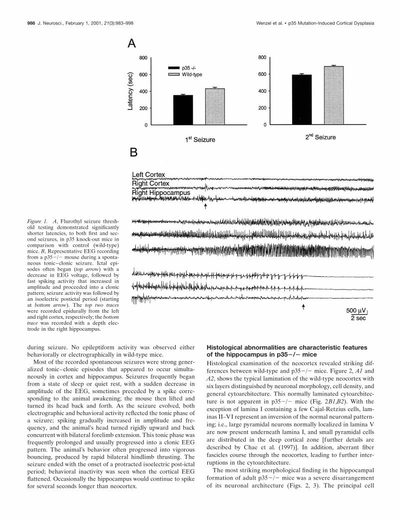
during seizure. No epileptiform activity was observed eitherbehaviorally or electrographically in wild-type mice.
Most of the recorded spontaneous seizures were strong gener-alized tonic–clonic episodes that appeared to occur simulta-neously in cortex and hippocampus. Seizures frequently beganfrom a state of sleep or quiet rest, with a sudden decrease inamplitude of the EEG, sometimes preceded by a spike corre-sponding to the animal awakening; the mouse then lifted andturned its head back and forth. As the seizure evolved, bothelectrographic and behavioral activity reflected the tonic phase ofa seizure; spiking gradually increased in amplitude and fre-quency, and the animal’s head turned rigidly upward and backconcurrent with bilateral forelimb extension. This tonic phase wasfrequently prolonged and usually progressed into a clonic EEGpattern. The animal’s behavior often progressed into vigorousbouncing, produced by rapid bilateral hindlimb thrusting. Theseizure ended with the onset of a protracted isoelectric post-ictalperiod; behavioral inactivity was seen when the cortical EEGflattened. Occasionally the hippocampus would continue to spikefor several seconds longer than neocortex.
Histological abnormalities are characteristic featuresof the hippocampus in p352/2 miceHistological examination of the neocortex revealed striking dif-ferences between wild-type and p352/2 mice. Figure 2, A1 andA2, shows the typical lamination of the wild-type neocortex withsix layers distinguished by neuronal morphology, cell density, andgeneral cytoarchitecture. This normally laminated cytoarchitec-ture is not apparent in p352/2 mice (Fig. 2B1,B2). With theexception of lamina I containing a few Cajal-Retzius cells, lam-inas II–VI represent an inversion of the normal neuronal pattern-ing; i.e., large pyramidal neurons normally localized in lamina Vare now present underneath lamina I, and small pyramidal cellsare distributed in the deep cortical zone [further details aredescribed by Chae et al. (1997)]. In addition, aberrant fiberfascicles course through the neocortex, leading to further inter-ruptions in the cytoarchitecture.
The most striking morphological finding in the hippocampalformation of adult p352/2 mice was a severe disarrangementof its neuronal architecture (Figs. 2, 3). The principal cell
Figure 1. A, Flurothyl seizure thresh-old testing demonstrated significantlyshorter latencies, to both first and sec-ond seizures, in p35 knock-out mice incomparison with control (wild-type)mice. B, Representative EEG recordingfrom a p352/2 mouse during a sponta-neous tonic–clonic seizure. Ictal epi-sodes often began (top arrow) with adecrease in EEG voltage, followed byfast spiking activity that increased inamplitude and proceeded into a clonicpattern; seizure activity was followed byan isoelectric postictal period (startingat bottom arrow). The top two traceswere recorded epidurally from the leftand right cortex, respectively; the bottomtrace was recorded with a depth elec-trode in the right hippocampus.
986 J. Neurosci., February 1, 2001, 21(3):983–998 Wenzel et al. • p35 Mutation-Induced Cortical Dysplasia
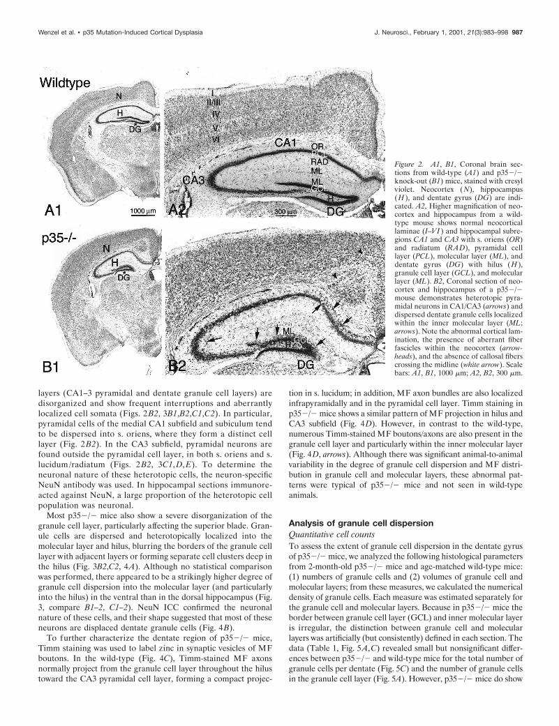
layers (CA1–3 pyramidal and dentate granule cell layers) aredisorganized and show frequent interruptions and aberrantlylocalized cell somata (Figs. 2 B2, 3B1,B2,C1,C2). In particular,pyramidal cells of the medial CA1 subfield and subiculum tendto be dispersed into s. oriens, where they form a distinct celllayer (Fig. 2 B2). In the CA3 subfield, pyramidal neurons arefound outside the pyramidal cell layer, in both s. oriens and s.lucidum/radiatum (Figs. 2 B2, 3C1, D, E). To determine theneuronal nature of these heterotopic cells, the neuron-specificNeuN antibody was used. In hippocampal sections immunore-acted against NeuN, a large proportion of the heterotopic cellpopulation was neuronal.
Most p352/2 mice also show a severe disorganization of thegranule cell layer, particularly affecting the superior blade. Gran-ule cells are dispersed and heterotopically localized into themolecular layer and hilus, blurring the borders of the granule celllayer with adjacent layers or forming separate cell clusters deep inthe hilus (Fig. 3B2,C2, 4A). Although no statistical comparisonwas performed, there appeared to be a strikingly higher degree ofgranule cell dispersion into the molecular layer (and particularlyinto the hilus) in the ventral than in the dorsal hippocampus (Fig.3, compare B1–2, C1–2). NeuN ICC confirmed the neuronalnature of these cells, and their shape suggested that most of theseneurons are displaced dentate granule cells (Fig. 4B).
To further characterize the dentate region of p352/2 mice,Timm staining was used to label zinc in synaptic vesicles of MFboutons. In the wild-type (Fig. 4C), Timm-stained MF axonsnormally project from the granule cell layer throughout the hilustoward the CA3 pyramidal cell layer, forming a compact projec-
tion in s. lucidum; in addition, MF axon bundles are also localizedinfrapyramidally and in the pyramidal cell layer. Timm staining inp352/2 mice shows a similar pattern of MF projection in hilus andCA3 subfield (Fig. 4D). However, in contrast to the wild-type,numerous Timm-stained MF boutons/axons are also present in thegranule cell layer and particularly within the inner molecular layer(Fig. 4D, arrows). Although there was significant animal-to-animalvariability in the degree of granule cell dispersion and MF distri-bution in granule cell and molecular layers, these abnormal pat-terns were typical of p352/2 mice and not seen in wild-typeanimals.
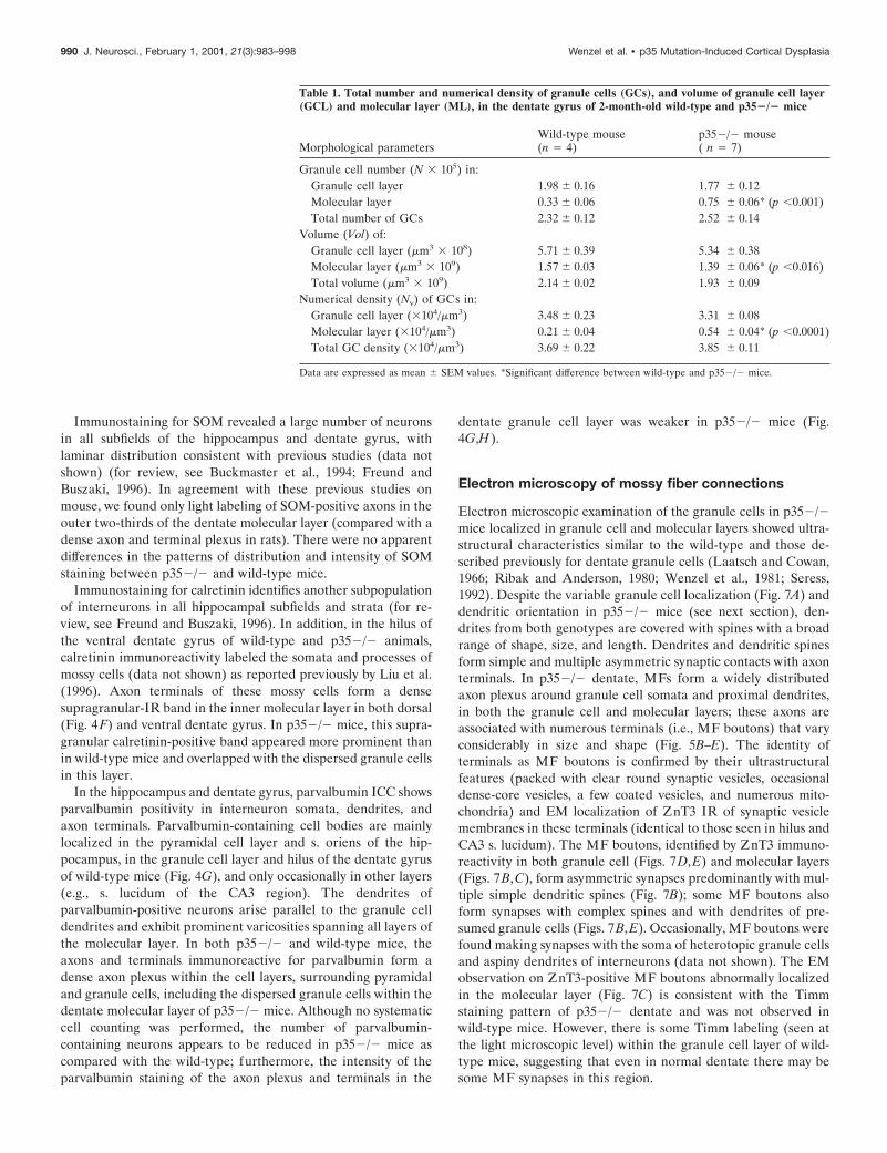
Analysis of granule cell dispersionQuantitative cell countsTo assess the extent of granule cell dispersion in the dentate gyrusof p352/2 mice, we analyzed the following histological parametersfrom 2-month-old p352/2 mice and age-matched wild-type mice:(1) numbers of granule cells and (2) volumes of granule cell andmolecular layers; from these measures, we calculated the numericaldensity of granule cells. Each measure was estimated separately forthe granule cell and molecular layers. Because in p352/2 mice theborder between granule cell layer (GCL) and inner molecular layeris irregular, the distinction between granule cell and molecularlayers was artificially (but consistently) defined in each section. Thedata (Table 1, Fig. 5A,C) revealed small but nonsignificant differ-ences between p352/2 and wild-type mice for the total number ofgranule cells per dentate (Fig. 5C) and the number of granule cellsin the granule cell layer (Fig. 5A). However, p352/2 mice do show
Figure 2. A1, B1, Coronal brain sec-tions from wild-type (A1) and p352/2knock-out (B1) mice, stained with cresylviolet. Neocortex (N), hippocampus(H ), and dentate gyrus (DG) are indi-cated. A2, Higher magnification of neo-cortex and hippocampus from a wild-type mouse shows normal neocorticallaminae (I–VI ) and hippocampal subre-gions CA1 and CA3 with s. oriens (OR)and radiatum (RAD), pyramidal celllayer (PCL), molecular layer (ML), anddentate gyrus (DG) with hilus (H ),granule cell layer (GCL), and molecularlayer (ML). B2, Coronal section of neo-cortex and hippocampus of a p352/2mouse demonstrates heterotopic pyra-midal neurons in CA1/CA3 (arrows) anddispersed dentate granule cells localizedwithin the inner molecular layer (ML;arrows). Note the abnormal cortical lam-ination, the presence of aberrant fiberfascicles within the neocortex (arrow-heads), and the absence of callosal fiberscrossing the midline (white arrow). Scalebars: A1, B1, 1000 mm; A2, B2, 300 mm.
Wenzel et al. • p35 Mutation-Induced Cortical Dysplasia J. Neurosci., February 1, 2001, 21(3):983–998 987

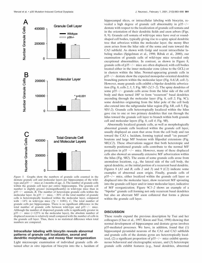
significantly more granule cells in the molecular layer than wild-type mice (Fig. 5B). Approximately 29.8% of all granule cells inp352/2 mice are localized within the molecular layer (comparedwith 14.4% in the wild-type). Statistical comparison of the meanvolume of the dentate layers revealed a difference between p352/2mice and wild-type mice: molecular layer and the total value of thegranule cell plus molecular layers of p352/2 animals are signifi-cant smaller ( p , 0.016 and p , 0.03, respectively), but thedifference in the granule cell layer is not significant ( p , 0.44).However, the numerical density of granule cells in the molecularlayer, the dispersed granule cells, is significantly higher in p352/2mice compared with the wild-type. The intra-animal CE for thenumber of cells counted and points counted (see Materials andMethods), as well as the between-subjects CV and CE2/CV2 ratio,were in the range that indicated adequate sampling (West et al.,1991).
Field potential analysisA laminar profile of synaptic responses, recorded throughout thegranule cell and molecular layers of the superior blade of the
dentate gyrus, revealed clear differences between p352/2 andwild-type mice (Fig. 6). The amplitude of the evoked fEPSPs wasmeasured at a fixed latency (Fig. 6A) and plotted as a function oflocation along a model granule soma–dendrite axis (Fig. 6C).Consistent with the morphological findings of a dispersed granulecell layer in p352/2 animals, the fEPSP positivity of p352/2dentate evoked by perforant path stimulation was much broaderin slices from p352/2 mice (Fig. 6B). This positivity is thought toreflect a “somal” current source [see Sutula et al. (1998) forsimilar analysis of “sprouted” dentate], suggesting that the hete-rotopic granule cells are activated by incoming perforant pathfibers.
Immunocytochemistry of astrocytes and interneuronsreveals cell-specific differences between p352/2 andwild-type miceICC techniques were used to examine the expression of severalmarkers specific for astrocytes and inhibitory interneurons (Fig.4E–H). Using an antibody against GFAP (Fig. 4E), we found noobvious differences in distribution and density of GFAP-positive
Figure 3. Transverse sections (cresyl vio-let) of the hippocampus (A1) and dentategyrus (A2) of the wild-type mouse. B1, B2,Transverse sections of the dorsal hip-pocampus of a p352/2 mouse demon-strate heterotopic CA3 pramidal cells (ar-rows) and dispersed granule cells withinthe dentate inner and middle molecularlayer (IML and MML) (arrows). C1, C2,Transverse sections of the ventral hip-pocampus of a p352/2 mouse show exten-sive granule cell dispersion in the dentategyrus (arrows) and a gap within the CA3pyramidal cell layer (arrow). Higher mag-nification of the granule cell layer (C2)shows displaced cells in both the molecularlayer and the hilus. D, E, Comparison ofthe CA3 subfield of wild-type and p352/2mice, showing pyramidal neurons hetero-topically localized within s. oriens (OR)and lucidum/radiatum (LU, RAD) (ar-rows). Scale bars: A1–C1, 400 mm; A2–D2,100 mm.
988 J. Neurosci., February 1, 2001, 21(3):983–998 Wenzel et al. • p35 Mutation-Induced Cortical Dysplasia

cells within the hippocampus proper containing heterotopic neu-rons. However, in the dentate gyrus, a characteristic populationof astrocytes that is normally localized along the hilar–granulecell border was absent in p352/2 mice; furthermore, occasionalaberrant astrocyte somata were seen between the granule cells.
The distribution of hippocampal inhibitory interneurons inp352/2 hippocampus was examined using an antibody againstGAD67 (synthesizing enzyme of the inhibitory neurotransmitterGABA) to stain all GABAergic neurons. In addition, we used aset of antibodies—calretinin, parvalbumin, and somatostatin—that were previously associated with various GABAergic sub-populations of interneurons. The pattern of GAD67-positive neu-ron distribution in p352/2 and wild-type mice was similar to thatseen in other ICC and in in situ hybridization studies on rodents,
including mice (data not shown) (for review, see Houser andEsclapez, 1994; Freund and Buszaki, 1996; Fukuda et al., 1997).In both p352/2 and wild-type mice, GAD immunoreactive neu-rons were found in the hippocampal pyramidal cell layer, s. oriensand s. radiatum/moleculare. In the dentate, GAD-positive cellswere seen in the dentate hilus, in the granule cell layer, andscattered in the molecular layer. GAD67 reveals consistent“punctate” (presumed axon terminals) immunostaining sur-rounding somata of principal cells, including the dispersed gran-ule cells in the dentate molecular layer (data not shown). Thisexpanded region of GABA-containing terminals surroundinggranule cells constitutes the major difference in the pattern ofGAD67 immunoreactivity distribution between p352/2 andwild-type animals.
Figure 4. Transverse sections of thedentate gyrus from p352/2 mice, show-ing cresyl violet staining (A) and NeuNimmunoreactivity (B). Most of the dis-persed cells within the inner molecularlayer (IML) are NeuN-positive. C, D,Timm’s histochemistry for zinc in mossyfiber boutons shows the normal stainingpattern in the dentate hilus (H ) of awild-type mouse (C). In p352/2 mice(D), Timm stain can be seen in the hilus(H ), the granule cell layer (GCL), andinner and middle molecular layers (IMLand MML), indicating the abnormal dis-tribution of mossy fibers associated withheterotopically dispersed granule cells(arrows). E, Immunocytochemistry forGFAP shows a relatively normal astro-cytic border between hilus and granulecell layer of the inferior blade (A, ar-rows). In contrast, note the absence ofGFAP-positive astrocytes at the hilus/granule cell layer border in the superiordentate blade. F1–F3, Sections immuno-reacted against calretinin antibody shownormal distribution of the calretinin-positive fiber plexus (CRP) within theIML of the wild-type mouse (inset F2)and, in p352/2 mice (F1 and inset F3),dispersed granule cells (dGC) in theIML surrounded by the CRP. G, H, Pho-tomicrographs show parvalbumin immu-noreactivity in dentate interneurons (ar-rows) and the axonal plexus (arrowheads)in the wild-type (G1, inset G2) andp352/2 mice (H1, inset H2). There isdramatically less parvalbumin immunore-activity in the axonal plexus surroundingthe granule cells in p352/2 mice (H2).Scale bars: A, D, E, F1, 100 mm; B, C, G1,H1, 200 mm; F2–3, G2, 50 mm.
Wenzel et al. • p35 Mutation-Induced Cortical Dysplasia J. Neurosci., February 1, 2001, 21(3):983–998 989
Immunostaining for SOM revealed a large number of neuronsin all subfields of the hippocampus and dentate gyrus, withlaminar distribution consistent with previous studies (data notshown) (for review, see Buckmaster et al., 1994; Freund andBuszaki, 1996). In agreement with these previous studies onmouse, we found only light labeling of SOM-positive axons in theouter two-thirds of the dentate molecular layer (compared with adense axon and terminal plexus in rats). There were no apparentdifferences in the patterns of distribution and intensity of SOMstaining between p352/2 and wild-type mice.
Immunostaining for calretinin identifies another subpopulationof interneurons in all hippocampal subfields and strata (for re-view, see Freund and Buszaki, 1996). In addition, in the hilus ofthe ventral dentate gyrus of wild-type and p352/2 animals,calretinin immunoreactivity labeled the somata and processes ofmossy cells (data not shown) as reported previously by Liu et al.(1996). Axon terminals of these mossy cells form a densesupragranular-IR band in the inner molecular layer in both dorsal(Fig. 4F) and ventral dentate gyrus. In p352/2 mice, this supra-granular calretinin-positive band appeared more prominent thanin wild-type mice and overlapped with the dispersed granule cellsin this layer.
In the hippocampus and dentate gyrus, parvalbumin ICC showsparvalbumin positivity in interneuron somata, dendrites, andaxon terminals. Parvalbumin-containing cell bodies are mainlylocalized in the pyramidal cell layer and s. oriens of the hip-pocampus, in the granule cell layer and hilus of the dentate gyrusof wild-type mice (Fig. 4G), and only occasionally in other layers(e.g., s. lucidum of the CA3 region). The dendrites ofparvalbumin-positive neurons arise parallel to the granule celldendrites and exhibit prominent varicosities spanning all layers ofthe molecular layer. In both p352/2 and wild-type mice, theaxons and terminals immunoreactive for parvalbumin form adense axon plexus within the cell layers, surrounding pyramidaland granule cells, including the dispersed granule cells within thedentate molecular layer of p352/2 mice. Although no systematiccell counting was performed, the number of parvalbumin-containing neurons appears to be reduced in p352/2 mice ascompared with the wild-type; furthermore, the intensity of theparvalbumin staining of the axon plexus and terminals in the
dentate granule cell layer was weaker in p352/2 mice (Fig.4G,H).
Electron microscopy of mossy fiber connections
Electron microscopic examination of the granule cells in p352/2mice localized in granule cell and molecular layers showed ultra-structural characteristics similar to the wild-type and those de-scribed previously for dentate granule cells (Laatsch and Cowan,1966; Ribak and Anderson, 1980; Wenzel et al., 1981; Seress,1992). Despite the variable granule cell localization (Fig. 7A) anddendritic orientation in p352/2 mice (see next section), den-drites from both genotypes are covered with spines with a broadrange of shape, size, and length. Dendrites and dendritic spinesform simple and multiple asymmetric synaptic contacts with axonterminals. In p352/2 dentate, MFs form a widely distributedaxon plexus around granule cell somata and proximal dendrites,in both the granule cell and molecular layers; these axons areassociated with numerous terminals (i.e., MF boutons) that varyconsiderably in size and shape (Fig. 5B–E). The identity ofterminals as MF boutons is confirmed by their ultrastructuralfeatures (packed with clear round synaptic vesicles, occasionaldense-core vesicles, a few coated vesicles, and numerous mito-chondria) and EM localization of ZnT3 IR of synaptic vesiclemembranes in these terminals (identical to those seen in hilus andCA3 s. lucidum). The MF boutons, identified by ZnT3 immuno-reactivity in both granule cell (Figs. 7D,E) and molecular layers(Figs. 7B,C), form asymmetric synapses predominantly with mul-tiple simple dendritic spines (Fig. 7B); some MF boutons alsoform synapses with complex spines and with dendrites of pre-sumed granule cells (Figs. 7B,E). Occasionally, MF boutons werefound making synapses with the soma of heterotopic granule cellsand aspiny dendrites of interneurons (data not shown). The EMobservation on ZnT3-positive MF boutons abnormally localizedin the molecular layer (Fig. 7C) is consistent with the Timmstaining pattern of p352/2 dentate and was not observed inwild-type mice. However, there is some Timm labeling (seen atthe light microscopic level) within the granule cell layer of wild-type mice, suggesting that even in normal dentate there may besome MF synapses in this region.
Table 1. Total number and numerical density of granule cells (GCs), and volume of granule cell layer(GCL) and molecular layer (ML), in the dentate gyrus of 2-month-old wild-type and p352/2 mice
Morphological parametersWild-type mouse(n 5 4)
p352/2 mouse( n 5 7)
Granule cell number (N 3 105) in:Granule cell layer 1.98 6 0.16 1.77 6 0.12Molecular layer 0.33 6 0.06 0.75 6 0.06* (p ,0.001)Total number of GCs 2.32 6 0.12 2.52 6 0.14
Volume (Vol) of:Granule cell layer (mm3 3 108) 5.71 6 0.39 5.34 6 0.38Molecular layer (mm3 3 109) 1.57 6 0.03 1.39 6 0.06* (p ,0.016)Total volume (mm3 3 109) 2.14 6 0.02 1.93 6 0.09
Numerical density (Nv) of GCs in:Granule cell layer (3104/mm3) 3.48 6 0.23 3.31 6 0.08Molecular layer (3104/mm3) 0.21 6 0.04 0.54 6 0.04* (p ,0.0001)Total GC density (3104/mm3) 3.69 6 0.22 3.85 6 0.11
Data are expressed as mean 6 SEM values. *Significant difference between wild-type and p352/2 mice.
990 J. Neurosci., February 1, 2001, 21(3):983–998 Wenzel et al. • p35 Mutation-Induced Cortical Dysplasia
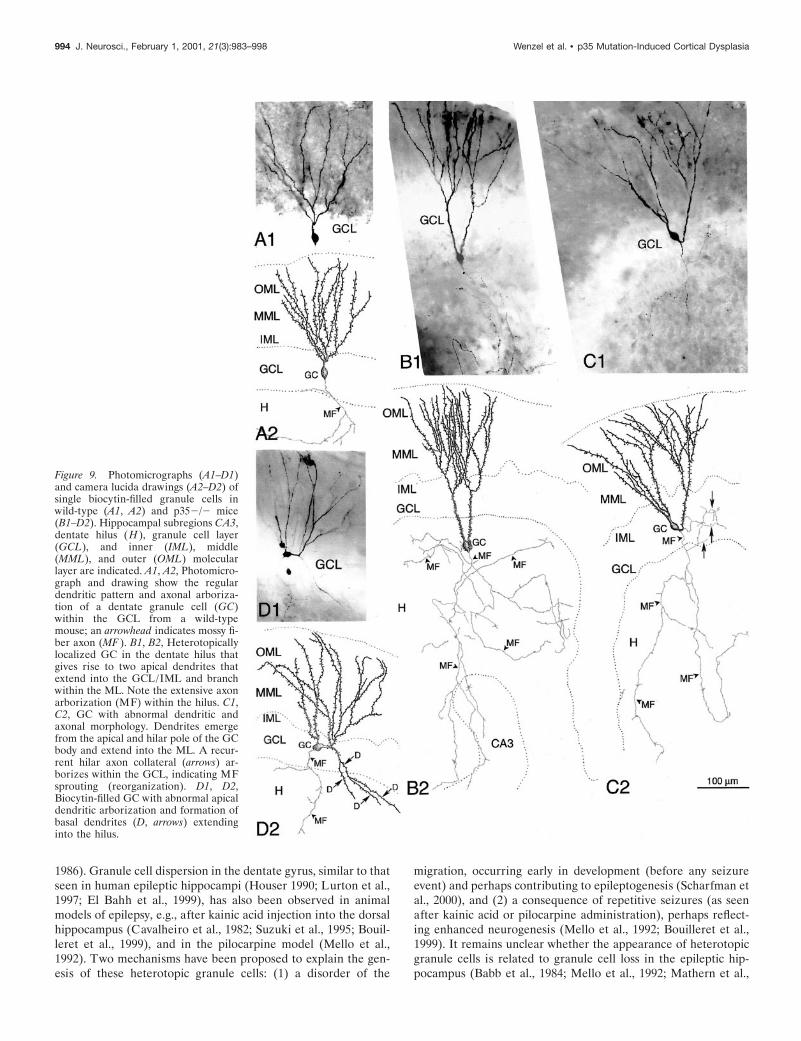
Intracellular labeling with biocytin reveals abnormalpatterns of granule cell localization, axonal anddendritic morphology, and mossy fiber reorganizationLight microscopic examination of individual granule cells ob-tained after in vitro injection of biocytin into the s. lucidum of
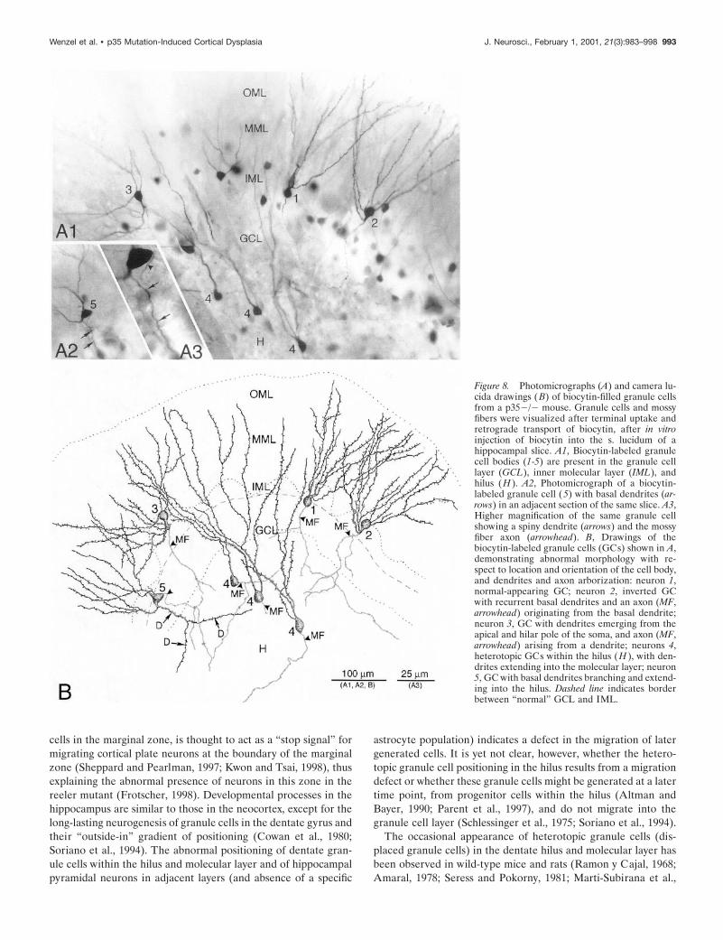
hippocampal slices, or intracellular labeling with biocytin, re-vealed a high degree of granule cell abnormality in p352/2dentate with respect to the localization of granule cell somata andin the orientation of their dendritic fields and axon arbors (Figs.8, 9). Granule cell somata of wild-type mice have oval or round-shaped cell bodies, typically giving rise to a spiny apical dendritictree that arborizes within the molecular layer; the mossy fiberaxon arises from the hilar side of the soma and runs toward theCA3 subfield. As shown with Golgi and recent intracellular la-beling studies (Spigelman et al., 1998; Ribak et al., 2000), ourexamination of granule cells of wild-type mice revealed onlyexceptional abnormalities. In contrast, as shown in Figure 8,granule cells of p352/2 mice are often displaced, with cell bodieslocated either in the inner molecular zone (close to the GCL) orin clusters within the hilus. Normal-appearing granule cells inp352/2 dentate show the expected monopolar-oriented dendriticbranching pattern within the molecular layer (Fig. 8A1,B, cell 1).However, many granule cells exhibit a bipolar dendritic arboriza-tion (Fig. 8, cells 2, 3, 5; Fig. 9B1–2,C1–2). The spiny dendrites ofsome p352/2 granule cells arose from the hilar side of the cellbody and then turned 180° to form “recurrent” basal dendritesascending through the molecular layer (Fig. 8, cell 2; Fig. 9C);some dendrites originating from the hilar pole of the cell bodyalso extend into the subgranular hilar region (Fig. 8B, cell 5; Fig.9D1-2). Granule cells heterotopically localized within the hilusgave rise to one or two primary dendrites that ran through thehilus toward the granule cell layer to branch within both granulecell and molecular layers (Fig. 8, cell 4; Fig. 9B).
Abnormally localized granule cells, as well as morphologicallyabnormal granule cells localized within the granule cell layer,usually displayed an axon that arose from the cell body and rantoward the CA3 s. lucidum, forming typical small “en passant”boutons and large MF boutons with filipodial extensions (Fig.9B2,C2). These observations suggest that both heterotopic andnormally positioned granule cells contribute to the normal MFprojection in p35 2/2 mice. However, many of these displacedcells also showed an unusually extensive MF arborization withinthe hilus (Fig. 9B2). The axons of some granule cells arose fromanomalous locations, e.g., the lateral side of the cell body, theapical dendrite, or the initial portion of a recurrent basal dendrite.Figures 8 (A1 and B, cells 2 and 3) and 9 (C2) indicate someexamples of abnormal axon origin. Finally, granule cells ofp352/2 mice, either localized within the granule cell layer ordisplaced into the molecular layer, show recurrent MF sproutinginto the granule cell layer and/or inner molecular layer, indicativeof MF reorganization. Figure 9C1–2 shows an example of a“bipolar” granule cell forming not only recurrent basal dendritesbut also an aberrant MF axon collateral that forms a plexuswithin the granule cell layer.
DISCUSSIONThese results expand the previous description by Tsai and hercolleagues (Chae et al., 1997; Kwon and Tsai, 1998) showing thatnormal development of hippocampus and dentate gyrus involvesp35-mediated processes. We have, in addition, found that (1)hippocampal pyramidal neurons of the CA1 and CA3 subfieldsand granule cells of the dentate gyrus are heterotopically local-ized, (2) a significant proportion of p352/2 mice exhibit sponta-neous behavioral and electrographic seizure, and (3) heterotopicgranule cells exhibit features (e.g., basal dendrites, abnormal
Figure 5. Graphs show the numbers of granule cells counted in thedentate granule cell and molecular layers per hippocampus of the wild-type and p352/2 mice at 2 months of age. A, The number of granule cellswithin the granule cell layer per entire hippocampus. The granule cellnumber is slightly greater (nonsignificantly) in wild-type mice than inp352/2 animals. B, The number of heterotopic granule cells within themolecular layer. In p352/2 mice, ;30% of the total number of granulecells is heterotopically localized within the molecular layer, comparedwith ;14% in wild-type mice (*p , 0.001). C, The total number ofgranule cells per hippocampus. There is no significant difference in thetotal number of granule cells between wild-type and p352/2 mice.Although the number of dispersed granule cells is significantly greater inp352/2 mice (1125% in the molecular layer), the absolute number ofdisplaced neurons is relatively small compared with the number of cells inthe granule cell layer. Thus, there is no statistical difference when totalnumbers are compared.
Wenzel et al. • p35 Mutation-Induced Cortical Dysplasia J. Neurosci., February 1, 2001, 21(3):983–998 991

axon origin, and aberrant mossy fiber collaterals and synapses)often observed in epileptic tissue.
The findings of Kwon and Tsai (1998) in p35 knock-out mice,together with similar cortical migration defects in cdk5-deficient
mice (Ohshima et al., 1996; Gilmore et al., 1998) suggest a criticalsignal transmission function of p35/cdk5 kinase for the migrationof cortical neurons (or glia) to their proper destinations. Incontrast, Reelin, a glycoprotein secreted by the Cajal-Retzius
Figure 6. A, Field EPSPs evoked by stim-ulation in the dentate molecular layer wererecorded from sites in the superior blade ofthe dentate gyrus in wild-type and p352/2mice. B, The amplitude of these EPSPs wasmeasured at a fixed latency [peak of EPSPrecorded in the granule cell layer (GCL)]and plotted as a function of recording loca-tion along an axis perpendicular to the GCL(wild-type, F; p352/2, �). C, Schematicdrawing showing location of normallyplaced granule cells (solid ovals) and dis-persed granule cells (dotted ovals). IML, In-ner molecular layer; MML, middle molecu-lar layer; OML, outer molecular layer.
Figure 7. Electron microscopy of mossyfiber boutons (MFB) heterotopically lo-calized within the granule cell layer(GCL) and inner molecular layer (IML)of p352/2 mice. MFBs are identifiedwith ZnT3 immunocytochemistry(black DAB-nickel reaction product inC and E). A, Low-power magnificationof the dentate gyrus (superior blade)with heterotopic GCs within the IML.B, In the IML (indicated by top box andarrow in A), an MFB forms asymmetricsynapses with spines (S) of granulecells. C, A ZnT3-positive MFB makesan asymmetric synapse with a simplespine. D, E, In the granule cell layer(indicated by bottom box and arrow), anonlabeled (D) and ZnT3-positive (E)MFB make asymmetric synapses withspines (S) and dendrites (D) of pre-sumed granule cells. Scale bars: B–E,0.25 mm.
992 J. Neurosci., February 1, 2001, 21(3):983–998 Wenzel et al. • p35 Mutation-Induced Cortical Dysplasia
cells in the marginal zone, is thought to act as a “stop signal” formigrating cortical plate neurons at the boundary of the marginalzone (Sheppard and Pearlman, 1997; Kwon and Tsai, 1998), thusexplaining the abnormal presence of neurons in this zone in thereeler mutant (Frotscher, 1998). Developmental processes in thehippocampus are similar to those in the neocortex, except for thelong-lasting neurogenesis of granule cells in the dentate gyrus andtheir “outside-in” gradient of positioning (Cowan et al., 1980;Soriano et al., 1994). The abnormal positioning of dentate gran-ule cells within the hilus and molecular layer and of hippocampalpyramidal neurons in adjacent layers (and absence of a specific
astrocyte population) indicates a defect in the migration of latergenerated cells. It is yet not clear, however, whether the hetero-topic granule cell positioning in the hilus results from a migrationdefect or whether these granule cells might be generated at a latertime point, from progenitor cells within the hilus (Altman andBayer, 1990; Parent et al., 1997), and do not migrate into thegranule cell layer (Schlessinger et al., 1975; Soriano et al., 1994).
The occasional appearance of heterotopic granule cells (dis-placed granule cells) in the dentate hilus and molecular layer hasbeen observed in wild-type mice and rats (Ramon y Cajal, 1968;Amaral, 1978; Seress and Pokorny, 1981; Marti-Subirana et al.,
Figure 8. Photomicrographs (A) and camera lu-cida drawings (B) of biocytin-filled granule cellsfrom a p352/2 mouse. Granule cells and mossyfibers were visualized after terminal uptake andretrograde transport of biocytin, after in vitroinjection of biocytin into the s. lucidum of ahippocampal slice. A1, Biocytin-labeled granulecell bodies (1-5) are present in the granule celllayer (GCL), inner molecular layer (IML), andhilus (H ). A2, Photomicrograph of a biocytin-labeled granule cell (5) with basal dendrites (ar-rows) in an adjacent section of the same slice. A3,Higher magnification of the same granule cellshowing a spiny dendrite (arrows) and the mossyfiber axon (arrowhead). B, Drawings of thebiocytin-labeled granule cells (GCs) shown in A,demonstrating abnormal morphology with re-spect to location and orientation of the cell body,and dendrites and axon arborization: neuron 1,normal-appearing GC; neuron 2, inverted GCwith recurrent basal dendrites and an axon (MF,arrowhead) originating from the basal dendrite;neuron 3, GC with dendrites emerging from theapical and hilar pole of the soma, and axon (MF,arrowhead) arising from a dendrite; neurons 4,heterotopic GCs within the hilus (H ), with den-drites extending into the molecular layer; neuron5, GC with basal dendrites branching and extend-ing into the hilus. Dashed line indicates borderbetween “normal” GCL and IML.
Wenzel et al. • p35 Mutation-Induced Cortical Dysplasia J. Neurosci., February 1, 2001, 21(3):983–998 993
1986). Granule cell dispersion in the dentate gyrus, similar to thatseen in human epileptic hippocampi (Houser 1990; Lurton et al.,1997; El Bahh et al., 1999), has also been observed in animalmodels of epilepsy, e.g., after kainic acid injection into the dorsalhippocampus (Cavalheiro et al., 1982; Suzuki et al., 1995; Bouil-leret et al., 1999), and in the pilocarpine model (Mello et al.,1992). Two mechanisms have been proposed to explain the gen-esis of these heterotopic granule cells: (1) a disorder of the
migration, occurring early in development (before any seizureevent) and perhaps contributing to epileptogenesis (Scharfman etal., 2000), and (2) a consequence of repetitive seizures (as seenafter kainic acid or pilocarpine administration), perhaps reflect-ing enhanced neurogenesis (Mello et al., 1992; Bouilleret et al.,1999). It remains unclear whether the appearance of heterotopicgranule cells is related to granule cell loss in the epileptic hip-pocampus (Babb et al., 1984; Mello et al., 1992; Mathern et al.,
Figure 9. Photomicrographs (A1–D1)and camera lucida drawings (A2–D2) ofsingle biocytin-filled granule cells inwild-type (A1, A2) and p352/2 mice(B1–D2). Hippocampal subregions CA3,dentate hilus (H ), granule cell layer(GCL), and inner (IML), middle(MML), and outer (OML) molecularlayer are indicated. A1, A2, Photomicro-graph and drawing show the regulardendritic pattern and axonal arboriza-tion of a dentate granule cell (GC)within the GCL from a wild-typemouse; an arrowhead indicates mossy fi-ber axon (MF ). B1, B2, Heterotopicallylocalized GC in the dentate hilus thatgives rise to two apical dendrites thatextend into the GCL/IML and branchwithin the ML. Note the extensive axonarborization (MF) within the hilus. C1,C2, GC with abnormal dendritic andaxonal morphology. Dendrites emergefrom the apical and hilar pole of the GCbody and extend into the ML. A recur-rent hilar axon collateral (arrows) ar-borizes within the GCL, indicating MFsprouting (reorganization). D1, D2,Biocytin-filled GC with abnormal apicaldendritic arborization and formation ofbasal dendrites (D, arrows) extendinginto the hilus.
994 J. Neurosci., February 1, 2001, 21(3):983–998 Wenzel et al. • p35 Mutation-Induced Cortical Dysplasia
1994; Suzuki et al., 1995). Granule cell dispersion has not beenobserved when cell loss is minimal (Lurton et al., 1997), but bothappear to be directly related to mossy fiber sprouting into themolecular layer (Cavazos and Sutula, 1990; Babb et al., 1991;Houser, 1999). The present study, using Timm staining and in-tracellular labeling with biocytin to vizualize mossy fiber boutons,demonstrates that heterotopic granule cells generate (and re-ceive) aberrant mossy fibers, in both granule cell and molecularlayers. In addition, heterotopic granule cells show basal dendrites(entering the hilus) and axons originating from the apical pole ofthe soma or the apical dendrite. These findings represent abnor-mal phenomena associated with epilepsy both in human hip-pocampus (Franck et al., 1995) and in experimental animal mod-els (Spigelman et al., 1998; Ribak et al., 2000). The behavioralobservations of the present study substantiate the enhanced sus-ceptibility of p352/2 mice to seizures. Simultaneous video/EEGrecordings confirmed spontaneous epileptiform activity, includ-ing both intermittent interictal discharge and spontaneous gener-alized seizures. Although no fatalities were observed duringrecorded seizures, the severity of observed seizures, together withoccasional sudden (unexplained) death of some animals, suggeststhat the mortality rate of ;10% [see also Chae et al. (1997)]might be attributable to fatal seizures. Previous experimentscomparing PTZ-induced seizures in p352/2 and wild-type miceshowed that generalized convulsions were only fatal in p352/2mice (Chae et al., 1997).
Early in postnatal development (before postnatal day 10), be-fore any spontaneous seizure, p352/2 mice exhibit heterotopicgranule cells, indicating that this abnormality is not a result ofseizure activity and suggesting a causal relationship betweenstructural abnormality and occurrence of spontaneous seizures.Other studies investigating animal models of cortical dysplasiawith experimentally induced disruption of cortical developmentalso suggest a causal link between cortical malformations andseizure activity (Chevassus-au-Louis et al., 1999; Walsh, 1999;Fleck et al., 2000; Chen et al., 2000). Such animal models includemigrational malformations induced by freezing (Dvorak and Feit,1977; Dvorak et al., 1978; Rosen et al., 1992, 1996; Jacobs et al.,1996, Hablitz and DeFazio, 1998), x-irradiation (Roper et al.,1995, 1997; Hicks et al., 1959), and chemical treatment (e.g.,methylazoxymethanol: Singh, 1977; Cattaneo et al., 1995; De Feoet al., 1995; Baraban and Schwartzkroin, 1996; Germano andSperber, 1997; Chevassus-au-Louis et al., 1998, 1999; Baraban etal., 2000). Animals with abnormalities paralleling human geneticneuronal migration disorders also show enhanced seizure-sensi-tivity: e.g., type 1 lissencephaly (Lis1 mutant) (Reiner et al., 1993;Hirotsune et al., 1998; Fleck et al., 2000), and band heterotopia(tish mutant) (Lee et al., 1997). Although many of these animalmodels exhibit increased excitability (Baraban and Schwartzk-roin, 1995; Jacobs et al., 1996, 1999; Luhmann and Raabe, 1996;Roper et al., 1997; Baraban et al., 2000) (for review, seeChevassus-au-Louis et al., 1999), the presence of a cortical mal-formation does not necessarily result in a spontaneous seizurephenotype. One might speculate that as in humans, a subtlepreexisting focal cortical dysgenesis (e.g., heterotopically dis-placed neurons) provides a substrate for seizure induction (e.g.,from a febrile episode) and may also predispose the animal to aseizure-related pathology (e.g., hippocampal sclerosis in TLE)(Lewis, 1999).
The underlying basis for hyperexcitability (and synchrony)associated with cortical disorganization and reorganization re-mains a topic of intense investigation. Numerous studies have
shown that alterations of GABAergic neurotransmission might becritically involved in underlying epileptic disorders (Schwartzk-roin, 1998). Although the present study did not reveal any obviousloss of specific interneuron populations in p352/2 mice, bothGAD67- and parvalbumin-containing axon terminals were abnor-mally distributed within the molecular layer, surrounding somataof heterotopic granule cells, reflecting correct target innervation(Fleck et al., 2000). However, the reduced parvalbumin immuno-reactivity within the terminal plexus around the granule cellssuggests the possibility of functional abnormality in these inhib-itory circuits, either as a direct result of the p352/2 disorgani-zation or as a result of earlier seizures. Loss of parvalbuminimmunoreactivity has also been observed after kainic acid-induced seizures (Sloviter 1991, 1994; Best et al., 1993; Buckmas-ter and Dudek, 1997), although transient parvalbumin loss maynot be related to cell death (Scotti et al., 1997). Subtle changes ofparvalbumin immunoreactivity in the dentate gyrus of p352/2mice could be of relevance for seizure sensitivity of these animals(Scotti and Nitsch, 1991). In addition, in both wild-type andp352/2 animals, the calcium binding protein calretinin is ex-pressed in excitatory hilar mossy cells (Liu et al., 1996; Blasco-Ibanez and Freund, 1997; Schurmans et al., 1997), which form adense axonal plexus within the dentate inner molecular layer(Buckmaster et al., 1996). In p352/2 mice, heterotopic granulecells within the inner molecular layer are surrounded by thesecalretinin-positive terminals; the abnormal position of excitatorymossy cell synapses onto granule cell bodies (data not shown)could undoubtedly alter the balance of excitation and inhibition inthis sensitive circuit.
Physiological studies in epileptic human tissue (Masukawa etal., 1991, 1992; Williamson, 1994; Franck et al., 1995) and exper-imental animal models (Scharfman et al., 1990; Ribak et al., 1992;Sloviter, 1994; Wuarin and Dudek, 1996; Patrylo and Dudek,1998; Okazaki et al., 1999) have shown that the dentate gyrus canplay an important role in epileptic conditions (Masukawa et al.,1999). Dentate granule cells are ideally situated to control thespread of excitation. Developmental malformations of the den-tate, whether genetically based or caused by early events such astrauma, fever or infection, may lead to various complex changes(e.g., cell loss, mossy fiber sprouting, receptor alterations, granulecell dispersion) that result in an imbalance between excitationand inhibition. The p352/2 mutant mouse represents one exam-ple of an experimental model in which anatomical disorganiza-tion is closely linked to seizures. Further studies are necessary toclarify how the p35 mutation and related structural abnormalitieslead to epileptiform activities.
REFERENCESAltman J, Bayer SA (1990) Mosaic organization of hippocampal neuro-
epithelium and the multiple germinal sources of dentate granule cells.J Comp Neurol 301:325–342.
Amaral DG (1978) A Golgi study of cell types in the hilar region ofthe hippocampus of the rat. J Comp Neurol 182:852–914.
Babb TL, Brown WJ, Pretorious J, Davenport C, Lieb JP, Crandall PH(1984) Temporal lobe volumetric cell densities in temporal lobeepilepsy. Epilepsia 25:729 –740.
Babb TL, Kupfer WR, Pretorius JK, Crandall PH, Levesque MF(1991) Synaptic reorganization by mossy fibers in human epilepticfascia dentata. Neuroscience 42:351–363.
Baraban SC, Schwartzkroin PA (1995) Electrophysiology of CA1 py-ramidal neurons in an animal model of neuronal migration disor-ders: prenatal methylazoxymethanol treatment. Epilepsy Res22:145–156.
Baraban SC, Schwartzkroin PA (1996) Flurothyl seizure susceptibil-
Wenzel et al. • p35 Mutation-Induced Cortical Dysplasia J. Neurosci., February 1, 2001, 21(3):983–998 995
ity in rats following prenatal methylazoxymethanol treatment. Epi-lepsy Res 23:189 –194.
Baraban SC, Wenzel HJ, Hochmann DW, Schwartzkroin PA (2000)Characterization of heterotopic cell clusters in the hippocampus ofrats exposed to methylazoxymethanol in utero. Epilepsy Res39:87–102.
Barkovich AJ, Kuzniecky RI, Dobyns WB, Jackson GD, Becker LE,Evrard P (1996) A classification scheme for malformations of cor-tical development. Neuropediatrics 27:59 – 63.
Best N, Mitchell J, Wheal HV (1993) Changes in parvalbumin-immunoreactive neurons in the CA1 area of the rat hippocampusfollowing a kainic acid lesion. Neurosci Lett 155:1– 6.
Blasco-Ibanez JM, Freund TF (1997) Distribution, ultrastructure,and connectivity of calretinin-immunoreactive mossy cells of themouse dentate gyrus. Hippocampus 7:307–320.
Bouilleret V, Ridoux V, Depaulis A, Marescaux C, Nehlig A, Le GalLa Salle G (1999) Recurrent seizures and hippocampal sclerosisfollowing intrahippocampal kainate injection in adult mice: electro-encephalography, histopathology and synaptic reorganization simi-lar to mesial temporal lobe epilepsy. Neuroscience 89:717–729.
Buckmaster PS, Dudek FE (1997) Neuron loss, granule cell axonreorganization, and functional changes in the dentate gyrus ofepileptic kainate-treated rats. J Comp Neurol 385:385– 404.
Buckmaster PS, Kunkel DD, Robbins RJ, Schwartzkroin PA (1994)Somatostatin-immunoreactivity in the hippocampus of mouse, rat,guinea pig, and rabbit. Hippocampus 4:167–180.
Buckmaster PS, Wenzel J, Kunkel DD, Schwartzkroin PA (1996)Axon arbors and synaptic connections of hippocampal mossy cells inthe rat in vivo. J Comp Neurol 366:270 –292.
Cattaneo E, Reinach B, Caputi A, Cattabeni F, Di Luca M (1995)Selective in vitro blockade of neuroepithelial cells proliferation bymethylazoxymethanol acetate, a molecule capable of inducing longlasting functional impairments. J Neurosci Res 41:640 – 647.
Cavalheiro EA, Riche D, Le Gal La Salle G (1982) Lon-term effcts ofintrahippocampal kainic acid injection in rats: a method for inducingspontaneous recurrent seizures. Electroencephalogr Clin Neuro-physiol 53:581–589.
Cavazos JE, Sutula TP (1990) Progressive neuronal loss induced bykindling: a possible mechanism for mossy fiber synaptic reorganiza-tion and hippocampal sclerosis. Brain Res 527:1– 6.
Caviness Jr VS (1976) Patterns of cell and fiber distribution in theneocortex of the reeler mutant mice. J Comp Neurol 170:435– 448.
Caviness Jr VS, Sidman RL (1973) Time of origin corresponding cellclasses in the cerebral cortex of normal and reeler mutant mice: anautoradiographic analysis. J Comp Neurol 148:141–152.
Chae T, Kwon YT, Bronson R, Dikkes P, Li E, Tsai L-H (1997) Micelacking p35, a neuronal specific activator of cdk5, display corticallamination defects, seizures, and adult lethality. Neuron 18:29 – 42.
Chen Z-F, Schottler F, Bertram E, Gall CM, Anzivino MJ, Lee KS(2000) Distribution and initiation of seizure activity in a rat brainwith subcortical band heterotopia. Epilepsia 41:493–501.
Chevassus-au-Louis N, Rafiki A, Jorquera I, Ben-Ari Y, Represa A(1998) Neocortex in the hippocampus: an anatomical and functionalstudy of CA1 heterotopias after prenatal treatment with methyla-zoxymethanol in rats. J Comp Neurol 394:520 –536.
Chevassus-au-Louis N, Baraban SC, Gaiarsa J-L, Ben-Ari Y (1999)Cortical malformations and epilepsy: new insights from animal mod-els. Epilepsia 40:811– 821.
Cowan WM, Stanfield BB, Kishi K (1980) The development of thedentate gyrus. Curr Topics Dev Biol 15:103–157.
De Feo MR, Mecarelli O, Ricci GF (1995) Seizure susceptibility inimmature rats with microencephaly induced by prenatal exposure tomethylazoxymethanol acetate. Pharmacol Res 31:109 –114.
Delalle I, Bhide PG, Caviness Jr VS, Tsai L-H (1997) Temporal andspatial patterns of expression of p35, a regulatory subunit of cyclin-dependent kinase 5, in the nervous system of the mouse. J Neuro-cytol 26:283–296.
Des Portes V, Pinard JM, Billuart P, Vinet MC, Koulakoff A, CarrieA, Gelot A, Dupuis E, Motte J, Berwald-Netter Y, Catala M, KahnA, Beldjord C, Chelly J (1998) A novel CNS gene required forneuronal migration and involved in X-linked subcortical laminarheterotopia and lissencephaly syndrome. Cell 92:51– 61.
Dvorak K, Feit J (1977) Migration of neuroblasts through partialnecrosis of the cerebral cortex in newborn rats: contribution to theproblems of morphological development and developmental periodof cerebral microgyria. Histological and autoradiographical study.Acta Neuropathol (Berl) 38:203–212.
Dvorak K, Feit J, Jurankova Z (1978) Experimental induced focalmicrogyria and status verrucosus deformis in rats- pathogenesis andinterrelation histological and autoradiographic study. Acta Neuro-pathol (Berl) 38:121–129.
El Bahh B, Lespinet V, Lurton D, Coussemacq M, Le Gal La Salle G,Rougier A (1999) Correlation between granule cell dispersion,mossy fiber sprouting, and hippocampal cell loss in temporal lobeepilepsy. Epilepsia 40:1393–1401.
Falconer D (1951) Two new mutants, “trembler” and “reeler,” withneurological actions in the house mouse. J Genet 50:192–201.
Fleck MW, Hirotsune S, Gambello MJ, Phillips-Tansey E, Suares G,Mervis RF, Wynshaw-Boris A, McBain CJ (2000) Hippocampalabnormalities and enhanced excitability in a murine model of humanlissencephaly. J Neurosci 20:2439 –2450.
Franck JE, Pokorny J, Kunkel DD, Schwartzkroin PA (1995) Physio-logic and morphologic characteristics of granule cell circuitry inhuman epileptic hippocampus. Epilepsia 36:543–558.
Freund TF, Buszaki G (1996) Interneurons of the hippocampus. Hip-pocampus 6:347– 470.
Frotscher M (1998) Cajal-Retzius cells, Reelin, and the formation oflayers. Curr Opin Neurobiol 8:570 –575.
Fukuda T, Heizmann CW, Kosaka T (1997) Quantitative analysis ofGAD65 and GAD67 immunoreactivities in somata of GABAergicneurons in the mouse hippocampus proper (CA1 and CA3 regions),with special reference to parvalbumin-containing neurons. BrainRes 764:237–243.
Germano IM, Sperber EF (1997) Increased seizure susceptibility in adultrats with neuronal migration disorders. Brain Res 777:219–222.
Gilmore EC, Ohshima T, Goffinet AM, Kulkarni AB, Herrup K(1998) Cyclin-dependent kinase 5-deficient mice demonstrate noveldevelopmental arrest in cerebral cortex. J Neurosci 18:6370 – 6377.
Guerrini R, Andermann E, Avoli M, Dobyns WB (1999) Corticaldysplasias, genetics, and epileptogenesis. In: Jasper’s basic mecha-nisms of the epilepsies, Ed 3, Advances in neurology, Vol 79(Delgado-Escueta AV, Wilson WA, Olsen RW, Porter RJ, eds), pp95–121. Philadelphia: Lippincott Williams & Wilkins.
Hablitz JJ, DeFazio T (1998) Excitability changes in freeze-inducedneocortical microgyria. Epilepsy Res 32:75– 82.
Hicks SP, D’Amato CJ, Lowe MJ (1959) The development of themammalian nervous system: I. Malformation of the brain, especiallythe cerebral cortex, induced in rats by radiation. II. Some mecha-nisms of the malformations of the cortex. J Comp Neurol113:435– 469.
Hirotsune S, Fleck MW, Gambello MJ, Bix GJ, Chen A, Clark GD,Ledbetter DH, McBain CJ, Wynshaw-Boris A (1998) Graded re-duction of Pafah 1b1 (Lis1) activity results in neuronal migrationdefects and early embryonic lethality. Nat Genet 19:333–339.
Honavar M, Meldrum BS (1997) Epilepsy. In: Greenfield’s neuropa-thology, Vol 2 (Graham DI, Lantos PL, eds), pp 931–971. London:Arnold.
Houser CR (1990) Granule cell dispersion in the dentate gyrus ofhumans with temporal lobe epilepsy. Brain Res 535:195–204.
Houser CR (1999) Neuronal loss and synaptic reorganization in tem-poral lobe epilepsy. In: Jasper’s basic mechanisms of the epilepsies,Ed 3, Advances in neurology, Vol 79 (Delgado-Escueta AV, WilsonWA, Olsen RW, Porter RJ, eds), pp 743–761. Philadelphia: Lippin-cott Williams & Wilkins.
Houser CR, Esclapez M (1994) Localization of mRNAs encoding twoforms of glutamic acid decarboxylase in the rat hippocampal forma-tion. Hippocampus 5:530 –545.
Hsu S-M, Raine L, Fanger H (1981) Use of avidin-biotin-peroxidasecomplex (ABC) in immunoperoxidase techniques: a comparisonbetween ABC and unlabeled antibody (PAP) procedures. J Histo-chem Cytochem 29:577–580.
Huttenlocher A, Sandborg R, Horwitz A (1995) Adhesion in cellmigration. Curr Opin Cell Biol 7:697–706.
Jacobs KM, Hwang BJ, Prince DA (1996) Focal epileptogenesis in arat model of polymicrogyria. J Neurophysiol 81:159 –173.
Kwon YT, Tsai L-H (1998) A novel disruption of cortical develop-ment in p352/2 mice distinct from reeler. J Comp Neurol395:510 –522.
Kwon YT, Tsai L-H, Crandall JE (1999) Callosal axon guidancedefects in p352/2 mice. J Comp Neurol 415:218 –229.
Laatsch RH, Cowan WM (1966) Electron microscopic studies of thedentate gyrus of the rat. I. Normal structure with special referenceto synaptic organization. J Comp Neurol 128:359 –396.
Lee KS, Schottler F, Collins JL, Lanzino G, Couture D, Rao A,Hiramatsu K, Goto Y, Hong S-C, Caner H, Yamamoto H, ChenZ-F, Bertram E, Berr S, Omary R, Scrable H, Jackson T, Goble J,Eisenman L (1997) A genetic animal model of human neocorticalheterotopia associated with seizures. J Neurosci 17:6236 – 6242.
Lewis DV (1999) Febrile convulsions and mesial temporal sclerosis.Curr Opin Neurol 12:197–201.
Liu Y, Fujise N, Kosaka T (1996) Distribution of calretinin immuno-reactivity in the mouse dentate gyrus. I. General description. ExpBrain Res 108:389 – 403.
Luhmann HJ, Raabe K (1996) Characterization of neuronal migra-tion disorders in neocortical structures: I. Expression of epilepti-form activity in an animal model. Epilepsy Res 26:67–74.
Lurton D, Sundstrom L, Brana C, Bloch B, Rougier A (1997) Possiblemechanisms inducing granule cell dispersion in humans with tem-poral lobe epilepsy. Epilepsy Res 26:351–361.
Marti-Subirana A, Soriano E, Garcia-Verdugo JM (1986) Morphologi-
996 J. Neurosci., February 1, 2001, 21(3):983–998 Wenzel et al. • p35 Mutation-Induced Cortical Dysplasia
cal aspects of the ectopic granule-like cellular populations in the albinorat hippocampal formation: a Golgi study. J Anat 144:31–47.
Masukawa LM, Higashima M, Hart GJ, Spencer DD, O’Connor MJ(1991) NMDA receptor activation during epileptiform responses inthe dentate gyrus of epileptic patients. Brain Res 562:176 –180.
Masukawa, LM, Urono K, Sperling M, O’Connor MJ, Burdette LJ(1992) The functional relationship between antidromically evokedfield responses of the dentate gyrus and mossy fiber reorganizationin temporal lobe epileptic patients. Brain Res 579:119 –127.
Masukawa LM, Burdette LJ, McGonigle P, Wang H, O’Connor W,Sperling MR, O’Connor MJ, Uruno K (1999) Physiological andanatomical correlates of the human dentate gyrus: consequences orcauses of epilepsy. In: Jasper’s basic mechanisms of the epilepsies,Ed 3, Advances in neurology, Vol 79 (Delgado-Escueta AV, WilsonWA, Olsen RW, Porter RJ, eds), pp 781–794. Philadelphia: Lippin-cott Williams & Wilkins.
Mathern GW, Leite JP, Pretorius JK, Quinn B, Peacock WJ, Babb TL(1994) Children with severe epilepsy: evidence of hippocampal neu-ron losses and aberrant mossy fiber sprouting during postnatalgranule cell migration and differentiation. Dev Brain Res 78:70 – 80.
Mello LM, Cavalheiro E, Tan A, Pretorius JK, Babb T, Finch D(1992) Granule cell dispersion in relation to mossy fiber sprouting,hippocampal cell loss, silent period and seizure frequency in thepilocarpine model of epilepsy. In: Molecular neurobiology of epi-lepsy (Engel Jr J, Wasterlain C, Cavalheiro EA, Heinemann U,Avanzini G, eds), pp 51– 60. Amsterdam: Elsevier.
Mischel PS, Nguyen LP, Vinters HV (1995) Cerebral cortical dyspla-sia associated with pediatric epilepsy. Review of neuropathologicfeatures and proposal for a grading system. J Neuropathol ExpNeurol 54:137–153.
Ohshima T, Ward JM, Huh C-G, Longenecker G, Veeranna, Pant HC,Brady RO, Martin LJ, Kulkarni AB (1996) Targeted disruption ofthe cyclin-dependent kinase 5 gene results in abnormal corticogen-esis, neuronal pathology and perinatal death. Proc Natl Acad SciUSA 93:11173–11178.
Okazaki MM, Evenson DA, Nadler JV (1995) Hippocampal mossyfiber sprouting and synapse formation after status epilepticus in rats:visualization after retrograde transport of biocytin. J Comp Neurol352:515–534.
Okazaki MM, Molnar P, Nadler JV (1999) Recurrent mossy fiber pathwayin rat dentate gyrus: synaptic currents evoked in presence and absence ofseizure-induced growth. J Neurophysiol 81:1645–1660.
Parent JM, Yu TW, Leibowitz RT, Geschwind DH, Sloviter RS,Lowenstein DH (1997) Dentate granule cell neurogenesis is in-creased by seizures and contributes to aberrant network reorgani-zation in the adult rat hippocampus. J Neurosci 17:3727–3738.
Patrylo PR, Dudek FE (1998) Physiological unmasking of new glu-tamtergic pathways in the dentate gyrus of hippocampal slices fromkainate-induced epileptic rats. J Neurophysiol 79:418 – 429.
Rakic P, Caviness VSJ (1995) Cortical development: view from neu-rological mutants two decades later. Neuron 14:1101–1104.
Ramon y Cajal S (1968) The structure of Ammon’s horn. Springfield,IL: Charles Thomas.
Raymond AA, Fish DR, Sisodya SM, Alsanjari N, Stevens JM, Shor-von SD (1995) Abnormalities of gyration, heterotopias, focal corti-cal dysplasia, microdysgenesis, dysembryoplastic neuroepithelial tu-mour and dysgenesis of the archicortex in epilepsy: clinical, EEGand neuroimaging features in 100 adult patients. Brain 118:629 – 660.
Reiner O, Carrozzo R, Shen Y, Wehnert M, Faustinella F, Dobyns W,Caskey T, Ledbetter D (1993) Isolation of a Miller-Dieker lissen-cephaly gene containing G protein b-subunit-like repeats. Nature364:717–721.
Rho JM, Kim DW, Robbins CA, Anderson GD, Schwartzkroin PA(1999) Age-dependent differences in flurothyl seizure sensitivity inmice treated with a ketogenic diet. Epilepsy Res 37:233–240.
Ribak CE, Anderson L (1980) Ultrastructure of the pyramidal basketcells in the dentate gyrus of the rat. J Comp Neurol 192:903–916.
Ribak CE, Gall CM, Mody I (1992) The dentate gyrus and its role inseizures. Amsterdam: Elsevier.
Ribak CE, Tran PH, Spigelman I, Okazaki MM, Nadler JV (2000)Status epilepticus-induced hilar basal dendrites on rodent granulecells contribute to recurrent excitatory circuitry. J Comp Neurol428:240 –253.
Robbins CA, Wenzel HJ, Tsai L-H, Schwartzkroin PA (1999) Structuralorganization and function in dentate gyrus of the p35 mutant modelassociated with spontaneous seizures. Epilepsia [Suppl 7]40:34.
Roper SN, Gilmore RL, Houser CR (1995) Experimentally induceddisorders of neuronal migration produce an increased propensity forelectrographic seizures in rats. Epilepsy Res 21:205–219.
Roper SN, Abraham LA, Streit WJ (1997) Exposure to in uteroirradiation produces disruption of radial glia in rats. Dev Neurosci19:521–528.
Rosen GD, Press DM, Sherman GF, Galaburda AM (1992) The de-velopment of induced cerebrocortical microgyria in the rat. J Neu-ropathol Exp Neurol 51:601– 611.
Rosen GD, Sherman GF, Galaburda AM (1996) Birthdates of neu-rons in induced microgyria. Brain Res 727:71–78.
Samoriski GM, Applegate CD (1997) Repeated generalized seizuresinduce time-dependent changes in the behavioral seizure responseindependent of continued seizure induction. J Neurosci17:5581–5590.
Scharfman HE, Kunkel DD, Schwartzkroin PA (1990) Synaptic con-nections of dentate granule cells and hilar neurons: results of pairedintracellular recordings and intracellular horseradish peroxidaseinjections. J Neurosci 37:693–707.
Scharfman HE, Goodman JH, Sollas AL (2000) Granule-like neuronsat the hilar/CA3 border after status epilepticus and their synchronywith area CA3 pyramidal cells: functional implications of seizure-induced neurogenesis. J Neurosci 20:6144 – 6158.
Schlessinger AR, Cowan WM, Gottlieb DI (1975) An autoradio-graphic study of the time of origin and the pattern of granule cellmigration in the dentate gyrus of the rat. J Comp Neurol159:149 –176.
Schurmans S, Schiffmann SN, Gurden H, Lemaire M, Lipp H-P, SchwamV, Pochet R, Imperato A, Boehme GA, Parmentier M (1997) Impairedlong-term potentiation induction in dentate gyrus of calretinin-deficientmice. Proc Natl Acad Sci USA 94:10415–10420.
Schwartzkroin PA (1998) GABA synapses enter the molecular bigtime. Nat Med 4:1115–1116.
Scotti AL, Nitsch C (1991) The perforant path in the seizure sensitivegerbil contains the Ca 21-binding protein parvalbumin. Exp BrainRes 85:137–143.
Scotti AL, Kalt G, Bollag O, Nitsch C (1997) Parvalbumin disappearsfrom GABAergic CA1 neurons of the gerbil hippocampus withseizure onset while its presence persists in the perforant path. BrainRes 760:109 –117.
Seress L (1992) Morphological variability and developmental aspectsof the monkey and human granule cells: diffences between therodent and primate dentate gyrus. In: The dentate gyrus and its rolein seizures (Ribak CE, Gall CM, Mody I, eds), Amsterdam, Elsevier.
Seress L, Pokorny J (1981) Structure of the granular layer of the ratdentate gyrus: a light microscopic and Golgi study. J Comp Neurol300:449 – 461.
Sheppard AM, Pearlman AL (1997) Abnormal reorganization of pre-plate neurons and their associated extracellular matrix: an earlymanifestation of altered neocortical development in the reeler mu-tant mouse. J Comp Neurol 378:173–179.
Singh SC (1977) Ectopic neurons in the hippocampus of the postnatalrat exposed to methylazoxymethanol during foetal development.Acta Neuropathol (Berl) 40:111–116.
Sloviter RS (1991) Permanently altered hippocampal structure, excitabil-ity, and inhibition after experimental status epilepticus in the rat: the“dormant basket cell” hypothesis and its possible relevance to temporallobe epilepsy. Hippocampus 1:41–66.
Sloviter RS (1994) The functional organization of the hippocampaldentate gyrus and its relevance to the pathogenesis of temporal lobeepilepsy. Ann Neurol 35:640 – 654.
Smith BN, Dudek FE, Roper SN (1999) Synaptic responses of neu-rons in heterotopic gray matter in an animal model of corticaldysgenesis. Dev Neurosci 21:365–373.
Soriano E, Del Rio JA, Martinez A, Super H (1994) Organization ofthe embryonic and early postnatal murine hippocampus. I. Immu-nocytochemical characterization of neuronal populations in the sub-plate and marginal zone. J Comp Neurol 342:571–595.
Spigelman I, Yan X-X, Obenaus A, Lee EY-S, Wasterlain CL, RibakCE (1998) Dentate granule cells form novel basal dendrites in a ratmodel of temporal lobe epilepsy. Neuroscience 86:109 –120.
Sutula T, Zhang P, Lynch M, Sayin U, Golarai G, Rod R (1998)Synaptic and axonal remodeling of mossy fibers in the hilus andsupragranular region of the dentate gyrus in kainate-treated rats.J Comp Neurol 390:578 –594.
Suzuki F, Junier MP, Guilhem D, Sorensen JC, Onteniente B (1995)Morphogenetic effect of kainate on adult hippocampal neuronsassociated with a prolonged expression of brain-derived neurotro-phic factor. Neuroscience 64:665– 674.
Tsai L-H, Delalle I, Caviness Jr VS, Chae T, Harlow E (1994) p35 isa neural-specific regulatory subunit of cyclin-dependent kinase 5.Nature 371:419 – 423.
Walsh CA (1999) Genetic malformations of the human cerebral cor-tex. Neuron 23:19 –29.
Wenzel HJ, Cole TB, Born DE, Schwartzkroin PA, Palmiter RD(1997) Ultrastructural localization of zinc transporter-3 (ZnT-3) tosynaptic vesicle membranes within mossy fiber boutons in the hip-pocampus of mouse and monkey. Proc Natl Acad Sci USA 94:12676 –12681.
Wenzel et al. • p35 Mutation-Induced Cortical Dysplasia J. Neurosci., February 1, 2001, 21(3):983–998 997

Wenzel HJ, Kwon YT, Tsai L-H, Schwartzkroin PA (1998) Loss ofp35, a neuronal-specific activator of cdk5, results in abnormal hip-pocampal development in mice. Abstr Soc Neurosci 24:918.
Wenzel HJ, Woolley CS, Robbins CA, Schwartzkroin PA (2000)Kainic acid-induced mossy fiber sprouting and synapse formation inthe dentate gyrus of rats. Hippocampus 10:244 –260.
Wenzel J, Stender G, Duwe G (1981) Zur Entwicklung der Neu-ronenstruktur der Fascia dentata bei der Ratte. Neurohistologisch-morphometrische, ultrastrukturelle und experimentelle Untersu-chungen. J Hirnforsch 22:629 – 683.
West MJ, Gundersen HJG (1990) Unbiased stereological estimationof the number of neurons in the human hippocampus. J Comp
Neurol 296:1–22.West MJ, Slomianka L, Gundersen HJG (1991) Unbiased stereologi-
cal estimation of the total number of neurons in the subdivisions ofthe rat hippocampus using the optical fractionator. Anat Rec231:482– 497.
Williamson A (1994) Electrophysiology of epileptic human neocorticaland hippocampal neurons maintained in vitro. Clin Neurosci 2:47–52.
Wuarin J-P, Dudek FE (1996) Electrographic seizures and new re-current excitatory circuits in the dentate gyrus of hippocampal slicesfrom kainate-treated epileptic rats. J Neurosci 16:4438 – 4448. “dor-mant basket cell” hypothesis and its possible relevance to temporallobe epilepsy. Hippocampus 1:41– 66.
998 J. Neurosci., February 1, 2001, 21(3):983–998 Wenzel et al. • p35 Mutation-Induced Cortical Dysplasia