Embed Size (px)
Citation preview
Abnormal Cytolytic Activity of Lymphocyte Function-associatedAntigen-1-deficient Human Cytolytic T Lymphocyte Clones
Steven J. Mentzer, Barbara E. Bierer, Donald C. Anderson, Timothy A. Springer, and Steven J. BurakoffDivisions ofPediatric Oncology and Membrane Immunochemistry, Dana-Farber Cancer Institute, Harvard Medical School,Boston, Massachusetts 02115; and Department ofMedicine, Baylor University, Houston, Texas 77030
Abstract
The involvement of the lymphocyte function-associated antigen-1 (LFA-1) membrane molecule in cytolytic T lymphocyte (CTL)interactions with lymphoid target cells was investigated usingCTL clones derived from two patients with a heritable deficiencyof LFA-1. LFA-1 surface expression on the CTL clones was 1%of the normal level of LFA-1, unchanged with prolonged culture,and identical on 14 different CTL clones. The function of theLFA-1 molecule was addressed using the LFA-1-deficient CTLclones and LFA-1-deficient lymphoid target cells. The lytic ac-tivity of the LFA-1-deficient CTL clones was 43% of controlwhen tested against a target cell line expressing normal levelsof LFA-1 and <10% of control when tested against an LFA-1-deficient target cell line. These results demonstrate a direct in-volvement of LFA-1 in CTL-mediated cytolysis and suggest amore general dependence on LFA-1 in lymphoid cell-cell inter-actions.
Introduction
Human lymphocyte function-associated antigen-1 (LFA-1)' isa 177- and 95-kD glycoprotein heterodimer that is expressed onall leukocytes, most thymocytes, and on a third ofbone marrowcells (1). The LFA-l molecule was initially identified by screeningmonoclonal antibodies (MAb) for their ability to inhibit cytolyticT lymphocyte (CTL)-mediated cytotoxicity in the absence ofcomplement (1, 2). MAb to LFA-l significantly inhibited notonly cell-mediated cytotoxicity but T cell proliferation as well(1). In contrast, MAb to a variety ofother cell surface molecules,often present at higher cell surface density than LFA- 1, did notinhibit T cell function. The ability ofanti-LFA-1 MAb to inhibita variety of T cell functions led to the suggestion that LFA-lwas an essential cell-cell interaction molecule.
A unique opportunity to assess the function of the LFA-lmolecule has been provided by patients with a genetic deficiencyof the LFA- I glycoprotein. The LFA- I deficiency syndrome isan immunodeficiency characterized by a susceptibility to re-
Address correspondence to Dr. Mentzer, 1630D, Dana-Farber CancerInstitute, 44 Binney St., Boston, MA 02115.
Receivedfor publication 8 May 1986.
1. Abbreviations used in this paper: CR3, complement receptor type 3;CTL, cytolytic T lymphocyte; EBV, Epstein-Barr virus; F-GAM, fluo-rescein-conjugated F(ab')2 goat anti-mouse antibody; IL-2, interleukin2; LFA-1, lymphocyte function-associated antigen-l; LU, lytic units;MAb, monoclonal antibodies.
current life-threatening infections (3). Biochemical analysis ofthe leukocytes from LFA- I -deficient patients have shown a de-ficiency of not only LFA-1, but of a structurally related familyof molecules (4). LFA-1, complement receptor type 3 (CR3),and p1 50,95 are heterodimers with different heavy chains but astructurally identical 95-kD light chain. All three molecules aredeficient in patients with the clinical syndrome (5). The advan-tage of studying CTL from these deficient patients is that sig-nificant levels ofCR3 and p 150,95 are not expressed on normalT cells. Thus, the CTL from these patients have a selective de-ficiency of the LFA- 1 membrane molecule.
Previous in vitro studies of LFA- 1-deficient CTL functionhave been inconclusive. Arnaout et al. (6) and Miedema et al.(7) have found no significant defect in CTL function. Krenskyet al. (8), using HLA-typed related and unrelated controls, dem-onstrated abnormalities in both cell-mediated cytotoxicity andT cell proliferation. Similarly, Fischer et al. (9) have found se-verely depressed cell-mediated immune function. The inconsis-tencies observed in vitro may be explained by the use of poly-clonal T cell responder populations to assess CTL function. Bulkcultures, even from the same individual, can demonstrate sig-nificant differences in cytolytic activity (10). Therefore, a com-parison of clones directed at the same alloantigen should providea more definitive evaluation of cytolytic activity.
In this report, we used the genetic deficiency of LFA-l toinvestigate the role of LFA-l in CTL-target cell interactions.The deficiency of LFA-1 on CTL clones was associated withonly a 43% decrease in the lysis of a lymphoid target cell due tothe ability ofthese CTL clones to utilize target cell LFA-1. WhenLFA-l was deficient on both the CTL and the target cell, lyticactivity was decreased by >90%. These results indicate a directinvolvement of LFA-l in CTL-mediated cytolysis, and suggestpossible mechanisms for CTL compensation in the LFA-l de-ficiency syndrome.
Methods
Peripheral blood lymphocytes. Peripheral blood lymphocytes were ob-tained from LFA-1-deficient patient 6 and his son (patient 8). Controllymphocytes were obtained from a healthy donor (Col). Lymphocytesobtained from Col demonstrated a normal response in vitro based ona variety of functional and phenotypic assays (10). HLA typing was per-formed by the Immunology Research Laboratory at Baylor Universityand the Histocompatibility Laboratory at the Dana-Farber Cancer In-stitute: patient 6 (HLA-Al, B8, B14, DR3, DR7), patient 8 (HLA-Al,A32, B8, Bw6l, DR3, DRw6), and Col (HLA-Al1, A32, B27,Bw5 1, DR7).
Culture medium. Cells were grown in culture medium consisting ofRPMI 1640 (M. A. Bioproducts, Walkersville, MD) supplemented with10% heat-inactivated fetal calf serum (Gibco, Grand Island, NY), 100U/ml penicillin (Gibco), 100 ,g/ml streptomycin (Gibco), 10 mM Hepes(Sigma Chemical Co., St. Louis, MO), and 25 uM 2-mercaptoethanol(Eastman Organic Chemicals, Rochester, NY). Long-term CTL culturesand CTL clones were maintained in 10% human conditioned medium.
Lymphocyte Function-associated Antigen-I Deficiency 1387
J. Clin. Invest.© The American Society for Clinical Investigation, Inc.0021-9738/86/11/1387/05 $1.00Volume 78, November 1986, 1387-1391
Interleukin 2 (IL-2) containing human conditioned medium. Themethod for IL-2 production has been described elsewhere (10). Briefly,peripheral blood leukocytes obtained from five different platelet donorswere mixed and washed three times. The cells were resuspended at aconcentration of 3 X 10' cells/ml in RPMI 1640 (M. A. Bioproducts)containing 3% fetal calf serum and 0.15% phytohemagglutinin (BactoPHA-P; Difco Laboratories, Detroit, MI). Special additives included 2.8,gM indomethacin, 3 mM lithium chloride, and 50 ,M hydroxyurea, allfrom Sigma Chemical Co. The cells were cultured for 72 h and thesupernatants were collected. The supernatants were ammonium sulfate-precipitated (50%, 75%) and dialyzed. The purified conditioned mediumwas filter sterilized and stored at -40C.
Cytotoxicity assay. CTL assays were performed in triplicate in V-bottom microtiter wells (Linbro, Flow Laboratories, Hamden, CT). Ef-fector cells were added in threefold dilutions. Target cells, preincubatedin 0.1 mCi of5"Cr (Na5lCrO4, New England Nuclear, Boston, MA) for2 h and washed three times, were added at 103 cells/well. Microtiterplates were centrifuged and incubated at 370C for 4 h. After incubation,the plates were again centrifuged and the supernatants assayed for 51Cr-release. Specific cytotoxicity was calculated as percent cytotoxicity = 100X (cpm experimental release - cpm spontaneous release)/(total cpm- cpm spontaneous release). When blocking monoclonal antibodies wereused, they were added to the wells at the start of the assay. The standarddeviation ofthe triplicate wells rarely exceeded 2-4% of the specific lysis.Lytic units (LU) were calculated as the number of effector cells requiredfor 50% lysis of I03 target cells. LU were based on effector titrations andconfirmed with the following formula: LU/l0' = I0'/(effector cells/targetcells) X [(plateau cpm - sample cpm)/(cpm sample)].
CTL clones. Peripheral blood mononuclear cells were separated ona Ficoll/Hypaque gradient (Lymphocyte Separation Medium, Bionetics,Kensington, MD). The mononuclear cells at 2 X 10⁶ cells/ml were co-cultured with irradiated JY at I X 105 cells/ml in 2-ml wells (Linbro) at370C in a 5% CO2 incubator. The bulk culture was maintained in 9.6-cm2 wells (Nunc, Vangard, Neptune, NJ) by stimulation every 1-2 wkwith irradiated stimulator cells. After 6 wk in culture, the cells were againisolated using a Ficoll-Hypaque gradient and cloned using limiting di-lution in 96-well round-bottom microtiter plates (Linbro) with 2 X 10⁴irradiated JY cells as a feeder layer. Culture medium containing 10%human conditioned medium was used for cloning; fresh medium wasadded every 3 d. Clones were obtained at a concentration of 1 celi/10wells. The clones chosen for further analysis were subcloned at <1 cell/well. The specificity ofeach clone was determined using a panel ofHLA-typed target cells and monoclonal antibody blocking. Col clones 350,349, and 354 were specific for HLA-A2; clones 147, 303, and 353 werespecific for HLA-B7. The CTL line MMJ was specific for the cell lineJY. These clones were shown to have comparable lytic activity in com-parisons with clones obtained from two other normal donors.
Cell surface immunofluorescence. Cells were washed twice withphosphate-buffered saline containing 2.5% fetal calf serum and 0.02%sodium azide. Approximately 106 cells were incubated on ice for 30 minwith an excess concentration of monoclonal antibody. The cells werewashed twice and stained with fluorescein-conjugated goat F(ab')2-anti-mouse (F-GAM) IgG antibody (Tago Inc., Burlingame, CA) diluted1:10. The cells were incubated on ice for another 30 min. After threewashes, the cells were fixed in 1% paraformaldehyde and analyzed on aFACS I (Becton-Dickinson & Co., Mountain View, CA).
Monoclonal antibodies. MAbto LFA-l were derived from subclonedhybridomas that produced antibodies to LFA-l as previously described(2). MAb to the LFA-l alpha chain were TS1/22, TS2/14, TSl/l 1, andTS2/4. MAb to the LFA-l beta chain was TSI/18. Titration curves wereused to establish saturating concentrations ofMAb. The HLA-A2-specificMAb PA2. 1 was described elsewhere (11).
Tumor cell lines. The human tumor cell lines were maintained inculture media and regularly passaged. All lines were typed by the HLAtissue-typing laboratory, Dana-Farber Cancer Institute: JY (HLA-A2,B7, DR4, DR6), Priess (HLA-A2, B15, DR4), 23.1 (HLA-A2, B27, DR8),AS (HLA-A2, A24, B27, DR4), PGF (HLA-A3, B7, DR2), Thal B (HLA-A3, AIO, B7, B51, DR2, DR5), Daudi (HLA- -, -, DR6), and BBN(HLA-A2, A32, B35, B5 1, DR3, DR4).
Results
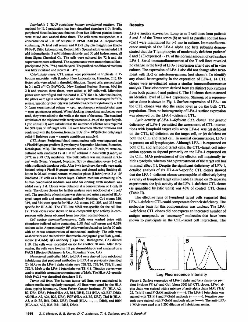
LFA-1 surface expression. Long-term T cell lines from patients6 and 8 of the Texas series (8) as well as parallel control lines(Col) were maintained for >6 mo in culture. Immunofluores-cence analysis of the LFA-1 alpha and beta subunits demon-strated that the T lymphocytes of moderately deficient patients'6 and 8(3) expressed -1% of the normal amount of cell surfaceLFA-1. Serial immunofluorescence of the T cell lines revealedno change in the level of LFA-l expression after 6 mo of in vitroculture. The expression ofLFA-I also did not change after treat-ment with IL-2 or interferon-gamma (not shown). To identifyany clonal heterogeneity in the expression of LFA-1, 14 CTLclones were investigated using a similar immunofluorescenceanalysis. These clones were derived from six distinct bulk culturesfrom both patient 6 and patient 8. The 14 clones demonstratedan identical level of LFA-l expression. Staining of a represen-tative clone is shown in Fig. 1. Surface expression of LFA- I onthe CTL clones was also the same level as on the bulk CTLpopulation. Thus, no heterogeneity ofLFA-l surface expressionwas observed on the LFA-l-deficient CTL.
Lytic activity of LFA-J-deficient CTL clones. The geneticdeficiency of LFA-1 permitted the assessment of CTL interac-tions with lymphoid target cells when LFA-1 was (a) deficienton the CTL, (b) deficient on the target cell, or (c) deficient onboth the CTL and target cell. In normal circumstances, LFA-Iis present on all lymphocytes. Although LFA- I is expressed onboth CTL and lymphoid target cells, the CTL-target cell inter-action appears to depend primarily on the LFA-l expressed onthe CTL. MAb pretreatment of the effector cell maximally in-hibits cytolysis, whereas MAb pretreatment of the target cell hasminimal effect (1). Despite the significant deficiency of LFA- 1,detailed analysis of six HLA-A2-specific CTL clones showedthat the LFA-l-deficient clones were capable of effectively lysinga variety of lymphoid target cells (Table I). Based on 26 separateexperiments, the lytic activity ofthe LFA-l-deficient CTL clones(as quantified by lytic units) was 43% of control CTL clones(Table II).
The effective lysis of lymphoid target cells suggested thatLFA-l-deficient CTL could compensate for their deficiency. Themolecular basis for this compensation was unclear. The LFA-1-deficient CTL clones did not express an increased number ofantigen nonspecific or "accessory" molecules that have beenshown to participate in the CTL-target cell interaction. The
A B
E -rz
Log Fluorescence IntensityFigure 1. Surface expression of LFA-l alpha and beta chains on pa-tient 6 (clone F4) (A) and Col (clone 350) (B) CTL clones. LFA-1 al-pha chain was stained with a mixture of anti-alpha chain MAb (Tsl/22, Tsl/ I1) and F-GAM antibody (-----). The LFA-I beta chain wasstained with TSI/18 and F-GAM antibody (- - - - -). Negative con-trols were stained with F-GAM antibody alone (-). The anti-LFA-1 MAb were used at a 1:200 dilution of hybridoma ascites.
1388 S. J. Mentzer, B. E. Bierer, D. C. Anderson, T. A. Springer, and S. J. Burakoff
F L-- b P--b--e-are ____I
0LL MPSt ---,
"I "-,]
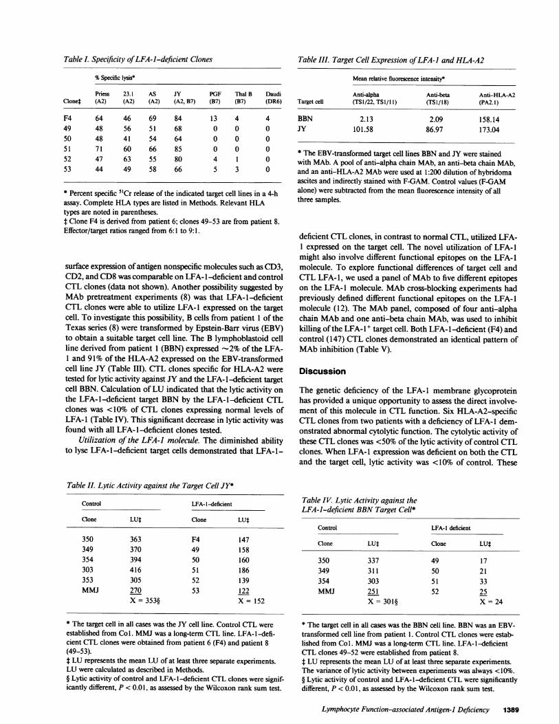
Table . Specificity ofLFA-i-deficient Clones
% Specific lysis*
Priess 23.1 AS JY PGF Thal B DaudiClonet (A2) (A2) (A2) (A2, B7) (B7) (B7) (DR6)
F4 64 46 69 84 13 4 449 48 56 51 68 0 0 050 48 41 54 64 0 0 051 71 60 66 85 0 0 052 47 63 55 80 4 1 053 44 49 58 66 5 3 0
* Percent specific 5"Cr release of the indicated target cell lines in a 4-hassay. Complete HLA types are listed in Methods. Relevant HLAtypes are noted in parentheses.t Clone F4 is derived from patient 6; clones 49-53 are from patient 8.Effector/target ratios ranged from 6: 1 to 9: 1.
surface expression ofantigen nonspecific molecules such as CD3,CD2, and CD8 was comparable on LFA- 1-deficient and controlCTL clones (data not shown). Another possibility suggested byMAb pretreatment experiments (8) was that LFA-l-deficientCTL clones were able to utilize LFA- I expressed on the targetcell. To investigate this possibility, B cells from patient 1 of theTexas series (8) were transformed by Epstein-Barr virus (EBV)to obtain a suitable target cell line. The B lymphoblastoid cellline derived from patient 1 (BBN) expressed -2% of the LFA-1 and 91% of the HLA-A2 expressed on the EBV-transformedcell line JY (Table III). CTL clones specific for HLA-A2 weretested for lytic activity against JY and the LFA- l-deficient targetcell BBN. Calculation ofLU indicated that the lytic activity onthe LFA-l-deficient target BBN by the LFA-l-deficient CTLclones was <10% of CTL clones expressing normal levels ofLFA- 1 (Table IV). This significant decrease in lytic activity wasfound with all LFA- 1-deficient clones tested.
Utilization of the LFA-i molecule. The diminished abilityto lyse LFA-l-deficient target cells demonstrated that LFA-l-
Table IL Lytic Activity against the Target Cell JY*
Control LFA-l-deficient
Clone LUt Clone LUt
350 363 F4 147349 370 49 158354 394 50 160303 416 51 186353 305 52 139MMJ 270 53 122
X = 353§ X = 152
* The target cell in all cases was the JY cell line. Control CTL wereestablished from Col. MMJ was a long-term CTL line. LFA- l-defi-cient CTL clones were obtained from patient 6 (F4) and patient 8(49-53).* LU represents the mean LU of at least three separate experiments.LU were calculated as described in Methods.§ Lytic activity of control and LFA- I -deficient CTL clones were signif-icantly different, P < 0.01, as assessed by the Wilcoxon rank sum test.
Table III. Target Cell Expression ofLFA-I and HLA-A2
Mean relative fluorescence intensity*
Anti-alpha Anti-beta Anti-HLA-A2Target cell (TSI/22, TSI/11) (TS1/18) (PA2. 1)
BBN 2.13 2.09 158.14JY 101.58 86.97 173.04
* The EBV-transformed target cell lines BBN and JY were stainedwith MAb. A pool of anti-alpha chain MAb, an anti-beta chain MAb,and an anti-HLA-A2 MAb were used at 1:200 dilution of hybridomaascites and indirectly stained with F-GAM. Control values (F-GAMalone) were subtracted from the mean fluorescence intensity of allthree samples.
deficient CTL clones, in contrast to normal CTL, utilized LFA-1 expressed on the target cell. The novel utilization of LFA-1might also involve different functional epitopes on the LFA-1molecule. To explore functional differences of target cell andCTL LFA- 1, we used a panel ofMAb to five different epitopeson the LFA-l molecule. MAb cross-blocking experiments hadpreviously defined different functional epitopes on the LFA-1molecule (12). The MAb panel, composed of four anti-alphachain MAb and one anti-beta chain MAb, was used to inhibitkilling ofthe LFA- l target cell. Both LFA- l-deficient (F4) andcontrol (147) CTL clones demonstrated an identical pattern ofMAb inhibition (Table V).
Discussion
The genetic deficiency of the LFA-l membrane glycoproteinhas provided a unique opportunity to assess the direct involve-ment of this molecule in CTL function. Six HLA-A2-specificCTL clones from two patients with a deficiency of LFA- I dem-onstrated abnormal cytolytic function. The cytolytic activity ofthese CTL clones was <50% of the lytic activity of control CTLclones. When LFA- 1 expression was deficient on both the CTLand the target cell, lytic activity was <10% of control. These
Table IV. Lytic Activity against theLFA-i-deficient BBN Target Cell*
Control LFA-1 deficient
Clone LU* Clone LUt
350 337 49 17349 311 50 21354 303 51 33MMJ 251 52 25
X = 301§ X = 24
* The target cell in all cases was the BBN cell line. BBN was an EBV-transformed cell line from patient 1. Control CTL clones were estab-lished from Col. MMJ was a long-term CTL line. LFA-l-deficientCTL clones 49-52 were established from patient 8.t LU represents the mean LU of at least three separate experiments.The variance of lytic activity between experiments was always <10%.§ Lytic activity of control and LFA- l-deficient CTL were significantlydifferent, P < 0.01, as assessed by the Wilcoxon rank sum test.
Lymphocyte Function-associated Antigen-I Deficiency 1389
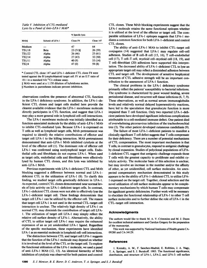
Table V. Inhibition ofCTL-mediated
Lysis by a Panel ofAnti-LFA-J MAb*
% Specific lysis
MAbt Subunit of LFA-lI Clone P4 Clone 147
Medium -47 64TSI/18 Beta 23 (51)§ 26 (59)TSI/22 Alpha 26 (46) 25 (61)TS2/14 Alpha 27 (43) 32 (50)TSl/llI Alpha 40 (9) 55 (14)TS2/4 Alpha 47 (0) 59 (8)
* Control CTL clone 147 and LFA-lI-deficient CTL clone F4 weretested against the B lymphoblastoid target cell JY at an E:T ratio of18:1 in a standard 4-h 51Cr-release assay.f MAb were used at a 1:150 dilution of hybridoma ascites.§ Numbers in parentheses indicate percent inhibition.
observations confirm the presence of abnormal CTL functionin the LFA-lI deficiency syndrome. In addition, the LFA-1I-de-ficient CTL clones and target cells studied here provide theclearest available evidence that the LFA- 1 membrane moleculedirectly participates in CTL function, and suggest that LFA-lmay play a more general role in lymphoid cell-cell interactions.
The LFA-1I membrane molecule was initially identified as afunction-associated molecule by the ability of anti-LFA-lI MAbto inhibit CTL-mediated lysis. Because LFA-lI is expressed onT cells as well as lymphoid target cells, MAb pretreatment wasrequired to identify the relative contributions of effector andtarget cell LFA-lI to the killing interaction. MAb pretreatmentdemonstrated that anti-LFA-lI MAb inhibited cytolysis at thelevel of the effector cell (1). The dominant role of effector cellLFA-lI was confirmed using nonlymphoid target cells. Endo-thelial cells and fibroblasts do not express LFA-l. When usedas target cells, endothelial cells and fibroblasts were effectivelylysed by human CTL clones, and this lysis was inhibited byanti-LFA- 1 MAb.
Previous experiments with LFA- 1-deficient CTL and MAbblocking suggested a difference between normal and LFA- 1-deficient CTL in the utilization of LFA-l1 (8). To clarify thisfinding, we studied target cells genetically deficient in LFA-l.As expected, control CTL clones demonstrated near normal lev-els of lytic activity on LFA-lI-deficient target cells. In contrast,LFA- 1-deficient CTL clones were not able to effectively lyse theLFA-lI-deficient target cell. These findings demonstrate thattarget cell LFA-lI can be utilized by the effector cell. The reasonthat target cell LFA- 1 is not used in the normal CTL-target cellinteraction is unclear. The relatively high density of LFA-lI onnormal CTL may diminish the contribution of target cell LFA-1. The utilization of target cell LFA-lI may simply reflect therelative cell surface density of LFA-l1. Alternatively, the abilityof CTL to utilize target cell LFA-lI may require the enhancedexpression of an as yet unidentified LFA-lI ligand. Regardlessof the specific mechanism, these experiments have identifiedLFA-lI as an essential molecule in lymphoid cell--cell interactions.
The distinction between CTL and target cell LFA-lI suggeststhat the function of the LFA-lI molecule may depend on whetherit is involved at the level ofthe CTL or the target cell. To explorethe functional utilization of the LFA-1I molecule, we used a panelof anti-LFA-lI MAb (12, 13). An identical hierarchy of MAbinhibition of cytolysis was observed for both patient and control
CTL clones. These MAb blocking experiments suggest that theLFA- 1 molecule retains the same functional epitopes whetherit is utilized at the level of the effector or target cell. The com-parable utilization of LFA-1 epitopes suggests that LFA-1 me-diates a common function for both LFA- 1-deficient and controlCTL clones.
The ability of anti-LFA- 1 MAb to inhibit CTL-target cellconjugates (14) suggested that LFA-l may regulate cell--celladhesion. Studies of B cell-B cell (15, 16), T cell-endothelialcell (17), T cell-T cell, myeloid cell-myeloid cell (18, 19), andT cell-fibroblast (20) adhesions have supported this interpre-tation. The decreased ability of LFA-lI-deficient CTL to lyse anappropriate target cell may reflect a diminished adhesion betweenCTL and target cell. The development of sensitive biophysicalmeasures of CTL adhesive strength will be an important con-tribution to the assessment of LFA- 1 function.
The clinical problems in the LFA-lI deficiency syndromeprimarily reflect the patients' susceptibility to bacterial infections.The syndrome is characterized by poor wound healing, severeperiodontal disease, and recurrent soft tissue infections (6, 7, 9).These observations, as well as normal serum immunoglobulinlevels and relatively normal delayed hypersensitivity reactions,have led to the speculation that granulocyte function is moreimpaired than T cell function. Only two reported LFA-lI-defi-cient patients have developed significant infectious complicationsattributable to a cell-mediated immune defect. One patient diedofoverwhelming picornavirus infection of the tracheobronchialtree (3). The other patient developed cutaneous candidiasis (9).
The failure of most LFA-lI-deficient patients to manifest aclinically significant T cell defect suggests that T cells compensatefor their deficiency. There are a number of potential mechanismsof CTL compensation. The most important mechanism is thatT cells, in contrast to granulocytes, respond to antigenic challengeby clonal expansion. Studies of polyclonal populations of LFA-1-deficient CTL suggest that antigenic challenge expands thoseT cells with the greatest capacity to proliferate and exhibit cy-tolytic activity. The molecular basis of this selection is unclear,but may involve an increase in the number and/or the affinityof other, as yet unidentified, cell surface molecules. An unex-pected compensatory mechanism demonstrated in this studyappears to be the ability of LFA-lI-deficient CTL to utilize LFA-1 expressed on the target cell. Together, clonal selection and thenovel utilization of cell surface molecules appear to be comple-mentary mechanisms by which human T cells may compensatefor significant genetic deficiencies. Further work will be necessaryto elucidate the functional relationship of LFA-lI to other T cellsurface molecules and to further define the role of LFA-1I in theCTL-target cell interaction.
Ac~knowledaments
The authors would like to thank M. A. V. Crimmins and M. J. Dunnfor excellent technical assistance and Carolyn Gregory for the preparationof the manuscript.
This work was supported by National Institutes ofHealth grants CA-09280 and CA-34129.
References
1. Krensky, A. M., F. Sanchez-Madrid, E. Robbins, J. A. Nagy,T. A. Springer, and S. J. Burakoffi 1983. The functional significance,distribution, and structure of LFA-l1, LFA-2, and LFA-3: cell surface
1390 S. J. Mentzer, B. E. Bierer, D. C. Anderson, T. A. Springer, and S. J. Burakoff
antigens associated with CTL-target interactions. J. Immunol. 131:611-616.
2. Sanchez-Madrid, F., A. M. Krensky, C. F. Ware, E. Robbins,J. L. Strominger, S. J. Burakoff, and T. A. Springer. 1982. Three distinctantigens associated with human T lymphocyte-mediated cytolysis: LFA-1, LFA-2, and LFA-3. Proc. Natl. Acad. Sci. USA. 79:7489-7493.
3. Anderson, D. C., F. C. Schmalstieg, M. J. Finegold, B. J. Hughes,R. Rothlein, L. J. Miller, S. Kohl, M. F. Tosi, R. L. Jacobs, A. Goldman,W. T. Shearer, and T. A. Springer. 1985. The severe and moderate phe-notypes ofheritable Mac-l, LFA-1 deficiency: their quantitative definitionand relation to leukocyte dysfunction and clinical features. J. Infect. Dis.152:668-689.
4. Sanchez-Madrid, F., J. A. Nagy, E. Robbins, P. Simon, and T. A.Springer. 1983. A human leukocyte differentiation antigen family withdistinct alpha subunits and common beta subunit: the lymphocyte func-tion-associated antigen (LFA-1), the C3bi complement receptor (OKMI/Macd) and the p150,95 molecule. J. Exp. Med. 158:1785-1803.
5. Springer, T. A., W. S. Thompson, L. J. Miller, F. C. Schmalstieg,and D. C. Anderson. 1984. Inherited deficiency of the Mac-i, LFA-1,p150,95 glycoprotein family and its molecular basis. J. Exp. Med. 160:1901-1918.
6. Arnaout, M. A., H. Spitz, C. Terhorst, J. Pitt, and R. F. Todd.1984. Deficiency ofa leukocyte surface glycoprotein in two patients withMol deficiency. J. Clin. Invest. 74:1291-1300.
7. Miedema, F., P. A. F. Tetteroo, F. G. Terpstra, G. Keizer, M.Roos, R. S. Weening, C. M. R. Weemaes, D. Roos, and C. J. M. Melief.1985. Immunologic studies with LFA-1 and Mol-deficient lymphocytesfrom a patient with recurrent bacterial infections. J. Immunol 134:3075-3081.
8. Krensky, A. M., S. J. Mentzer, C. Clayberger, F. C. Schmalstieg,D. C. Anderson, S. J. Burakoff, and T. A. Springer. 1985. Heritablelymphocyte function-associated antigen-l deficiency: abnormalities ofcytotoxicity and proliferation associated with abnormal expression ofLFA-1. J. Immunol. 135:3102-3108.
9. Lisowska-Grospierre, B., M.-C. Bohler, A. Fischer, C. Mawas,T. A. Springer, and C. Giscelli. 1986. Defective membrane expressionof the LFA-1 complex may be secondary to the absence of the # chainin a child with recurrent bacterial infection. Eur. J. Immunol. 16:205-208.
10. Krensky, A. M., S. J. Mentzer, J. L. Greenstein, M. Crimmins,C. Clayberger, T. A. Springer, and S. J. Burakoff. 1985. Human cytolyticT lymphocyte clones and their function-associated cell surface molecules.In Hybridoma Technology in the Biosciences and Medicine. T. A.Springer, editor. Plenum Press, New York. 559-573.
11. Parham, P., and W. F. Bodmer. 1978. Monoclonal antibody toa human histocompatibility antigen, HLA-A2. Nature (Lond.). 276:397-399.
12. Ware, C. F., F. Sanchez-Madrid, A. M. Krensky, S. J. Burakoff,J. L. Strominger, and T. A. Springer. 1983. Human lymphocyte function-associated antigen- I (LFA- 1): identification of multiple antigenic epitopesand their relationship to CTL-mediated cytotoxicity. J. Immunol. 131:1182-1187.
13. Mentzer, S. J., A. M. Krensky, and S. J. Burakoff. 1986. Mappingfunctional epitopes of the LFA-1 glycoprotein: monoclonal antibodyinhibition ofNK and CTL effectors. Hum. Immunol. In press.
14. Krensky, A. M., E. Robbins, T. A. Springer, and S. J. Burakoff.1984. LFA-1, LFA-2, and LFA-3 antigens are involved in CTL-targetconjugation. J. Immunol. 132:2180-2182.
15. Mentzer, S. J., S. H. Gromkowski, A. M. Krensky, S. J. Burakoff,and E. Martz. 1985. LFA-1 membrane molecule in the regulation ofhomotypic adhesions ofhuman B lymphocytes. J. Immunol. 135:9-11.
16. Springer, T. A., R. Rothlein, D. C. Anderson, S. J. Burakoff, andA. M. Krensky. 1985. The function of LFA-I in cell-mediated killingand adhesion: studies on heritable LFA-l, Mac-l deficiency and p150,95on lymphoid cell self-aggregation. Adv. Exp. Med. Biol. 184:311-320.
17. Mentzer, S. J., S. J. Burakoff, and D. V. Faller. 1986. T celladhesion to endothelial cells is regulated by the LFA-1 membrane mol-ecule. J. Cell. Physiol. 126:285-290.
18. Rothlein, R., and T. A. Springer. 1986. The requirement forLFA-1 in homotypic leukocyte adhesion stimulated by phorbol ester. J.Exp. Med. 163:1132-1149.
19. Mentzer, S. J., D. V. Faller, and S. J. Burakoff. 1986. Interferon-gamma induction of LFA-1 mediated homotypic adhesions of humanmonocytes. J. Immunol. 137:108-113.
20. Dustin, M. L., R. Rothlein, A. K. Bhan, C. A. Dinarello, andT. A. Springer. 1986. Induction by IL 1 and interferon-gamma: tissuedistribution, biochemistry, and function of a natural adherence molecule(ICAM-1). J. Immunol. In press.
Lymphocyte Function-associated Antigen-i Deficiency 1391