Embed Size (px)
Citation preview
Department of Medicine
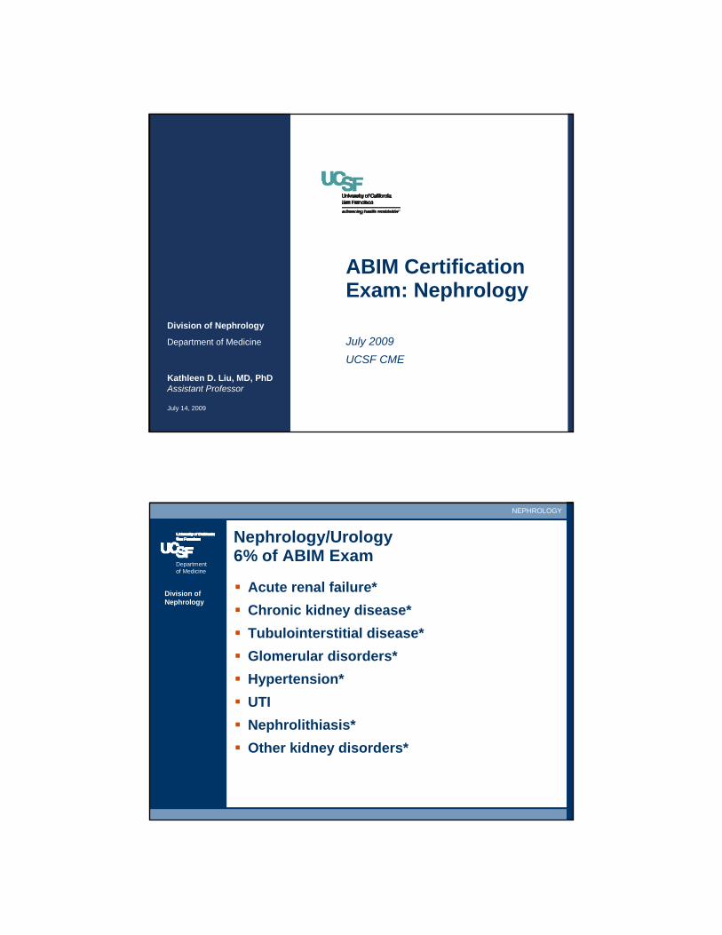
ABIM Certification Exam: Nephrology
July 2009UCSF CME
Kathleen D. Liu, MD, PhDAssistant Professor
July 14, 2009
Division of Nephrology
Department of Medicine
NEPHROLOGY
Division of Nephrology
Nephrology/Urology6% of ABIM Exam
Acute renal failure*Chronic kidney disease*Tubulointerstitial disease*Glomerular disorders*Hypertension*UTINephrolithiasis*Other kidney disorders*

Department of Medicine
NEPHROLOGY
Division of Nephrology
Nephrology/Urology6% of ABIM Exam
Urologic cancerProstate disordersOther urologyUrinary IncontinenceWater and electrolyte balance*Miscellaneous bladder and kidney disorders
Department of Medicine
NEPHROLOGY
Division of Nephrology
Acute Renal Failure/Kidney Injury
Pre-Renal = Decreased kidney perfusionIntra-Renal = Intrinsic kidney diseasePost-Renal = Obstructive nephropathy

Department of Medicine
NEPHROLOGY
Division of Nephrology
Pre-Renal ARF:Kidney Hypoperfusion
Dehydration, overdiuresis, hypovolemiaHemorrhageHemodynamic effect: ACE/ARB and NSAIDsHeart failure
– Cardiorenal syndrome
Cirrhosis/End-stage liver disease– Hepatorenal syndrome
Department of Medicine
NEPHROLOGY
Division of Nephrology
Pre-Renal ARF:Kidney Hypoperfusion
Diagnosis– +/- Oliguria– High BUN:Creatinine ratio > 20 – Bland urine sediment, normal kidney US– Low FENa < 1% and low urine Na < `10 mEq/L– High specific gravity, high urine osmolality– Rapid renal recovery with resuscitation
Therapy: Restore renal perfusionPrognosis: Good, often rapid renal recovery
– Exceptions: Cardiorenal and hepatorenal syndromes
Department of Medicine
NEPHROLOGY
Division of Nephrology
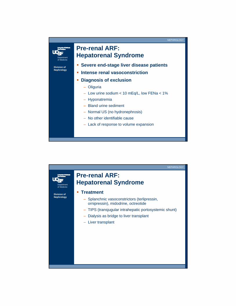
Pre-renal ARF: Hepatorenal Syndrome
Severe end-stage liver disease patientsIntense renal vasoconstrictionDiagnosis of exclusion
– Oliguria– Low urine sodium < 10 mEq/L, low FENa < 1%– Hyponatremia– Bland urine sediment– Normal US (no hydronephrosis)– No other identifiable cause– Lack of response to volume expansion
Department of Medicine
NEPHROLOGY
Division of Nephrology
Pre-renal ARF: Hepatorenal Syndrome
Treatment– Splanchnic vasoconstrictors (terlipressin,
ornipressin), midodrine, octreotide– TIPS (transjugular intrahepatic portosystemic shunt)– Dialysis as bridge to liver transplant– Liver transplant
Department of Medicine
NEPHROLOGY
Division of Nephrology
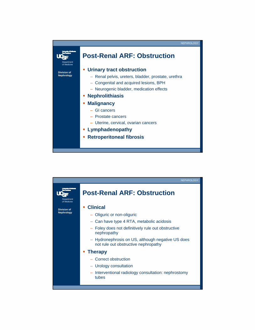
Post-Renal ARF: Obstruction
Urinary tract obstruction– Renal pelvis, ureters, bladder, prostate, urethra– Congenital and acquired lesions, BPH– Neurogenic bladder, medication effects
NephrolithiasisMalignancy
– GI cancers– Prostate cancers– Uterine, cervical, ovarian cancers
LymphadenopathyRetroperitoneal fibrosis
Department of Medicine
NEPHROLOGY
Division of Nephrology
Post-Renal ARF: Obstruction
Clinical– Oliguric or non-oliguric– Can have type 4 RTA, metabolic acidosis– Foley does not definitively rule out obstructive
nephropathy– Hydronephrosis on US, although negative US does
not rule out obstructive nephropathy
Therapy– Correct obstruction– Urology consultation– Interventional radiology consultation: nephrostomy
tubes
Department of Medicine
NEPHROLOGY
Division of Nephrology
Post-Renal ARF: Obstruction
Prognosis– More rapid recovery with rapid correction of
obstruction– Can recover kidney function after prolonged
obstruction– Post-obstructive diuresis from urinary concentrating
defect
Department of Medicine
NEPHROLOGY
Division of Nephrology
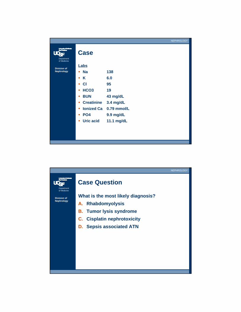
Case
A 65 year-old woman is admitted to the hospital with newly diagnosed diffuse B cell lymphoma for induction chemotherapy. 24 hours after induction chemotherapy, she is noted to be oliguric.
Physical examT 38.4, BP 95/60, HR 94, RR 24Heart is normal.Lungs are clear, though she is mildly tachypneicTrace-1+ pitting edema
Department of Medicine
NEPHROLOGY
Division of Nephrology
Case
LabsNa 138K 6.0Cl 95HCO3 19BUN 43 mg/dLCreatinine 3.4 mg/dLIonized Ca 0.79 mmol/LPO4 9.9 mg/dLUric acid 11.1 mg/dL
Department of Medicine
NEPHROLOGY
Division of Nephrology
Case Question
What is the most likely diagnosis?A. RhabdomyolysisB. Tumor lysis syndromeC. Cisplatin nephrotoxicityD. Sepsis associated ATN
Department of Medicine
NEPHROLOGY
Division of Nephrology
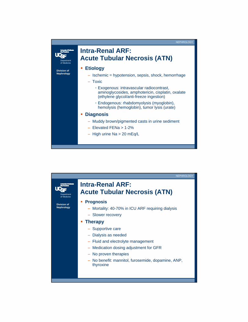
Intra-Renal ARF:Acute Tubular Necrosis (ATN)
Etiology– Ischemic = hypotension, sepsis, shock, hemorrhage– Toxic
• Exogenous: intravascular radiocontrast, aminoglycosides, amphotericin, cisplatin, oxalate (ethylene glycol/anti-freeze ingestion)
• Endogenous: rhabdomyolysis (myoglobin), hemolysis (hemoglobin), tumor lysis (urate)
Diagnosis– Muddy brown/pigmented casts in urine sediment– Elevated FENa > 1-2%– High urine Na > 20 mEq/L
Department of Medicine
NEPHROLOGY
Division of Nephrology
Intra-Renal ARF:Acute Tubular Necrosis (ATN)
Prognosis– Mortality: 40-70% in ICU ARF requiring dialysis– Slower recovery
Therapy– Supportive care– Dialysis as needed– Fluid and electrolyte management– Medication dosing adjustment for GFR– No proven therapies– No benefit: mannitol, furosemide, dopamine, ANP,
thyroxine

Department of Medicine
NEPHROLOGY
Division of Nephrology
Intra-Renal ARF:Radiocontrast Nephropathy
Etiology– Iodine-based radiocontrast– Intravenous or intraarterial injection– CT, angiography, cardiac catheterization
Risk factors– Pre-existing chronic kidney disease– Proteinuria– Age– Diabetes mellitus– Multiple myeloma– Dehydration
Department of Medicine
NEPHROLOGY
Division of Nephrology
Intra-Renal ARF:Radiocontrast Nephropathy
Presentation– Rise in creatinine 24-48 hours post-exposure– Patient with risk factors– Low FENa < 1%– Bland sediment (mild forms with vasoconstriction) or
muddy brown casts of ATN (severe forms with toxic injury)
Prognosis– Mild cases resolve within 2-5 days, likely
vasoconstriction mediated ARF– Severe cases resolve slowly over days to weeks,
require dialysis, and may be irreversible due to toxin-induced ATN
Department of Medicine
NEPHROLOGY
Division of Nephrology
Intra-Renal ARF:Radiocontrast Nephropathy
Prevention– Avoid radiocontrast (US, nuclear medicine)– Minimize dose of radiocontrast– Use iso-osmolar or hypo-osmolar contrast (as
opposed to hyperosmolar contrast)– N-Acetylcysteine– IVF: Isotonic sodium bicarbonate vs. normal saline– Hold diuretics peri-contrast, avoid hypovolemia– No clear benefit of post-contrast dialysis
Recent reviews– Barrett BJ, Parfrey PS. NEJM 2006.– Pannu N et al. JAMA 2006.
Department of Medicine
NEPHROLOGY
Division of Nephrology
Gadolinium based MRI agents – a word of caution
Nephrogenic systemic fibrosis– Recently described syndrome associated with MRI
based gadolinium administration– Patients with acute renal failure/kidney injury and
chronic kidney disease are at risk– Studies to ascertain incidence are ongoing– Rarer than radiocontrast nephropathy, but can be
fatal
Recent reviews– Perazella Clin Journal Amer Soci Neph 2009.
Department of Medicine
NEPHROLOGY
Division of Nephrology
Intra-Renal ARF:Rhabdomyolysis
Etiology– Crush injury, muscle trauma/ischemia/inflammation – Prolonged immobilization: coma, ethanol, earthquake
victims– Fevers/rigors, seizures– Toxic injury: statins, cocaine, reverse transcriptase
inhibitors– Metabolic: Hypokalemia, hypophosphatemia– Genetic: McArdle disease
Department of Medicine
NEPHROLOGY
Division of Nephrology
Intra-Renal ARF:Rhabdomyolysis
Diagnosis– High serum uric acid, phosphate, potassium– Hypocalcemia– Elevated serum CK (along with AST/ALT)– Dipstick hematuria from myoglobinuria– Negative microanalysis for RBCs– ATN urine sediment, muddy brown casts
Treatment– Aggressive and early hydration– Alkalinization of urine vs. NS hydration alone?– Stop offending medications
Department of Medicine
NEPHROLOGY
Division of Nephrology
Intra-Renal ARF:Acute Interstitial Nephritis (AIN)
Etiology– Medications = antibiotics, NSAIDs, diuretics, PPIs,
others– Infections = bacterial, fungal, viral, others– Immune disorders = SLE, Sjogrens, sarcoidosis
Presentation– Triad: Fever, drug rash, eosinophilia– Minority of patients have complete triad– Arthralgias– NSAID-AIN may have proteinuria from concomitant
minimal change disease– AIN is often occult, should be suspected if no
apparent etiology of ARF or new medication started
Department of Medicine
NEPHROLOGY
Division of Nephrology
Intra-Renal ARF:Acute Interstitial Nephritis (AIN)
Diagnosis– Sterile pyuria, WBC casts, eosinophilia– Clinical diagnosis; kidneys improve after stopping
offending drug (which may be a chronic medication or one tolerated safely in the past)
– Kidney biopsy– Skin biopsy (leukocytoclastic vasculitis)
Therapy– Stop offending drugs– Treat underlying infection– Consider oral steroids (e.g., prednisone 60 mg PO
daily), lack of large randomized controlled trials showing efficacy
Department of Medicine
NEPHROLOGY
Division of Nephrology
Intra-Renal ARF:Atheroembolic Disease (AED)
Etiology– Spontaneous/idiopathic– Anticoagulation– Instrumentation: aortic surgery/cross-clamping,
CABG, angiography, cardiac catheterization
Presentation– Stuttering, inexorable rise in serum creatinine– Livedo reticularis, embolic stigmata– Non-specific urine sediment– Often occult, should be considered if no obvious
etiology
Department of Medicine
NEPHROLOGY
Division of Nephrology
Intra-Renal ARF:Atheroembolic Disease (AED)
Diagnosis– Often clinical diagnosis, embolic skin findings– Low complements C3 and C4– Eosinophilia and eosinophiluria– Retinal embolization (Hollenhorst plaques)– Skin biopsy, kidney biopsy
Therapy– Supportive. Stop anticoagulation?
Prognosis– Poor, generally irreversible– Heavy burden of cardiovascular disease
Department of Medicine
NEPHROLOGY
Division of Nephrology
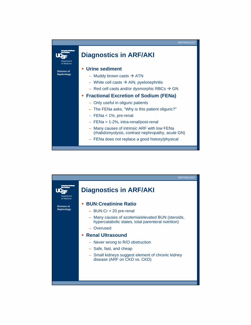
Diagnostics in ARF/AKI
Urine sediment– Muddy brown casts ATN– White cell casts AIN, pyelonephritis– Red cell casts and/or dysmorphic RBCs GN
Fractional Excretion of Sodium (FENa)– Only useful in oliguric patients– The FENa asks, “Why is this patient oliguric?”– FENa < 1%, pre-renal– FENa > 1-2%, intra-renal/post-renal– Many causes of intrinsic ARF with low FENa
(rhabdomyolysis, contrast nephropathy, acute GN)– FENa does not replace a good history/physical
Department of Medicine
NEPHROLOGY
Division of Nephrology
Diagnostics in ARF/AKI
BUN:Creatinine Ratio– BUN:Cr > 20 pre-renal– Many causes of azotemia/elevated BUN (steroids,
hypercatabolic states, total parenteral nutrition)– Overused
Renal Ultrasound– Never wrong to R/O obstruction– Safe, fast, and cheap– Small kidneys suggest element of chronic kidney
disease (ARF on CKD vs. CKD)
Department of Medicine
NEPHROLOGY
Division of Nephrology
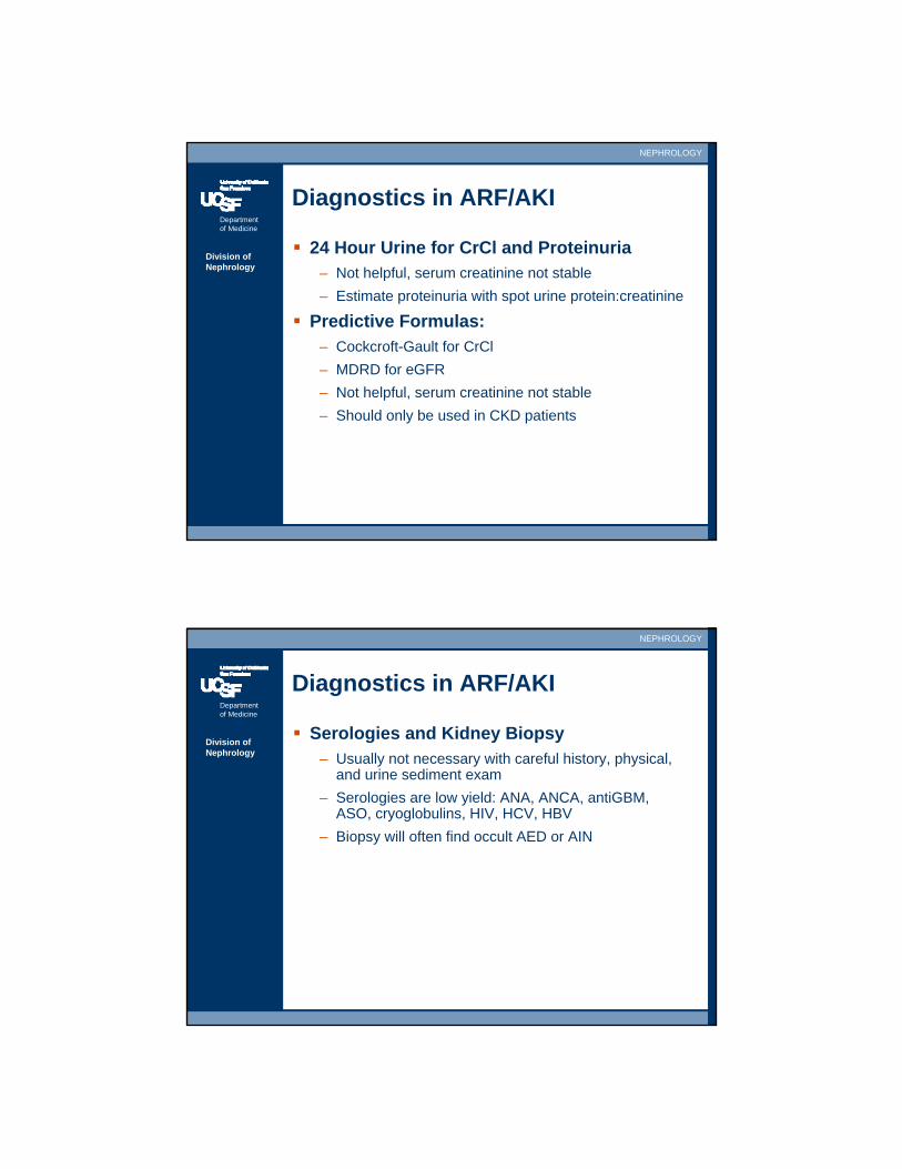
Diagnostics in ARF/AKI
24 Hour Urine for CrCl and Proteinuria– Not helpful, serum creatinine not stable– Estimate proteinuria with spot urine protein:creatinine
Predictive Formulas: – Cockcroft-Gault for CrCl– MDRD for eGFR– Not helpful, serum creatinine not stable– Should only be used in CKD patients
Department of Medicine
NEPHROLOGY
Division of Nephrology
Diagnostics in ARF/AKI
Serologies and Kidney Biopsy– Usually not necessary with careful history, physical,
and urine sediment exam– Serologies are low yield: ANA, ANCA, antiGBM,
ASO, cryoglobulins, HIV, HCV, HBV– Biopsy will often find occult AED or AIN
Department of Medicine
NEPHROLOGY
Division of Nephrology
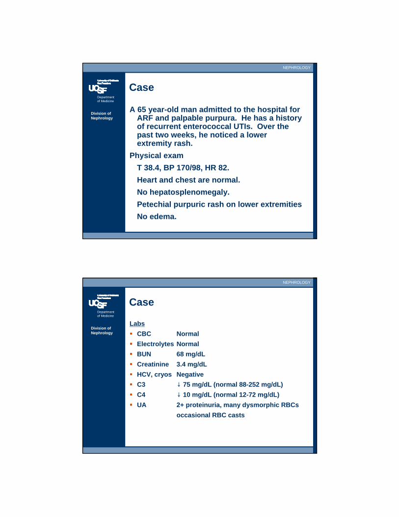
Case
A 65 year-old man admitted to the hospital for ARF and palpable purpura. He has a history of recurrent enterococcal UTIs. Over the past two weeks, he noticed a lower extremity rash.
Physical examT 38.4, BP 170/98, HR 82.Heart and chest are normal.No hepatosplenomegaly.Petechial purpuric rash on lower extremitiesNo edema.
Department of Medicine
NEPHROLOGY
Division of Nephrology
Case
LabsCBC NormalElectrolytes NormalBUN 68 mg/dLCreatinine 3.4 mg/dLHCV, cryos NegativeC3 ↓ 75 mg/dL (normal 88-252 mg/dL)C4 ↓ 10 mg/dL (normal 12-72 mg/dL)UA 2+ proteinuria, many dysmorphic RBCs
occasional RBC casts
Department of Medicine
NEPHROLOGY
Division of Nephrology
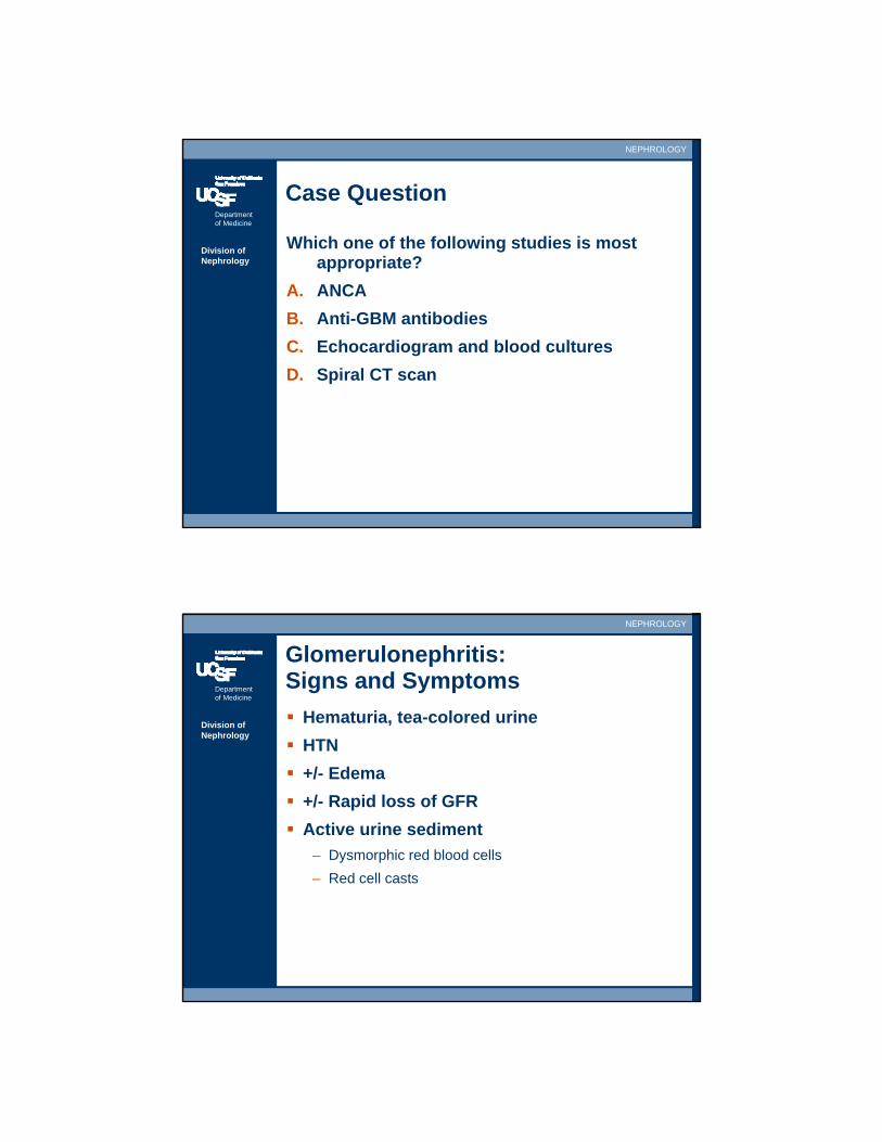
Case Question
Which one of the following studies is most appropriate?
A. ANCAB. Anti-GBM antibodiesC. Echocardiogram and blood culturesD. Spiral CT scan
Department of Medicine
NEPHROLOGY
Division of Nephrology
Glomerulonephritis: Signs and Symptoms
Hematuria, tea-colored urineHTN+/- Edema+/- Rapid loss of GFRActive urine sediment
– Dysmorphic red blood cells– Red cell casts
Department of Medicine
NEPHROLOGY
Division of Nephrology
Non-glomerular Hematuria
Bloody/pink urineBlood clotsComplete absence of proteinuriaBland urine sediment
– Non-dysmorphic red cells– No red cells– No red cell casts
Department of Medicine
NEPHROLOGY
Division of Nephrology
Chronic Hematuria
Benign Familial HematuriaAlport Syndrome/Hereditary NephritisIgA nephropathySLENephrolithiasis
Department of Medicine
NEPHROLOGY
Division of Nephrology
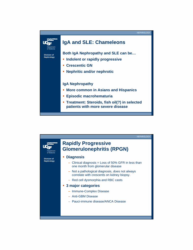
IgA and SLE: Chameleons
Both IgA Nephropathy and SLE can be…Indolent or rapidly progressiveCrescentic GNNephritic and/or nephrotic
IgA NephropathyMore common in Asians and HispanicsEpisodic macrohematuriaTreatment: Steroids, fish oil(?) in selected patients with more severe disease
Department of Medicine
NEPHROLOGY
Division of Nephrology
Rapidly Progressive Glomerulonephritis (RPGN)
Diagnosis– Clinical diagnosis = Loss of 50% GFR in less than
one month from glomerular disease– Not a pathological diagnosis, does not always
correlate with crescents on kidney biopsy.– Red cell dysmorphia and RBC casts
3 major categories– Immune-Complex Disease– Anti-GBM Disease– Pauci-immune disease/ANCA Disease
Department of Medicine
NEPHROLOGY
Division of Nephrology
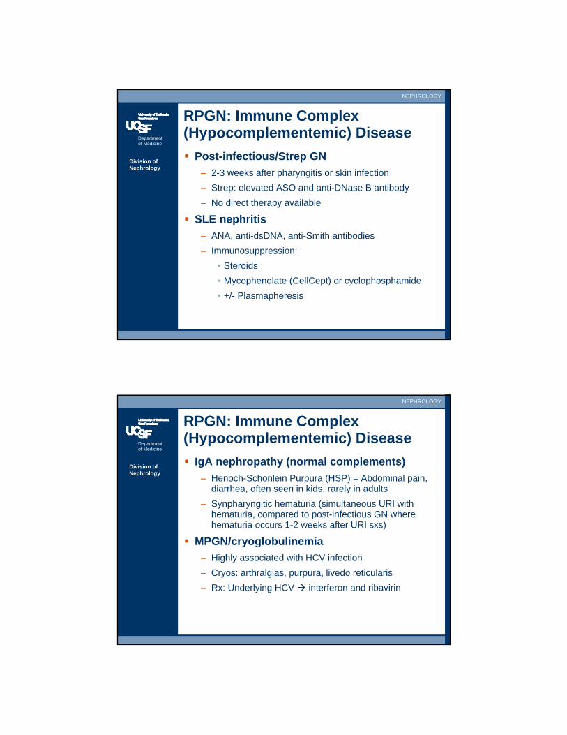
RPGN: Immune Complex (Hypocomplementemic) Disease
Post-infectious/Strep GN– 2-3 weeks after pharyngitis or skin infection– Strep: elevated ASO and anti-DNase B antibody– No direct therapy available
SLE nephritis– ANA, anti-dsDNA, anti-Smith antibodies– Immunosuppression:
• Steroids• Mycophenolate (CellCept) or cyclophosphamide• +/- Plasmapheresis
Department of Medicine
NEPHROLOGY
Division of Nephrology
RPGN: Immune Complex (Hypocomplementemic) Disease
IgA nephropathy (normal complements)– Henoch-Schonlein Purpura (HSP) = Abdominal pain,
diarrhea, often seen in kids, rarely in adults– Synpharyngitic hematuria (simultaneous URI with
hematuria, compared to post-infectious GN where hematuria occurs 1-2 weeks after URI sxs)
MPGN/cryoglobulinemia– Highly associated with HCV infection– Cryos: arthralgias, purpura, livedo reticularis– Rx: Underlying HCV interferon and ribavirin
Department of Medicine
NEPHROLOGY
Division of Nephrology
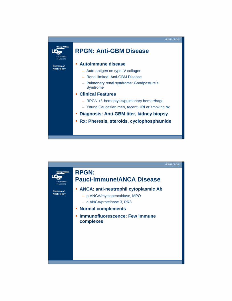
RPGN: Anti-GBM Disease
Autoimmune disease– Auto-antigen on type IV collagen– Renal limited: Anti-GBM Disease– Pulmonary renal syndrome: Goodpasture’s
Syndrome
Clinical Features– RPGN +/- hemoptysis/pulmonary hemorrhage– Young Caucasian men, recent URI or smoking hx
Diagnosis: Anti-GBM titer, kidney biopsyRx: Pheresis, steroids, cyclophosphamide
Department of Medicine
NEPHROLOGY
Division of Nephrology
RPGN:Pauci-Immune/ANCA Disease
ANCA: anti-neutrophil cytoplasmic Ab– p-ANCA/myeloperoxidase, MPO– c-ANCA/proteinase 3, PR3
Normal complementsImmunofluorescence: Few immune complexes

Department of Medicine
NEPHROLOGY
Division of Nephrology
RPGN:Pauci-Immune/ANCA Disease
Microscopic polyangiitis (p-ANCA)– Steroids/cyclophosphamide
Wegener’s granulomatosis (c-ANCA)– Lung disease, upper airway disease– Granulomas– Steroids/cyclophosphamide, +/- pheresis if severe
Churg-Strauss Disease (p-ANCA)– Eosinophilia, asthma, sinus disease, peripheral
neuropathy– Granulomas– Steroids/cyclophosphamide
Aorta (large artery)
Renal artery (medium sized artery)
Lobar Artery (medium sized artery)
Arcuate artery (small artery)
Interlobular Artery(small artery)
Arteriole
Glomerulus
Giant Cell (Temporal)Arteritis & TakayasuArteritis
Polyarteritis nodosa & Kawasaki Disease
ANCA = Microscopic polyangiitis, Wegener’s, Churg-Strauss
Henoch-Schonlein Purpura, Cryoglobulinemic vasculitis, Lupus and Rheumatoid vasculitis
Nomenclature of Systemic Vasculitides
Department of Medicine
NEPHROLOGY
Division of Nephrology
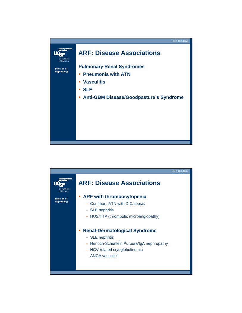
ARF: Disease Associations
Pulmonary Renal Syndromes Pneumonia with ATNVasculitisSLEAnti-GBM Disease/Goodpasture’s Syndrome
Department of Medicine
NEPHROLOGY
Division of Nephrology
ARF: Disease Associations
ARF with thrombocytopenia– Common: ATN with DIC/sepsis– SLE nephritis– HUS/TTP (thrombotic microangiopathy)
Renal-Dermatological Syndrome– SLE nephritis– Henoch-Schonlein Purpura/IgA nephropathy– HCV-related cryoglobulinemia– ANCA vasculitis
Department of Medicine
NEPHROLOGY
Division of Nephrology
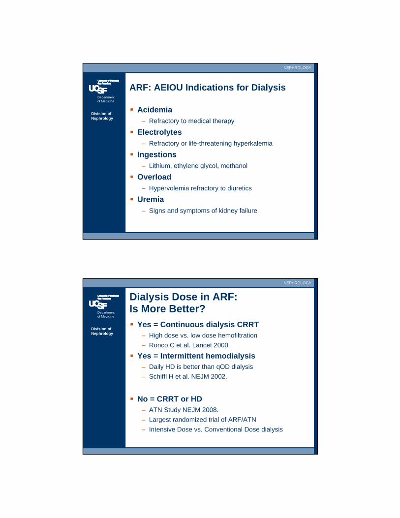
ARF: AEIOU Indications for Dialysis
Acidemia– Refractory to medical therapy
Electrolytes– Refractory or life-threatening hyperkalemia
Ingestions– Lithium, ethylene glycol, methanol
Overload– Hypervolemia refractory to diuretics
Uremia– Signs and symptoms of kidney failure
Department of Medicine
NEPHROLOGY
Division of Nephrology
Dialysis Dose in ARF:Is More Better?
Yes = Continuous dialysis CRRT– High dose vs. low dose hemofiltration– Ronco C et al. Lancet 2000.
Yes = Intermittent hemodialysis– Daily HD is better than qOD dialysis– Schiffl H et al. NEJM 2002.
No = CRRT or HD– ATN Study NEJM 2008.– Largest randomized trial of ARF/ATN– Intensive Dose vs. Conventional Dose dialysis
Department of Medicine
NEPHROLOGY
Division of Nephrology
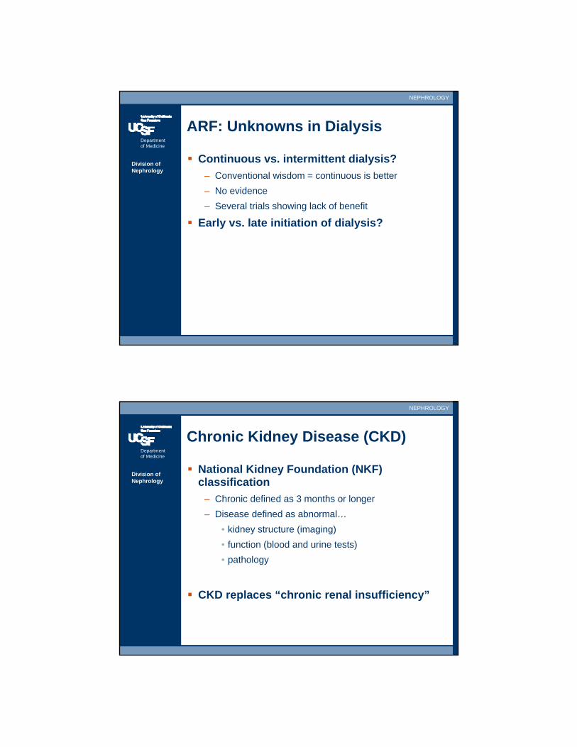
ARF: Unknowns in Dialysis
Continuous vs. intermittent dialysis?– Conventional wisdom = continuous is better– No evidence– Several trials showing lack of benefit
Early vs. late initiation of dialysis?
Department of Medicine
NEPHROLOGY
Division of Nephrology
Chronic Kidney Disease (CKD)
National Kidney Foundation (NKF) classification
– Chronic defined as 3 months or longer– Disease defined as abnormal…
• kidney structure (imaging)• function (blood and urine tests)• pathology
CKD replaces “chronic renal insufficiency”
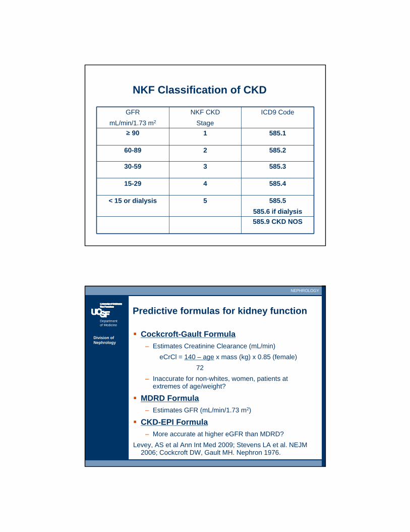
NKF Classification of CKD
GFRmL/min/1.73 m2
NKF CKDStage
ICD9 Code
≥ 90 1 585.1
60-89 2 585.2
30-59 3 585.3
15-29 4 585.4
< 15 or dialysis 5 585.5585.6 if dialysis585.9 CKD NOS
Department of Medicine
NEPHROLOGY
Division of Nephrology
Predictive formulas for kidney function
Cockcroft-Gault Formula– Estimates Creatinine Clearance (mL/min)
eCrCl = 140 – age x mass (kg) x 0.85 (female)72
– Inaccurate for non-whites, women, patients at extremes of age/weight?
MDRD Formula– Estimates GFR (mL/min/1.73 m2)
CKD-EPI Formula– More accurate at higher eGFR than MDRD?
Levey, AS et al Ann Int Med 2009; Stevens LA et al. NEJM 2006; Cockcroft DW, Gault MH. Nephron 1976.
Department of Medicine
NEPHROLOGY
Division of Nephrology
MDRD estimated GFR (eGFR)
Modification of Diet in Renal Disease Study4 variables predict GFR, weight not neededRecommended by NKF and NIDDK
– Reported by clinical labs– PDA calculators or Google “MDRD”
eGFR (mL/min/1.73 m2) =186 x (SCr)-1.154 x age-0.203
x 0.742 (female) x 1.210 (black)
Klahr S et al. NEJM 1994.Levey AS et al. Ann Intern Med 1999.
Department of Medicine
NEPHROLOGY
Division of Nephrology
Estimating Proteinuria
24-hour urine collection– Time consuming, inconvenient– Inaccurate/inadequate urine collections– Difficult to follow serially
Spot Urine Protein:Creatinine ratio– Ratio correlates to grams/day/1.73 m2 of proteinuria– Quick, easy to follow serially– Assess response to therapy, e.g. ACE inhibitors/ARB– Recommended by NKF

Department of Medicine
NEPHROLOGY
Division of Nephrology
Albuminuria in DM Nephropathy
Normal urinary albumin excretion (UAE)– < 30 mg/24hr or < 20 μg/min– < 30 mg/gram creatinine (albumin:creatinine ratio)
Microalbuminuria– 30-300 mg/day or 20-200 μg/min– 30-300 mg/gram creatinine (albumin:creatinine ratio)– Incipient diabetic nephropathy
Macroalbuminuria– > 300 mg/day or > 200 μg/min– > 300 mg/gram creatinine (albumin:creatinine ratio)– Overt diabetic nephropathy
Department of Medicine
NEPHROLOGY
Division of Nephrology
Renoprotective Therapy in CKD
HTN control– Goal BP < 130/80 mm Hg
Proteinuria suppression– ACE inhibitors ± ARB– Goal urine protein:creatinine ratio < 0.5– Dietary protein restriction controversial
Glycemic/metabolic control– HbA1c < 7%
Smoking cessationAvoid nephrotoxins
Department of Medicine
NEPHROLOGY
Division of Nephrology
Adjuvant Therapy in CKD
Lipid management – LDL < 100 mg/dL and triglycerides < 200– Cardiovascular risk reduction– Emerging evidence for kidney function preservation
Primary CV prophylaxis with ASA
Independent cardiovascular risk factors– Kidney disease– Proteinuria/albuminuria– Elevated cystatin C
Department of Medicine
NEPHROLOGY
Division of Nephrology
HTN Definitions and Goals
JNC 7 (2003)– Normal BP < 120/80– Pre-hypertension 120-139/80-89– Stage 1 140-159/90-99– Stage 2 >160/100
BP Goals for patients on therapy– < 140/90 (JNC 7)– < 130/80 CKD or DM (JNC 7, ADA, NKF)– < 125/75 for proteinuric CKD (MDRD, AASK)?
Department of Medicine
NEPHROLOGY
Division of Nephrology
ACE inhibitors/ARB in Diabetes
Preferred Rx in DM patientsHTNDiabetic nephropathy +/- HTNPrevention of microalbuminuria in hypertensive DM pts
– Ruggenenti P et al. NEJM 2004.
Regression of microalbuminuria in DM1– Perkins BA et al. NEJM 2003.
Unknown: prevention of microalbuminuria in non-HTN diabetic patients?
Department of Medicine
NEPHROLOGY
Division of Nephrology
ACE inhibitors/ARB in Diabetes
Strongest evidenceType 1 DM pts ACE inhibitors
– Lewis EJ et al. NEJM 1993.
Type 2 DM pts ARB– NEJM 2001;IDNT, RENAAL, IRMA 2.
Preliminary DataACE and ARB clinically equivalent
– Barnett AH et al. NEJM 2004;351:1952-1961.Combination of ACE and ARB may be superior to either alone
– Blood pressure and proteinuria, unknown clinical endpoints
Department of Medicine
NEPHROLOGY
Division of Nephrology
Anemia in CKD
Early manifestation of CKD, GFR < 60 mL/minEvaluation
Rule-out GI bleed Check iron status (iron sat > 20%, ferritin > 100)Reticulocyte indexSerum/urine protein electrophoresisShould you check Epo levels? No.
TreatmentEpoetin alfa or Darbepoetin if Hb < 10 g/dLGoal Hb < 10-12 g/dL Increased CV events in CHOIR Study, NEJM 2006
Department of Medicine
NEPHROLOGY
Division of Nephrology
Renal Osteodystrophy
Renal osteodystrophy includes a variety of different bone diseases in CKD
– Adynamic bone disease– Osteomalacia (often assoc. with aluminum toxicity)– Osteitis fibrosa cystica
Need bone biopsy to differentiate bone diseases
– Rarely done– Not widely available

Vitamin D Deficiency
25-OH Vit D(ng/mL) [nmol/L]
Definition Ergocalciferol(Vitamin D2)
Follow-up
< 5[12]
Severe deficiency
50,000 units weekly x 12, then monthly
Treat 6 months then repeat25-OH Vit D
5-15[12-37]
Milddeficiency
50,000 units weekly x 4,
then monthly
Treat 6 months then repeat25-OH Vit D
16-30[40-75]
Insufficiency 50,000 units monthly
Treat 6 months then repeat25-OH Vit D
NKF KDOQI Guidelines, Am J. Kidney Diseases 2004.NKF KDOQI Guidelines, Am J. Kidney Diseases 2004.
Screening for 2° Hyperparathyroidism
GFR mL/min/1.73 m2
CKD Stage Serum PTH Ca and PO4
30-59 3 Annual Annual
15-29 4 Quarterly Quarterly
< 15 or dialysis 5 Quarterly Monthly
NKF KDOQI Guidelines, Am J. Kidney Diseases 2004.NKF KDOQI Guidelines, Am J. Kidney Diseases 2004.
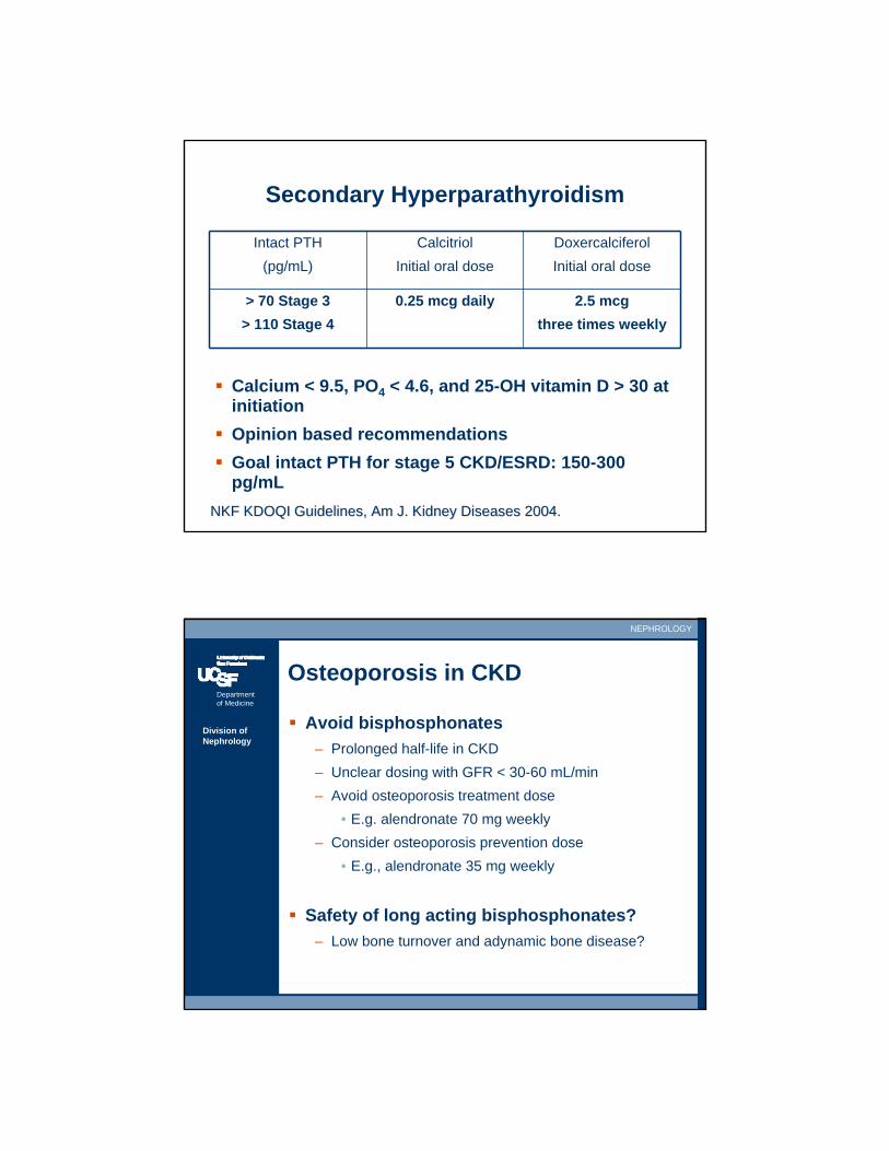
Secondary Hyperparathyroidism
Calcium < 9.5, PO4 < 4.6, and 25-OH vitamin D > 30 at initiationOpinion based recommendationsGoal intact PTH for stage 5 CKD/ESRD: 150-300 pg/mL
Intact PTH(pg/mL)
CalcitriolInitial oral dose
DoxercalciferolInitial oral dose
> 70 Stage 3> 110 Stage 4
0.25 mcg daily 2.5 mcgthree times weekly
NKF KDOQI Guidelines, Am J. Kidney Diseases 2004.NKF KDOQI Guidelines, Am J. Kidney Diseases 2004.
Department of Medicine
NEPHROLOGY
Division of Nephrology
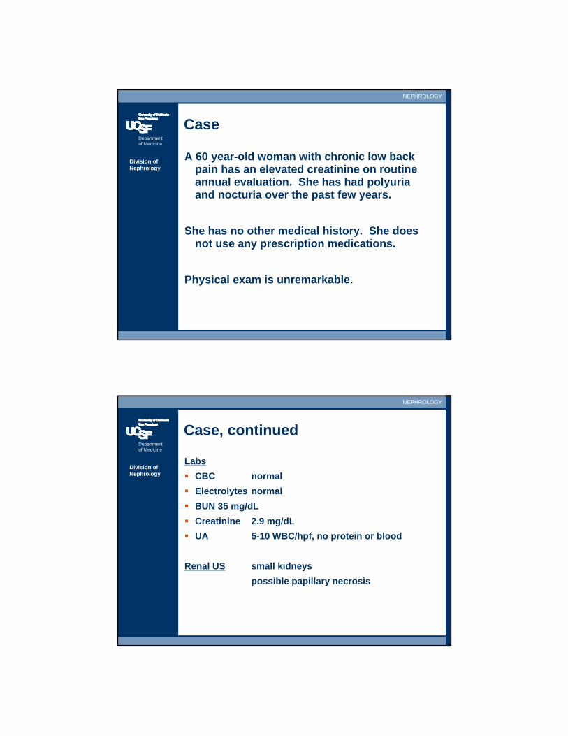
Osteoporosis in CKD
Avoid bisphosphonates– Prolonged half-life in CKD– Unclear dosing with GFR < 30-60 mL/min– Avoid osteoporosis treatment dose
• E.g. alendronate 70 mg weekly– Consider osteoporosis prevention dose
• E.g., alendronate 35 mg weekly
Safety of long acting bisphosphonates?– Low bone turnover and adynamic bone disease?
Department of Medicine
NEPHROLOGY
Division of Nephrology
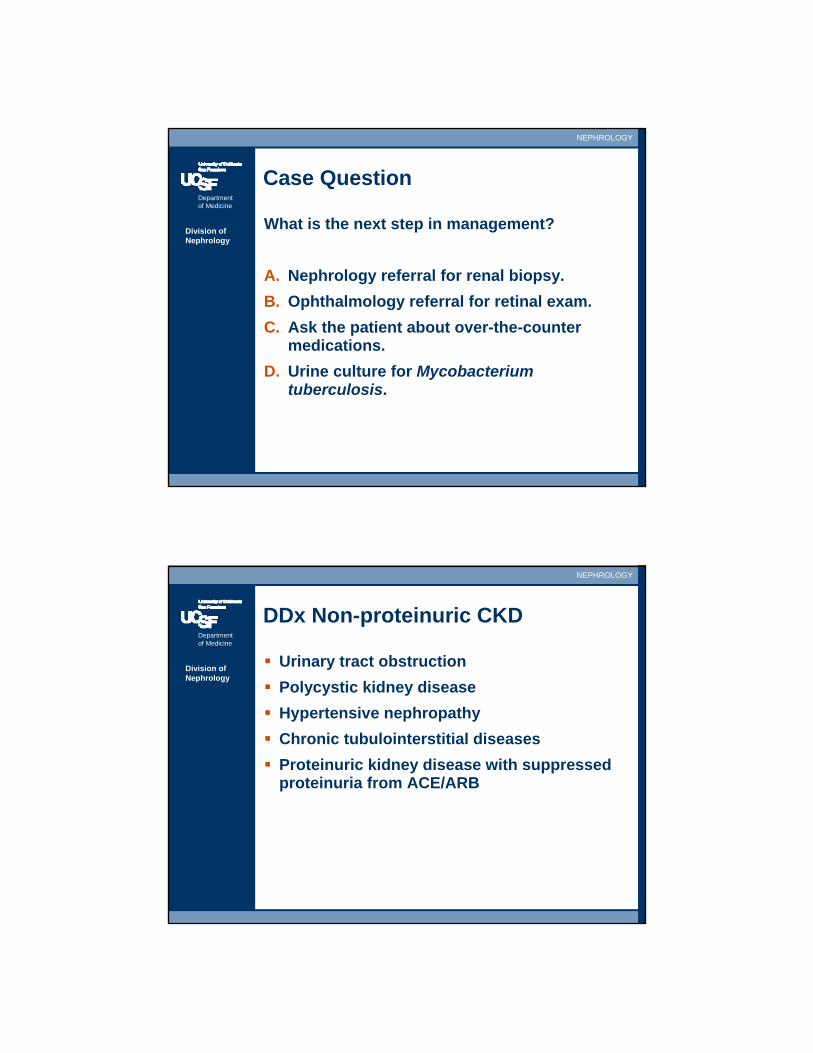
Case
A 60 year-old woman with chronic low back pain has an elevated creatinine on routine annual evaluation. She has had polyuria and nocturia over the past few years.
She has no other medical history. She does not use any prescription medications.
Physical exam is unremarkable.
Department of Medicine
NEPHROLOGY
Division of Nephrology
Case, continued
LabsCBC normalElectrolytes normalBUN 35 mg/dLCreatinine 2.9 mg/dLUA 5-10 WBC/hpf, no protein or blood
Renal US small kidneyspossible papillary necrosis
Department of Medicine
NEPHROLOGY
Division of Nephrology
Case Question
What is the next step in management?
A. Nephrology referral for renal biopsy.B. Ophthalmology referral for retinal exam.C. Ask the patient about over-the-counter
medications.D. Urine culture for Mycobacterium
tuberculosis.
Department of Medicine
NEPHROLOGY
Division of Nephrology
DDx Non-proteinuric CKD
Urinary tract obstructionPolycystic kidney disease Hypertensive nephropathyChronic tubulointerstitial diseasesProteinuric kidney disease with suppressed proteinuria from ACE/ARB
Department of Medicine
NEPHROLOGY
Division of Nephrology
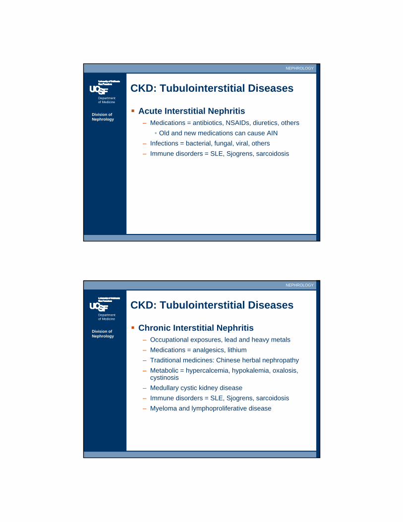
CKD: Tubulointerstitial Diseases
Acute Interstitial Nephritis– Medications = antibiotics, NSAIDs, diuretics, others
• Old and new medications can cause AIN– Infections = bacterial, fungal, viral, others– Immune disorders = SLE, Sjogrens, sarcoidosis
Department of Medicine
NEPHROLOGY
Division of Nephrology
CKD: Tubulointerstitial Diseases
Chronic Interstitial Nephritis– Occupational exposures, lead and heavy metals– Medications = analgesics, lithium– Traditional medicines: Chinese herbal nephropathy– Metabolic = hypercalcemia, hypokalemia, oxalosis,
cystinosis– Medullary cystic kidney disease– Immune disorders = SLE, Sjogrens, sarcoidosis– Myeloma and lymphoproliferative disease
Department of Medicine
NEPHROLOGY
Division of Nephrology
CKD: Interstitial Disease
Often asymptomatic– May not have the fever, rash, and arthralgias of acute
interstitial nephritis
Minimal proteinuria/hematuriaSterile pyuriaUrine sediment: +/- WBC, WBC castsLate manifestations: hypertension and anemia
Department of Medicine
NEPHROLOGY
Division of Nephrology
CKD: Interstitial Disease
Tubular abnormalities – Urinary concentrating defects and nephrogenic
diabetes insipidus polyuria, nocturia– Fanconi syndrome
• Impaired tubular reabsorption: amino acids, bicarbonate, phosphate, glucose in urine
• Glucosuria with normal serum glucose• Proximal (type 2) RTA/metabolic acidosis from
bicarbonate spilling• Distal (type 1) RTA/metabolic acidosis from
inability to acidify urine
Department of Medicine
NEPHROLOGY
Division of Nephrology
CKD: Interstitial Disease
Definitive diagnosis by kidney biopsy– Diagnosis often made clinically– Biopsy may not alter therapy
Therapy– Eliminate or treat underlying cause– Mainly supportive therapy– Steroids for AIN? Controversial, lack of good trials– Steroids NOT used for chronic interstitial nephritis
Department of Medicine
NEPHROLOGY
Division of Nephrology
CKD: Analgesic Nephropathy (AN)
Phenacetin– Previously widely available outside United States– Incidence of AN dropped after taken off market
Acetaminophen – Metabolite of phenacetin– Conflicting data on nephrotoxicity
Aspirin– Potentiates toxicity of phenacetin and acetaminophen
Department of Medicine
NEPHROLOGY
Division of Nephrology
CKD: Analgesic Nephropathy (AN)
Usually seen in womenHistory of chronic back pain or headachesRadiology findings
– IVP: Papillary necrosis in severe cases– Ultrasound: Atrophic kidneys– CT: Papillary calcifications, atrophic kidneys with
“bumpy" or lobulated/irregular contours
Department of Medicine
NEPHROLOGY
Division of Nephrology
NSAIDs and Kidney Disease
ARF: Hemodynamic acute renal failure– Prostaglandins vasodilate afferent arteriole
ARF: Acute interstitial nephritis +/- minimal change disease
– Sterile pyuria with proteinuria
CKD: Analgesic nephropathy– Cumulative nephrotoxicity, high doses over years
CKD: Membranous nephropathy– Heavy proteinuria, nephrotic syndrome– Hypercoagulability
Department of Medicine
NEPHROLOGY
Division of Nephrology
Case
A 32 year-old African-American man with a recent diagnosis of HIV presents with nausea and vomiting for 2 months. He notes frothy urine for 6 months.
He is afebrile with blood pressure 100/62 and heart rate 72. Physical exam is normal without pedal edema.
Department of Medicine
NEPHROLOGY
Division of Nephrology
Case, continued
LabsHematocrit 32%BUN 104 mg/dLSerum Cr 14.2 mg/dLCD4 132/mm3
U/A 3+ protein, no hematuria24-hr urine 12 gm protein
Renal US large kidneys with marked echogenicity
He starts hemodialysis and has a kidney biopsy.
Department of Medicine
NEPHROLOGY
Division of Nephrology
Case Question
Kidney biopsy shows focal segmental glomerulosclerosis of the collapsing variant, interstitial inflammation, and tubular microcyst formation.
Which of the following is the most appropriate therapy for this patient’s disease?
A. Pulse IV methylprednisoloneB. CyclophosphamideC. CyclosporineD. Highly active antiretroviral therapy
(HAART)
Department of Medicine
NEPHROLOGY
Division of Nephrology
Case
A 72 year-old woman is admitted to the hospital for new onset nephrotic syndrome. She had been healthy until the past year when she noticed a decrease in appetite, constipation, and a ten pound weight loss. Over the past month, she has noticed face, arm, and leg swelling.
Physical examination reveals a chronically ill-appearing woman with anasarca.
Department of Medicine
NEPHROLOGY
Division of Nephrology
Case, continued
LabsHematocrit 29% with MCV 72 fLBUN 54 mg/dLCreatinine 3.1 mg/dLHBV, HCV, cryo negativeComplementsnormalUA 4+ protein, no hematuria24-hr urine 4.5 g protein
Renal US: Normal sized kidneys with mild echogenicity.
Renal Bx: Thickened glomerular capillary walls with subepithelial deposits consistent with membranous nephropathy.
Department of Medicine
NEPHROLOGY
Division of Nephrology
Case Question
Which of the following studies are most appropriate in light of the renal biopsy results?
A. ANCA antibodiesB. Anti-GBM antibodiesC. ANA and double-stranded DNA antibodiesD. EchocardiogramE. Colonoscopy
Department of Medicine
NEPHROLOGY
Division of Nephrology
Nephrotic Syndrome
Proteinuria > 3.5 g/dayDyslipidemiaEdemaHypoalbuminemiaLipiduria (oval fat bodies in urine, Maltese cross with polarized light)
Caveat: – Many patients do not have all 5 features; nephrotic
proteinuria without nephrotic syndrome
Associated Feature: Hypercoagulability
Department of Medicine
NEPHROLOGY
Division of Nephrology
Nephrotic Diseases
Focal Segmental Glomerulosclerosis (FSGS)Membranous Nephropathy (MN)Minimal Change Disease (MCD)AmyloidosisDiabetic nephropathyOthers = SLE, IgA nephropathy, MPGN
Department of Medicine
NEPHROLOGY
Division of Nephrology
Nephrotic Disease: Focal Segmental Glomerulosclerosis (FSGS)
Idiopathic/Primary– African-Americans and patients < 40 years– Can be treated with steroids – Can recur explosively post-kidney transplant
Secondary– HIV-associated nephropathy (HIVAN), almost
exclusively in African-Americans, large kidneys– Chronic kidney disease, reduced nephron mass,
hyperfiltration injury– Heroin, morbid obesity, drugs (lithium, pamidronate)– Sickle cell disease– Typically not steroid responsive
Department of Medicine
NEPHROLOGY
Division of Nephrology
Nephrotic Disease:Membranous Nephropathy
Idiopathic/Primary– Caucasians, most common cause of nephrotic
syndrome by a primary glomerular disease
Secondary– Malignancy
• Typically solid (colon, lung, breast), also non-Hodgkin’s
• 5-10% have malignancy, but <1-2% are occult – Chronic infections, HBV > HCV, syphilis, leprosy,
schistosomiasis– SLE and autoimmune/connective tissue diseases– Drugs: NSAIDs, gold, penicillamine– Sickle cell disease
Department of Medicine
NEPHROLOGY
Division of Nephrology
Nephrotic Disease: Membranous Nephropathy
Clinical– Renal vein thrombosis and hypercoagulability– Secondary prophylaxis with warfarin– Malignancy and age-appropriate cancer screening
Prognosis: Mixed– Third get better, third stay same, third get worse
Treatment: – Carefully selected patients with poor prognostic
features (older age, men, chronic kidney disease, symptomatic proteinuria/nephrotic syndrome)
– Immunosuppression: steroids AND (cyclophosphamide or chlorambucil)
Department of Medicine
NEPHROLOGY
Division of Nephrology
Nephrotic Disease:Minimal Change Disease (MCD)
Idiopathic/Primary– Second peak in 60-70 year old patients– More steroid resistance/dependence and higher
relapse rate in adults than in children
Secondary– Drugs– NSAID-induced AIN with MCD, pyuria with
proteinuria– Infections– Neoplasm, Hodgkin’s and others– Allergy and toxins (bee stings, mercury, lead)
Rx: Steroids typically first-line
Department of Medicine
NEPHROLOGY
Division of Nephrology
Nephrotic Disease: Amyloidosis
Pathology– β pleated structure that forms 8-10 nm fibrils– Congo Red stain has apple-green birefringence with
polarized light
Classification– ~ 20 unique amyloidoses– AL (primary) amyloidosis
• myeloma and monoclonal gammopathies– AA (secondary) amyloidosis
• chronic infections, inflammatory states (inflammatory bowel disease, rheumatoid arthritis, familial Mediterranean fever)
Department of Medicine
NEPHROLOGY
Division of Nephrology
Nephrotic Disease: Amyloidosis
Clinical findings– Renal involvement is common in amyloidoses– Large kidneys and massive proteinuria– Multi-organ involvement
• Periorbital hemorrhage (raccoon sign), macroglossia
• Cardiac deposits• GI involvement, hepatomegaly• Carpal tunnel syndrome, neuropathy• Shoulder pad sign = amyloid deposits in deltoids
– Cardiac and kidney disease are poor prognostic signs

Department of Medicine
NEPHROLOGY
Division of Nephrology
Nephrotic Disease: Amyloidosis
Treatment– AA Amyloidosis: Treat underlying infection or
inflammation, colchicine for Familial Mediterranean Fever
– AL Amyloidosis: Treat underlying myeloma, melphalan, prednisone, stem-cell transplant
– Adjuvant therapy: ACE/ARB, blood pressure control, diuretics, sodium/water restriction
Department of Medicine
NEPHROLOGY
Division of Nephrology
Kidney Disease in Multiple Myeloma
Amyloidosis– Lambda > kappa light chains
Light chain deposition disease– Kappa > lambda light chains
Cast nephropathyHypercalcemia and vasoconstrictive ARFHypercalcemia and nephrogenic DI with pre-renal ARFATN from sepsis
Department of Medicine
NEPHROLOGY
Division of Nephrology
Diabetic Nephropathy
Common cause of proteinuriaUnusual cause of massive proteinuria and nephrotic syndrome.Early hyperfiltration phase with preserved creatinine and large kidneysDiagnosis
– Usually clinical diagnosis without kidney biopsy– Compatible clinical history
• Duration and severity of DM,• Evidence of end-organ disease from DM
(retinopathy, neuropathy) • No suspicious features for alternative diagnosis
Department of Medicine
NEPHROLOGY
Division of Nephrology
Diabetic Nephropathy
Untreated DM patients will lose GFR at rate of 1 mL/min/month or 12 mL/min/year Rapid deterioration of function and/or unexplained rise in proteinuria suggest non-diabetic disease

Department of Medicine
NEPHROLOGY
Division of Nephrology
Serologies for Secondary Causes of Nephrotic Syndrome
FSGS– HIV
Membranous Nephropathy– HBV, HCV, VDRL/RPR, ANA, RF
Minimal change disease– None
Amyloidosis– SPEP/UPEP/IFE for primary/AL amyloidosis
Diabetic Nephropathy– None, HbA1c for glycemic control
Department of Medicine
NEPHROLOGY
Division of Nephrology
3 Approaches to Nephrotic Proteinuria
1. All serologies on all patients– Expensive, time-consuming, low yield
2. Biopsy first, ask questions later– Serologies based on pathology to r/o 2° causes
3. Some serologies on all patients– C3 C4: low vs. normal complements– ANA: vaguely rheumatologic vs. non-rheumatologic– SPEP/UPEP/IFE: multiple myeloma and MGUS– Other serologies based on clinical suspicion– Low threshold for kidney biopsy
Department of Medicine
NEPHROLOGY
Division of Nephrology
Serologies in Nephrotic Syndrome
Serologies are suggestive, not definitiveStill require kidney biopsy for diagnosis
Nephritic DiseasesSerologies and clinical hx can be definitive
– SLE (ANA, anti-DS DNA)– ANCA-related disease (ANCA)– Anti-GBM disease (anti-GBM)– Post-infectious (ASO/antiDNase)
Kidney bx for prognosis
Department of Medicine
NEPHROLOGY
Division of Nephrology
DDx Enlarged Kidneys
Hydronephrosis/ObstructionPolycystic kidney diseaseInfiltrative disease (lymphoma)HIVANAmyloidosisEarly diabetic nephropathy
Department of Medicine
NEPHROLOGY
Division of Nephrology
Adjuvant Rx in Nephrotic Disease
HTN control– Goal BP < 130/80 mm Hg or even 125/75
Proteinuria suppression– ACE inhibitors ± ARB– Goal urine protein:creatinine ratio < 0.5– Dietary protein restriction controversial
Loop diuretics for edemaSodium/fluid restrictionNo clear role for primary prophylaxis with anticoagulation for hypercoagulability
Department of Medicine
NEPHROLOGY
Division of Nephrology
Secondary HTN: When to Suspect
Clinical Features– Age at onset < 30 yrs (unless + family history)– Age at onset > 50 yrs– Rapid onset of severe HTN– Refractory HTN– Worsening of previously well controlled HTN– Hypokalemia
Department of Medicine
NEPHROLOGY
Division of Nephrology
Secondary HTN: DDx
Kidney– Renovascular disease, Liddle syndrome
Endocrine– Hyper/hypothyroidism– Aldosteronism (plasma aldo:renin ratio, 24 hr urine
aldosterone)– Cushing Syndrome (dexamethasone suppression
test, 24 hr urine cortisol)– Pheochromocytoma (24 hr urine catecholamines and
metanephrines)– Congenital adrenal hyperplasia– Hypercalcemia– Syndrome apparent mineralocorticoid excess
Department of Medicine
NEPHROLOGY
Division of Nephrology
Secondary HTN: DDx
Drugs– Prescription: estrogen, cyclosporine, steroids– Over the counter: NSAIDs, pseudoephedrine– Smoking, ethanol, cocaine
Neurogenic– Increased intracranial pressure, spinal cord injury
Miscellaneous– Aortic coarctation– Obstructive sleep apnea– Polycythemia vera
Department of Medicine
NEPHROLOGY
Division of Nephrology
Renal Artery Stenosis/Disease
Clinical Features– Secondary HTN– Flash pulmonary edema– Hypokalemia– Kidney size asymmetry > 1.5 cm– ARF after initiation of ACE inhibitor/ARB
Diagnosis– CTA, MRA, conventional angiograph– Ultrasound: highly operator/institution dependent
Department of Medicine
NEPHROLOGY
Division of Nephrology
Renal Artery Stenosis/Disease
Atherosclerosis– Men and women, age > 50– Proximal/ostial lesions– Complete occlusion and renal atrophy are common– Medical management
Fibromuscular Dysplasia– Women, younger, 15-40– Mid-vessel disease, can affect multiple vessels– String of beads appearance on angiography– Complete occlusion and renal atrophy are rare– Often reversible with angioplasty
Department of Medicine
NEPHROLOGY
Division of Nephrology
HIV and Kidney Disease
ARF/ATN– Immunodeficiency and sepsis– Drug nephrotoxicity
• Tenofovir, foscarnet, pentamidine• Acyclovir, aminoglycosides, amphotericin B
AIN– NSAIDs, rifampin, trimethoprim-sulfamethoxazole
Nephrolithiasis– Indinavir, acyclovir, sulfadiazine– Drug crystals on urine sediment
Department of Medicine
NEPHROLOGY
Division of Nephrology
HIV and Kidney Disease
HIV associated diseases– HIVAN (African-Americans)– Immune complex GN (all others)– HUS/TTP/Thrombotic Microangiopathy– IgA Nephropathy
Other co-morbidities causing kidney disease
– HBV: Membranous > MPGN– HCV: MPGN, cryoglobulinemia > Membranous
Department of Medicine
NEPHROLOGY
Division of Nephrology
HIV-Associated Nephropathy (HIVAN)
African-Americans, unusual in Caucasians– Other HIV kidney diseases (immune-complex
glomerulonephritis, IgA nephropathy)Usually late manifestation of AIDS
– Low CD4 count
Often asymptomaticLack of hypertension and edemaNephrotic proteinuriaBland urine sedimentNormal or enlarged kidneysOften rapid deterioration of renal function
Department of Medicine
NEPHROLOGY
Division of Nephrology
Treatment of HIVAN
Highly active antiretroviral therapy (HAART)ACE inhibitors and/or ARBsLack of randomized controlled trials
Department of Medicine
NEPHROLOGY
Division of Nephrology
Case
A 21 year-old woman in the third trimester of her first pregnancy comes to the ED after a minor car collision.
Medical history is unremarkable.
On physical examination, she is anxious. Respiratory rate is 18/min, and blood pressure is 110/70 mmHg. Otherwise her exam is unremarkable.
Department of Medicine
NEPHROLOGY
Division of Nephrology
Case, continued
LabsHematocrit 33%WBC 9000/uLPlatelet count 122,000/uLSerum sodium 132 mEq/LSerum potassium 3.8 mEq/LSerum chloride 100 mEq/LSerum bicarbonate 20 mEq/LBUN 5 mg/dLCreatinine 0.4 mg/dLUA pH 6.0, SG 1.020, no proteinuria or hematuria
Department of Medicine
NEPHROLOGY
Division of Nephrology
Case Question
Which of the following is the most likely explanation for her serum electrolytes?
A. Acute respiratory alkalosis due to a possible pneumothorax
B. Normal electrolyte values in a pregnant woman
C. Metabolic acidosis from hypoperfusionand tissue hypoxia
D. Renal tubular acidosis
Department of Medicine
NEPHROLOGY
Division of Nephrology
Kidney Physiology during Pregnancy
Kidney length increases by 1 cmDilatation of the calyces, pelves, uretersDuring 1st trimester, renal plasma flow increases 50-80%, GFR increases 50%Creatinine decreases from 0.8 mg/dL non-pregnant to 0.5 mg/dL in 3rd trimesterSlight increase in urine protein excretion, upper limit of normal increases from 150 to 300 mg/day
Department of Medicine
NEPHROLOGY
Division of Nephrology
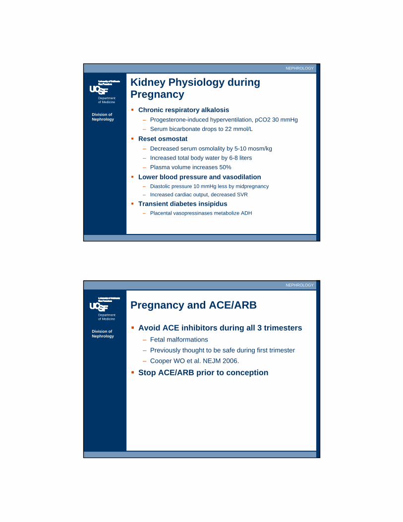
Kidney Physiology during Pregnancy
Chronic respiratory alkalosis– Progesterone-induced hyperventilation, pCO2 30 mmHg– Serum bicarbonate drops to 22 mmol/L
Reset osmostat– Decreased serum osmolality by 5-10 mosm/kg– Increased total body water by 6-8 liters– Plasma volume increases 50%
Lower blood pressure and vasodilation– Diastolic pressure 10 mmHg less by midpregnancy– Increased cardiac output, decreased SVR
Transient diabetes insipidus– Placental vasopressinases metabolize ADH
Department of Medicine
NEPHROLOGY
Division of Nephrology
Pregnancy and ACE/ARB
Avoid ACE inhibitors during all 3 trimesters– Fetal malformations– Previously thought to be safe during first trimester– Cooper WO et al. NEJM 2006.
Stop ACE/ARB prior to conception
Department of Medicine
NEPHROLOGY
Division of Nephrology
HTN and Pregnancy
Chronic hypertension– Elevated BP prior to pregnancy– Documented before 20 weeks of pregnancy– Persists 12 weeks after pregnancy
Gestational hypertension– Elevated BP without proteinuria after 20 weeks– BP returns to normal within 12 weeks after
pregnancy– 25% progress to preeclampsia (develop proteinuria)– Increased risk for developing HTN postpartum
Department of Medicine
NEPHROLOGY
Division of Nephrology
Preeclampsia:Definition and Risk Factors
Hypertension and proteinuria after 20 weeks– BP > 140/90 mmHg in pts with previously normal BP– Proteinuria > 0.3 g/day (roughly 1+ dipstick protein)– Unusual in first trimester/20 weeks
Risk factors– Age > 35 or < 20 years, African-American, family/pt
hx preeclampsia, nulliparity, gestational DM, type 1 DM, obesity, chronic HTN, kidney disease, thrombophilias, vascular and connective tissue disease, antiphospholipid antibody syndrome, elevated serum uric acid
Department of Medicine
NEPHROLOGY
Division of Nephrology
Preeclampsia: Clinical Features
May develop before, during, or after deliveryInsidious or fulminant+/- Symptoms (visual disturbances, headache, upper abdominal pain)5-15% with HELLP (worse prognosis)Complications
– Eclampsia, intrauterine growth retardation (IUGR)– Placental abruption, DIC, acute renal failure– CVA and cardiovascular complications– Maternal death
Department of Medicine
NEPHROLOGY
Division of Nephrology
Preeclampsia: Treatment
Delivery, vaginal preferred over CesareanSeizure prophylaxis – magnesium sulfateHospitalization for non-compliant patients, poor access to medical care, progressive or severe preeclampsiaTertiary center and/or high-risk obstetrician
Department of Medicine
NEPHROLOGY
Division of Nephrology
Pregnancy and CKD
High-risk CKD patients– Baseline creatinine > 1.4 mg/dL– Pre-existing hypertension
Pregnancy effects on CKD– Worsening HTN and proteinuria– Temporary or permanent decline in kidney function
CKD effects on Pregnancy– Intrauterine growth retardation, prematurity, fetal loss– Increased risk for preeclampsia, eclampsia
Department of Medicine
NEPHROLOGY
Division of Nephrology
Case
83 year-old woman falls and fractures her right hip. Medical history includes hypertension and diabetes.
Medications include an ACE inhibitor.
On physical exam, she is slightly confused. BP 140/90, HR 80. JVP is 8 cm, normal heart sounds, and clear chest exam. No hepatosplenomegaly. No pedal edema. Deep tendon reflexes are normal.
Department of Medicine
NEPHROLOGY
Division of Nephrology
Case, continued
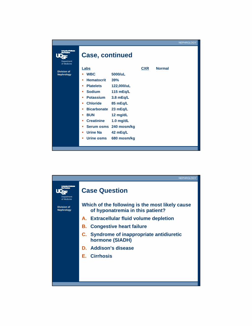
Labs CXR NormalWBC 5000/uL Hematocrit 39%Platelets 122,000/uLSodium 115 mEq/LPotassium 3.8 mEq/LChloride 85 mEq/LBicarbonate 23 mEq/LBUN 12 mg/dLCreatinine 1.0 mg/dLSerum osms 240 mosm/kgUrine Na 42 mEq/LUrine osms 680 mosm/kg
Department of Medicine
NEPHROLOGY
Division of Nephrology
Case Question
Which of the following is the most likely cause of hyponatremia in this patient?
A. Extracellular fluid volume depletionB. Congestive heart failureC. Syndrome of inappropriate antidiuretic
hormone (SIADH)D. Addison’s diseaseE. Cirrhosis
Department of Medicine
NEPHROLOGY
Division of Nephrology
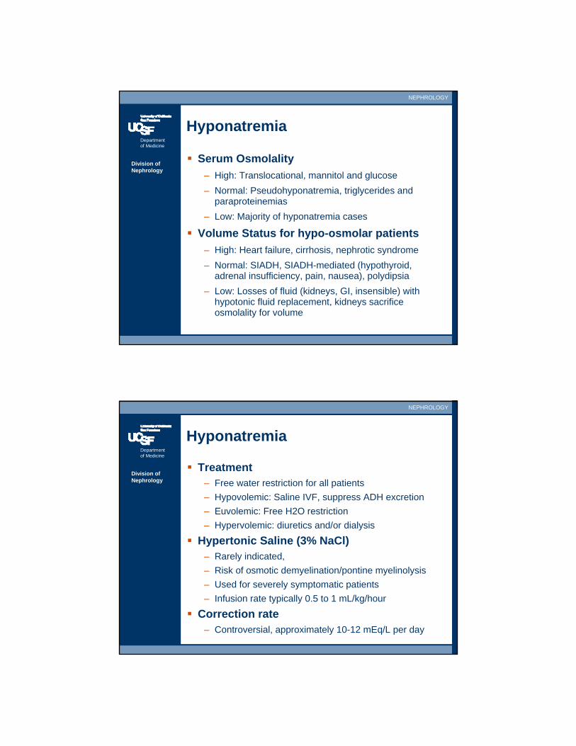
Hyponatremia
Serum Osmolality– High: Translocational, mannitol and glucose– Normal: Pseudohyponatremia, triglycerides and
paraproteinemias– Low: Majority of hyponatremia cases
Volume Status for hypo-osmolar patients– High: Heart failure, cirrhosis, nephrotic syndrome– Normal: SIADH, SIADH-mediated (hypothyroid,
adrenal insufficiency, pain, nausea), polydipsia – Low: Losses of fluid (kidneys, GI, insensible) with
hypotonic fluid replacement, kidneys sacrifice osmolality for volume
Department of Medicine
NEPHROLOGY
Division of Nephrology
Hyponatremia
Treatment– Free water restriction for all patients– Hypovolemic: Saline IVF, suppress ADH excretion– Euvolemic: Free H2O restriction– Hypervolemic: diuretics and/or dialysis
Hypertonic Saline (3% NaCl)– Rarely indicated, – Risk of osmotic demyelination/pontine myelinolysis– Used for severely symptomatic patients– Infusion rate typically 0.5 to 1 mL/kg/hour
Correction rate– Controversial, approximately 10-12 mEq/L per day
Department of Medicine
NEPHROLOGY
Division of Nephrology
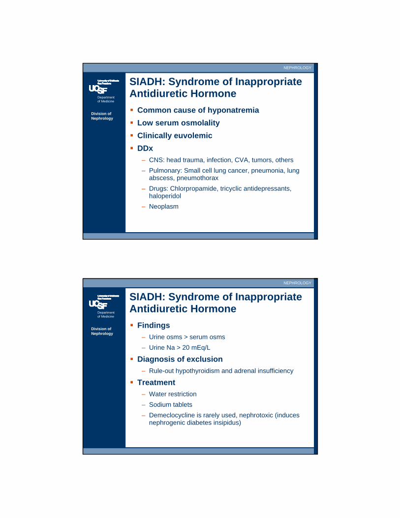
SIADH: Syndrome of Inappropriate Antidiuretic Hormone
Common cause of hyponatremiaLow serum osmolalityClinically euvolemicDDx
– CNS: head trauma, infection, CVA, tumors, others– Pulmonary: Small cell lung cancer, pneumonia, lung
abscess, pneumothorax– Drugs: Chlorpropamide, tricyclic antidepressants,
haloperidol– Neoplasm
Department of Medicine
NEPHROLOGY
Division of Nephrology
SIADH: Syndrome of Inappropriate Antidiuretic Hormone
Findings– Urine osms > serum osms– Urine Na > 20 mEq/L
Diagnosis of exclusion– Rule-out hypothyroidism and adrenal insufficiency
Treatment– Water restriction– Sodium tablets– Demeclocycline is rarely used, nephrotoxic (induces
nephrogenic diabetes insipidus)
Department of Medicine
NEPHROLOGY
Division of Nephrology
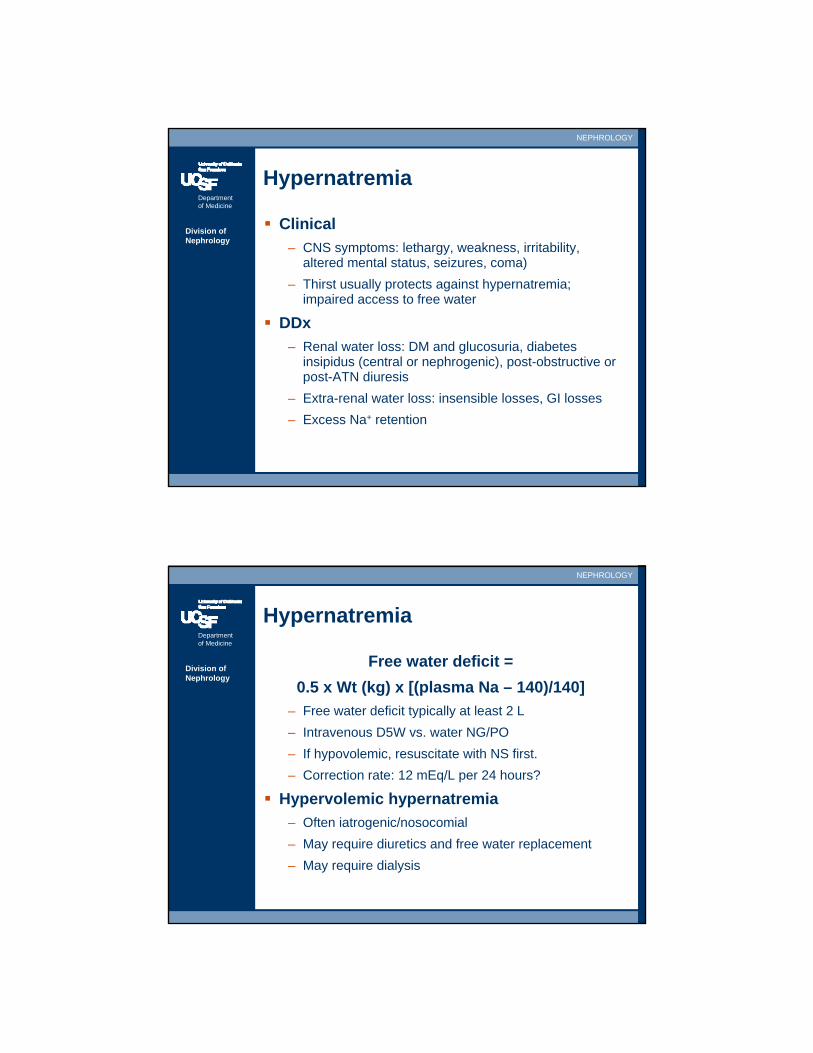
Hypernatremia
Clinical– CNS symptoms: lethargy, weakness, irritability,
altered mental status, seizures, coma)– Thirst usually protects against hypernatremia;
impaired access to free water
DDx– Renal water loss: DM and glucosuria, diabetes
insipidus (central or nephrogenic), post-obstructive or post-ATN diuresis
– Extra-renal water loss: insensible losses, GI losses– Excess Na+ retention
Department of Medicine
NEPHROLOGY
Division of Nephrology
Hypernatremia
Free water deficit = 0.5 x Wt (kg) x [(plasma Na – 140)/140]
– Free water deficit typically at least 2 L– Intravenous D5W vs. water NG/PO– If hypovolemic, resuscitate with NS first.– Correction rate: 12 mEq/L per 24 hours?
Hypervolemic hypernatremia– Often iatrogenic/nosocomial– May require diuretics and free water replacement– May require dialysis
Department of Medicine
NEPHROLOGY
Division of Nephrology
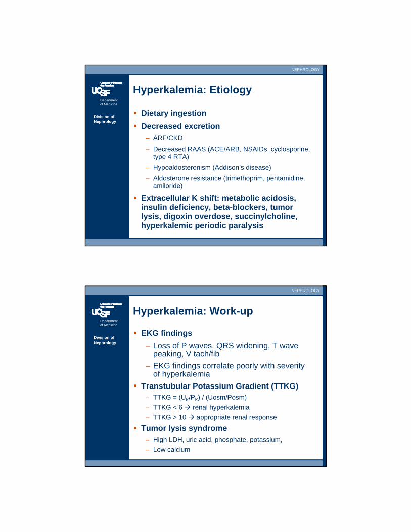
Hyperkalemia: Etiology
Dietary ingestion Decreased excretion
– ARF/CKD– Decreased RAAS (ACE/ARB, NSAIDs, cyclosporine,
type 4 RTA) – Hypoaldosteronism (Addison’s disease)– Aldosterone resistance (trimethoprim, pentamidine,
amiloride)
Extracellular K shift: metabolic acidosis, insulin deficiency, beta-blockers, tumor lysis, digoxin overdose, succinylcholine, hyperkalemic periodic paralysis
Department of Medicine
NEPHROLOGY
Division of Nephrology
Hyperkalemia: Work-up
EKG findings– Loss of P waves, QRS widening, T wave
peaking, V tach/fib– EKG findings correlate poorly with severity
of hyperkalemiaTranstubular Potassium Gradient (TTKG)
– TTKG = (UK/PK) / (Uosm/Posm)– TTKG < 6 renal hyperkalemia– TTKG > 10 appropriate renal response
Tumor lysis syndrome– High LDH, uric acid, phosphate, potassium,– Low calcium
Department of Medicine
NEPHROLOGY
Division of Nephrology
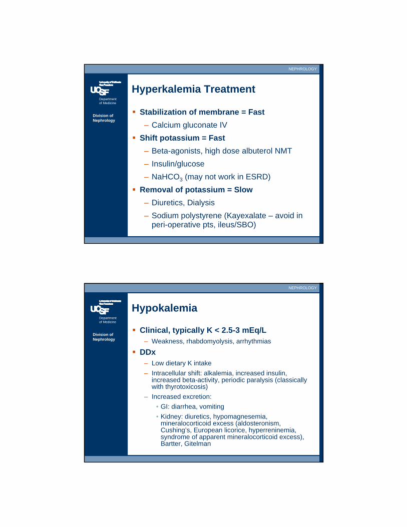
Hyperkalemia Treatment
Stabilization of membrane = Fast– Calcium gluconate IV
Shift potassium = Fast– Beta-agonists, high dose albuterol NMT– Insulin/glucose– NaHCO3 (may not work in ESRD)
Removal of potassium = Slow– Diuretics, Dialysis– Sodium polystyrene (Kayexalate – avoid in
peri-operative pts, ileus/SBO)
Department of Medicine
NEPHROLOGY
Division of Nephrology
Hypokalemia
Clinical, typically K < 2.5-3 mEq/L– Weakness, rhabdomyolysis, arrhythmias
DDx– Low dietary K intake– Intracellular shift: alkalemia, increased insulin,
increased beta-activity, periodic paralysis (classically with thyrotoxicosis)
– Increased excretion:• GI: diarrhea, vomiting• Kidney: diuretics, hypomagnesemia,
mineralocorticoid excess (aldosteronism, Cushing’s, European licorice, hyperreninemia, syndrome of apparent mineralocorticoid excess), Bartter, Gitelman
Department of Medicine
NEPHROLOGY
Division of Nephrology
Hypokalemia
DiagnosticsTranstubular Potassium Gradient (TTKG)
– TTKG < 2 GI losses, TTKG > 4 renal loss
24 hr urine– < 25 mEq/day extrarenal loss– > 25 mEq/day renal losses
TreatmentPotassium
– Difficult to estimate deficit, usually at least 200 mEq– IV: 10 mEq/hr peripherally, 20 mEq/hr centrally
Department of Medicine
NEPHROLOGY
Division of Nephrology
Metabolic Acidosis:Increased Anion Gap
Increased Anion Gap– MUDPILES (methanol, uremia, DKA, paraldehyde,
isoniazid, lactic acidosis, ethylene glycol, salicylate)– AG > 20 implies metabolic acidosis regardless of
serum bicarbonate or pH
Serum Anion Gap = Na – Cl – HCO3
Normal AG < 12Add 2.5 to the AG for every 1 g/dL drop in albumin
Department of Medicine
NEPHROLOGY
Division of Nephrology
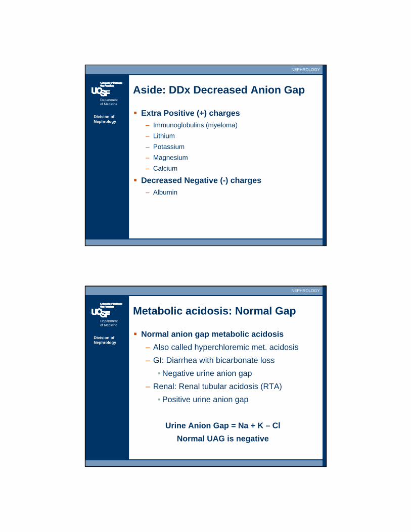
Aside: DDx Decreased Anion Gap
Extra Positive (+) charges– Immunoglobulins (myeloma)– Lithium– Potassium– Magnesium– Calcium
Decreased Negative (-) charges– Albumin
Department of Medicine
NEPHROLOGY
Division of Nephrology
Metabolic acidosis: Normal Gap
Normal anion gap metabolic acidosis– Also called hyperchloremic met. acidosis– GI: Diarrhea with bicarbonate loss
• Negative urine anion gap– Renal: Renal tubular acidosis (RTA)
• Positive urine anion gap
Urine Anion Gap = Na + K – ClNormal UAG is negative
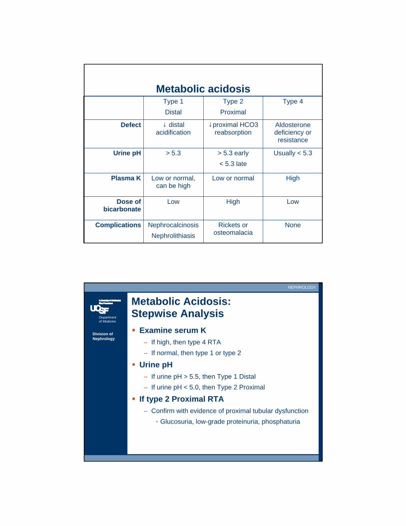
Metabolic acidosisType 1 Distal
Type 2 Proximal
Type 4
Defect ↓ distal acidification
↓proximal HCO3 reabsorption
Aldosterone deficiency or resistance
Urine pH > 5.3 > 5.3 early < 5.3 late
Usually < 5.3
Plasma K Low or normal, can be high
Low or normal High
Dose of bicarbonate
Low High Low
Complications NephrocalcinosisNephrolithiasis
Rickets or osteomalacia
None
Department of Medicine
NEPHROLOGY
Division of Nephrology
Metabolic Acidosis:Stepwise Analysis
Examine serum K– If high, then type 4 RTA– If normal, then type 1 or type 2
Urine pH– If urine pH > 5.5, then Type 1 Distal– If urine pH < 5.0, then Type 2 Proximal
If type 2 Proximal RTA– Confirm with evidence of proximal tubular dysfunction
• Glucosuria, low-grade proteinuria, phosphaturia
Department of Medicine
NEPHROLOGY
Division of Nephrology
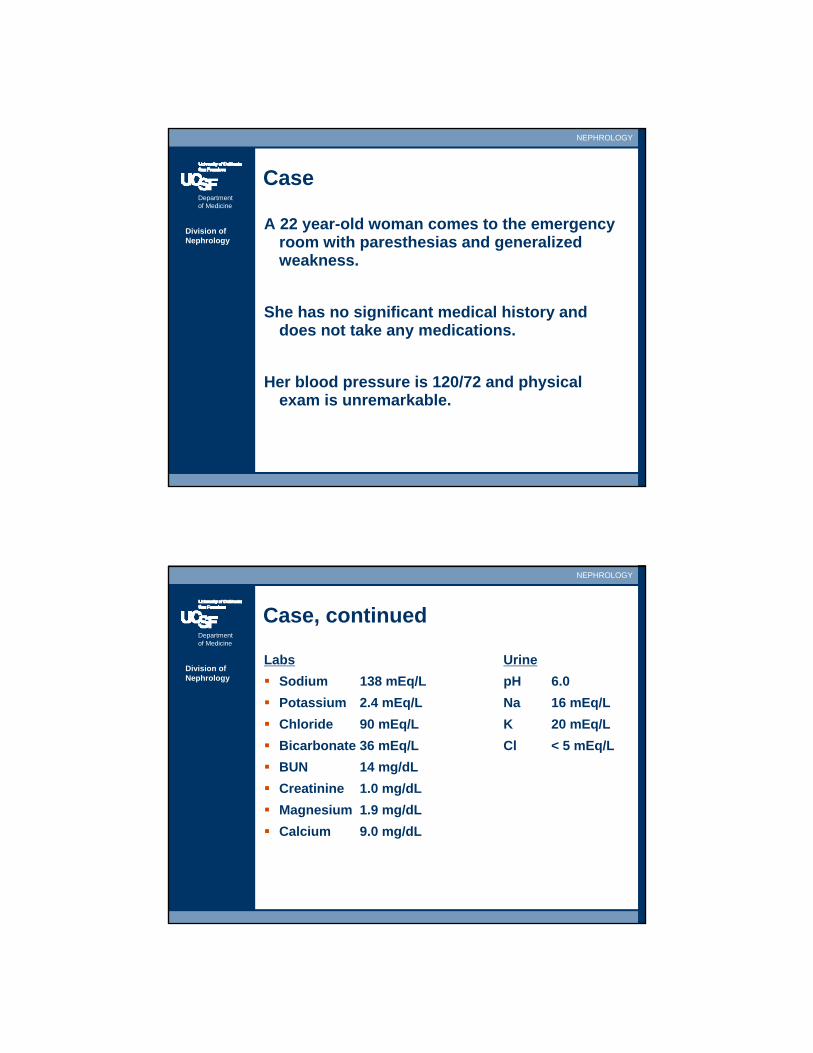
Case
A 22 year-old woman comes to the emergency room with paresthesias and generalized weakness.
She has no significant medical history and does not take any medications.
Her blood pressure is 120/72 and physical exam is unremarkable.
Department of Medicine
NEPHROLOGY
Division of Nephrology
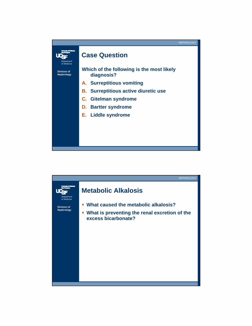
Case, continued
Labs UrineSodium 138 mEq/L pH 6.0Potassium 2.4 mEq/L Na 16 mEq/LChloride 90 mEq/L K 20 mEq/LBicarbonate 36 mEq/L Cl < 5 mEq/LBUN 14 mg/dLCreatinine 1.0 mg/dLMagnesium 1.9 mg/dLCalcium 9.0 mg/dL
Department of Medicine
NEPHROLOGY
Division of Nephrology
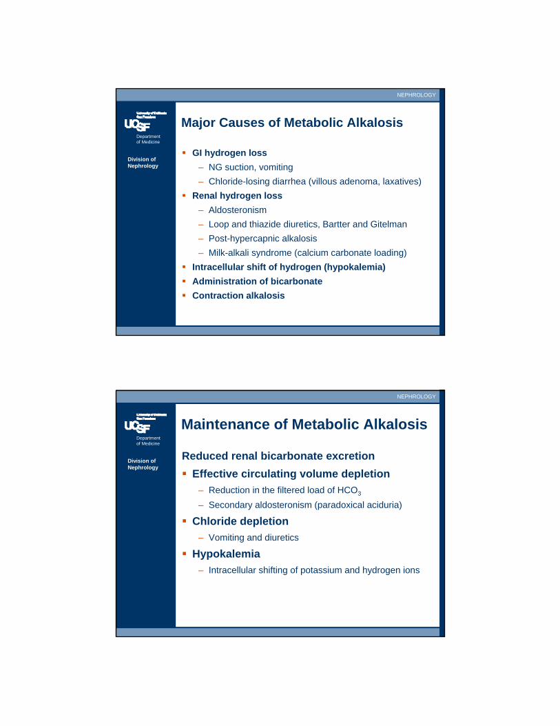
Case Question
Which of the following is the most likely diagnosis?
A. Surreptitious vomitingB. Surreptitious active diuretic useC. Gitelman syndromeD. Bartter syndromeE. Liddle syndrome
Department of Medicine
NEPHROLOGY
Division of Nephrology
Metabolic Alkalosis
What caused the metabolic alkalosis?What is preventing the renal excretion of the excess bicarbonate?
Department of Medicine
NEPHROLOGY
Division of Nephrology
Major Causes of Metabolic Alkalosis
GI hydrogen loss– NG suction, vomiting– Chloride-losing diarrhea (villous adenoma, laxatives)
Renal hydrogen loss– Aldosteronism– Loop and thiazide diuretics, Bartter and Gitelman– Post-hypercapnic alkalosis– Milk-alkali syndrome (calcium carbonate loading)
Intracellular shift of hydrogen (hypokalemia)Administration of bicarbonateContraction alkalosis
Department of Medicine
NEPHROLOGY
Division of Nephrology
Maintenance of Metabolic Alkalosis
Reduced renal bicarbonate excretionEffective circulating volume depletion
– Reduction in the filtered load of HCO3
– Secondary aldosteronism (paradoxical aciduria)
Chloride depletion– Vomiting and diuretics
Hypokalemia– Intracellular shifting of potassium and hydrogen ions

Department of Medicine
NEPHROLOGY
Division of Nephrology
Urine Chloride in Metabolic Alkalosis
Vomiting and long term diuretic use– Depleted body chloride stores – Kidneys will conserve/reabsorb chloride– Urine Cl < 15 mEq/L – Urine Cl will be elevated with ACTIVE diuretic use
Primary aldosteronism – Volume expanded – Urine Cl > 20 mEq/L
Department of Medicine
NEPHROLOGY
Division of Nephrology
Saline and Alkalosis
Saline Responsive = Low urine Cl < 15– Vomiting or nasogastric suction– Diuretics– Post-hypercapnic alkalosis– Low dietary chloride intake
Saline Unresponsive = High urine Cl > 20– Mineralocorticoid excess– Severe hypokalemia– Edematous disorders, e.g. CHF
Department of Medicine
NEPHROLOGY
Division of Nephrology
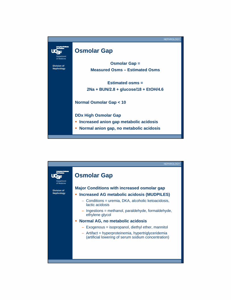
Osmolar Gap
Osmolar Gap = Measured Osms – Estimated Osms
Estimated osms = 2Na + BUN/2.8 + glucose/18 + EtOH/4.6
Normal Osmolar Gap < 10
DDx High Osmolar Gap Increased anion gap metabolic acidosisNormal anion gap, no metabolic acidosis
Department of Medicine
NEPHROLOGY
Division of Nephrology
Osmolar Gap
Major Conditions with increased osmolar gapIncreased AG metabolic acidosis (MUDPILES)
– Conditions = uremia, DKA, alcoholic ketoacidosis, lactic acidosis
– Ingestions = methanol, paraldehyde, formaldehyde, ethylene glycol
Normal AG, no metabolic acidosis– Exogenous = isopropanol, diethyl ether, mannitol– Artifact = hyperproteinemia, hypertriglyceridemia
(artificial lowering of serum sodium concentration)
Department of Medicine
NEPHROLOGY
Division of Nephrology
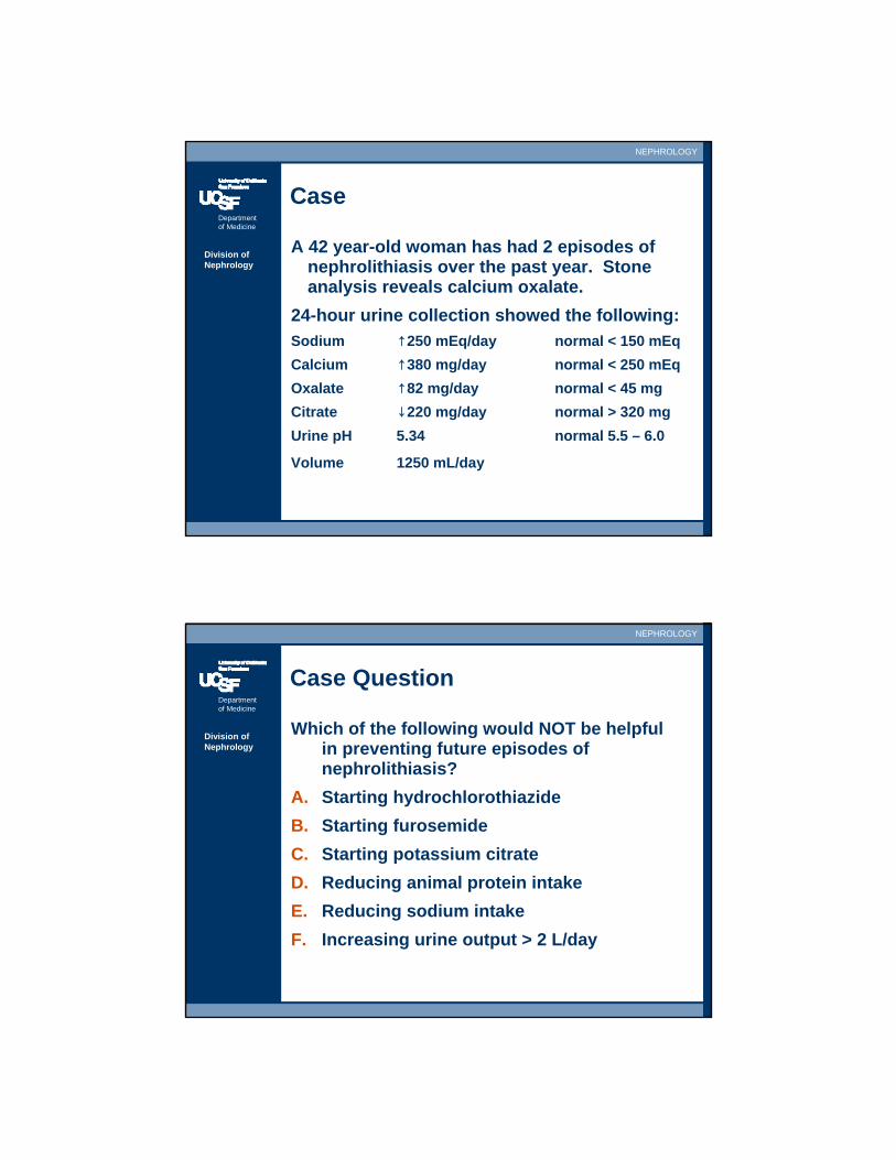
Case
A 42 year-old woman has had 2 episodes of nephrolithiasis over the past year. Stone analysis reveals calcium oxalate.
24-hour urine collection showed the following:Sodium ↑250 mEq/day normal < 150 mEqCalcium ↑380 mg/day normal < 250 mEqOxalate ↑82 mg/day normal < 45 mgCitrate ↓220 mg/day normal > 320 mgUrine pH 5.34 normal 5.5 – 6.0
Volume 1250 mL/day
Department of Medicine
NEPHROLOGY
Division of Nephrology
Case Question
Which of the following would NOT be helpful in preventing future episodes of nephrolithiasis?
A. Starting hydrochlorothiazideB. Starting furosemideC. Starting potassium citrateD. Reducing animal protein intakeE. Reducing sodium intakeF. Increasing urine output > 2 L/day
Department of Medicine
NEPHROLOGY
Division of Nephrology
Nephrolithiasis
Common80% stones contain calcium (usually Ca-oxalate)Assessment
– Stone: analysis (if stone available)– Blood: routine electrolytes, calcium, uric acid, PTH if
hypercalcemic– Urine: UA, sediment exam for crystals– 24 hour urine: metabolic analysis for volume, sodium,
calcium, uric acid, citrate, oxalate, creatinine, pH
Department of Medicine
NEPHROLOGY
Division of Nephrology
Nephrolithiasis: Risk Factors
Hypercalciuria (50%)– Absorptive hypercalciuria
• Most common• Increase in intestinal calcium absorption
– Fasting hypercalciuria– Renal hypercalciuria
• Defect in renal tubular calcium reabsorption
Hyperoxaluria (15-60%)– Low calcium diet– Increased intestinal calcium absorption– Small bowel disease (enteric hyperoxaluria)

Department of Medicine
NEPHROLOGY
Division of Nephrology
Nephrolithiasis: Risk Factors
Hypocitraturia– Urinary citrate inhibits crystal formation
Hyperuricosuria– Nidus for calcium oxalate precipitation– High purine diet
Dietary factors– Vitamin D supplements (increase risk)– High calcium intake (reduces risk)– High fluid intake (reduces risk)– High sodium intake (increases risk)– High protein intake (increases risk)
Department of Medicine
NEPHROLOGY
Division of Nephrology
Nephrolithiasis: Treatment
Dietary Modification– Increase urine output to >2 liters/day– Reduce protein intake to 1 g/kg/day– Reduce sodium intake to 80-100 mEq/day– Avoid low calcium intake
Drug therapy– Thiazides for hypercalciuria– Potassium citrate and allopurinol for hyperuricosuria– Potassium citrate for hypocitraturia– Calcium carbonate for enteric hyperoxaluria

Department of Medicine
NEPHROLOGY
Division of Nephrology
Genetic Diseases
Autosomal Dominant Polycystic Kidney– PKD1/polycystin 1– PKD2/polycystin 2– Enlarged kidneys– Mitral valve prolapse, polycystic liver disease– Intracranial aneurysms, screen with MRA if affected
family members with CVA, sudden death– No direct therapy, ACE/ARB for HTN/LVH– Kidney transplantation
Department of Medicine
NEPHROLOGY
Division of Nephrology
Genetic Diseases
Alport’s Syndrome– Type IV collagen of kidney, lens, cochlea– Findings
• Hematuria in female carriers and men • ESRD/CKD in men• Sensorineural hearing loss• Ocular defects, anterior lenticonus
– Treatment• Kidney transplant• Anti-GBM disease can occur in transplant kidney