Embed Size (px)
Citation preview
Title:Endoscopic ultrasound (EUS) guided fineneedle biopsy (FNB) with the Procore™needle provides inadequate material forthe histological diagnosis of early chronicpancreatitis
Authors:Julio Iglesias García , José Lariño-Noia, IhabAbdulkader Nallib, Björn Lindkvist, J. EnriqueDomínguez-Muñoz
DOI: 10.17235/reed.2018.5164/2017Link: PubMed (Epub ahead of print)
Please cite this article as:Iglesias García Julio, Lariño-Noia José,Abdulkader Nallib Ihab, Lindkvist Björn,Domínguez-Muñoz J. Enrique. Endoscopicultrasound (EUS) guided fine needle biopsy(FNB) with the Procore™ needle providesinadequate material for the histologicaldiagnosis of early chronic pancreatitis. RevEsp Enferm Dig 2018. doi:10.17235/reed.2018.5164/2017.
This is a PDF file of an unedited manuscript that has been accepted for publication. As a service to ourcustomers we are providing this early version of the manuscript. The manuscript will undergocopyediting, typesetting, and review of the resulting proof before it is published in its final form.Please note that during the production process errors may be discovered which could affect thecontent, and all legal disclaimers that apply to the journal pertain.

OR 5164
Endoscopic ultrasound (EUS) guided fine needle biopsy (FNB) with the Procore™
needle provides inadequate material for the histological diagnosis of early chronic
pancreatitis
Julio Iglesias-García1, Jose Lariño-Noia1, Ihab Abdulkader-Nallib2, Björn Lindkvist3 and J.
Enrique Domínguez-Muñoz1
Departments of 1Gastroenterology and Hepatology and 2Pathology. Health Research
Institute of Santiago (IDIS). Hospital Universitario de Santiago. Santiago de
Compostela, Spain. 3Institute of Medicine. Sahlgrenska Academy. University of
Gothenburg. Gothenburg, Sweden
Received: 14/07/2017
Accepted: 16/02/2018
Correspondence: Julio Iglesias-García. Department of Gastroenterology and
Hepatology. Hospital Universitario de Santiago. C/Choupana, s/n. 15706 Santiago de
Compostela, Spain
e-mail: [email protected]
CONFLICT OF INTEREST
Julio Iglesias-García has acted as a speaker for Pentax Medical. J. Enrique Domínguez-
Muñoz has acted as an advisor for Pentax Medical. The needles for EUS-guided FNB
used in this study were provided by Cook-Medical at no cost. The other authors have
no conflicts of interest to disclose.
ABSTRACT
Background: diagnosis of early chronic pancreatitis (CP) is hampered due to the low
accuracy of current imaging techniques and the absence of methods for histological
confirmation. We aimed to evaluate the efficacy of endoscopic ultrasound-guided fine
needle biopsy (EUS-FNB) for the histological diagnosis of early CP.
Methods: a prospective, cross-sectional, single-center study was designed.
Consecutive patients referred for EUS with a clinical suspicion of CP were evaluated for
inclusion into the study. Inclusion criteria were age > 18 years and indeterminate EUS
findings for the diagnosis of CP according to the Rosemont classification. EUS-FNB of
the body of the pancreas was performed with Procore™ needles. Tissue samples were
immersed into a methanol-based buffered preservative solution for cytohistological
evaluation. The quality of the samples obtained and the histological findings were
evaluated. Procedure-related complications were recorded.
Results: the study was stopped after eleven patients were included due to safety
concerns and poor diagnostic yield. The mean age of the patients was 50.3 years
(range 33-70 years) and six were male. Samples were of poor quality in five cases, but
were sufficient for cell-block evaluation. An inflammatory infiltration with mild fibrosis
was identified in two cases and neither inflammatory infiltration nor fibrosis was
identified in three cases. With regard to the other six cases, isolated inflammatory cells
were observed in one case, although the cellularity was poor and unsuitable for
cytological evaluation in five cases. There was one major complication (9.1%) of acute
pancreatitis that required hospitalization for 48 hours.
Conclusion: EUS-FNB is technically feasible in patients with EUS findings categorized as
indeterminate for a CP diagnosis. However, the diagnostic yield is poor and there is a
non-negligible risk of complications.
Key words: Tissue acquisition. Chronic pancreatitis. Endoscopic ultrasound.
INTRODUCTION
Chronic pancreatitis (CP) is defined as the presence of chronic inflammation of the
pancreas, fibrosis and loss of function of the parenchymal cells (1,2). Diagnosis of CP is
usually easy during late stages of the disease. Severe morphological changes such as
pancreatic calcifications, atrophy and a disturbed morphology of the pancreatic duct
can be demonstrated by routine imaging techniques. These include computer
tomography (CT), magnetic resonance imaging (MRI), transabdominal ultrasound and
endoscopic ultrasound (EUS) (3,4). However, these methods are not sensitive enough
for the detection of early changes that occur in CP. In addition, mild morphological
changes that may be detected are not specific to CP, thus making early diagnosis a
challenge (4,5).
A safe and simple method for the acquisition and evaluation of pancreatic tissue in
patients with suspected CP could be useful for an early diagnosis of CP, as well as for
differentiating this condition from other pancreatopathies (6,7). A histological grading
of pancreatic inflammation and fibrosis has the additional potential benefit of being
used as an endpoint in future therapeutic trials. The implementation of tissue
acquisition via EUS in solid pancreatic lesions has had a major impact on the diagnosis
and subsequent management of pancreatic cancer (8-12). Moreover, EUS-guided fine
needle aspiration (FNA) or biopsy (FNB) is safe and reliable (13-15). Data on the role of
EUS-FNB in early CP are scarce (16).
Different types of needles for EUS-FNB have been developed over the last few years.
The Procore™ needle (Cook Endoscopy, Winston-Salem, NC) provides adequate
samples for the histological evaluation of different lesions, including solid pancreatic
tumors (17-19).
The aim of the present study was to evaluate the feasibility, safety and sample quality
of a pancreatic EUS-FNB performed with standard Procore™ needles in patients with a
clinical suspicion of CP and minimal changes of the disease on EUS.
MATERIAL AND METHODS
Study design
This was a pilot, prospective, cross-sectional, single-center study.
Patients
Patients referred to the Endoscopy Unit of the Gastroenterology Department, Hospital
Universitario de Santiago de Compostela in Spain, due to epigastric pain and the
clinical suspicion of CP were eligible for inclusion into the study. Additional inclusion
criteria were age > 18 years and EUS findings classified as “indeterminate for CP”
according to the Rosemont classification (20). Exclusion criteria included the following:
prior pancreatic or upper-GI surgery, thrombocytopenia (platelet levels < 100,000/mm3

), anemia (Hb < 10 g/dl), advanced heart disease, chronic obstructive pulmonary
disease, chronic liver disease with portal hypertension, known systemic malignancy,
coagulopathy (international normalized ratio ≥ 1.3), use of anticoagulants, inability to
discontinue platelet aggregation inhibitors, acute pancreatitis during the previous
twelve weeks, active infection or fever during the previous seven days, pregnancy,
breast-feeding or the inability to provide informed consent. Complications such as
pain, bleeding and acute pancreatitis, were monitored in-hospital for 24 hours after
the procedure and seven days thereafter via a phone call.
Endoscopic ultrasound (EUS) and tissue acquisition
EUS was performed using the slim linear echoendoscope (EG-3270-UK; Pentax Europe
GmbH, Hamburg, Germany) and the HITACHI-Preirus platform (Hitachi Medical
Systems Europe, Zug, Switzerland). All procedures were performed under
anesthesiologist-guided propofol sedation by two experienced endosonographers (JIG
and JLN), with more than 2,500 and 1,200 EUS-guided FNA/FNB procedures performed
respectively at the time of the study. The Rosemont criteria for the diagnosis of CP
were evaluated and the presence of vessels that may impede EUS-guided puncture
was investigated using the doppler function. EUS-guided tissue acquisition from the
body of the pancreas was performed from the stomach with 19, 22 and 25 gauge
Procore™ histological needles (Wilson Cook Procore Echotip; Cook Endoscopy,
Winston-Salem, NC) with the stylet in place. The selection of the needle size was left to
the discretion of the endosonographer. Special attention was given to avoid puncture
of the main pancreatic duct (Fig. 1). When the needle was in place within the pancreas,
the stylet was pushed in order to expel any mucosal tissue and was then removed. A
10-ml syringe was attached to the hub of the needle to apply a negative suction. Five
to ten to-and-fro movements were performed within the pancreas. The needle was
withdrawn into the sheath, followed by the withdrawal of the entire system from the
biopsy channel. The specimen was expelled into a methanol-based buffered
preservative solution (ThinPrep® CytoLyt solution, Hologic, Marlborough, MA) by
flushing the needle with 2-3 ml of the buffer solution. Tissue samples were sent
immediately for pathological evaluation.
After each biopsy, the pancreatic parenchyma was observed by real-time EUS for at
least one minute for any evidence of hemorrhage. In the case that hemorrhaging was
observed, the period of observation continued until its resolution. Patients were
hospitalized for 24 hours after completion of the biopsy and discharged thereafter
provided that there were complications. Patients were called seven days later in order
to control any clinically relevant late complications.
Cytohistological evaluation
Samples were processed at the Department of Pathology for histological evaluation.
Tissue was embedded in paraffin and stained with hematoxylin-eosin. Samples were
processed as a cell block for cytological evaluation when a tissue core was not
obtained. All samples were analyzed by one expert pathologist with more than 1,500
pancreatic samples from EUS-guided tissue acquisition evaluated. The quality of the
samples was classified as: a) non-diagnostic, no pancreatic tissue or pancreatic cells; b)
scant pancreatic tissue, only suitable for cytological evaluation; and c) a pancreatic
core sample suitable for histological evaluation. Pathological diagnostic criteria for CP
were scored based on a modification of pathological classification proposed by Amman
et al. (21). The degree of fibrosis (1, mild; 2, moderate; and 3, severe) and the degree
of lymphocyte and plasma cell infiltration (1, mild; 2, moderate; and 3, severe) were
evaluated. Figure 2 shows an example of the sample obtained.
Statistical analysis
A descriptive analysis of the data was performed. Data are shown as mean or median
and percentage, as appropriate. Based on a prevalence of chronic pancreatitis of 50
cases per 100,000 inhabitants, one third will have indeterminate changes of the
disease on EUS. The expected proportion of adequate tissue samples was 85%,
therefore a sample size of 33 patients was calculated for a confidence level of 0.95 and
a precision of 10%.
Ethical aspects
The study was approved by the local ethical committee and conducted in accordance
with the Declaration of Helsinki and its amendments and the Good Clinical Practice
guidelines. All patients provided written informed consent for the study. Register code
of the ethical committee of Galicia: 2011/281.
RESULTS
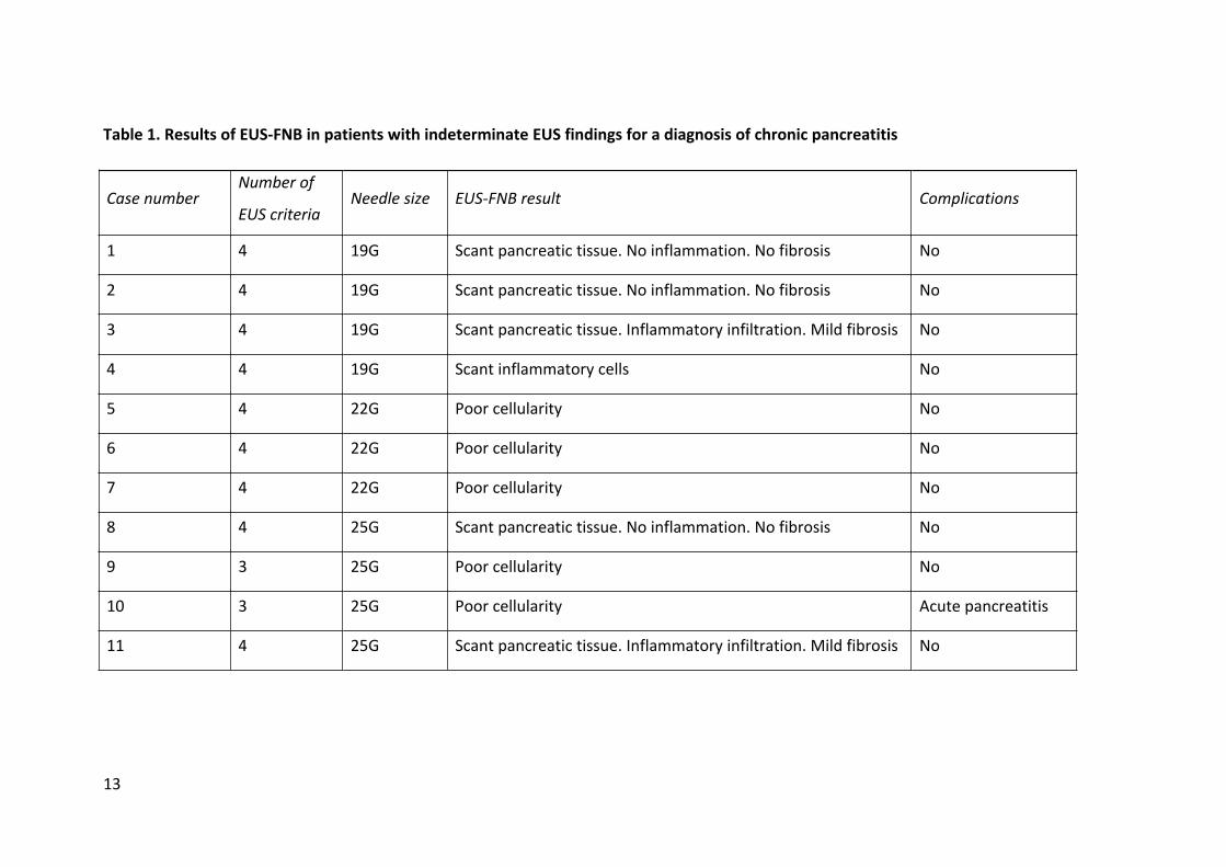
The investigators decided to interrupt the study after the inclusion of eleven patients
due to the poor yield of EUS-FNB and the development of a major complication in one
patient (complication rate of 9.1%). The mean age of patients was 50.3 years (range
33-70 years), six were male. Four and three Rosemont features of CP were observed
via EUS in nine and two patients respectively. Eight patients were mild drinkers and
smokers, whereas the remaining three patients had no risk factor for CP (4,22).
The EUS-guided tissue acquisition procedure was considered to be feasible in all cases.
A 19 gauge needle was used in four cases, 22 gauge in three and 25 gauge in four
cases. The general impression of the endosonographers was the fact that the softness
of the pancreatic parenchyma made it difficult to puncture and move the needle inside
the parenchyma. A tissue core was not obtained in any of the cases. Microscopic
evaluation demonstrated a generally poor sample quality. Scant pancreatic cells that
were sufficient for cell-block evaluation was obtained in five cases; three cases
punctured with a 19-gauge needle and two cases with a 25-gauge needle.
Inflammatory infiltration with mild fibrosis was found in two cases; one punctured
with the 19-gauge needle and one, with the 25-gauge needle. Thus, supporting the
diagnosis of CP (Fig. 2). In the remaining three cases, neither inflammatory infiltration
nor fibrosis was detected and thus a CP diagnosis could not be confirmed. Among the
remaining six patients, inflammatory cells but no pancreatic cells were observed in one
case punctured with the 19-gauge needle. However, the cellularity of samples was
poor and did not allow for a cytological evaluation in the other five cases; three
punctured with a 22-gauge needle and two, with a 25-gauge needle. The results are
summarized in table 1.
As previously mentioned, one patient with three Rosemont features for a CP diagnosis
developed mild acute pancreatitis after the procedure that required hospitalization for
48 hours. There were no other complications related to the EUS-FNB.
DISCUSSION
This study shows that EUS-guided tissue acquisition with Procore™ needles in patients
with indeterminate EUS findings for a CP diagnosis is technically feasible, but provides
inadequate material for cytohistological analysis. It was not possible to obtain a core
biopsy for histological evaluation in any of the patients. Even though both
endosonographers and the pathologist involved in this study are experts in obtaining
and processing EUS-guided pancreatic tissue samples, adequate samples for
cytological evaluation were only obtained in five of the patients (45.4%). Furthermore,
the development of one major complication of acute pancreatitis in one case
questions the safety of this procedure in patients with suspected early CP. Solid
pancreatic lesions usually have dense fibrosis and the safety of EUS-guided tissue
acquisition in this scenario is well documented (23-25). However, the cases in the
current study represent a different risk category. Until the safety and efficacy of EUS-
guided FNB in mildly altered pancreas is well documented, ideally in animal studies,
EUS-guided biopsy should be avoided in these patients, even for research purposes.
The diagnosis of CP is a clinical challenge, mainly during early stages of the disease
(1,4,5). Chronic pancreatitis is characterized by the presence of a chronic infiltration of
inflammatory cells and the progressive loss of acinar cells which are replaced by
fibrosis. However, due to the absence of histology, the diagnosis of chronic
pancreatitis relies on the demonstration of morphological abnormalities suggestive of
pancreatic fibrosis (20,26,27). This makes the diagnosis at an early stage of the disease
challenging, as well as the differential diagnosis with regard to similar conditions like
pancreatopathy (7). In addition, the lack of histological analysis renders the
appropriate evaluation of any potential new therapy of the disease difficult.
EUS-guided FNA and FNB are safe and accurate techniques for the cytohistological
evaluation of solid pancreatic lesions (13-15,24,25,28). The use of reverse-beveled
Procore™ needles provides a high diagnostic yield in this setting (17-19,29,30).
However, data with regard to the cytohistological diagnosis of chronic pancreatitis are
scarce. We previously reported on the accuracy of EUS-guided FNA for the
cytohistological evaluation of moderate to severe chronic pancreatitis (31). DeWitt et
al. obtained an appropriate tissue sample for a histological diagnosis of chronic
pancreatitis in only one of nine patients with mild chronic pancreatitis (16). In the
present study, we also failed to obtain adequate pancreatic tissue by EUS-guided FNB
for the evaluation of indeterminate EUS findings for a diagnosis of chronic pancreatitis.
The needles used in the DeWitt’ study and our study were different (QuickCore® and
Procore™ needles, respectively), even though the diagnostic yield was similar.
Difficulties to obtain a core biopsy was probably related to the softness of the
pancreatic parenchyma in early chronic pancreatitis.
Furthermore, the accuracy of a histological diagnosis of chronic pancreatitis based on
single biopsies may not be high due to the patchy distribution of the disease (32).
However, since the FNB is guided by EUS to target the most abnormal looking area,
this cannot be confirmed as appropriate needles to obtain core biopsies from the
pancreatic parenchyma are not available. Finally, there are still safety concerns related
with EUS-FNB. In the present study, one case developed acute pancreatitis after the
procedure. This, together with the limited success of the technique led to the study
being interrupted prematurely.
The strengths of the current study include the prospective design with a consecutive
inclusion of patients. During the study period, no patient that fulfilled inclusion and
exclusion criteria was excluded. The skills and experience of endosonographers and
the pathologist involved in the study should also be highlighted. The open study design
may be considered as a limitation.
In conclusion, EUS-guided FNB for the histological diagnosis of chronic pancreatitis in
patients with indeterminate EUS findings of the disease is technically feasible.
However, the yield is poor. In addition, the safety of this technique is also a concern.
Based on the present data, EUS-FNB for mild chronic pancreatitis should be avoided in
humans until the efficacy and safety of this technique has been demonstrated in
animal studies.
REFERENCES
1. Martínez J, Abad-González A, Aparicio JR, et al. The Spanish Pancreatic Club
recommendations for the diagnosis and treatment of chronic pancreatitis: part 1
(diagnosis). Pancreatology 2013;13(1):8-17. DOI: 10.1016/j.pan.2012.11.309
2. Hoffmeister A, Mayerle J, Beglinger C, et al. English language version of the S3-
consensus guidelines on chronic pancreatitis: definition, aetiology, diagnostic
examinations, medical, endoscopic and surgical management of chronic pancreatitis. Z
Gastroenterol 2015;53(12):1447-95. DOI: 10.1055/s-0041-107379
3. Issa Y, Kempeneers MA, Van Santvoort HC, et al. Diagnostic performance of
imaging modalities in chronic pancreatitis: a systematic review and meta-analysis. Eur
Radiol 2017;27(9):3820-44. DOI: 10.1007/s00330-016-4720-9
4. Conwell DL, Lee LS, Yadav D, et al. American Pancreatic Association Practice
Guidelines in Chronic Pancreatitis: evidence-based report on diagnostic guidelines.
Pancreas 2014;43(8):1143-62. DOI: 10.1097/MPA.0000000000000237
5. Duggan SN, Ní Chonchubhair HM, Lawal O, et al. Chronic pancreatitis: a
diagnostic dilemma. World J Gastroenterol 2016;22(7):2304-13. DOI:
10.3748/wjg.v22.i7.2304
6. Whitcomb DC, Frulloni L, Garg P, et al. Chronic pancreatitis: an international
draft consensus proposal for a new mechanistic definition. Pancreatology
2016;16(2):218-24. DOI: 10.1016/j.pan.2016.02.001
7. Chari ST. Defining chronic pancreatitis. Pancreatology 2016;16(5):694-5. DOI:
10.1016/j.pan.2016.08.008
8. Iglesias-García J, Lariño-Noia J, Domínguez-Muñoz JE. When to puncture, when
not to puncture: pancreatic masses. Endosc Ultrasound 2014;3(2):91-7. DOI:
10.4103/2303-9027.123007
9. Iglesias-García J, Lindkvist B, Lariño-Noia J, et al. The role of EUS in relation to
other imaging modalities in the differential diagnosis between mass forming chronic
pancreatitis, autoimmune pancreatitis and ductal pancreatic adenocarcinoma. Rev Esp
Enferm Dig 2012;104(6):315-21.
10. Tsutsumi H, Hara K, Mizuno N, et al. Clinical impact of preoperative endoscopic
ultrasound-guided fine-needle aspiration for pancreatic ductal adenocarcinoma.
Endosc Ultrasound 2016;5(2):94-100. DOI: 10.4103/2303-9027.180472
11. Sun B, Yang X, Ping B, et al. Impact of inconclusive endoscopic ultrasound-
guided fine-needle aspiration results in the management and outcome of patients with
solid pancreatic masses. Dig Endosc 2015;27(1):130-6. DOI: 10.1111/den.12315
12. Puli SR, Bechtold ML, Buxbaum JL, et al. How good is endoscopic ultrasound-
guided fine-needle aspiration in diagnosing the correct etiology for a solid pancreatic
mass?: a meta-analysis and systematic review. Pancreas 2013;42(1):20-6. DOI:
10.1097/MPA.0b013e3182546e79
13. Thomas T, Kaye PV, Ragunath K, et al. Efficacy, safety, and predictive factors for
a positive yield of EUS-guided Trucut biopsy: a large tertiary referral center experience.
Am J Gastroenterol 2009;104(3):584-91. DOI: 10.1038/ajg.2008.97
14. Fisher L, Segarajasingam DS, Stewart C, et al. Endoscopic ultrasound guided fine
needle aspiration of solid pancreatic lesions: performance and outcomes. J
Gastroenterol Hepatol 2009;24(1):90-6. DOI: 10.1111/j.1440-1746.2008.05569.x
15. O’Toole D, Palazzo L, Arotçarena R, et al. Assessment of complications of EUS-
guided fine-needle aspiration. Gastrointest Endosc 2001;53(4):470-4. DOI:
10.1067/mge.2001.112839
16. DeWitt J, McGreevy K, LeBlanc J, et al. EUS-guided Trucut biopsy of suspected
nonfocal chronic pancreatitis. Gastrointest Endosc 2005;62(1):76-84. DOI:
10.1016/S0016-5107(05)00504-3
17. Iglesias-García J, Poley J-W, Larghi A, et al. Feasibility and yield of a new EUS
histology needle: results from a multicenter, pooled, cohort study. Gastrointest Endosc
2011;73(6):1189-96. DOI: 10.1016/j.gie.2011.03.193
18. Larghi A, Iglesias-García J, Poley J-W, et al. Feasibility and yield of a novel 22-
gauge histology EUS needle in patients with pancreatic masses: a multicenter
prospective cohort study. Surg Endosc 2013;27(10):3733-8.
19. Attili F, Petrone G, Abdulkader I, et al. Accuracy and inter-observer agreement
of the Procore™ 25 gauge needle for endoscopic ultrasound-guided tissue core biopsy.
Dig Liver Dis 2015;47(11):943-9. DOI: 10.1016/j.dld.2015.07.003
20. Catalano MF, Sahai A, Levy M, et al. EUS-based criteria for the diagnosis of
chronic pancreatitis: the Rosemont classification. Gastrointest Endosc
2009;69(7):1251-61. DOI: 10.1016/j.gie.2008.07.043
21. Ammann RW, Heitz PU, Klöppel G. Course of alcoholic chronic pancreatitis: a
prospective clinicomorphological long-term study. Gastroenterology 1996;111(1):224-
31. DOI: 10.1053/gast.1996.v111.pm8698203
22. Etemad B, Whitcomb DC. Chronic pancreatitis: diagnosis, classification, and
new genetic developments. Gastroenterology 2001;120(3):682-707. DOI:
10.1053/gast.2001.22586
23. Lakhtakia S. Complications of diagnostic and therapeutic endoscopic
ultrasound. Best Pract Res Clin Gastroenterol 2016;30(5):807-23. DOI:
10.1016/j.bpg.2016.10.008
24. Eloubeidi MA, Tamhane A. Prospective assessment of diagnostic utility and
complications of endoscopic ultrasound-guided fine needle aspiration. Results from a
newly developed academic endoscopic ultrasound program. Dig Dis Basel Switz
2008;26(4):356-63. DOI: 10.1159/000177022
25. Dumonceau J-M, Polkowski M, Larghi A, et al. Indications, results, and clinical
impact of endoscopic ultrasound (EUS)-guided sampling in gastroenterology: European
Society of Gastrointestinal Endoscopy (ESGE) Clinical Guideline. Endoscopy
2011;43(10):897-912. DOI: 10.1055/s-0030-1256754
26. Ashkar M, Gardner TB. Role of endoscopic ultrasound in pancreatic diseases: a
systematic review. Minerva Gastroenterol Dietol 2014;60(4):227-45.
27. Löhr JM, Domínguez-Muñoz E, Rosendahl J, et al. United European
Gastroenterology evidence-based guidelines for the diagnosis and therapy of chronic
pancreatitis (HaPanEU). United Eur Gastroenterol J 2017;5(2):153-99. DOI:
10.1177/2050640616684695
28. Eloubeidi MA, Tamhane A, Varadarajulu S, et al. Frequency of major
complications after EUS-guided FNA of solid pancreatic masses: a prospective
evaluation. Gastrointest Endosc 2006;63(4):622-9. DOI: 10.1016/j.gie.2005.05.024
29. Dwyer J, Pantanowitz L, Ohori NP, et al. Endoscopic ultrasound-guided FNA and
ProCore biopsy in sampling pancreatic and intra-abdominal masses. Cancer Cytopathol
2016;124(2):110-21. DOI: 10.1002/cncy.21623
30. Alatawi A, Beuvon F, Grabar S, et al. Comparison of 22G reverse-beveled versus
standard needle for endoscopic ultrasound-guided sampling of solid pancreatic lesions.
United Eur Gastroenterol J 2015;3(4):343-52. DOI: 10.1177/2050640615577533
31. Iglesias-García J, Abdulkader I, Lariño-Noia J, et al. Histological evaluation of
chronic pancreatitis by endoscopic ultrasound-guided fine needle biopsy. Gut
2006;55(11):1661-2.
32. Klöppel G, Maillet B. Pseudocysts in chronic pancreatitis: a morphological
analysis of 57 resection specimens and 9 autopsy pancreata. Pancreas 1991;6(3):266-
74.
13
Case numberNumber of
EUS criteriaNeedle size EUS-FNB result Complications
1 4 19G Scant pancreatic tissue. No inflammation. No fibrosis No
2 4 19G Scant pancreatic tissue. No inflammation. No fibrosis No
3 4 19G Scant pancreatic tissue. Inflammatory infiltration. Mild fibrosis No
4 4 19G Scant inflammatory cells No
5 4 22G Poor cellularity No
6 4 22G Poor cellularity No
7 4 22G Poor cellularity No
8 4 25G Scant pancreatic tissue. No inflammation. No fibrosis No
9 3 25G Poor cellularity No
10 3 25G Poor cellularity Acute pancreatitis
11 4 25G Scant pancreatic tissue. Inflammatory infiltration. Mild fibrosis No
Table 1. Results of EUS-FNB in patients with indeterminate EUS findings for a diagnosis of chronic pancreatitis
14
Fig. 1. EUS image of the pancreatic parenchyma accessed for EUS guided tissue
acquisition.
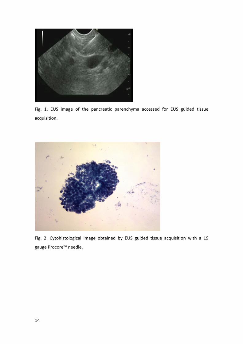
Fig. 2. Cytohistological image obtained by EUS guided tissue acquisition with a 19
gauge Procore™ needle.