Embed Size (px)
Citation preview
Intra-abdominal Hypertension and
Compartment Syndrome
in the Trauma Patient:
Is Your Patient at Risk?
John J. Gallagher MSN, RN, CCNS, CCRN, RRT
Trauma Program Coordinator/Clinical Nurse Specialist
Hospital of the University of Pennsylvania
Objective
Brief review of pathophysiology
Definitions of IAH/ACS
Define the risk in trauma patients
IAP measurement
Prevention and treatment strategies
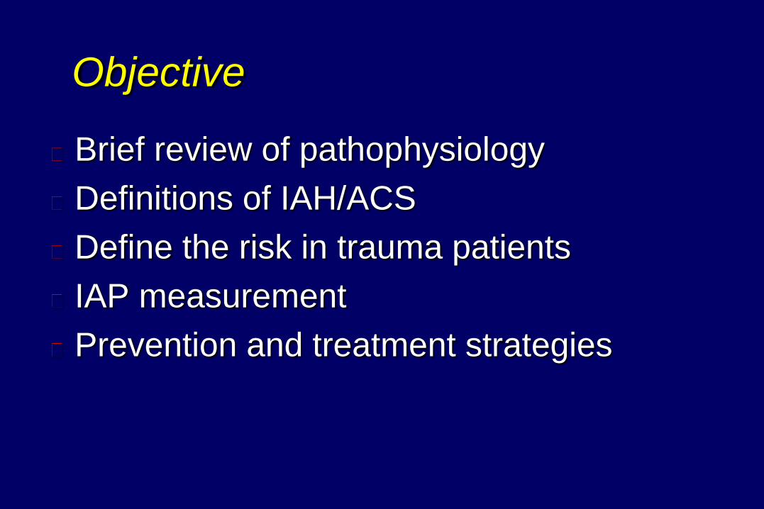
World Society of Abdominal Compartment Syndrome
Guidelines 2013
www. WSACS.org
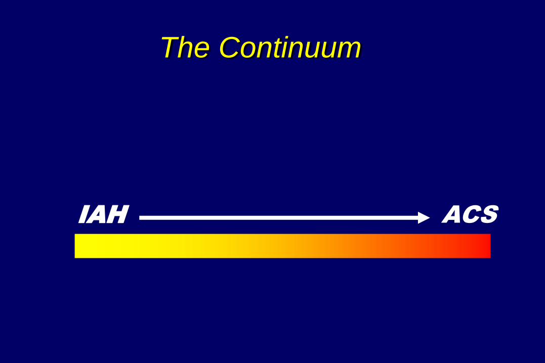
The Continuum
IAH ACS
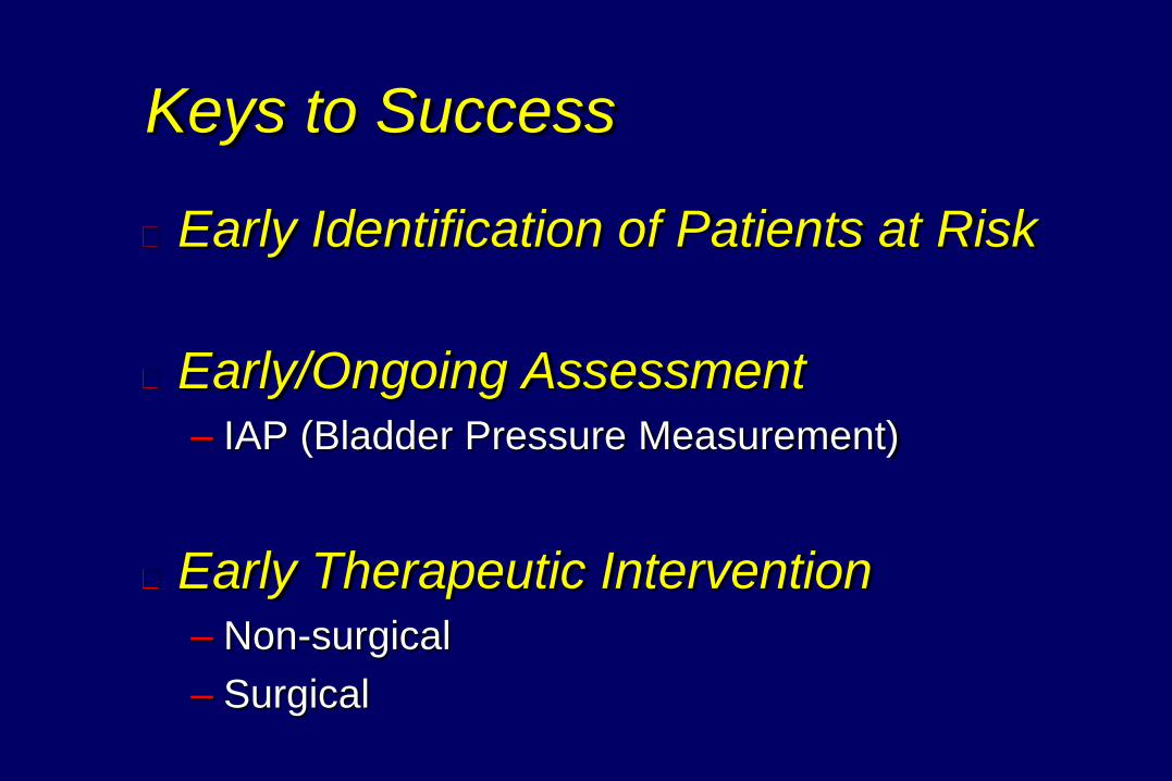
Keys to Success
Early Identification of Patients at Risk
Early/Ongoing Assessment – IAP (Bladder Pressure Measurement)
Early Therapeutic Intervention – Non-surgical
– Surgical

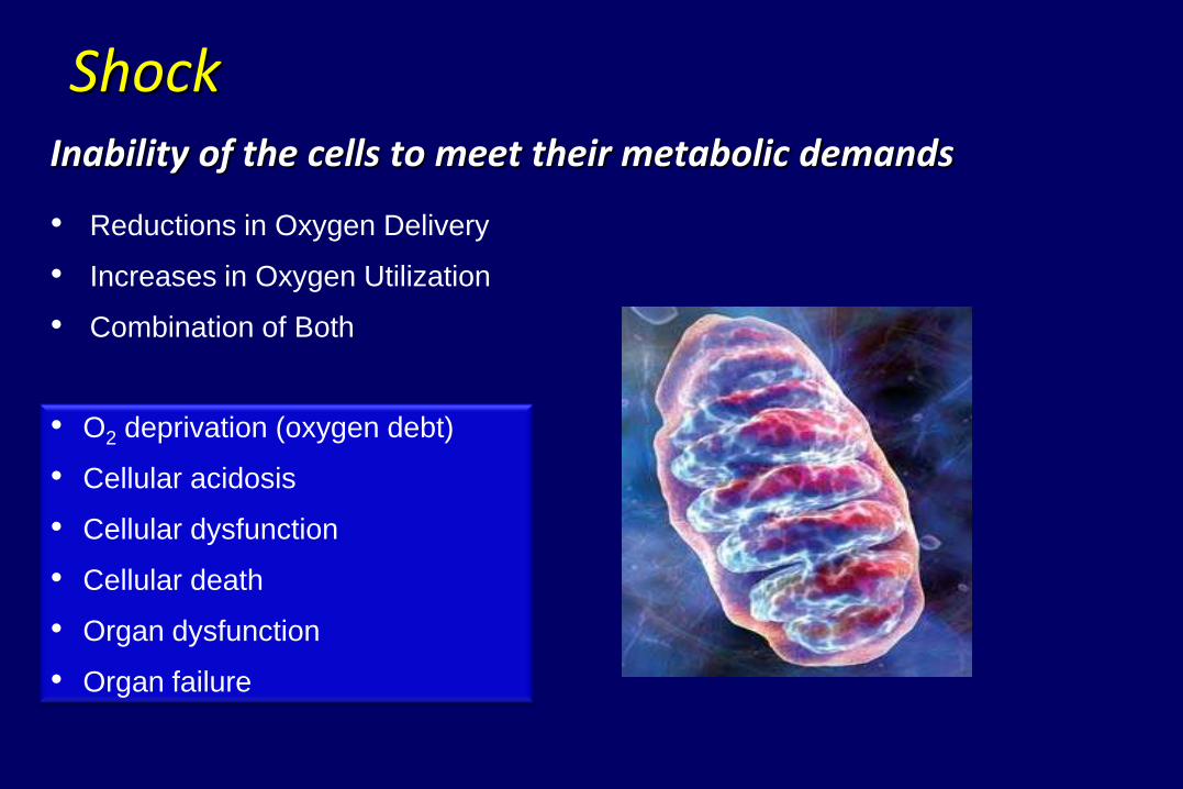
Shock Inability of the cells to meet their metabolic demands
• Reductions in Oxygen Delivery
• Increases in Oxygen Utilization
• Combination of Both
• O2 deprivation (oxygen debt)
• Cellular acidosis
• Cellular dysfunction
• Cellular death
• Organ dysfunction
• Organ failure
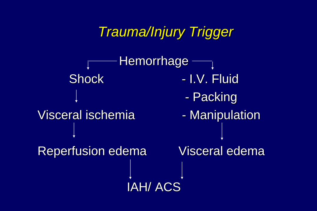
Trauma/Injury Trigger
Hemorrhage
Shock - I.V. Fluid
- Packing
Visceral ischemia - Manipulation
Reperfusion edema Visceral edema
IAH/ ACS
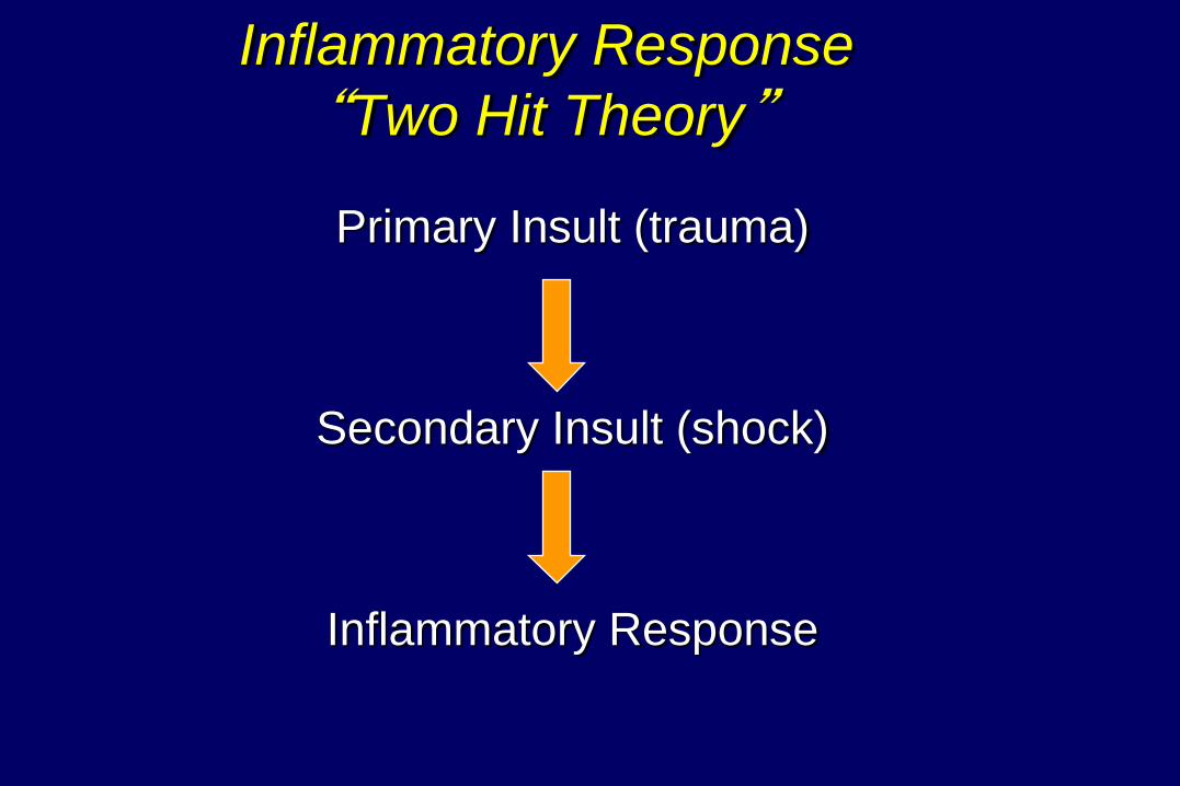
Inflammatory Response
“Two Hit Theory”
Primary Insult (trauma)
Secondary Insult (shock)
Inflammatory Response
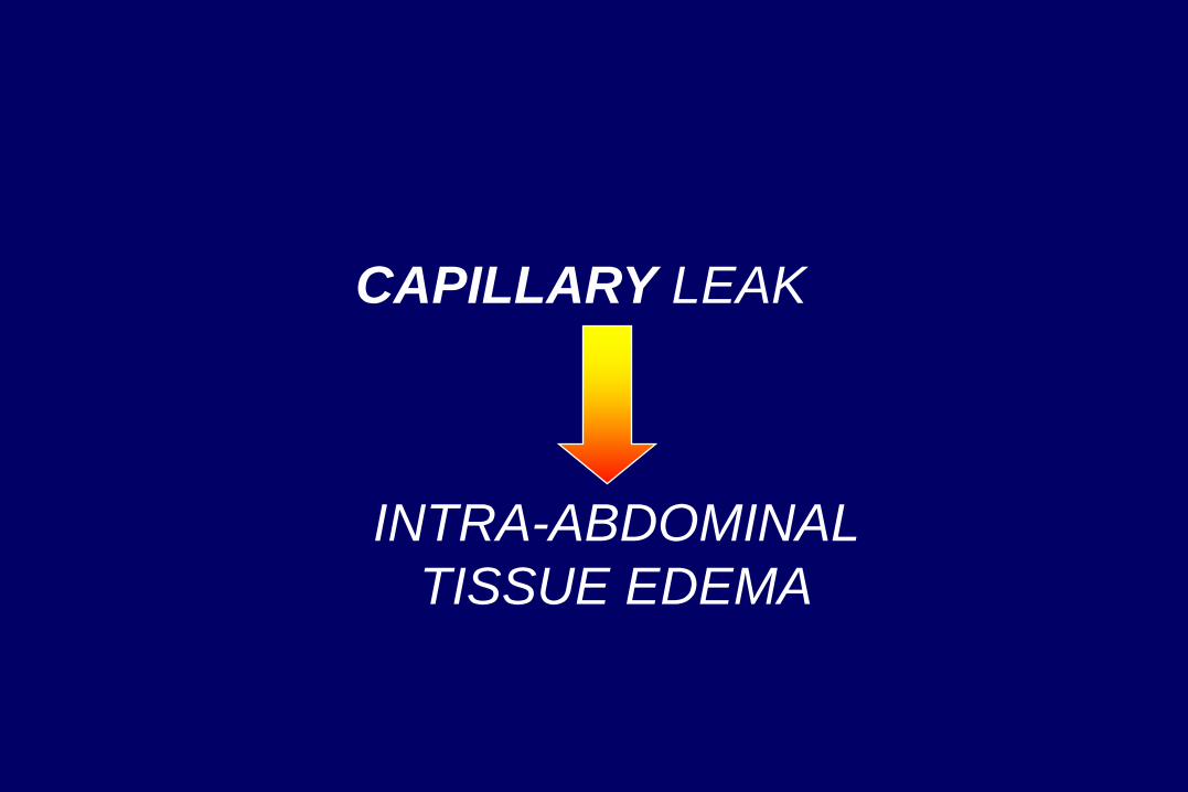
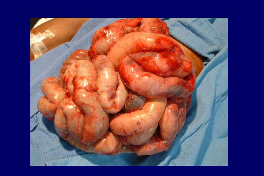
CAPILLARY LEAK
INTRA-ABDOMINAL
TISSUE EDEMA
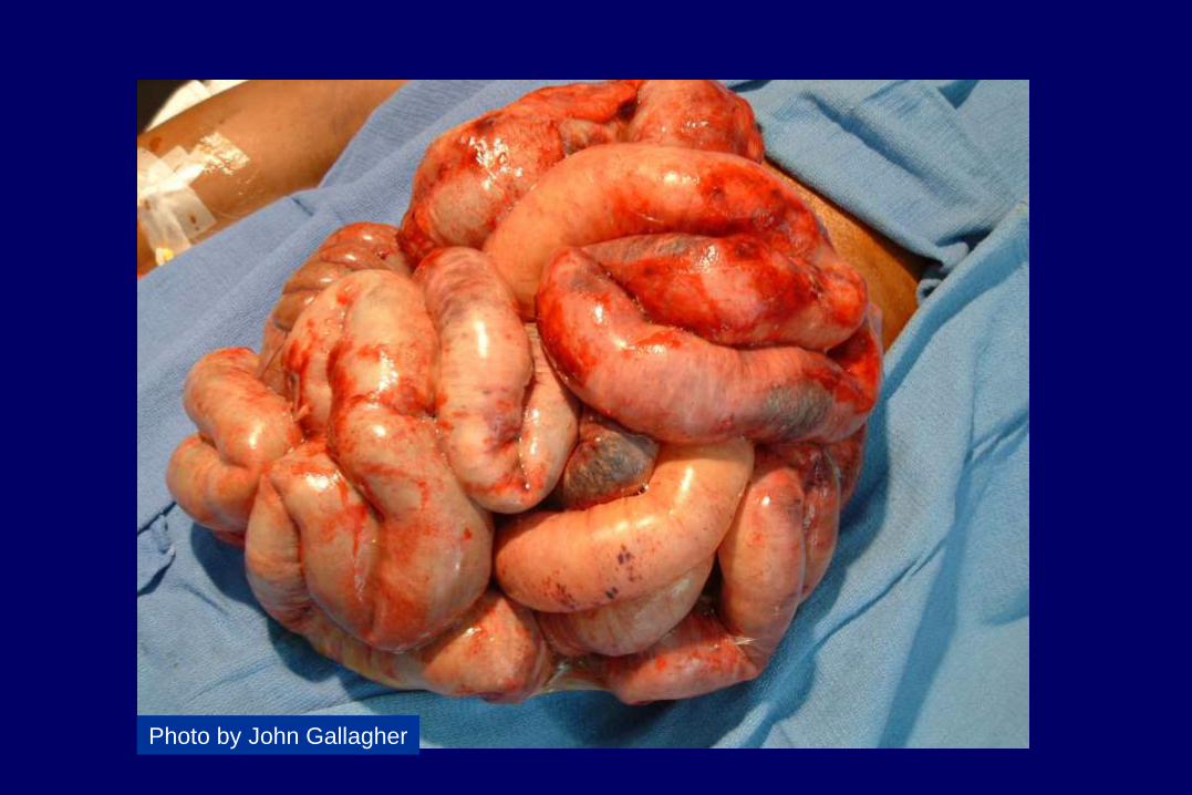
Photo by John Gallagher
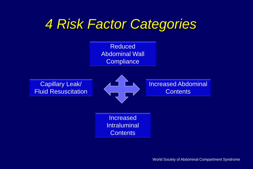
4 Risk Factor Categories
Reduced
Abdominal Wall
Compliance
Increased Abdominal
Contents
Increased
Intraluminal
Contents
Capillary Leak/
Fluid Resuscitation
World Society of Abdominal Compartment Syndrome

Risk Factors
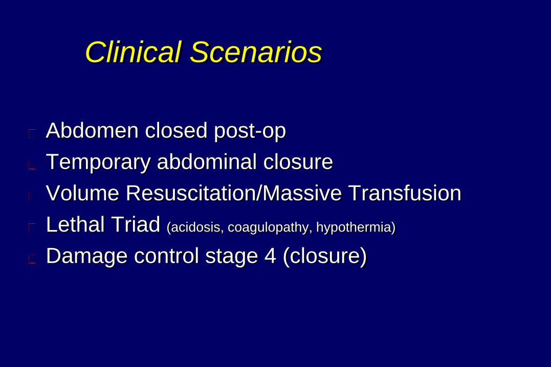
Clinical Scenarios
Abdomen closed post-op
Temporary abdominal closure
Volume Resuscitation/Massive Transfusion
Lethal Triad (acidosis, coagulopathy, hypothermia)
Damage control stage 4 (closure)
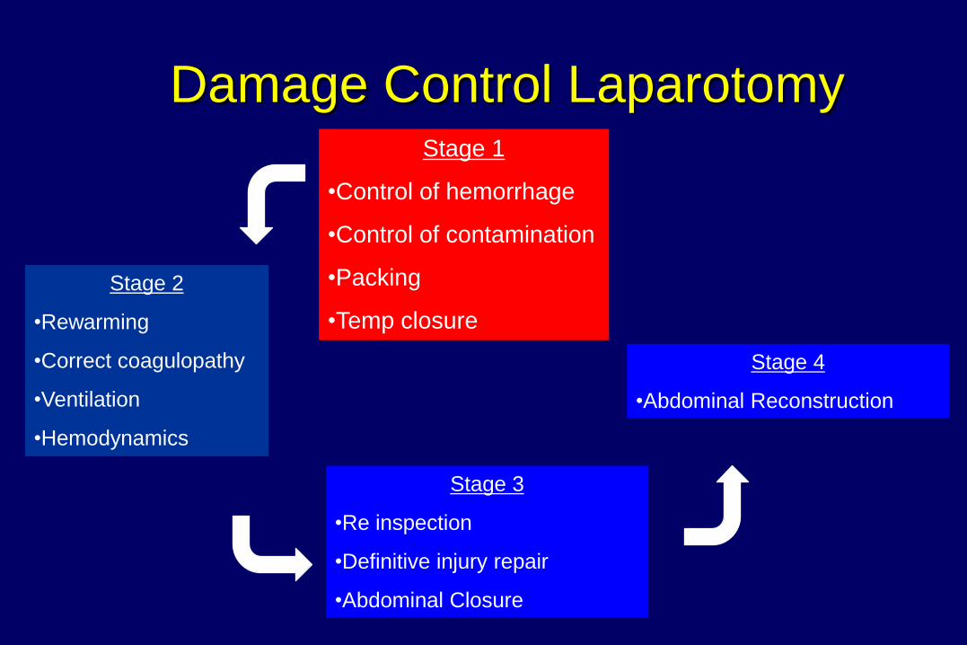
Damage Control Laparotomy Stage 1
•Control of hemorrhage
•Control of contamination
•Packing
•Temp closure
Stage 2
•Rewarming
•Correct coagulopathy
•Ventilation
•Hemodynamics
Stage 3
•Re inspection
•Definitive injury repair
•Abdominal Closure
���
Stage 4
•Abdominal Reconstruction
���
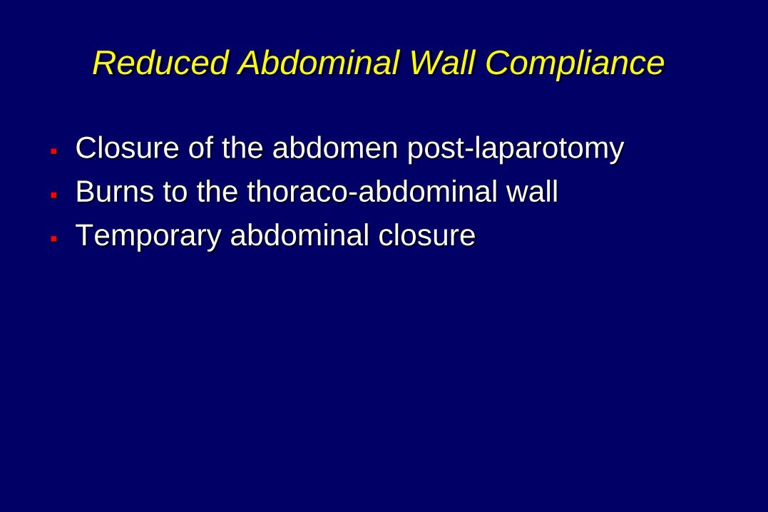
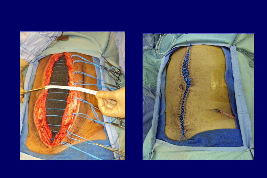
Reduced Abdominal Wall Compliance
Closure of the abdomen post-laparotomy
Burns to the thoraco-abdominal wall
Temporary abdominal closure
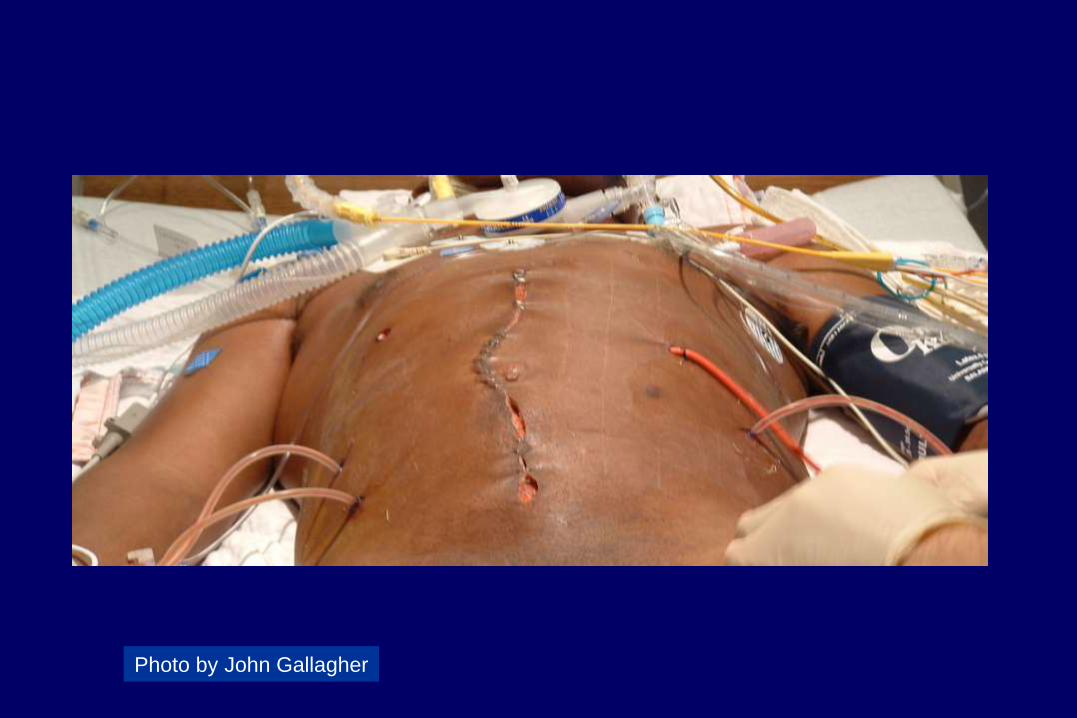
Photo by John Gallagher
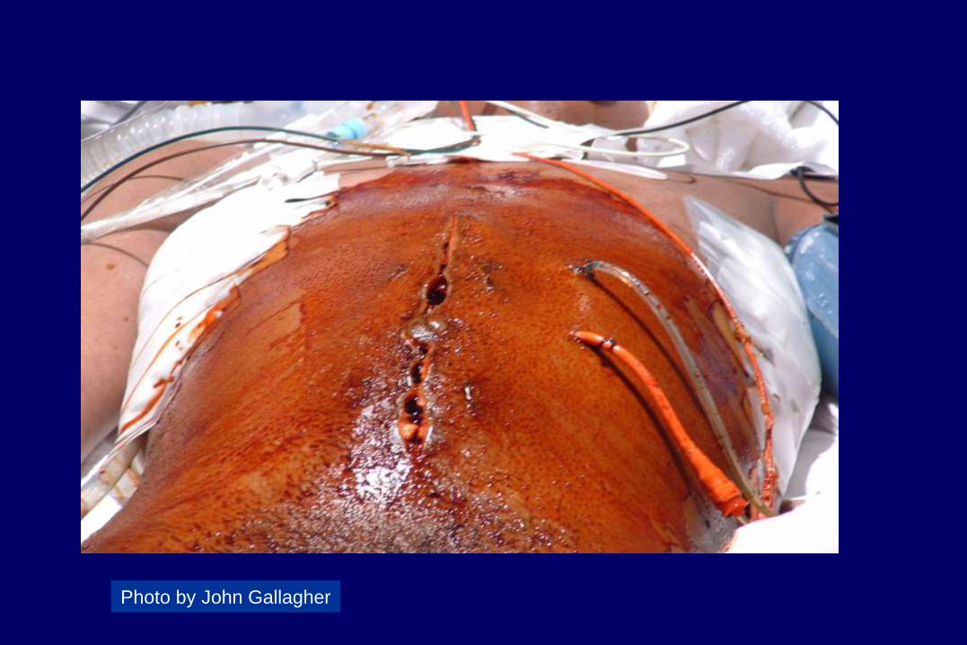
Photo by John Gallagher

Photo by John Gallagher
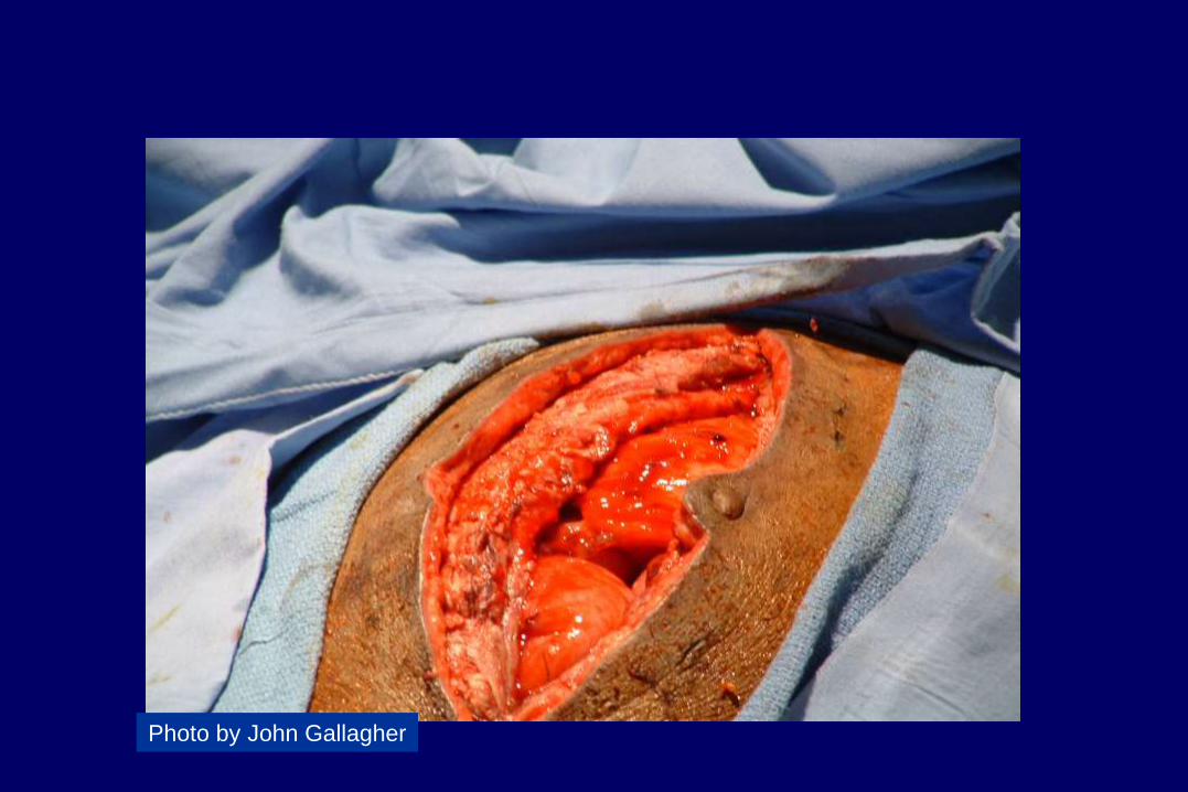
Photo by John Gallagher
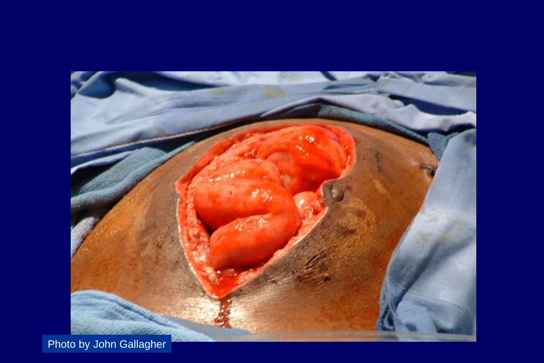
Photo by John Gallagher
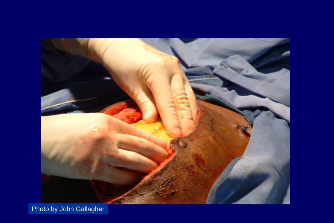
Photo by John Gallagher
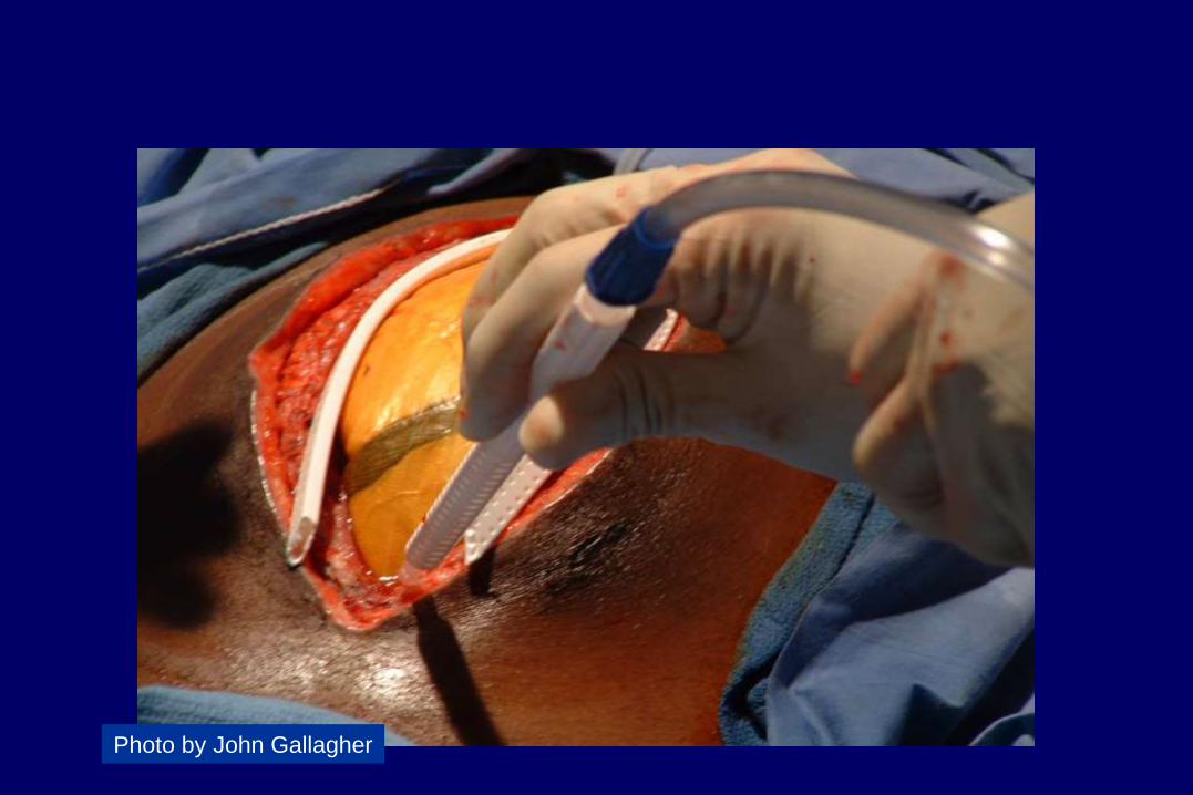
Photo by John Gallagher
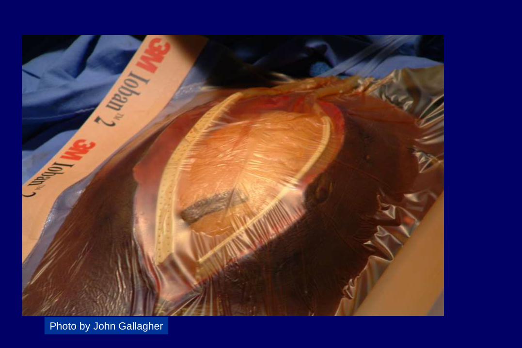
Photo by John Gallagher
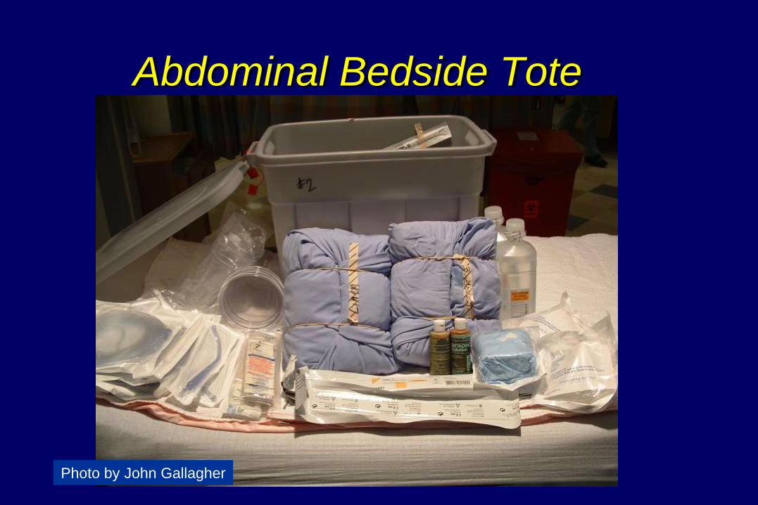
Abdominal Bedside Tote
Photo by John Gallagher

Bogota/Silo Bag Closure
trauma.org Image Bank
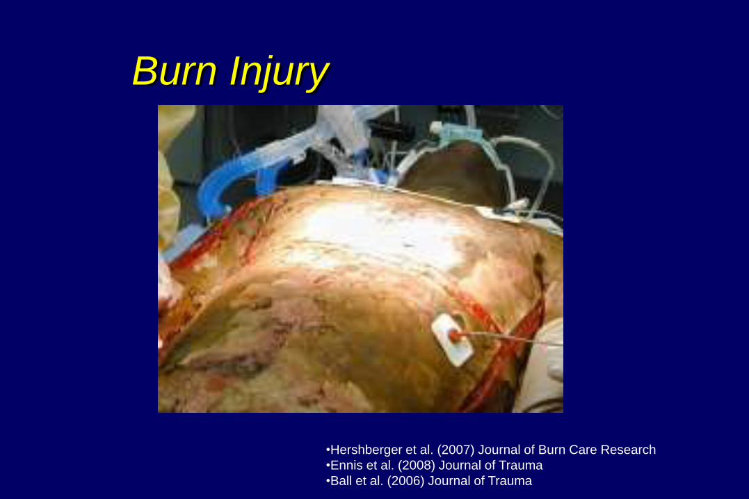
Burn Injury
•Hershberger et al. (2007) Journal of Burn Care Research
•Ennis et al. (2008) Journal of Trauma
•Ball et al. (2006) Journal of Trauma
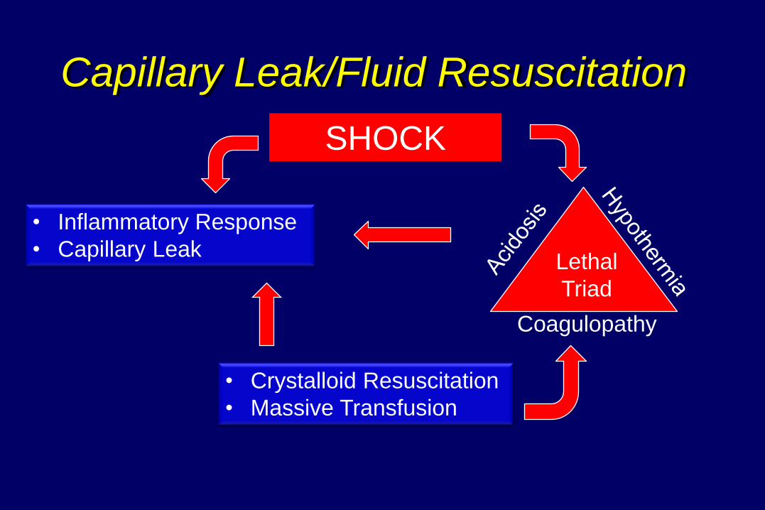
Capillary Leak/Fluid Resuscitation
• Crystalloid Resuscitation
• Massive Transfusion
• Inflammatory Response
• Capillary Leak
SHOCK
Coagulopathy
Lethal
Triad
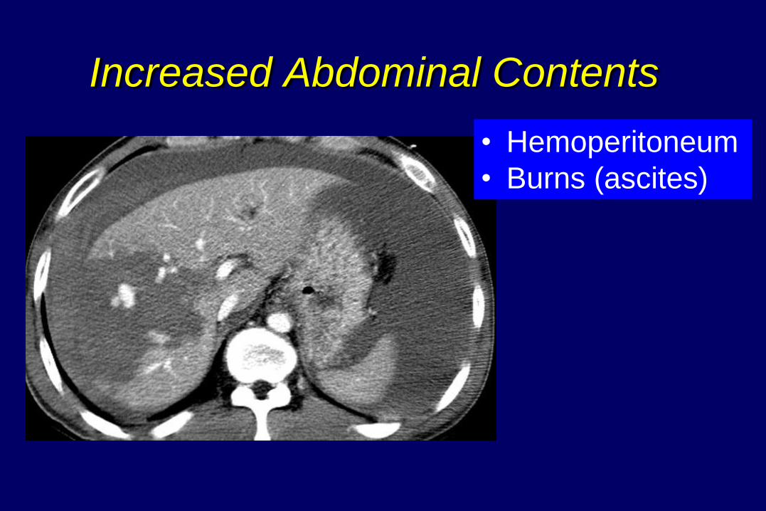
Increased Abdominal Contents
• Hemoperitoneum
• Burns (ascites)
Increase Intraluminal Contents
Ileus
Gastroparesis
Obstructions
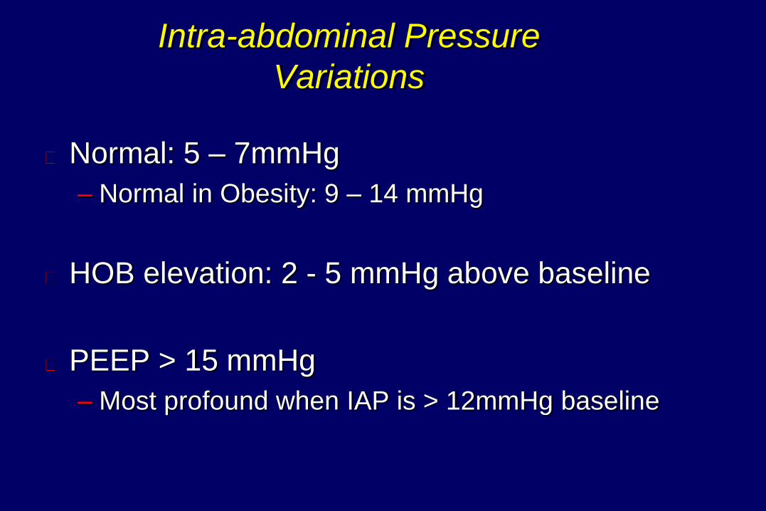
Intra-abdominal Pressure
Variations
Normal: 5 – 7mmHg
– Normal in Obesity: 9 – 14 mmHg
HOB elevation: 2 - 5 mmHg above baseline
PEEP > 15 mmHg
– Most profound when IAP is > 12mmHg baseline
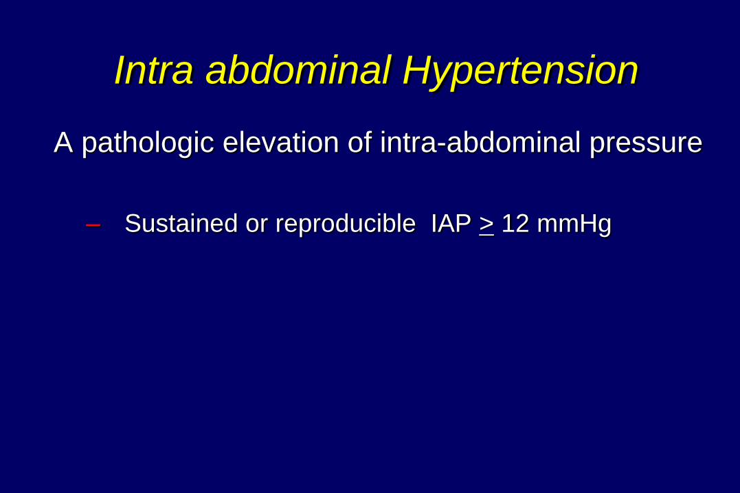
Intra abdominal Hypertension
A pathologic elevation of intra-abdominal pressure
– Sustained or reproducible IAP > 12 mmHg
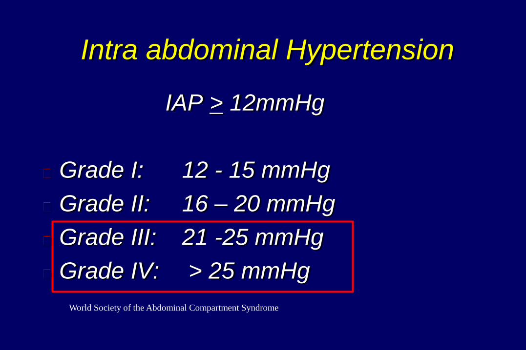
Intra abdominal Hypertension
IAP > 12mmHg
Grade I: 12 - 15 mmHg
Grade II: 16 – 20 mmHg
Grade III: 21 -25 mmHg
Grade IV: > 25 mmHg
World Society of the Abdominal Compartment Syndrome
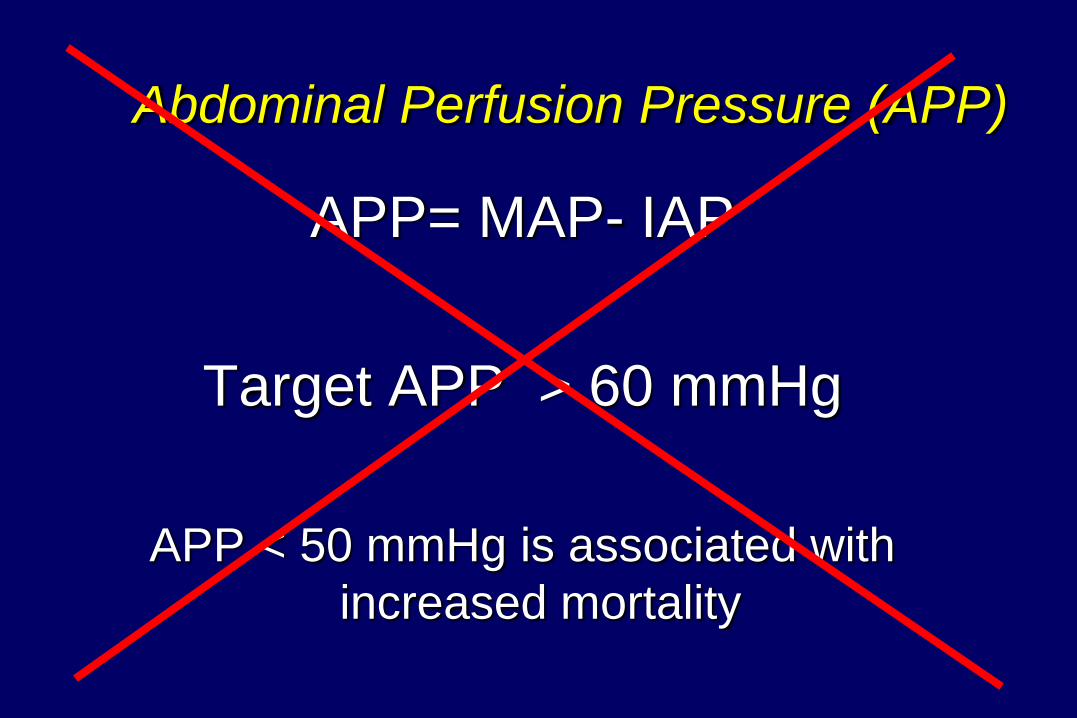
Abdominal Perfusion Pressure (APP)
APP= MAP- IAP
Target APP > 60 mmHg
APP < 50 mmHg is associated with
increased mortality
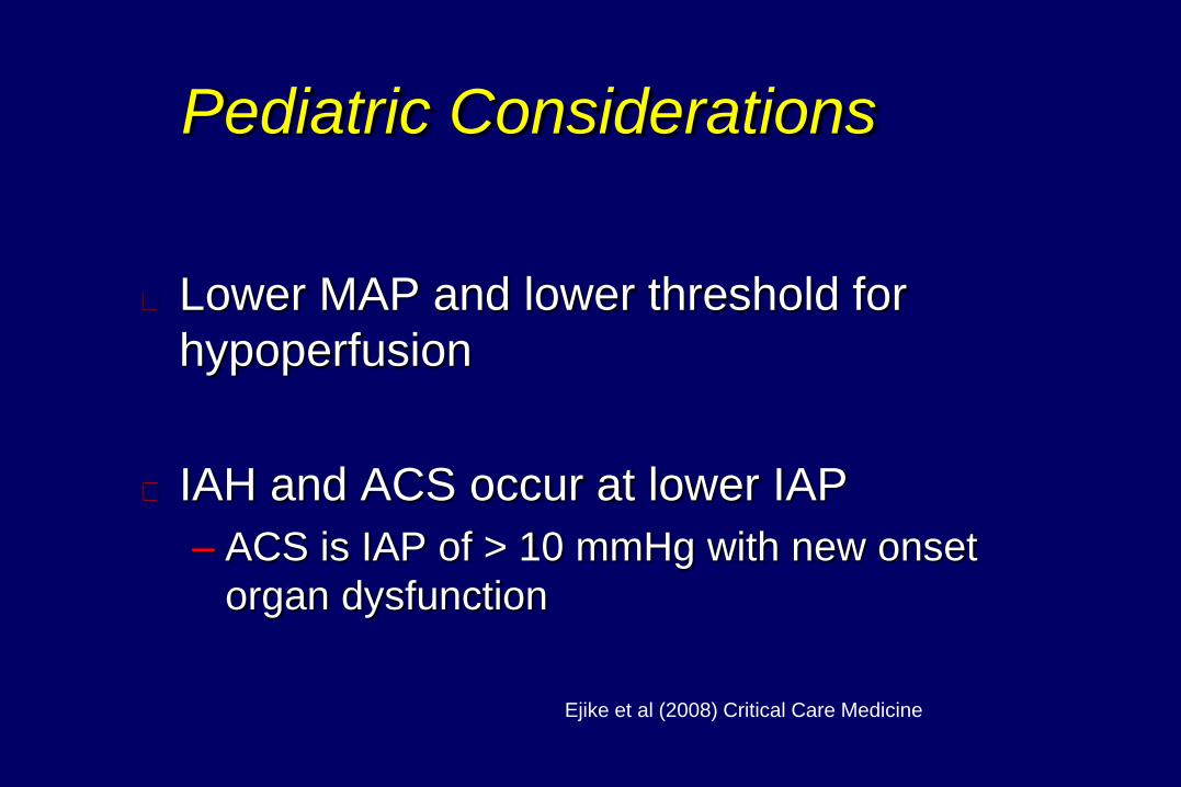
Pediatric Considerations
Lower MAP and lower threshold for
hypoperfusion
IAH and ACS occur at lower IAP
– ACS is IAP of > 10 mmHg with new onset
organ dysfunction
Ejike et al (2008) Critical Care Medicine
Abdominal Compartment Syndrome
An increase in intra-abdominal
pressure that exceeds the capacity of the
compartment, resulting in the impaired
perfusion and function of multiple organ systems
Abdominal Compartment Syndrome
Presence of both….
IAP > 20 mm Hg Regardless of APP
New onset single/multiple organ system failure
Abdominal Compartment Syndrome
Primary
Secondary
Recurrent
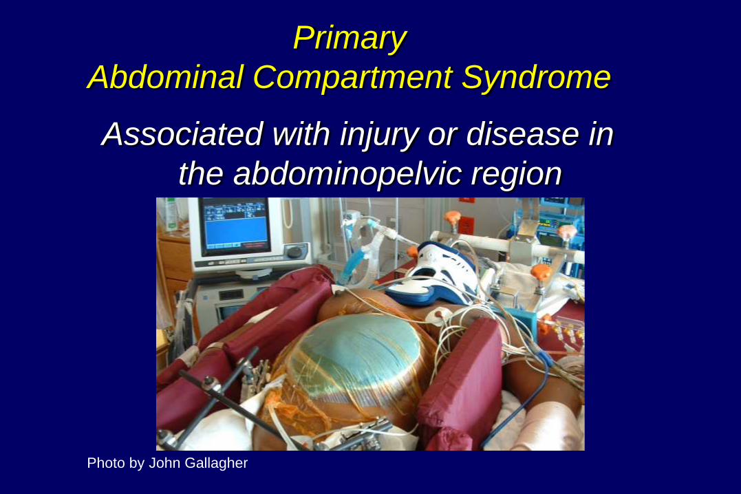
Primary
Abdominal Compartment Syndrome
Associated with injury or disease in
the abdominopelvic region
Photo by John Gallagher
Secondary
Abdominal Compartment Syndrome
Develops from
conditions outside the abdomen
–Massive fluid resuscitation
–Burns
–Sepsis
SACS Post Injury
Lower SBP
Penetrating chest
Vascular Injuries
Multiple extremity fractures
Secondary
Abdominal Compartment Syndrome
Inflammatory process that may be more subtle
– Trauma:
» SACS represents 58% of post injury ACS
» 38 – 68 % mortality
Resuscitation outside the OR
– IR and ICU
– More crystalloid
– Longer times to control of bleeding
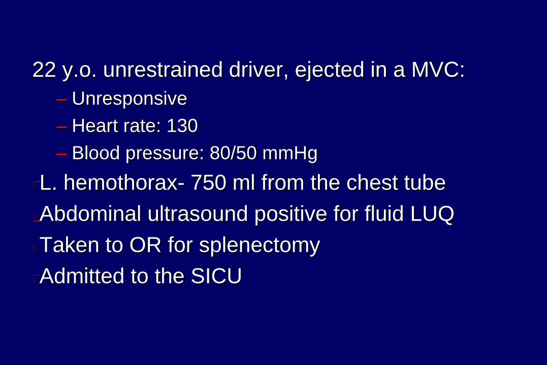
22 y.o. unrestrained driver, ejected in a MVC:
– Unresponsive
– Heart rate: 130
– Blood pressure: 80/50 mmHg
L. hemothorax- 750 ml from the chest tube
Abdominal ultrasound positive for fluid LUQ
Taken to OR for splenectomy
Admitted to the SICU
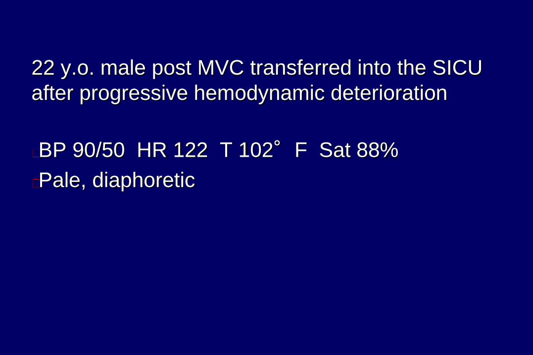
22 y.o. male post MVC transferred into the SICU
after progressive hemodynamic deterioration
BP 90/50 HR 122 T 102°F Sat 88%
Pale, diaphoretic
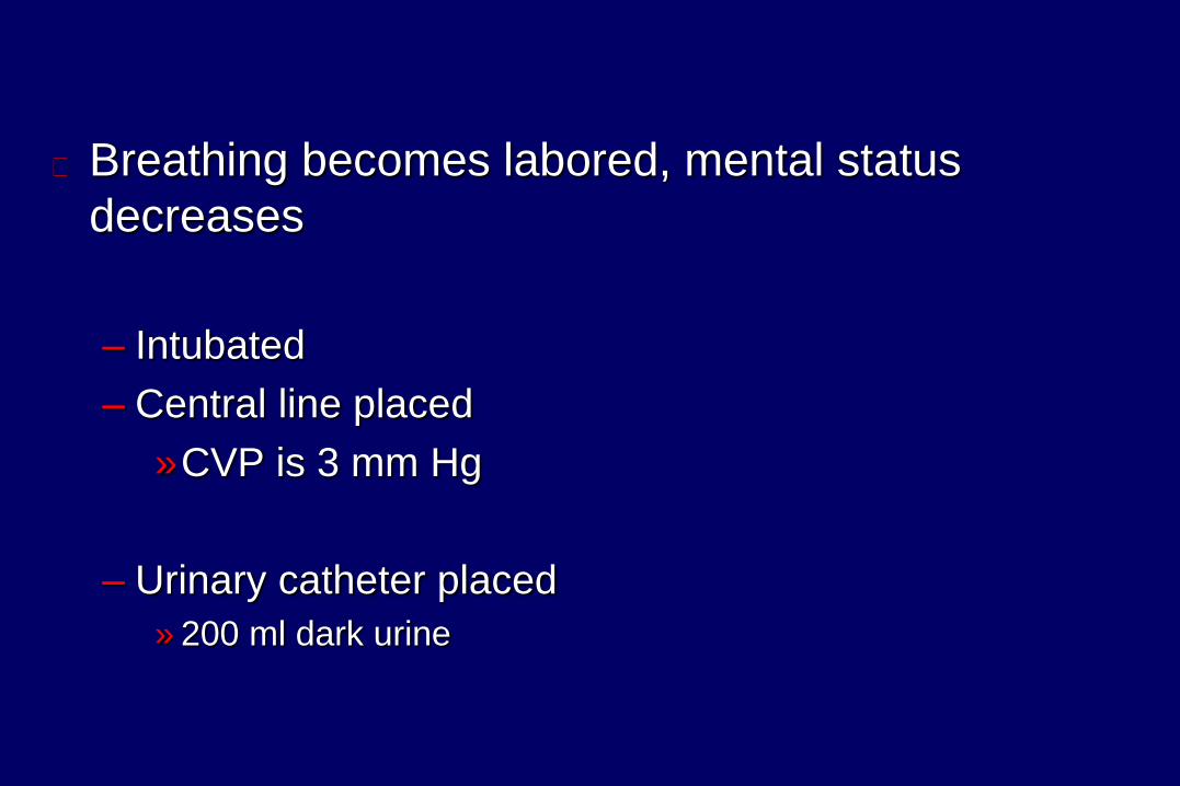
Breathing becomes labored, mental status
decreases
– Intubated
– Central line placed
»CVP is 3 mm Hg
– Urinary catheter placed
» 200 ml dark urine
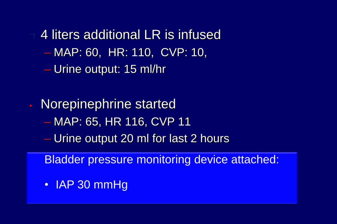
4 liters additional LR is infused
– MAP: 60, HR: 110, CVP: 10,
– Urine output: 15 ml/hr
• Norepinephrine started
– MAP: 65, HR 116, CVP 11
– Urine output 20 ml for last 2 hours
Would you give this patient volume?
Bladder pressure monitoring device attached:
• IAP 30 mmHg
Recurrent
Abdominal Compartment Syndrome
Abdominal Compartment Syndrome
that re-develops after previous medical
or surgical treatment
Recurrent
Abdominal Compartment Syndrome
Can develop even with an “expanded” (open)
abdomen with temporary closure
Increased morbidity and mortality
Gracias, V.H. et al. 2002. Archives of Surgery
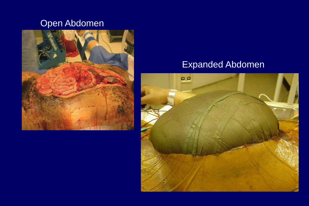
Open Abdomen
Expanded Abdomen
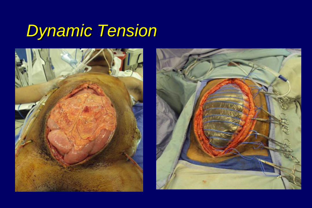
Dynamic Tension

Organ Systems Compromised
Gastrointestinal
Pulmonary
Cardiovascular
Renal
Neurological
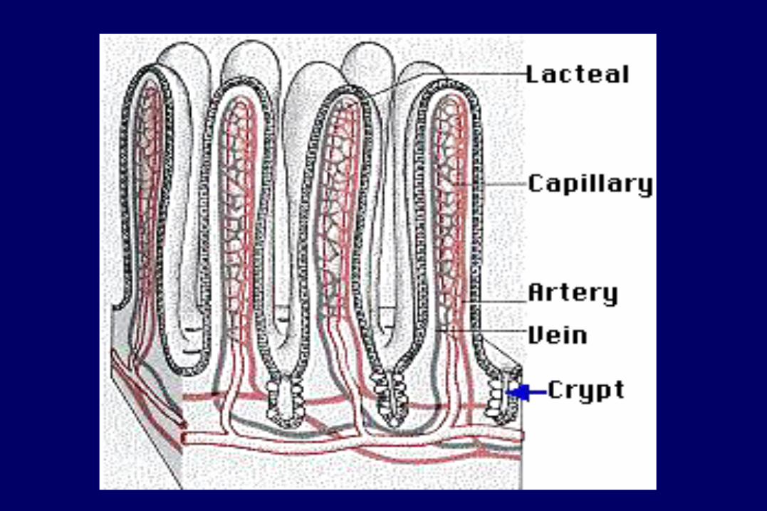
Gastrointestinal Effects
Increased IAP
Compression of mesenteric vessels
Gastric mucosal acidosis (pHi)
Mesenteric ischemia
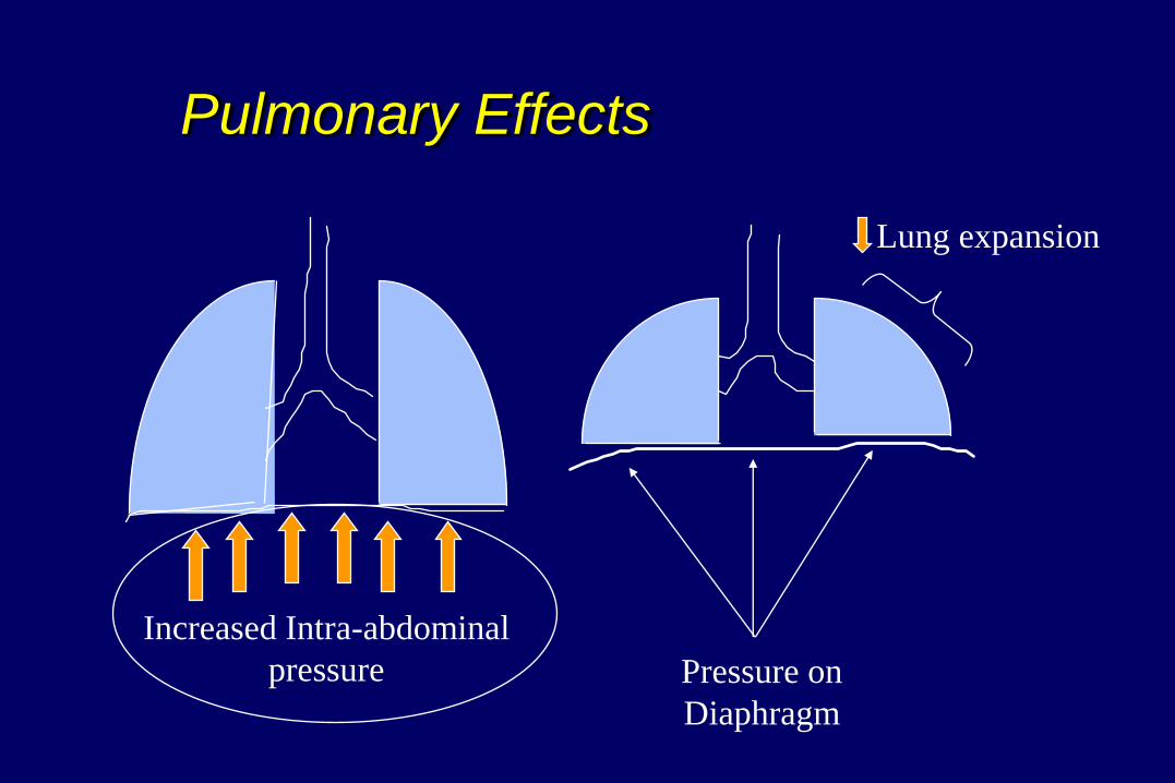
Pulmonary Effects
Increased Intra-abdominal
pressure Pressure on
Diaphragm
Lung expansion
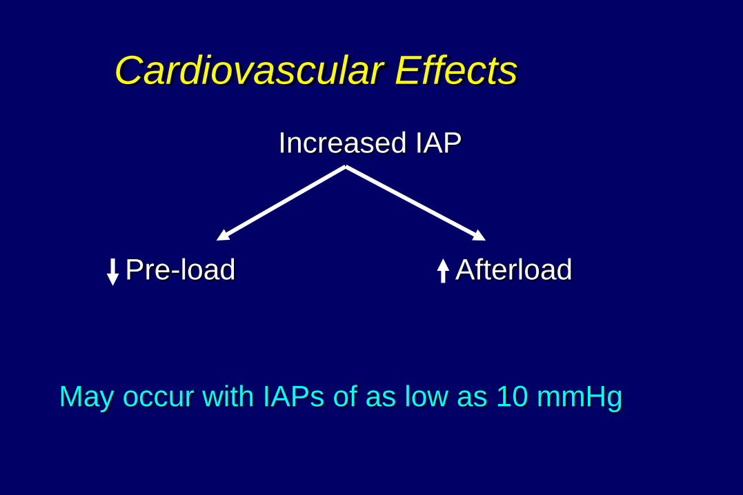
Cardiovascular Effects
Increased IAP
Pre-load Afterload
May occur with IAPs of as low as 10 mmHg

Cardiovascular Assessment Findings
Normotensive
CVP, PA, PCOP, SVR, PVR
C.O.
CVP corrected = CVP measured - IAP/2
PCOP corrected = PCOP measured - IAP/2
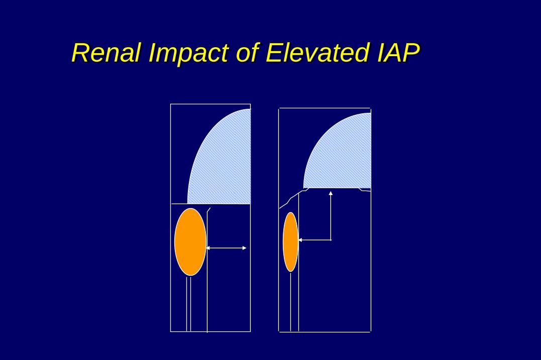
Renal Impact of Elevated IAP
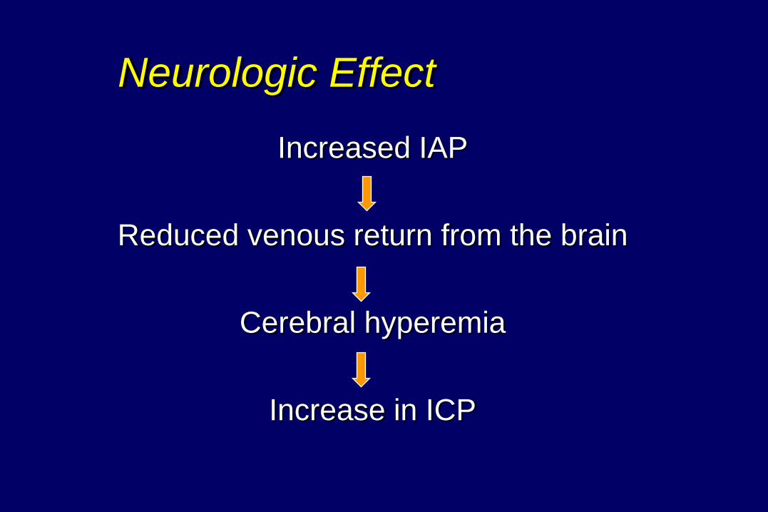
Neurologic Effect
Increased IAP
Reduced venous return from the brain
Cerebral hyperemia
Increase in ICP
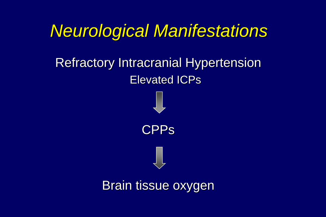
Neurological Manifestations
Refractory Intracranial Hypertension
Elevated ICPs
CPPs
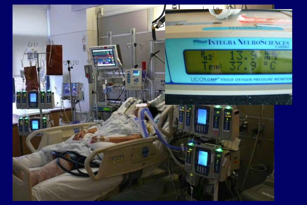
Brain tissue oxygen
Early/Ongoing Assessment
Early Initiation of IAP monitoring in patients
with 2 or more risk factors
Serial measurement until IAH risk is past
Bladder Pressure Monitoring
– The current standard for monitoring IAP
– Comparable to direct intraperitoneal pressure
measurements, but is non-invasive (Bailey, Crit Care 2000)
– More reliable and reproducible than clinical judgment (Kirkpatrick, CJS 2000; Sugrue World J Surg 2002)
Intra-Abdominal Pressure Measurement
Performed in the supine position
Zero at the level of the mid-axillary line (pelvis)
Expressed in mmHg (1 mmHg= 1.36 cmH2O)
Measured at End-expiration
Measure 30 – 60 sec after instillation of saline
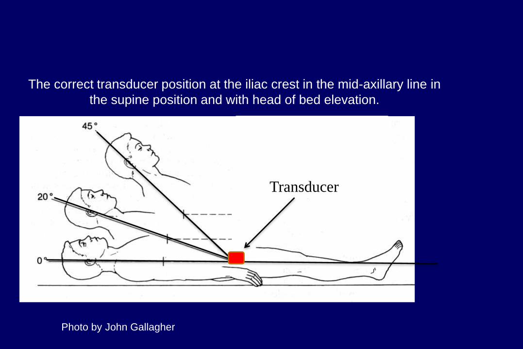
Transducer
location
The correct transducer position at the iliac crest in the mid-axillary line in
the supine position and with head of bed elevation.
Photo by John Gallagher
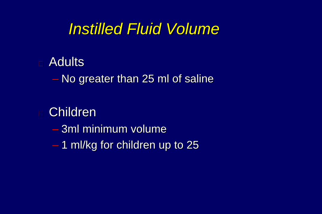
Instilled Fluid Volume
Adults
– No greater than 25 ml of saline
Children
– 3ml minimum volume
– 1 ml/kg for children up to 25
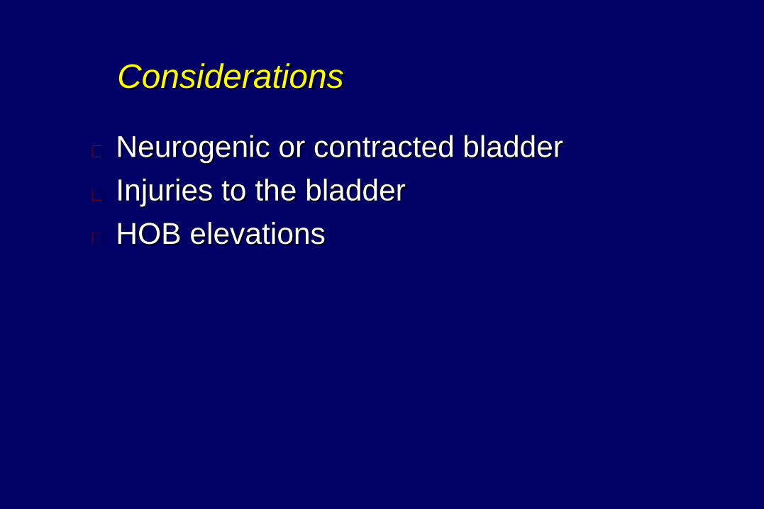
Considerations
Neurogenic or contracted bladder
Injuries to the bladder
HOB elevations
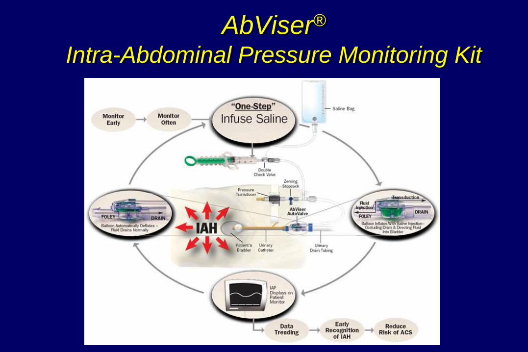
AbViser® Intra-Abdominal Pressure Monitoring Kit
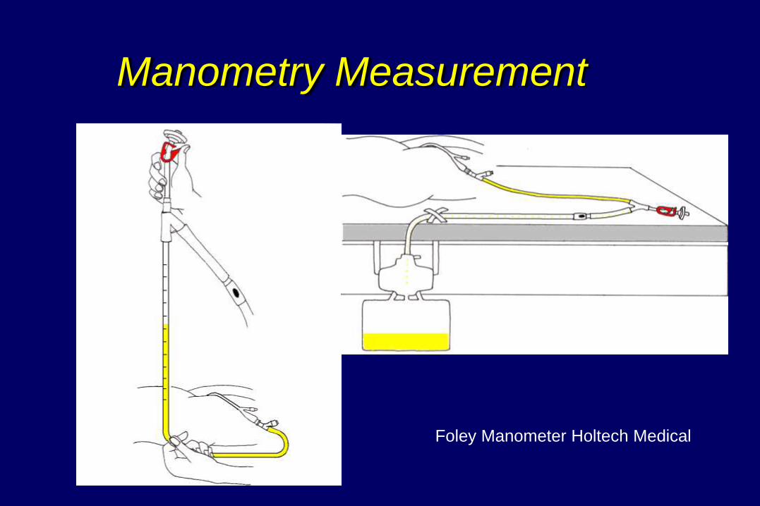
Manometry Measurement
Foley Manometer Holtech Medical
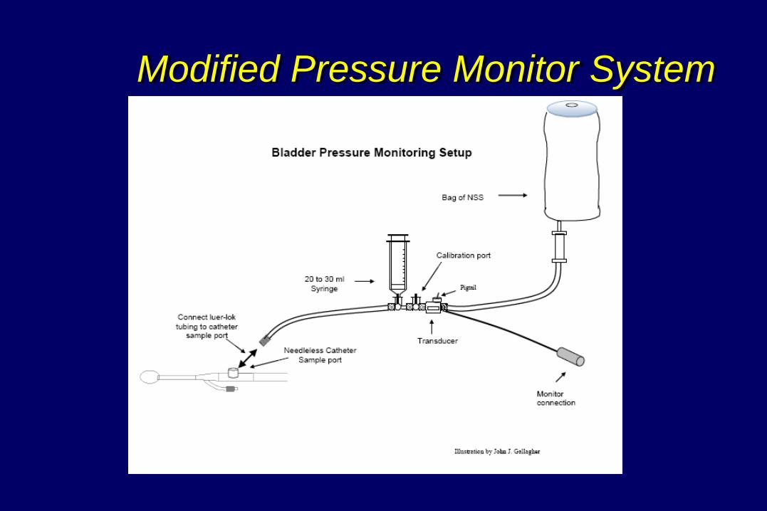
Modified Pressure Monitor System
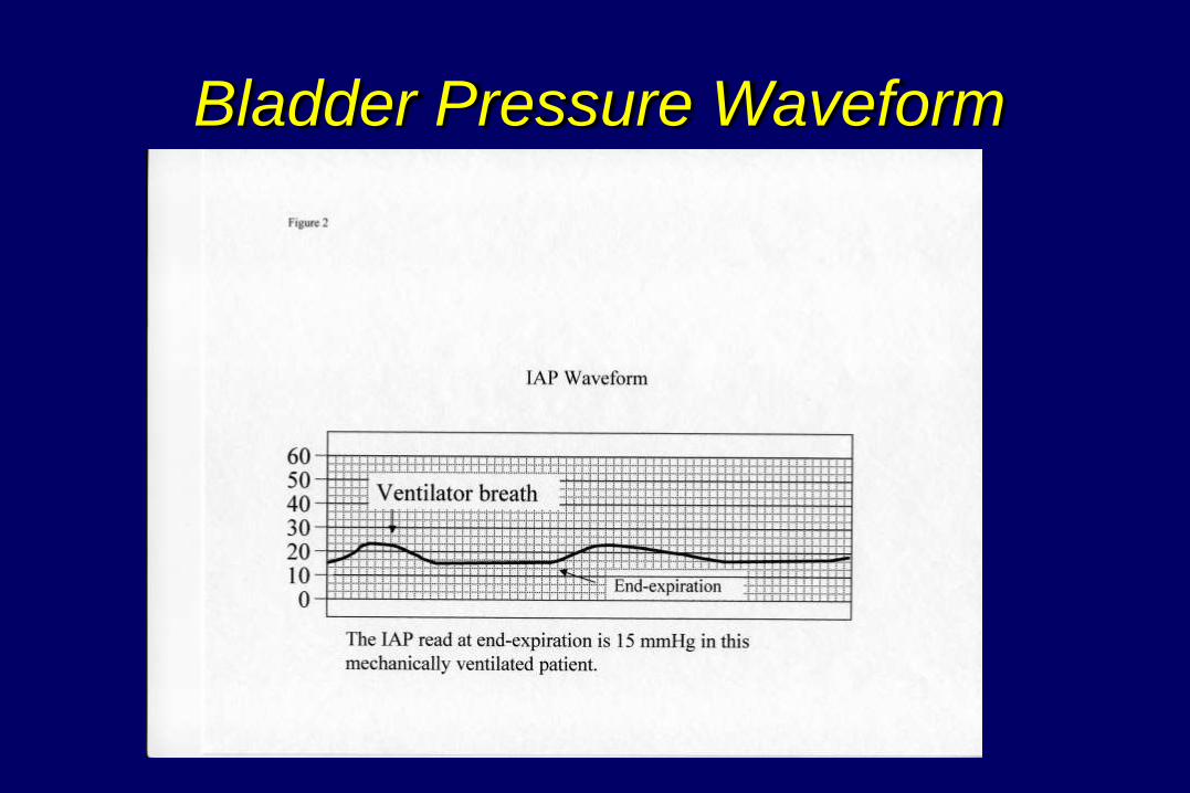
Bladder Pressure Waveform
Keys to Accuracy
Standardized measurement device
Standardized clinical protocol
Kimball et al. (2007) Intensive Care Medicine
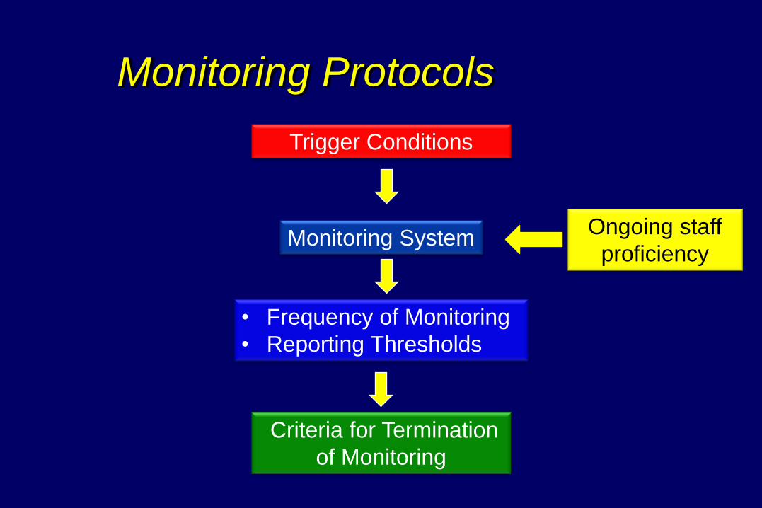
Monitoring Protocols
Trigger Conditions
Monitoring System
• Frequency of Monitoring
• Reporting Thresholds
Criteria for Termination
of Monitoring
Ongoing staff
proficiency
Pitfalls
Failing to identify patients at risk
– Temporary abdominal closure
– Volume resuscitation
– Post-op abdominal closure
Requiring an order to monitor IAP
Staff unfamiliar with the monitoring procedure
Terminating monitoring too soon
Management Strategies
Optimize systemic perfusion/organ function
Non-surgical interventions to reduce IAP
Surgical decompression
Ventilation Strategies
Decreased thoracic compliance
Normal lung tissue compliance
Lung Protective Strategies
– Limit ventilation pressures
– Optimize PEEP
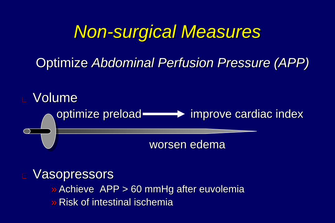
Non-surgical Measures
Optimize Abdominal Perfusion Pressure (APP)
Volume
optimize preload improve cardiac index
worsen edema
Vasopressors » Achieve APP > 60 mmHg after euvolemia
» Risk of intestinal ischemia
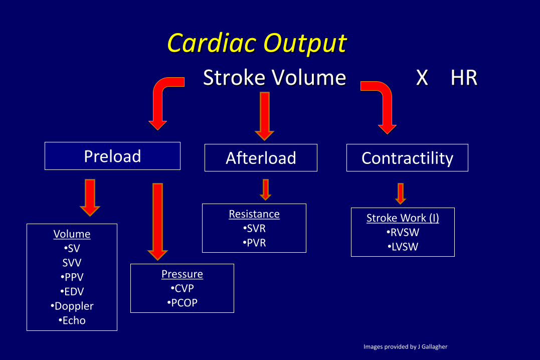
Cardiac Output Stroke Volume X HR
Preload Afterload Contractility
Volume •SV SVV •PPV •EDV
•Doppler •Echo
Pressure •CVP
•PCOP
Resistance •SVR •PVR
Stroke Work (I) •RVSW •LVSW
Images provided by J Gallagher
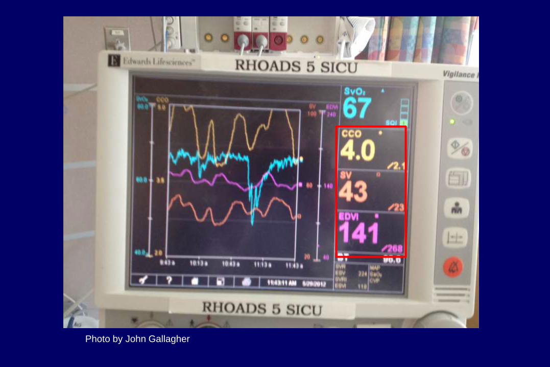
Photo by John Gallagher
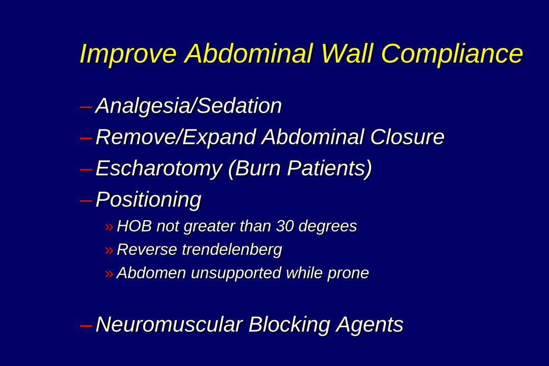
Improve Abdominal Wall Compliance
– Analgesia/Sedation
– Remove/Expand Abdominal Closure
– Escharotomy (Burn Patients)
– Positioning » HOB not greater than 30 degrees
» Reverse trendelenberg
» Abdomen unsupported while prone
– Neuromuscular Blocking Agents
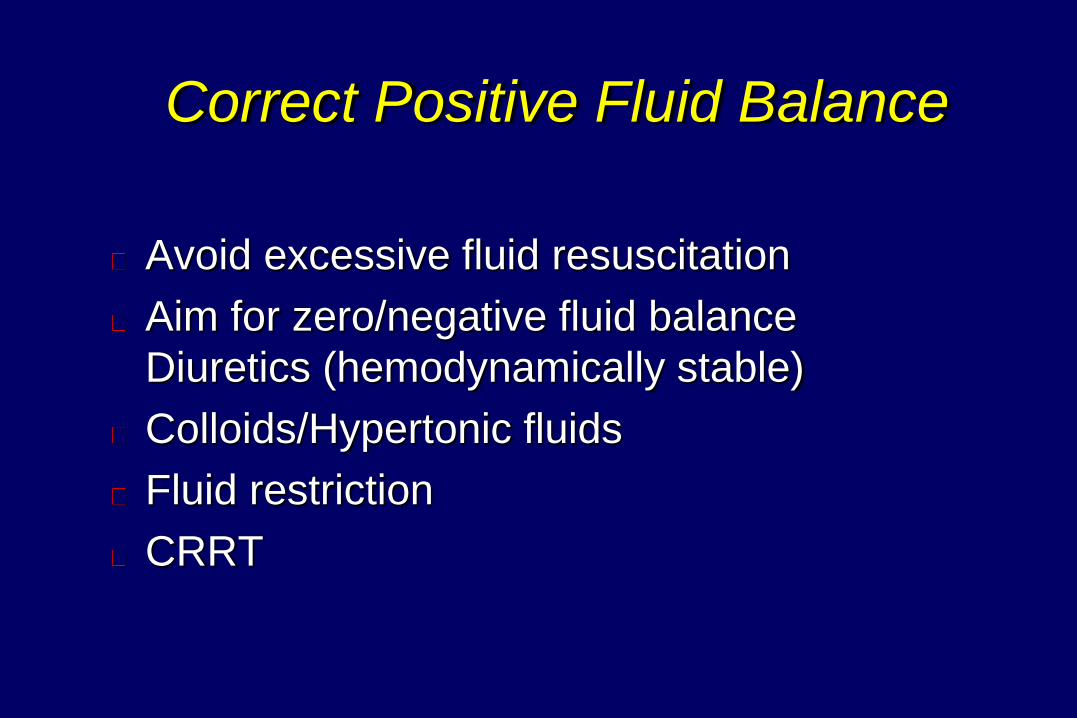
Correct Positive Fluid Balance
Avoid excessive fluid resuscitation
Aim for zero/negative fluid balance
Diuretics (hemodynamically stable)
Colloids/Hypertonic fluids
Fluid restriction
CRRT
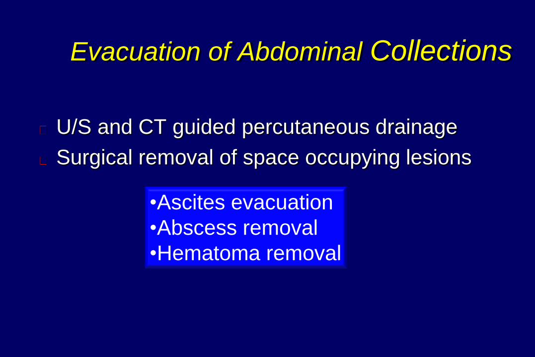
Evacuation of Abdominal Collections
U/S and CT guided percutaneous drainage
Surgical removal of space occupying lesions
•Ascites evacuation
•Abscess removal
•Hematoma removal
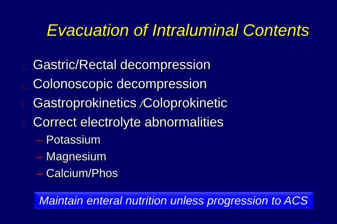
Evacuation of Intraluminal Contents
Gastric/Rectal decompression
Colonoscopic decompression
Gastroprokinetics /Coloprokinetic
Correct electrolyte abnormalities
– Potassium
– Magnesium
– Calcium/Phos
Maintain enteral nutrition unless progression to ACS
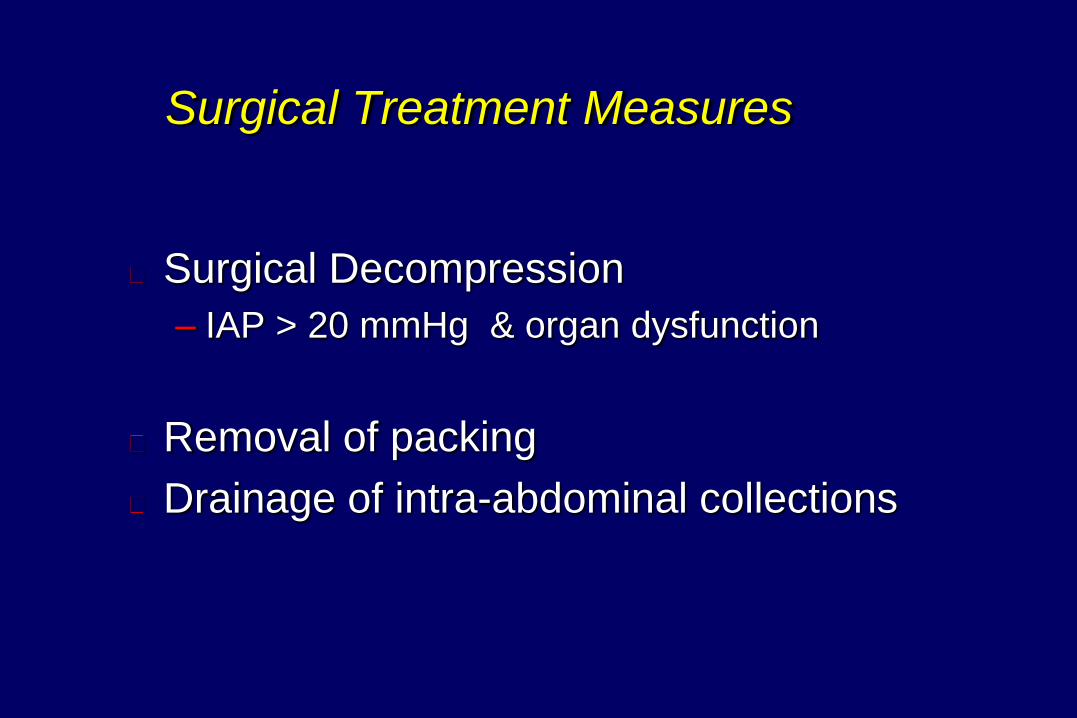
Surgical Treatment Measures
Surgical Decompression
– IAP > 20 mmHg & organ dysfunction
Removal of packing
Drainage of intra-abdominal collections
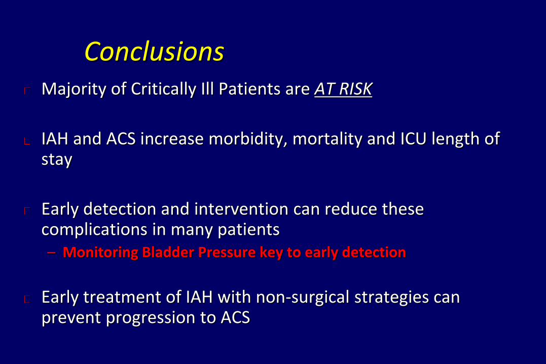
Conclusions Majority of Critically Ill Patients are AT RISK
IAH and ACS increase morbidity, mortality and ICU length of stay
Early detection and intervention can reduce these complications in many patients – Monitoring Bladder Pressure key to early detection
Early treatment of IAH with non-surgical strategies can prevent progression to ACS
Resources
World Society of Abdominal
Compartment Syndrome
www.wsacs.org