Embed Size (px)
Citation preview
www.elsevier.com/locate/yabbi
Archives of Biochemistry and Biophysics 430 (2004) 247–255
ABB
Investigation of the role of a second conserved serinein carboxylesterases via site-directed mutagenesis
Jeanette E. Stok, Andrey Goloshchapov, Cheng Song, Craig E. Wheelock,Maher B.H. Derbel, Christophe Morisseau, Bruce D. Hammock*
Department of Entomology and University of California Davis Cancer Research Center, University of California, Davis, CA 95616, USA
Received 20 May 2004, and in revised form 16 June 2004
Available online 4 August 2004
Abstract
Carboxylesterases are enzymes that catalyze the hydrolysis of ester and amide moieties. These enzymes have an active site that is
composed of a nucleophile (Ser), a base (His), and an acid (Glu) that is commonly known as a catalytic triad. It has previously been
observed that the majority of carboxylesterases and lipases contain a second conserved serine in their active site [Proteins, 34 (1999)
184]. To investigate whether this second serine is also involved in the catalytic mechanism, it was mutated to an alanine, a glycine or
a cysteine. Site-directed mutagenesis of this conserved serine resulted in a loss of specific activity, in both the S247G and S247A
mutants (5- to 15-fold), which was due to a decrease in the rate of catalysis (kcat). Due to the instability of the S247C mutant no
reliable data could be attained. A carbamate inhibitor, carbaryl, was then employed to investigate whether this decrease in the kcatwas due to the rate of formation of the acyl–enzyme intermediate (k2) or the rate of deacylation (k3). The S247A mutant was found
only to alter k2 (2.5-fold decrease), with no effect on k3. Together with information inferred from a human carboxylesterase crystal
structure, it was concluded that this serine provides an important structural support for the spatial orientation of the glutamic acid,
stabilizing the catalytic triad so that it can perform the hydrolysis.
� 2004 Elsevier Inc. All rights reserved.
Keywords: Carboxylesterase; Site-directed mutagenesis; Catalytic triad; Serine
Carboxylesterases (EC 3.1.1.1) are members of the a/b hydrolase fold family and are enzymes that catalyze
the hydrolysis of a wide range of endogenous and xeno-
biotic ester-containing compounds [2]. The a/b hydro-
lase fold family all contain a catalytic triad consisting
of a nucleophile, a base, and an orientating acid [2].
The existence of the catalytic triad was first establishedwith acetylcholinesterase (AchE)1 a well-studied mem-
ber of the a/b hydrolase fold family. It has been known
0003-9861/$ - see front matter � 2004 Elsevier Inc. All rights reserved.
doi:10.1016/j.abb.2004.06.020
* Corresponding author. Fax: 1-530-752-1537.
E-mail address: [email protected] (B.D. Hammock).1 Abbreviations used: AchE, acetylcholinesterase; PCR, polymerase
chain reaction; pNPA, p-nitrophenyl acetate; IEF, isoelectrofocusing;
PAGE, polyacrylamide gel electrophoresis; BSA, bovine serum
albumin.
for some time that both the serine and the histidine are
members of the catalytic triad in AchE [3–5]. Further
investigation via mutagenesis of these amino acids in
AchE (Torpedo californica) confirmed that they were
essential for catalysis [6]. The third member of the cata-
lytic triad, the glutamic acid, was identified by crystal
structure analysis of AchE [7]. This was again supportedbiochemically via the mutation of the Glu334 in human
AchE in which all catalytic activity was lost [8]. This
procedure has been repeated for many lipases and ester-
ases (e.g., [9–11]), including the highly specific juvenile
hormone esterase [12].
Carboxylesterases cleave esters via a two-step process
that involves the formation and degradation of an acyl–
enzyme intermediate. The hydrolysis initially occursthrough a nucleophilic attack via the serine onto the
248 J.E. Stok et al. / Archives of Biochemistry and Biophysics 430 (2004) 247–255
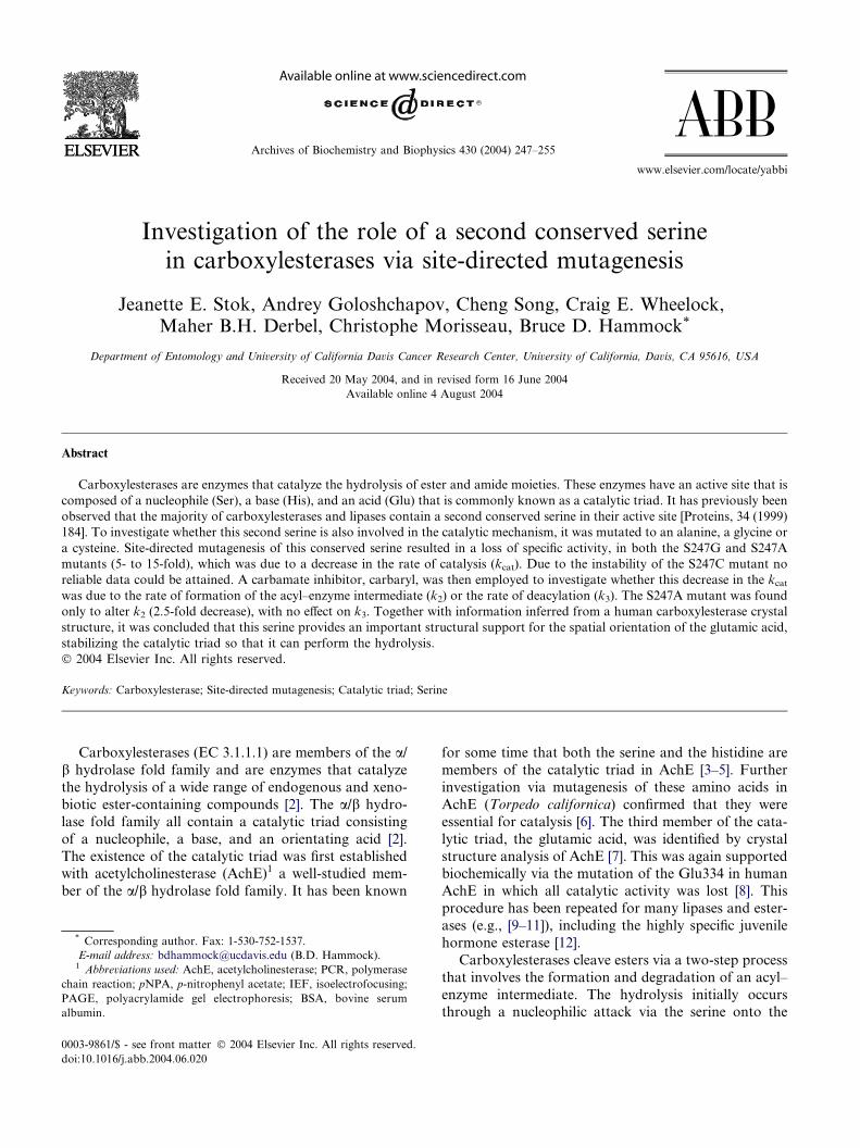
carbon of the carbonyl group (Fig. 1A) [2]. The catalytic
serine is able to accomplish this with the assistance of the
histidine acting as a general base. In turn, the protonated-
histidine is stabilized via a hydrogen bond to the glutamic
acid. These two catalytic amino acids, His–Glu, are often
called a �charge relay� system because they work togetherto activate the serine. The initial nucleophilic attack pro-
duces what is thought to be the first of two tetrahedral
intermediates (Fig. 1A, 1) that are stabilized by the pres-
ence of two glycines in the active site (oxyanion hole).
This tetrahedral intermediate rapidly collapses, aided
Fig. 1. (A) Detailed mechanism of the hydrolysis of esters by carboxylesteras
model [25]. Compounds in boxes are the products of the hydrolysis.
by the protonated-histidine acting as a general acid, to
produce an acyl–enzyme complex (Fig. 1A, 2) while the
alcohol component of the ester is released. The acyl–en-
zyme complex then undergoes an attack by a histidine-ac-
tivated water molecule (Fig. 1A, 3) which produces the
second tetrahedral intermediate (Fig. 1A, 4). After rapidrearrangement of this intermediate, the enzyme is regen-
erated and the acid component released. Much debate
still surrounds the exact nature of the mechanism de-
scribed above, which includes the possibility of a short,
strong bond or low barrier hydrogen bond (LBHB)
es. (B) Mechanism of the two-step carbamate cholinesterase inhibition
J.E. Stok et al. / Archives of Biochemistry and Biophysics 430 (2004) 247–255 249
between the glutamic acid and the histidine during catal-
ysis [13–16]. By lowering the activation energy required
for the formation of the first tetrahedral intermediate
(Fig. 1A, 1), the LBHB is believed to assist in the activa-
tion of the catalytic serine.
During a study involving the comparison of severalesterase and lipase sequences, a second absolutely con-
served serine was identified in all the sequences analyzed
[1]. It was therefore hypothesized that this serine may be
positioning the water molecule required for the second
nucleophilic attack [1]. Not only was this second serine
conserved in sequence but its absolute three-dimensional
position was conserved in all published structures [1]. In
this present study, rat carboxylesterase pI 6.1 (ES10) [17]was employed to explore the role of this second serine
by converting it via mutagenesis to an alanine, a glycine
or a cysteine. This carboxylesterase was chosen because
it was one of the first carboxylesterases to be isolated,
and it is a well-characterized enzyme [17–19]. It also
appears to be a good representative of carboxylesterases
in general. To directly explore the hypothesis that
the serine orients the water molecule, both the cata-lytic and kinetic properties of these expressed mutants
were measured and compared to that of the recombi-
nant wildtype enzyme. In particular, a carboxylesterase
inhibitor, carbaryl, was used to measure the specific
kinetic parameters, Kd, k2, and k3. We selected these
compounds as models because the decarbamylation rate
constant (k3; Fig. 1B) is slow compared to the deacyla-
tion rate constant (k2). Thus, we hoped to use the carba-mates to dissect the role of the second serine.
Materials and methods
Chemicals
Carbaryl was purchased from Chem Services (WestChester, PA). All other chemicals used in this study were
purchased from Sigma–Aldrich (St. Louis, MO), Fisher
Scientific (Pittsburgh, PA), or synthesized as described
below.
Synthesis of naphthalen-5-yl ethylcarbamate
Naphthol (1.0g, 6.94mmol) was ground to a finepowder using a mortar and pestle and placed into a
small round bottomed flask, followed by the addition
of ethyl isocyanate (0.5mL, 6.32mmol). A catalytic
amount of triethylamine was then added, producing an
immediate exothermic reaction. The resulting solid was
recrystallized twice from methanol to give the product
in 39% yield (0.53g, 2.46mmol): mp 100–101 �C; 1H
NMR (DMSO-d6) d 1.12 (t, 3H, J=7.2Hz), 3.14 (m,2H), 3.33 (t, 1H, J=6.9Hz), 7.26 (d, 1H, J=7.5Hz)
7.57 (m, 3H), 7.86 (m, 3H).
Site-directed mutagenesis
Rat esterase pI 6.1 [17] was isolated by PCR screen-
ing a cDNA library prepared from Sprague–Dawley
rat livers using specific primers (5 0-CACAATGCGCC
TCTACCCTCTG-3 0 and 5 0-AACATGGCTGACCCTCCTGATC-3 0) and a high fidelity Taq polymerase,
Advantage-HF (BD Biosciences, San Jose, CA). The
PCR product (1.7kb) was cloned into a pT-Adv vector
and sequenced to verify that it was the correct gene
(DNA Sequencing Facility, UC Davis). Mutants of rat
esterase pI 6.1 (S247G, S247C or S247A) were
constructed using the QuikChange Site-directed Muta-
genesis Kit (Stratagene, La Jolla, CA). Briefly, thisintroduced the desired mutations using the following
primers:
For
S247G (5 0-GCCATTTCTGAGGGTGGTGTGGTCC
TC-3 0),
S247C (5 0-GCCATTTCTGAGTGTGGTGTGGTCC
TC-3 0), andS247A (5 0-GCCATTTCTGAGGCTGGTGTGGTCC
TC-3 0) and their complementary strands.
These primers were used in a Pfu polymerase initiated
PCR. The original methylated, un-mutated DNA strand
was digested with DpnI, and the remaining DNA was
transformed into Escherichia coli. These mutated clones
were verified by sequencing (DNA Sequencing Facility,UC Davis). The modified gene was then ligated into a
pACUW21 baculovirus expression vector via the EcoRI
site.
Expression and preliminary analysis
The recombinant baculovirus was generated in Sf21
cells [20]. The viral DNA was extracted using the HighPure Viral Nucleic acid Kit (Roche, Indianapolis, IN)
and a PCR was performed to ensure that the gene had
been incorporated. The recombinant baculovirus was
expressed in Trichoplusia ni High five cells (2L,
1 · 106/mL) with a multiplicity of infection of approxi-
mately 0.1 at 28 �C. At 72h post-infection the cells were
pelleted (4000g, 20min) and resuspended in 50mM Tris–
HCl, pH 8.0, containing 1mM dithiothreitol, 1mMEDTA, and 1mM 1-phenyl-2-thiourea (buffer A). The
cells were then homogenized (2 · 30s; 10,000rpm, Poly-
tron homogenizer, Brinkmann, Westbury, NY) and the
microsomes and cellular debris were pelleted by centrifu-
gation (100,000g, 1h). The pellet was resuspended in
buffer A including 1% (w/v) of n-octyl-b-DD-glycopyrano-side and placed on a rotating wheel for 1h according to
the method of Huang [21]. The solution was centrifuged(100,000g, 1h) and the supernatant, dialyzed over 24h
(4 �C). The wildtype rat carboxylesterase 6.1 and each
250 J.E. Stok et al. / Archives of Biochemistry and Biophysics 430 (2004) 247–255
of the mutations (30lg) were analyzed by SDS–PAGE/
Western.
Carboxylesterase purification
All procedures were performed at 4 �C unless other-wise stated. Following the expression and homogeniza-
tion of the rat esterase 6.1 as described above, the
cellular debris and microsomes were pelleted by centrifu-
gation (100,000g, 1h). The supernatant was diluted
5-fold loaded onto a DEAE ion exchange column
(3 · 15cm, Amersham Biosciences, Piscataway, NJ),
and washed with buffer B (20mM Tris–HCl, pH 8.0,
and 1mM dithiothreitol). This was followed by a secondwash with 30mM NaCl in buffer B and the carboxylest-
erase was eluted with 50mM NaCl in buffer B. The carb-
oxylesterase containing fractions were detected by
measuring p-nitrophenyl acetate (pNPA) hydrolysis
activity at 405nm according to methods of Ljungquist
and Augustinsson [22] as described in Wheelock et al.
[23]. Following concentration by filtration (50kDa cut-
off; Millipore, Billerica, MA), the protein was furtherpurified using a preparative isoelectrofocusing unit
(Bio-Rad, Hercules, CA; pH 5–8). The esterase contain-
ing fractions were re-applied onto the preparative IEF
unit for refocusing and then loaded onto a gel filtration
column (Superose 12 10/300, FPLC, Amersham Bio-
sciences, Piscataway, NJ). The protein was eluted in buf-
fer B, fractions were pooled, concentrated by filtration
(50kDa cutoff; Millipore, Billerica, MA), and stored at�80 �C until used.
Antibody preparation
Rat carboxylesterase pI 6.1 was purified from rat li-
ver microsomes according to a previously reported pro-
cedure [19]. The purified protein was N-terminally
sequenced (Molecular Structure Facility, UC Davis) toconfirm its identity. Antibodies were raised against this
purified rat esterase 6.1 (ES10) according to the method
previously described [24].
Protein analysis
Protein concentrations were determined using the
Pierce BCA assay (Pierce, Rockford, IL) with BSAas the standard. SDS–PAGE and native PAGE were
performed using 12% Tris–glycine gels (Invitrogen,
Carlsbad, CA) whilst IEF–PAGE was performed
using IEF 3–7 gels (Invitrogen). Westerns were ana-
lyzed with polyclonal antibodies raised against rat
esterase 6.1 (ES10) (Rabbit No. 13053). Esterase
activity was detected by the hydrolysis of pNPA to
its corresponding alcohol by measuring the produc-tion of p-nitrophenolate anion at 405nm as described
above [22,23].
Kinetics
All kinetic analysis was performed in 0.1mg/mL BSA
in 20mM Tris–HCl, pH 8.0. Km and kcat were calculated
for both the wildtype and mutants by measuring pNPA
hydrolysis activity at various concentrations of the sub-strate. Other kinetic parameters were determined using
the classical two-step carbamate cholinesterase inhibi-
tion model outlined by Main [25] (Eq. (1)).
ð1Þ
where E is the enzyme and IX is the inhibitor.
For calculation of the inhibitor dissociation constant
(Kd) and the rate of carbamylation (k2), the enzyme was
inhibited with a range of inhibitor (carbaryl) concentra-
tions (0–250lMfinal concentration) in the presence of ex-
cess substrate (pNPA). The substrate was added either 10,
20, 30 or 60s after the introduction of the inhibitor. Theinitial rates of the acyl–enzyme formation at t0 (q) werecalculated by plotting each concentration of ln (At=0/
At) vs. time. The slopes (q) that were generated were then
plotted against the inhibitor concentration,where k2 is the
maximum rate of formation at t0 (q) (s�1) and Kd is
the inhibitor concentration at a half of the qmax (lM).
The rate of decarbamylation (k3) was calculated by
measuring pNPA activity of a 1:1 enzyme to inhibitorsolution 20, 40, 60, and 90min after the introduction of
the inhibitor, where k3 (s�1) is the slope of the plot of ln
(A0�At) vs time.
To determine the pH profile for the wildtype and
mutations, specific activity (pNPA) was measured every
0.2 pH units using 20mM Tris–HCl from pH 7.0 to 8.4.
The inhibitor dissociation constant (Kd) and rate of car-
bamylation (k2) were also calculated at pH 7.0, 7.5, 8.0,and 8.5 of 20mM Tris–HCl buffer.
Results
Construction of rat carboxylesterase 6.1 (ES 10) mutants
All mutants were generated using site-directed muta-genesis which incorporated the desired mutation
(S247A, G or C) via PCR. Pfu, a high fidelity polymer-
ase, was employed to limit the possible sequencing error
produced during PCR. Verification by DNA sequencing
was performed both after cloning into pACUW21 and
following production of the baculovirus. All baculovi-
ruses produced had incorporated the desired mutation
with no unwanted changes.
Preliminary analysis of mutants
The rat esterase 6.1 and each of the mutants were
expressed in insect cells (T. ni) that had been infected
J.E. Stok et al. / Archives of Biochemistry and Biophysics 430 (2004) 247–255 251
with the recombinant baculovirus. The recombinant
rat esterase mutants were initially analyzed by SDS–
PAGE and IEF–PAGE/Westerns to verify that they
were behaving in the same manner as the recombinant
wildtype esterase (data not shown). SDS–PAGE/Wes-
tern blot revealed one band at approximately 60kDain all the expressed proteins, that corresponded to
the size of rat carboxylesterase 6.1 (ES10) [17]. IEF/
Western showed two bands of approximately pI 6.0
in the wildtype with an extra band in each of the mu-
tants. As based on both IEF and SDS–PAGE, it was
therefore judged that the mutants were apparently in-
tact and could be used for comparison to the
wildtype.Following expression of the recombinant carboxy-
lesterase, microsomes were produced, solubilized, and
the soluble fraction was analyzed by SDS–PAGE/Wes-
tern blot (Fig. 2). Activity assays (pNPA) performed
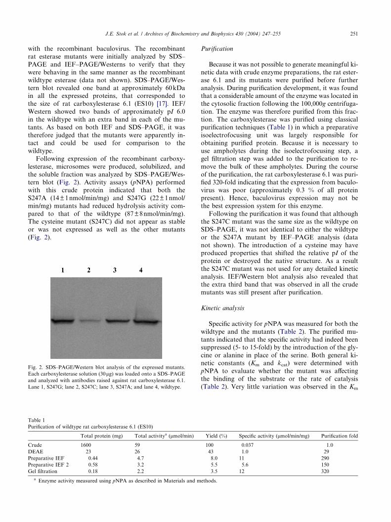
with this crude protein indicated that both the
S247A (14±1nmol/min/mg) and S247G (22±1nmol/
min/mg) mutants had reduced hydrolysis activity com-
pared to that of the wildtype (87±8nmol/min/mg).The cysteine mutant (S247C) did not appear as stable
or was not expressed as well as the other mutants
(Fig. 2).
Fig. 2. SDS–PAGE/Western blot analysis of the expressed mutants.
Each carboxylesterase solution (30lg) was loaded onto a SDS–PAGE
and analyzed with antibodies raised against rat carboxylesterase 6.1.
Lane 1, S247G; lane 2, S247C; lane 3, S247A; and lane 4, wildtype.
Table 1
Purification of wildtype rat carboxylesterase 6.1 (ES10)
Total protein (mg) Total activitya (lmol/min)
Crude 1600 59
DEAE 23 26
Preparative IEF 0.44 4.7
Preparative IEF 2 0.58 3.2
Gel filtration 0.18 2.2
a Enzyme activity measured using pNPA as described in Materials and m
Purification
Because it was not possible to generate meaningful ki-
netic data with crude enzyme preparations, the rat ester-
ase 6.1 and its mutants were purified before further
analysis. During purification development, it was foundthat a considerable amount of the enzyme was located in
the cytosolic fraction following the 100,000g centrifuga-
tion. The enzyme was therefore purified from this frac-
tion. The carboxylesterase was purified using classical
purification techniques (Table 1) in which a preparative
isoelectrofocusing unit was largely responsible for
obtaining purified protein. Because it is necessary to
use ampholytes during the isoelectrofocusing step, agel filtration step was added to the purification to re-
move the bulk of these ampholytes. During the course
of the purification, the rat carboxylesterase 6.1 was puri-
fied 320-fold indicating that the expression from baculo-
virus was poor (approximately 0.3 % of all protein
present). Hence, baculovirus expression may not be
the best expression system for this enzyme.
Following the purification it was found that althoughthe S247C mutant was the same size as the wildtype on
SDS–PAGE, it was not identical to either the wildtype
or the S247A mutant by IEF–PAGE analysis (data
not shown). The introduction of a cysteine may have
produced properties that shifted the relative pI of the
protein or destroyed the native structure. As a result
the S247C mutant was not used for any detailed kinetic
analysis. IEF/Western blot analysis also revealed thatthe extra third band that was observed in all the crude
mutants was still present after purification.
Kinetic analysis
Specific activity for pNPA was measured for both the
wildtype and the mutants (Table 2). The purified mu-
tants indicated that the specific activity had indeed beensuppressed (5- to 15-fold) by the introduction of the gly-
cine or alanine in place of the serine. Both general ki-
netic constants (Km and kcat) were determined with
pNPA to evaluate whether the mutant was affecting
the binding of the substrate or the rate of catalysis
(Table 2). Very little variation was observed in the Km
Yield (%) Specific activity (lmol/min/mg) Purification fold
100 0.037 1.0
43 1.0 29
8.0 11 290
5.5 5.6 150
3.5 12 320
ethods.
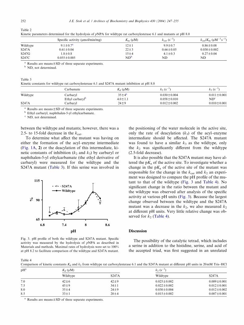
Table 2
Kinetic parameters determined for the hydrolysis of pNPA for wildtype rat carboxylesterase 6.1 and mutants at pH 8.0
Specific activity (lmol/min/mg) Km (lM) kcat (s�1) kcat/Km (lM�1 s�1)
Wildtype 9.1±0.7a 12±1 9.9±0.7 0.86±0.08
S247A 0.61±0.04 22±3 0.66±0.05 0.030±0.002
S247G 1.8±0.8 15±4 4.1±0.3 0.27±0.04
S247C 0.055±0.005 NDb ND ND
a Results are means±SD of three separate experiments.b ND, not determined.
Table 3
Kinetic constants for wildtype rat carboxylesterase 6.1 and S247A mutant inhibition at pH 8.0
Carbamate Kd (lM) k2 (s�1) k3 (s
�1)
Wildtype Carbaryl 35±4a 0.030±0.004 0.011±0.001
Ethyl carbarylb 4.0±1.1 0.058±0.010 NDc
S247A Carbaryl 24±9 0.012±0.002 0.010±0.001
a Results are means±SD of three separate experiments.b Ethyl carbaryl, naphthalen-5-yl ethylcarbamate.c ND, not determined.
252 J.E. Stok et al. / Archives of Biochemistry and Biophysics 430 (2004) 247–255
between the wildtype and mutants; however, there was a
2.5- to 15-fold decrease in the kcat.
To determine what affect the mutant was having on
either the formation of the acyl–enzyme intermediate
(Fig. 1A, 2) or the deacylation of this intermediate, ki-
netic constants of inhibition (k2 and k3) by carbaryl or
naphthalen-5-yl ethylcarbamate (the ethyl derivative of
carbaryl) were measured for the wildtype and theS247A mutant (Table 3). If this serine was involved in
Fig. 3. pH profile of both the wildtype and S247A mutant. Specific
activity was measured by the hydrolysis of pNPA as described in
Materials and methods. Maximal rates of hydrolysis were set to 100%
at pH 8.2 to facilitate comparison of the wildtype and S247A mutant.
Table 4
Comparison of kinetic constants Kd and k2 from wildtype rat carboxylestera
pHa Kd (lM)
Wildtype S247A
7.0 42±6 42±9
7.5 45±9 34±1
8.0 35±4 24±9
8.5 33±1 20±4
a Results are means±SD of three separate experiments.
the positioning of the water molecule in the active site,
only the rate of deacylation (k3) of the acyl–enzyme
intermediate should be affected. The S247A mutant
was found to have a similar k3 as the wildtype, only
the k2 was significantly different from the wildtype
(2.5-fold decrease).
It is also possible that the S247A mutant may have al-
tered the pKa of the active site. To investigate whether achange in the pKa of the active site of the mutant was
responsible for the change in the kcat and k2 an experi-
ment was designed to compare the pH profile of the mu-
tant to that of the wildtype (Fig. 3 and Table 4). No
significant change in the ratio between the mutant and
the wildtype was observed after analysis of the specific
activity at various pH units (Fig. 3). Because the largest
change observed between the wildtype and the S247Amutant was a decrease in the k2, we also measured k2at different pH units. Very little relative change was ob-
served for k2 (Table 4).
Discussion
The possibility of the catalytic tetrad, which includesa serine in addition to the histidine, serine, and acid of
the accepted triad, was first suggested in an unrelated
se 6.1 and the S247A mutant at different pH units in 20mM Tris–HCl
k2 (s�1)
Wildtype S247A
0.025±0.002 0.009±0.001
0.022±0.002 0.012±0.001
0.030±0.004 0.012±0.002
0.015±0.002 0.007±0.001
J.E. Stok et al. / Archives of Biochemistry and Biophysics 430 (2004) 247–255 253
family of enzymes called the serine proteases [26,27].
Serine proteases are enzymes that have a catalytic site
similar to that of esterases except the catalytic acid
and base are towards the N-terminal of the nucleophilic
serine rather than towards the C-terminal [28]. In almost
all chymotrypsin-like proteases, the acid (Asp in chymo-trypsin) is hydrogen-bonded to this second serine [29].
Investigation into the role of this serine in serine prote-
ases has produced a number of contradictory observa-
tions. Mutagenesis of at least two different serine
proteases revealed that although the rate of catalysis
(kcat) is affected by the substitution of this second serine
it may not be essential for catalysis [26,30]. From these
two studies it was proposed that the serine was mostlikely to be a structural feature, stabilizing the spatial
orientation of the catalytic triad enabling it to perform
the hydrolysis. In contrast, a more recent study con-
cluded from mutagenesis of this serine in human a-thrombin that the catalytic function (kcat) is not affected
but rather the serine is involved in the substrate binding
to enzyme (Km) [31]. It has been additionally elucidated
that the main chain carbonyl of this serine is hydrogen-bonded to the catalytic histidine [32,33]. This interaction
is also thought to stabilize the histidine and thus facili-
tate catalysis. Overall, these investigations conclude that
this second serine is not involved directly in catalysis but
is important for either, the stability and correct orienta-
tion of the catalytic triad or, the correct spatial environ-
ment for optimal substrate binding.
In a study analyzing a number of esterase and lipasesequences, it was observed that this conserved second
serine could also be found in these enzymes, including
AchE [1]. It was proposed in this study that this second
serine could be involved in the positioning of the water
that is required for the deacylation of the acyl–enzyme
complex (Fig. 1A, 2). Although this second serine is of-
ten depicted as being hydrogen-bonded to the glutamic
acid of the catalytic triad [34], there has been no directwork in carboxylesterases that has explored the role of
this amino acid. Hence, we were interested in exploring
the ramifications of removing this serine and replacing it
with either, a glycine, an alanine or a cysteine. Because it
was hypothesized that this serine could be important in
positioning a water molecule during deacylation of the
acyl–enzyme intermediate [1], we wanted to compare
the kinetic parameters of both the wildtype esteraseand its mutants. We anticipated that the nucleophilic at-
tack of the acyl–enzyme complex (Fig. 1A, 3) by the
water would be slower in the mutants than with the wild-
type. Hence, we would expect a decrease in k3, the rate
of deacylation.
Initial kinetic analysis revealed as expected that kcatrather than Km was suppressed in the S247A and
S247G mutants in comparison to the wildtype enzyme(Table 2). These observations support work done with
a-lytic protease where the introduction of the mutation
altered the kcat with very little affect on the Km [30].
Employing the classical two-step carbamate cholinester-
ase inhibition model outlined by Main [25] (Fig. 1B),
carbamate inhibitors were then employed to measure
the kinetic parameters Kd, k2, and k3 for both the wild-
type and the S247A mutant (Table 3). Some carbamates,including carbaryl, are used as insecticidal inhibitors of
AchE. The ethyl-homolog is used as the nontoxic inhib-
itor of general esterase activity. These compounds form
a carbamylated-enzyme (Fig. 1B, 2) analogous to the
acyl–enzyme complex (Fig. 1A, 2). Because no difference
in k3 was observed between these two proteins, it is un-
likely that this serine is involved in positioning the water
for the final step of the hydrolysis. Instead, an unex-pected 2.5-fold decrease in k2 was observed in the
S247A mutant as compared to the wildtype (Table 3).
Because kcat is really a measure of both k2 and k3, the
observed decrease in kcat appears to be a direct reflection
of the decrease in k2. The stability of the ethyl carbaryl
was too high to be of value in this experimental ap-
proach (Table 3).
The rate of formation of the acyl–enzyme complex(k2) can also dictate the relative value of Km [35]. When
k2 is fast, Km may not reflect the true affinity for the sub-
strate due to the large impact of k2 on its value. Hence, in
this case, as there is no significant change in the in Km
even after a change in k2 induced by the S247A mutant,
Km would appear to be a good approximation for sub-
strate affinity. Therefore, for this enzyme, it does not ap-
pear that this second serine is involved directly insubstrate binding. Although these findings are supported
by work performed with a-lytic protease [30], these
observations are slightly different to the analysis of
thrombin [31]. The large impact on the Km after the
substitution of alanine for the second serine in thrombin
implies a significant role for this amino acid in substrate–
enzyme binding (10-fold increase in Km). Comparison of
kcat/Km values from this study reveals that a-thrombinalanine mutant is approximately 18-fold less than the
wildtype which is similar to that observed in the S247A
mutant (28-fold decrease in kcat/Km; Table 2). Detailed
kinetic information revealed a smaller 2-fold decrease
in k2 in this same thrombin mutant. Comparison of these
two enzymes suggests that this serine plays a minor role
in both substrate-binding and stabilizing the spatial
orientation of the catalytic triad for optimal hydrolysis.The small changes in k2 or kcat/Km in SfiA mutants
(either 2-fold decrease or increase as seen in trypsin
[26]) appear to be the only factor that remains consistent
in both serine proteases and this esterase. As substrates
may vary widely in both these two families of enzymes
perhaps this spatial orientation provided by this second
serine is the predominant reason for its conservation.
The S247G mutant is slightly more active than theS247A mutant (Table 2), although both glycine and ala-
nine are incapable of hydrogen-bonding to the catalytic
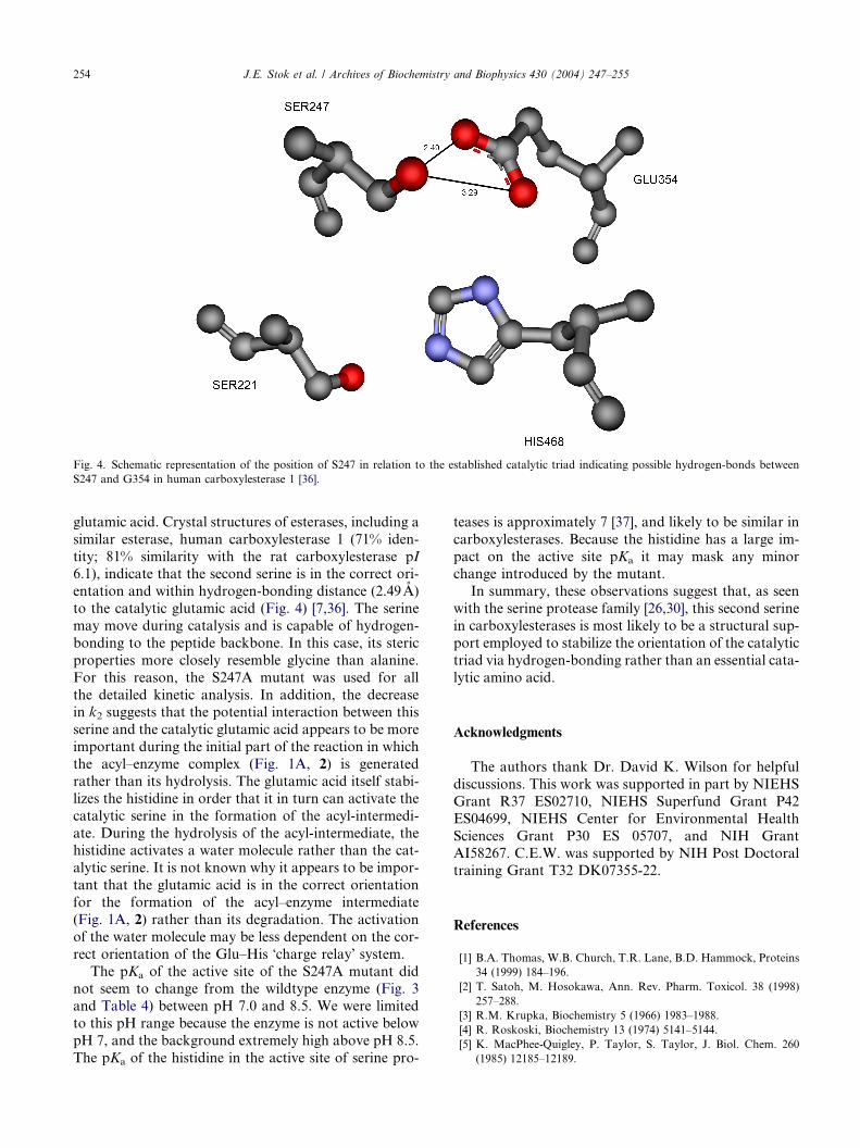
Fig. 4. Schematic representation of the position of S247 in relation to the established catalytic triad indicating possible hydrogen-bonds between
S247 and G354 in human carboxylesterase 1 [36].
254 J.E. Stok et al. / Archives of Biochemistry and Biophysics 430 (2004) 247–255
glutamic acid. Crystal structures of esterases, including a
similar esterase, human carboxylesterase 1 (71% iden-
tity; 81% similarity with the rat carboxylesterase pI6.1), indicate that the second serine is in the correct ori-
entation and within hydrogen-bonding distance (2.49A)
to the catalytic glutamic acid (Fig. 4) [7,36]. The serine
may move during catalysis and is capable of hydrogen-
bonding to the peptide backbone. In this case, its steric
properties more closely resemble glycine than alanine.
For this reason, the S247A mutant was used for all
the detailed kinetic analysis. In addition, the decreasein k2 suggests that the potential interaction between this
serine and the catalytic glutamic acid appears to be more
important during the initial part of the reaction in which
the acyl–enzyme complex (Fig. 1A, 2) is generated
rather than its hydrolysis. The glutamic acid itself stabi-
lizes the histidine in order that it in turn can activate the
catalytic serine in the formation of the acyl-intermedi-
ate. During the hydrolysis of the acyl-intermediate, thehistidine activates a water molecule rather than the cat-
alytic serine. It is not known why it appears to be impor-
tant that the glutamic acid is in the correct orientation
for the formation of the acyl–enzyme intermediate
(Fig. 1A, 2) rather than its degradation. The activation
of the water molecule may be less dependent on the cor-
rect orientation of the Glu–His �charge relay� system.
The pKa of the active site of the S247A mutant didnot seem to change from the wildtype enzyme (Fig. 3
and Table 4) between pH 7.0 and 8.5. We were limited
to this pH range because the enzyme is not active below
pH 7, and the background extremely high above pH 8.5.
The pKa of the histidine in the active site of serine pro-
teases is approximately 7 [37], and likely to be similar in
carboxylesterases. Because the histidine has a large im-
pact on the active site pKa it may mask any minorchange introduced by the mutant.
In summary, these observations suggest that, as seen
with the serine protease family [26,30], this second serine
in carboxylesterases is most likely to be a structural sup-
port employed to stabilize the orientation of the catalytic
triad via hydrogen-bonding rather than an essential cata-
lytic amino acid.
Acknowledgments
The authors thank Dr. David K. Wilson for helpful
discussions. This work was supported in part by NIEHS
Grant R37 ES02710, NIEHS Superfund Grant P42
ES04699, NIEHS Center for Environmental Health
Sciences Grant P30 ES 05707, and NIH GrantAI58267. C.E.W. was supported by NIH Post Doctoral
training Grant T32 DK07355-22.
References
[1] B.A. Thomas, W.B. Church, T.R. Lane, B.D. Hammock, Proteins
34 (1999) 184–196.
[2] T. Satoh, M. Hosokawa, Ann. Rev. Pharm. Toxicol. 38 (1998)
257–288.
[3] R.M. Krupka, Biochemistry 5 (1966) 1983–1988.
[4] R. Roskoski, Biochemistry 13 (1974) 5141–5144.
[5] K. MacPhee-Quigley, P. Taylor, S. Taylor, J. Biol. Chem. 260
(1985) 12185–12189.
J.E. Stok et al. / Archives of Biochemistry and Biophysics 430 (2004) 247–255 255
[6] G. Gibney, S. Camp, M. Dionne, K. Mac-Phee-Quigley, P.
Taylor, Proc. Natl. Acad. Sci. USA 87 (1990) 7546–7550.
[7] J.L. Sussman, M. Harel, F. Frolow, C. Oefner, A. Goldman, L.
Toker, I. Silman, Science 253 (1991) 872–879.
[8] A. Shafferman, C. Kronman, Y. Flashner, M. Leitner, H.
Grosfeld, A. Ordentlich, Y. Gozes, S. Cohen, N. Ariel, D. Barak,
M. Harel, I. Silman, J.L. Sussman, B. Velan, J. Biol. Chem. 267
(1992) 17640–17648.
[9] J. Emmerich, O.U. Beg, J. Peterson, L. Previato, J.D. Brunzell,
H.B.J. Brewer, S. Santamarina-Fojo, J. Biol. Chem. 267 (1992)
4161–4165.
[10] M. Haruki, Y. Oohashi, S. Mizuguchi, Y. Matsuo, M. Morikawa,
S. Kanaya, FEBS Lett. 454 (1999) 262–266.
[11] P. Lohse, S. Chahrokh-Zadeh, P. Lohse, D. Seidel, J. Lipid Res.
38 (1997) 892–903.
[12] V.K. Ward, B.C. Bonning, Q. Huang, T. Shiotsuki, V.N. Griffeth,
B.D. Hammock, Int. J. Biochem. 24 (1992) 1933–1941.
[13] P.A. Frey, S.A. Whitt, J.B. Tobin, Science 264 (1994) 1927–1930.
[14] A. Warshel, A. Papazyan, P.A. Kollman, Science 269 (1995) 102–
106.
[15] C. Viragh, T.K. Harris, P.M. Reddy, M.A. Massiah, A.S.
Mildvan, I.M. Kovach, Biochemistry 39 (2000) 16200–16205.
[16] M.A. Massiah, C. Viragh, P.M. Reddy, I.M. Kovach, J. Johnson,
T.L.Rosenberry,A.S.Mildvan, Biochemistry 40 (2001) 5682–5690.
[17] M. Robbi, H. Beaufay, J.-N. Octave, Biochem. J. 269 (1990) 451–
458.
[18] R. Mentlein, A. Ronai, M. Robbi, E. Heymann, O.v. Deimling,
Biochim. Biophys. Acta 913 (1987) 27–38.
[19] M. Hosokawa, T. Maki, T. Satoh, Arch. Biochem. Biophys. 277
(1990) 219–227.
[20] D.R. O�Reilly, L.K. Miller, V.A. Luckow (Eds.), Baculovirus
Expression vectors: A Laboratory Manual, W.H. Freeman and
Co., New York, 1994, p. 347.
[21] T.L. Huang, T. Shiotsuki, T. Uematsu, B. Borhan, Q.X. Li, B.D.
Hammock, Pharm. Res. 13 (1996) 1495–1500.
[22] A. Ljungquist, K.B. Augustinsson, Eur. J. Biochem. 23 (1971)
303–313.
[23] C.E. Wheelock, T.F. Severson, B.D. Hammock, Chem. Res.
Toxicol. 14 (2001) 1563–1572.
[24] S.J. Gee, T. Miyamoto, M.H. Goodrow, D. Buster, B.D.
Hammock, J. Agric. Food Chem. 36 (1988) 863–870.
[25] A.R. Main, in: E. Hodgson, F.E. Guthrie (Eds.), Introduction
to Biochemical Toxicology, Elsevier, New York, 1980, pp. 193–
223.
[26] M.E. McGrath, J.R. Vasquez, C.S. Craik, A.S. Yang, B. Honig,
R.J. Fletterick, Biochemistry 31 (1992).
[27] M.M. Krem, E. Di Cera, EMBO J. 20 (2001) 3036–3045.
[28] G. Dodson, A. Wlodawer, Trends Biochem. Sci. 23 (1998) 347–
352.
[29] L. Hedstrom, Chem. Rev. 102 (2002) 4501–4523.
[30] D.M. Epstein, R.H. Abeles, Biochemistry 31 (1992) 11216–
11223.
[31] M.M. Krem, S. Prasad, E. Di Cera, J. Biol. Chem. 277 (2002)
40260–40294.
[32] Z.S. Derewenda, U. Derewenda, P.M. Kobos, J. Mol. Biol. 241
(1994).
[33] P.A. Molina, R.S. Sikorski, J.H. Jensen, Theor. Chem. Accounts
109 (2003) 100–107.
[34] C.B. Millard, G. Koellner, A. Ordentlich, A. Shafferman, I.
Silman, J.L. Sussman, J. Am. Chem. Soc. 121 (1999) 9883–
9884.
[35] R.N. Armstrong, C.S. Cassidy, Drug Metab. Rev. 32 (2000) 327–
338.
[36] S. Bencharit, C.L. Morton, J.L. Hyatt, P. Kuhn, M.K. Danks,
P.M. Potter, M.R. Redinbo, Chem. Biol. 10 (2003) 341–349.
[37] W.W. Bachovchin, Magn. Reson. Chem. 39 (2001) S199–S213.