Embed Size (px)
Citation preview

ABA Is an Essential Signal for Plant Resistance to PathogensAffecting JA Biosynthesis and the Activation of Defensesin Arabidopsis W
Bruce A.T. Adie,a Julian Perez-Perez,a Manuel M. Perez-Perez,a Marta Godoy,a Jose-J. Sanchez-Serrano,a
Eric A. Schmelz,b and Roberto Solanoa,1
a Departamento de Genetica Molecular de Plantas, Centro Nacional de Biotecnologıa–Consejo Superior de Investigaciones
Cientıficas, Campus Universidad Autonoma, 28049 Madrid, Spainb U.S. Department of Agriculture–Agricultural Research Service, Center for Medical, Agricultural, and Veterinary Entomology,
Gainesville, Florida 32608-1067
Analyses of Arabidopsis thaliana defense response to the damping-off oomycete pathogen Pythium irregulare show that
resistance to P. irregulare requires a multicomponent defense strategy. Penetration represents a first layer, as indicated by
the susceptibility of pen2 mutants, followed by recognition, likely mediated by ERECTA receptor-like kinases. Subsequent
signaling of inducible defenses is predominantly mediated by jasmonic acid (JA), with insensitive coi1 mutants showing
extreme susceptibility. In contrast with the generally accepted roles of ethylene and salicylic acid cooperating with or
antagonizing, respectively, JA in the activation of defenses against necrotrophs, both are required to prevent disease
progression, although much less so than JA. Meta-analysis of transcriptome profiles confirmed the predominant role of JA
in activation of P. irregulare–induced defenses and uncovered abscisic acid (ABA) as an important regulator of defense gene
expression. Analysis of cis-regulatory sequences also revealed an unexpected overrepresentation of ABA response
elements in promoters of P. irregulare–responsive genes. Subsequent infections of ABA-related and callose-deficient
mutants confirmed the importance of ABA in defense, acting partly through an undescribed mechanism. The results support
a model for ABA affecting JA biosynthesis in the activation of defenses against this oomycete.
INTRODUCTION
The success of plants in colonizing so many different environ-
ments where they have to cope with a plethora of biotic and
abiotic challenges indicates that evolution has provided them
with efficient defense mechanisms. Plants possess both pre-
formed and inducible layers of defense to resist pathogen
invasion. Constitutive physical and chemical barriers prevent
the establishment of most plant–pathogen interactions. How-
ever, should the pathogen overcome these constitutive de-
fenses, its recognition leads to the induction of a multitude of
defenses through the genetic reprogramming of the cell.
Our understanding of the complex mechanisms by which
plants first detect, and then defend against, different microbial
pathogens has advanced considerably over the last few de-
cades. Central to this progress has been the identification and
characterization of plant disease resistance genes that facilitate
pathogen strain–specific recognition and the identification of
signal transduction pathways that link pathogen recognition with
a targeted response (Nimchuk et al., 2003; Glazebrook, 2005).
Three phytohormones, salicylic acid (SA), jasmonic acid (JA),
and ethylene (ET), have been shown to play a major role in the
regulation of these signal transduction pathways. This regulation
is not achieved through the isolated activation of each single
hormonal pathway but rather through a complex regulatory
network that connects the different pathways enabling each to
assist or antagonize the others as required to fine-tune the
defense response to the individual pathogen. Thus, it is generally
accepted (although may be oversimplified) that SA plays a major
role in activation of defenses against biotrophic pathogens,
whereas JA and ET are more usually associated with defense
against necrotrophic pathogen attack. Additionally, SA and JA/
ET defense pathways are mutually antagonistic (Thomma et al.,
2001; Kunkel and Brooks, 2002; Turner et al., 2002; Rojo et al.,
2003; Glazebrook, 2005; Lorenzo and Solano, 2005; van Loon
et al., 2006).
Although mechanistic explanations of this antagonistic and
cooperative crosstalk are scarce, several examples suggest its
regulation through the differential modulation of transcription
factor (and cofactors) activity by the different hormones. Thus,
antagonism between JA and SA pathways requires the activation
of proteins such as NPR1 and WRKY70 that activate expression
of SA-responsive genes while repressing JA-responsive genes
(Spoel et al., 2003; Li et al., 2004). Conversely, cooperation of JA
and ET in the activation of defenses against necrotrophs can be
explained by their concerted activation of Ethylene Response
Factor1 (ERF1), which induces defense gene expression and
plant resistance (Berrocal-Lobo et al., 2002; Lorenzo et al.,
2003). However, in contrast with the case of pathogen infection,
1 To whom correspondence should be addressed. E-mail [email protected]; fax 34-91-5854506.The author responsible for distribution of materials integral to thefindings presented in this article in accordance with the policy describedin the Instructions for Authors (www.plantcell.org) is: Roberto Solano([email protected]).W Online version contains Web-only data.www.plantcell.org/cgi/doi/10.1105/tpc.106.048041
The Plant Cell, Vol. 19: 1665–1681, May 2007, www.plantcell.org ª 2007 American Society of Plant Biologists

ET and JA antagonize one another in the activation of responses
to wounding. The fine-tune regulation of this antagonism de-
pends on the balance of activation by both hormones of ERF1
and MYC2, another transcription factor differentially regulating
two branches of the JA signaling pathway (Lorenzo et al., 2004).
The current understanding of plant defense responses de-
scribed above has been achieved through the study of a limited
number of models, which may be constraining our view of the plant–
pathogen interaction and the true capacity of plants to defend
against pathogens. In fact, random sequencing approaches of
microbial populations from seawater samples have recently
demonstrated that we still only know a relatively small percent-
age of gene functions existing in nature (Venter et al., 2004).
Applied to the plant–pathogen field, discovery of new plant
defense mechanisms should be favored by broadening the range
of pathogens and plant species under study. With this aim, we
have characterized the interaction between Pythium irregulare
and Arabidopsis thaliana.
Pythium is commonly regarded as a soil-borne vascular path-
ogen. It is particularly virulent in seedlings, although it can infect
mature aboveground tissue in several plant species, including
Arabidopsis (van der Plaats-Niterink, 1981; Martin, 1995). Spe-
cies within this genus range from virulent to opportunistic path-
ogens, producing seedling damping-off and root and stem rots
that may cause economically important losses in crops and
ornamental plants (Miller and Sauve, 1975; Farr et al., 1989).
P. irregulare has been reported to produce both lytic enzymes
(Deacon, 1979) and phytotoxins (Brandenburg, 1950) that would
degrade plant tissue, thus behaving as a necrotrophic pathogen.
Being an oomycete, and thus phylogenetically distant from the
most extensively studied species of fungi (Kamoun et al., 1999),
the supposed necrotrophic lifestyle, and the fact that JA and ET
are required for effective defenses against different species from
this genus also differentiate Pythium from other oomycetes
(Knoester et al., 1998; Staswick et al., 1998; Vijayan et al., 1998).
To gain a deeper insight of the Arabidopsis–P. irregulare host–
pathogen interaction, it is imperative to understand both the
physical infection process and the molecular consequences. To
that end, we have (1) characterized the infection process and (2)
studied the plant molecular defense pathways involved in resis-
tance through genetic and genomic analysis. In addition to the
comprehensive characterization of the interaction, this com-
bined analysis has identified ABA as a signal required for plant
resistance to P. irregulare and other necrotrophic pathogens.
RESULTS
Characterization of P. irregulare Colonization of
Arabidopsis Tissue
Appresoria were observed during early stages of infection of
both roots and leaves of mature plants (Figure 1A; see Supple-
mental Figure 1A online). Following penetration of the first host
cell, hyphal ramification gave rise to multidigitate haustoria-like
structures (Figure 1B; see Supplemental Figure 1B online). The
individual lobes of the haustoria-like structures continued to
lengthen until an opposing cell wall was encountered, where-
upon constricted hyphae attempted penetration of the adjoining
host cell wall (Figure 1C; see Supplemental Figure 1C online).
Further ramification of hyphal tissue occurred frequently, giving
rise to a dense network that penetrated all host tissue types. This
colonization eventually lead to tissue collapse and ultimately wet
rot. Fungal growth was predominantly but not exclusively intra-
cellular (Figure 1C; see Supplemental Figure 1C online).
Hyphal swellings were occasionally produced where fungal
growth was prolific and allowed to progress for 48 h (see
Figure 1. Microscopy Analysis of Arabidopsis Infection by P. irregulare.
(A) Appresorial (ap) formation on leaf epidermis. Penetration pegs (pp)
may also be observed (Trypan blue stain). Bar ¼ 30 mm.
(B) Multidigitate haustoria-like structure (hl) formed upon invasion of the
epidermal cell by runner hyphae (rh). The cell wall (cw) is also fluorescent
due to callose deposition (Analine blue stain). Bar ¼ 25 mm.
(C) Constricted hyphae (hy) penetration through the root cell wall (cw)
(Trypan blue stain). Bar ¼ 5 mm.
(D) Localized plant cell death upon direct infection of P. irregulare via
runner hyphae (rh). Bar ¼ 25 mm.
(E) Trailing necrosis (tn) of infected mesophyll cells. Bar ¼ 25 mm.
1666 The Plant Cell

Supplemental Figure 1D online). Oogonium and oospores were
only observed in culture media (see Supplemental Figure 1E on-
line), presumably because in planta infections were rarely allowed
to progress beyond 24 h, this being the optimal time for discern-
ment of differential resistance between defense mutants.
Trypan blue stain indicated that only infected cells in physical
contact with the hyphae died (Figure 1D), suggesting that this
oomycete does not produce lytic enzymes or phytotoxins during
infection. Consistent with this, culture filtrates were not able to
reproduce disease symptoms in Arabidopsis (data not shown).
Thus, despite reports that P. irregulare produces both lytic
enzymes (Deacon, 1979) and phytotoxins (Brandenburg, 1950),
no evidence of extracellular toxin production was seen during
this pathogen–host interaction.
Additionally, trailing necrosis occurred during infection of sus-
ceptible tissue, where hyphae were observed to extend ahead of
dead cells (i.e., those retaining the Trypan blue stain) (Figure 1E)
similar to that witnessed for Phytophthora infestans during in-
fection of partially resistant plants (Kamoun et al., 1999). If toxins
or lytic enzymes were being produced and dissipated in func-
tionally important quantities within the plant tissue, cell death
might be expected to extend ahead of hyphal growth, similar to
that of Botrytis cinerea (Clark and Lorbeer, 1976; Govrin and
Levine, 2000).
Characterization of Plant Defense Response
Results described above showed that P. irregulare infection of
Arabidopsis has neither typical necrotrophic nor biotrophic char-
acteristics. Additionally, oomycetes are phylogenetically distinct
from many of the more extensively studied fungal pathogens
(Kamoun et al., 1999). Thus, to understand how the plant coun-
teracts hyphal invasion, we used mutants affected in hormonal
and nonhormonal defense pathways to dissect the involvement
of known plant defense mechanisms.
Contribution of the JA, ET, and SA Pathways
Consistent with previous analyses of JA-deficient or -insensitive
mutants (Staswick et al., 1998; Vijayan et al., 1998) and the de-
scribed role of JA in the activation of responses against oomy-
cetes (Coego et al., 2005), the JA-insensitive coi1-1 mutants were
highly susceptible to P. irregulare (Figure 2A), indicating that JA is
a major signal for activation of defenses against this oomycete.
Consistent with results in transgenic tobacco (Nicotiana taba-
cum) plants (Geraats et al., 2002), ET-insensitive ein2-5 mutants
showed a significantly increased susceptibility to the oomycete,
indicating that ET is required to achieve fully active plant de-
fenses. However, the weaker effect of ein2 compared with that of
coi1 within this study indicates that ET is not as critical to the P.
irregulare defense response as it is for other necrotrophs.
Interestingly, SA-related mutants (npr1 and sid2-1) showed a
susceptibility similar to ET-insensitive mutants, indicating that
SA, instead of antagonizing JA-dependent defenses, also con-
tributes to overall resistance (Figure 2A).
To further understand this apparent cooperation between
JA and SA, the levels of both hormones were measured in
wild-type and JA/SA/ET-related mutants after P. irregulare in-
fection. Supporting their role in defense against the oomycete,
levels of both hormones increased rapidly in wild-type plants
after infection (Figure 2B). The increase in JA levels was com-
parable in wild-type and sid2-1 plants and lower in coi1-16
mutants, indicating that SA does not prevent JA biosynthesis. In
contrast with the assumed cooperation between ET and JA, but
consistent with previous reports of ET-mediated repression of
JA-regulated gene expression (Rojo et al., 2003; Lorenzo et al.,
2004), JA levels were dramatically increased in ein2-5 mutants
than in wild-type plants. In contrast with wild-type plants, an
increase in SA levels was not observed in the biosynthetic mutant
Figure 2. P. irregulare Infection of Arabidopsis Defense Mutants.
(A) Infection of JA/ET/SA-related mutants. Mean disease area (MDA) per
infected leaf (mm2) 24 h after inoculation with a mycelial plug. Asterisks
indicate significant difference (P < 0.05) from Col-0 using the Student’s t
test.
(B) Endogenous JA and SA levels (ng/g fresh weight [FW]) in Arabidopsis
wild-type (Col-0) and JA/ET/SA-related mutant plants (coi1-16, ein2-5,
and sid2-1) after infection with P. irregulare for 0, 6, and 12 h (h after
infection [HAI]). Seedlings were grown on Johnson’s media plates for 7 d
prior to infection. Values are means 6 SE of three independent replicate
experiments.
(C) Infection of nonhormonal mutants. Asterisks indicate significant
difference (P < 0.05) from Col-0 (or Landsberg in the case of Ler) using
the Student’s t test.
ABA Requirement for Plant Resistance to Pathogens 1667
sid2-1. Furthermore, and consistent with the negative regulation
of SA biosynthesis by JA/ET, the levels of SA in coi1-16 and ein2-5
mutants increased over wild-type levels.
These results support a role for both defense pathways (JA/ET
and SA) in the resistance against P. irregulare.
Involvement of Other Nonhormonal Defense Pathways
We analyzed the susceptibility of nonhormonal defense-related
mutants, including nonhost-related mutants (pen1 and pen2;
Collins et al., 2003; Assaad et al., 2004), mutants affected in
responses to biotrophs (edr1, pad4, and eds1; Frye and Innes,
1998; Falk et al., 1999; Jirage et al., 1999), and the erecta
mutation (essential for resistance to some necrotrophs; Godiard
et al., 2003; Llorente et al., 2005). Among them, only pen2
and erecta (in Columbia-0 [Col-0] and Landsberg erecta [Ler]
backgrounds, respectively) showed a significantly increased
susceptibility to P. irregulare colonization (Figure 2C). This sug-
gests that plant resistance to the initial establishment and pro-
gression of disease by P. irregulare depends on avoidance of
penetration mediated by PEN2 and, very likely, other primary
defense layers, since pen2 mutants are not fully susceptible to
the oomycete. Additionally, signaling of defense activation may
start from ERECTA (and other partially redundant proteins).
Transcriptomic Profiling of Arabidopsis Infection
by P. irregulare
To further characterize the plant molecular response to this
oomycete, whole-genome transcriptional profiles of infected
wild-type, coi1-1, ein2-5, and sid2-1 plants were obtained using
two-color long-oligonucleotide microarrays (see Methods).
Seven transcriptome comparisons were directly made in the
two-color chips, four within-genotype comparisons between in-
fected versus noninfected seedlings from each genotype (Col-0,
coi1-1, ein2-5, and sid2-1), and three counter genotype com-
parisons between infected wild-type seedlings and each of the
infected hormone-related mutants (Col-0 infected versus coi1-1
infected, Col-0 infected versus ein2-5 infected, and Col-0 in-
fected versus sid2-1 infected). A cluster view of all genes dif-
ferentially expressed in at least one of the seven comparisons
can be found in Supplemental Figure 2 online. The complete list
of genes can be found in Supplemental Table 1 online.
The combination of these two types of comparison (within
genotype and counter genotype) allowed for the differentiation of
two gene classes of interest: (1) JA/ET/SA-dependent genes that
are differentially expressed following P. irregulare infection in
Col-0 and are also dependent on at least one of the three hor-
monal pathways being studied (129 genes) and (2) JA/ET/SA-
independent genes that are differentially expressed following P.
irregulare infection in wild-type plants but appear independent of
the three hormonal pathways being studied (1385 genes).
In addition to these two groups, 217 genes appear to be
dependent upon at least one of the three hormonal pathways
tested but are not differentially expressed in the wild type fol-
lowing P. irregulare infection. Most of these genes, being constitu-
tively over/underexpressed in hormonal mutants, are of limited
interest to this pathogen defense study and will not be consid-
ered further.
In addition to the statistical methods described elsewhere in
this article, validation of microarray data was achieved by RNA
gel blot analysis. As shown in Supplemental Figure 3 online,
expression of all randomly chosen genes correlated well with the
microarray data.
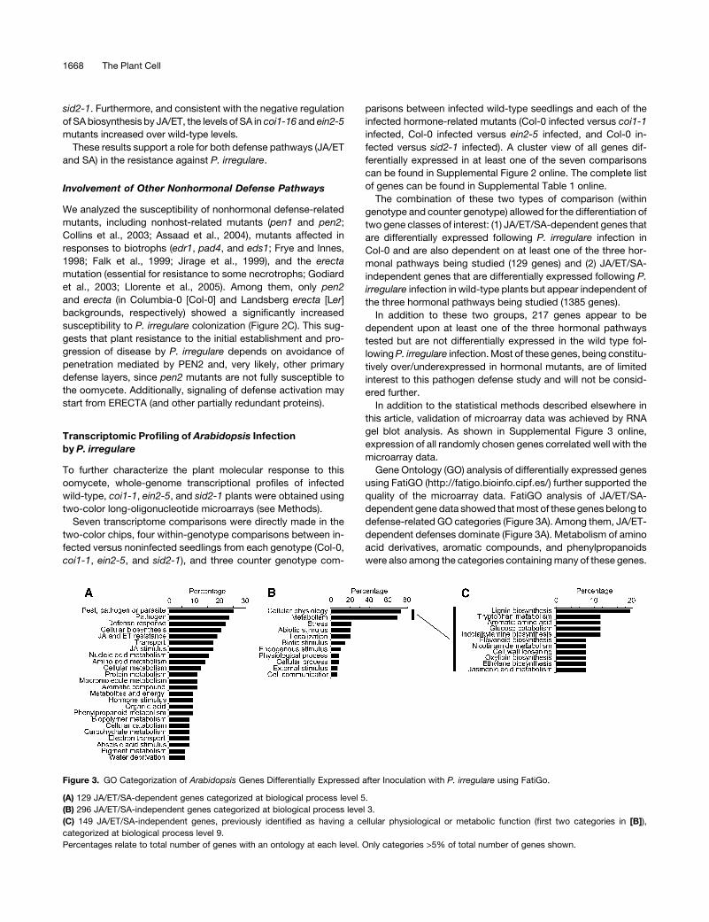
Gene Ontology (GO) analysis of differentially expressed genes
using FatiGO (http://fatigo.bioinfo.cipf.es/) further supported the
quality of the microarray data. FatiGO analysis of JA/ET/SA-
dependent gene data showed that most of these genes belong to
defense-related GO categories (Figure 3A). Among them, JA/ET-
dependent defenses dominate (Figure 3A). Metabolism of amino
acid derivatives, aromatic compounds, and phenylpropanoids
were also among the categories containing many of these genes.
Figure 3. GO Categorization of Arabidopsis Genes Differentially Expressed after Inoculation with P. irregulare using FatiGo.
(A) 129 JA/ET/SA-dependent genes categorized at biological process level 5.
(B) 296 JA/ET/SA-independent genes categorized at biological process level 3.
(C) 149 JA/ET/SA-independent genes, previously identified as having a cellular physiological or metabolic function (first two categories in [B]),
categorized at biological process level 9.
Percentages relate to total number of genes with an ontology at each level. Only categories >5% of total number of genes shown.
1668 The Plant Cell
Interestingly, many genes within this group (JA/ET/SA-depen-
dent genes) belong to two categories related to ABA (response to
ABA stimulus and response to water deprivation), suggesting a
role for ABA in the plant response to P. irregulare (Figure 3A).
Analysis of JA/ET/SA-independent genes showed that most of
these genes belong to two major categories: cellular physiological
process and metabolism (Figure 3B). Significantly, a deeper anal-
ysis of the genes within these two categories (using more detailed
GO levels) showed that the majority of the genes belong to met-
abolic processes involved in the biosynthesis of defensive sec-
ondary metabolites, such as lignin, indol-glucosinolates (Trp and
glucose derivatives), flavonoids, and nicotinamide (Figure 3C).
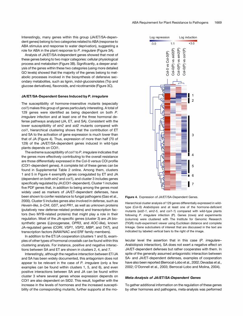
JA/ET/SA-Dependent Genes Induced by P. irregulare
The susceptibility of hormone-insensitive mutants (especially
coi1) makes this group of genes particularly interesting. A total of
129 genes were identified as being dependent on both P.
irregulare infection and at least one of the three hormonal de-
fense pathways analyzed (JA, ET, and SA). Consistent with the
lower susceptibility of ein2 and sid2 mutants compared with
coi1, hierarchical clustering shows that the contribution of ET
and SA to the activation of gene expression is much lower than
that of JA (Figure 4). Thus, expression of more than half (70 of
129) of the JA/ET/SA-dependent genes induced in wild-type
plants depends on COI1.
The extreme susceptibility of coi1 to P. irregulare indicates that
the genes more effectively contributing to the overall resistance
are those differentially expressed in the Col-0 versus COI profile
(COI1-dependent genes). A complete list of these genes can be
found in Supplemental Table 2 online. Among them, clusters
1 and 5 in Figure 4 exemplify genes coregulated by ET and JA
(dependent on both ein2 and coi1), and cluster 3 includes genes
specifically regulated by JA (COI1-dependent). Cluster 1 includes
five PDF genes that, in addition to being among the genes most
widely used as markers of JA/ET-dependent defenses, have
been shown to confer resistance to fungal pathogens (Gao et al.,
2000). Cluster 5 includes genes also involved in defense, such as
Hevein-like, b-CHI, GST, and PR1, as well as unknown proteins
(putatively new defense-related proteins) and transcription fac-
tors (two MYB-related proteins) that might play a role in their
regulation. Most of the JA-specific genes (cluster 3) are JA bio-
synthetic genes (Lipoxygenase, OPR3, and AOC-like), known
JA-regulated genes (CORI, VSP1, VSP2, MBP, and TAT), and
transcription factors (NAM/NAC and ERF family members).
In addition to the ET/JA cooperation (clusters 1 and 5), exam-
ples of other types of hormonal crosstalk can be found within this
clustering analysis. For instance, positive and negative interac-
tions between SA and ET are shown in clusters 2, 4, and 7.
Interestingly, although the negative interaction between ET/JA
and SA has been widely documented, this antagonism does not
appear to be relevant in the case of P. irregulare (only a few
examples can be found within clusters 1, 5, and 6), and even
positive interactions between SA and JA can be found within
cluster 3 where several genes whose expression depends on
COI1 are also dependent on SID2. This result, together with the
increase in the levels of hormones and the increased suscepti-
bility of the corresponding mutants, further supports at the mo-
lecular level the assertion that in this case (P. irregulare–
Arabidopsis interaction), SA does not exert a negative effect on
JA/ET-dependent defenses but rather cooperates with them. In
spite of the generally assumed antagonistic interaction between
SA- and JA/ET-dependent defenses, examples of cooperation
have also been reported (Berrocal-Lobo et al., 2002; Devadas et al.,
2002; O’Donnell et al., 2003; Berrocal-Lobo and Molina, 2004).
Meta-Analysis of JA/ET/SA-Dependent Genes
To gather additional information on the regulation of these genes
by other hormones and pathogens, meta-analysis was performed
Figure 4. Expression of JA/ET/SA-Dependent Genes.
Hierarchical cluster analysis of 129 genes differentially expressed in wild-
type (Col-0) Arabidopsis and at least one of the hormone-deficient
mutants (sid2-1, ein2-5, and coi1-1) compared with wild-type plants
following P. irregulare infection (P). Genes (rows) and experiments
(columns) were clustered with The Institute for Genomic Research
(TIGR) multi-experiment viewer using Euclidean distance and complete
linkage. Gene subclusters of interest that are discussed in the text are
indicated by labeled vertical bars to the right of the image.
ABA Requirement for Plant Resistance to Pathogens 1669

by clustering JA/ET/SA-dependent gene data obtained in this
work together with available expression data for these genes
following hormonal and pathogen treatments in the GENEVES-
TIGATOR database (www.genevestigator.ethz.ch/; Zimmermann
et al., 2004). Of the nine hormonal treatment experiments available
(ABA, 1-aminocyclopropane-1-carboxylic acid, brassinolide, ET,
gibberellic acid, indole-3-acetic acid, methyl jasmonate [MeJA],
SA, and Zeatin), MeJA clustered most closely to the wild-type re-
sponse to P. irregulare infection, confirming that JA is the major
signal activating responses to this oomycete (Figure 5; see
Supplemental Table 3 online). In accordance with this, the COI1-
dependent profile is, in general, the opposite to the MeJA profile,
with only minor differences likely due to the availability of only one
time point of the MeJA treatment, indicating that most (if not all) of
the JA-activated responses to P. irregulare are mediated by COI1.
Interestingly, besides JA, the second signal more closely
related to the pattern of gene activation by P. irregulare in wild-
type plants is ABA. More than one-third (39 of 119) of the genes
upregulated by P. irregulare are also upregulated by the hormone,
suggesting that it may be an important signal in the activation
of defenses against this oomycete. Furthermore, approximately
half of these genes (19 of 39 ABA-regulated genes) are also reg-
ulated by JA, suggesting that either both signals cooperate or
one hormone precedes the other in the activation of this set of
genes.
In addition to the ABA and JA signals, P. irregulare’s infection
profile also clusters with profiles of responses to herbivory (Pieris
rapae) and a necrotrophic fungal pathogen (B. cinerea), indicat-
ing that the hormone-dependent response to P. irregulare is
similar to that of necrotrophic pathogens and chewing insects.
By contrast, profiles from two biotrophic fungal pathogens
(Erysiphe orontii and Erysiphe cichoracearum) cluster together,
distant from P. irregulare, indicating that plants regulate the
expression of most of the genes in this set (JA/ET/SA-dependent
Figure 5. Meta-Analysis of JA/ET/SA-Dependent Gene Data.
Cluster of JA/ET/SA-dependent genes with available expression data from hormonal and pathogen treatments within the GENEVESTIGATOR database
(expression data available for only 107 of the 129 genes; Zimmermann et al., 2004). Genes (rows) and experiments (columns) were clustered with the
TIGR multi-experiment viewer using Euclidean distance and complete linkage. Gene subclusters of interest that are discussed in the text are indicated
by labeled vertical bars. Overrepresented cis-elements within each gene cluster were identified by TAIR motif analysis, Botany Beowulf Cluster
Promomer, and Gibbs motif sampler. Statistical significance (P value from binomial distribution) is shown in parenthesis where possible.
1670 The Plant Cell
induced by P. irregulare) in a different way (in many cases the
opposite) depending on the lifestyle of the pathogen. This result
fully agrees with the general view that defenses against biotrophs
and necrotrophs are essentially antagonistic.
Identification of cis-Regulatory Elements
To gain further insight into the transcriptional regulation of JA/ET/
SA-dependent responses to P. irregulare, we analyzed the
presence of putative cis-regulatory elements and trans-acting
factors responsible for the transcriptional regulation of the genes
in each gene cluster by searching for statistically overrepre-
sented sequences in their promoters. Using motif analysis (The
Arabidopsis Information Resource [TAIR]), Promomer (http://
bbc.botany.utoronto.ca), and motif sampler (Thijs et al., 2002),
several statistically significant candidate cis-regulatory elements
were found for each cluster (only known boxes are shown; Figure
5). Consistent with the regulation by ET and JA of genes in cluster
1a (see Supplemental Table 3 online), the GCC-box was the
predominant element found in this cluster. This element is
specifically recognized by members of the ERF family of tran-
scription factors, some of which are well-known defense regu-
lators (Lorenzo et al., 2003; Gutterson and Reuber, 2004). Also
consistent with the regulation of clusters 2 and 4 by JA, the
G-box and related elements (T/G-box) were the most signifi-
cantly overrepresented sequences. These elements are regulated
by basic domain/leucine zipper or MYC transcription factors, some
of which are also involved in defense (Siberil et al., 2001; Boter
et al., 2004; Lorenzo et al., 2004; Nickstadt et al., 2004). Inter-
estingly, the ABA response element (ABRE) is overrepresented in
clusters 2 and 4 where most of the ABA-regulated genes con-
centrate. In addition to ABRE, another cis-element related to
dehydration was identified, the dehydration-responsive element.
The presence of the ABRE and dehydration-responsive elements
further supports an important role for ABA and dehydration in the
regulation of responses to P. irregulare. Additional boxes with
likely functional importance in the regulation of these genes are
an H-box– and a P-box–related element (both MYB-related
binding sites) and a W-box (Figure 5). Candidate transcription
factors regulating these cis-elements can be identified within the
genes induced by the oomycete.
JA/ET/SA-Independent Genes Induced by P. irregulare
It is noteworthy that the vast majority of differentially expressed
genes (1385) are independent of the three hormones studied. GO
analysis showed that the majority of the genes with a GO
description are related to the metabolism of defensive com-
pounds (Figure 3B), indicating that many of these inducible
defenses may be activated by other, so far unknown, processes/
signals.
To further understand the role of these genes in plant defense
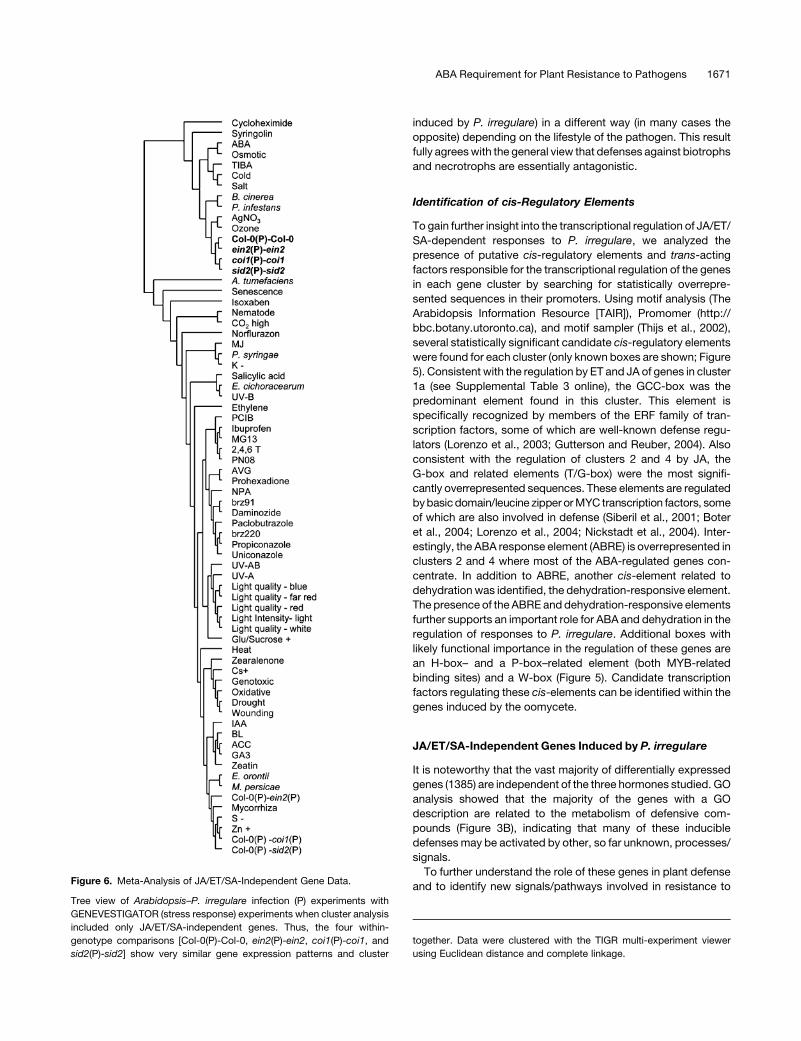
and to identify new signals/pathways involved in resistance toFigure 6. Meta-Analysis of JA/ET/SA-Independent Gene Data.
Tree view of Arabidopsis–P. irregulare infection (P) experiments with
GENEVESTIGATOR (stress response) experiments when cluster analysis
included only JA/ET/SA-independent genes. Thus, the four within-
genotype comparisons [Col-0(P)-Col-0, ein2(P)-ein2, coi1(P)-coi1, and
sid2(P)-sid2] show very similar gene expression patterns and cluster
together. Data were clustered with the TIGR multi-experiment viewer
using Euclidean distance and complete linkage.
ABA Requirement for Plant Resistance to Pathogens 1671

P. irregulare, meta-analysis was performed by clustering JA/ET/
SA-independent gene data obtained in this work together with
available expression data from the GENEVESTIGATOR data-
base. A full view of the cluster including all genes can be found in
Supplemental Figure 4 online. Figure 6 shows a clustered tree
view of the experiments, and Figure 7 shows a full cluster view of
the most related profiles to P. irregulare expression patterns. To
facilitate the analysis, only genes with a false discovery rate <1%
and log ratio >1.5 are shown.
Consistent with the JA/ET/SA-independent nature of the reg-
ulation of these genes, the four within-genotype profiles cluster
tightly together, showing that once JA/ET/SA-dependent genes
are eliminated, the response of all four genotypes is similar,
further supporting the quality of the data (Figures 6 and 7).
Similar to JA/ET/SA-dependent genes, P. irregulare response
profiles clustered with the necrotroph B. cinerea and the only
other oomycete (P. infestans) profile included within the analysis
and show little similarity to the profiles of responses to biotrophs.
This result is consistent with our previous conclusions from the
genetic analysis and further supports the plant’s necrotrophic-
like response to this oomycete (Figure 6).
In addition to B. cinerea and P. infestans, several profiles of
responses to ABA and abiotic stresses, such as those induced
by ozone and osmotic and salt treatments, clustered within this
group (Figures 6 and 7). In fact, most of the genes up- or down-
regulated by P. irregulare infection are also regulated by these
stresses (Figure 7). For instance, clusters 1 and 3 (a, b, c, and d)
in Figure 7, representing >90% of the genes analyzed, are com-
monly regulated by most of the stresses in the cluster.
Interestingly, the only hormone profile clustering close to P.
irregulare (and B. cinerea and P. infestans) profiles is ABA
treatment. In fact, when analyzed in more detail, ABA was found
to be the main signal regulating the expression of many of the P.
irregulare–responsive genes, particularly those included within
clusters 3b and 3d, which represent ;30% of the genes in this
analysis. These data support the previous observation using JA/
ET/SA-dependent genes that ABA may play a role in the activa-
tion of effective defenses against P. irregulare.
While the vast majority of gene clusters are nonspecific for P.
irregulare infection, clusters specifically regulated by this oomy-
cete can be identified (cluster 2, Figure 7; see Supplemental
Table 4 online). The specificity of the regulation of these genes by
P. irregulare suggests that they may have a defined role in the
determination of particular responses to this oomycete.
Figure 7. Meta-Analysis of JA/ET/SA-Independent Gene Data.
Cluster view of 296 JA/ET/SA-independent genes (selected with a false
discovery rate <1% and log ratio >1.5) with available expression data
from the most closely related experiments within the GENEVESTIGATOR
database (Zimmermann et al., 2004). Genes (rows) and experiments
(columns) were clustered with the TIGR multi-experiment viewer using
the Pearsons uncentered distance and complete linkage. Gene subclus-
ters of interest that are discussed in the text are indicated by labeled
vertical bars. Overrepresented (black lettering) and underrepresented
(green lettering) cis-elements within each gene cluster were identified by
TAIR motif analysis, Botany Beowulf Cluster Promomer, and Gibbs motif
sampler. WBS, WRKY binding site; ARE, anthocyanin regulatory ele-
ment. Statistical significance (z-score significance [WRKY binding site
and ARE] and P value from binomial distribution [all others]) is shown in
parenthesis.
1672 The Plant Cell

As with JA/ET/SA-dependent genes, a search for sequences
statistically overrepresented within promoters of JA/ET/SA-
independent genes identified several candidate cis-regulatory
elements (Figure 7). Among them, two are associated with ABA-
regulated responses, ABRE and RY (an element recognized by
B3 transcription factors, including ABI3, that regulate seed-
specific promoters; Vicente-Carbajosa and Carbonero, 2005). In
addition, WRKY binding sites seem also to be particularly rele-
vant since they appeared overrepresented in several clusters (2,
3a, and 3c). Candidate transcription factors recognizing these
elements have been found within each cluster, and the corre-
sponding KO mutants are being analyzed for susceptibility/
resistance to P. irregulare.
ABA Is Required for Plant Defense
To further discriminate if, as suggested above, ABA may be
required for overall plant resistance, ABA hormone levels were
measured in wild-type and JA/ET/SA/ABA-related mutants after
P. irregulare infection, and ABA-deficient mutants were tested for
their susceptibility to P. irregulare. As shown in Figure 8A, ABA
levels increased rapidly in the wild type and all mutants tested
(except in aba2-12) after infection, further supporting a role for
this hormone in activation of defenses. Moreover, all three ABA
mutants tested, either impaired in ABA biosynthesis (aao3-2 and
aba2-12) or insensitive to ABA (abi4), showed an increased
susceptibility to P. irregulare compared with the wild-type back-
ground, indicating that ABA is a positive signal involved in the
activation of effective defenses against this pathogen (Figure 8B).
Figure 8. Role of ABA and Callose in Resistance to P. irregulare.
(A) Endogenous ABA levels (ng/g fresh weight) in Arabidopsis wild-type
(Col-0) and JA/ET/SA/ABA-related mutant plants (coi1-16, ein2-5, sid2-1,
and aba2-12) after infection with P. irregulare for 0, 6, and 12 h. Seedlings
were grown on Johnson’s media plates for 7 d prior to infection. Values are
means 6 SE of three independent replicate experiments.
(B) Susceptibility of Arabidopsis ABA-related mutants (biosynthesis,
aba2-12 and aao3-2; insensitive, abi4-1) and a callose-deficient mutant
(pmr4) to P. irregulare compared with the wild type (Col-0). Mean disease
area (mm2) per infected leaf 24 h after infection. Asterisks indicate
significant difference (P < 0.05) from Col-0 using the Student’s t test.
(C) B. cinerea (open bars) and A. brassicicola (closed bars) infection of
Arabidopsis ABA-related mutants (biosynthesis, aba2-12 and aao3-2;
insensitive, abi4-1) compared with the wild type (Col-0). Mean disease
area (mm2) per infected leaf either 4 (B. cinerea) or 10 (A. brassicicola) d
after infection. Asterisks indicate significant difference (P < 0.05) from
Col-0 using the Student’s t test.
(D) to (G) Callose deposition in Arabidopsis following infection with P.
irregulare.
(D) Localized callose deposition (apposition [app]) around site of path-
ogen contact. Plant cell wall (cw) and P. irregulare hyphae (hy) are also
indicated. Bar ¼ 5 mm.
(E) Normal callose deposition in wild-type (Col-0) Arabidopsis following
P. irregulare infection (haustoria-like infection structure [hl]) encom-
passed the entire plant cell wall (cw). Bar ¼ 30 mm.
(F) Callose deposition in the Arabidopsis ABA mutant aao3-2 following P.
irregulare infection (haustoria-like infection structure [hl]) encompassed
the entire plant cell wall (cw). Bar ¼ 15 mm.
(G) Spotted callose deposition (sp) within the Arabidopsis callose mutant
(pmr4) cell wall (cw) following infection with P. irregulare and production
of haustoria-like infection structure (hl). Bar ¼ 30 mm.
ABA Requirement for Plant Resistance to Pathogens 1673

Two additional necrotrophs (Alternaria brassicicola and to B.
cinerea) have been tested to further understand the breadth of
ABA’s role in pathogen resistance. As shown in Figure 8C, all
three ABA-related mutants tested were more susceptible to A.
brassicicola but (surprisingly) more resistant to B. cinerea.
These results demonstrate that although the role of ABA in
pathogen resistance is not restricted to P. irregulare, ABA is not a
positive signal for plant defense against all necrotrophs. Thus,
other properties of the pathogenic infection may be determinant
of the role of ABA in each plant–pathogen interaction, rather than
the pathogen’s assigned lifestyle.
ABA has been previously proposed to play a role in priming of
callose biosynthesis after pathogen recognition, which suggests
a putative mechanism explaining the role of ABA in defense
activation (Ton and Mauch-Mani, 2004). Callose (b-1,3-glucan) is
normally associated with cell wall appositions (papillae) and has
been suggested to be a fortifying agent deposited rapidly after
pathogen recognition to inhibit pathogen penetration of the cell
(Aist, 1976; Jacobs et al., 2003; Ton and Mauch-Mani, 2004). To
ascertain the role of callose formation in the case of P. irregulare,
we studied the deposition of this compound and its effect on
resistance after inoculation with the oomycete of wild-type plants
and callose-deficient mutants (pmr4).
Aniline blue staining for 1/3-b-D-glucan (callose) suggested
that the deposition of this compound occurred in recently
infected wild-type cells. This was infrequently only concentrated
in appositions or papilla around the area of penetration (Figure
8D; see Supplemental Figure 1F online), more normally being
seen to encompass the entire cell wall and possibly the walls of
adjoining cells (Figure 8E). By contrast, pmr4 mutants, impaired
in callose biosynthesis, deposited callose only in specks around
the cell wall (Figure 8G). Moreover, pmr4 mutants showed an
enhanced susceptibility to the oomycete compared with wild-
type plants (Figure 8B), thus indicating that callose deposition
plays a role in defense against P. irregulare.
Taken altogether, these results suggest that ABA may exert its
role, at least in part, through priming of callose production (Ton
and Mauch-Mani, 2004). Nevertheless, the susceptibility of pmr4
mutants was lower than that of ABA-deficient mutants, suggest-
ing that priming of callose is not the only defense mechanism
regulated by ABA. Moreover, aniline blue staining demonstrated
that whereas no biosynthesis of callose was observed in pmr4
after infection with P. irregulare (Figure 8G), the level of callose
production in ABA-deficient mutants was indistinguishable from
that of the wild type (Figure 8F). Therefore, in addition to its role in
callose production, ABA-dependent resistance has to be exerted
through a callose-independent mechanism.
To further understand this mechanism, microarray profiles of
ABA biosynthesis mutants (aba2-12) were compared with those
of wild-type plants following the infection of both with P. irreg-
ulare. This identified 38 ABA-dependent genes among those
regulated by the pathogen in wild-type plants (Figure 9; see
Supplemental Table 5 online). Meta-analysis showed that within
this ABA-dependent group, two major classes of genes could be
identified: ABA-regulated genes and JA-regulated genes.
These results reinforce the previous conclusion that ABA is an
important signal in the activation of plant defenses through the
transcriptional reprogramming of the cell. Moreover, the defi-
ciency in the activation of JA-induced genes in aba2-12 indicates
that ABA either precedes or cooperates with JA in the activation
of this set of defense genes.
To discriminate between these two possibilities, JA hormone
levels as well as a JA precursor (12-oxo-phytodienoic acid) were
measured in wild-type plants and aba2-12 mutants after P.
irregulare infection. As shown in Figure 10, the increase in JA (or
its precursor) following infection (12 h after infection) is much
lower in aba2-12 mutants than in wild-type plants. This indicates
that ABA synthesis is required for JA production and the activa-
tion of plant defenses against P. irregulare.
Figure 9. Meta-Analysis of ABA-Dependent Gene Data.
Cluster view of 38 aba2-12–dependent genes also induced/repressed by
P. irregulare in wild-type (Col-0) plants, with available expression data
from JA- and ABA-treated experiments within the GENEVESTIGATOR
database (Zimmermann et al., 2004). Genes (rows) were clustered with
the TIGR multi-experiment viewer using the Pearsons uncentered dis-
tance and complete linkage. Gene subclusters of interest that are
discussed in the text are indicated by labeled vertical bars.
1674 The Plant Cell

DISCUSSION
The study of Arabidopsis–P. irregulare host–pathogen interac-
tion reported here highlights the importance of broadening our
understanding of the interaction of diverse pathogens with this
important model species.
Microscopy analysis has shown that P. irregulare infection of
Arabidopsis has neither purely typical necrotrophic nor biotro-
phic characteristics. The infection process starts with the pro-
duction of appressoria and haustoria-like structures. Compatible
host–pathogen interactions show further hyphal ingression to be
primarily intracellular, moving more rapidly through the vascula-
ture and invading all tissues. Although no lytic enzymes or toxins
have been detected, this progressive invasion ultimately pro-
vokes plant cell death.
Analysis of defense-related mutants has shown that Arabi-
dopsis resistance to the establishment and progression of dis-
ease by P. irregulare depends initially on avoidance of
penetration, mediated by PEN2 and, very likely, other primary
defense layers, since pen2 mutants are not fully susceptible to
the pathogen. These results are in line with previous reports
demonstrating that PEN2 (encoding a glucosyl hydrolase) con-
trols the ingress of a broader range of pathogens, including both
biotrophs and necrotrophs, than other penetration proteins
(Lipka et al., 2005; Nurnberger and Lipka, 2005).
Recognition and signaling of defense activation may start
from ERECTA (and other partially redundant proteins, since
erecta mutants are not fully susceptible). Due to its structural
similarity to receptor-like kinases, ERECTA has been suggested
to recognize a pathogen-associated molecular pattern (Godiard
et al., 2003; Llorente et al., 2005). The susceptibility of erecta
mutants suggests the likely importance of this receptor-like
kinase in the recognition of P. irregulare.
Role of JA, ET, and SA in Defense against P. irregulare
While P. irregulare has biotrophic-like infection structures, the
genetic and genomic characterization of defense-related hor-
mone-signaling mutants has shown that the plant’s response to
P. irregulare is more similar to responses against necrotrophic
pathogens. Thus, expression profiles in response to P. irregulare
are closely related to those of B. cinerea and different from those
of biotrophic pathogens such as Erisyphe spp.
In accordance with this and as shown by the extreme suscep-
tibility of the JA-insensitive coi1 mutants, the main signal acti-
vating defenses is JA, the role of which in necrotrophic pathogen
defense has been widely documented (Rojo et al., 2003; Reymond
et al., 2004; Lorenzo and Solano, 2005). Additionally, although of
lesser importance than JA, ET and SA play significant roles in
disease resistance against P. irregulare. Cooperation of SA with
JA/ET-dependent defenses has been previously reported
(Berrocal-Lobo et al., 2002; Devadas et al., 2002; O’Donnell et al.,
2003; Berrocal-Lobo and Molina, 2004). However, these results
contrast with the generally assumed roles of SA and JA/ET in the
activation of two independent and antagonistic defense path-
ways and highlight the flaws inherent within oversimplified
models. Pathogen lifestyles are not often readily attributable to
purely biotrophic or necrotrophic classes. Thus, in our view,
depending on the particular characteristics of each pathogen,
the complex regulatory network involving JA, ET, and SA will
establish the required interactions to fine-tune the appropriate
defenses. Thus, cooperation or antagonism may be regulated
(and has been selected through evolution) to adapt to the
specific pathogen.
Transcriptomic analysis and meta-analysis confirmed these
conclusions showing that JA regulates more than half the JA/ET/
SA-dependent genes, whereas the contribution of ET and SA is
more modest.
It is noteworthy, however, that profiles of responses to hemi-
biotrophic bacteria (both virulent and avirulent strains of Pseu-
domonas syringae pv maculicola ES4326) are very closely related
to the P. irregulare–wild type profile, being particularly evident in
the case of genes regulated by JA or ABA (clusters 2a and 4
in Figure 5). Interestingly, this particular P. syringae strain pro-
duces a JA analog (coronatine) as a mechanism of pathogenicity
Figure 10. JA and JA-Precursor Levels in Wild-Type and ABA Mutant
Plants.
Endogenous JA and 12-oxo-phytodienoic acid (OPDA) (ng/g fresh
weight) levels in Arabidopsis wild-type (Col-0) and ABA mutant (aba2-
12) plants following infection with P. irregulare for 0, 6, and 12 h. Seed-
lings were grown on Johnson’s media plates for 7 d prior to infection.
Values are means 6 SE of three independent replicate experiments.
ABA Requirement for Plant Resistance to Pathogens 1675

(Hendrickson et al., 2000). The activation of JA-dependent re-
sponses by coronatine is supposed to counteract SA-dependent
defenses facilitating bacterial infection (Cui et al., 2005). Coro-
natine also promotes stomatal reopening, favoring bacterial entry
into the plant (Melotto et al., 2006). Thus, although P. irregulare
and P. syringae have different lifestyles and promote very differ-
ent defense responses, they are coincident in this particular set of
genes likely because of the coronatine mimicry of JA. By analogy,
the fact that ABA regulates a set of these genes (i.e., cluster 2a)
suggests that the coronatine mimicry of JA may not be the only
mechanism inhibiting SA-dependent defenses by P. syringae and
that ABA might also play a role. In fact, this is consistent with
previous reports showing that ABA-deficient Arabidopsis mu-
tants have higher levels of salicylate-induced genes and are more
resistant to P. syringae, whereas ABA treatment promotes the
opposite behavior (Mohr and Cahill, 2003; Thaler and Bostock,
2004). The SA-dependent inhibition of these genes is evident
from their upregulated expression in sid2 mutants infected with
P. irregulare (see clusters 2a and 3 in Figure 5). In line with our
results, this ABA/SA antagonism could be explained by an in-
direct effect based on the ABA induction of JA biosynthesis.
JA-, ET-, and SA-Independent Defenses
Transcriptome analysis indicated that the majority of genes
differentially expressed following P. irregulare infection were
independent of the three defense hormones, JA, ET, and SA, but
more related to secondary metabolism (i.e., biosynthesis of
defensive compounds), highlighting the importance of these
metabolites in defense. The striking overlap of expression pat-
terns between JA/ET/SA-independent genes and profiles from
abiotic stress (osmotic, salt, and ozone) may suggest that plant
cells use similar mechanisms (the same type of genes) to cope
with different stress situations or that some of these stresses are
part of the infection process. In fact, among abiotic stresses,
ozone exposure, which promotes plant cell death, is the closest
profile to P. irregulare, suggesting that many of the common
genes in this analysis maybe associated with the cell damage
accompanying the infection. In addition, Pythium has been
shown to be a vascular pathogen promoting clogging of the
vasculature and wilting of the plant (Martin, 1995; Kamoun,
2003). Thus, it should not be surprising that osmotic/salt-related
stress profiles and ABA profiles are closely related to P. irregulare
infection profiles. A similar hypothesis has been proposed in the
case of wounding, where dehydration is a major component of
the response (Reymond et al., 2000, 2004). The fact that B.
cinerea and P. infestans profiles are also included within this
experimental cluster suggests that the abiotic and cell death
components of the stress induced after pathogen infection is not
exclusive to P. irregulare but rather common to other pathogens.
Role of ABA in Pathogen Defense
The role of ABA in pathogen defense is, so far, poorly understood
and even controversial. Whereas several reports have shown an
inverse correlation between ABA levels and resistance to path-
ogens with different lifestyles in several plant species, others
have suggested a positive role of this hormone in activation of
defense gene expression and pathogen resistance (Mauch-Mani
and Mauch, 2005). In the case of bacterial leaf pathogens, ABA
plays an important role in the activation of stomatal closure that,
as part of the innate immune system, represents a barrier against
bacterial infection (Melotto et al., 2006). Thus, ABA-deficient
mutants are more susceptible to P. syringae infection. However,
it has also been shown that (1) ABA increases susceptibility by
counteracting SA-dependent defenses, and (2) ABA-dependent
priming of callose biosynthesis promotes enhanced resistance
to some pathogens (Ton and Mauch-Mani, 2004). Since callose
has been shown to have a detrimental effect on SA-dependent
defenses (Nishimura et al., 2003), both issues (1 and 2) support
that ABA should be expected to have a negative effect on
resistance against biotrophs. However, the effect of ABA on
resistance to necrotrophs remains unclear.
Interestingly, meta-analysis of transcriptomic data showed
that ABA upregulated approximately one-third of the plant genes
induced by P. irregulare, suggesting an important role of this hor-
mone in defense activation. This hypothesis contrasts with pre-
vious analysis showing that induction of some ET/JA-regulated
defense genes, such as PDF1.2, HEL, and b-CHI, is prevented by
ABA (Anderson et al., 2004). However, the genomic view in our
work shows that the ABA repression described by Anderson
et al. (2004) affects only a reduced group of ET/JA-regulated
genes, whereas the major effect of ABA is the opposite, activating
many ABA-specific and ABA/JA-related defense genes.
The indication that ABA is a signal required for necrotrophic
pathogen resistance was substantiated by the increase in ABA
levels after infection and when analysis of ABA-deficient or -insen-
sitive mutants showed them to be more sensitive to P. irregulare and
A. brassicicola than wild-type plants. In accordance with these
results, several groups have reported mutants (or transgenic plants)
with altered resistance to both pathogens and ABA-dependent
abiotic stresses, further supporting our hypothesis (Mengiste et al.,
2003; Chini et al., 2004). Moreover, treatment with ABA protects
Arabidopsis plants against A. brassicicola and Plectosphaerella
cucumerina (Ton and Mauch-Mani, 2004), further indicating that
ABA is necessary and sufficient to enhance defense responses
against several necrotrophic pathogens.
However, ABA is not a positive signal for plant defense against
all necrotrophs since it has a negative effect on plant resistance
against B. cinerea and Fusarium oxysporum (Audenaert et al.,
2002; Anderson et al., 2004; Abuqamar et al., 2006; this work).
Thus, other properties of the pathogenic infection, rather than the
pathogen lifestyle, have to be determinant of the role of ABA in
each plant-pathogen interaction.
The increased susceptibility of pmr4 mutants, impaired in
callose biosynthesis, suggests that ABA may exert its role, at
least in part, through priming of callose production (Ton and
Mauch-Mani, 2004). Nevertheless, ABA-deficient mutants do not
show a significant defect in callose production compared with
wild-type plants in response to P. irregulare infection and still
show higher susceptibility to this oomycete than pmr4 mutants.
These results strongly suggest that priming of callose is not the
only defense mechanism regulated by ABA. In support of this
hypothesis, microarray data and measurements of ABA levels in
aba2-12 biosynthetic mutants demonstrated that ABA is re-
quired for JA biosynthesis and JA-dependent defense gene
1676 The Plant Cell

expression after infection with P. irregulare. Thus, similar to the
proposed wound response mechanism in solanaceous plants
(Hildmann et al., 1992; Pena-Cortes et al., 1995; Leon et al.,
2001), ABA affects JA biosynthesis, suggesting that it precedes
JA in the activation of defenses against this oomycete.
In summary, our results indicate that ABA is a component of
the signaling network activating plant defenses necessary for
resistance against some (but not all) necrotrophic pathogens.
ABA enhances defenses through at least two independent
mechanisms: callose priming and regulation of defense gene
expression through activation of JA biosynthesis. The differential
role of the hormone within different plant–pathogen interactions
suggests that ABA levels may be key to the fine-tuning of plant
defenses against particular pathogens, a modulation system
previously suggested for ET (Pierik et al., 2006).
METHODS
Genetic Backgrounds of Material
Pythium irregulare was identified by microscopy and the genotype
confirmed by sequencing of the internal transcribed spacer (ITS) re-
gion using primers ITS1 (59-TCCGTAGGTGAACCTGCGG-39) and ITS4
(59-TCCTCCGCTTATTGATATGC-39) (Kageyama et al., 1997).
With the exception of Ler, all mutants were in a Col-0 background.
Seed Sterilization and Growth
Seeds were surface sterilized with 75% bleach prior to plating on
sterile Johnson’s growth medium as described by Lorenzo et al. (2003).
Homozygous coi1 was selected using 50 mM JA amended Johnson’s
media. In vitro growth of seedlings was at 218C with a photoperiod of 16 h
light/8 h dark. While seedlings for soil inoculation were transferred at 7 d
old, those for transcriptome analysis were transferred to three concentric
rings with diameters measuring 55, 69, and 90 mm on Murashige and
Skoog media at 4 d old. In vitro growth conditions remained as previously
described, but soil-transferred seedlings were moved to a phytochamber
with a 12-h-light/12-h-dark photoperiod, 218C, and 70% RH. To encour-
age a fuller rosette, ABA mutants were grown in a phytochamber with 8 h
light/16 h dark, 218C, and 70% RH.
P. irregulare Growth Conditions
P. irregulare was cultured on potato dextrose agar (Oxoid CM0139) at
218C and plugs taken from the growing edge stored underwater at 48C.
Fresh cultures from these stocks were used for inoculation of soil-grown
plants and transcriptome seedlings. All plugs for inoculation were taken
from the growing edge of a fresh P. irregulare colony. Periodically
(approximately every 6 months), fresh 48C stocks were established using
infected leaf tissue to grow an aseptic colony.
Disease Inoculation and Assessment of Soil-Grown Plants
Four weeks after being transferred to soil, plants were inoculated. A single
1.5-mm-diameter agar plug, taken from a fresh-growing colony edge,
was placed centrally on the upper surface of each of two lower rosette
leaves per plant. This was then covered with 50 to 100 mL of sterilized
distilled water. Clear plastic lids covered the inoculated plants to maintain
100% humidity, and infections were allowed to progress for either 24 or
48 h. Leaves were collected and immediately either stained with Trypan
blue (Koch and Slusarenko, 1990) or Analine blue (Dietrich et al., 1994).
Leaves were mounted in glycerol solutions and Leica DMR and Leica
MZFLIII microscopes with fluorescence capabilities used for the exam-
ination of infected tissue.
Two diameters, perpendicular to each other, were measured for each
disease lesion and a mean obtained. The production of runner hyphae
and concomitant establishment of distinct lesions per single inoculum
necessitated the conversion of lesion radii to area, thereby enabling total
disease areas per leaf to be calculated. Thus, disease area per leaf was
calculated as the sum of all disease lesion areas:
S Areai; Areai ¼ pR1 þ R2
2
� �2
All infections of Arabidopsis thaliana mutant genotypes were repeated
at least three times with similar results. Between six and nine plants of
each genotype were infected per replicate experiment.
Hormone Analysis
Seeds were plated on Johnson’s media in a grid formation so that each
was 1 cm from the next. Seed sterilization and growth conditions were as
described above. After 7 d, plates were inoculated with P. irregulare
mycelium taken from the growing edge of a fresh colony. Multiple points
of inoculation that were equidistant from each seedling ensured seedlings
were infected at the same time. Tissue was collected 0, 6, and 12 h after
infection (0, 18, and 24 h after inoculation, respectively), immediately
weighed, and frozen until hormone quantification. Pooled whole-plant
samples (n ¼ 3) containing 100 mg of tissue were homogenized, de-
rivatized, vapor phase extracted, and analyzed by isobutane chemical-
ionization gas chromatography–mass spectrometryas previously described
(Schmelz et al., 2004). The initial extraction solution was modified to
contain a 62:100:1 ratio of H20:1-propanol:concentrated HCl.
Microarray Hybridization and Analysis
Seedling Inoculation and Tissue Collection
Three days after seedling transfer, MS plates were centrally inoculated
with a 4.5-mm P. irregulare–containing agar plug. From the central point,
growth of the pathogen took 24 h to reach the first ring of seedlings and
48 h to reach the outer ring. Thus, plants growing on the inner ring had
been in contact with P. irregulare for ;24 h and the middle ring for ;10 h
before the outer ring plants were infected. Whole seedlings from tran-
scriptome plates were harvested and immediately frozen when P. irregulare
hyphae infected the outer ring of plants.
Four Arabidopsis genotypes were included in the initial transcriptome
analysis, comprising the wild type (Col-0) and three mutants (coi1-1, ein2-5,
and sid2-1). The four genotypes were represented in each independent
plate, divided into four equal sectors each containing 18 seedlings equally
divided between the three rings. Nine replicate experiments were done on
different days, each with an identical, but noninoculated, control plate.
Additional transcriptome analyses of aba2-12 mutant plants were done
identically to the initial analysis although with only one of the four plate
sectors occupied.
RNA Gel Blot Analysis
RNA was extracted from the nine individual experiments and then pooled,
in equal proportions, into three groups of three. RNAwiz was used to
extract the RNA from frozen tissue according to the manufacturer’s
instructions (Ambion). Following purification with RNeasy (Qiagen), RNAs
were subjected to electrophoresis on 1.5% formaldehyde/agarose gels
and blotted to Hybond Nþ membranes (Amersham). All probes were
labeled with 50 mCi of [a-32P]dCTP. Blots were exposed for 12 to 24 h on a
PhosphorImager screen (Molecular Dynamics).
ABA Requirement for Plant Resistance to Pathogens 1677

RNA Quantification and Quality Determination
RNA was quantified with a NanoDrop ND-100 spectrophotometer (Nano-
Drop Technologies). RNA quality was assessed with a 2100 Bioanalyzer
from Agilent Technologies.
RNA Amplification and Labeling
RNA was amplified with the MessageAmp aRNA amplification kit from
Ambion following the instruction manual. To allow later labeling with Cy
fluorophores, aminoallyl UTP (Ambion) was added to the mix of the T7
RNA polymerase-driven aRNA amplification reaction. The amount and
quality of aRNA obtained was assessed as before. The aminoallyl-labeled
aRNA (10 mg) was incubated in 1 M Na2CO3 with 8 nmol of dye
monofunctional NHS ester (Cy3/Cy5) RPN 5661 (Amersham Biosciences)
at room temperature in the dark for 1 h. Then, 35 mL of 0.1 M sodium
acetate, pH 5.2, was added and incubated for a further 5 min in the dark.
The Cy-labeled aRNA was purified with the Megaclear kit from Ambion
and measured with the Nanodrop ND-100 spectrophotometer.
Hybridization
Three biological replicates were independently hybridized for each tran-
scriptomic comparison.
Microarray slides were composed of synthetic 70-mer oligonucleotides
from the Operon Arabidopsis Genome Oligo Set Version 1.0 (Qiagen)
spotted on aminosilane-coated slides (Telechem) by the University of
Arizona. Slides were rehydrated and UV cross-linked according to the
supplier’s website (http://ag.arizona.edu/microarray/methods.html). The
slides were then washed twice for 2 min in 0,1% SDS and in ethanol for
30 s. Arrays were drained with a 2000-rpm spin for 2 min. Slides were
prehybridized in 63 SSC, 0.5% SDS (w/v), and 1% BSA (w/v) at 428C for
1 h, followed by five rinses with milliQ water. Excess water was drained
with a 2000-rpm spin for 2 min.
For the hybridization, equal amounts of dye of each aRNA labeled with
either Cy3 or Cy5, ranging from 200 to 300 pmol, were mixed with 20 ug of
poly(A) and 20 mg of yeast tRNA (Sigma-Aldrich) in a volume of 9 mL. To
this volume, 1 mL of RNA fragmentation buffer was added (RNA frag-
mentation reagents; Ambion) and after 15 min at 708C, 1 mL of stop so-
lution. Formamide, 203 SSC, 503 Denhardt’s, and 20% SDS were added
to a final concentration of 50% formamide, 63 SSC, 53 Denhardt’s, and
0.5% SDS. This mix was boiled for 3 min at 958C and then added to the
prehybridized slide. Hybridization took place overnight at 378C in a hy-
bridization chamber. Arrays were then washed for 5 min at 378C in 0.53
SSC and 0.1% SDS, twice for 5 min at room temperature (218C) with 0.53
SSC and 0.1% SDS, three times with 0.53 SSC at room temperature, and
5 min with 0.13 SSC. The slides were then drained with a 2000-rpm spin
for 2 min. The slides were stored in darkness until they were scanned.
The scanning was done with a GenePix 400B scanner (Molecular De-
vices) at 10-mm resolution. The images were quantified with GenePix Pro 5.1.
Images from Cy3 and Cy5 channels were equilibrated and captured
with a GenePix 4000B (Axon) and spots quantified using GenPix Pro 5.1
software (Axon). The data from each scanned slide were first scaled and
normalized using the Lowess method, before being log-transformed. The
mean of the three replicate log-ratio intensities and their standard
deviations were generated.
Microarray Analysis
The expression data were normalized and statistically analyzed using the
LIMMA package (Smyth and Speed, 2003). LIMMA is part of Bioconduc-
tor, an R language project (Ihaka and Gentleman, 1996). First, the data set
was filtered based on the spot quality. A strategy of adaptive background
correction was used that avoids exaggerated variability of log ratios for
low-intensity spots. For local background correction, the normexp
method in LIMMA to adjust the local median background was used.
The resulting log ratios were print-tip loess normalized for each array
(Smyth and Speed, 2003). To have similar distribution across arrays and
to achieve consistency among arrays, log-ratio values were scaled using
as scale estimator the median absolute value (Smyth and Speed, 2003).
Assessment of Differentially Expressed Genes
Linear model methods were used for determining differentially expressed
genes. Each probe was tested for changes in expression over replicates
using an empirical Bayes moderated t statistic (Smyth, 2004). To control
the false discovery rate, P values were corrected using the method of
Benjamani and Hochberg (1995) and Berrocal-Lobo and Molina (2004).
The expected false discovery rate was controlled to be <5% (or 1% where
specified). Genes were considered to be differentially expressed if the
corrected P values were <0.05 (or <0.01 where specified). In addition,
only genes with a fold change more than twofold were considered for
further analysis. After removing repetition, 1731 differentially expressed
genes (in at least one experiment) were merged. Data from Affymetrix
microarrays were downloaded from the meta-analysis program of the
GENEVESTIGATOR database. Previously, and just for clustering pur-
poses, a scaling factor was applied to gain consistency between the two
platforms. To assure a normal distribution of mean 0 and SD equal to 1, a
z-score transformation was performed for each gene. First, the mean and
SD were calculated for each row. Subsequently, each value (of the row)
was transformed by subtracting the mean and dividing by the SD. All
hierarchical clusters were calculated and drawn using the TIGR MeV
(Saeed et al., 2003) software provided by TIGR.
Three biological replicates were made for each transcriptomic com-
parison.
Accession Number
Microarray data were deposited with MIAMEXPRESS under accession
number E-TABM-258
Supplemental Data
The following materials are available in the online version of this article.
Supplemental Figure 1. Infection of Arabidopsis Tissue by P.
irregulare.
Supplemental Figure 2. Cluster of All Genes Significantly Induced or
Repressed following Infection.
Supplemental Figure 3. Verification of Microarray Data.
Supplemental Figure 4. Meta-Analysis of JA/ET/SA-Independent
Gene Data.
Supplemental Table 1. All Arabidopsis Genes Differentially Ex-
pressed in at Least One of the Seven Comparisons following P.
irregulare Infection.
Supplemental Table 2. JA/ET/SA- and P. irregulare–Dependent
Arabidopsis Genes Included in Figure 4.
Supplemental Table 3. Gene Data Included in Meta-Analysis of JA/
ET/SA-Dependent Genes in Figure 5.
Supplemental Table 4. JA/ET/SA-Independent Genes Specifically
Upregulated in Arabidopsis by P. irregulare Infection in Gene Cluster 2
in Figure 7.
Supplemental Table 5. ABA- and P. irregulare–Dependent Arabi-
dopsis Genes Included in Figure 9.
1678 The Plant Cell

ACKNOWLEDGMENTS
We thank C. Castresana, J. Paz-Ares, and members of the R.S. lab for
critical reading of the manuscript and stimulating discussions. We also
thank P.L. Rodriguez-Egea who kindly provided seeds of ABA-related
mutants. pmr4 mutants were kindly supplied by M. Nishimura and S.
Sommerville. edr1 and pen mutants were kindly supplied by R. Innes
and P. Schulze-Lefert, respectively. This work was financed by grants to
R.S. from the Spanish Ministerio de Ciencia y Tecnologıa (BIO2001-
0567, BIO2004-02502, and GEN2003-20218-C02-02) and from the
Comunidad de Madrid (07G/0048/2000, 07B/0044/2002, and GR/SAL/
0674/2004) and by European Union Grant HPRN-CT-2000-00093 to
J.-J.S.-S. B.A.T.A. was supported by postdoctoral fellowships from
the European Union (CRISP project HPRN-CT-2000-00093) and by the
Spanish Ministerio e Educacion y Ciencia (GEN2003-20218-C02-02).
Received October 19, 2006; revised March 14, 2007; accepted April 30,
2007; published May 18, 2007.
REFERENCES
Abuqamar, S., Chen, X., Dhawan, R., Bluhm, B., Salmeron, J., Lam,
S., Dietrich, R.A., and Mengiste, T. (2006). Expression profiling and
mutant analysis reveals complex regulatory networks involved in
Arabidopsis response to Botrytis infection. Plant J. 48: 28–44.
Aist, J. (1976). Papillae and related wound plugs of plant cells. Annu.
Rev. Phytopathol. 14: 145–163.
Anderson, J.P., Badruzsaufari, E., Schenk, P.M., Manners, J.M.,
Desmond, O.J., Ehlert, C., Maclean, D.J., Ebert, P.R., and Kazan,
K. (2004). Antagonistic interaction between abscisic acid and jasm-
onate-ethylene signaling pathways modulates defense gene expres-
sion and disease resistance in Arabidopsis. Plant Cell 16: 3460–3479.
Assaad, F.F., Qiu, J.L., Youngs, H., Ehrhardt, D., Zimmerli, L., Kalde,
M., Wanner, G., Peck, S.C., Edwards, H., Ramonell, K., Somerville,
C.R., and Thordal-Christensen, H. (2004). The PEN1 syntaxin de-
fines a novel cellular compartment upon fungal attack and is required
for the timely assembly of papillae. Mol. Biol. Cell 15: 5118–5129.
Audenaert, K., De Meyer, G.B., and Hofte, M.M. (2002). Abscisic acid
determines basal susceptibility of tomato to Botrytis cinerea and
suppresses salicylic acid-dependent signaling mechanisms. Plant
Physiol. 128: 491–501.
Benjamani, Y., and Hochberg, Y. (1995). Controlling the false discov-
ery rate. J. R. Stat. Soc. [Ser A] 57: 289–300.
Berrocal-Lobo, M., and Molina, A. (2004). Ethylene response factor
1 mediates Arabidopsis resistance to the soilborne fungus Fusarium
oxysporum. Mol. Plant Microbe Interact. 17: 763–770.
Berrocal-Lobo, M., Molina, A., and Solano, R. (2002). Constitutive
expression of ETHYLENE-RESPONSE-FACTOR1 in Arabidopsis con-
fers resistance to several necrotrophic fungi. Plant J. 29: 23–32.
Boter, M., Ruiz-Rivero, O., Abdeen, A., and Prat, S. (2004). Conserved
MYC transcription factors play a key role in jasmonate signaling both
in tomato and Arabidopsis. Genes Dev. 18: 1577–1591.
Brandenburg, E. (1950). Uber die bildung von toxinen in der gattung
Pythium und ihre wirkung auf die pflanzen. Nachrichtenbl. Deut.
Pflanzenschutzdienst. 2: 69–70.
Chini,A.,Grant,J.J.,Seki,M.,Shinozaki,K.,andLoake,G.J. (2004). Drought
tolerance established by enhanced expression of the CC-NBS-LRR
gene, ADR1, requires salicylic acid, EDS1 and ABI1. Plant J. 38: 810–822.
Clark, C.A., and Lorbeer, J.W. (1976). Comparative histopathology of
Botrytis squamosa and B. cinerea on onion leaves. Phytopathology
66: 1279–1289.
Coego, A., Ramirez, V., Gil, M.J., Flors, V., Mauch-Mani, B., and Vera,
P. (2005). An Arabidopsis homeodomain transcription factor, OVER-
EXPRESSOR OF CATIONIC PEROXIDASE 3, mediates resistance to
infection by necrotrophic pathogens. Plant Cell 17: 2123–2137.
Collins, N.C., Thordal-Christensen, H., Lipka, V., Bau, S., Kombrink,
E., Qiu, J.L., Huckelhoven, R., Stein, M., Freialdenhoven, A.,
Somerville, S.C., and Schulze-Lefert, P. (2003). SNARE-protein-
mediated disease resistance at the plant cell wall. Nature 425: 973–977.
Cui, J., Bahrami, A.K., Pringle, E.G., Hernandez-Guzman, G.,
Bender, C.L., Pierce, N.E., and Ausubel, F.M. (2005). Pseudomonas
syringae manipulates systemic plant defenses against pathogens and
herbivores. Proc. Natl. Acad. Sci. USA 102: 1791–1796.
Deacon, J.W. (1979). Cellulose decomposition by Pythium and its rele-
vance to substrate-groups of fungi. Trans. Br. Mycol. Soc. 72: 469–477.
Devadas, S.K., Enyedi, A., and Raina, R. (2002). The Arabidopsis hrl1
mutation reveals novel overlapping roles for salicylic acid, jasmonic
acid and ethylene signalling in cell death and defence against path-
ogens. Plant J. 30: 467–480.
Dietrich, R.A., Delaney, T.P., Uknes, S.J., Ward, E.R., Ryals, J.A., and
Dangl, J.L. (1994). Arabidopsis mutants simulating disease resistance
response. Cell 77: 565–577.
Falk, A., Feys, B.J., Frost, L.N., Jones, J.D., Daniels, M.J., and
Parker, J.E. (1999). EDS1, an essential component of R gene-mediated
disease resistance in Arabidopsis has homology to eukaryotic lipases.
Proc. Natl. Acad. Sci. USA 96: 3292–3297.
Farr, D.F., Bills, G.F., Chamuris, G.P., and Rossman, A.Y. (1989).
Fungi on Plants and Plant Products in the United States. (St. Paul,
MN: American Phytopathological Society Press).
Frye, C.A., and Innes, R.W. (1998). An Arabidopsis mutant with en-
hanced resistance to powdery mildew. Plant Cell 10: 947–956.
Gao, A.G., Hakimi, S.M., Mittanck, C.A., Wu, Y., Woerner, B.M.,
Stark, D.M., Shah, D.M., Liang, J., and Rommens, C.M. (2000).
Fungal pathogen protection in potato by expression of a plant
defensin peptide. Nat. Biotechnol. 18: 1307–1310.
Geraats, B.P., Bakker, P.A., and van Loon, L.C. (2002). Ethylene
insensitivity impairs resistance to soilborne pathogens in tobacco and
Arabidopsis thaliana. Mol. Plant Microbe Interact. 15: 1078–1085.
Glazebrook, J. (2005). Contrasting mechanisms of defense against
biotrophic and necrotrophic pathogens. Annu. Rev. Phytopathol. 43:
205–227.
Godiard, L., Sauviac, L., Torii, K.U., Grenon, O., Mangin, B., Grimsley,
N.H., and Marco, Y. (2003). ERECTA, an LRR receptor-like kinase
protein controlling development pleiotropically affects resistance to
bacterial wilt. Plant J. 36: 353–365.
Govrin, E.M., and Levine, A. (2000). The hypersensitive response
facilitates plant infection by the necrotrophic pathogen Botrytis cine-
rea. Curr. Biol. 10: 751–757.
Gutterson, N., and Reuber, T.L. (2004). Regulation of disease resis-
tance pathways by AP2/ERF transcription factors. Curr. Opin. Plant
Biol. 7: 465–471.
Hendrickson, E.L., Guevera, P., Penaloza-Vazquez, A., Shao, J.,
Bender, C., and Ausubel, F.M. (2000). Virulence of the phytopath-
ogen Pseudomonas syringae pv. maculicola is rpoN dependent.
J. Bacteriol. 182: 3498–3507.
Hildmann, T., Ebneth, M., Pena-Cortes, H., Sanchez-Serrano, J.J.,
Willmitzer, L., and Prat, S. (1992). General roles of abscisic and
jasmonic acids in gene activation as a result of mechanical wounding.
Plant Cell 9: 1157–1170.
Ihaka, R., and Gentleman, R. (1996). R: A language for data analysis
and graphics. J. Comput. Graph. Statist. 5: 299–314.
Jacobs, A.K., Lipka, V., Burton, R.A., Panstruga, R., Strizhov, N.,
Schulze-Lefert, P., and Fincher, G.B. (2003). An Arabidopsis callose
synthase, GSL5, is required for wound and papillary callose formation.
Plant Cell 15: 2503–2513.
ABA Requirement for Plant Resistance to Pathogens 1679
Jirage, D., Tootle, T.L., Reuber, T.L., Frost, L.N., Feys, B.J., Parker,
J.E., Ausubel, F.M., and Glazebrook, J. (1999). Arabidopsis thaliana
PAD4 encodes a lipase-like gene that is important for salicylic acid
signaling. Proc. Natl. Acad. Sci. USA 96: 13583–13588.
Kageyama, K., Ohyama, A., and Hyakumachi, M. (1997). Detection
of Pythium ultimum using polymerase chain reaction with species-
specific primers. Plant Dis. 81: 1155–1160.
Kamoun, S. (2003). Molecular genetics of pathogenic oomycetes.
Eukaryot. Cell 2: 191–199.
Kamoun, S., Huitema, E., and Vleeshouwers, V.G. (1999). Resistance
to oomycetes: A general role for the hypersensitive response? Trends
Plant Sci. 4: 196–200.
Knoester, M., van Loon, L.C., van den Heuvel, J., Hennig, J., Bol,
J.F., and Linthorst, H.J.M. (1998). Ethylene-insensitive tobacco lacks
nonhost resistance against soil-borne fungi. Proc. Natl. Acad. Sci.
USA 95: 1933–1937.
Koch, E., and Slusarenko, A. (1990). Arabidopsis is susceptible to
infection by a downy mildew fungus. Plant Cell 2: 437–445.
Kunkel, B.N., and Brooks, D.M. (2002). Cross talk between signaling
pathways in pathogen defense. Curr. Opin. Plant Biol. 5: 325–331.
Leon, J., Rojo, E., and Sanchez-Serrano, J.J. (2001). Wound signaling
in plants. J. Exp. Bot. 52: 1–9.
Li, J., Brader, G., and Palva, E.T. (2004). The WRKY70 transcription
factor: A node of convergence for jasmonate-mediated and salicylate-
mediated signals in plant defense. Plant Cell 16: 319–331.
Lipka, V., et al. (2005). Pre- and postinvasion defenses both contribute
to nonhost resistance in Arabidopsis. Science 310: 1180–1183.
Llorente, F., Alonso-Blanco, C., Sanchez-Rodriguez, C., Jorda, L., and
Molina, A. (2005). ERECTA receptor-like kinase and heterotrimeric G
protein from Arabidopsis are required for resistance to the necrotro-
phic fungus Plectosphaerella cucumerina. Plant J. 43: 165–180.
Lorenzo, O., Chico, J.M., Sanchez-Serrano, J.J., and Solano, R.
(2004). JASMONATE-INSENSITIVE1 encodes a MYC transcription
factor essential to discriminate between different jasmonate-regulated
defense responses in Arabidopsis. Plant Cell 16: 1938–1950.
Lorenzo, O., Piqueras, R., Sanchez-Serrano, J.J., and Solano, R.
(2003). ETHYLENE RESPONSE FACTOR1 integrates signals from eth-
ylene and jasmonate pathways in plant defense. Plant Cell 15: 165–178.
Lorenzo, O., and Solano, R. (2005). Molecular players regulating the
jasmonate signalling network. Curr. Opin. Plant Biol. 8: 532–540.
Martin, F.N. (1995). Pythium. In Pathogenesis and Host Specificity in
Plant Diseases: Histopathological, Biochemical, Genetic and Molec-
ular Bases, Vol. 2, U.S. Singh, K. Kohmoto, and R.P. Singh, eds
(Oxford, UK: Elsevier), pp. 17–36.
Mauch-Mani, B., and Mauch, F. (2005). The role of abscisic acid in
plant-pathogen interactions. Curr. Opin. Plant Biol. 8: 409–414.
Melotto, M., Underwood, W., Koczan, J., Nomura, K., and He, S.Y.
(2006). Plant stomata function in innate immunity against bacterial
invasion. Cell 126: 969–980.
Mengiste, T., Chen, X., Salmeron, J., and Dietrich, R. (2003). The
BOTRYTIS SUSCEPTIBLE1 gene encodes an R2R3MYB transcription
factor protein that is required for biotic and abiotic stress responses in
Arabidopsis. Plant Cell 15: 2551–2565.
Miller, H.M., and Sauve, R.J. (1975). Etiology and control of Pythium
stem rot of geranium. Plant Dis. Reptr. 59: 122–126.
Mohr, P., and Cahill, D. (2003). Abscisic acid influences the suscep-
tibility of Arabidopsis thaliana to Pseudomonas syringae pv. tomato
and Peronospora parasitica. Funct. Plant Biol. 30: 461–469.
Nickstadt, A., Thomma, B.P.H.J., Feussner, I., Kangasjarvi, J., Zeier,
J., Loffler, C., Scheel, D., and Berger, S. (2004). The jasmonate-
insensitive mutant jin1 shows increased resistance to biotrophic as
well as necrotrophic pathogens. Mol. Plant Pathol. 5: 425–434.
Nimchuk, Z., Eulgem, T., Holt, B.F., and Dangl, J.L. (2003). Recog-
nition and response in the plant immune system. Annu. Rev. Genet.
37: 579–609.
Nishimura, M.T., Stein, M., Hou, B.H., Vogel, J.P., Edwards, H., and
Somerville, S.C. (2003). Loss of a callose synthase results in salicylic
acid-dependent disease resistance. Science 301: 969–972.
Nurnberger, T., and Lipka, V. (2005). Non-host resistance in plants:
New insights into an old phenomenon. Mol. Plant Pathol. 6: 335–345.
O’Donnell, P.J., Schmelz, E., Block, A., Miersch, O., Wasternack, C.,
Jones, J.B., and Klee, H.J. (2003). Multiple hormones act sequen-
tially to mediate a susceptible tomato pathogen defense response.
Plant Physiol. 133: 1181–1189.
Pena-Cortes, H., Fisahn, J., and Willmitzer, L. (1995). Signals involved
in wound-induced proteinase inhibitor II gene expression in tomato
and potato plants. Proc. Natl. Acad. Sci. USA 92: 4106–4113.
Pierik, R., Tholen, D., Poorter, H., Visser, E.J., and Voesenek, L.A.
(2006). The Janus face of ethylene: growth inhibition and stimulation.
Trends Plant Sci. 11: 176–183.
Reymond, P., Bodenhausen, N., Van Poecke, R.M., Krishnamurthy,
V., Dicke, M., and Farmer, E.E. (2004). A conserved transcript
pattern in response to a specialist and a generalist herbivore. Plant
Cell 16: 3132–3147.
Reymond, P., Weber, H., Damond, M., and Farmer, E.E. (2000).
Differential gene expression in response to mechanical wounding and
insect feeding in Arabidopsis. Plant Cell 12: 707–720.
Rojo, E., Solano, R., and Sanchez-Serrano, J.J. (2003). Interactions
between signaling compounds involved in plant defense. J. Plant
Growth Regul. 22: 82–98.
Saeed, A.I., et al. (2003). TM4: A free, open-source system for micro-
array data management and analysis. Biotechniques 34: 374–378.
Schmelz, E.A., Engelberth, J., Tumlinson, J.H., Block, A., and
Alborn, H.T. (2004). The use of vapor phase extraction in metabolic
profiling of phytohormones and other metabolites. Plant J. 39: 790–808.
Siberil, Y., Doireau, P., and Gantet, P. (2001). Plant bZIP G-box
binding factors. Modular structure and activation mechanisms. Eur. J.
Biochem. 268: 5655–5666.
Smyth, G.K. (2004). Linear models and empirical bayes methods for
assessing differential expression in microarray experiments. Stat.
Appl. Genet. Mol. Biol. 3, Article3.
Smyth, G.K., and Speed, T. (2003). Normalization of cDNA microarray
data. Methods 31: 265–273.
Spoel, S.H., et al. (2003). NPR1 modulates cross-talk between
salicylate- and jasmonate-dependent defense pathways through a
novel function in the cytosol. Plant Cell 15: 760–770.
Staswick, P.E., Yuen, G.Y., and Lehman, C.C. (1998). Jasmonate
signaling mutants of Arabidopsis are susceptible to the soil fungus
Pythium irregulare. Plant J. 15: 747–754.
Thaler, J.S., and Bostock, R.M. (2004). Interactions between abscisic-
acid-mediated responses and plant resistance to pathogens and
insects. Ecology 85: 48–58.
Thijs, G., Marchal, K., Lescot, M., Rombauts, S., De Moor, B.,
Rouze, P., and Moreau, Y. (2002). A Gibbs sampling method to
detect overrepresented motifs in the upstream regions of coex-
pressed genes. J. Comput. Biol. 9: 447–464.
Thomma, B.P., Penninckx, I.A., Broekaert, W.F., and Cammue, B.P.
(2001). The complexity of disease signaling in Arabidopsis. Curr. Opin.
Immunol. 13: 63–68.
Ton, J., and Mauch-Mani, B. (2004). Beta-amino-butyric acid-induced
resistance against necrotrophic pathogens is based on ABA-depen-
dent priming for callose. Plant J. 38: 119–130.
Turner, J.G., Ellis, C., and Devoto, A. (2002). The jasmonate signal
pathway. Plant Cell 14(Suppl): S153–S164.
van der Plaats-Niterink, A.J. (1981). Monograph of the genus Pythium.
Stud. Mycol. 21: 1–242.
1680 The Plant Cell

van Loon, L.C., Geraats, B.P., and Linthorst, H.J. (2006). Ethylene
as a modulator of disease resistance in plants. Trends Plant Sci. 11:
184–191.
Venter, J.C., et al. (2004). Environmental genome shotgun sequencing
of the Sargasso Sea. Science 304: 66–74.
Vicente-Carbajosa, J., and Carbonero, P. (2005). Seed maturation:
Developing an intrusive phase to accomplish a quiescent state. Int. J.
Dev. Biol. 49: 645–651.
Vijayan, P., Shockey, J., Levesque, C.A., Cook, R.J., and Browse, J.
(1998). A role for jasmonate in pathogen defense of Arabidopsis. Proc.
Natl. Acad. Sci. USA 95: 7209–7214.
Zimmermann, P., Hirsch-Hoffmann, M., Hennig, L., and Gruissem,
W. (2004). GENEVESTIGATOR. Arabidopsis microarray database and
analysis toolbox. Plant Physiol. 136: 2621–2632.
NOTE ADDED IN PROOF
When this work was in press, Hernandez-Blanco et al. (2007) published
results concerning the role of ABA in disease resistance that support
results described in this article.
Hernandez-Blanco, C., et al. (2007). Impairment of cellulose synthases
required for Arabidopsis secondary cell wall formation enhances
disease resistance. Plant Cell 19: 890–903.
ABA Requirement for Plant Resistance to Pathogens 1681

DOI 10.1105/tpc.106.048041; originally published online May 18, 2007; 2007;19;1665-1681Plant Cell
Eric A. Schmelz and Roberto SolanoBruce A.T. Adie, Julián Pérez-Pérez, Manuel M. Pérez-Pérez, Marta Godoy, José-J. Sánchez-Serrano,
ArabidopsisActivation of Defenses in ABA Is an Essential Signal for Plant Resistance to Pathogens Affecting JA Biosynthesis and the
This information is current as of June 19, 2018
Supplemental Data /content/suppl/2007/05/17/tpc.106.048041.DC1.html
References /content/19/5/1665.full.html#ref-list-1
This article cites 76 articles, 29 of which can be accessed free at:
Permissions https://www.copyright.com/ccc/openurl.do?sid=pd_hw1532298X&issn=1532298X&WT.mc_id=pd_hw1532298X
eTOCs http://www.plantcell.org/cgi/alerts/ctmain
Sign up for eTOCs at:
CiteTrack Alerts http://www.plantcell.org/cgi/alerts/ctmain
Sign up for CiteTrack Alerts at:
Subscription Information http://www.aspb.org/publications/subscriptions.cfm
is available at:Plant Physiology and The Plant CellSubscription Information for
ADVANCING THE SCIENCE OF PLANT BIOLOGY © American Society of Plant Biologists