Embed Size (px)
Citation preview

Version 3a Last Updated 28 February 2020
Instructions for Use
For rapid, sensitive and accurate measurement of creatine levels in various samples.
This product is for research use only and is not intended for diagnostic use.
ab65339Creatine Assay Kit (Colorimetric/Fluorometric)
Discover more at www.abcam.com 1
Table of Contents
INTRODUCTION1. BACKGROUND 22. ASSAY SUMMARY 3
GENERAL INFORMATION3. PRECAUTIONS 44. STORAGE AND STABILITY 45. MATERIALS SUPPLIED 56. MATERIALS REQUIRED, NOT SUPPLIED 57. LIMITATIONS 68. TECHNICAL HINTS 7
ASSAY PREPARATION9. REAGENT PREPARATION 810. STANDARD PREPARATION 911. SAMPLE PREPARATION 11
ASSAY PROCEDURE and DETECTION12. ASSAY PROCEDURE and DETECTION 14
DATA ANALYSIS13. CALCULATIONS 1614. TYPICAL DATA 17
RESOURCES15. QUICK ASSAY PROCEDURE 1916. TROUBLESHOOTING 2017. FAQ 2218. INTERFERENCES 2319. NOTES 24

Discover more at www.abcam.com 2
INTRODUCTION
1. BACKGROUND
Creatine Assay Kit (Colorimetric/Fluorometric) (ab65339) provides an accurate, convenient measure of creatine in a variety of biological samples. In the assay, creatine is enzymatically converted to sarcosine which is then specifically oxidized to generate a product that converts a colorless probe to an intensely red color (λmax = 570nm), and highly fluorescent (Ex/Em = 538/587 nm) product. Creatine is therefore easily detected by either colorimetric or fluorometric methods. Detection range 0.001 – 10 mM Creatine.
Creatine is an endogenous compound whose function is to maintain a high ATP/ADP ratio, by way of its phosphorylated form and creatine kinase. Creatine supplementation has been used in the treatment of muscular, neurological and neurodegenerative diseases, as well as a sport performance enhancer. Detection of creatine level has wide applications in research and development.

Discover more at www.abcam.com 3
INTRODUCTION
2. ASSAY SUMMARY
Standard curve preparation
Sample preparation*
Add reaction mix and incubate 37°C for 60 min
Measure optical density (OD 570 nm) or
fluorescence (Ex/Em = 535/587 nm)
*Samples might require deproteinization.

Discover more at www.abcam.com 4
GENERAL INFORMATION
3. PRECAUTIONSPlease read these instructions carefully prior to beginning the assay.All kit components have been formulated and quality control tested to function successfully as a kit. Modifications to the kit components or procedures may result in loss of performance.
4. STORAGE AND STABILITYStore kit at -20ºC in the dark immediately upon receipt. Kit has a storage time of 1 year from receipt, providing components have not been reconstituted.Refer to list of materials supplied for storage conditions of individual components. Observe the storage conditions for individual prepared components in section 5.Aliquot components in working volumes before storing at the recommended temperature. Reconstituted components are stable for 2 months.

Discover more at www.abcam.com 5
GENERAL INFORMATION
5. MATERIALS SUPPLIED
Item AmountStorage
Condition(Before
Preparation)
StorageCondition
(After Preparation)
Creatine Assay Buffer 25 mL -20°C 4°C; -20°CCreatine Probe 200 µL -20°C -20°CCreatinase (lyophilized) 1 vial -20°C -20°CCreatine Enzyme Mix (lyophilized) 1 vial -20°C -20°CCreatine Standard (10 µmol) (lyophilized) 1 vial -20°C -20°C
6. MATERIALS REQUIRED, NOT SUPPLIEDThese materials are not included in the kit, but will be required to successfully perform this assay:
MilliQ water or other type of double distilled water (ddH2O)
PBS
Microcentrifuge
Pipettes and pipette tips
Colorimetric or fluorescent microplate reader – equipped with filter for OD 570 nm or Ex/Em = 535/587 nm (respectively)
96 well plate: clear plates for colorimetric assay; black plates (clear bottoms) for fluorometric assay
Heat block or water bath
Dounce homogenizer or pestle (if using tissue)For deproteinization step, additional reagents are required:
Perchloric acid (PCA) 4M, ice cold
Potassium Hydroxide (KOH) 2M
10 kD Spin Columns (ab93349) – for fluid samples, if not performing PCA precipitation

Discover more at www.abcam.com 6
GENERAL INFORMATION
7. LIMITATIONS Assay kit intended for research use only. Not for use in diagnostic
procedures.
Do not use kit or components if it has exceeded the expiration date on the kit labels.
Do not mix or substitute reagents or materials from other kit lots or vendors. Kits are QC tested as a set of components and performance cannot be guaranteed if utilized separately or substituted.

Discover more at www.abcam.com 7
GENERAL INFORMATION
8. TECHNICAL HINTS This kit is sold based on number of tests. A ‘test’ simply
refers to a single assay well. The number of wells that contain sample, control or standard will vary by product. Review the protocol completely to confirm this kit meets your requirements. Please contact our Technical Support staff with any questions.
Keep enzymes, heat labile components and samples on ice during the assay.
Make sure all buffers and solutions are at room temperature before starting the experiment.
Samples generating values higher than the highest standard should be further diluted in the appropriate sample dilution buffers.
Avoid foaming or bubbles when mixing or reconstituting components.
Avoid cross contamination of samples or reagents by changing tips between sample, standard and reagent additions.
Ensure plates are properly sealed or covered during incubation steps.
Make sure you have the right type of plate for your detection method of choice.
Make sure the heat block/water bath and microplate reader are switched on.

Discover more at www.abcam.com 8
ASSAY PREPARATION
9. REAGENT PREPARATION Briefly centrifuge small vials at low speed prior to opening.
9.1 Creatine Assay Buffer:Ready to use as supplied. Equilibrate to room temperature before use. Store at 4°C or -20°C.
9.2 Creatine Probe – in DMSO:9.3 Ready to use as supplied. Warm by placing in a 37°C bath
for 1 – 5 minutes to thaw the DMSO solution before use. NOTE: DMSO tends to be solid when stored at -20°C, even when left at room temperature, so it needs to melt for few minutes at 37°C. Aliquot probe so that you have enough volume to perform the desired number of assays. Store at -20°C protected from light and moisture. Once probe is thawed, use within two months. Keep on ice while in use.Creatinase:Reconstitute in 220 µL Assay Buffer. Aliquot enzyme so that you have enough volume to perform the desired number of assays. Freeze/thaw should be limited to one time. Store at -20°C. Keep on ice while in use.
9.4 Creatine Enzyme Mix:Reconstitute in 220 µL Assay Buffer. Aliquot enzyme mix so that you have enough volume to perform the desired number of assays. Freeze/thaw should be limited to one time. Store at -20°C. Keep on ice while in use.
9.5 Creatine Standard:Reconstitute the Creatine Standard (10 µmol) in 100 µL of ddH2O to generate a 100 nmol/µL standard stock solution. Pipette up and down to dissolve completely. Aliquot standard so that you have enough volume to perform the desired number of assays. Store at -20°C. Use within 2 months. Keep on ice while in use.

ASSAY PRE
Discover more at www.abcam.com 9
ASSAY PREPARATION
10.STANDARD PREPARATION Always prepare a fresh set of standards for every use.
Diluted standard solution is unstable and must be used within 4 hours.
10.1 For the colorimetric assay:10.1.1 Prepare 500 µL of 1 nmol/µL Creatine standard by diluting
5 µL of the reconstituted standard with 495 µL of Assay Buffer.
10.1.2 Using 1 nmol/µL Creatine standard, prepare standard curve dilution as described in the table in a microplate or microcentrifuge tubes:
Standard#
Volume of Standard
(µL)Assay Buffer
(µL)Final volume standard in
well (µL)End [Creatine]
in well
1 0 150 50 0 nmol/well2 6 144 50 2 nmol/well3 12 138 50 4 nmol/well4 18 132 50 6 nmol/well5 24 126 50 8 nmol/well6 30 120 50 10 nmol/well
Each dilution has enough amount of standard to set up duplicate readings (2 x 50 µL).

ASSAY PRE
Discover more at www.abcam.com 10
ASSAY PREPARATION
10.2 For the flurometric assay:10.2.1 Prepare a 1 nmol/µL standard as described in Section
10.1.1.10.2.2 Prepare 500 µL of 0.1 nmol/µL Creatine standard by diluting
50 µL of 1 nmol/µL Standard with 450 µL of Assay Buffer.10.2.3 Using 0.1 nmol/µL standard, prepare standard curve dilution
as described in the table in a microplate or microcentrifuge tubes:
Standard#
Volume of Standard
(µL)Assay Buffer
(µL)Final volume standard in
well (µL)End [Creatine] in
well
1 0 150 50 02 6 144 50 0.2 nmol/well3 12 138 50 0.4 nmol/well4 18 132 50 0.6 nmol/well5 24 126 50 0.8 nmol/well6 30 120 50 1.0 nmol/well
Each dilution has enough amount of standard to set up duplicate readings (2 x 50 µL).
NOTE: If your sample readings fall out the range of your fluorometric standard curve, you might need to adjust the dilutions and create a new standard curve. Alternatively, further dilute the standard.

ASSAY PRE
Discover more at www.abcam.com 11
ASSAY PREPARATION
11.SAMPLE PREPARATIONGeneral Sample information:
We recommend performing several dilutions of your sample to ensure the readings are within the standard value range.
We recommend that you use fresh samples. If you cannot perform the assay at the same time, we suggest that you complete the Sample Preparation step as well as the deproteinization step before storing the samples. Alternatively, if that is not possible, we suggest that you snap freeze cells or tissue in liquid nitrogen upon extraction and store the samples immediately at -80°C. When you are ready to test your samples, thaw them on ice. Be aware however that this might affect the stability of your samples and the readings can be lower than expected.
11.1 Cell (adherent or suspension) samples:11.1.1 Harvest the amount of cells necessary for each assay (initial
recommendation = 2 x 10⁶ cells).11.1.2 Wash cells with cold PBS.11.1.3 Resuspend cells in 100 µL of Assay Buffer.11.1.4 Homogenize cells quickly by pipetting up and down a few
times. Incubate on ice 10 – 30 minutes.11.1.5 Centrifuge sample for 2 – 5 minutes at 4°C at top speed
using a cold microcentrifuge to remove any insoluble material.
11.1.6 Collect supernatant and transfer to a clean tube.11.1.7 Keep on ice.11.1.8 Perform deproteinization step as described in section 11.4.
11.2 Tissue samples:11.2.1 Harvest the amount of tissue necessary for each assay
(initial recommendation = 10 mg).11.2.2 Wash tissue in cold PBS.11.2.3 Resuspend tissue in 100 µL of Assay Buffer.

ASSAY PRE
Discover more at www.abcam.com 12
ASSAY PREPARATION
11.2.4 Homogenize tissue with a Dounce homogenizer sitting on ice, with 10 – 15 passes. Incubate on ice 10 – 30 minutes.
11.2.5 Centrifuge samples for 2 – 5 minutes at 4°C at top speed using a cold microcentrifuge to remove any insoluble material.
11.2.6 Collect supernatant and transfer to a clean tube.11.2.7 Keep on ice.11.2.8 Perform deproteinization step as described in section 11.4.
11.3 Plasma and Serum samples: Plasma and serum samples generally contain high amount of proteins, so they should be deproteinized as described in section 11.4. Alternatively, you can use 10kD Spin column (ab93349) to deproteinize biological fluids.
11.4 Deproteinization step:Prepare samples as specified in protocol. You should have a clear protein sample after homogenization and centrifugation. Keep your samples on ice.
11.4.1 Add ice cold PCA 4 M to a final concentration of 1 M in the homogenate solution and vortex briefly to mix well. NOTE: high protein concentration samples might need more PCA.
NOTE: high protein concentration samples might need more PCA.
11.4.2 Incubate on ice for 5 minutes.11.4.3 Centrifuge samples at 13,000 x g for 2 minutes at 4°C in a
cold centrifuge and transfer supernatant to a fresh tube. Measure volume of supernatant.
11.4.4 Precipitate excess PCA by adding ice-cold 2 M KOH that equals 34% of the supernatant to your sample (for instance, 34 µL of 2 M KOH to 100 µL sample) and vortexing briefly. This will neutralize the sample and precipitate excess PCA.
11.4.5 After neutralization, it is very important that pH equals 6.5 – 8 (use pH paper to test 1 µL of sample). Any left over PCA

ASSAY PRE
Discover more at www.abcam.com 13
ASSAY PREPARATION
will interfere with the assay. If necessary, adjust pH with 0.1 M KOH.
11.4.6 Centrifuge at 13,000 x g for 15 minutes at 4°C and collect supernatant.Samples are now deproteinized, neutralized and PCA has been removed. The samples are now ready to use in the assay.
Sample RecoveryThe deproteinized samples will be diluted from the original concentration.
To calculate the dilution factor of your final sample, simply apply the following formula:
% original concentration =Initial sample volume
(initial sample volume + vol PCA + vol KOH)
NOTE: We suggest using different volumes of sample to ensure readings are within the Standard Curve range.
X100

Discover more at www.abcam.com 14
ASSAY PROCEDURE and DETECTION
12.ASSAY PROCEDURE and DETECTION● Equilibrate all materials and prepared reagents to room
temperature prior to use.● It is recommended to assay all standards, controls and
samples in duplicate.
12.1 Set up Reaction wells:- Standard wells = 50 µL standard dilutions.- Sample wells = 0 – 50 µL samples (adjust volume to
50 µL/well with Assay Buffer).- Background control sample wells= 0 – 50 µL samples (adjust
volume to 50 µL/well with Assay Buffer). NOTE: for samples with high sarcosine content as it will generate background.
12.2 Reaction Mix (COLORIMETRIC ASSAY):Prepare 50 µL of Reaction Mix for each reaction
Mix enough reagents for the number of assays (samples, standards and background control) to be performed. Prepare a master mix of the Reaction Mix to ensure consistency. We recommend the following calculation:X µL component x (Number samples + standards +1)
Component Reaction Mix (µL)Background
Control Reaction Mix (µL)
Creatine Assay Buffer 44 46Creatinase 2 0Creatine Enzyme Mix 2 2Creatine Probe 2 2

ASSAY PRE
Discover more at www.abcam.com 15
ASSAY PROCEDURE and DETECTION
12.3 Reaction Mix (FLUOROMETRIC ASSAY):Prepare 50 µL of Reaction Mix for each reaction
*For fluorometric readings, using 0.4 μL/well of the probe decreases the background readings, therefore increasing detection sensitivity.Mix enough reagents for the number of assays (samples, standards and background control) to be performed. Prepare a master mix of the Reaction Mix to ensure consistency. We recommend the following calculation:X µL component x (Number samples + Standards +1)
12.4 Add 50 µL of appropriate Reaction Mix to standard and sample wells.
12.5 Mix well. Incubate at 37°C for 60 minutes protected from light.
12.6 Measure output on a microplate reader.- Colorimetric assay: measure OD 570 nm.- Fluorometric assay: measure Ex/Em = 535/587 nm.
Component Reaction Mix (µL)Background
Control Reaction Mix (µL)
Creatine Assay Buffer 45.6 47.6Creatinase 2 0Creatine Enzyme Mix 2 2Creatine Probe 0.4 0.4

Discover more at www.abcam.com 16
DATA ANALYSIS
13.CALCULATIONS Samples producing signals greater than that of the highest
standard should be further diluted in appropriate buffer and reanalyzed, then multiplying the concentration found by the appropriate dilution factor.
For statistical reasons, we recommend each sample should be assayed with a minimum of two replicates (duplicates).
13.1 Average the duplicate reading for each standard and sample.
13.2 If the sample background control is significant, then subtract the sample background control from sample reading.
13.3 Subtract the mean absorbance value of the blank (Standard #1) from all standard and sample readings. This is the corrected absorbance.
13.4 Plot the corrected absorbance values for each standard as a function of the final concentration of creatine.
13.5 Draw the best smooth curve through these points to construct the standard curve. Most plate reader software or Excel can plot these values and curve fit. Calculate the trendline equation based on your standard curve data (use the equation that provides the most accurate fit).
13.6 Concentration of creatine (nmol/µL or mM) in the test samples is calculated as:
𝐶𝑟𝑒𝑎𝑡𝑖𝑛𝑒 𝑐𝑜𝑛𝑐𝑒𝑛𝑡𝑟𝑎𝑡𝑖𝑜𝑛 = (𝐴𝐵) ∗ 𝐷
Where:A = Amount of creatine in the sample well (µmol).B = Sample volume added into the reaction well (µL).D = Sample dilution factor.Creatine molecular weight: 131.13 g/mol.

Discover more at www.abcam.com 17
DATA ANALYSIS
14.TYPICAL DATA
TYPICAL STANDARD CURVE – Data provided for demonstration purposes only. A new standard curve must be generated for each assay performed.
Figure 1. Typical sarcosine standard calibration curve using colorimetric reading.
Discover more at www.abcam.com 18
DATA ANALYSIS
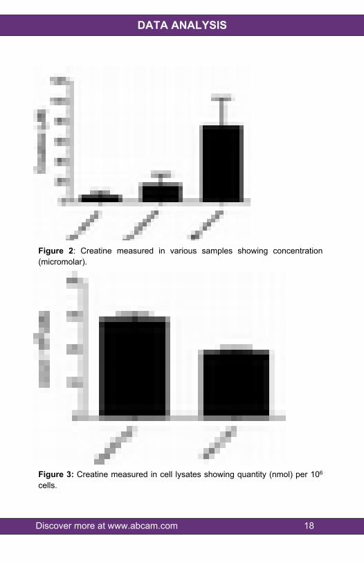
Figure 2: Creatine measured in various samples showing concentration (micromolar).
Figure 3: Creatine measured in cell lysates showing quantity (nmol) per 106 cells.

Discover more at www.abcam.com 19
RESOURCES
15.QUICK ASSAY PROCEDURENOTE: This procedure is provided as a quick reference for experienced users. Follow the detailed procedure when performing the assay for the first time.
Prepare standard, probe, enzyme and enzyme mix (aliquot if necessary); get equipment ready.
Prepare appropriate standard curve for your detection method of choice (colorimetric or fluorometric).
Prepare samples in duplicate (find optimal dilutions to fit standard curve readings), including deproteinization step.
Set up plate for standard (50 µL), samples (50 µL) and background wells (50 µL).
Prepare Creatine Reaction Mix (Number samples + background control + standards + 1).
Component Colorimetric Reaction Mix (µL)
Background Control Reaction Mix (µL)
Creatine Assay Buffer 44 46Creatinase 2 0Creatine Enzyme Mix 2 2Creatine Probe 2 2
Component FluorometricReaction Mix (µL)
Background Control Reaction Mix (µL)
Creatine Assay Buffer 45.6 47.6Creatinase 2 0Creatine Enzyme Mix 2 2Creatine Probe 0.4 0.4
Add 50 µL of Reaction Mix to the standard, sample and background control wells.
Incubate plate at 37°C 60 minutes protected from light.
Measure plate at OD 570 nm for colorimetric assay or Ex/Em= 535/587 nm for fluorometric assay.

Discover more at www.abcam.com 20
RESOURCES
16.TROUBLESHOOTING
Problem Cause Solution
Use of ice-cold buffer Buffers must be at room temperature
Plate read at incorrect wavelength
Check the wavelength and filter settings of instrument
Assay not
workingUse of a different 96-
well plate
Colorimetric: Clear platesFluorometric: black wells/clear
bottom plateSamples not
deproteinized (if indicated on protocol)
Use PCA precipitation protocol for deproteinization
Cells/tissue samples not homogenized
completely
Use Dounce homogenizer, increase number of strokes
Samples used after multiple free/ thaw
cycles
Aliquot and freeze samples if needed to use multiple times
Use of old or inappropriately stored
samples
Use fresh samples or store at - 80°C (after snap freeze in liquid
nitrogen) till use
Sample with erratic readings
Presence of interfering substance
in the sample
Check protocol for interfering substances; deproteinize samples
Improperly thawed components
Thaw all components completely and mix gently before use
Allowing reagents to sit for extended times
on ice
Always thaw and prepare fresh reaction mix before use
Lower/ Higher readings in samples and Standards Incorrect incubation
times or temperaturesVerify correct incubation times and temperatures in protocol

Discover more at www.abcam.com 21
RESOURCES
Problem Cause SolutionPipetting errors in
standard or reaction mix
Avoid pipetting small volumes (< 5 µL) and prepare a master mix
whenever possibleAir bubbles formed in
wellPipette gently against the wall of
the tubes
Standard readings do not follow a linear pattern Standard stock is at
incorrect concentration
Always refer to dilutions on protocol
Measured at incorrect wavelength Check equipment and filter setting
Samples contain interfering
substances
Troubleshoot if it interferes with the kit
Unanticipated results
Sample readings above/ below the
linear range
Concentrate/ Dilute sample so it is within the linear range

Discover more at www.abcam.com 22
RESOURCES
17.FAQWhat is the sensitivity of this assay and how can plasma/whole blood samples be processed for this assay?The assay has a detection sensitivity of 0.5 mU/ml of glutathione peroxidase in samples.
Which protein assay is compatible with this kit?We suggest you use a detergent compatible BCA assay kit: (ab102536).
What is the sample volume to be used with this kit for plasma samples from rat?This depends on the amount of active CK enzyme in the sample. The sample volume per well would need to be optimized to make sure that the values obtained are within the linear range of the std. curve.
All the standards and the samples, including ones without creatine turned dark pink. What could be wrong?If the probe was exposed to light/air it might get oxidized and yield color.
Is it known if pyruvic acid, glucose, albumin, bilirubin and ascorbic acid interfere in this assay?We are not aware of any interference by pyruvic acid, glucose, albumin, bilirubin or ascorbic acid. There might be cause-effect relationship between the creatine level in the sample and the concentration of these moieties but there is no reported interference with this assay per se that we are aware of. Sarcosine causes interference in this assay, which can be resolved by running side by side controls and subtracting the value from the results.
If there is no significant background, is it necessary to run background control for every sample?
Discover more at www.abcam.com 23
RESOURCES
Sarcosine creates background in this assay. If there is no significant background seen during the pilot assay, it is not essential to run background controls for every sample.
Discover more at www.abcam.com 24
RESOURCES
18.INTERFERENCESThese chemicals or biological materials will cause interferences in this assay causing compromised results or complete failure:
Sarcosine.

Discover more at www.abcam.com 25
RESOURCES
19.NOTES

Discover more at www.abcam.com 26
RESOURCES

RESOURCES 27
UK, EU and ROWEmail: [email protected] | Tel: +44-(0)1223-696000
AustriaEmail: [email protected] | Tel: 019-288-259
FranceEmail: [email protected] | Tel: 01-46-94-62-96 GermanyEmail: [email protected] | Tel: 030-896-779-154 SpainEmail: [email protected] | Tel: 911-146-554 SwitzerlandEmail: [email protected] Tel (Deutsch): 0435-016-424 | Tel (Français): 0615-000-530
US and Latin AmericaEmail: [email protected] | Tel: 888-77-ABCAM (22226)
CanadaEmail: [email protected] | Tel: 877-749-8807
China and Asia Pacific Email: [email protected] | Tel: 400 921 0189 / +86 21 2070 0500 JapanEmail: [email protected] | Tel: +81-(0)3-6231-0940
www.abcam.com | www.abcam.cn | www.abcam.co.jp
Copyright © 2020 Abcam, All Rights Reserved. The Abcam logo is a registered trademark.
All information / detail is correct at time of going to print.