Embed Size (px)
Citation preview
Version 9 Last Updated 11 November 2016
ab113851DCFDA Cellular ROS Detection Assay Kit
Instructions for use:
For quantitative measurement of cellular reactive oxygen species (ROS) in cells.
This product is for research use only and is not intended for diagnostic use.
Table of Contents INTRODUCTION 11. BACKGROUND 12. ASSAY SUMMARY - MICROPLATE 23. ASSAY SUMMARY - FLOW 3GENERAL INFORMATION 44. PRECAUTIONS 45. STORAGE AND STABILITY 46. LIMITATIONS 57. MATERIALS SUPPLIED 58. MATERIALS REQUIRED, NOT SUPPLIED 69. TECHNICAL HINTS 7ASSAY PREPARATION 810. REAGENT PREPARATION 8ASSAY PROCEDURE 1111. ASSAY PROCEDURE 11DATA ANALYSIS 1612. CALCULATIONS 1613. TYPICAL DATA 16RESOURCES 1814. QUICK ASSAY – SUSPENSION CELL MICROPLATE ASSAY 1815. QUICK ASSAY – ADHERENT CELL MICROPLATE ASSAY 1916. QUICK ASSAY – FLOW ASSAY 2017. INTERFERENCES 2118. FAQS 2119. NOTES 25
ab113851 DCFDA Cellular ROS Detection Assay Kit 1
INTRODUCTION
INTRODUCTION
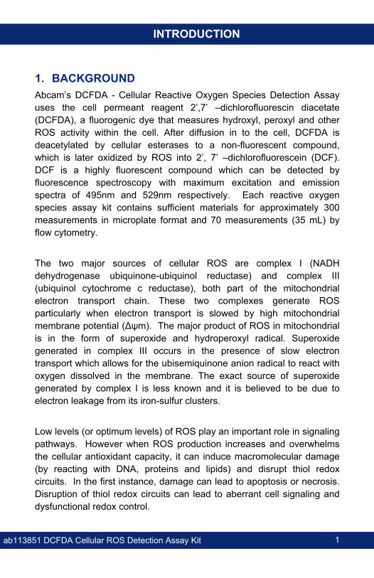
1. BACKGROUNDAbcam’s DCFDA - Cellular Reactive Oxygen Species Detection Assay uses the cell permeant reagent 2’,7’ –dichlorofluorescin diacetate (DCFDA), a fluorogenic dye that measures hydroxyl, peroxyl and other ROS activity within the cell. After diffusion in to the cell, DCFDA is deacetylated by cellular esterases to a non-fluorescent compound, which is later oxidized by ROS into 2’, 7’ –dichlorofluorescein (DCF). DCF is a highly fluorescent compound which can be detected by fluorescence spectroscopy with maximum excitation and emission spectra of 495nm and 529nm respectively. Each reactive oxygen species assay kit contains sufficient materials for approximately 300 measurements in microplate format and 70 measurements (35 mL) by flow cytometry.
The two major sources of cellular ROS are complex I (NADH dehydrogenase ubiquinone-ubiquinol reductase) and complex III (ubiquinol cytochrome c reductase), both part of the mitochondrial electron transport chain. These two complexes generate ROS particularly when electron transport is slowed by high mitochondrial membrane potential (Δψm). The major product of ROS in mitochondrial is in the form of superoxide and hydroperoxyl radical. Superoxide generated in complex III occurs in the presence of slow electron transport which allows for the ubisemiquinone anion radical to react with oxygen dissolved in the membrane. The exact source of superoxide generated by complex I is less known and it is believed to be due to electron leakage from its iron-sulfur clusters.
Low levels (or optimum levels) of ROS play an important role in signaling pathways. However when ROS production increases and overwhelms the cellular antioxidant capacity, it can induce macromolecular damage (by reacting with DNA, proteins and lipids) and disrupt thiol redox circuits. In the first instance, damage can lead to apoptosis or necrosis. Disruption of thiol redox circuits can lead to aberrant cell signaling and dysfunctional redox control.
ab113851 DCFDA Cellular ROS Detection Assay Kit 2
INTRODUCTION
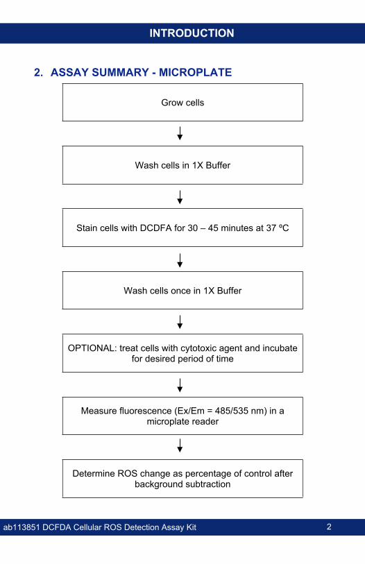
2. ASSAY SUMMARY - MICROPLATE
Grow cells
Wash cells in 1X Buffer
Stain cells with DCDFA for 30 – 45 minutes at 37 ºC
Wash cells once in 1X Buffer
OPTIONAL: treat cells with cytotoxic agent and incubate for desired period of time
Measure fluorescence (Ex/Em = 485/535 nm) in a microplate reader
Determine ROS change as percentage of control after background subtraction
ab113851 DCFDA Cellular ROS Detection Assay Kit 3
INTRODUCTION
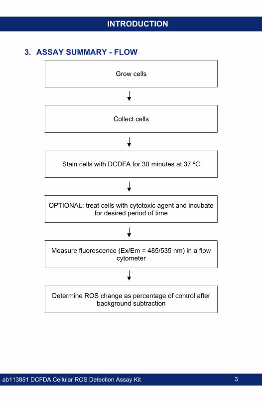
3. ASSAY SUMMARY - FLOW
Grow cells
Collect cells
Stain cells with DCDFA for 30 minutes at 37 ºC
OPTIONAL: treat cells with cytotoxic agent and incubate for desired period of time
Measure fluorescence (Ex/Em = 485/535 nm) in a flow cytometer
Determine ROS change as percentage of control after background subtraction
ab113851 DCFDA Cellular ROS Detection Assay Kit 4
GENERAL INFORMATION
GENERAL INFORMATION
4. PRECAUTIONSPlease read these instructions carefully prior to beginning the assay.
All kit components have been formulated and quality control tested to function successfully as a kit.
We understand that, occasionally, experimental protocols might need to be modified to meet unique experimental circumstances. However, we cannot guarantee the performance of the product outside the conditions detailed in this protocol booklet.
Reagents should be treated as possible mutagens and should be handle with care and disposed of properly. Please review the Safety Datasheet (SDS) provided with the product for information on the specific components.
Observe good laboratory practices. Gloves, lab coat, and protective eyewear should always be worn. Never pipet by mouth. Do not eat, drink or smoke in the laboratory areas.
All biological materials should be treated as potentially hazardous and handled as such. They should be disposed of in accordance with established safety procedures.
5. STORAGE AND STABILITY Store kit at 4ºC in the dark immediately upon receipt. For longer term storage, keep at -20 ºC or -80 ºC. Kit has a storage time of 1 year from receipt, providing components have not been reconstituted.Refer to list of materials supplied for storage conditions of individual components. Observe the storage conditions for individual prepared components in the Materials Supplied section.Aliquot components in working volumes before storing at the recommended temperature.
ab113851 DCFDA Cellular ROS Detection Assay Kit 5
GENERAL INFORMATION
6. LIMITATIONS Assay kit intended for research use only. Not for use in
diagnostic procedures.
Do not mix or substitute reagents or materials from other kit lots or vendors. Kits are QC tested as a set of components and performance cannot be guaranteed if utilized separately or substituted.
7. MATERIALS SUPPLIED Item Amount Storage
Condition (Before
Preparation)
Storage Condition
(After Preparation)
20 mM DCFDA (in DMSO) 35 µL 4°C 4°C10X Buffer 10 mL 4°C 4°C55 mM Tert-Butyl Hydrogen Peroxide (TBHP)
50 µL 4°C 4°C
ab113851 DCFDA Cellular ROS Detection Assay Kit 6
GENERAL INFORMATION
8. MATERIALS REQUIRED, NOT SUPPLIED These materials are not included in the kit, but will be required to successfully perform this assay:
Fluorescence microplate reader or flow cytometer. Suggested wavelengths: Ex/Em = 485 nm/535 nm (use similar settings to those used to detect FITC).
MilliQ water or other type of double distilled water (ddH2O)
Pipettes and pipette tips, including multichannel pipette
General tissue culture supplies
PBS
Fetal Bovine Serum (FBS)
DMSO (Cell culture grade)
Sterile, tissue culture treated, clear bottom, dark sided 96-well microplates.
(Optional) Test compounds/diluents of interest
(Optional) Other ROS inducing control compounds such as doxorubicin, idarubicin or antimycin
(Optional) Decane: Solvent used for the preparation and stabilization of TBHP
ab113851 DCFDA Cellular ROS Detection Assay Kit 7
GENERAL INFORMATION
9. TECHNICAL HINTS This kit is sold based on number of tests. A ‘test’ simply
refers to a single assay well. The number of wells that contain sample, control or standard will vary by product. Review the protocol completely to confirm this kit meets your requirements. Please contact our Technical Support staff with any questions.
Selected components in this kit are supplied in surplus amount to account for additional dilutions, evaporation, or instrumentation settings where higher volumes are required. They should be disposed of in accordance with established safety procedures.
Avoid foaming or bubbles when mixing or reconstituting components.
Avoid cross contamination of samples or reagents by changing tips between sample, standard and reagent additions.
Ensure plates are properly sealed or covered during incubation steps.
Ensure all reagents and solutions are at the appropriate temperature before starting the assay.
Samples which generate values that are greater than the most concentrated standard should be further diluted in the appropriate sample dilution buffer.
Ensure plates are properly sealed or covered during incubation steps.
Make sure you have the right type of plate for your detection method of choice. Clear bottom, dark sided microplates are recommended with this assay. Clear sided microplates have not been tested with this kit.
Make sure all necessary equipment is switched on and set at the appropriate temperature.
ab113851 DCFDA Cellular ROS Detection Assay Kit 8
ASSAY PREPARATION
ASSAY PREPARATION
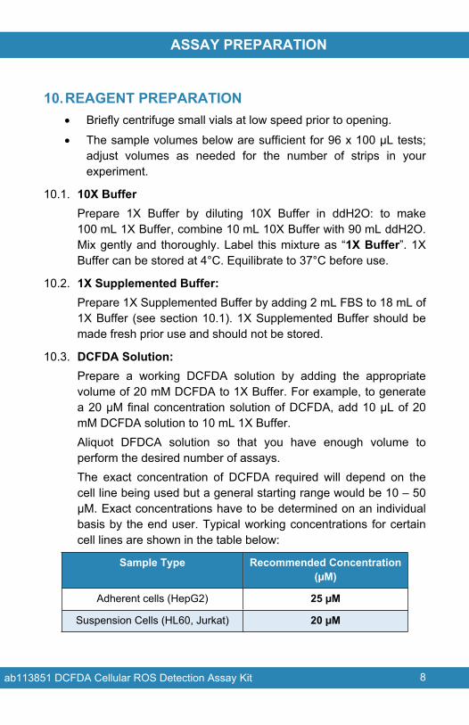
10.REAGENT PREPARATION Briefly centrifuge small vials at low speed prior to opening.
The sample volumes below are sufficient for 96 x 100 µL tests; adjust volumes as needed for the number of strips in your experiment.
10.1. 10X BufferPrepare 1X Buffer by diluting 10X Buffer in ddH2O: to make 100 mL 1X Buffer, combine 10 mL 10X Buffer with 90 mL ddH2O. Mix gently and thoroughly. Label this mixture as “1X Buffer”. 1X Buffer can be stored at 4°C. Equilibrate to 37°C before use.
10.2. 1X Supplemented Buffer:Prepare 1X Supplemented Buffer by adding 2 mL FBS to 18 mL of 1X Buffer (see section 10.1). 1X Supplemented Buffer should be made fresh prior use and should not be stored.
10.3. DCFDA Solution:Prepare a working DCFDA solution by adding the appropriate volume of 20 mM DCFDA to 1X Buffer. For example, to generate a 20 µM final concentration solution of DCFDA, add 10 µL of 20 mM DCFDA solution to 10 mL 1X Buffer.Aliquot DFDCA solution so that you have enough volume to perform the desired number of assays.The exact concentration of DCFDA required will depend on the cell line being used but a general starting range would be 10 – 50 μM. Exact concentrations have to be determined on an individual basis by the end user. Typical working concentrations for certain cell lines are shown in the table below:
Sample Type Recommended Concentration (µM)
Adherent cells (HepG2) 25 µM
Suspension Cells (HL60, Jurkat) 20 µM
ab113851 DCFDA Cellular ROS Detection Assay Kit 9
ASSAY PREPARATION
10.4. Tert-Butyl Hydrogen Peroxide (TBHP) Solution (Positive control):
10.4.1. TBHP Solution for ADHERENT CELLS:Prepare a 50 - 250 µM TBHP* working solution by diluting 55 mM TBHP stock solution in the 1X Supplemented Buffer (see section 10.2). Working TBHP solution should be made fresh every time and should not be stored for future use (storage may lead to TBHP degradation).TBHP may also be diluted in complete media with 10% FBS without phenol red.* The concentration of TBHP to use will depend on the sensitivity of the cell line: HL60 and Jurkat cells are very sensitive to TBHP whereas HepG2 cells are very insensitive.
10.4.2.TBHP Solution for SUSPENSION CELLS:Prepare a 100 - 250 µM TBHP* working solution by diluting 55 mM TBHP stock solution in the 1X Supplemented Buffer (see section 10.2). Working TBHP solution should be made fresh every time and should not be stored for future use (storage may lead to TBHP degradation).TBHP may also be diluted in complete media with 10% FBS without phenol red.* The concentration of TBHP to use will depend on the sensitivity of the cell line: HL60 and Jurkat cells are very sensitive to TBHP whereas HepG2 cells are very insensitive.
10.5. Optional: 5 mM DecaneDilute Decane (TBHP diluent; not included in the kit) 1,100X times in 1X Supplemented Buffer or in complete media with 10% FBS without phenol red.
ab113851 DCFDA Cellular ROS Detection Assay Kit 10
ASSAY PREPARATION
10.6. Optional: Compounds/Diluents of interestIf performing toxicity assays, dilute compounds of interest in 1X Supplemented Buffer to final desired concentration for the experiment. A 96-well deep well microplate may be use in this step. Compounds may also be diluted in complete media with 10% FBS without phenol red.
ab113851 DCFDA Cellular ROS Detection Assay Kit 11
ASSAY PROCEDURE
ASSAY PROCEDURE
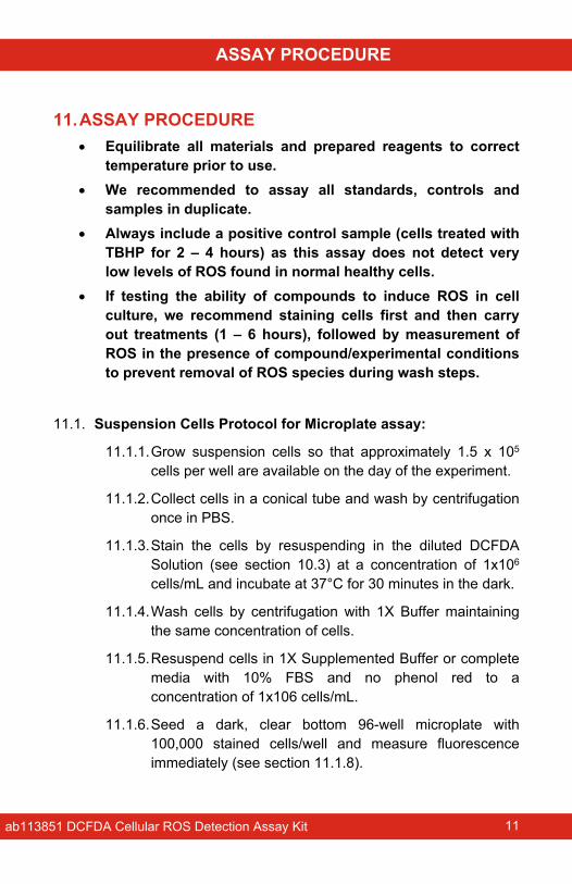
11.ASSAY PROCEDURE Equilibrate all materials and prepared reagents to correct
temperature prior to use. We recommended to assay all standards, controls and
samples in duplicate. Always include a positive control sample (cells treated with
TBHP for 2 – 4 hours) as this assay does not detect very low levels of ROS found in normal healthy cells.
If testing the ability of compounds to induce ROS in cell culture, we recommend staining cells first and then carry out treatments (1 – 6 hours), followed by measurement of ROS in the presence of compound/experimental conditions to prevent removal of ROS species during wash steps.
11.1. Suspension Cells Protocol for Microplate assay:
11.1.1.Grow suspension cells so that approximately 1.5 x 10⁵ cells per well are available on the day of the experiment.
11.1.2.Collect cells in a conical tube and wash by centrifugation once in PBS.
11.1.3.Stain the cells by resuspending in the diluted DCFDA Solution (see section 10.3) at a concentration of 1x10⁶ cells/mL and incubate at 37°C for 30 minutes in the dark.
11.1.4.Wash cells by centrifugation with 1X Buffer maintaining the same concentration of cells.
11.1.5.Resuspend cells in 1X Supplemented Buffer or complete media with 10% FBS and no phenol red to a concentration of 1x10⁶ cells/mL.
11.1.6.Seed a dark, clear bottom 96-well microplate with 100,000 stained cells/well and measure fluorescence immediately (see section 11.1.8).
ab113851 DCFDA Cellular ROS Detection Assay Kit 12
ASSAY PROCEDURE
11.1.7. If performing toxicity assays, overlay each well with previously diluted 2X compounds and treat cells for desired period of time (1 – 6 hours). NOTE: include background wells (untreated or diluent treated stained cells) as well as blank wells (media or buffer only).
11.1.8. Measure plate on a fluorescence plate reader at Ex/Em= 485/535 nm in end point mode in the presence of compounds, media or buffer.
11.2. Adherent Cells Protocol for Microplate assay:
11.2.1.Grow adherent cells in standard cell culture media so that 3 x 10⁶ - 4 x 10⁶ cells are obtained the day before the experiment.
11.2.2.Harvest cells and seed a dark, clear bottom 96-well microplate with 25,000 cells per well. Allow cells to adhere overnight.
11.2.3.Remove the media and add 100 μL/well of 1X Buffer.
11.2.4.Remove 1X Buffer and stain cells by adding 100 μL/well of the diluted DCFDA Solution.
11.2.5.Incubate cells with the diluted DCFDA Solution for 45 minutes at 37°C in the dark.
11.2.6.Remove DCFDA Solution; add 100 μL/well of 1X Buffer or 1X PBS and measure fluorescence immediately (see Section 11.2.8).
11.2.7.If performing toxicity assays, remove 1X Buffer/PBS and add 100 μL of previously 1X diluted compound(s) of interest. Treat cells for desired period of time (1–6 hours). NOTE: include background wells (untreated or diluent treated stained cells) as well as blank wells (media or buffer only).
11.2.8.Measure plate on a fluorescence plate reader at Ex/Em= 485/535 nm in end point mode in the presence of compounds, media or buffer.
ab113851 DCFDA Cellular ROS Detection Assay Kit 13
ASSAY PROCEDURE
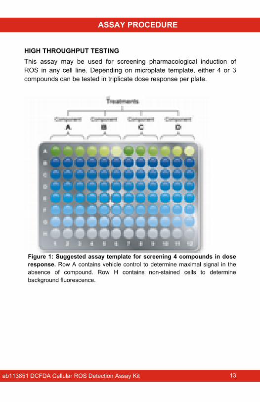
HIGH THROUGHPUT TESTINGThis assay may be used for screening pharmacological induction of ROS in any cell line. Depending on microplate template, either 4 or 3 compounds can be tested in triplicate dose response per plate.
Figure 1: Suggested assay template for screening 4 compounds in dose response. Row A contains vehicle control to determine maximal signal in the absence of compound. Row H contains non-stained cells to determine background fluorescence.
ab113851 DCFDA Cellular ROS Detection Assay Kit 14
ASSAY PROCEDURE
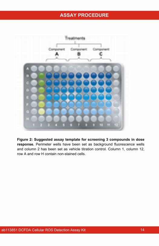
Figure 2: Suggested assay template for screening 3 compounds in dose response. Perimeter wells have been set as background fluorescence wells and column 2 has been set as vehicle titration control. Column 1, column 12, row A and row H contain non-stained cells.
ab113851 DCFDA Cellular ROS Detection Assay Kit 15
ASSAY PROCEDURE
11.3. Flow cytometry Measurement
11.3.1.Grow cells (adherent or suspension) in glucose based media so that on the day of the experiment there are at least 1.5 x 10⁴ cells per assayed condition (treatment, dose, time). Include in the calculation enough cells for control signal (control compound, control vehicle and non-stained control cells). This number takes into account any cell loss experienced during processing.
11.3.2.Harvest cells and ensure a single cell suspension by (1) gently pipetting up and down suspension cells or (2) by fully detaching adherent cells (e.g. trypsinize and quench with media).
11.3.3.Stain cells in culture media with 20 µM DCFDA and incubate for 30 minutes at 37°C. Once the incubation is completed, DO NOT wash the cells.
11.3.4.After staining, treat the cells with compound(s) of interest and ensure appropriate controls are included. If using THBP as positive control, optimal signal is obtained after 4 hours of treatment.
11.3.5.Gently pipette cells up/down to ensure single cell suspension.
11.3.6.Analyze on flow cytometer. Establish forward and side scatter gates to exclude debris and cellular aggregates from analysis.
11.3.7.DCF should be excited by the 488 nm laser and should be detected at 535 nm (typically FL1).
11.3.8.Ideally 10,000 cells should be analyzed per experimental condition. Cells should not be overly dense during the experiment (< 1 x 10⁶ cells/mL).
ab113851 DCFDA Cellular ROS Detection Assay Kit 16
DATA ANALYSIS
DATA ANALYSIS
12.CALCULATIONS Fluorescent Microplate Measurement
Subtract blank readings from all measurements and determine fold change from assay control (diluents treated cells if performing toxicity studies).
Flow cytometry MeasurementExclude debris and isolate cell population of interest with gating. Using mean fluorescent intensity, determine fold change between control and treated samples.
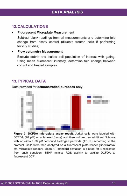
13.TYPICAL DATAData provided for demonstration purposes only.
Figure 3: DCFDA microplate assay result. Jurkat cells were labeled with DCFDA (20 µM) or unlabeled (none) and then cultured an additional 3 hours with or without 50 µM tert-butyl hydrogen peroxide (TBHP) according to the protocol. Cells were then analyzed on a fluorescent plate reader (SpectraMax M4 Microplate reader). Mean +/- standard deviation is plotted for 4 replicates from each condition. TBHP mimics ROS activity to oxidize DCFDA to fluorescent DCF.
ab113851 DCFDA Cellular ROS Detection Assay Kit 17
DATA ANALYSIS
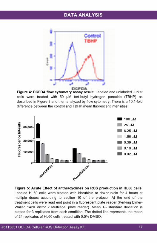
Figure 4: DCFDA flow cytometry assay result. Labeled and unlabeled Jurkat cells were treated with 50 µM tert-butyl hydrogen peroxide (TBHP) as described in Figure 3 and then analyzed by flow cytometry. There is a 10.1-fold difference between the control and TBHP mean fluorescent intensities.
IDARUBICIN
DOXORUBICIN0
10,000
20,000
60,000
Fluo
resc
ence
Inte
sity
100 M
25 M
6.25 M
1.56 M
0.39 M
0.10 M
0.02 M
Figure 5: Acute Effect of anthracyclines on ROS production in HL60 cells. Labeled HL60 cells were treated with idarubicin or doxorubicin for 4 hours at multiple doses according to section 10 of the protocol. At the end of the treatment cells were read end point in a fluorescent plate reader (Perking Elmer-Wallac 1420 Victor 2 Multilabel plate reader). Mean +/- standard deviation is plotted for 3 replicates from each condition. The dotted line represents the mean of 24 replicates of HL60 cells treated with 0.5% DMSO.
ab113851 DCFDA Cellular ROS Detection Assay Kit 18
RESOURCES
RESOURCES
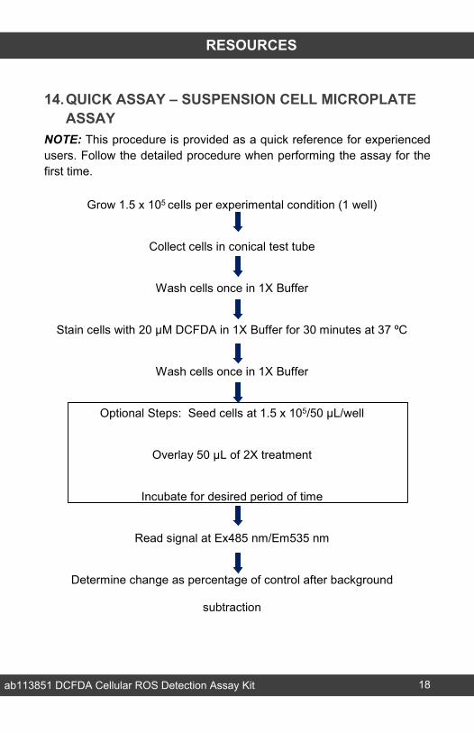
14.QUICK ASSAY – SUSPENSION CELL MICROPLATE ASSAY
NOTE: This procedure is provided as a quick reference for experienced users. Follow the detailed procedure when performing the assay for the first time.
Grow 1.5 x 10⁵ cells per experimental condition (1 well)
Collect cells in conical test tube
Wash cells once in 1X Buffer
Stain cells with 20 µM DCFDA in 1X Buffer for 30 minutes at 37 ºC
Wash cells once in 1X Buffer
Optional Steps: Seed cells at 1.5 x 10⁵/50 µL/well
Overlay 50 µL of 2X treatment
Incubate for desired period of time
Read signal at Ex485 nm/Em535 nm
Determine change as percentage of control after background
subtraction
ab113851 DCFDA Cellular ROS Detection Assay Kit 19
RESOURCES
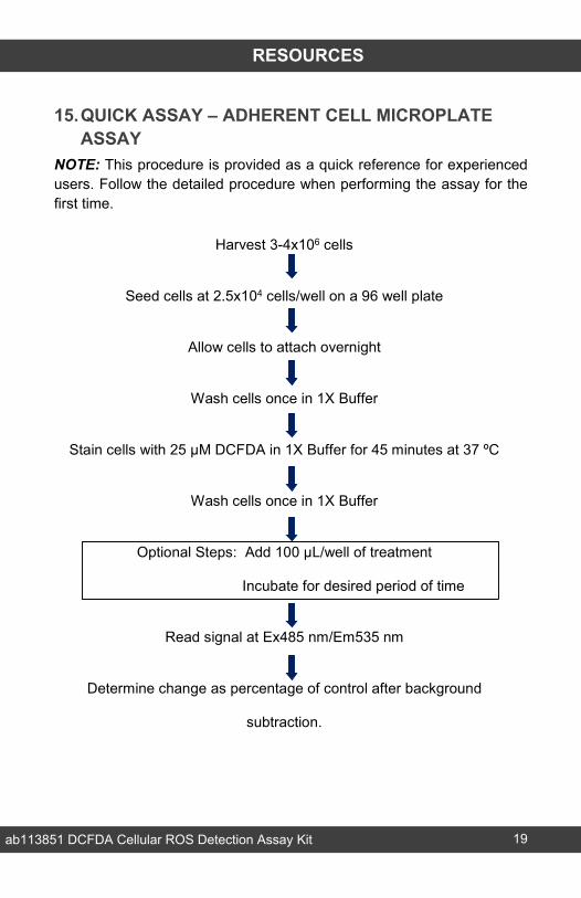
15.QUICK ASSAY – ADHERENT CELL MICROPLATE ASSAY
NOTE: This procedure is provided as a quick reference for experienced users. Follow the detailed procedure when performing the assay for the first time.
Harvest 3-4x10⁶ cells
Seed cells at 2.5x10⁴ cells/well on a 96 well plate
Allow cells to attach overnight
Wash cells once in 1X Buffer
Stain cells with 25 µM DCFDA in 1X Buffer for 45 minutes at 37 ºC
Wash cells once in 1X Buffer
Optional Steps: Add 100 µL/well of treatment
Incubate for desired period of time
Read signal at Ex485 nm/Em535 nm
Determine change as percentage of control after background
subtraction.
ab113851 DCFDA Cellular ROS Detection Assay Kit 20
RESOURCES
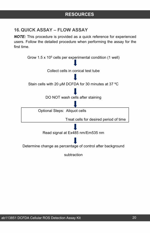
16.QUICK ASSAY – FLOW ASSAYNOTE: This procedure is provided as a quick reference for experienced users. Follow the detailed procedure when performing the assay for the first time.
Grow 1.5 x 10⁵ cells per experimental condition (1 well)
Collect cells in conical test tube
Stain cells with 20 µM DCFDA for 30 minutes at 37 ºC
DO NOT wash cells after staining
Optional Steps: Aliquot cells
Treat cells for desired period of time
Read signal at Ex485 nm/Em535 nm
Determine change as percentage of control after background
subtraction
ab113851 DCFDA Cellular ROS Detection Assay Kit 21
RESOURCES
17. INTERFERENCESThese chemicals or biological materials will cause interference in this assay causing compromised results or complete failure:
Phenol red: phenol red present in media can interfere with the assay, therefore we recommend using cell media without phenol red.
18.FAQsWhich concentration of TBHP shall I use for my cells?The amount of TBHP to use will depend on the sensitivity of the cell line: HL60 and Jurkat cells are very sensitive to TBHP whereas HepG2 cells are very insensitive. We suggest starting with 50 - 250 µM of TBHP as positive control, but you will probably have to optimize this concentration depending on your cells’ sensitivity to TBHP.
I’m using HepG2, which doesn’t seem to be responding to TBHP treatment. Can you suggest other components to induce ROS production in HepG2 and HL60 cells?ROS induction in HepG2 (cultured in glucose based media) – 50 µM antimycin, 50 µM clozapine, 50 µM fluvastatin, 50 µM camptothecin.ROS induction in HL60 (cultured in glucose based media) – doxorubicin, idarubicin, menadione, methyl aminolevulinate, TNHP.ROS induction in HL60 (cultured in galactose based media – glucose free) – idarubicin, menadione.TBHP is generally able to induce in most cultured cell lines like Jurkat.
ab113851 DCFDA Cellular ROS Detection Assay Kit 22
RESOURCES
I want to treat my cells on a microplate for 24 – 48 hours. Will DCFDA be stable inside the cells for this period of time?We don’t know whether DCFDA is stable for more than 6 hours. This kit has not been tested with prolonged treatments. However, in this situation we recommend to follow the steps below:
Dilute compounds of interest in complete media without phenol red. Make twice the volume required.
Treat suspension or adhered cells for the desired period of time. If treating cells for microplate measurements, treat with 100 µL per well.
Include blank wells with no cells but with compound at the same concentration used for treatment.
Include at least 2 positive control wells, to be reserved for TBHP treatment, containing cells but none of the test compounds.
4 hours prior to completion of treatment, dilute TBHP to 10X of the final concentration (500 µM) and spike 10X TBHP into the reserved positive control wells by adding 11 µL per well.
1 hour prior to completion of the treatment, dilute DCFDA at 2X of the final concentration desired in the same media used for treatment (containing experimental compounds) and warm to 37°C.
30 – 45 minutes prior to completion of the treatment, overlay 2X DCFDA dilution on top of the treated cells. If treating cells for microplate measurements, overlay 100 µL of 2X DCFDA dilution per well.
Incubate DCFDA and compounds for the desired period of time (30 – 45 minutes).
Transfer the plate to the microplate reader without washing and read end point in the presence of compounds and DCFDA with Ex/Em = 485/535 nm.
Ratio the relative fluorescence intensity of control and treated wells to the relative fluorescence intensity of the blank wells.
ab113851 DCFDA Cellular ROS Detection Assay Kit 23
RESOURCES
Should cells be washed after incubation with the cytotoxicity compound?Cells should NOT be washed after treatment with the TBHP or other compounds of interest.
Will this kit work with fixed cells?Cells must be alive to work with this kit, therefore fixation is not recommended.
How long is DCFDA stable for in cells?We do not know how long DCFDA is stable in cells. We do know that it is stable for 4 hours and have tested DCFDA with treatments up to this point. However as we have not tested beyond this we do not know whether the signal is detectable beyond this point
Is it possible to dilute the reagents and then freeze them for another time?The 1X Buffer can be frozen or kept at 4ºC for use on a different experiment. It is recommended that the customer aliquot the DCFDA into as many experiments as they will run. It is probably only practical to aliquot in 8 – 10 µL volumes to prevent loss of reagent by aliquoting in too small a volume. We cannot ensure the stability long term (weeks/months) of the DCFDA once it is diluted in the dilution buffer. Enough is given for a 96 well plate, but there is no need to run all 96 data points at once. The TBHP will not be stable if diluted long term. There is no need to freeze TBHP, simply keep in the fridge and use as necessary. This is very stable if kept as it is supplied.
Can FBS be included in the buffer whilst incubating the DCFDA? It should work, but we have not tested it. Under the conditions stated in the current protocol, we have not found an increase in background signal.
ab113851 DCFDA Cellular ROS Detection Assay Kit 24
RESOURCES
Why is TBHP being used in this kit instead of H2O2?Either TBHP or H2O2 would work as positive controls.
Can the culture media contain phenol red?The microplate assay should be performed without phenol red both in suspension and adherent cells. If this is not possible we suggest to include all the relevant negative controls (including media + DCFDA with no cells) to ensure that the media does not affect DCFDA reading.
How long can treated cells be stored prior to analyzing via flow cytometry?After treatment, cells cannot be stored as the flow cytometry reading of DCFDA must be run on live cells
Can I use this product to measure ROS in serum or plasma?No, DCFDA is an intracellular label and therefore it is not suitable to use in serum or plasma.
ab113851 DCFDA Cellular ROS Detection Assay Kit 25
RESOURCES
19.NOTES
Discover more at www.abcam.com 26
UK, EU and ROWEmail: [email protected] | Tel: +44-(0)1223-696000
AustriaEmail: [email protected] | Tel: 019-288-259
FranceEmail: [email protected] | Tel: 01-46-94-62-96 GermanyEmail: [email protected] | Tel: 030-896-779-154 SpainEmail: [email protected] | Tel: 911-146-554 SwitzerlandEmail: [email protected] Tel (Deutsch): 0435-016-424 | Tel (Français): 0615-000-530
US and Latin AmericaEmail: [email protected] | Tel: 888-77-ABCAM (22226)
CanadaEmail: [email protected] | Tel: 877-749-8807
China and Asia Pacific Email: [email protected] | Tel: 108008523689 (中國聯通) JapanEmail: [email protected] | Tel: +81-(0)3-6231-0940
www.abcam.com | www.abcam.cn | www.abcam.co.jp
Copyright © 2016 Abcam, All Rights Reserved. The Abcam logo is a registered trademark.
All information / detail is correct at time of going to print.


![Oxidative stress-induced cellular senescence desensitizes cell … · 2019-10-15 · theories to explain possible causes of aging [1, 2]. Intracellular reactive oxygen species (ROS)](https://img.pdfslide.us/doc/110x75/5eaa448dd86ba957c94e5c58/oxidative-stress-induced-cellular-senescence-desensitizes-cell-2019-10-15-theories.jpg)


![Oxygen availability strongly affects chronological lifespan and thermotolerance … · related cellular deterioration in many organisms [10]. However, ROS have also been implicated](https://img.pdfslide.us/doc/110x75/612d0edd1ecc51586941f3d0/oxygen-availability-strongly-affects-chronological-lifespan-and-thermotolerance.jpg)