Embed Size (px)
Citation preview
AACN PCCN Webinar
Session 1
Cardiovascular
Presenter: Carol A. Rauen, RN, MS, CCNS, CCRN, PCCN, CEN
Independent Clinical Nurse Specialist & Education Consultant [email protected]

Session 1: Cardiovascular
1
Table of Contents Cardiovascular ................................................................................................................................. 2
Heart Failure ................................................................................................................................... 8
Acute Coronary Syndromes .......................................................................................................... 15
Session 1: Cardiovascular
2
Cardiovascular
I. INTRODUCTION
PCCN Test Plan
Cardiovascular: 33% a. Acute Coronary Syndromes
Non-ST Segment Elevation MI
ST Segment Elevation MI
Unstable Angina b. Acute Inflammatory Disease (e.g. myocarditis, endocarditis, pericarditis) c. Aneurysm
Dissecting
Repair d. Cardiac Surgery (e.g. open chest surgery) – more than 48 hrs post-op e. Cardiac Tamponade f. Cardiogenic Shock g. Cardiomyopathies
Dilated (e.g. ischemic/non-ischemic)
Hypertrophic
Stress-Induced (e.g. Takotsubo) h. Dysrhythmia
Bradydysrhythmias
Conduction Defects & Blocks
Device-related (e.g. ICD and Pacemaker)
Lethal Ventricular Dysrhythmias
Tachydysrhythmias i. Genetic Cardiac Disease (e.g. long QT syndrome, Brugada syndrome) j. Heart Failure
Acute Exacerbations (e.g., pulmonary edema)
Chronic k. Hypertensive Crisis l. Minimally-Invasive Cardiac Surgery (i.e., non-sternal approach) m. Septal Defects (congenital and acquired)
Session 1: Cardiovascular
3
n. Valvular Heart Disease
Aortic
Mitral o. Vascular disease
carotid artery stenosis
minimally-invasive interventions (e.g., stents, endografts)
peripheral arterial occlusions
peripheral surgical interventions
peripheral venous thrombosis
Cardiovascular Testable Nursing Actions a. Perform a comprehensive cardiovascular assessment b. Identify, interpret, and monitor
Dysrhythmias
ST segments
QTc intervals c. Select leads for cardiac monitoring for the indicated disease process d. Recognize indications for and manage patients requiring hemodynamic monitoring using
non-invasive hemodynamic monitoring e. Monitor hemodynamic status and recognize signs and symptoms of hemodynamic
instability
Pacemakers
Defibrillation
Arterial/venous sheaths
Transesophageal echocardiogram (TEE) f. Monitor patients pre- and post-procedure
Cardioversion
Pericardiocentesis
Cardiac catheterization
Ablation
Arterial closure devices g. Monitor normal and abnormal cardiovascular diagnostic test results h. Administer cardiovascular medications and monitor response i. Titrate vasoactive medications j. Recognize signs and symptoms of cardiovascular emergencies, initiate standardized
interventions, and seek assistance as needed k. Monitor and manage patient following coronary intervention
Session 1: Cardiovascular
4
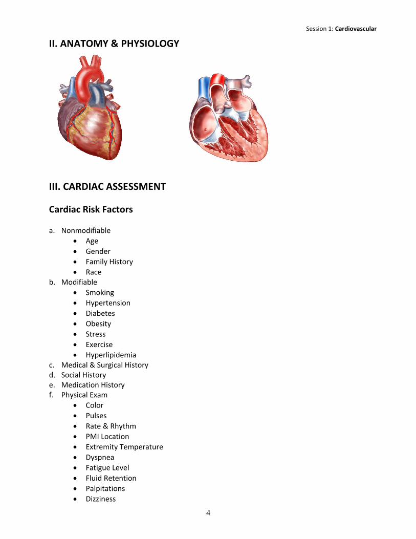
II. ANATOMY & PHYSIOLOGY
III. CARDIAC ASSESSMENT
Cardiac Risk Factors a. Nonmodifiable
Age
Gender
Family History
Race b. Modifiable
Smoking
Hypertension
Diabetes
Obesity
Stress
Exercise
Hyperlipidemia c. Medical & Surgical History d. Social History e. Medication History f. Physical Exam
Color
Pulses
Rate & Rhythm
PMI Location
Extremity Temperature
Dyspnea
Fatigue Level
Fluid Retention
Palpitations
Dizziness
Session 1: Cardiovascular
5
g. Chest Pain Exam
PQRST Assessment o P: Pain, Placement, Provocation o Q: Quality (sharp, stabbing, pressure) Quantity o R: Radiation, Relief o S: Severity, Systems (nausea, sweaty, dizziness) o T: Timing (when it started, how long did it last, what makes it better
or worse) h. Hemodynamic Stability
Vital Signs: supine, sitting and standing
Work of Breathing, Breath Sounds (congestion)
LOC
Noninvasive Cardiac Output Monitoring
Diagnostic Tests & Procedures a. 12 Lead ECG b. Echocardiography (Transthoracic and Transesophageal) c. Stress Test d. Cardiac Catheterization e. Doppler Ultrasound f. Blood Work
Acute Coronary Syndrome o Cardiac Enzymes: CK-MB o Amino Acids: Troponins o Heme Proteins: Myoglobin
Lipid Profile o Triglycerides o Cholesterol o Low Density Lipoproteins o High Density Lipoproteins
Coagulation Profile o PT/INR o aPTT o ACT
Miscellaneous o B Type Natriuertic Peptide (BNP) o C Reactive Protein o Homocysteine
Cardiac Assessment, Test Prep and Monitoring are
listed under Nursing Actions. Remember to
Review!
Session 1: Cardiovascular
6
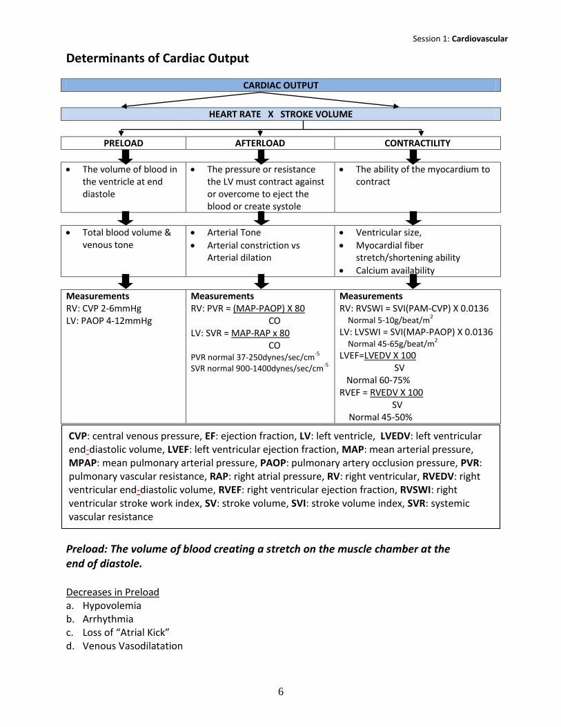
Determinants of Cardiac Output
CARDIAC OUTPUT
HEART RATE X STROKE VOLUME
PRELOAD AFTERLOAD CONTRACTILITY
The volume of blood in the ventricle at end diastole
The pressure or resistance the LV must contract against or overcome to eject the blood or create systole
The ability of the myocardium to contract
Total blood volume & venous tone
Arterial Tone
Arterial constriction vs Arterial dilation
Ventricular size,
Myocardial fiber stretch/shortening ability
Calcium availability
Measurements RV: CVP 2-6mmHg LV: PAOP 4-12mmHg
Measurements RV: PVR = (MAP-PAOP) X 80 CO LV: SVR = MAP-RAP x 80 CO PVR normal 37-250dynes/sec/cm
-5
SVR normal 900-1400dynes/sec/cm-5
Measurements RV: RVSWI = SVI(PAM-CVP) X 0.0136 Normal 5-10g/beat/m
2
LV: LVSWI = SVI(MAP-PAOP) X 0.0136 Normal 45-65g/beat/m
2
LVEF=LVEDV X 100 SV Normal 60-75% RVEF = RVEDV X 100 SV Normal 45-50%
Preload: The volume of blood creating a stretch on the muscle chamber at the end of diastole. Decreases in Preload a. Hypovolemia b. Arrhythmia c. Loss of “Atrial Kick” d. Venous Vasodilatation
CVP: central venous pressure, EF: ejection fraction, LV: left ventricle, LVEDV: left ventricular end-diastolic volume, LVEF: left ventricular ejection fraction, MAP: mean arterial pressure, MPAP: mean pulmonary arterial pressure, PAOP: pulmonary artery occlusion pressure, PVR: pulmonary vascular resistance, RAP: right atrial pressure, RV: right ventricular, RVEDV: right ventricular end-diastolic volume, RVEF: right ventricular ejection fraction, RVSWI: right ventricular stroke work index, SV: stroke volume, SVI: stroke volume index, SVR: systemic vascular resistance

Session 1: Cardiovascular
7
Increases in Preload a. Left Heart
LV Failure/Dysfunction
Mitral Valve Disease
Aortic Valve Disease
Cardiac Tamponade/Effusion
Volume Overload
Decreased Compliance b. Right Heart
RV Failure Due to Ischemia
Increased Pulmonary Vascular Resistance
Cardiac Tamponade/Effusion
Volume Overload
LV Failure
Afterload: The pressure or resistance the LV must contract against or overcome to eject the blood or create systole. Decreases in Afterload a. Vasodilation b. Sepsis c. Vasodilator Therapies Increases in Afterload a. Right Heart
Pulmonary Hypertension
Hypoxemia
Pulmonic Stenosis b. Left Heart
Vasoconstriction
Vasopressors
Hypothermia
Aortic Stenosis
Contractility: The ability of the myocardium to contract. Decreased Contractility a. Parasympathetic Stimulation b. Negative Inotropic Therapies
Beta Blockers
Calcium Channel Blockers c. Metabolic States
Hyperkalemia
Myocardial Ischemia/Infarct
Acidosis
Session 1: Cardiovascular
8
Increased Contractility a. Sympathetic Stimulation b. Inotropic Therapies
Epinephrine
Dopamine
Digoxin
Calcium c. Metabolic States
Hypercalcemia
Heart Failure
l. PATHOPHYSIOLOGY OF HEART FAILURE
Definitions a. Heart Failure is the inability of the heart to adequately supply blood to meet the metabolic
demands of the tissues resulting in inadequate tissue perfusion and volume overload. b. Acute Heart Failure occurs when the inability to meet the demands of the tissues takes
place abruptly, frequently without time for compensatory mechanisms to be activated. If the failure is severe or rapid enough the result will be cardiogenic shock.
Cause Heart failure is a potential complication of most cardiac conditions and many organic and systemic problems. Acute failure is frequently the result of a new event or progression of a preexisting heart failure state. a. Cardiac Anatomical Causes b. Cardiac Physiological Causes c. Non-Cardiac Causes
Mechanism of Failure a. Although failure may be caused by a variety of cardiac and non-cardiac pathologies, the
outcome is the same - decline in cardiac function leads to a drop in cardiac output (CO). Low CO stimulates initial and progressive adaptation phases.
b. Initial Adaptation to Low CO
Drop in CO Drop in Ejection Fraction (EF)
Increase in End Diastolic Volume Myocardial Fiber Stretch
Increase contractility (augmented sarcomere sensitivity to Ca++)
Session 1: Cardiovascular
9
Activation of the Neurohormonal Systems o Adrenergic System o Renin-Angiotensin-Aldosterone System o Hypothalamic-Neurohypophyseal System o Endothelium Activated Mediators
Activation of the Neurohormonal Systems
Adrenergic + Renin-Angiotensin + Hypothalamic-Neurohypophyseal
+ Endothelium
Baroreceptors:
Low BP/CO
Activation of Sympathetic Nervous System (SNS)
Norepinephrine & Epinephrine
Beta Stimulation Heart Rate Contractility
Alpha1 Stimulation Vasoconstriction
Low BP/CO
GFR
Renin Release
Angiotensin I
Angiotensin II
Vasoconstriction
Aldosterone Release
Na+ & H20 Retention
Low BP/CO
Release of Vasopressin (ADH) from Posterior Pituitary
Vasoconstriction & Na+ & H20 Retention
Low BP/CO
Endothelin –1 = Vaso-constriction
Nitric Oxide, Endothelium-Derived Relaxing Factor (EDRF) = Vasodilation
Increased HR Increased Contractility
Vasoconstriction Sodium & H20 Retention
Increased CO & BP
c. Progression of Heart Failure
Continued Activation of the Sympathetic System Causes Increased Afterload
Release of Natriuretic Peptides: ANP & BNP o Atrial Natriuretic Peptide (ANP): produced by the stretched atria
promotes diuresis, vasodilation. Not strong enough to counteract the vasoconstricting mechanisms of the initial compensatory response
o Brain Natriuretic Peptide (BNP): produced by the ventricles, is a marker for ventricular dysfunction and produces the same response as ANP
Release of Cytokines
o Tumor Necrosis Factor (TNF-): produced secondary to hypervolemia, triggers both systemic and cardiac inflammatory responses
Cardiac Hypertrophy & Remodeling: initially adaptive in nature, eventually leads to hypertrophy, ventricular dilation and increased 02 demands leading to CO & ischemia
Reflex Response from the Baroreceptors, Stretch Receptors
Increase Demand and Decrease Function Progressive Failure
Session 1: Cardiovascular
10
Classifications of Heart Failure a. Systolic vs Diastolic b. Right vs Left c. High-Output vs Low-Output d. Compensated vs Decompensated e. New York Heart Association Classification of Congestive Heart Failure
Class I No Symptoms
Class II Symptoms on Maximal Exertion
Class III Symptoms on Minimal Exertion
Class IV Symptoms occur at Rest f. ACC/AHA Evolution & Progression Classification System
Stage A At high risk for heart failure but without structural heart disease of symptoms of HF
Stage B Structural heart disease but without symptoms of HF
Stage C Structural heart disease with prior or current symptoms of HF
Stage D Refractory HF requiring specialized interventions
Signs & Symptoms of Heart Failure a. Cardiac
Tachycardia
Weak Pulses
Low CO & BP
Jugular Venous Distention
S3 Diastolic Gallop
Displaced PMI
Chest X-ray: cardiomegaly and vascular prominence
2D Echo: valvular abnormalities, cardiac enlargement
Peripheral Edema
Positive Hepatojugular Reflux b. Pulmonary
Dyspnea
Bibasilar Rales
Paroxysmal Nocturnal Dyspnea c. Neurological
Fatigue, Weakness &/or Dizziness
Change in LOC
Feeling of Impending Doom
Session 1: Cardiovascular
11
II. HEART FAILURE MANAGEMENT
Goals of Therapy a. Prevent and/or Reverse Cause of Failure b. Decrease the Negative Spiral of the Compensatory Mechanisms c. Decrease Demands on the Heart d. Decrease Ectopy or Maintain Electrical Stability e. Focus on Quality of Life
Prevent and/or Reverse Cause of Failure a. If the primary cause is poor coronary perfusion the tx should be directed towards opening
the arteries and revascularization of the myocardium.
Thrombolytics
Percutaneous Coronary Interventions (PCI)
Coronary Artery Bypass Graft Surgery (CABG) b. Surgery to repair anatomical problem c. Treat the physiological cause: CAD, HTN, Dysrhythmia
Decrease the Negative Spiral of the Compensatory Mechanisms. The major treatment modalities in this category are pharmacologic
Vasodilators Venodilators and arteriodilators are both helpful in the management of heart failure. They can decrease preload, decrease afterload, increase renal perfusion and improve symptoms of both failure and those related to the compensatory response. a. Angiotensin-Converting Enzyme (ACE) Inhibitors: ACE inhibitors are now the flagship agent
in drug management for heart failure. Utilized as a solo agent or in combination with other drugs.
Dilate Arterioles
Dilate Veins
Decrease Release of Aldosterone (decreasing the need for high doses of diuretics) b. Angiotensin II Receptor Blockers (ARB): block the actions of Angiotensin II. Same
pharmacological affects as ACE inhibitors without adverse effects of cough, hyperkalemia & angioedema. Used with ACE Inhibitors are not tolerated.
c. Hydralazine (Apresoline) a selective arteriole dilator and Isosorbide dinitrate (Isordil, sorbitrate) a nitrate that is a selective venous dilator, are commonly used together to create a similar effect as the ACE inhibiting agents. ACE inhibitors should be used first.
d. Nitroglycerin and the nitrate class drugs work directly on the vascular smooth muscle causing venodilation with only minimal arteriole dilation.
e. Calcium Channel Blockers: It would seem logical that Ca++ blockers because of their vasodilating effect would be useful in the tx of heart failure. Clinical trials have shown just the opposite. These agents have either shown not to be helpful or in some causes to actually be harmful to pts with heart failure. They are not recommended for use in the tx of heart failure.
Session 1: Cardiovascular
12
f. B-Type Natriuretic Peptide: Nesiritide (Natrecor) simulates cGMP production and binds to the receptors in vasculature and kidneys. Increases cardiac output and GFR, decreases aldosterone levels, in order to promote diuresis. Main side effect is hypotension.
Diuretics One of the first string management agents for heart failure that will decrease both preload and afterload by reducing water retention. The therapeutic goal is to decrease the work of the failing heart muscle. Diuretics are recommended for use with all patients who have symptomatic heart failure. a. Potassium Sparing Diuretics b. Thiazide Diuretics c. Loop Diuretics Inotropic Agents Increase the force of myocardial contraction enhancing stroke volume increasing cardiac output. a. Cardiac Glycosides: Digoxin (Lanoxin) promotes the accumulation of Ca++ within the cardiac
cell and therefore contractility by inhibiting Na+/K+ATPase. It also decreases heart rate by slowing conduction through the AV node.
b. Sympathomimetics: Dobutamine (Dobutrex) a synthetic catecholamine with selective beta-adrenergic agonist properties
c. Phosphodiesterase Inhibitors: Amrinone (Inocor) and Milrinone (Primacor) are non-catecholamine agents that increase contractility by increasing cyclic adenosine monophosphate (cAMP) which enhances Ca++ entry into the cell. By blocking PDE III they block sympathetic vasoconstriction causing vasodilation.
Beta Blockers The normal response to beta blockage (in the non heart failure pt) is decreased heart rate, reduced forced of contraction, and decreased velocity of impulse conduction through the AV node. Blocking the activation of the sympathetic nervous system has an effect on the negative spiral of compensatory responses in heart failure.
Decrease Demands on the Heart a. Intra-Aortic Balloon Pump (IABP) b. Ventricular Assist Devices (VAD)
Session 1: Cardiovascular
13
Decrease Ectopy and Maintain Electrical Stability a. Approximately half of deaths from heart failure occur suddenly and are most likely the
result of a dysrhythmia. b. Treatments:
Oral Antidysrhythmic Agents: Amiodarone, Beta Blockers, Digoxin
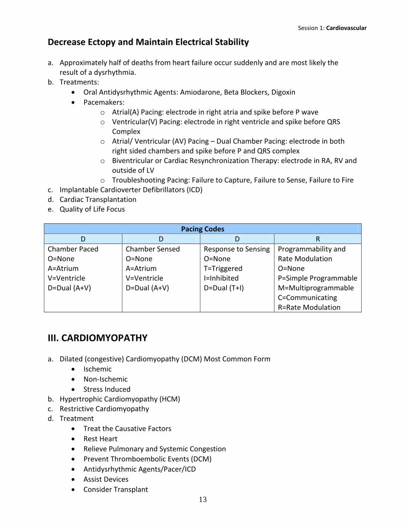
Pacemakers: o Atrial(A) Pacing: electrode in right atria and spike before P wave o Ventricular(V) Pacing: electrode in right ventricle and spike before QRS
Complex o Atrial/ Ventricular (AV) Pacing – Dual Chamber Pacing: electrode in both
right sided chambers and spike before P and QRS complex o Biventricular or Cardiac Resynchronization Therapy: electrode in RA, RV and
outside of LV o Troubleshooting Pacing: Failure to Capture, Failure to Sense, Failure to Fire
c. Implantable Cardioverter Defibrillators (ICD) d. Cardiac Transplantation e. Quality of Life Focus
Pacing Codes
D D D R
Chamber Paced O=None A=Atrium V=Ventricle D=Dual (A+V)
Chamber Sensed O=None A=Atrium V=Ventricle D=Dual (A+V)
Response to Sensing O=None T=Triggered I=Inhibited D=Dual (T+I)
Programmability and Rate Modulation O=None P=Simple Programmable M=Multiprogrammable C=Communicating R=Rate Modulation
III. CARDIOMYOPATHY a. Dilated (congestive) Cardiomyopathy (DCM) Most Common Form
Ischemic
Non-Ischemic
Stress Induced b. Hypertrophic Cardiomyopathy (HCM) c. Restrictive Cardiomyopathy d. Treatment
Treat the Causative Factors
Rest Heart
Relieve Pulmonary and Systemic Congestion
Prevent Thromboembolic Events (DCM)
Antidysrhythmic Agents/Pacer/ICD
Assist Devices
Consider Transplant
Session 1: Cardiovascular
14
Pharmacology o Digitalis * o Diuretics* o Beta-Blockers, Ace Inhibitors o Vasodilators o Inotropic Agents* o Antidysrhythmics o Anticoagulants *caution with HCM
Session 1: Cardiovascular
15
Acute Coronary Syndromes
I. INTRODUCTION The rupture or disruption of the plaque is caused from internal and/or external factors or triggers.
Definitions of chest pain syndromes
Angina Myocardial Anoxia
Exertional Angina Pain that is brought on during times of increased myocardial oxygen demand like exertion, eating, extreme emotions and exposure to cold temperatures, the four Es. These symptoms are typically caused by or a sign of atherosclerosis.
Prinzmetal’s Angina or Variant Angina Pain that occurs at rest, during sleep or without evidence of provocation. Symptoms are thought to be caused by coronary vasospasm.
Stable Angina Exertional angina with consistent symptoms which is typically relieved with rest or cessation of cause and possibly nitroglycerine administration.
Unstable Angina Aka crescendo or pre-infarction angina. Angina that: a. Has a recent onset (within 2 months) and severely limits activity b. Newly occurs at rest c. Differs in characters or symptoms from the person’s ‘typical exertional angina’ (it occurs
with less exertion, has a greater intensity or longer duration, requires more interventions before obtaining relief)
Non-CAD Causes Non-ischemic causes of chest pain must be ruled out, such as:
Cardiac Causes a. Acute Pericarditis b. Cardiac Tamponade c. Acute Myocarditis d. Aortic Stenosis e. Myocardial Contusion f. Mitral Valve Prolapse g. Cardiomyopathies
Session 1: Cardiovascular
16
Non Cardiac Causes a. Panic Attack/Anxiety b. Illicit Drug Use c. Gastrointestinal Disorders d. Spontaneous Pneumothorax e. Pulmonary Embolism f. Pulmonary Hypertension g. Esophageal Rupture h. Costochondritis i. Hypovolemia
II. UNSTABLE ANGINA
Pathophysiology Partially occluding thrombus
Assessment
History a. Assessment of Angina: PQRST Assessment P: Pain, Placement, Provocation Q: Quality, Quantity R: Radiation, Relief S: Severity, Systems (nausea, sweaty, dizziness) T: Timing (when it started, how long did it last) b. Medical History c. Medications: Prescription, Over the Counter, Dietary Supplements d. Social History e. Family History f. Major Risk Factors of Atherosclerosis
12-Lead Electrocardiogram (ECG) Note: Patient can have a ST Segment Elevation Myocardial Infarction (STEMI) or a non-ST Segment Elevation Myocardial Infarction (non-STEMI). All situations are unique and the treatment team must look at entire presentation.
a. Ischemia: ST Segment Depression b. Injury: ST Segment Elevation c. Infarction: Q waves d. Brugada Syndrome: Rare genetic cardiac rhythm disease. Intermittent ST segment elevation in
V1-V3 (Brugada’s sign). May lead to syncope, and even sudden cardiac death. More common to present during sleep. More common in males. Tx is AICD
Session 1: Cardiovascular
17
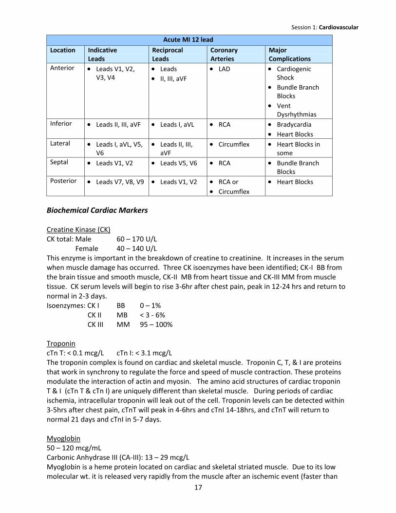
Acute MI 12 lead
Location Indicative Leads
Reciprocal Leads
Coronary Arteries
Major Complications
Anterior Leads V1, V2, V3, V4
Leads II, III, aVF
LAD Cardiogenic Shock
Bundle Branch Blocks
Vent Dysrhythmias
Inferior Leads II, III, aVF Leads I, aVL RCA Bradycardia
Heart Blocks Lateral Leads I, aVL, V5,
V6 Leads II, III,
aVF Circumflex Heart Blocks in
some Septal Leads V1, V2 Leads V5, V6 RCA Bundle Branch
Blocks Posterior Leads V7, V8, V9 Leads V1, V2 RCA or
Circumflex
Heart Blocks
Biochemical Cardiac Markers
Creatine Kinase (CK) CK total: Male 60 – 170 U/L Female 40 – 140 U/L This enzyme is important in the breakdown of creatine to creatinine. It increases in the serum when muscle damage has occurred. Three CK isoenzymes have been identified; CK-I BB from the brain tissue and smooth muscle, CK-II MB from heart tissue and CK-III MM from muscle tissue. CK serum levels will begin to rise 3-6hr after chest pain, peak in 12-24 hrs and return to normal in 2-3 days. Isoenzymes: CK I BB 0 – 1% CK II MB < 3 - 6%
CK III MM 95 – 100% Troponin cTn T: < 0.1 mcg/L cTn I: < 3.1 mcg/L The troponin complex is found on cardiac and skeletal muscle. Troponin C, T, & I are proteins that work in synchrony to regulate the force and speed of muscle contraction. These proteins modulate the interaction of actin and myosin. The amino acid structures of cardiac troponin T & I (cTn T & cTn I) are uniquely different than skeletal muscle. During periods of cardiac ischemia, intracellular troponin will leak out of the cell. Troponin levels can be detected within 3-5hrs after chest pain, cTnT will peak in 4-6hrs and cTnI 14-18hrs, and cTnT will return to normal 21 days and cTnI in 5-7 days. Myoglobin 50 – 120 mcg/mL Carbonic Anhydrase III (CA-III): 13 – 29 mcg/L Myoglobin is a heme protein located on cardiac and skeletal striated muscle. Due to its low molecular wt. it is released very rapidly from the muscle after an ischemic event (faster than
Session 1: Cardiovascular
18
troponin or CK-MB). Serum levels will rise within 2 hrs of chest pain, peak in 3-15hrs and return to normal levels in 2 days. Because there is not a cardiac specific myoglobin, many non-cardiac events may cause an elevation. Carbonic Anydydrase III (CA-III) is another cytoplasmic protein found primarily in skeletal muscle. In skeletal muscle damage both CA III and myoglobin rise. In cardiac muscle damage there is only a rise in myoglobin. Therefore a rise in the myoglobin/CA III ratio is more indicative of an AMI than just an elevated myoglobin. A ratio of > 3.21 is considered abnormal and indicates for cardiac damage.
Early Risk Stratification Early identification of the cause and severity of the pain is essential in determining triage and appropriate therapy. The five factors from the patient’s history that increase the likelihood that the ischemia is from CAD are: a. Nature of Symptoms b. Prior History of CAD c. Gender & Age d. Number of CAD Risk Factors
Treatment Treatment should be initiated as quickly as possible, while assessment is being completed. Immediate general-treatment includes: a. Oxygen at 4L/min b. Aspirin 160-325mg (chewed) c. Nitroglycerin SL or spray d. Morphine IV (if pain not relieved by NTG) e. “MONA” meets the patient (Morphine, Oxygen, Nitroglycerin, Aspirin) f. Clopidogrel (Plavix) 600 mg, or prasugel (Effient) 60mg, or Ticagrelor (Brilinta) 180mg now
part of AHA ACS guidelines (2013)
Once assessment is complete, patient is identified as having characteristics for one of four categories: a. Non-Cardiac Diagnosis b. Chronic Stable Angina c. Possible Acute Coronary Syndrome (ACS) d. Definite ACS
Possible ACS a. Give Aspirin – may have already done so b. Consider Primary Coronary Intervention in Cath Lab c. Consider Antithrombin Tx
ASA
Glycoprotein IIb/IIIa Inhibitor: Abciximab (ReoPro), Eptifibatide (Integrelin), Tirofiban (Aggrastat)
Heparin d. Consider Beta Blocker
Session 1: Cardiovascular
19
III. ACUTE MYOCARDIAL INFARCTION
Pathophysiology Completely occlusive thrombus
Assessment a. History b. Physical Examination c. 12-lead Electrocardiogram: ST elevation d. Biochemical Cardiac Markers
Treatment a. Give triple anti-thrombin tx b. NTG if pain present c. If ST-segment elevation - evaluate for reperfusion
Thrombolytics
Percutaneous Coronary Interventions
Coronary Artery Bypass Grafting
Thrombolytic Agents Thrombolytic agents have been proven to decrease mortality and complications of acute MI. Therapeutic Uses Being given IV, directly into peripheral clot, intracoronary & intracerebral a. Acute Coronary Thrombosis b. DVT c. Massive Pulmonary Emboli d. Adjunct to PCI e. Thrombotic Stroke f. Combination tx of thrombolytic agents and GP IIb/IIIa, UFH, & LMWH have been shown to
increase long term perfusion, mortality & morbidity Absolute Contraindications a. Active Bleeding b. Aortic Dissection c. Cerebral Neoplasm d. History of Intracranial Hemorrhage e. Recent (within 2 mo) intracranial or intraspinal surgery or trauma f. Cerebral Vascular Disease (aneurysm, arteriovenous malformation) g. Bleeding Diathesis h. Severe Uncontrolled Hypertension (> 180/110)
Session 1: Cardiovascular
20
Relative Contraindications a. Recent (within 10 mo) Major Surgery b. Recent (within 10 days) GI or GU bleeding c. High likelihood of Left Heart Thrombus (mitral stenosis or A-fib) d. Acute Pericarditis or Sub acute Bacterial endocarditis e. Significant Liver Dysfunction f. Pregnancy g. Diabetic Hemorrhagic Retinopathy Adverse Effects a. Major Risk is for Bleeding b. Should major bleeding occur
Stop infusion & other anticoagulants
Anticipate immediate head CT if ICH suspected
Administer cryoprecipitate, FFP, platelets
Aminocaproic acid (Amicar)
Interventional Cardiology Percutaneous coronary interventions have increased in both number of procedures and success rates since the first balloon angioplasty was performed in 1977. Percutaneous Coronary Interventions (PCI) a. Diagnostic Coronary Angiography b. Percutaneous Transluminal Coronary Angioplasty (PTCA) c. Coronary Stents
Nursing Care Concerns a. Pre Procedure
BUN/Cret Levels
Dye Allergy
Hydration Status
Anticoagulation and Antiplatelet Medications
Rate & Rhythm
Electrolyte Balance: Especially Potassium
Limb Circulation b. Post Procedure/Potential Complications
Myocardial Ischemia
Stroke
Groin Site Bleeding o Arterial/venous sheaths o Arterial Closure Devices
Distal Circulation
Dysrhythmias
Coronary Artery Spasm
Abrupt Closure/Restenosis
Session 1: Cardiovascular
21
Coronary Artery Dissection
Peripheral Vascular Complication
Discharge Education
Coronary Artery Bypass Grafting Purpose Revascularize the Heart. Procedure a. With the use of cardiopulmonary bypass, hypothermia and cardioplegia the heart is made
motionless and bloodless. b. Grafts are used to supply blood distal to occlusion. c. Cannulation sites for bypass are typically the aorta & RA. d. Minimally-invasive approach. e. Smaller sternal incisions and non-sternal approaches. On and off bypass. Graft Option a. Saphenous Vein b. Internal Mammary c. Radial Artery d. Gastric Artery Post Op Care: (for PCCN >48 hr post op care) a. Pain b. Volume Overload c. MI d. Stroke e. Dysrhythmias f. Infection g. Decrease CO h. Impaired Gas Exchange i. Impaired Work of Breathing j. Hypoperfusion Complications
Additional Nursing Concerns a. Pain b. Immobility c. Risk for Infection d. Life Style Modification e. Discharge Education f. Nutrition Treatment Continued
a. Give -blocker b. Once reperfused evaluate myocardial damage and provide post MI care
Session 1: Cardiovascular
22
Complications of AMI a. Cardiogenic Shock: infarction of > 40% of the left ventricle
Hypotension: SPB < 100mmHg
Pulmonary Edema
Low Cardiac Output
Cardiogenic Pulmonary Edema
S&S of Poor Peripheral Perfusion b. Arrhythmias Associated with Ischemia, Infarction & Reperfusion Treatment Goals for Cardiogenic Shock a. Assist Contractility b. Alleviate Cause of Failure c. Fluid d. Pharmacological Agents e. Coronary Reperfusion f. Mechanical Assist