Embed Size (px)
Citation preview
121
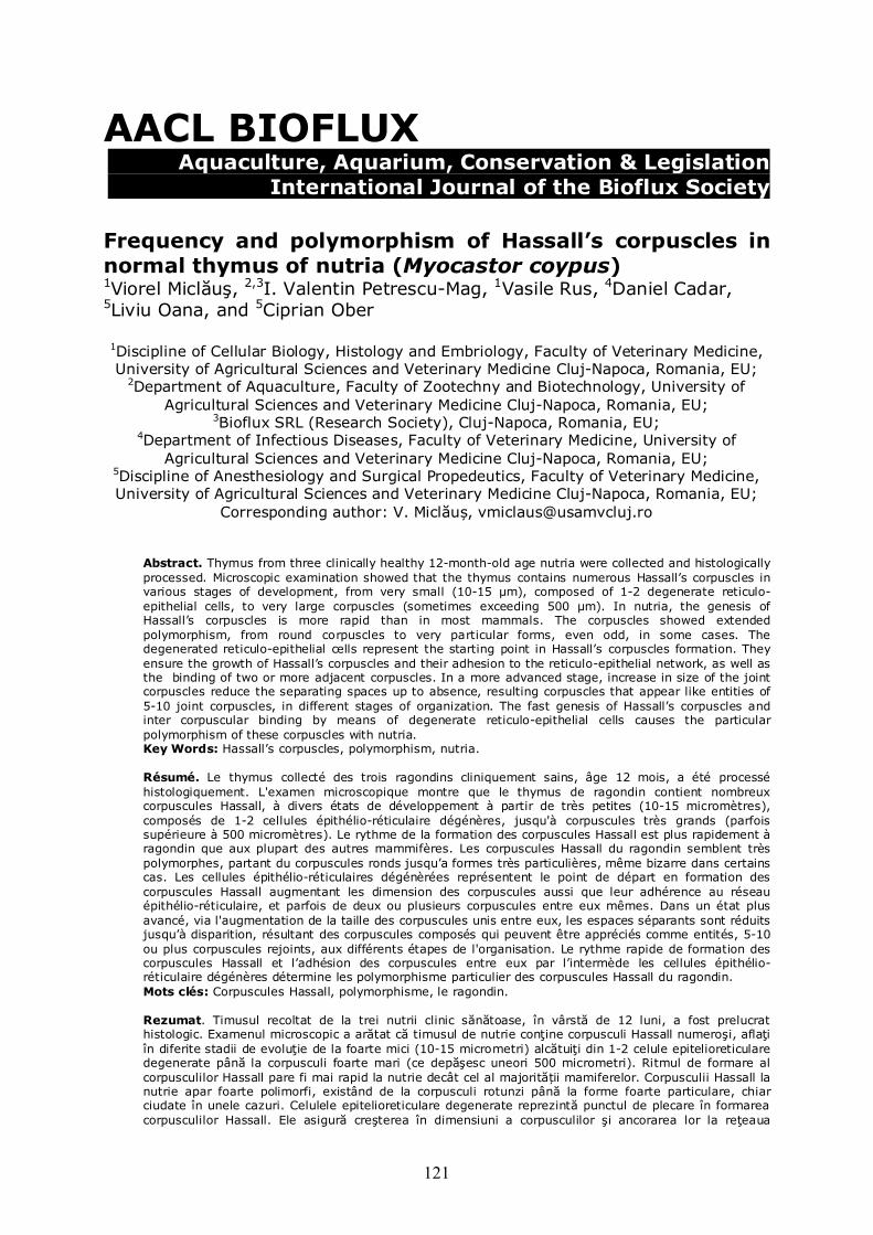
AACL BIOFLUX Aquaculture, Aquarium, Conservation & Legislation International Journal of the Bioflux Society Frequency and polymorphism of Hassall’s corpuscles in normal thymus of nutria (Myocastor coypus) 1Viorel Miclăuş, 2,3I. Valentin Petrescu-Mag, 1Vasile Rus, 4Daniel Cadar, 5Liviu Oana, and 5Ciprian Ober 1Discipline of Cellular Biology, Histology and Embriology, Faculty of Veterinary Medicine, University of Agricultural Sciences and Veterinary Medicine Cluj-Napoca, Romania, EU;
2Department of Aquaculture, Faculty of Zootechny and Biotechnology, University of Agricultural Sciences and Veterinary Medicine Cluj-Napoca, Romania, EU;
3Bioflux SRL (Research Society), Cluj-Napoca, Romania, EU; 4Department of Infectious Diseases, Faculty of Veterinary Medicine, University of
Agricultural Sciences and Veterinary Medicine Cluj-Napoca, Romania, EU; 5Discipline of Anesthesiology and Surgical Propedeutics, Faculty of Veterinary Medicine, University of Agricultural Sciences and Veterinary Medicine Cluj-Napoca, Romania, EU;
Corresponding author: V. Miclăuș, [email protected]
Abstract. Thymus from three clinically healthy 12-month-old age nutria were collected and histologically processed. Microscopic examination showed that the thymus contains numerous Hassall’s corpuscles in various stages of development, from very small (10-15 µm), composed of 1-2 degenerate reticulo-epithelial cells, to very large corpuscles (sometimes exceeding 500 µm). In nutria, the genesis of Hassall’s corpuscles is more rapid than in most mammals. The corpuscles showed extended polymorphism, from round corpuscles to very particular forms, even odd, in some cases. The degenerated reticulo-epithelial cells represent the starting point in Hassall’s corpuscles formation. They ensure the growth of Hassall’s corpuscles and their adhesion to the reticulo-epithelial network, as well as the binding of two or more adjacent corpuscles. In a more advanced stage, increase in size of the joint corpuscles reduce the separating spaces up to absence, resulting corpuscles that appear like entities of 5-10 joint corpuscles, in different stages of organization. The fast genesis of Hassall’s corpuscles and inter corpuscular binding by means of degenerate reticulo-epithelial cells causes the particular polymorphism of these corpuscles with nutria. Key Words: Hassall’s corpuscles, polymorphism, nutria. Résumé. Le thymus collecté des trois ragondins cliniquement sains, âge 12 mois, a été processé histologiquement. L'examen microscopique montre que le thymus de ragondin contient nombreux corpuscules Hassall, à divers états de développement à partir de très petites (10-15 micromètres), composés de 1-2 cellules épithélio-réticulaire dégénères, jusqu'à corpuscules très grands (parfois supérieure à 500 micromètres). Le rythme de la formation des corpuscules Hassall est plus rapidement à ragondin que aux plupart des autres mammifères. Les corpuscules Hassall du ragondin semblent très polymorphes, partant du corpuscules ronds jusqu’a formes très particulières, même bizarre dans certains cas. Les cellules épithélio-réticulaires dégénèrées représentent le point de départ en formation des corpuscules Hassall augmentant les dimension des corpuscules aussi que leur adhérence au réseau épithélio-réticulaire, et parfois de deux ou plusieurs corpuscules entre eux mêmes. Dans un état plus avancé, via l'augmentation de la taille des corpuscules unis entre eux, les espaces séparants sont réduits jusqu’à disparition, résultant des corpuscules composés qui peuvent être appréciés comme entités, 5-10 ou plus corpuscules rejoints, aux différents étapes de l'organisation. Le rythme rapide de formation des corpuscules Hassall et l’adhésion des corpuscules entre eux par l’intermède les cellules épithélio-réticulaire dégénères détermine les polymorphisme particulier des corpuscules Hassall du ragondin. Mots clés: Corpuscules Hassall, polymorphisme, le ragondin. Rezumat. Timusul recoltat de la trei nutrii clinic sănătoase, în vârstă de 12 luni, a fost prelucrat histologic. Examenul microscopic a arătat că timusul de nutrie conţine corpusculi Hassall numeroşi, aflaţi în diferite stadii de evoluţie de la foarte mici (10-15 micrometri) alcătuiţi din 1-2 celule epitelioreticulare degenerate până la corpusculi foarte mari (ce depăşesc uneori 500 micrometri). Ritmul de formare al corpusculilor Hassall pare fi mai rapid la nutrie decât cel al majorității mamiferelor. Corpusculii Hassall la nutrie apar foarte polimorfi, existând de la corpusculi rotunzi până la forme foarte particulare, chiar ciudate în unele cazuri. Celulele epitelioreticulare degenerate reprezintă punctul de plecare în formarea corpusculilor Hassall. Ele asigură creşterea în dimensiuni a corpusculilor şi ancorarea lor la reţeaua
122
epitelio-reticulară, dar şi ancorarea a doi sau mai mulţi corpusculi învecinaţi între ei. Într-un stadiu mai avansat, prin creşterea în dimensiuni a corpusculilor ancoraţi între ei, spaţiile care îi separă se reduc până la dispariţie, rezultând corpusculi compuşi care pot fi apreciaţi ca entităţi de 5-10 sau chiar mai mulţi corpusculi alăturaţi, aflaţi în diferite stadii de organizare. Ritmul alert de formare al corpusculilor Hassall cât şi ancorarea corpusculilor între ei prin celule epitelioreticulare degenerate, determină polimorfismul deosebit al corpusculilor Hassall la nutrie. Cuvinte cheie: corpusculi Hassall, polimorfism, nutrie.
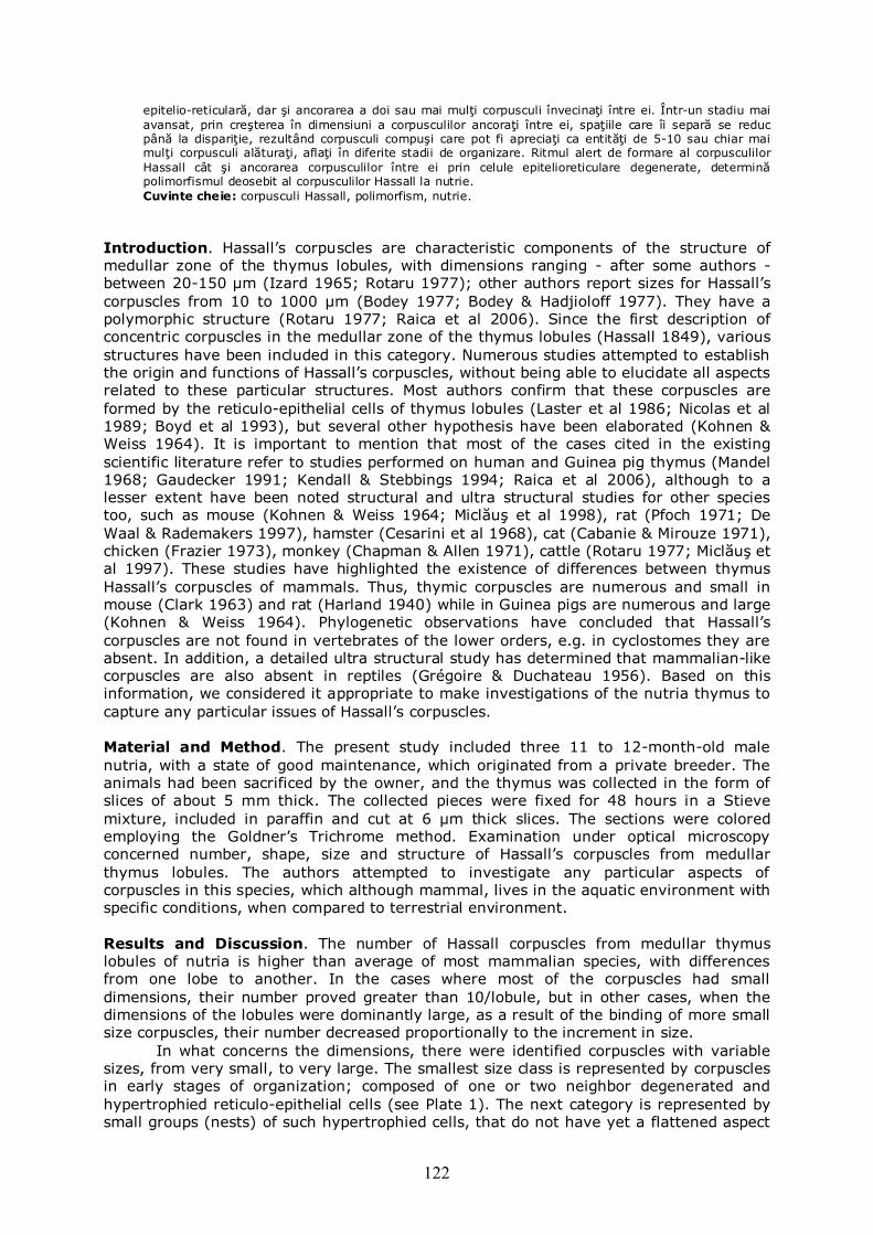
Introduction. Hassall’s corpuscles are characteristic components of the structure of medullar zone of the thymus lobules, with dimensions ranging - after some authors - between 20-150 μm (Izard 1965; Rotaru 1977); other authors report sizes for Hassall’s corpuscles from 10 to 1000 μm (Bodey 1977; Bodey & Hadjioloff 1977). They have a polymorphic structure (Rotaru 1977; Raica et al 2006). Since the first description of concentric corpuscles in the medullar zone of the thymus lobules (Hassall 1849), various structures have been included in this category. Numerous studies attempted to establish the origin and functions of Hassall’s corpuscles, without being able to elucidate all aspects related to these particular structures. Most authors confirm that these corpuscles are formed by the reticulo-epithelial cells of thymus lobules (Laster et al 1986; Nicolas et al 1989; Boyd et al 1993), but several other hypothesis have been elaborated (Kohnen & Weiss 1964). It is important to mention that most of the cases cited in the existing scientific literature refer to studies performed on human and Guinea pig thymus (Mandel 1968; Gaudecker 1991; Kendall & Stebbings 1994; Raica et al 2006), although to a lesser extent have been noted structural and ultra structural studies for other species too, such as mouse (Kohnen & Weiss 1964; Miclăuş et al 1998), rat (Pfoch 1971; De Waal & Rademakers 1997), hamster (Cesarini et al 1968), cat (Cabanie & Mirouze 1971), chicken (Frazier 1973), monkey (Chapman & Allen 1971), cattle (Rotaru 1977; Miclăuş et al 1997). These studies have highlighted the existence of differences between thymus Hassall’s corpuscles of mammals. Thus, thymic corpuscles are numerous and small in mouse (Clark 1963) and rat (Harland 1940) while in Guinea pigs are numerous and large (Kohnen & Weiss 1964). Phylogenetic observations have concluded that Hassall’s corpuscles are not found in vertebrates of the lower orders, e.g. in cyclostomes they are absent. In addition, a detailed ultra structural study has determined that mammalian-like corpuscles are also absent in reptiles (Grégoire & Duchateau 1956). Based on this information, we considered it appropriate to make investigations of the nutria thymus to capture any particular issues of Hassall’s corpuscles. Material and Method. The present study included three 11 to 12-month-old male nutria, with a state of good maintenance, which originated from a private breeder. The animals had been sacrificed by the owner, and the thymus was collected in the form of slices of about 5 mm thick. The collected pieces were fixed for 48 hours in a Stieve mixture, included in paraffin and cut at 6 µm thick slices. The sections were colored employing the Goldner’s Trichrome method. Examination under optical microscopy concerned number, shape, size and structure of Hassall’s corpuscles from medullar thymus lobules. The authors attempted to investigate any particular aspects of corpuscles in this species, which although mammal, lives in the aquatic environment with specific conditions, when compared to terrestrial environment. Results and Discussion. The number of Hassall corpuscles from medullar thymus lobules of nutria is higher than average of most mammalian species, with differences from one lobe to another. In the cases where most of the corpuscles had small dimensions, their number proved greater than 10/lobule, but in other cases, when the dimensions of the lobules were dominantly large, as a result of the binding of more small size corpuscles, their number decreased proportionally to the increment in size.
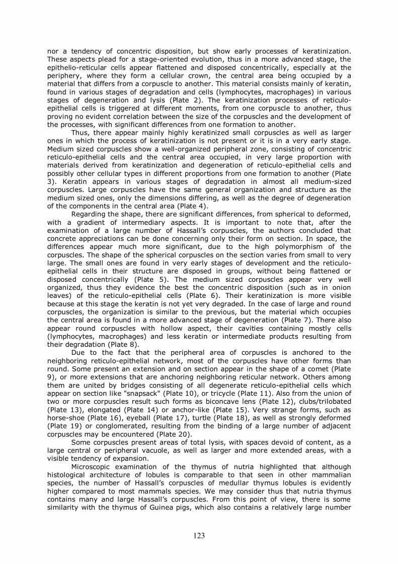
In what concerns the dimensions, there were identified corpuscles with variable sizes, from very small, to very large. The smallest size class is represented by corpuscles in early stages of organization; composed of one or two neighbor degenerated and hypertrophied reticulo-epithelial cells (see Plate 1). The next category is represented by small groups (nests) of such hypertrophied cells, that do not have yet a flattened aspect
123
nor a tendency of concentric disposition, but show early processes of keratinization. These aspects plead for a stage-oriented evolution, thus in a more advanced stage, the epithelio-reticular cells appear flattened and disposed concentrically, especially at the periphery, where they form a cellular crown, the central area being occupied by a material that differs from a corpuscle to another. This material consists mainly of keratin, found in various stages of degradation and cells (lymphocytes, macrophages) in various stages of degeneration and lysis (Plate 2). The keratinization processes of reticulo-epithelial cells is triggered at different moments, from one corpuscle to another, thus proving no evident correlation between the size of the corpuscles and the development of the processes, with significant differences from one formation to another.
Thus, there appear mainly highly keratinized small corpuscles as well as larger ones in which the process of keratinization is not present or it is in a very early stage. Medium sized corpuscles show a well-organized peripheral zone, consisting of concentric reticulo-epithelial cells and the central area occupied, in very large proportion with materials derived from keratinization and degeneration of reticulo-epithelial cells and possibly other cellular types in different proportions from one formation to another (Plate 3). Keratin appears in various stages of degradation in almost all medium-sized corpuscles. Large corpuscles have the same general organization and structure as the medium sized ones, only the dimensions differing, as well as the degree of degeneration of the components in the central area (Plate 4).
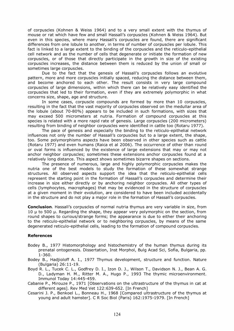
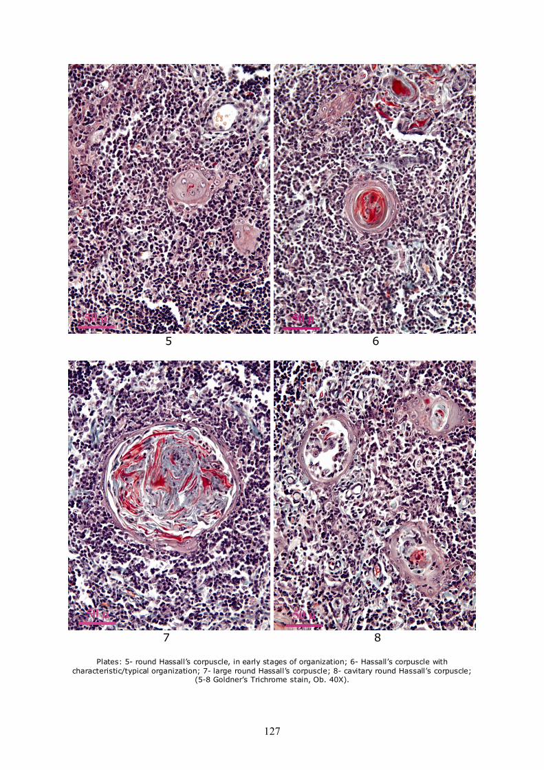
Regarding the shape, there are significant differences, from spherical to deformed, with a gradient of intermediary aspects. It is important to note that, after the examination of a large number of Hassall’s corpuscles, the authors concluded that concrete appreciations can be done concerning only their form on section. In space, the differences appear much more significant, due to the high polymorphism of the corpuscles. The shape of the spherical corpuscles on the section varies from small to very large. The small ones are found in very early stages of development and the reticulo-epithelial cells in their structure are disposed in groups, without being flattened or disposed concentrically (Plate 5). The medium sized corpuscles appear very well organized, thus they evidence the best the concentric disposition (such as in onion leaves) of the reticulo-epithelial cells (Plate 6). Their keratinization is more visible because at this stage the keratin is not yet very degraded. In the case of large and round corpuscles, the organization is similar to the previous, but the material which occupies the central area is found in a more advanced stage of degeneration (Plate 7). There also appear round corpuscles with hollow aspect, their cavities containing mostly cells (lymphocytes, macrophages) and less keratin or intermediate products resulting from their degradation (Plate 8).
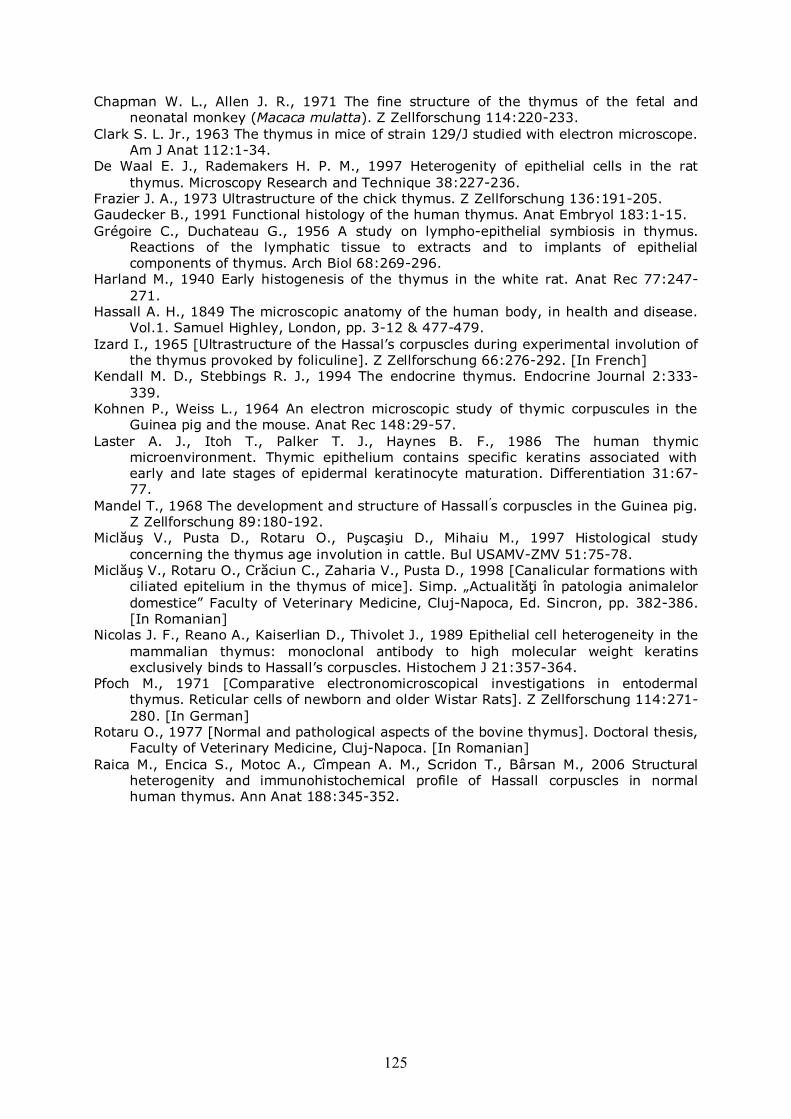
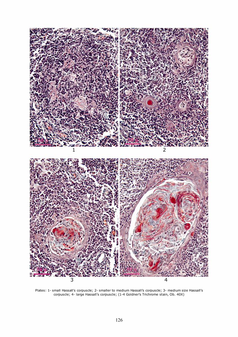
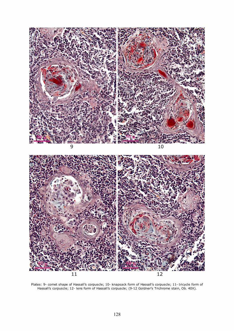
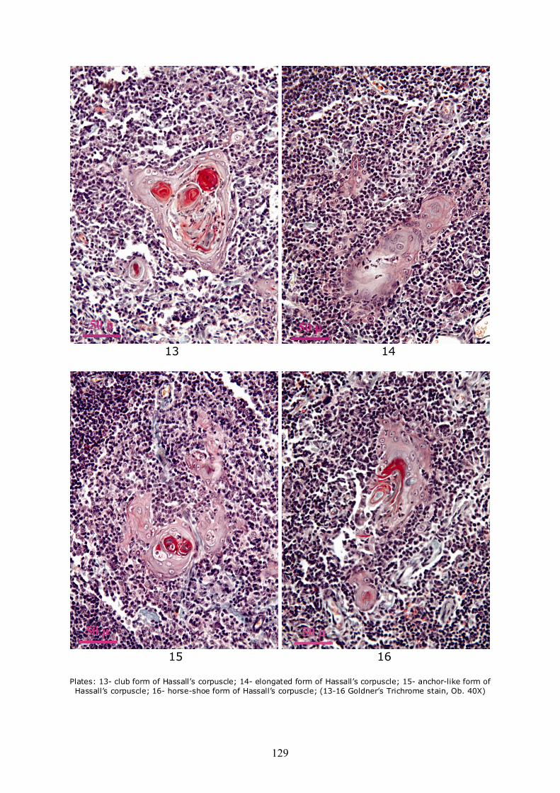
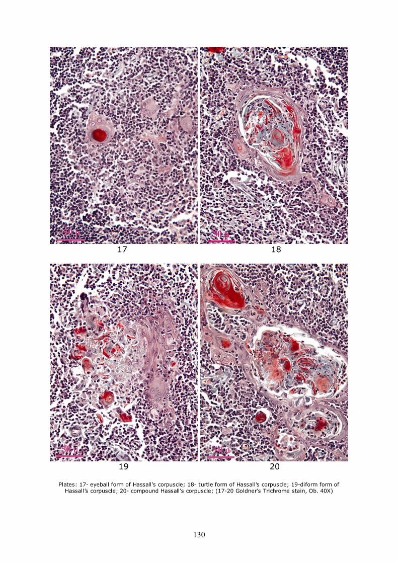
Due to the fact that the peripheral area of corpuscles is anchored to the neighboring reticulo-epithelial network, most of the corpuscles have other forms than round. Some present an extension and on section appear in the shape of a comet (Plate 9), or more extensions that are anchoring neighboring reticular network. Others among them are united by bridges consisting of all degenerate reticulo-epithelial cells which appear on section like "snapsack" (Plate 10), or tricycle (Plate 11). Also from the union of two or more corpuscles result such forms as biconcave lens (Plate 12), clubs/trilobated (Plate 13), elongated (Plate 14) or anchor-like (Plate 15). Very strange forms, such as horse-shoe (Plate 16), eyeball (Plate 17), turtle (Plate 18), as well as strongly deformed (Plate 19) or conglomerated, resulting from the binding of a large number of adjacent corpuscles may be encountered (Plate 20).
Some corpuscles present areas of total lysis, with spaces devoid of content, as a large central or peripheral vacuole, as well as larger and more extended areas, with a visible tendency of expansion.
Microscopic examination of the thymus of nutria highlighted that although histological architecture of lobules is comparable to that seen in other mammalian species, the number of Hassall’s corpuscles of medullar thymus lobules is evidently higher compared to most mammals species. We may consider thus that nutria thymus contains many and large Hassall’s corpuscles. From this point of view, there is some similarity with the thymus of Guinea pigs, which also contains a relatively large number
124
of corpuscles (Kohnen & Weiss 1964) and to a very small extent with the thymus of mouse or rat which have few and small Hassall’s corpuscles (Kohnen & Weiss 1964). But even in this species, where many Hassall’s corpuscles are found, there are significant differences from one lobule to another, in terms of number of corpuscles per lobule. This fact is linked to a large extent to the binding of the corpuscles and the reticulo-epithelial cell network and as the number of cells that degenerate or initiate the formation of new corpuscles, or of those that directly participate in the growth in size of the existing corpuscles increases, the distance between them is reduced by the union of small or sometimes large corpuscles.
Due to the fact that the genesis of Hassall’s corpuscles follows an evolutive pattern, more and more corpuscles initially spaced, reducing the distance between them, and become anchored to each other. The result consists in very large compound corpuscles of large dimensions, within which there can be relatively easy identified the corpuscles that led to their formation, even if they are extremely polymorphic in what concerns size, shape, age and structure.
In some cases, corpuscle compounds are formed by more than 10 corpuscles, resulting in the fact that the vast majority of corpuscles observed on the medullar area of the lobule (about 70-80%) appears to be included in such formations, with sizes that may exceed 500 micrometers at nutria. Formation of compound corpuscles at this species is related with a more rapid rate of genesis. Large corpuscles (200 micrometers) resulting from binding of neighbor corpuscles were identified in cattle too (Rotaru 1977).
The pace of genesis and especially the binding to the reticulo-epithelial network influences not only the number of Hassall’s corpuscles but to a large extent, the shape, too. Some polymorphisms aspects have been observed in other species such as cattle (Rotaru 1977) and even humans (Raica et al 2006). The occurrence of other than round or oval forms is influenced by the existence of large extensions that may or may not anchor neighbor corpuscles; sometimes these extensions anchor corpuscles found at a relatively long distance. This aspect shows sometimes bizarre shapes on sections.
The presence of numerous, large and highly polymorphic corpuscles makes the nutria one of the best models to study the formation of these somewhat strange structures. All observed aspects support the idea that the reticulo-epithelial cells represent the starting point in the formation of Hassall’s corpuscles and determine their increase in size either directly or by anchoring neighbor corpuscles. All other types of cells (lymphocytes, macrophages) that may be evidenced in the structure of corpuscles at a given moment in their evolution, are considered to have been included accidentally in the structure and do not play a major role in the formation of Hassall’s corpuscles. Conclusion. Hassall’s corpuscles of normal nutria thymus are very variable in size, from 10 μ to 500 μ. Regarding the shape, they appear very polymorphic on the section, from round shapes to curious/strange forms; the appearance is due to either their anchoring to the reticulo-epithelial network or to neighboring corpuscles, by means of the same degenerated reticulo-epithelial cells, leading to the formation of compound corpuscles. References Bodey B., 1977 Histomorphology and histochemistry of the human thymus during its
prenatal ontogenesis. Dissertation, Inst Morphol, Bulg Acad Sci, Sofia, Bulgaria, pp. 1-360.
Bodey B., Hadjioloff A. I., 1977 Thymus development, structure and function. Nature (Bulgaria) 26:11-19.
Boyd R. L., Tucek C. L., Godfrey D. I., Izon D. J., Wilson T., Davidson N. J., Bean A. G. D., Ladyman H. M., Ritter M. A., Hugo P., 1993 The thymic microenvironment. Immunol Today 14:445-459.
Cabanie P., Mirouze P., 1971 [Observations on the ultrastructure of the thymus in cat at different ages]. Rev Med Vet 122:639-652. [In French]
Cesarini J. P., Benkoel L., Bonneau H., 1968 [Compared ultrastructure of the thymus at young and adult hamster]. C R Soc Biol (Paris) 162:1975-1979. [In French]
125
Chapman W. L., Allen J. R., 1971 The fine structure of the thymus of the fetal and neonatal monkey (Macaca mulatta). Z Zellforschung 114:220-233.
Clark S. L. Jr., 1963 The thymus in mice of strain 129/J studied with electron microscope. Am J Anat 112:1-34.
De Waal E. J., Rademakers H. P. M., 1997 Heterogenity of epithelial cells in the rat thymus. Microscopy Research and Technique 38:227-236.
Frazier J. A., 1973 Ultrastructure of the chick thymus. Z Zellforschung 136:191-205. Gaudecker B., 1991 Functional histology of the human thymus. Anat Embryol 183:1-15. Grégoire C., Duchateau G., 1956 A study on lympho-epithelial symbiosis in thymus.
Reactions of the lymphatic tissue to extracts and to implants of epithelial components of thymus. Arch Biol 68:269-296.
Harland M., 1940 Early histogenesis of the thymus in the white rat. Anat Rec 77:247-271.
Hassall A. H., 1849 The microscopic anatomy of the human body, in health and disease. Vol.1. Samuel Highley, London, pp. 3-12 & 477-479.
Izard I., 1965 [Ultrastructure of the Hassal’s corpuscles during experimental involution of the thymus provoked by foliculine]. Z Zellforschung 66:276-292. [In French]
Kendall M. D., Stebbings R. J., 1994 The endocrine thymus. Endocrine Journal 2:333-339.
Kohnen P., Weiss L., 1964 An electron microscopic study of thymic corpuscules in the Guinea pig and the mouse. Anat Rec 148:29-57.
Laster A. J., Itoh T., Palker T. J., Haynes B. F., 1986 The human thymic microenvironment. Thymic epithelium contains specific keratins associated with early and late stages of epidermal keratinocyte maturation. Differentiation 31:67-77.
Mandel T., 1968 The development and structure of Hassall’s corpuscles in the Guinea pig. Z Zellforschung 89:180-192.
Miclăuş V., Pusta D., Rotaru O., Puşcaşiu D., Mihaiu M., 1997 Histological study concerning the thymus age involution in cattle. Bul USAMV-ZMV 51:75-78.
Miclăuş V., Rotaru O., Crăciun C., Zaharia V., Pusta D., 1998 [Canalicular formations with ciliated epitelium in the thymus of mice]. Simp. „Actualităţi în patologia animalelor domestice” Faculty of Veterinary Medicine, Cluj-Napoca, Ed. Sincron, pp. 382-386. [In Romanian]
Nicolas J. F., Reano A., Kaiserlian D., Thivolet J., 1989 Epithelial cell heterogeneity in the mammalian thymus: monoclonal antibody to high molecular weight keratins exclusively binds to Hassall’s corpuscles. Histochem J 21:357-364.
Pfoch M., 1971 [Comparative electronomicroscopical investigations in entodermal thymus. Reticular cells of newborn and older Wistar Rats]. Z Zellforschung 114:271-280. [In German]
Rotaru O., 1977 [Normal and pathological aspects of the bovine thymus]. Doctoral thesis, Faculty of Veterinary Medicine, Cluj-Napoca. [In Romanian]
Raica M., Encica S., Motoc A., Cîmpean A. M., Scridon T., Bârsan M., 2006 Structural heterogenity and immunohistochemical profile of Hassall corpuscles in normal human thymus. Ann Anat 188:345-352.
126
1 2
3 4
Plates: 1- small Hassall’s corpuscle; 2- smaller to medium Hassall’s corpuscle; 3- medium size Hassall’s
corpuscle; 4- large Hassall’s corpuscle; (1-4 Goldner’s Trichrome stain, Ob. 40X)
127
5 6
7 8
Plates: 5- round Hassall’s corpuscle, in early stages of organization; 6- Hassall’s corpuscle with
characteristic/typical organization; 7- large round Hassall’s corpuscle; 8- cavitary round Hassall’s corpuscle; (5-8 Goldner’s Trichrome stain, Ob. 40X).
128
9 10
11 12
Plates: 9- comet shape of Hassall’s corpuscle; 10- knapsack form of Hassall’s corpuscle; 11- tricycle form of
Hassall’s corpuscle; 12- lens form of Hassall’s corpuscle; (9-12 Goldner’s Trichrome stain, Ob. 40X).
129
13 14
15 16
Plates: 13- club form of Hassall’s corpuscle; 14- elongated form of Hassall’s corpuscle; 15- anchor-like form of Hassall’s corpuscle; 16- horse-shoe form of Hassall’s corpuscle; (13-16 Goldner’s Trichrome stain, Ob. 40X)
130
17 18
19 20
Plates: 17- eyeball form of Hassall’s corpuscle; 18- turtle form of Hassall’s corpuscle; 19-diform form of
Hassall’s corpuscle; 20- compound Hassall’s corpuscle; (17-20 Goldner’s Trichrome stain, Ob. 40X)
131
Received: 25 February 2009. Accepted: 10 April 2009. Published online: 11 April 2009. Authors: Viorel Miclăuş, Discipline of Cellular Biology, Histology and Embriology, Faculty of Veterinary Medicine, University of Agricultural Sciences and Veterinary Medicine, Calea Mănăștur Street, No 3-5, Cluj-Napoca 400372, Romania, EU, e-mail [email protected] Ioan Valentin Petrescu-Mag, Department of Aquaculture, Faculty of Zootechny and Biotechnology, University of Agricultural Sciences and Veterinary Medicine, Calea Mănăștur Street, No 3-5, Cluj-Napoca 400372, Romania, EU; Bioflux SRL, Ceahlău Street No 54, Cluj-Napoca 400488, Romania, EU, e-mail: [email protected] Vasile Rus, Discipline of Cellular Biology, Histology and Embriology, Faculty of Veterinary Medicine, University of Agricultural Sciences and Veterinary Medicine, Calea Mănăștur Street, No 3-5, Cluj-Napoca 400372, Romania, EU, e-mail: [email protected] Daniel Cadar, Department of Infectious Diseases, Faculty of Veterinary Medicine, University of Agricultural Sciences and Veterinary Medicine, Calea Mănăștur Street, No 3-5, Cluj-Napoca 400372, Romania, EU; Liviu Oana, Discipline of Anesthesiology and Surgical Propedeutics, Faculty of Veterinary Medicine, University of Agricultural Sciences and Veterinary Medicine, Calea Mănăștur Street, No 3-5, Cluj-Napoca 400372, Romania, EU; Ciprian Ober, Discipline of Anesthesiology and Surgical Propedeutics, Faculty of Veterinary Medicine, University of Agricultural Sciences and Veterinary Medicine, Calea Mănăștur Street, No 3-5, Cluj-Napoca 400372, Romania, EU. How to cite this article: Miclăuș V., Petrescu-Mag I. V., Rus V., Cadar D., Oana L., Ober C., 2009 Frequency and polymorphism of Hassall’s corpuscles in normal thymus of nutria (Myocastor coypus). AACL Bioflux 2(2):121-131. Printed version: ISSN 1844-8143 Online version: ISSN 1844-9166 available at: http://www.bioflux.com.ro/docs/2009.2.121-131.pdf Submitted and accepted also in the official program of International Symposium ACVAPEDIA 2009.

132