Embed Size (px)
Citation preview

Bull. Soc. Nat. luxemb. 115 (2014) 141
Aabaarnia and Normanogalla, two new lichenicolous genera of Ostropales, Ascomycota
Paul DiederichMusée national d’histoire naturelle, 25 rue Munster, L-2160 Luxembourg, Luxembourg ([email protected])
Diederich, P., 2014. Aabaarnia and Normanogalla, two new lichenicolous genera of Ostropales, Ascomycota. Bulletin de la Société des naturalistes luxembourgeois 115 : 141–149.
Abstract. The new genus and species Aabaarnia siphulicola is described for a lichenicolous, gall-inducing ascomycete growing on Siphula decumbens in Australia (Tasmania) and New Zealand (Auckland Islands). The species is characterized by pale, immersed, cleistohymenial ascomata, an ascomatal wall without crystals, missing periphysoids, a K/I+ blue hymenium, subcylindrical, 4–6-spored, K/I– asci with a massive apical cap, and oval to short-cylindrical, 3-septate ascospores. The new genus Normanogalla is described for the lichenicolous, gall-inducing N. cribriformis (previously known as Unguiculariopsis cribriformis), known on terricolous Pertusaria in Scandinavia and Greenland. It is characterized by pale, immersed, cleistohymenial ascomata, an ascomatal wall without crystals, abundant periphysoids, a K/I– hymenium, subcylindrical, 8-spored, K/I– asci with an apically not or slightly thicker wall, and subspherical, aseptate ascospores.
IntroductionThe ostropalean fungi, as circumscribed by Sherwood (1977), included ascomyce-tes with apothecioid (rarely perithecioid) ascomata that are usually immersed and closed when young, opening by an irre-gular pore or a slit (cleistohymenial asco-matal development), a hamathecium of true paraphyses, periphysoids often pre-sent, subcylindrical asci that are thick-walled when young, but functionally not bitunicate, with a distinct I– apical thicke-ning, long-cylindrical to filiform ascospo-res and an ascomatal wall frequently with crystalline inclusions (Ostropales s.str.), or ovoid ascospores or wall without cry-stalline inclusions (Ostropales s. lat.). In addition to the Stictidaceae, Hawksworth & Sherwood (1982) described a second family of Ostropales, the Odontotre-mataceae, that were eventually revised by Sherwood-Pike (1987). One genus of Stic-didaceae, Nanostictis, and three genera of Odontotremataceae, Odontotrema, Paralethariicola and Spirographa, included obligate lichenicolous species. Recent molecular studies allowed enlarging the concept of Ostropales, which now also
include the Coenogoniaceae, Gomphilla-ceae, Graphidaceae, Gyalectaceae, Myelo-conidaceae, Phaneromycetaceae, Phlycti-daceae, Porinaceae and Thelotremataceae (Lumbsch & Huhndorf 2007). On the other hand, Baloch et al. (2013) showed that Odontotrema (Odontotremataceae) is poly-phyletic, and most taxa, including all liche-nicolous species, were transferred to Sphaeropezia (Stictidaceae).In this paper we describe two new genera of lichenicolous ascomycetes that we include in the Ostropales. As the type species of both have rarely been collected, and as no recent collections are available, no molecular data could be obtained, hence they cannot be attributed to a family at this moment.
Material and MethodsMaterial from BR and UPS has been exa-mined. Dry herbarium specimens were examined and measured under a binocu-lar microscope Leica MZ 7.5 (magnifica-tion up to 50×). Macroscopic photographs were done using a Canon 40D camera with a Canon MP-E 65 mm macro lens, Stack
142 Bull. Soc. Nat. luxemb. 115 (2014)
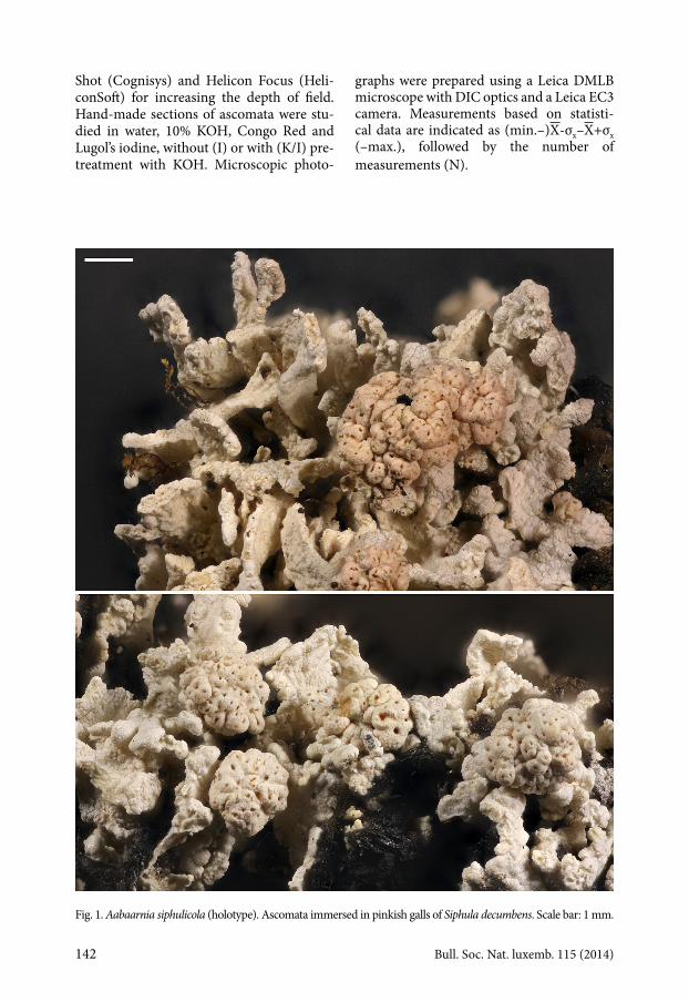
Shot (Cognisys) and Helicon Focus (Heli-conSoft) for increasing the depth of field. Hand-made sections of ascomata were stu-died in water, 10% KOH, Congo Red and Lugol’s iodine, without (I) or with (K/I) pre-treatment with KOH. Microscopic photo-
graphs were prepared using a Leica DMLB microscope with DIC optics and a Leica EC3 camera. Measurements based on statisti-cal data are indicated as (min.–)X-σx–X+σx (–max.), followed by the number of measurements (N).
Fig. 1. Aabaarnia siphulicola (holotype). Ascomata immersed in pinkish galls of Siphula decumbens. Scale bar: 1 mm.

Bull. Soc. Nat. luxemb. 115 (2014) 143
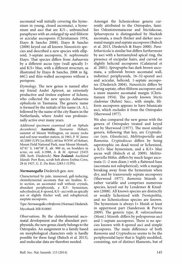
Fig. 2. Aabaarnia siphulicola (holotype). A, Section through closed, immature ascoma, in water. B, Section through immature, opened ascoma in lactophenol-cotton blue. C, The same, in polarized light, showing crystals of host thallus present in the ascomatal wall. D, Section through mature ascoma, in 10% KOH. E, The same, in polarized light, with crystals absent in ascomatal wall. F, Magnification of B, showing hamathecium near ostiole in immature ascoma. G, Section through mature ascoma, filled with asci containing ascospores (DIC). H, The same, showing reaction of Lugol’s reagent entering hamathecium, after pre-treatment with 10% KOH. I, Mature ascospores in 10% KOH, showing perispore. J, Mature ascospores in water (DIC), showing perispore. K, Asci (DIC). L, Ascus and paraphyses (DIC). M, Asci in Lugol’s reagent after pre-treatment with 10% KOH. Scale bars: A–E, G–H = 50 µm; F, I–M = 10 µm.
144 Bull. Soc. Nat. luxemb. 115 (2014)
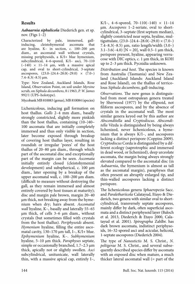
ResultsAabaarnia siphulicola Diederich gen. et sp. nov. (Figs 1–2)Characterized by pale, immersed, gall-inducing, cleistohymenial ascomata that are hyaline, K– in section, c. 100–200 µm diam., an ascomatal wall without crystals, missing periphysoids, a K/I+ blue hymenium, subcylindrical, 4–6-spored, K/I– asci, 70–110 (–140) × 11–14 µm, with a massive apical cap, and oval to short-cylindrical, 3-septate ascospores, (23.0–)24.4–28.0(–29.0) × (7.0–) 7.4–8.3(–8.5) µm.Type: New Zealand, Auckland Islands, Rose Island, Observation Point, on soil under Myrsine scrub, on Siphula decumbens, 8 i 1963, P. W. James 992/1 (UPS–holotype).Mycobank MB 810083 (genus), MB 810084 (species)
Lichenicolous, inducing gall formation on host thallus. Galls 2–4 mm diam., basally strongly constricted, slightly more pinkish than the host thallus, containing (10–)40–100 ascomata that are initially completely immersed and thus only visible in section, later become exposed through breakup of covering host thallus layer, resulting in roundish or irregular ‘pores’ of the host thallus of 20–80 µm diam., through which part of the ascomatal disc and exceptionally part of the margin can be seen. Ascomata initially entirely closed (cleistohymenial development) and subspherical, c. 100 µm diam., later opening by a breakup of the upper ascomatal wall, c. 100–200 µm diam. (difficult to measure without destroying the gall, as they remain immersed and almost entirely covered by host tissues at maturity); disc and margin pale brown, margin 20–40 µm thick, not breaking away from the hyme-nium when dry; hairs absent. Ascomatal wall hyaline, K–, basally and laterally 55–65 µm thick, of cells 3–6 µm diam., without crystals (but sometimes filled with crystals from the host thallus). Periphysoids absent. Hymenium hyaline, filling the entire asco-matal cavity, 130–170 µm tall, I–, K/I+ blue. Epihymenium hyaline, K–. Subhymenium hyaline, 5–10 µm thick. Paraphyses septate, simple or occasionally branched, 1.7–2.5 µm thick, apically not or slightly swollen. Asci subcylindrical, unitunicate, wall laterally thin, with a massive apical cap, entirely I–,
K/I–, 4–6-spored, 70–110(–140) × 11–14 µm. Ascospores 1–2-seriate, oval to short-cylindrical, 3–septate (first septum median), slightly constricted near septa, hyaline, mul-tiguttulate, (23.0–)24.4–28.0(–29.0) × (7.0–) 7.4–8.3(–8.5) µm, ratio length/width (3.0–) 3.1–3.6(–4.0) [N = 20], wall 0.5 –1 µm thick, perispore present, hyaline, appearing verru-cose with DIC optics, c. 1 µm thick, in KOH up to 2–3 µm thick. Pycnidia unknown.Distribution and host. The species is known from Australia (Tasmania) and New Zea-land (Auckland Islands: Auckland Island and Rose Island), on the thallus of terrico-lous Siphula decumbens, gall-inducing.Observations. The new genus is distinguis-hed from most ostropalean genera treated by Sherwood (1977) by the ellipsoid, not filiform ascospores, and by the absence of crystals in the ascomatal wall. The most similar genera keyed out by this author are Absconditella and Cryptodiscus. Absconditella Vězda is distinguished by being always lichenized, never lichenicolous, a hyme-nium that is always K/I–, and ascospores lacking a distinct perispore (Coppins 2009). Cryptodiscus Corda is distinguished by a dif-ferent ecology (saprotrophic and immersed in dead wood, or lichenized), deep urceolate ascomata, the margin being always strongly elevated compared to the ascomatal disc (in Aabaarnia, the hymenium is almost as tall as the ascomatal margin), paraphyses that often present an abruptly enlarged tip, and thin-walled ascospores lacking a distinct perispore.The lichenicolous genera Sphaeropezia Sacc. and Paralethariicola Calatayud, Etayo & Die-derich, two genera with similar oval to short-cylindrical, transversely septate ascospores, mainly differ by dark, often superficial asco-mata and a distinct periphysoid layer (Baloch et al. 2013, Diederich & Etayo 2000, Cala-tayud et al. 2001). Spirographa Zahlbr. has dark brown ascomata, indistinct periphyso-ids, 16–32-spored asci and acicular, helicoid, 1-septate ascospores (Diederich 2004).The type of Nanostictis M. S. Christ., N. peltigerae M. S. Christ., and several subse-quently described species differ by ascomata with an exposed disc when mature, a much thicker lateral ascomatal wall (= part of the
Bull. Soc. Nat. luxemb. 115 (2014) 145
ascomatal wall initially covering the hyme-nium in young, closed ascomata), a hyme-nium and asci that are K/I–, unbranched paraphyses with an enlarged tip and filiform or acicular ascospores (Christiansen 1954, Etayo & Sancho 2008). Etayo & Sancho (2008) keyed out all known Nanostictis spe-cies and described a new species with ellip-soid, 3-septate ascospores, N. nephromatis Etayo. That species differs from Aabaarnia by a different ascus type (wall apically I+ and K/I+ blue, with a different structure, as illustrated by Etayo & Sancho, 2008 in fig. 66C) and thin-walled ascospores without a perispore. Etymology. The new genus is named after my friend André Aptroot, an extremely productive and tireless explorer of lichens, who collected the new species Aabaarnia siphulicola in Tasmania. The generic name is formed by the initials of his name (A. A.), followed by the name of the city Baarn in the Netherlands, where André was professio-nally active over many years.Additional specimens examined (all on Siphula decumbens): Australia: Tasmania: Hobart, summit of Mount Wellington, on mossy rocks and soil near weather station, iii 1963, P. W. James AU2085 (UPS [ex BM]); 60 km WNW of Hobart, Mount Field National Park, near Mount Monash, 42°41’ S, 146°38’ E, alt. 900 m, on boulders in scree, on soil, 4.1988, A. & M. Aptroot 23435 (BR, herb. Diederich). New Zealand: Auckland Islands: Port Ross, scrub belt above Erebus Cove, 28 iii 1927, G. E. Du Rietz 2283:1 (UPS).
Normanogalla Diederich gen. nov.Characterized by pale, immersed, gall-inducing, cleistohymenial ascomata that are hyaline, K– in section, an ascomatal wall without crystals, abundant periphysoids, a K/I– hymenium, subcylindrical, 8-spored, K/I– asci with an apically not or slightly thicker wall, and subspherical, aseptate ascospores.Type: Normanogalla cribriformis (Norman) Diederich.Mycobank MB 810085
Observations. By the cleistohymenial asco-matal development and the abundant peri-physoids, the new genus clearly belongs to the Ostropales. An assignment to a family based on morphological characters only is hardly possible for these fungi (Baloch et al. 2013), and molecular data are therefore needed.
Amongst the lichenicolous genera cur-rently attributed to the Ostropales, fami-lies Odonto tremataceae and Stictidaceae, Sphaero pezia is distinguished by blackish ascomata, a much thicker and darker asco-matal margin and septate ascospores (Baloch et al. 2013, Diederich & Etayo 2000). Para-lethariicola is similar but differs furthermore by asci with a hemiamyloid apical ring, the presence of excipular hairs, and curved or slightly helicoid ascospores (Calatayud et al. 2001). Spirographa has dark brown asco-mata, a yellowish brown ascomatal wall, indistinct periphysoids, 16–32-spored asci and acicular, helicoid, 1-septate ascospo-res (Diederich 2004). Nanostictis differs by having septate, often filiform ascospores and a more massive ascomatal margin (Chris-tiansen 1954). The poorly known Stictis cladoniae (Rehm) Sacc., with simple, fili-form ascospores appears to have bitunicate asci, which excludes it from the Ostropales (Sherwood 1977). We also compared the new genus with the genera of Ostropales treated and keyed out by Sherwood (1977). The most similar genera, following that key, are Cryptodiscus (syn. Gloeolecta), Melittosporiella and Ramonia. Cryptodiscus differs by being saprotrophic on dead wood or lichenized, a K/I+ blue hymenium, and a K/I+ blue ascus wall (Baloch et al. 2009). Melittosporiella Höhn. differs by much larger asco-mata (1–2 mm diam.) with a flattened base (ascomata not subspherical), with a margin breaking away from the hymenium when dry, and by transversely septate ascospores (Sherwood 1977). Ramonia Stizenb. is rather variable and comprises numerous species, keyed out by Lendemer & Knud-sen (2008). All known species are distinctly or weakly lichenized with Trentepohlia, and no lichenicolous species are known. The hymenium is always I+ bluish at least in uppermost part (Sanderson & Purvis 2009). The generic type, R. valenzueliana (Mont.) Stizenb. differs by polysporous asci and 1-septate ascospores. There is no spe-cies known with 8-spored asci and simple ascospores. The main difference of both Ramonia and Cryptodiscus seems to be the periphysoidal layer that is ‘highly modified, consisting, not of distinct filaments, but of
146 Bull. Soc. Nat. luxemb. 115 (2014)
a compact, fleshy, pseudoparenchymatous matrix in which the cells are oriented per-pendicularly to the surface of the ascocarp’
(Sherwood 1977: 17), whereas in Normanogalla, this layer consists of distinct peri-physoids.
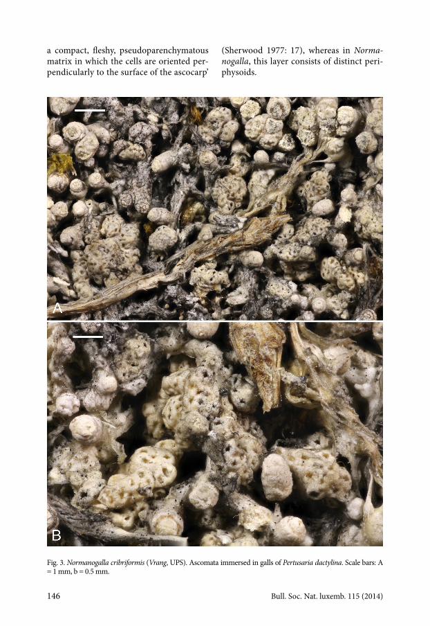
Fig. 3. Normanogalla cribriformis (Vrang, UPS). Ascomata immersed in galls of Pertusaria dactylina. Scale bars: A = 1 mm, b = 0.5 mm.

Bull. Soc. Nat. luxemb. 115 (2014) 147
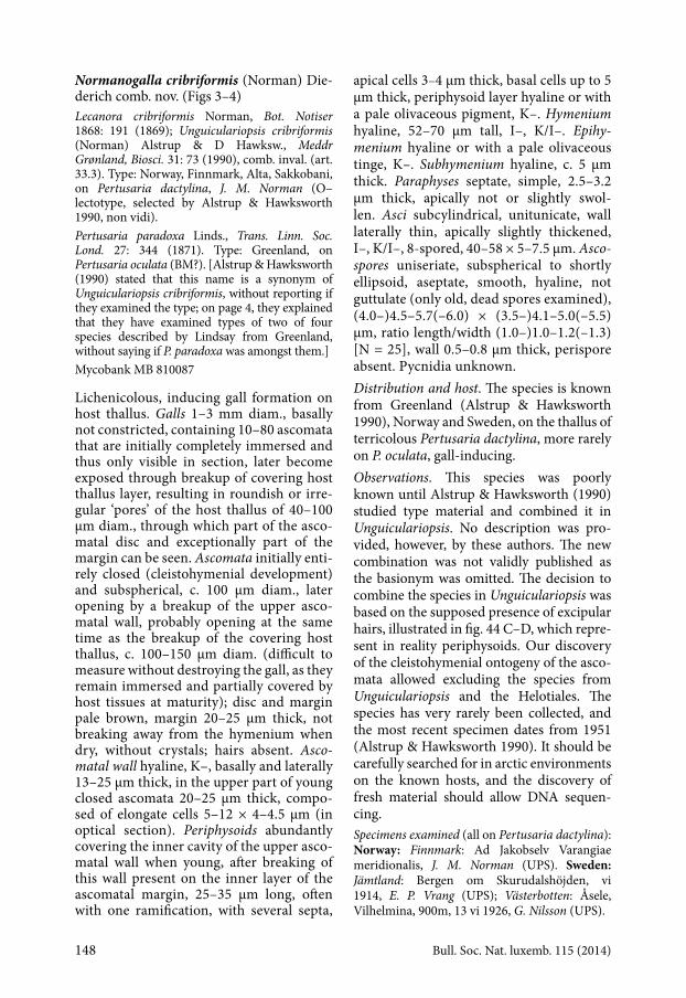
Fig. 4. Normanogalla cribriformis (Vrang, UPS). A, Section through closed ascoma, showing hymenium (left, bottom and right) and periphysoid layer (top). B, Periphysoids (centre) and paraphyses (right) in squash preparation. C, Section through opened ascoma, with periphysoid layer along the ascending margins. D, Hymenium and subhymenium, with periphysoid layer visible on the top left. E, Asci. F, Ascospores. [C, D: in water. A, B, F: in a mixture of Congo Red and 10% KOH. E: in Lugol after KOH-pretreatment. A, B, D, F: DIC optics.]. Scale bars: A, C = 50 µm; B, D = 20 µm; E–F = 5 µm.
148 Bull. Soc. Nat. luxemb. 115 (2014)
Normanogalla cribriformis (Norman) Die-derich comb. nov. (Figs 3–4)Lecanora cribriformis Norman, Bot. Notiser 1868: 191 (1869); Unguiculariopsis cribriformis (Norman) Alstrup & D Hawksw., Meddr Grønland, Biosci. 31: 73 (1990), comb. inval. (art. 33.3). Type: Norway, Finnmark, Alta, Sakkobani, on Pertusaria dactylina, J. M. Norman (O–lectotype, selected by Alstrup & Hawksworth 1990, non vidi).Pertusaria paradoxa Linds., Trans. Linn. Soc. Lond. 27: 344 (1871). Type: Greenland, on Pertusaria oculata (BM?). [Alstrup & Hawksworth (1990) stated that this name is a synonym of Unguiculariopsis cribriformis, without reporting if they examined the type; on page 4, they explained that they have examined types of two of four species described by Lindsay from Greenland, without saying if P. paradoxa was amongst them.]Mycobank MB 810087
Lichenicolous, inducing gall formation on host thallus. Galls 1–3 mm diam., basally not constricted, containing 10–80 ascomata that are initially completely immersed and thus only visible in section, later become exposed through breakup of covering host thallus layer, resulting in roundish or irre-gular ‘pores’ of the host thallus of 40–100 µm diam., through which part of the asco-matal disc and exceptionally part of the margin can be seen. Ascomata initially enti-rely closed (cleistohymenial development) and subspherical, c. 100 µm diam., later opening by a breakup of the upper asco-matal wall, probably opening at the same time as the breakup of the covering host thallus, c. 100–150 µm diam. (difficult to measure without destroying the gall, as they remain immersed and partially covered by host tissues at maturity); disc and margin pale brown, margin 20–25 µm thick, not breaking away from the hymenium when dry, without crystals; hairs absent. Ascomatal wall hyaline, K–, basally and laterally 13–25 µm thick, in the upper part of young closed ascomata 20–25 µm thick, compo-sed of elongate cells 5–12 × 4–4.5 μm (in optical section). Periphysoids abundantly covering the inner cavity of the upper asco-matal wall when young, after breaking of this wall present on the inner layer of the ascomatal margin, 25–35 µm long, often with one ramification, with several septa,
apical cells 3–4 µm thick, basal cells up to 5 µm thick, periphysoid layer hyaline or with a pale olivaceous pigment, K–. Hymenium hyaline, 52–70 µm tall, I–, K/I–. Epihymenium hyaline or with a pale olivaceous tinge, K–. Subhymenium hyaline, c. 5 µm thick. Paraphyses septate, simple, 2.5–3.2 µm thick, apically not or slightly swol-len. Asci subcylindrical, unitunicate, wall laterally thin, apically slightly thickened, I–, K/I–, 8-spored, 40–58 × 5–7.5 µm. Ascospores uniseriate, subspherical to shortly ellipsoid, aseptate, smooth, hyaline, not guttulate (only old, dead spores examined), (4.0–)4.5–5.7(–6.0) × (3.5–)4.1–5.0(–5.5) µm, ratio length/width (1.0–)1.0–1.2(–1.3) [N = 25], wall 0.5–0.8 µm thick, perispore absent. Pycnidia unknown.Distribution and host. The species is known from Greenland (Alstrup & Hawksworth 1990), Norway and Sweden, on the thallus of terricolous Pertusaria dactylina, more rarely on P. oculata, gall-inducing. Observations. This species was poorly known until Alstrup & Hawksworth (1990) studied type material and combined it in Unguiculariopsis. No description was pro-vided, however, by these authors. The new combination was not validly published as the basionym was omitted. The decision to combine the species in Unguiculariopsis was based on the supposed presence of excipular hairs, illustrated in fig. 44 C–D, which repre-sent in reality periphysoids. Our discovery of the cleistohymenial ontogeny of the asco-mata allowed excluding the species from Unguiculariopsis and the Helotiales. The species has very rarely been collected, and the most recent specimen dates from 1951 (Alstrup & Hawksworth 1990). It should be carefully searched for in arctic environments on the known hosts, and the discovery of fresh material should allow DNA sequen-cing.Specimens examined (all on Pertusaria dactylina): Norway: Finnmark: Ad Jakobselv Varangiae meridionalis, J. M. Norman (UPS). Sweden: Jämtland: Bergen om Skurudalshöjden, vi 1914, E. P. Vrang (UPS); Västerbotten: Åsele, Vilhelmina, 900m, 13 vi 1926, G. Nilsson (UPS).
Bull. Soc. Nat. luxemb. 115 (2014) 149
AcknowledgementsI warmly thank the curators of BR and UPS for the loan of specimens in their care, André Aptroot and Rolf Santesson for putting at my disposal unidentified specimens of Aabaarnia siphulae, and Brian Coppins for critically reading the manuscript.
ReferencesAlstrup, V. & D. L. Hawksworth, 1990. The
lichenicolous fungi of Greenland. Meddelelser om GrØnland, Bioscience 31: 1–90.
Baloch, E., G. Gilenstam & M. Wedin, 2009. Phylogeny and classification of Cryptodiscus, with a taxonomic synopsis of the Swedish species. Fungal Diversity 38: 51–68.
Baloch, E., G. Gilenstam & M. Wedin, 2013. The relationships of Odontotrema (Odontotre-mataceae) and the resurrected Sphaeropezia (Stictidaceae) – new combinations and three new Sphaeropezia species. Mycologia 105: 384–397.
Calatayud, V., J. Etayo & P. Diederich, 2001. Paralethariicola aspiciliae (Ostropales, Odon-totremataceae), a new genus and species of lichenicolous fungi. Lichenologist 33: 477–482.
Christiansen, M. S., 1954. Nanostictis, a new genus of scolecosporous Discomycetes. Bot. Tidskr. 51: 59–65.
Coppins, B. J., 2009. Absconditella Vězda (1965). In: C. W. Smith, A. Aptroot, B. J. Coppins, A. Fletcher, O. L. Gilbert, P. W. James & P. A. Wolseley (eds): The lichens of Great Britain
and Ireland. British Lichen Society, London, pp. 123–124.
Diederich, P., 2004. Spirographa. In: T. H. Nash III, B. D. Ryan, P. Diederich, C. Gries, F. Bun-gartz (eds.): Lichen flora of the Greater Sono-ran Desert Region, Vol. 2. Lichens Unlimited, Arizona State University, Tempe, Arizona, pp. 702–703.
Diederich, P. & J. Etayo, 2000. A synopsis of the genera Skyttea, Llimoniella and Rhymbocarpus (lichenicolous Ascomycota, Leotiales). Lichenologist 32: 423–485.
Etayo, J. & L. G. Sancho, 2008. Hongos liquení-colas del Sur de Sudamérica, especialmente de Isla Navarino (Chile). Bibliotheca Lichenologica 98: 302 pp.
Hawksworth, D. L. & M. A. Sherwood, 1982. Two new families in the Ascomycotina. Mycotaxon 16: 262–264.
Lendemer, J. C. & K. Knudsen, 2008. Ramonia vermispora, a new species from the Sonoran Desert region of southwestern North Amer-ica. Opuscula Philolichenum 5: 83–88.
Lumbsch, H. T. & S. M. Huhndorf, 2007. Outline of Ascomycota – 2007. Myconet 13: 1–58.
Sanderson, N. A. & O. W. Purvis, 2009. Ramonia Stizenb. (1862). In: C. W. Smith, A. Aptroot, B. J. Coppins, A. Fletcher, O. L. Gilbert, P. W. James & P. A. Wolseley (eds): The lichens of Great Britain and Ireland. British Lichen Society, London, pp. 788–790.
Sherwood, M. A., 1977. The ostropalean fungi. Mycotaxon 5: 1–277.
Sherwood-Pike, M. A., 1987. The Ostropalean Fungi III: the Odontotremataceae. Mycotaxon 28: 137–177.