Embed Size (px)
Citation preview
Biochemical and Biophysical Research Communications 443 (2014) 677–682
Contents lists available at ScienceDirect
Biochemical and Biophysical Research Communications
journal homepage: www.elsevier .com/locate /ybbrc
A unique F-type H+-ATPase from Streptococcus mutans: An active H+
pump at acidic pH
0006-291X/$ - see front matter � 2013 Elsevier Inc. All rights reserved.http://dx.doi.org/10.1016/j.bbrc.2013.12.025
Abbreviations: SFOF1, S. mutans FOF1 expressed in E. coli membranes; EFOF1, E. coliFOF1 expressed in E. coli membranes; SFOEF1, hybrid FOF1 formed from S. mutans FO
and E. coli F1.⇑ Corresponding author. Fax: +81 749 64 8140.
E-mail address: [email protected] (A. Iwamoto-Kihara).
Yuka Sasaki a, Eri Nogami a, Masatomo Maeda b, Mayumi Nakanishi-Matsui b, Atsuko Iwamoto-Kihara a,⇑a Department of Bioscience, Nagahama Institute of Bioscience and Technology, 1266 Tamura, Nagahama, Shiga 526-0829, Japanb Department of Molecular Biology, School of Pharmacy, Iwate Medical University, 2-1-1 Nishitokuta, Yahaba, Iwate 028-3694, Japan
a r t i c l e i n f o a b s t r a c t
Article history:Received 2 December 2013Available online 11 December 2013
Keywords:H+-ATPaseATP synthaseFOF1
c SubunitS. mutansDental caries
We have shown previously that the Streptococcus mutans F-type H+-ATPase (FOF1) c subunit gene couldcomplement Escherichia coli defective in the corresponding gene, particularly at acidic pH (Araki et al.,(2013) [14]). In this study, the entire S. mutans FOF1 was functionally assembled in the E. coli plasmamembrane (SFOF1). Membrane SFOF1 ATPase showed optimum activity at pH 7, essentially the same asthat of the S. mutans, although the activity of E. coli FOF1 (EFOF1) was optimum at pH P 9. The membranesshowed detectable ATP-dependent H+-translocation at pH 5.5–6.5, but not at neutral conditions(pH P 7), consistent with the role of S. mutans FOF1 to pump H+ out of the acidic cytoplasm. A hybridFOF1, consisting of membrane-integrated FO and -peripheral F1 sectors from S. mutans and E. coli (SFOEF1),respectively, essentially showed the same pH profile as that of EFOF1 ATPase. However, ATP-driven H+-transport was similar to that by SFOF1, with activity at acidic pH. Replacement of the conserved c subunitGlu53 in SFOF1 abolished H+-transport at pH 6 or 7, suggesting its role in H+ transport. Mutations in theSFOF1 c subunit, Ser17Ala or Glu20Ile, changed the pH dependency of H+-transport, and the FO couldtransport H+ at pH 7, as the membranes with EFOF1. Ser17, Glu20, and their vicinity were suggested tobe involved in H+-transport in S. mutans at acidic pH.
� 2013 Elsevier Inc. All rights reserved.
1. Introduction
The F-type H+-ATPase (FOF1) family includes ATP synthasesfound in membranes of bacteria, mitochondria and chloroplasts[1–3]. An electrochemical proton gradient generated by a respira-tory chain is converted to the chemical energy currency ATPthrough FOF1 [1–3]. The same enzyme can hydrolyze ATP togenerate the electrochemical proton gradient, which is aphysiological role of the enzyme in anaerobic bacteria [1].
Plasma membranes of various bacteria contain the simplest ver-sion of the enzyme that consists of membrane embedded FO
(ab2c10–15) and peripheral F1 (a3b3cde) portions. F1 has three cata-lytic sites formed from residues of a and b subunits, and FO has aproton pathway at the interface of a and c subunits. The c subunithas a hairpin structure with two transmembrane helices 1 and 2(TM1 and TM2), and 10–15 of them assemble to form a ring struc-ture (c-ring) [1–3]. A conserved acidic Glu or Asp residue in TM2 isessential for the H+-transport [3].
Intra-molecular rotation of FOF1 couples between H+-transportand ATP synthesis. Direct observations of bacterial FOF1 has estab-lished ATP hydrolysis dependent rotation of the cec10–15 subunitassembly against the a3b3dab2, functioning mechanically as a rotorand a stator, respectively [4,5]. Each catalytic site in the three bsubunits changes its conformation accompanying ATP hydrolysis.The sequential ATP hydrolysis directs the c subunit rotation inthe central space of a3b3 subunit hexamer. Simultaneously, c-ringin the cec10–15 complex rotates against the ab2, leading to contin-uous H+-transport through the interface of a and c subunits, inwhich the essential Glu or Asp residue in the c subunit carries H+
between the cytoplasmic and periplasmic half channels [2].Streptococcus mutans is implicated as the principal causative
agent of human dental caries, which is one of the most commoninfectious diseases [6,7]. The bacterium metabolizes dietary sugars,producing lactic acids that are excreted by a lactate/proton co-transporter [8]. Acidification (pH < 5.5) causes demineralizationof calcium phosphate from the enamel layer of teeth that initiatescaries [6]. Thus, S. mutans is able to grow anaerobically and surviveat an acidic pH environment. The acid tolerance system involvesmaintenance of macromolecules such as DNA and protein, regula-tion of phospholipid composition of the plasma membrane, and iontransport including H+ efflux [7].
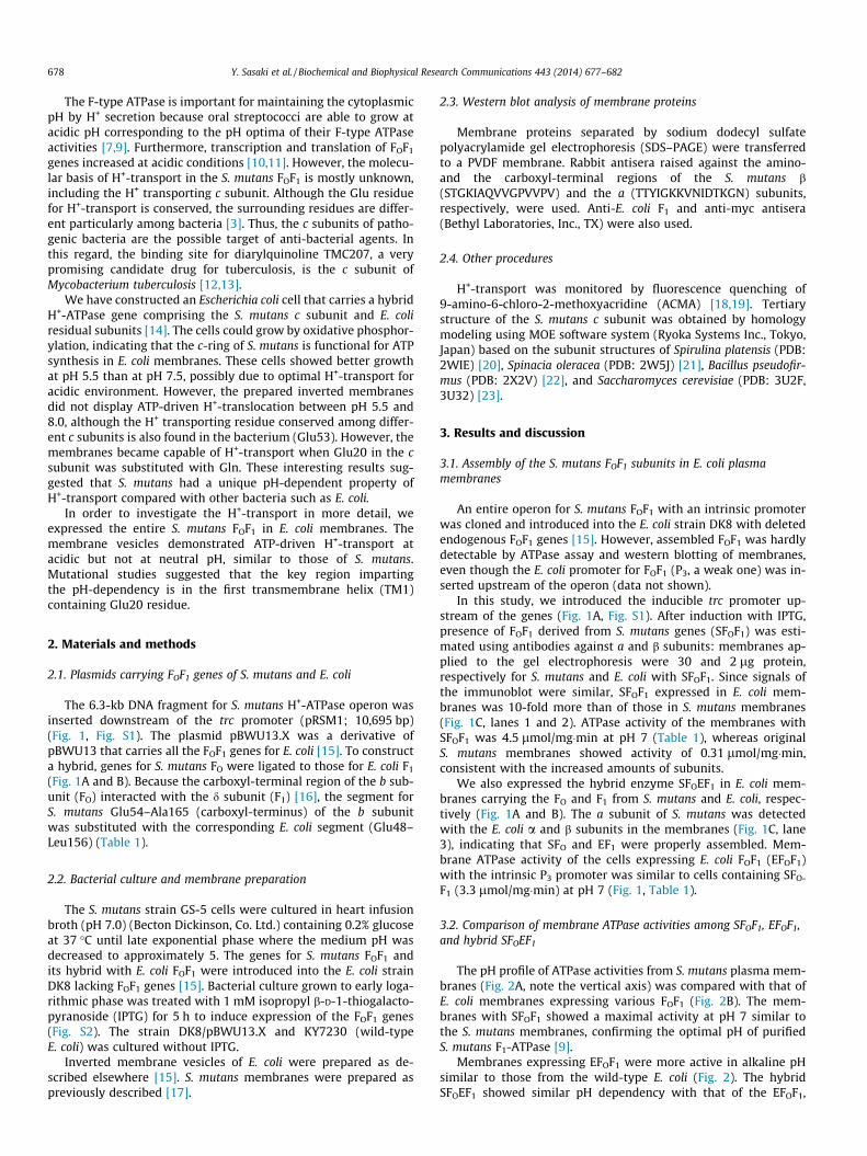
678 Y. Sasaki et al. / Biochemical and Biophysical Research Communications 443 (2014) 677–682
The F-type ATPase is important for maintaining the cytoplasmicpH by H+ secretion because oral streptococci are able to grow atacidic pH corresponding to the pH optima of their F-type ATPaseactivities [7,9]. Furthermore, transcription and translation of FOF1
genes increased at acidic conditions [10,11]. However, the molecu-lar basis of H+-transport in the S. mutans FOF1 is mostly unknown,including the H+ transporting c subunit. Although the Glu residuefor H+-transport is conserved, the surrounding residues are differ-ent particularly among bacteria [3]. Thus, the c subunits of patho-genic bacteria are the possible target of anti-bacterial agents. Inthis regard, the binding site for diarylquinoline TMC207, a verypromising candidate drug for tuberculosis, is the c subunit ofMycobacterium tuberculosis [12,13].
We have constructed an Escherichia coli cell that carries a hybridH+-ATPase gene comprising the S. mutans c subunit and E. coliresidual subunits [14]. The cells could grow by oxidative phosphor-ylation, indicating that the c-ring of S. mutans is functional for ATPsynthesis in E. coli membranes. These cells showed better growthat pH 5.5 than at pH 7.5, possibly due to optimal H+-transport foracidic environment. However, the prepared inverted membranesdid not display ATP-driven H+-translocation between pH 5.5 and8.0, although the H+ transporting residue conserved among differ-ent c subunits is also found in the bacterium (Glu53). However, themembranes became capable of H+-transport when Glu20 in the csubunit was substituted with Gln. These interesting results sug-gested that S. mutans had a unique pH-dependent property ofH+-transport compared with other bacteria such as E. coli.
In order to investigate the H+-transport in more detail, weexpressed the entire S. mutans FOF1 in E. coli membranes. Themembrane vesicles demonstrated ATP-driven H+-transport atacidic but not at neutral pH, similar to those of S. mutans.Mutational studies suggested that the key region impartingthe pH-dependency is in the first transmembrane helix (TM1)containing Glu20 residue.
2. Materials and methods
2.1. Plasmids carrying FOF1 genes of S. mutans and E. coli
The 6.3-kb DNA fragment for S. mutans H+-ATPase operon wasinserted downstream of the trc promoter (pRSM1; 10,695 bp)(Fig. 1, Fig. S1). The plasmid pBWU13.X was a derivative ofpBWU13 that carries all the FOF1 genes for E. coli [15]. To constructa hybrid, genes for S. mutans FO were ligated to those for E. coli F1
(Fig. 1A and B). Because the carboxyl-terminal region of the b sub-unit (FO) interacted with the d subunit (F1) [16], the segment forS. mutans Glu54–Ala165 (carboxyl-terminus) of the b subunitwas substituted with the corresponding E. coli segment (Glu48–Leu156) (Table 1).
2.2. Bacterial culture and membrane preparation
The S. mutans strain GS-5 cells were cultured in heart infusionbroth (pH 7.0) (Becton Dickinson, Co. Ltd.) containing 0.2% glucoseat 37 �C until late exponential phase where the medium pH wasdecreased to approximately 5. The genes for S. mutans FOF1 andits hybrid with E. coli FOF1 were introduced into the E. coli strainDK8 lacking FOF1 genes [15]. Bacterial culture grown to early loga-rithmic phase was treated with 1 mM isopropyl b-D-1-thiogalacto-pyranoside (IPTG) for 5 h to induce expression of the FOF1 genes(Fig. S2). The strain DK8/pBWU13.X and KY7230 (wild-typeE. coli) was cultured without IPTG.
Inverted membrane vesicles of E. coli were prepared as de-scribed elsewhere [15]. S. mutans membranes were prepared aspreviously described [17].
2.3. Western blot analysis of membrane proteins
Membrane proteins separated by sodium dodecyl sulfatepolyacrylamide gel electrophoresis (SDS–PAGE) were transferredto a PVDF membrane. Rabbit antisera raised against the amino-and the carboxyl-terminal regions of the S. mutans b(STGKIAQVVGPVVPV) and the a (TTYIGKKVNIDTKGN) subunits,respectively, were used. Anti-E. coli F1 and anti-myc antisera(Bethyl Laboratories, Inc., TX) were also used.
2.4. Other procedures
H+-transport was monitored by fluorescence quenching of9-amino-6-chloro-2-methoxyacridine (ACMA) [18,19]. Tertiarystructure of the S. mutans c subunit was obtained by homologymodeling using MOE software system (Ryoka Systems Inc., Tokyo,Japan) based on the subunit structures of Spirulina platensis (PDB:2WIE) [20], Spinacia oleracea (PDB: 2W5J) [21], Bacillus pseudofir-mus (PDB: 2X2V) [22], and Saccharomyces cerevisiae (PDB: 3U2F,3U32) [23].
3. Results and discussion
3.1. Assembly of the S. mutans FOF1 subunits in E. coli plasmamembranes
An entire operon for S. mutans FOF1 with an intrinsic promoterwas cloned and introduced into the E. coli strain DK8 with deletedendogenous FOF1 genes [15]. However, assembled FOF1 was hardlydetectable by ATPase assay and western blotting of membranes,even though the E. coli promoter for FOF1 (P3, a weak one) was in-serted upstream of the operon (data not shown).
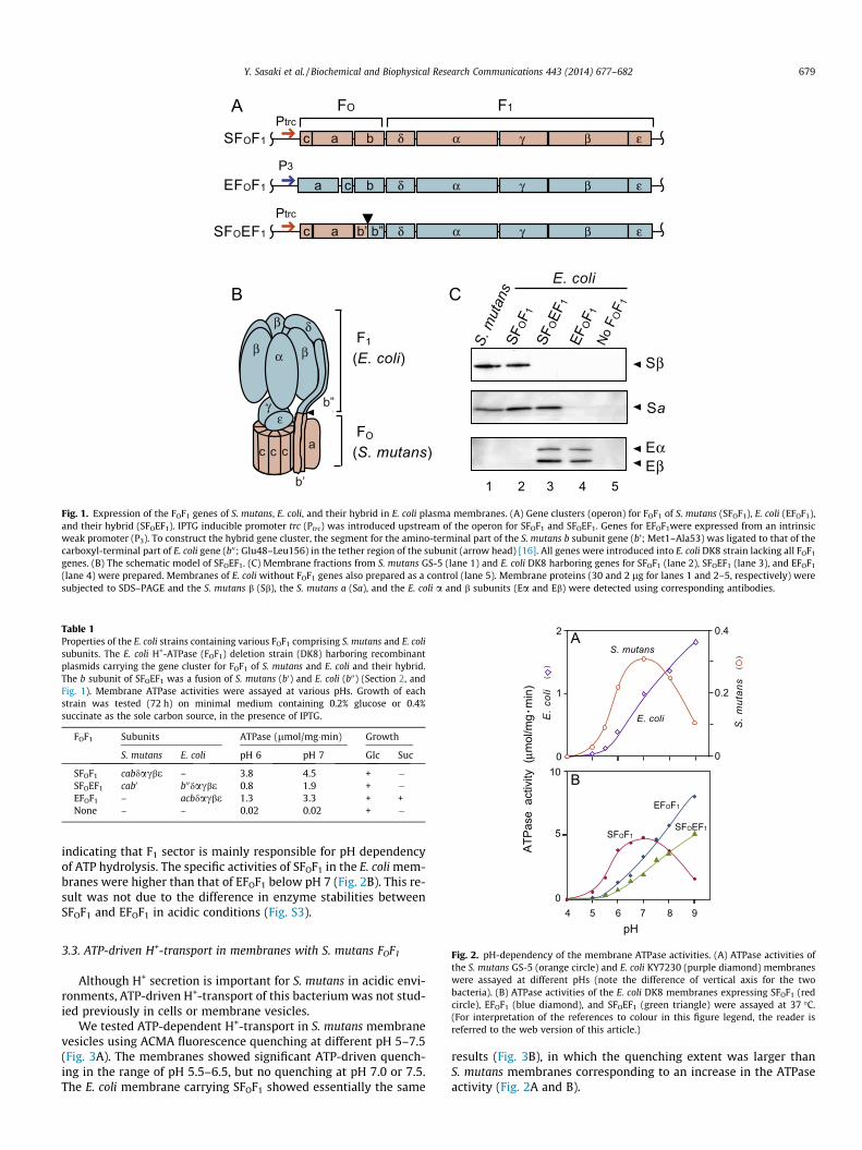
In this study, we introduced the inducible trc promoter up-stream of the genes (Fig. 1A, Fig. S1). After induction with IPTG,presence of FOF1 derived from S. mutans genes (SFOF1) was esti-mated using antibodies against a and b subunits: membranes ap-plied to the gel electrophoresis were 30 and 2 lg protein,respectively for S. mutans and E. coli with SFOF1. Since signals ofthe immunoblot were similar, SFOF1 expressed in E. coli mem-branes was 10-fold more than of those in S. mutans membranes(Fig. 1C, lanes 1 and 2). ATPase activity of the membranes withSFOF1 was 4.5 lmol/mg�min at pH 7 (Table 1), whereas originalS. mutans membranes showed activity of 0.31 lmol/mg�min,consistent with the increased amounts of subunits.
We also expressed the hybrid enzyme SFOEF1 in E. coli mem-branes carrying the FO and F1 from S. mutans and E. coli, respec-tively (Fig. 1A and B). The a subunit of S. mutans was detectedwith the E. coli a and b subunits in the membranes (Fig. 1C, lane3), indicating that SFO and EF1 were properly assembled. Mem-brane ATPase activity of the cells expressing E. coli FOF1 (EFOF1)with the intrinsic P3 promoter was similar to cells containing SFO-
F1 (3.3 lmol/mg�min) at pH 7 (Fig. 1, Table 1).
3.2. Comparison of membrane ATPase activities among SFOF1, EFOF1,and hybrid SFOEF1
The pH profile of ATPase activities from S. mutans plasma mem-branes (Fig. 2A, note the vertical axis) was compared with that ofE. coli membranes expressing various FOF1 (Fig. 2B). The mem-branes with SFOF1 showed a maximal activity at pH 7 similar tothe S. mutans membranes, confirming the optimal pH of purifiedS. mutans F1-ATPase [9].
Membranes expressing EFOF1 were more active in alkaline pHsimilar to those from the wild-type E. coli (Fig. 2). The hybridSFOEF1 showed similar pH dependency with that of the EFOF1,
c a δ α γ βb ε
ca δ α γ βb ε
c a δ α γ βb’ b” ε
SFOF1
EFOF1
SFOEF1
FO F1
S. m
utan
sSF
OF1
SFOEF
1
Sa
A
CB
γε
c c c a
b’
b”
F1
(E. coli)
FO
(S. mutans) Eα
Sβ
Eβ
EFOF1
No F
OF1
1 2 3 4 5
E. coli
Ptrc
Ptrc
P3
α
δ
β
β
β
Fig. 1. Expression of the FOF1 genes of S. mutans, E. coli, and their hybrid in E. coli plasma membranes. (A) Gene clusters (operon) for FOF1 of S. mutans (SFOF1), E. coli (EFOF1),and their hybrid (SFOEF1). IPTG inducible promoter trc (Ptrc) was introduced upstream of the operon for SFOF1 and SFOEF1. Genes for EFOF1were expressed from an intrinsicweak promoter (P3). To construct the hybrid gene cluster, the segment for the amino-terminal part of the S. mutans b subunit gene (b’; Met1–Ala53) was ligated to that of thecarboxyl-terminal part of E. coli gene (b00; Glu48–Leu156) in the tether region of the subunit (arrow head) [16]. All genes were introduced into E. coli DK8 strain lacking all FOF1
genes. (B) The schematic model of SFOEF1. (C) Membrane fractions from S. mutans GS-5 (lane 1) and E. coli DK8 harboring genes for SFOF1 (lane 2), SFOEF1 (lane 3), and EFOF1
(lane 4) were prepared. Membranes of E. coli without FOF1 genes also prepared as a control (lane 5). Membrane proteins (30 and 2 lg for lanes 1 and 2–5, respectively) weresubjected to SDS–PAGE and the S. mutans b (Sb), the S. mutans a (Sa), and the E. coli a and b subunits (Ea and Eb) were detected using corresponding antibodies.
Table 1Properties of the E. coli strains containing various FOF1 comprising S. mutans and E. colisubunits. The E. coli H+-ATPase (FOF1) deletion strain (DK8) harboring recombinantplasmids carrying the gene cluster for FOF1 of S. mutans and E. coli and their hybrid.The b subunit of SFOEF1 was a fusion of S. mutans (b0) and E. coli (b00) (Section 2, andFig. 1). Membrane ATPase activities were assayed at various pHs. Growth of eachstrain was tested (72 h) on minimal medium containing 0.2% glucose or 0.4%succinate as the sole carbon source, in the presence of IPTG.
FOF1 Subunits ATPase (lmol/mg�min) Growth
S. mutans E. coli pH 6 pH 7 Glc Suc
SFOF1 cabdacbe – 3.8 4.5 + �SFOEF1 cab0 b00dacbe 0.8 1.9 + �EFOF1 – acbdacbe 1.3 3.3 + +None – – 0.02 0.02 + �
( )
( )
SFOF1SFOEF1
EFOF1
S. mutans
10
5
0
ATP
ase
act
ivity
(μm
ol/m
g m
in)
A
B
E. coli
2
0
1
0.4
0.3
0.1
0.2
0
S. m
utan
s
E. c
oli
4 5 6 7 8 9
Y. Sasaki et al. / Biochemical and Biophysical Research Communications 443 (2014) 677–682 679
indicating that F1 sector is mainly responsible for pH dependencyof ATP hydrolysis. The specific activities of SFOF1 in the E. coli mem-branes were higher than that of EFOF1 below pH 7 (Fig. 2B). This re-sult was not due to the difference in enzyme stabilities betweenSFOF1 and EFOF1 in acidic conditions (Fig. S3).
pH
Fig. 2. pH-dependency of the membrane ATPase activities. (A) ATPase activities ofthe S. mutans GS-5 (orange circle) and E. coli KY7230 (purple diamond) membraneswere assayed at different pHs (note the difference of vertical axis for the twobacteria). (B) ATPase activities of the E. coli DK8 membranes expressing SFOF1 (redcircle), EFOF1 (blue diamond), and SFOEF1 (green triangle) were assayed at 37 �C.(For interpretation of the references to colour in this figure legend, the reader isreferred to the web version of this article.)
3.3. ATP-driven H+-transport in membranes with S. mutans FOF1
Although H+ secretion is important for S. mutans in acidic envi-ronments, ATP-driven H+-transport of this bacterium was not stud-ied previously in cells or membrane vesicles.
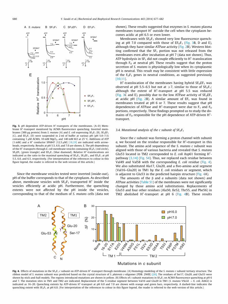
We tested ATP-dependent H+-transport in S. mutans membranevesicles using ACMA fluorescence quenching at different pH 5–7.5(Fig. 3A). The membranes showed significant ATP-driven quench-ing in the range of pH 5.5–6.5, but no quenching at pH 7.0 or 7.5.The E. coli membrane carrying SFOF1 showed essentially the same
results (Fig. 3B), in which the quenching extent was larger thanS. mutans membranes corresponding to an increase in the ATPaseactivity (Fig. 2A and B).
5.5, 6.0
7.0
7.0
5.5
6.0
7.0
5.5
6.06.0
5.5
7.0
A S. mutans
ATP
SF6847
ATP
SF6847
ATP
SF6847
ATP
SF6847
1 min1 min
5%ΔF
20%ΔF
1 min
10%ΔF
1 min
10%ΔF
B SFOF1 C SFOEF1 D EFOF1
E
0
1.0
0.5
5 6 7 8
Rel
ativ
e H
+ tra
nslo
catio
n
pH
SFOEF1
EFOF1
SFOF1
Fig. 3. pH dependent ATP-driven H+-transports of the membranes. (A–D) Mem-brane H+-transport monitored by ACMA fluorescence quenching. Inverted mem-branes (300 lg protein) from S. mutans (A) and E. coli expressing SFOF1 (B), SFOEF1
(C), and EFOF1 (D) were suspended in 2 ml of buffer at various pH (pH 5.0–7.5)containing 1 lM ACMA, 10 mM MgCl2, and 140 mM KCl at 25 �C. Additions of ATP(1 mM) and a H+ conductor SF6847 (12.5 lM) [18,19] are indicated with arrow-heads, respectively. Results at pH 5.5, 6.0, and 7.0 are shown. E. The pH-dependencyof the H+-transports through E. coli membrane vesicles containing SFOF1 (red circle),SFOEF1 (green triangle) and EFOF1 (blue diamond). Relative H+-translocations areindicated as the ratio to the maximal quenching of SFOF1, SFOEF1, and EFOF1 at pH5.5, 6.0, and 6.5, respectively. (For interpretation of the references to colour in thisfigure legend, the reader is referred to the web version of this article.)
680 Y. Sasaki et al. / Biochemical and Biophysical Research Communications 443 (2014) 677–682
Since the membrane vesicles tested were inverted (inside-out),pH of the buffer corresponds to that of the cytoplasm. As describedabove, membrane vesicles with SFOF1 transported H+ inside thevesicles efficiently at acidic pH. Furthermore, the quenchingextents were not affected by the pH inside the vesicles,corresponding to that of the medium of S. mutans cells (data not
BA
Ser-17
NC
Glu-20
Glu-53
TM1TM2
0
10
20
30
S17A
16-2
0
ΔF
(%)
None
TM 1
Fig. 4. Effects of mutations in the SFOF1 c subunit on ATP-driven H+-transport through mribbon model of S. mutans subunit was predicted based on the crystal structure of S. plshown by stick-and-ball models. The regions introduced mutations are shown in yellow.and 7. The mutation sites in TM1 and TM2 are indicated. Replacement of the 5-residuindicated as 16–20. Quenching extents by ATP-driven H+-transport at pH 6.0 and 7.0quenching extent with SFOF1 at pH 6.0. (For interpretation of the references to colour in
shown). These results suggested that enzymes in S. mutans plasmamembranes transport H+ outside the cell when the cytoplasm be-comes acidic at pH 6.5 or even lower.
Membranes with SFOF1 showed very low fluorescence quench-ing at pH 7.0 compared with those of EFOF1 (Fig. 3B, D and E),although they have similar ATPase activity (Fig. 2B). Western blot-ting confirmed that the SF1 portion was not released from themembranes even after incubation at pH 7 (data not shown). Thus,ATP hydrolysis in SF1 did not couple efficiently to H+-translocationthrough FO at neutral pH. These results suggest that the protonsecretion of S. mutans is physiologically low when its cytoplasmicpH is neutral. This result may be consistent with little expressionof the FOF1 genes in neutral conditions, as suggested previously[10,11].
H+-translocation of the membranes having hybrid SFOEF1 wasobserved at pH 5.5–6.5 but not at P7, similar to those of SFOF1;although the extent of H+-transport at pH 5.5 was reduced(Fig. 3C and E), possibly due to the low ATPase activity of SFOEF1
at acidic pH (Fig. 2B). A similar amount of EF1 was found inmembranes treated at pH 6 or 7. These results suggest that pHdependencies of ATPase and H+-transport were due to F1 and FO
portions, respectively. These findings prompted us to study the do-mains of FO responsible for the pH dependence of ATP-driven H+-transport.
3.4. Mutational analysis of the c subunit of SFOF1
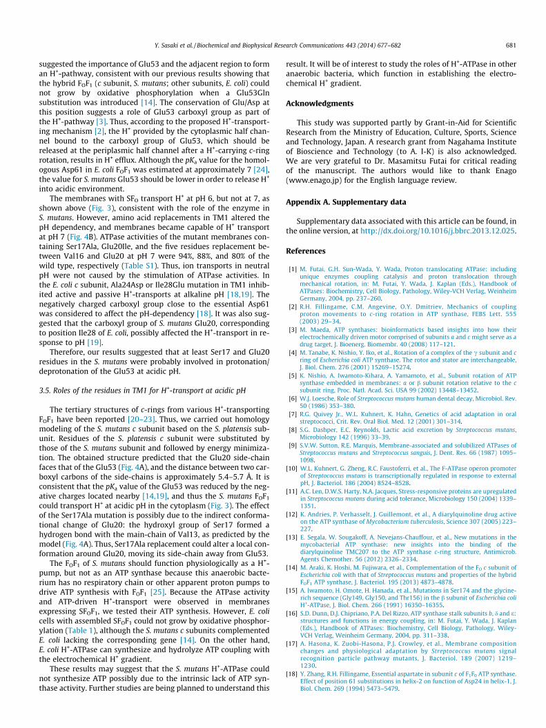
Since the c subunit was forming a proton channel with subunita, we focused on the residue responsible for H+-transport in thissubunit. The amino acid sequence of the S. mutans c subunit wasaligned with those of various bacteria and revealed that S. mutansGlu53 located in TM2 corresponded to E. coli Asp61 forming H+-pathway [3,14] (Fig. S4). Thus, we replaced each residue betweenVal49 and Val58 with the corresponding E. coli residue (Fig. 4).We also substituted Ala17, Glu20, and a five-amino acid segment(Val16–Glu20) in TM1 by the E. coli residues or segment, whichis adjacent to Glu53 in the predicted hairpin structure (Fig. 4A).
The amounts of the b and a subunits (data not shown) andATPase activities (Table S1) of the membranes were not significantlychanged by those amino acid substitutions. Replacements ofGlu53 and four other residues (Ala50, Ile52, Thr55, and Phe56) inTM2 abolished H+-transport at pH 6 (Fig. 4B). These results
E20I
V49M
A50G
F51L
I52V
E53D
G54A
T55I
F56P
F57M
V58I
Mutations in c subunit
pH 6.0pH 7.0
TM 2
embrane. (A) Homology modeling of the S. mutans c subunit tertiary structure. Theatensis c-oligomer (PDB: 2WIE) [20]. The residues of Ser17, Glu20, and Glu53 were(B) Effects of c subunit mutations in SFOF1 on ACMA fluorescence quenching at pH 6e segment between Val16 and Glu20 in TM1 (S. mutans VSLGE ? E. coli, AAIGI) isare shown with orange and green bars, respectively. A dashed-line indicates thethis figure legend, the reader is referred to the web version of this article.)
Y. Sasaki et al. / Biochemical and Biophysical Research Communications 443 (2014) 677–682 681
suggested the importance of Glu53 and the adjacent region to forman H+-pathway, consistent with our previous results showing thatthe hybrid FOF1 (c subunit, S. mutans; other subunits, E. coli) couldnot grow by oxidative phosphorylation when a Glu53Glnsubstitution was introduced [14]. The conservation of Glu/Asp atthis position suggests a role of Glu53 carboxyl group as part ofthe H+-pathway [3]. Thus, according to the proposed H+-transport-ing mechanism [2], the H+ provided by the cytoplasmic half chan-nel bound to the carboxyl group of Glu53, which should bereleased at the periplasmic half channel after a H+-carrying c-ringrotation, results in H+ efflux. Although the pKa value for the homol-ogous Asp61 in E. coli FOF1 was estimated at approximately 7 [24],the value for S. mutans Glu53 should be lower in order to release H+
into acidic environment.The membranes with SFO transport H+ at pH 6, but not at 7, as
shown above (Fig. 3), consistent with the role of the enzyme inS. mutans. However, amino acid replacements in TM1 altered thepH dependency, and membranes became capable of H+ transportat pH 7 (Fig. 4B). ATPase activities of the mutant membranes con-taining Ser17Ala, Glu20Ile, and the five residues replacement be-tween Val16 and Glu20 at pH 7 were 94%, 88%, and 80% of thewild type, respectively (Table S1). Thus, ion transports in neutralpH were not caused by the stimulation of ATPase activities. Inthe E. coli c subunit, Ala24Asp or Ile28Glu mutation in TM1 inhib-ited active and passive H+-transports at alkaline pH [18,19]. Thenegatively charged carboxyl group close to the essential Asp61was considered to affect the pH-dependency [18]. It was also sug-gested that the carboxyl group of S. mutans Glu20, correspondingto position Ile28 of E. coli, possibly affected the H+-transport in re-sponse to pH [19].
Therefore, our results suggested that at least Ser17 and Glu20residues in the S. mutans were probably involved in protonation/deprotonation of the Glu53 at acidic pH.
3.5. Roles of the residues in TM1 for H+-transport at acidic pH
The tertiary structures of c-rings from various H+-transportingFOF1 have been reported [20–23]. Thus, we carried out homologymodeling of the S. mutans c subunit based on the S. platensis sub-unit. Residues of the S. platensis c subunit were substituted bythose of the S. mutans subunit and followed by energy minimiza-tion. The obtained structure predicted that the Glu20 side-chainfaces that of the Glu53 (Fig. 4A), and the distance between two car-boxyl carbons of the side-chains is approximately 5.4–5.7 Å. It isconsistent that the pKa value of the Glu53 was reduced by the neg-ative charges located nearby [14,19], and thus the S. mutans FOF1
could transport H+ at acidic pH in the cytoplasm (Fig. 3). The effectof the Ser17Ala mutation is possibly due to the indirect conforma-tional change of Glu20: the hydroxyl group of Ser17 formed ahydrogen bond with the main-chain of Val13, as predicted by themodel (Fig. 4A). Thus, Ser17Ala replacement could alter a local con-formation around Glu20, moving its side-chain away from Glu53.
The FOF1 of S. mutans should function physiologically as a H+-pump, but not as an ATP synthase because this anaerobic bacte-rium has no respiratory chain or other apparent proton pumps todrive ATP synthesis with FOF1 [25]. Because the ATPase activityand ATP-driven H+-transport were observed in membranesexpressing SFOF1, we tested their ATP synthesis. However, E. colicells with assembled SFOF1 could not grow by oxidative phosphor-ylation (Table 1), although the S. mutans c subunits complementedE. coli lacking the corresponding gene [14]. On the other hand,E. coli H+-ATPase can synthesize and hydrolyze ATP coupling withthe electrochemical H+ gradient.
These results may suggest that the S. mutans H+-ATPase couldnot synthesize ATP possibly due to the intrinsic lack of ATP syn-thase activity. Further studies are being planned to understand this
result. It will be of interest to study the roles of H+-ATPase in otheranaerobic bacteria, which function in establishing the electro-chemical H+ gradient.
Acknowledgments
This study was supported partly by Grant-in-Aid for ScientificResearch from the Ministry of Education, Culture, Sports, Scienceand Technology, Japan. A research grant from Nagahama Instituteof Bioscience and Technology (to A. I-K) is also acknowledged.We are very grateful to Dr. Masamitsu Futai for critical readingof the manuscript. The authors would like to thank Enago(www.enago.jp) for the English language review.
Appendix A. Supplementary data
Supplementary data associated with this article can be found, inthe online version, at http://dx.doi.org/10.1016/j.bbrc.2013.12.025.
References
[1] M. Futai, G.H. Sun-Wada, Y. Wada, Proton translocating ATPase: includingunique enzymes coupling catalysis and proton translocation throughmechanical rotation, in: M. Futai, Y. Wada, J. Kaplan (Eds.), Handbook ofATPases: Biochemistry, Cell Biology, Pathology, Wiley-VCH Verlag, WeinheimGermany, 2004, pp. 237–260.
[2] R.H. Fillingame, C.M. Angevine, O.Y. Dmitriev, Mechanics of couplingproton movements to c-ring rotation in ATP synthase, FEBS Lett. 555(2003) 29–34.
[3] M. Maeda, ATP synthases: bioinformaticts based insights into how theirelectrochemically driven motor comprised of subunits a and c might serve as adrug target, J. Bioenerg. Biomembr. 40 (2008) 117–121.
[4] M. Tanabe, K. Nishio, Y. Iko, et al., Rotation of a complex of the c subunit and cring of Escherichia coli ATP synthase. The rotor and stator are interchangeable,J. Biol. Chem. 276 (2001) 15269–15274.
[5] K. Nishio, A. Iwamoto-Kihara, A. Yamamoto, et al., Subunit rotation of ATPsynthase embedded in membranes: a or b subunit rotation relative to the csubunit ring, Proc. Natl. Acad. Sci. USA 99 (2002) 13448–13452.
[6] W.J. Loesche, Role of Streptococcus mutans human dental decay, Microbiol. Rev.50 (1986) 353–380.
[7] R.G. Quivey Jr., W.L. Kuhnert, K. Hahn, Genetics of acid adaptation in oralstreptococci, Crit. Rev. Oral Biol. Med. 12 (2001) 301–314.
[8] S.G. Dashper, E.C. Reynolds, Lactic acid excretion by Streptococcus mutans,Microbiology 142 (1996) 33–39.
[9] S.V.W. Sutton, R.E. Marquis, Membrane-associated and solubilized ATPases ofStreptococcus mutans and Streptococcus sanguis, J. Dent. Res. 66 (1987) 1095–1098.
[10] W.L. Kuhnert, G. Zheng, R.C. Faustoferri, et al., The F-ATPase operon promoterof Streptococcus mutans is transcriptionally regulated in response to externalpH, J. Bacteriol. 186 (2004) 8524–8528.
[11] A.C. Len, D.W.S. Harty, N.A. Jacques, Stress-responsive proteins are upregulatedin Streptococcus mutans during acid tolerance, Microbiology 150 (2004) 1339–1351.
[12] K. Andries, P. Verhasselt, J. Guillemont, et al., A diarylquinoline drug activeon the ATP synthase of Mycobacterium tuberculosis, Science 307 (2005) 223–227.
[13] E. Segala, W. Sougakoff, A. Nevejans-Chauffour, et al., New mutations in themycobacterial ATP synthase: new insights into the binding of thediarylquinoline TMC207 to the ATP synthase c-ring structure, Antimicrob.Agents Chemother. 56 (2012) 2326–2334.
[14] M. Araki, K. Hoshi, M. Fujiwara, et al., Complementation of the FO c subunit ofEscherichia coli with that of Streptococcus mutans and properties of the hybridFOF1 ATP synthase, J. Bacteriol. 195 (2013) 4873–4878.
[15] A. Iwamoto, H. Omote, H. Hanada, et al., Mutations in Ser174 and the glycine-rich sequence (Gly149, Gly150, and Thr156) in the b subunit of Escherichia coliH+-ATPase, J. Biol. Chem. 266 (1991) 16350–16355.
[16] S.D. Dunn, D.J. Chipriano, P.A. Del Rizzo, ATP synthase stalk subunits b, d and e:structures and functions in energy coupling, in: M. Futai, Y. Wada, J. Kaplan(Eds.), Handbook of ATPases: Biochemistry, Cell Biology, Pathology, Wiley-VCH Verlag, Weinheim Germany, 2004, pp. 311–338.
[17] A. Hasona, K. Zuobi-Hasona, P.J. Crowley, et al., Membrane compositionchanges and physiological adaptation by Streptococcus mutans signalrecognition particle pathway mutants, J. Bacteriol. 189 (2007) 1219–1230.
[18] Y. Zhang, R.H. Fillingame, Essential aspartate in subunit c of F1F0 ATP synthase.Effect of position 61 substitutions in helix-2 on function of Asp24 in helix-1, J.Biol. Chem. 269 (1994) 5473–5479.
682 Y. Sasaki et al. / Biochemical and Biophysical Research Communications 443 (2014) 677–682
[19] P.C. Jones, Introduction of a carboxyl group in the first transmembrane helix ofEscherichia coli F1FO ATPase subunit c and cytoplasmic pH regulation, J.Bacteriol. 183 (2001) 1524–1530.
[20] D. Pogoryelov, O. Yildiz, J.D. Faraldo-Gómez, et al., High-resolution structure ofthe rotor ring of a proton-dependent ATP synthase, Nat. Struct. Mol. Biol. 16(2009) 1068–1073.
[21] M. Vollmar, D. Schlieper, M. Winn, et al., Structure of the c14 rotor ring of theproton translocating chloroplast ATP synthase, J. Biol. Chem. 284 (2009)18228–18235.
[22] L. Preiss, O. Yildiz, D.B. Hicks, et al., A new type of proton coordination in anF1FO-ATP synthase rotor ring, PLoS Biol. 8 (2010) e1000443.
[23] J. Symersky, V. Pagadala, D. Osowski, et al., Structure of the c10 ring of the yeastmitochondrial ATP synthase in the open conformation, Nat. Struct. Mol. Biol.19 (2012) 485–491.
[24] F.M. Assadi-Porter, R.H. Fillingame, Proton-translocating carboxyl of subunit cof F1FO H+-ATP synthase: the unique environment suggested by the pKadetermined by 1H NMR, Biochemistry 34 (1995) 16186–16193.
[25] D. Ajdic, W.M. McShan, R.E. McLaughlin, et al., Genome sequence ofStreptococcus mutans UA159, a cariogenic dental pathogen, Proc. Natl. Acad.Sci. USA 99 (2002) 14434–14439.