Embed Size (px)
Citation preview

8/3/2019 a Types
http://slidepdf.com/reader/full/a-types 1/86

8/3/2019 a Types
http://slidepdf.com/reader/full/a-types 2/86
INTRODUCTIONINTRODUCTION
Lesions derived from the epithelial and or Lesions derived from the epithelial and or
mesenchymal remnants of the tooth formingmesenchymal remnants of the tooth forming
apparatusapparatus
Found exclusively in the mandible and maxillaFound exclusively in the mandible and maxilla
( occasionally – gingiva)( occasionally – gingiva)
Originate from an aberration from the normalOriginate from an aberration from the normal
pattern of odontogenesispattern of odontogenesis A complex group of lesions of diverse A complex group of lesions of diverse
histopathologic types and clinical behavior histopathologic types and clinical behavior

8/3/2019 a Types
http://slidepdf.com/reader/full/a-types 3/86
INTRODUCTIONINTRODUCTION
Range from hamartomatous proliferations toRange from hamartomatous proliferations tomalignant neoplasms with metastatic capabilitiesmalignant neoplasms with metastatic capabilities
Exhibit varying inductive interactions betweenExhibit varying inductive interactions between
odontogenic epithelium and odontogenicodontogenic epithelium and odontogenic
ectomesenchymeectomesenchyme

8/3/2019 a Types
http://slidepdf.com/reader/full/a-types 4/86
PresentationPresentation
Asymptomatic Asymptomatic
Jaw expansionJaw expansion Movement of teethMovement of teeth
Root resorptionRoot resorption
Bone lossBone loss
8/3/2019 a Types
http://slidepdf.com/reader/full/a-types 5/86

8/3/2019 a Types
http://slidepdf.com/reader/full/a-types 6/86

8/3/2019 a Types
http://slidepdf.com/reader/full/a-types 7/86
MicroscopyMicroscopy
Mimic cell / tissue of originMimic cell / tissue of origin
Resemble the soft tissues of the enamel organResemble the soft tissues of the enamel organ
or dental pulp or may contain hard tissueor dental pulp or may contain hard tissue
elements of enamel, dentin, & or cementumelements of enamel, dentin, & or cementum

8/3/2019 a Types
http://slidepdf.com/reader/full/a-types 8/86
CLASSIFICATIONCLASSIFICATION
WHO (1992)WHO (1992)
ODONTOGENIC TUMORS
BENIGN MALIGNANT

8/3/2019 a Types
http://slidepdf.com/reader/full/a-types 9/86
MODIFIED WHO CLASSIFICATIONMODIFIED WHO CLASSIFICATION
BENIGNBENIGN
I Odontogenic epithelium withoutI Odontogenic epithelium without
odontogenic ectomesenchymeodontogenic ectomesenchymeAmeloblastomaAmeloblastoma
Squamous odontogenic tumor Squamous odontogenic tumor
Calcifying epithelial odontogenic tumor Calcifying epithelial odontogenic tumor
Adenomatoid odontogenic tumor Adenomatoid odontogenic tumor

8/3/2019 a Types
http://slidepdf.com/reader/full/a-types 10/86
CLASSIFICATIONCLASSIFICATION
II Odontogenic epithelium with odontogenicII Odontogenic epithelium with odontogenic
ectomesenchyme with or without hard tissueectomesenchyme with or without hard tissue
formationformation
Ameloblastic fibromaAmeloblastic fibroma Ameloblastic fibrodentinomaAmeloblastic fibrodentinoma
Ameloblastic fibro-odontomaAmeloblastic fibro-odontoma
OdontoameloblastomaOdontoameloblastoma
Calcifying odontogenic cystCalcifying odontogenic cyst
Complex odontomaComplex odontoma
Compound odontomaCompound odontoma

8/3/2019 a Types
http://slidepdf.com/reader/full/a-types 11/86
III Odontogenic ectomesenchyme with or III Odontogenic ectomesenchyme with or
without included odontogenicwithout included odontogenic
epitheliumepitheliumOdontogenic fibromaOdontogenic fibroma
Myxoma (myxofibroma)Myxoma (myxofibroma)
Cementoblastoma (benignCementoblastoma (benign
cementoblastoma, true cementoma)cementoblastoma, true cementoma)
CLASSIFICATIONCLASSIFICATION

8/3/2019 a Types
http://slidepdf.com/reader/full/a-types 12/86
B. MALIGNANTB. MALIGNANT
I Odontogenic CarcinomasI Odontogenic Carcinomas
Malignant ameloblastomaMalignant ameloblastomaPrimary intraosseous carcinomaPrimary intraosseous carcinoma
Clear cell odontogenic carcinomaClear cell odontogenic carcinoma
Ghost cell odontogenic carcinomaGhost cell odontogenic carcinoma
CLASSIFICATIONCLASSIFICATION

8/3/2019 a Types
http://slidepdf.com/reader/full/a-types 13/86
II Odontogenic SarcomasII Odontogenic SarcomasAmeloblastic fibrosarcomaAmeloblastic fibrosarcoma
Ameloblastic fibro dentinosarcomaAmeloblastic fibro dentinosarcoma
Ameloblastic fibro odontosarcomaAmeloblastic fibro odontosarcoma
8/3/2019 a Types
http://slidepdf.com/reader/full/a-types 14/86
2005 WHO2005 WHO
HISTOLOGICALHISTOLOGICAL
CLASSIFICATION OFCLASSIFICATION OF
ODONTOGENIC TUMOURSODONTOGENIC TUMOURS

8/3/2019 a Types
http://slidepdf.com/reader/full/a-types 15/86
BENIGN TUMOURS1. Odontogenic epithelium with mature, fibrous
stroma without odontogenicectomesenchyme
Ameloblastoma, solid / multicystic type extraosseous / peripheral type desmoplastic type unicystic type
Squamous odontogenic tumour Calcifying epithelial odontogenic tumour Adenomatoid odontogenic tumour Keratocystic odontogenic tumour

8/3/2019 a Types
http://slidepdf.com/reader/full/a-types 16/86
2. Odontogenic epithelium with odontogenicectomesenchyme,with or without hard tissueformation
Ameloblastic fibroma Ameloblastic fibrodentinoma
Ameloblastic fibro-odontomaComplex OdontomaCompound OdontomaOdontoameloblastomaCalcifying cystic odontogenic tumour Dentinogenic ghost cell tumour

8/3/2019 a Types
http://slidepdf.com/reader/full/a-types 17/86
3. Mesenchyme and/or odontogenic
ectomesenchyme with or without
odontogenic epithelium
Odontogenic fibroma
Odontogenic myxoma / myxofibroma
Cementoblastoma

8/3/2019 a Types
http://slidepdf.com/reader/full/a-types 18/86
MALIGNANT TUMOURS
1. Odontogenic carcinomas
Metastasizing (malignant) ameloblastoma
Ameloblastic carcinomaPrimary intraosseous squamous cell
carcinoma
Clear cell odontogenic carcinomaGhost cell odontogenic carcinoma

8/3/2019 a Types
http://slidepdf.com/reader/full/a-types 19/86
2. Odontogenic sarcomas
Ameloblastic fibrosarcoma
Ameloblastic fibrodentino andfibro-odontosarcoma

8/3/2019 a Types
http://slidepdf.com/reader/full/a-types 20/86
AMELOBLASTOMA AMELOBLASTOMA
Introduction & definitionIntroduction & definitionTypesTypes
IncidenceIncidenceClinical featuresClinical featuresRadiographic featuresRadiographic features
HistogenesisHistogenesisHistopathology – subtypesHistopathology – subtypesTreatment and PrognosisTreatment and Prognosis

8/3/2019 a Types
http://slidepdf.com/reader/full/a-types 21/86
UNICYSTIC AMELOBLASTOMAUNICYSTIC AMELOBLASTOMA
Peripheral AmeloblastomaPeripheral Ameloblastoma
Pitutary AmeloblastomaPitutary Ameloblastoma Adamntinoma of long bones Adamntinoma of long bones

8/3/2019 a Types
http://slidepdf.com/reader/full/a-types 22/86
AMELOBLASTOMA AMELOBLASTOMA
SYNONYMSSYNONYMS
Adamantinoma Adamantinoma
Adamantoblastoma AdamantoblastomaMultilocular cystMultilocular cyst

8/3/2019 a Types
http://slidepdf.com/reader/full/a-types 23/86
Definition (Robinson)Definition (Robinson)
It is a tumor of odontogenic origin (derivedIt is a tumor of odontogenic origin (derived
from enamel organ type tissue which doesfrom enamel organ type tissue which does
not undergo differentiation to the point of not undergo differentiation to the point of
enamel formation) usually unicentric, non-enamel formation) usually unicentric, non-functional, intermittent in growth,functional, intermittent in growth,
anatomically benign and clinicallyanatomically benign and clinically
persistentpersistent
Ivy and Churchill – ameloblastoma (1934)Ivy and Churchill – ameloblastoma (1934)
8/3/2019 a Types
http://slidepdf.com/reader/full/a-types 24/86
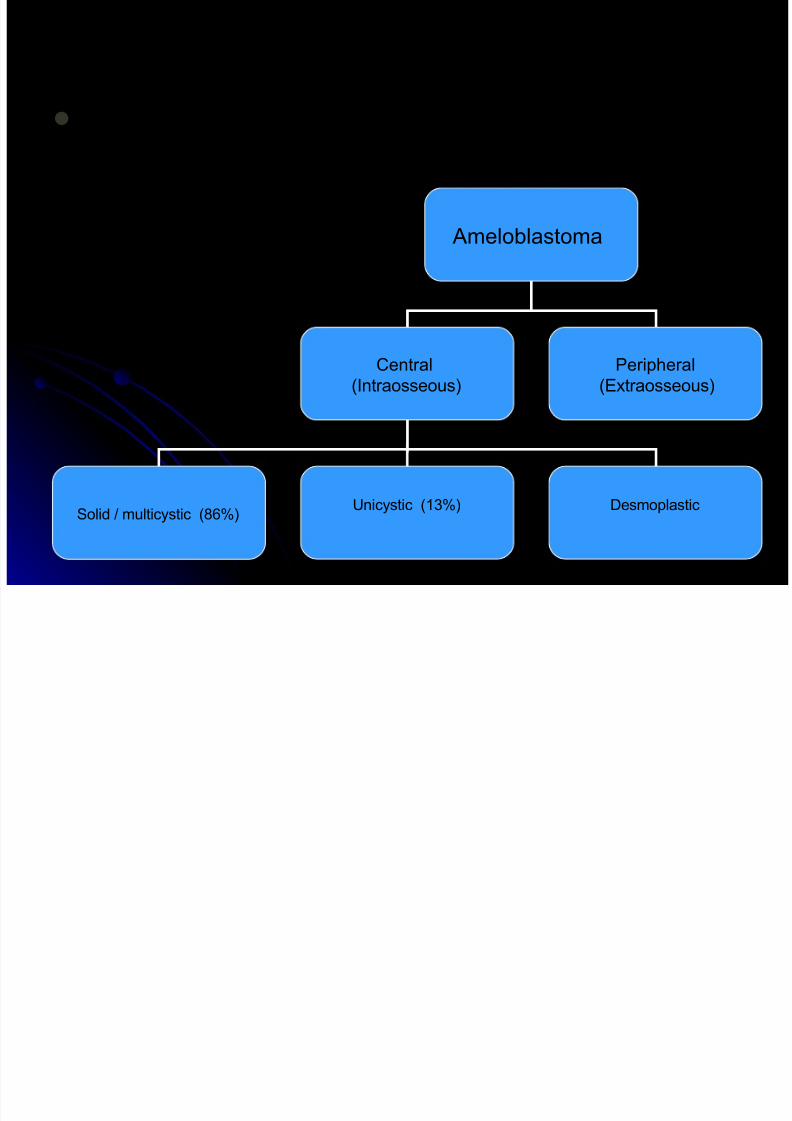
TypesTypes
Certain behaviour patterns, anatomic locations,Certain behaviour patterns, anatomic locations,
histologic and radiologic featureshistologic and radiologic features
Ameloblastoma
Central
(Intraosseous)
Peripheral
(Extraosseous)
Solid / multicystic (86%)Unicystic (13%) Desmoplastic

8/3/2019 a Types
http://slidepdf.com/reader/full/a-types 25/86
IncidenceIncidence
Common odontogenic tumor Common odontogenic tumor
11% of all tumors and cysts of jaws
18 % of odontogenic neoplasms

8/3/2019 a Types
http://slidepdf.com/reader/full/a-types 26/86
Conventional Solid / multicystic
Intraosseous Ameloblastoma
Most frequently encounteredMost frequently encountered
Demographics :Demographics :
Age : Age : wide age rangewide age range
3-73-7thth decades of lifedecades of life
Sex : no gender predilectionSex : no gender predilectionRace : blacksRace : blacks

8/3/2019 a Types
http://slidepdf.com/reader/full/a-types 27/86
Clinical featuresClinical features
Site :Site :
8585 % - mandible – molar – ascending
ramus area
15 % - maxilla – posterior region
Usually asymptomatic
Pain & paresthesia – uncommonSmaller lesions – only on routine R/F

8/3/2019 a Types
http://slidepdf.com/reader/full/a-types 28/86

8/3/2019 a Types
http://slidepdf.com/reader/full/a-types 29/86
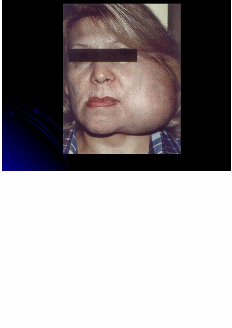
A painless swelling / expansion of the A painless swelling / expansion of the
jaw ; tends to expand the bone rather than jaw ; tends to expand the bone rather than
perforate itperforate it
Very thin bone – ‘EGG – SHELLVery thin bone – ‘EGG – SHELL
CRACKLING’ on palpationCRACKLING’ on palpation
Tooth mobilityTooth mobility
Unhealed extraction sitesUnhealed extraction sites
aggressiveaggressive

8/3/2019 a Types
http://slidepdf.com/reader/full/a-types 30/86
MaxillaMaxilla
Facial swelling, nasal obstructionFacial swelling, nasal obstruction
Sinus involvement, extension into the orbitSinus involvement, extension into the orbitor nasopharynxor nasopharynx
Sinusitis, pre auricular painSinusitis, pre auricular pain
Foul smelling dischargeFoul smelling discharge

8/3/2019 a Types
http://slidepdf.com/reader/full/a-types 31/86
Radiographic featuresRadiographic features
Often – multilocular radiolucent lesionOften – multilocular radiolucent lesion
Rarely – unilocular Rarely – unilocular
Large loculations – ‘soap bubble’Large loculations – ‘soap bubble’appearanceappearance
Small loculations – ‘honey combed’Small loculations – ‘honey combed’
appearanceappearance

8/3/2019 a Types
http://slidepdf.com/reader/full/a-types 32/86

8/3/2019 a Types
http://slidepdf.com/reader/full/a-types 33/86

8/3/2019 a Types
http://slidepdf.com/reader/full/a-types 34/86

8/3/2019 a Types
http://slidepdf.com/reader/full/a-types 35/86
Radiographic featuresRadiographic features
Buccal & lingual cortical expansion andBuccal & lingual cortical expansion and
thinningthinning
Resorption of rootsResorption of roots
Displacement of teethDisplacement of teeth
Association with an unerupted tooth Association with an unerupted tooth
(mandibular 3(mandibular 3rdrd
molar)molar)
Irregular scalloping of the margins of R/LIrregular scalloping of the margins of R/L
lesionslesions
Sinus – antral clouding / opacitySinus – antral clouding / opacity

8/3/2019 a Types
http://slidepdf.com/reader/full/a-types 36/86
Differential diagnosisDifferential diagnosis
OKCOKC
ABC ABCCentral hemangiomaCentral hemangioma
Brown tumor Brown tumor
Radiographic featuresRadiographic features

8/3/2019 a Types
http://slidepdf.com/reader/full/a-types 37/86
HISTOGENESISHISTOGENESIS
Tumor may be derived fromTumor may be derived from Cell rests of enamel organCell rests of enamel organ Cell rests of SerreCell rests of Serre
Cell rests of MalassezCell rests of Malassez Epithelium of Odontogenic cystsEpithelium of Odontogenic cysts Disturbances in developing enamel organDisturbances in developing enamel organ Basal cells of surface epitheliumBasal cells of surface epithelium Heterotopic epithelium in other parts of the bodyHeterotopic epithelium in other parts of the body
(extragnathic ameloblastoma)(extragnathic ameloblastoma) E.g. pituitary gland and long bonesE.g. pituitary gland and long bones

8/3/2019 a Types
http://slidepdf.com/reader/full/a-types 38/86
PathologyPathology
Macroscopy / grossMacroscopy / gross
Grayish white or grayish yellow cylindricalGrayish white or grayish yellow cylindrical
or fusiform massor fusiform mass
Small or large cystic spacesSmall or large cystic spaces

8/3/2019 a Types
http://slidepdf.com/reader/full/a-types 39/86
Microscopy (h/p)Microscopy (h/p)
6 subtypes / variants6 subtypes / variants
Follicular Follicular
PlexiformPlexiform Acantomatous Acantomatous
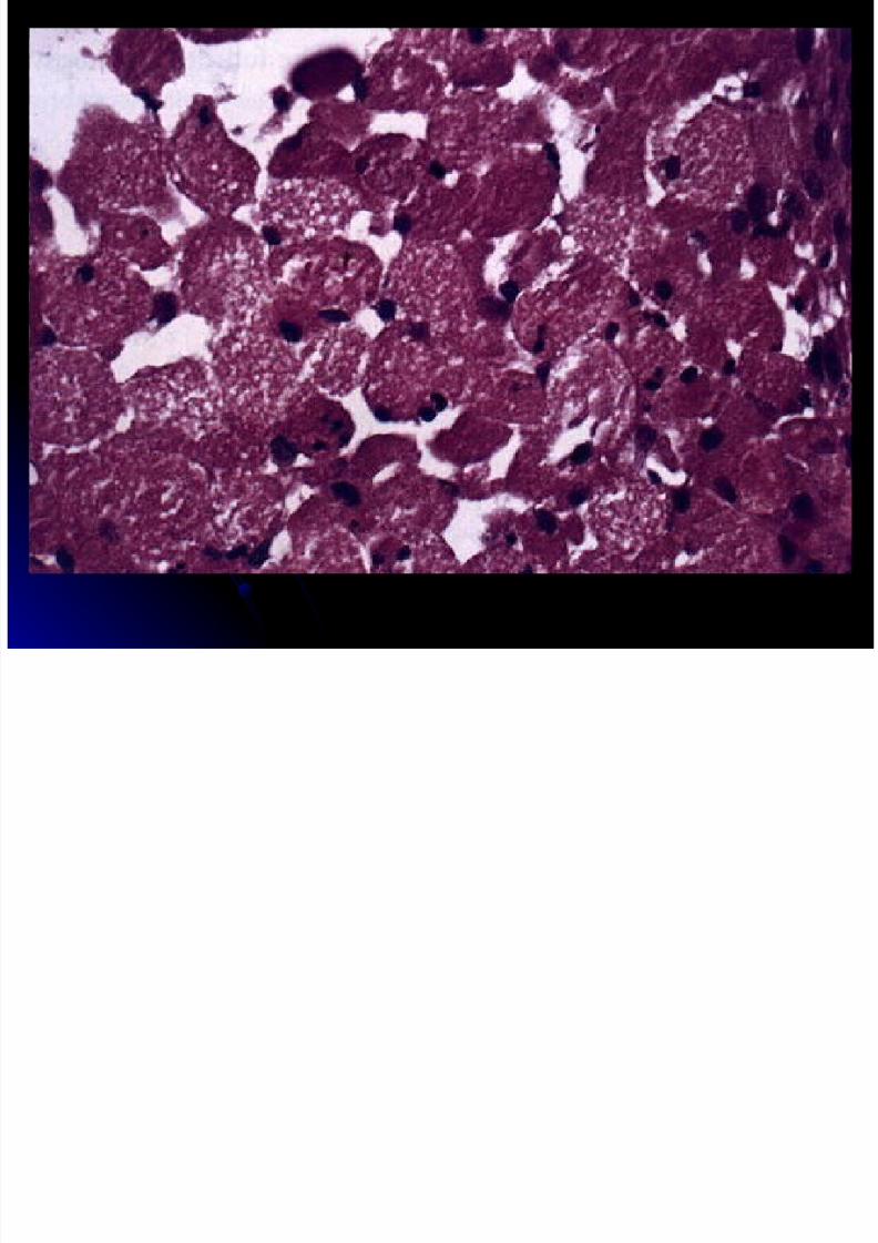
Granular cellGranular cell
Basal cellBasal cellDesmoplasticDesmoplastic

8/3/2019 a Types
http://slidepdf.com/reader/full/a-types 40/86
MicroscopyMicroscopy
Follicular patternFollicular patternMost common and recognizable patternMost common and recognizable pattern
Discrete islands or follicles of epithelialDiscrete islands or follicles of epithelial
cells in a mature connective tissue stromacells in a mature connective tissue stromaEpithelial islands – resemble enamelEpithelial islands – resemble enamel
organ of the developing tooth germ ; :organ of the developing tooth germ ; :

8/3/2019 a Types
http://slidepdf.com/reader/full/a-types 41/86
Consist of:Consist of:
Peripheral cells (ameloblast like cells)Peripheral cells (ameloblast like cells)
Coumnar basal cells with hyperchromaticCoumnar basal cells with hyperchromatic
nucleinuclei
Nuclear palisading with polarizationNuclear palisading with polarization
Cytoplasmic vacuolationCytoplasmic vacuolation
Central cells – loosely arranged and resembleCentral cells – loosely arranged and resemble
stellate reticulumstellate reticulum

8/3/2019 a Types
http://slidepdf.com/reader/full/a-types 42/86
Cyst formation –Cyst formation – commoncommonMicrocyst – large macroscopic cystsMicrocyst – large macroscopic cysts
Hyalinization around the follicles – because of Hyalinization around the follicles – because of induction phenomenoninduction phenomenon
StromaStroma
Mature fibrous connective tissue in variableMature fibrous connective tissue in variableamountsamounts
Plentiful or very minimalPlentiful or very minimal
MicroscopyMicroscopy

8/3/2019 a Types
http://slidepdf.com/reader/full/a-types 43/86

8/3/2019 a Types
http://slidepdf.com/reader/full/a-types 44/86
Mi

8/3/2019 a Types
http://slidepdf.com/reader/full/a-types 45/86
Plexiform patternPlexiform pattern
Epithelium –Epithelium –Long, anastomosing cords or larger Long, anastomosing cords or larger
sheets bounded by single layer columnar sheets bounded by single layer columnar or cuboidal ameloblast like cellsor cuboidal ameloblast like cellssurrounding more loosely arrangedsurrounding more loosely arranged
epithelial cellsepithelial cellsStroma –Stroma –Loosely arranged and vascular Loosely arranged and vascular
Cyst formation -- uncommonCyst formation -- uncommon
MicroscopyMicroscopy

8/3/2019 a Types
http://slidepdf.com/reader/full/a-types 46/86

8/3/2019 a Types
http://slidepdf.com/reader/full/a-types 47/86

8/3/2019 a Types
http://slidepdf.com/reader/full/a-types 48/86
8/3/2019 a Types
http://slidepdf.com/reader/full/a-types 49/86
Acanthomatous pattern Acanthomatous pattern
Extensive squamous metaplasia, oftenExtensive squamous metaplasia, often
with keratin formation in the centralwith keratin formation in the central
portions of a follicleportions of a follicle
Mistaken for Mistaken for Squamous cell carcinomaSquamous cell carcinoma
Squamous odontogenic tumor Squamous odontogenic tumor
MicroscopyMicroscopy

8/3/2019 a Types
http://slidepdf.com/reader/full/a-types 50/86

8/3/2019 a Types
http://slidepdf.com/reader/full/a-types 51/86

8/3/2019 a Types
http://slidepdf.com/reader/full/a-types 52/86
8/3/2019 a Types
http://slidepdf.com/reader/full/a-types 53/86
MiMi

8/3/2019 a Types
http://slidepdf.com/reader/full/a-types 54/86
Basal cell patternBasal cell pattern
Least common typeLeast common type
Nests of uniform basaloid cellsNests of uniform basaloid cellsH/p – similar to BCC of skinH/p – similar to BCC of skin
No stellate reticulum in the central portionsNo stellate reticulum in the central portions
of the nestsof the nestsPeripheral cells – cuboidal rather thanPeripheral cells – cuboidal rather than
columnar columnar
MicroscopyMicroscopy

8/3/2019 a Types
http://slidepdf.com/reader/full/a-types 55/86

8/3/2019 a Types
http://slidepdf.com/reader/full/a-types 56/86
Desmoplastic ameloblastoma
Eversole et al, 1984
Considered as a separate type:Different clinical features
Different radiological features
Different histopathological features

8/3/2019 a Types
http://slidepdf.com/reader/full/a-types 57/86
Desmoplastic ameloblastomaDesmoplastic ameloblastoma
Desmoplasia – extensive stromalDesmoplasia – extensive stromal
collagenization( hyalinization)collagenization( hyalinization)
Hypocellular Hypocellular
Tendency to grow in thin strands and cords of Tendency to grow in thin strands and cords of epi rather than in an island like patteren.epi rather than in an island like patteren.
Epithelium compressed and fragmentedEpithelium compressed and fragmented
Scant central cells with peripheral flat cells.Scant central cells with peripheral flat cells. Site : maxilla > mandibleSite : maxilla > mandible
Histogenesis : cell rests of MalassezHistogenesis : cell rests of Malassez

8/3/2019 a Types
http://slidepdf.com/reader/full/a-types 58/86
RADIOLOGICAL FEATURES
Mixed radiolucent & radio-opaque
lesion
Unilocular or Multilocular Borders poorly defined
DIFFERENTIAL DIAGNOSIS:Fibro-osseous lesions

8/3/2019 a Types
http://slidepdf.com/reader/full/a-types 59/86
HISTOPATHOLOGY
STROMA:
Desmoplasia
Thick collagen bundles squeeze theepithelial islands
New bone formation

8/3/2019 a Types
http://slidepdf.com/reader/full/a-types 60/86
HISTOPATHOLOGY
ODONTOGENIC EPITHELIUM:
Islands compressed by the collagen
bundles (ANIMAL-LIKE; KITE-LIKE)
Peripheral cells: cuboidal
No ameloblast-like cells
Central cells: spindle / polygonalNo stellate reticulum-like cells

8/3/2019 a Types
http://slidepdf.com/reader/full/a-types 61/86

8/3/2019 a Types
http://slidepdf.com/reader/full/a-types 62/86

8/3/2019 a Types
http://slidepdf.com/reader/full/a-types 63/86
Treatment and prognosisTreatment and prognosis
A variety of treatment modalities A variety of treatment modalities
Simple enucleation and curettage to en blocSimple enucleation and curettage to en bloc
resectionresection Curettage – higher recurrence rate (50Curettage – higher recurrence rate (50 – 90 %) – 90 %)
Marginal / bloc resection – most widely usedMarginal / bloc resection – most widely used
Recurrence rate -- 15 %Recurrence rate -- 15 %
Treatment and prognosisTreatment and prognosis

8/3/2019 a Types
http://slidepdf.com/reader/full/a-types 64/86
Treatment and prognosisTreatment and prognosis
Conventional ameloblastomaConventional ameloblastoma A persistent, infiltrative neoplasm A persistent, infiltrative neoplasm
Progressive spread to vital structuresProgressive spread to vital structures
DeathDeath
Many of these tumors – not lifeMany of these tumors – not life
threatening lesionsthreatening lesions
rarely – frank malignant behavior rarely – frank malignant behavior

8/3/2019 a Types
http://slidepdf.com/reader/full/a-types 65/86
UNICYSTIC AMELOBLASTOMAUNICYSTIC AMELOBLASTOMA
A unilocular cystic lesion whose clinical A unilocular cystic lesion whose clinical
features resemble those of a non neoplasticfeatures resemble those of a non neoplastic
cystcyst
A distinct entity based on clinical, A distinct entity based on clinical,
radiographic & pathologic features and itsradiographic & pathologic features and its
response to treatmentresponse to treatment
10 - 1510 - 15% of all intraosseous% of all intraosseousameloblastomasameloblastomas
UNICYSTIC AMELOBLASTOMAUNICYSTIC AMELOBLASTOMA

8/3/2019 a Types
http://slidepdf.com/reader/full/a-types 66/86
UNICYSTIC AMELOBLASTOMAUNICYSTIC AMELOBLASTOMA
Clinical featuresClinical features Age : Age :Younger patientsYounger patients
22ndnd
decadedecadeSex : no predilectionSex : no predilection
Site : posterior mandible (90Site : posterior mandible (90 % cases)% cases)
Presentation :Presentation : Asymptomatic Asymptomatic
Large lesions – painless swelling of the jawsLarge lesions – painless swelling of the jaws
UNICYSTIC AMELOBLASTOMAUNICYSTIC AMELOBLASTOMA

8/3/2019 a Types
http://slidepdf.com/reader/full/a-types 67/86
Radiographic featuresRadiographic features
May or may not be associated with anMay or may not be associated with an
impacted toothimpacted tooth
Well – defined radiolucencies ; may or Well – defined radiolucencies ; may or
may not be demarcated by a perilesionalmay not be demarcated by a perilesional
corticated rimcorticated rim

8/3/2019 a Types
http://slidepdf.com/reader/full/a-types 68/86

8/3/2019 a Types
http://slidepdf.com/reader/full/a-types 69/86
UNICYSTIC AMELOBLASTOMAUNICYSTIC AMELOBLASTOMA

8/3/2019 a Types
http://slidepdf.com/reader/full/a-types 70/86
HistogenesisHistogenesis
Proposed theoriesProposed theoriesCystic degeneration of solidCystic degeneration of solid
ameloblastomasameloblastomas Ameloblastomatous change in an Ameloblastomatous change in an
preexisting cystpreexisting cyst
Co – existence of non-neoplastic andCo – existence of non-neoplastic andneoplastic epitheliumneoplastic epithelium
De novoDe novo
U C S C O S O
UNICYSTIC AMELOBLASTOMAUNICYSTIC AMELOBLASTOMA

8/3/2019 a Types
http://slidepdf.com/reader/full/a-types 71/86
Association with a cyst Association with a cyst
Most common --Most common -- dentigerous cystdentigerous cyst
Other cysts --Other cysts --Parakeratinized OKCParakeratinized OKC
Radicular cystRadicular cyst
Residual cystResidual cyst
COCCOC
GOCGOC
UNICYSTIC AMELOBLASTOMAUNICYSTIC AMELOBLASTOMA
UNICYSTIC AMELOBLASTOMAUNICYSTIC AMELOBLASTOMA

8/3/2019 a Types
http://slidepdf.com/reader/full/a-types 72/86
HistopathologyHistopathology
3 distinct patterns3 distinct patterns
1.1. LuminalLuminal
2.2. IntraluminalIntraluminal
3.3. MuralMural

8/3/2019 a Types
http://slidepdf.com/reader/full/a-types 73/86
UNICYSTIC AMELOBLASTOMAUNICYSTIC AMELOBLASTOMA

8/3/2019 a Types
http://slidepdf.com/reader/full/a-types 74/86
LuminalLuminal
Unilocular cystic lesion lined by epithelium (basalUnilocular cystic lesion lined by epithelium (basal
cells –cells – VICKERS-GORLIN CRITERIAVICKERS-GORLIN CRITERIA))
No infiltrating neoplastic epitheliumNo infiltrating neoplastic epithelium Tumor confined to luminal surface of the cystTumor confined to luminal surface of the cyst
luminal unicystic ameloblastomaluminal unicystic ameloblastoma
Cells overlying the basal layer – looselyCells overlying the basal layer – looselycohesive and resemble stellate reticulumcohesive and resemble stellate reticulum

8/3/2019 a Types
http://slidepdf.com/reader/full/a-types 75/86
VICKERS-GORLIN CRITERIA
Columnar basal cells with
hyperchromatic nuclei
Nuclear palisading with polarization
Cytoplasmic vacuolation withintercellular spacing
Subepithelial hyalanization
UNICYSTIC AMELOBLASTOMAUNICYSTIC AMELOBLASTOMA

8/3/2019 a Types
http://slidepdf.com/reader/full/a-types 76/86
IntraluminalIntraluminalUnilocular cystic lesion in which a noduleUnilocular cystic lesion in which a nodule
arises from the epithelium and projectsarises from the epithelium and projects
into the lumen of the cystinto the lumen of the cystNodules – odontogenic epithelium thatNodules – odontogenic epithelium that
may sometimes resemble plexiformmay sometimes resemble plexiformameloblastomaameloblastoma
Part of the lining – V & G criteriaPart of the lining – V & G criteriaNo infiltration into the cyst wallNo infiltration into the cyst wall
UNICYSTIC AMELOBLASTOMAUNICYSTIC AMELOBLASTOMA

8/3/2019 a Types
http://slidepdf.com/reader/full/a-types 77/86
MuralMural
Unilocular cystic lesion – islands of Unilocular cystic lesion – islands of
ameloblastomatous epithelium (follicular /ameloblastomatous epithelium (follicular /
plexiform) in the fibrous cyst wallplexiform) in the fibrous cyst wallMay or may not be connected to the liningMay or may not be connected to the lining
Part of cystic lining – V & G criteriaPart of cystic lining – V & G criteria
Mural + Intraluminal prolierationsMural + Intraluminal prolierations

8/3/2019 a Types
http://slidepdf.com/reader/full/a-types 78/86

8/3/2019 a Types
http://slidepdf.com/reader/full/a-types 79/86
UNICYSTIC AMELOBLASTOMAUNICYSTIC AMELOBLASTOMA
8/3/2019 a Types
http://slidepdf.com/reader/full/a-types 80/86
DIAGNOSIS OF UA – ONLY AFTERDIAGNOSIS OF UA – ONLY AFTER
MICROSCOPIC EXAMINATIONMICROSCOPIC EXAMINATION
Treatment and PrognosisTreatment and Prognosis
Luminal & intraluminal variants –Luminal & intraluminal variants –
conservative approachconservative approach
Mural -- radical resectionMural -- radical resection
Recurrence rates – 10 - 20Recurrence rates – 10 - 20% after % after
enucleation & curettageenucleation & curettage
UNICYSTIC AMELOBLASTOMA

8/3/2019 a Types
http://slidepdf.com/reader/full/a-types 81/86

8/3/2019 a Types
http://slidepdf.com/reader/full/a-types 82/86

8/3/2019 a Types
http://slidepdf.com/reader/full/a-types 83/86

8/3/2019 a Types
http://slidepdf.com/reader/full/a-types 84/86

8/3/2019 a Types
http://slidepdf.com/reader/full/a-types 85/86

8/3/2019 a Types
http://slidepdf.com/reader/full/a-types 86/86


![Linear Dependent Types · Linear types and dependent types Linear types (Girard 1987): A− B, … Dependent types (Martin-Löf 1970s): x:A.B[x], … How to combine them? In most](https://img.pdfslide.us/doc/110x75/5f0480067e708231d40e46ee/linear-dependent-types-linear-types-and-dependent-types-linear-types-girard-1987.jpg)