Embed Size (px)
Citation preview

ARTICLE
A tumor-targeted trimeric 4-1BB-agonistic antibodyinduces potent anti-tumor immunity withoutsystemic toxicityMarta Compte1, Seandean Lykke Harwood2, Ines G. Muñoz3, Rocio Navarro4, Manuela Zonca1,
Gema Perez-Chacon5,6, Ainhoa Erce-Llamazares1, Nekane Merino 7, Antonio Tapia-Galisteo4,
Angel M. Cuesta4, Kasper Mikkelsen2, Eduardo Caleiras8, Natalia Nuñez-Prado4, M. Angela Aznar9,
Simon Lykkemark2, Jorge Martínez-Torrecuadrada3, Ignacio Melero9,10,11,12, Francisco J. Blanco 7,13,
Jorge Bernardino de la Serna 14,15, Juan M. Zapata5,6, Laura Sanz 4 & Luis Alvarez-Vallina2,16,17
The costimulation of immune cells using first-generation anti-4-1BB monoclonal antibodies
(mAbs) has demonstrated anti-tumor activity in human trials. Further clinical development,
however, is restricted by significant off-tumor toxicities associated with FcγR interactions.
Here, we have designed an Fc-free tumor-targeted 4-1BB-agonistic trimerbody, 1D8N/CEGa1,
consisting of three anti-4-1BB single-chain variable fragments and three anti-EGFR single-
domain antibodies positioned in an extended hexagonal conformation around the collagen
XVIII homotrimerization domain. The1D8N/CEGa1 trimerbody demonstrated high-avidity
binding to 4-1BB and EGFR and a potent in vitro costimulatory capacity in the presence of
EGFR. The trimerbody rapidly accumulates in EGFR-positive tumors and exhibits anti-tumor
activity similar to IgG-based 4-1BB-agonistic mAbs. Importantly, treatment with 1D8N/CEGa1
does not induce systemic inflammatory cytokine production or hepatotoxicity associated with
IgG-based 4-1BB agonists. These results implicate FcγR interactions in the 4-1BB-agonist-
associated immune abnormalities, and promote the use of the non-canonical antibody pre-
sented in this work for safe and effective costimulatory strategies in cancer immunotherapy.
DOI: 10.1038/s41467-018-07195-w OPEN
1 Department of Antibody Engineering, Leadartis SL, 28008 Madrid, Spain. 2 Immunotherapy and Cell Engineering Laboratory, Department of Engineering,Aarhus University, 8000C Aarhus, Denmark. 3 Crystallography and Protein Engineering Unit, Spanish National Cancer Research Centre (CNIO), 28029Madrid, Spain. 4Molecular Immunology Unit, Hospital Universitario Puerta de Hierro Majadahonda, 28222 Majadahonda, Madrid, Spain. 5 Instituto deInvestigaciones Biomédicas Alberto Sols (IIBm), CSIC-UAM, 28029 Madrid, Spain. 6 Instituto de Investigación Sanitaria La Paz (IdiPaz), 28029 Madrid,Spain. 7 Structural Biology Unit, CIC bioGUNE, Parque Tecnológico de Bizkaia, 48160 Derio, Spain. 8 Histopathology Unit, Spanish National Cancer ResearchCentre (CNIO), 28029 Madrid, Spain. 9 Department of Immunology and Immunotherapy, Center for Applied Medical Research (CIMA), University ofNavarra, 31008 Pamplona, Spain. 10 Department of Immunology, University Clinic, University of Navarra, 31008 Pamplona, Spain. 11 Instituto de InvestigaciónSanitaria de Navarra (IdISNA), 31008 Pamplona, Spain. 12 CIBERONC-Centro virtual de Investigación Biomédica en red de Oncología, 28029 Madrid, Spain.13 IKERBASQUE, Basque Foundation for Science, 48013 Bilbao, Spain. 14 Central Laser Facility, Science and Technology Facilities Council, Rutherford AppletonLaboratory, Research Complex at Harwell, OX11 0QX Harwell-Oxford, UK. 15 Department of Physics, King’s College London, WC2R 2LS London, UK.16 Cancer Immunotherapy Unit (UNICA), Department of Immunology, Hospital Universitario 12 de Octubre, 28041 Madrid, Spain. 17 Immuno-Oncology andImmunotherapy Group, Instituto de Investigación Sanitaria 12 de Octubre (i+12), 28041 Madrid, Spain. Correspondence and requests for materials should beaddressed to L.A.-V. (email: [email protected])
NATURE COMMUNICATIONS | (2018) 9:4809 | DOI: 10.1038/s41467-018-07195-w |www.nature.com/naturecommunications 1
1234
5678
90():,;
Modulating immune responses using monoclonal anti-bodies (mAbs) is a promising approach to cancertherapy. Antagonistic mAbs directed against checkpoint
inhibitors such as cytotoxic T-lymphocyte–associated antigen 4and programmed cell death 1/programmed cell death ligand 1(PD-L1) have been clinically approved, and agonistic mAbs tar-geting costimulatory receptors are undergoing clinical trials1.Costimulatory receptors of the tumor necrosis factor (TNF)receptor superfamily (TNFRSF), such as CD40, OX40, and 4‐1BB,are particularly interesting targets, as these receptors are notconstitutively expressed on resting naive T cells but acquiredupon activation2–4. This limits the potential deleterious sideeffects of the treatment5.
4-1BB (CD137, TNFRSF9) has only one confirmed ligand[4-1BB-Ligand (4-1BBL), TNFSF9], which is expressed on mac-rophages, activated B cells, and dendritic cells6. Engagement of4-1BB by its ligand or an agonistic antibody promotes T cellproliferation, cytokine production, and cytolytic effector func-tions and protects lymphocytes from programmed cell death7,8.Furthermore, engagement of 4-1BB on natural killer cellsenhances cytokine release (including interferon (IFN)-γ)9andantibody-dependent cellular cytotoxicity10,11. Indeed, treatmentof mice with 4-1BB-agonistic mAbs was found to induce tumorregression of established and poorly immunogenic tumors asearly as 199712. Since then, a large body of accumulated pre-clinical data has been gathered that supports the induction of 4-1BB signaling in cancer immunotherapy, both as a single agentand in combination therapies13.
The effect of 4-1BB-agonistic mAbs is not spatially restricted tothe tumor, and peripheral toxicities can therefore reduce thetherapeutic window for 4-1BB-targeting therapies. In mice, 4-1BBmAbs have been shown to cause immune anomalies, notablypolyclonal activation of CD8+ T cells and secretion of inflam-matory cytokines, which affected the function of liver, spleen, andbone marrow14,15. In clinical studies, an anti-4-1BB mAb (BMS-663513, urelumab) showed tolerable side effects in an initialPhase I trial, but a follow-up Phase II trial revealed severe livertoxicity in ≈10% of the patients that resulted in two fatalities16. Asa consequence, trials with urelumab were terminated17. Recently,data were presented on a dose-escalation study with urelumab asmonotherapy and in combination with nivolumab18. The reduceddose ameliorated liver toxicity; however, the clinical activity ofurelumab at the tolerated dose was limited. An integrated safetyanalysis of patients treated with urelumab confirmed a clearassociation between transaminitis and urelumab dose19. Utomi-lumab is another anti-41BB mAb in clinical trials with a bettersafety profile than urelumab but is a relatively less potent 4-1BBagonist20.
As it stands, costimulation by 4-1BB-agonistic mAbs is anotherwise viable therapeutic approach held back by off-tumortoxicities and could therefore benefit greatly from the addition oftumor-targeting functionality to restrict its effect to the tumordeposits. Furthermore, if this is conveyed by binding domainsspecific to cell surface tumor-associated antigens (TAAs), theanti-4-1BB antibodies will then cluster on the surface of cancercells. This may allow the antibodies to mimic physiological 4-1BBL and could have a major impact on the induction of 4-1BBsignaling. Importantly, 4-1BBL is a trimeric membrane proteinand can be proteolytically processed into soluble trimeric ligandswith a significantly reduced signaling activity compared to theirtransmembrane counterparts21. Signaling can be restored byhigher-order oligomerization21,22, cell surface display of anti-4-1BB single chain antibody fragments (scFv) expressed by tumorcells in fusion with membrane proteins23,24, or antibody-mediated display by fusing the extracellular domain of 4-1BBLto a TAA-specific scFv25. Another strategy is the use of anti-4-
1BB oligonucleotide aptamers instead of 4-1BBL26,27. In animalmodels, systemic delivery of a 4-1BB-agonistic aptamer con-jugated to a prostate-specific membrane antigen aptamer led tosuperior therapeutic effect compared to immunoglobulin G(IgG)-based 4-1BB-agonistic antibodies26. It has also beenrecently reported that anchoring anti-4-1BB F(ab′)2 fragmentsand interleukin (IL)-2 on the surface of liposomes inducedeffective antitumor immunity without systemic toxicity28.
In this article, we describe the adaptation of the first-generation4-1BB agonistic IgG 1D8 to a recombinant antibody format, thetrimerbody. This format is based on the fusion of antibody-derived binding domains to the small homotrimerizationregion from murine collagen XVIII, which yields trimericantibodies29–31. Trimerbodies have two major advantages com-pared to the IgG mAb: they lack the fragment crystallizable (Fc)region involved in 4-1BB-mediated toxicity20; and they are tri-meric, as is physiological 4-1BBL. We generated a panel of 1D8scFv-based N-terminal trimerbodies (1D8N) using linkers ofdifferent lengths and identified 1D8N18, the trimerbody with thelongest linker, as showing the most potent costimulatory activity.1D8N18 was therefore used as the basis for a bispecific tumor-targeted trimerbody, 1D8N/CEGa1, by adding the epidermalgrowth factor receptor (EGFR)-binding EGa1 single-domainantibody32. Compared to both 1D8 IgG and 1D8N18, 1D8N/CEGa1 was a more potent costimulator in vitro and showedenhanced tumor homing and tumor inhibition in vivo, with noindication of the 4-1BB mAb-associated toxicity.
ResultsDesign of 4-1BB-agonistic trimerbodies. Using the scFv derivedfrom the rat IgG2a, anti-4-1BB 1D8 mAb33 (Fig. 1a), we designeda panel of 1D8 scFv-based N-terminal trimerbodies (1D8N).Three candidates were generated with varied lengths of the flex-ible linker connecting the 1D8 scFv to a murine collagen XVIII-derived homotrimerization (TIEXVIII) domain: 1D8N0 has nolinker, while 1D8N5 and 1D8N18 have 5- and 18-residue-longlinkers, respectively (Fig. 1b). All three constructs were expressedby transfected HEK293 cells at similar levels to MFE-23N18, abenchmark N-terminal trimerbody based on the anti-CEA MFE-23 scFv29. In western blot analysis under reducing conditions, the1D8N trimerbodies were single-chain-type molecules with amigration pattern consistent with the molecular weights calcu-lated from their amino acid sequences (34.4, 34.7, and 36.8 kDa,in the order of increasing linker length) (Supplementary Fig-ure 1a). Additionally, they specifically recognized murine 4-1BBin fusion with human Fc (m4-1BB) immobilized on plastic, asdetermined by enzyme-linked immunosorbent assay (ELISA;Supplementary Figure 1b).
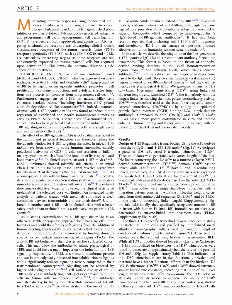
The three 4-1BB-specific trimerbodies were produced in stablytransfected HEK293 cells and purified by immobilized metalaffinity chromatography, with a yield of roughly 1 mg/l ofconditioned medium (Supplementary Figure 2a). Their bindingkinetics were then studied using biolayer interferometry (BLI).While all 1D8 antibodies showed low picomolar-range KD towardm4-1BB immobilized on biosensors, the 1D8N trimerbodies werefound to dissociate at approximately half the rate of the parental1D8 mAb (Fig. 2a, Supplementary Table 1). This indicates thatthe 1D8N trimerbodies are in fact functionally trivalent andtherefore have a higher functional affinity than the bivalent 1D8IgG. Furthermore, 1D8N18, 1D8N5, and 1D8N0 showed extremelysimilar kinetic rate constants, indicating that none of the linkerlength variations structurally compromise the 1D8 scFv orsterically hinder its access to antigen. The ability of 1D8N
trimerbodies to detect m4-1BB in a cellular context was studiedby flow cytometry. All 1D8N trimerbodies bound to HEK293 cells
ARTICLE NATURE COMMUNICATIONS | DOI: 10.1038/s41467-018-07195-w
2 NATURE COMMUNICATIONS | (2018) 9:4809 | DOI: 10.1038/s41467-018-07195-w |www.nature.com/naturecommunications

transfected to express murine 4-1BB on their cell surface(HEK293m4-1BB) but not to untransfected HEK293 cells (Supple-mentary Figure 2b). The binding of 1D8N0 to HEK293m4-1BB cellswas less efficient than that of the 1D8N5 and 1D8N18
(Supplementary Figure 2b). 1D8N5, 1D8N18, and 1D8 IgG allbound to activated mouse CD8a+ T cells but did not bind theunstimulated T cells (Fig. 2b). The binding of 1D8N5 and 1D8N18
was competitively inhibited by 1D8 IgG (SupplementaryFigure 2c). These results show that the 1D8N trimerbodies retainthe ability to bind to endogenous murine 4-1BB.
We proceeded to investigate the costimulatory capability of the1D8N trimerbodies by testing their effect on the proliferation, IFN-γ secretion, and viability of mouse CD8a+ T cells in the presence ofa suboptimal dose of anti-CD3 mAb. 1D8 IgG and 1D8N18
increased proliferation (P = 0.0163 and P = 0.0013, respectively)and IFN-γ secretion (P = 0.0092 and P = 0.0101, respectively)similarly to each other (Fig. 2c, d), and 1D8N18 was significantlymore potent than 1D8N5 and 1D8N0 (P = 0.0234 and P = 0.0016,respectively; Fig. 2c). After 72 h, a statistically significant increasedviability of CD8a+ T cells stimulated with 1D8 IgG and 1D8N18
was observed (P = 0.005 and P = 0.015, respectively; Fig. 2e).Furthermore, 1D8N18 was significantly more potent than 1D8 IgG(P = 0.046; Fig. 2e). The recombinant soluble mouse 4-1BBL (m4-1BBL) was essentially inactive (Fig. 2c, e). The m4-1BBL migratesat approximately 40 kDa in reducing conditions and atapproximately 70 kDa in non-reducing conditions, compatiblewith a trimer (Supplementary Figure 3a). The binding to m4-1BBexpressed on the cell surface was less efficient than the 1D8N18
(Supplementary Figure 3b), and competition ELISA demonstratedthat m4-1BBL and 1D8N18 recognize different regions of the m4-1BB (Supplementary Figure 3c). Next, we investigated the spatio-temporal distribution and dynamics of the interactions betweencell surface m4-1BB and CF488A-labeled m4-1BBL, 1D8 IgG or1D8N18 (Supplementary Figure 4) in living HEK293m4-1BB-S cellsdisplaying homogenous expression of the receptor (Supplementary
Figure 5). Employing raster imaging correlation spectroscopy(RICS)34, we observed and quantified receptor clustering and itsmolecular mobility upon binding. We observed that m4-1BBL doesnot induce receptor clustering but rather internalization of thereceptor into the cytoplasm (≈70 μm2/s) (Supplementary Figure6a). In contrast, both 1D8 IgG and 1D8N18 induce clusterformation, reducing the lateral mobility drastically at the plasmamembrane upon binding, from ≈1.5 to 0.35 and from ≈1.0 to 0.15μm2/s, respectively (Fig. 2f and Supplementary Figure 6b, c).1D8N18 formed larger and more numerous membrane clusters,which consequently impeded the lateral diffusion to a greaterdegree, which indicates more effective and extensive crosslinking(Fig. 2g).
As the 1D8N18 demonstrated improved T cell costimulatoryactivity and induced receptor clustering to a greater degree, wechose this particular configuration for subsequent studies. Weused size exclusion chromatography with multi-angle lightscattering (SEC-MALS) to investigate the oligomeric state of1D8N18. It eluted as a major symmetric peak with a mass of 112kDa (Supplementary Figure 7a), close to the predicted 110.1 kDaof 1D8N18 without signal sequence (mass spectrometry by matrixassisted laser desorption/ionization (MALDI) confirmed itsabsence). Two minor peaks were also detected, the smallest beinga protein impurity, as seen by sodium dodecyl sulfate-polyacrylamide gel electrophoresis (SDS-PAGE; SupplementaryFigure 7b). The other peak migrates in SDS-PAGE as 1D8N18 buthas a native mass of 244 kDa, probably corresponding totrimer–dimers. The two species can be separated by SEC, butreinjection of the major peak gives another minor trimer–dimerpeak (Supplementary Figure 7c), indicating an equilibrium wherethe trimeric species are predominant (85% and 97% at 1.0 and0.26 g/l, respectively). The circular dichroism spectrum of 1D8N18
(Supplementary Figure 7d) is typical of proteins with predomi-nantly β-sheet structure. The 1D8N18 is folded into a stable three-dimensional structure, as shown by the cooperative thermaldenaturation (Tm ≈ 57 °C; Supplementary Figure 7e). Tounderstand the behavior and structure of 1D8N18 in solution,we analyzed it in the absence of substrate using small-angle X-rayscattering (SAXS) (Fig. 2h and Supplementary Figure 7f, g,Supplementary Table 2). The ab initio model shows howthe homotrimer adopts a pyramidal conformation stabilizedby direct interactions between the C-terminal TIEXVIII domains,as has previously been described for human collagenXVIII35. The1D8 scFvs point orthogonally away from the plane defined by thetrimerization domain without directly contacting each other, in amanner resembling an open tripod (Fig. 2h).
Design of an EGFR-targeted 4-1BB-agonistic trimerbody. Weproceeded to generate a bispecific trimerbody by fusing the anti-human EGFR single-domain antibody (VHH; EGa1)32to the C-terminus of 1D8N18 through a 17-residue-long linker giving the1D8N/CEGa1 trimerbody (Fig. 1c). The construct was designedwith a FLAG and a StrepII tag at the N-terminus of the 1D8 scFv.The 1D8N/CEGa1 was produced in stably transfected HEK293cells (5 mg/l), followed by Strep-Tactin affinity chromatography.SDS-PAGE analysis, under reducing conditions, of the purifiedprotein revealed a single band with a molecular mass of 55.6 kDaconsistent with the calculated from its amino acid sequence (52.9kDa without the signal sequence; Supplementary Figure 8a). Theoligomeric state of the purified 1D8N/CEGa1 was examined bySEC-MALS measurements. The sample eluted as a major sym-metric peak with a mass of 160 kDa, close to the calculated massfor a trimer without the signal sequence (158.7 kDa), and aminor peak with a mass of 309 kDa (Supplementary Figure 8b),which is indistinguishable from 1D8N/CEGa1 by SDS-PAGE
1D8 IgG103 kDa
104 kDa
110 kDa
150 kDa
150 kDa
1D8N0
S T
T
T
VL
VL
VL
VL VH VHH
VH
VH
VH
S
S
S T
TIE
5TIE
18TIE
1D8N5
1D8N18
1D8N/CEGa118TIE18
a b
c
Fig. 1 Schematic diagrams of 4-1BB-agonistic trimerbodies. Proteinstructure of the anti-4-1BB IgG (a) and the gene layout (left) and proteinstructure (right) of monospecific (b) and bispecific trimerbodies (c). Thevariable regions derived from 1D8 antibody are represented in orange, theanti-EGFR VHH EGa1 in violet, the structural domains in light blue, and thelinker regions in gray. The 1D8 scFv-based N-terminal trimerbodies’ (1D8N)gene constructs (b) contain a signal peptide from oncostatin M (white box)and the 1D8 scFv gene (VL-VH) connected directly or through flexiblelinkers to the mouse TIEXVIII domain. In the bispecific 1D8N/CEGa1trimerbody (c), the anti-human EGFR VHH EGa1 is fused to the C-terminusof 1D8N18 through a flexible linker. Arrows indicate the direction oftranscription. His6-myc tag (green box) and FLAG-strep tags (dark bluebox) were appended for immunodetection
NATURE COMMUNICATIONS | DOI: 10.1038/s41467-018-07195-w ARTICLE
NATURE COMMUNICATIONS | (2018) 9:4809 | DOI: 10.1038/s41467-018-07195-w |www.nature.com/naturecommunications 3
(Supplementary Figure 8c). These results again indicate the for-mation of a minor population of dimers of trimers, as seen for1D8N18. The two oligomeric species can be separated by SEC, andreinjection of the isolated major peak of trimers yields only a veryminor peak at the elution volume of the trimer–dimers (Sup-plementary Figure 8d). 1D8N/CEGa1 performed very similarly to1D8N18 in CD and cooperative thermal denaturation studies(Supplementary Figure 8e, f). SAXS showed that 1D8N/CEGa1contains the same trimerized TIEXVIII core seen for 1D8N18. Thebinding domains, however, are distributed evenly around theplane defined by the TIEXVIII core (Fig. 3a and SupplementaryFigure 8g, h, Supplementary Table 2), resembling a playgroundroundabout. Using the SAXS data and structural informationfrom the protein data bank (PDB), we created a homology modelintended to clarify the layout of the six binding domains aroundthe core, as the linkers’ length and flexibility permit severalconfigurations, e.g., with the three 1D8 scFvs opposite the threeEGa1 VHH (as shown in Fig. 3a), or with alternating 1D8 andEGa1 domains side-by-side each other. Unfortunately, the
obtained resolution is insufficient to distinguish between thesepossibilities, although the overall hexagonal structure is clearlydefined in the ab initio SAXS model.
The functionality of the 1D8N/CEGa1 was demonstrated byBLI. The 1D8N/CEGa1 trimerbody has kinetic rate constants thatare very similar to the 1D8N trimerbodies in its interaction withimmobilized m4-1BB (Fig. 3b, Supplementary Table 1). Thebinding kinetics of 1D8N/CEGa1 to immobilized human EGFR-Fcchimera (hEGFR) was also investigated by BLI, and theinteraction was found to also have a low picomolar KD (Fig. 3b,Supplementary Table 1). Our previous comparison of EGa1 VHH
and EGa1-derived N-trimerbody (EGa1N) kinetics showed a lownanomolar KD for the EGa1 VHH and a low picomolar KD forEGa1N 36. These kinetics are easily distinguishable, and 1D8N/CEGa1 showed comparable kinetics to EGa1N, indicating that ittrivalently binds hEGFR. The 1D8N/CEGa1 was found to becapable of binding immobilized m4-1BB and hEGFR simulta-neously (Fig. 3c). This further demonstrates the bispecificityof 1D8N/CEGa1 and shows a lack of steric hindrance between
0.0
0.00 0.00
0.05
0.10
0.15
0.20
0.050.100.150.200.250.300.35
0 1 2 3 4 0 1 2 3 4
0 1 2 3 4 0 1 2 3 4
0.10.20.30.40.50.6
0.000.050.100.150.200.250.300.35
Time (h)
4
Fol
d ch
ange
– –
3
2
1
0
IgG
m4-
1BB
L
m4-
1BB
L
m4-
1BB
L
1D8
IgG
1D8N
18
1D8N
5
1D8N
0
****
**
**
** 5
4
Fol
d ch
ange
3
2
1
0
–
IgG
1D8
IgG
1D8N
18
MF
E-2
3N18– –
IgG
1D8
IgG
1D8N
18
1D8N
5
1D8N
0
MF
E-2
3N18
MF
E-2
3N18
2500
IFN
γ [p
g/m
l]
2000
1500
500
1000
0
Rel
ativ
e ce
ll nu
mbe
r
Res
pons
e (n
m)
Log fluorescence intensity
Uns
timul
ated
Stim
ulat
ed
Diff
usio
n co
effic
ient
(μm
2 /s)
2.0
0.0
0.5
1.5
1.0
N-CR CR N-CR CR
ii
ii
ii
ii
iiii
0.43 μm2/s
1.2 μm2/s
0.11 μm2/s
0.8 μm2/s
68
51
34
17
0100 101 102 103 104
100 101 102 103 104 100 101 102 103 104
100 101 102 103 104
64
48
32
16
0
68
51
34
17
0
72
54
36
18
0
**
**
*
1D8 IgG 1D8N18
1D8N5 1D8N0
b ha
c
Anti-CD3 mAb
e
Anti-CD3 mAb
d
Anti-CD3 mAb
1D8 IgG 1D8N18
1D8N5
1D8 IgG 1D8N18
1D8N5
scFv
scFv
scFvscFv
scFv
scFv
scFv
scFv
scFv
90°90°
f g
1D8
IgG
1D8N
18
1D8 IgG
1D8N18
Fig. 2 Characterization of anti-4-1BB trimerbodies. a Sensorgrams (black curves) and fitting curves for 1D8 antibodies (2 and 4 nM), obtained by biolayerinterferometry with surface-immobilized m4-1BB. b The binding to 4-1BB on the cell surface of stimulated mouse CD8a+ T cells measured by FACS.c–e Costimulatory activity of anti-4-1BB antibodies. Mouse CD8a+ T cells were stimulated with immobilized anti-CD3 mAb in the presence of m4-1BBL,1D8N5, 1D8N18, or 1D8 IgG, and proliferation (c) and secretion of IFN-γ (d) were measured after 48 h and cell viability (e) after 72 h. Data are reported asfold change relative to the values obtained from anti-CD3 mAb-stimulated CD8a+ T cells. Rat IgG2a and MFE-23N18 were used as controls. Data are mean± SD (n = 3), *P ≤ 0.05, **P ≤ 0.01, Student's t test. f RICS analyses performed in living HEK293m4-1BB-S cells at regions containing clusters formed upon1D8 IgG or 1D8N18 addition and at regions where clusters where not present (insert and zoomed-in regions ii, and i, respectively). Representative maximumintensity projection maps showing the RICS analyzed regions of interest. Values in the zoomed-in regions show the diffusion coefficient of bound antibody.The color heat map indicates in blueish tones the lower intensity and in redder tones the higher intensity. g Statistical analysis of the quantified diffusioncoefficient obtained from 5 to 7 independent live cell experiments and 3–5 different regions of interest per cell (N-CR non-clustered region, CR clusteredregion). Data are presented as median (center line), upper and lower quartiles (boxes), and minimum-maximum (whiskers). h Arrangement of 1D8N18
trimerbody in solution, as determined by SAXS. Rigid-body overlaying of the ab initio-determined SAXS envelope for 1D8N18. The generated model (whereeach chain is colored in blue, magenta, or cyan) fits into the envelope (colored in pale gray)
ARTICLE NATURE COMMUNICATIONS | DOI: 10.1038/s41467-018-07195-w
4 NATURE COMMUNICATIONS | (2018) 9:4809 | DOI: 10.1038/s41467-018-07195-w |www.nature.com/naturecommunications

its interactions with hEGFR and m-4-1BB. The ability of1D8N/CEGa1 to detect its antigens as cell surface proteins wasstudied by flow cytometry. The 1D8N/CEGa1 trimerbody boundto HEK293 (EGFR+), to HEK293m4-1BB cells, and to mouse 3T3cells expressing human EGFR (3T3hEGFR) but not to wild-type3T3 cells (Supplementary Figure 9). Furthermore, 1D8N/CEGa1bound to activated mouse CD8a+ T cells as efficiently as the1D8N18 (Fig. 3d). To further assess the multivalent binding of1D8N/CEGa1, we studied its capacity to inhibit proliferation andEGFR phosphorylation in A431 cells32. 1D8N/CEGa1 andcetuximab, an EGF-competitive inhibitor37, but neither theanti-human CD20 rituximab nor 1D8 IgG, inhibited A431proliferation, in a dose-dependent manner (P ≤ 0.0001 for thehigher doses of both antibodies, vs. equimolar doses of controlantibodies; Supplementary Figure 10a), and EGFR phosphoryla-tion (Supplementary Figure 10b).
We then wanted to determine whether 1D8N/CEGa1 retainedthe baseline costimulatory capacity seen for 1D8N18 and whetherthis was improved by the crosslinking provided through EGa1’sbinding to hEGFR. CD8a+ T cells were stimulated withimmobilized anti-CD3 mAb and the panel of costimulatoryagents in solution, in the presence or absence of plastic-immobilized hEGFR. The 1D8N/CEGa1 had a costimulatoryeffect similar to 1D8N18 in the absence of hEGFR, butproliferation (P = 0.0008) and IFN-γ levels (P = 0.0198) weregreatly enhanced when hEGFR was included (Fig. 3e, f). This
effect was further confirmed by co-culture assays using EGFR-negative and EGFR-positive target cells. The IFN-γ levels weresignificantly higher when CD8a+ T cells were co-cultured with3T3hEGFR in the presence of the 1D8N/CEGa1, as compared to thenon-targeted 1D8 molecules (P = 0.0344 1D8 IgG, and P =0.0009 1D8N18; Fig. 3g). We then investigated the effect of EGFR-targeted 4-1BB costimulation on cell viability. After 72 h, astatistically significant increased viability of CD8a+ T cellscostimulated with 1D8N/CEGa1 in the presence of plastic-immobilized hEGFR was observed (P = 0.0326), as comparedto cells costimulated with 1D8N18 (P = 0.0088; Fig. 3h).
The EGFR-targeted trimerbody shows high tumor localization.First, we studied the serum stability of 1D8N18 and 1D8N/CEGa1,and no significant loss of 4-1BB- or EGFR-binding activity wasdetected even after 7 days in mouse serum at 37 °C (Supple-mentary Figure 11a, b). Pharmacokinetic studies were performedin immunocompetent mice, which received a single intravenously(i.v.) injection of the anti-4-1BB antibodies. The serum con-centrations of active protein were determined by ELISA withimmobilized m4-1BB. In CD-1 mice, the 1D8N18 was rapidlycleared from circulation with a terminal half-life of 1.3 h, whereasthe 1D8N/CEGa1 showed a prolonged circulatory half-life of16.1 h (Fig. 4a, Supplementary Table 3). 1D8N/CEGa1 serum half-life was not influenced by the genetic background of the mice, and
Time (h)0
0.0
0.1
0.2
0.3
0.4
1 2 3Log fluorescence intensity
Res
pons
e (n
m)
0.0
0.1
0.2
40 1 2 3Time (h)
4
Res
pons
e (n
m)
Res
pons
e (n
m)
0.00 1 2 3
0.1
0.2
Time (h)
100 104103102101
68
51
34
17
0
68
51
34
17
0
68
51
34
17
0
Rel
ativ
e ce
ll nu
mbe
r
EGFR1D8N/CEGa1
b
c
d
scFv
scFv
VHH
scFv
VHH
90° 90°
a
hEGFR
1D8 IgG
1D8N18
1D8N/CEGa1
m4-1BB
1D8N18
100 104103102101
100 104103102101
Fol
d ch
ange
0
– –
IgG
1D8
IgG
1D8N
18
1D8N
/CE
Ga1
MF
E-2
3N18
2
4
6
8
e hg
IFN
γ [p
g/m
l]
0
500
1000
1500
2000 ***
– –
IgG
1D8
IgG
1D8N
18
1D8N
/CE
Ga1
MF
E-2
3N18
f
Anti-CD3 mAb
hEGFRBSA
Anti-CD3 mAb
hEGFRBSA
********
– –
IgG
1D8
IgG
1D8N
18
1D8N
/CE
Ga1
MF
E-2
3N18
1500
IFN
γ [p
g/m
l]
0
500
1000
*
Anti-CD3 mAb
3T3hEGFR
3T3
***
–
IgG
1D8
IgG
1D8N
18
1D8N
/CE
Ga1
MF
E-2
3N18
Fol
d ch
ange
0
2
4
6
8*
Anti-CD3 mAb
hEGFRBSA **
Fig. 3 Characterization of the EGFR-targeted 4-1BB-agonistic trimerbody. a Arrangement of 1D8N/CEGa1 trimerbody in solution by SAXS. Rigid-body fittingof the model corresponding to 1D8NCEGa1 inside the SAXS envelope (colored in pale gray). Each chain has been colored in blue, magenta, or cyan.b Sensorgrams (black curves) and the results of fitting to a 1:1 model (red curves) obtained using biolayer interferometry for the interaction of 1D8N/CEGa1(2 and 4 nM) with immobilized m4-1BB and the interaction of 1D8N/CEGa1 (0.5 and 1 nM) with immobilized hEGFR. c Simultaneous binding to both m4-1BB and hEGFR was demonstrated for 1D8N/CEGa1 but not 1D8N18. Biosensors were coated with m4-1BB, after which 4 nM of 1D8N/CEGa1 (black curves)or 1D8N18 (blue curves) were added. d The binding of anti-4-1BB antibodies to m4-1BB on the cell surface of stimulated mouse CD8a+ T cells measured byFACS. e, –f Mouse CD8a+ T cells were plated with immobilized anti-CD3 mAb and hEGFR or BSA in the presence of 1D8N18, 1D8N/CEGa1, or 1D8 IgG, andproliferation (e) and IFN-γ secretion (f) were determined after 48 h. EGFR-negative 3T3 cells or EGFR-positive 3T3hEGFR cells were co-cultured with mouseCD8a+ T cells in the presence of anti-CD3 mAb and 1D8N18, 1D8N/CEGa1, or 1D8 IgG. IFN-γ secretion analyzed after 48 h (g) and cell viability after 72 h(h). Data are represented as fold change relative to the values obtained from anti-CD3 mAb stimulated cells. Rat IgG2a and MFE-23N18 were used asnegative controls. Data are mean ± SD (n = 3), *P ≤ 0.05, **P ≤ 0.01, ***P ≤ 0.001, Student's t test
NATURE COMMUNICATIONS | DOI: 10.1038/s41467-018-07195-w ARTICLE
NATURE COMMUNICATIONS | (2018) 9:4809 | DOI: 10.1038/s41467-018-07195-w |www.nature.com/naturecommunications 5

a similar pharmacokinetic profile was observed in BALB/c mice(Fig. 4b, Supplementary Table 4). The half‐life of the anti-4-1BBmAb 3H333was 4.8 days, consistent with published data38(Fig. 4b,Supplementary Table 4). After a single intraperitoneal (i.p.)injection in BALB/c mice, the half-lives of 1D8N/CEGa1 and 3H3IgG were 32 h and 7 days, respectively (Fig. 4c, SupplementaryTable 5). For in vivo imaging, 3H3 IgG, 1D8N/CEGa1, and theanti-EGFR mAb cetuximab were labeled with near-infrared (NIR)fluorochromes, which did not change their SDS-PAGE migrationor compromise their binding to cell surface antigen (Supple-mentary Figure 12). Athymic nude mice bearing hEGFR-positiveA431 tumor xenografts subcutaneously (s.c.) implanted into theright flank were i.v. injected in the tail vein with NIR-labeledantibodies and imaged 24 h later (Fig. 4d). The 1D8N/CEGa1trimerbody showed high tumor localization with a tumor tonormal tissue (T/N) ratio of 4.85 ± 0.13 (mean ± SD), as com-pared to that of cetuximab (2.54 ± 0.34) (P ≤ 0.01) and 3H3 IgG(1.29 ± 0.06) (P ≤ 0.0001), which corresponds to little to nospecific tumor accumulation (Fig. 4d, e).
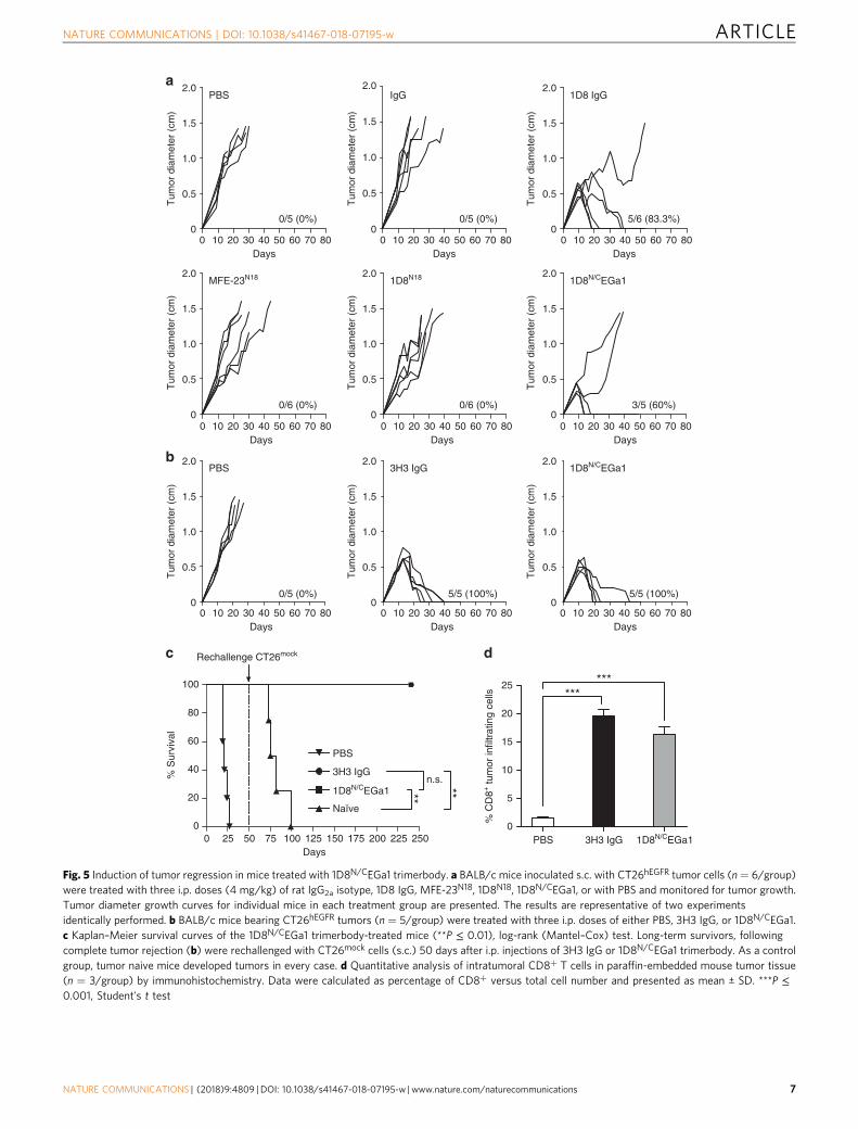
Antitumor activity of EGFR-targeted 4-1BB-agonistic trimer-body. To study the antitumor effects of the EGFR-targeted 4-1BB-agonistic trimerbody in immune competent mice, we usedmurine CT26 colorectal carcinoma (H-2d) cells infected withretrovirus encoding human EGFR (CT26hEGFR) (SupplementaryFigure 13a). The in vitro cell proliferation rates and the in vivotake rate and growth curves in BALB/c mice of CT26hEGFR cellsand CT26mock cells infected with empty vector retroviruses weresimilar (Supplementary Figure 13b, c), suggesting that theexpression of hEGFR did not significantly alter the poor immu-nogenicity of the CT26 tumor cells. Furthermore, ex vivo isolatedCT26hEGFR cells from 3-week-old s.c. tumors expressed sig-nificant levels of surface hEGFR (Supplementary Figure 13d). Toelucidate the functionality of the EGFR pathway in CT26hEGFR
cells, we studied the capacity of cetuximab and 1D8N/CEGa1 toinhibit their proliferation. As shown in Supplementary Figure
13e, neither cetuximab nor 1D8N/CEGa1 had a significant effecton CT26hEGFR proliferation (P = 0.6647 and P = 0.0760respectively, for higher dose, vs. equimolar dose of control anti-body). Therefore, the potential therapeutic effect of 1D8N/CEGa1is not contributed by an EGa1-mediated antiproliferative effect.We used an established regimen to administer IgG-based 4-1BB-agonistic mAbs and the EGFR-targeted 4-1BB-agonistic trimer-body, with three i.p. injections at 2-day intervals15. Injection ofpurified 1D8N/CEGa1 in mice bearing established CT26hEGFR
tumors (average diameter of 0.4 cm) induced tumor regression in8 out of 10 (80%) mice in two separate experiments (Fig. 5a, b).Treatment with the IgG-based 4-1BB agonist antibodies 1D8(Fig. 5a) or 3H3 (Fig. 5b) resulted in complete regression in 10out of 11 (91%) mice bearing CT26hEGFR tumors. All mice treatedwith phosphate-buffered saline (PBS), control antibodies (isotypecontrol rat IgG2a and MFE-23N18), and 1D8N18 were sacrificedwithin 4–5 weeks after tumor cell implantation (Fig. 5a, b). It iswell established that mice cured by IgG-based 4-1BB-agonisticmAb treatment have long-lasting and tumor-specific immu-nity39–41. To investigate whether the EGFR-targeted 4-1BB-agonistic trimerbody can generate a similar effect, mice thatrejected the implanted CT26hEGFR tumor by treatment with 3H3IgG or 1D8N/CEGa1 (Fig. 5b) were rechallenged 50 days laterwith CT26mock cells. 3H3 IgG- and 1D8N/CEGa1-cured mice(P = 0.0027 and P = 0.0067, respectively), but not age-matchednaive mice, were resistant to a rechallenge with CT26mock tumorcells (Fig. 5c and Supplementary Figure 14), showing that thetrimerbody-mediated EGFR-targeted 4-1BB costimulation caninduce long-term protective immunological memory againstCT26 tumors that do not express hEGFR. In order to understandthe antitumor immune response generated with 4-1BB anti-bodies, tumors from 3H3 IgG- and 1D8N/CEGa1-treatedmice and control mice were extracted 2 days after receiving thethird i.p dose (day 13 after tumor inoculation) (SupplementaryFigure 15) for immunohistochemistry to quantify tumor-infiltrating CD8 + T lymphocytes (TILs). The percentage ofCD8+ TILs was an order of magnitude higher in 3H3 IgG- and
10
1
0.1
0.010 12 24 36 48 0 24 48 72 96 120 144 168 0 24 48 72 96 120 144 168 192 216
10
1
0.1
0.01
10
1
0.1
0.01
×10
8
p/sec/cm2/sr
×10
8
p/sec/cm2/sr
2.0
4.0
6.0
2.0
4.0
6.0
2.0
4.0
6.0
×10
8
p/sec/cm2/sr
0
1
2
3
4
5
6***
**
cbaS
erum
con
cent
ratio
n[μ
g/m
l]
Ser
um c
once
ntra
tion
[μg/
ml]
Ser
um c
once
ntra
tion
[μg/
ml]
Time (h) Time (h) Time (h)
d e3H3 IgGPBS PBSCetuximabPBS PBS PBS PBS1D8N/CEGa1
3H3 IgGCetuximab 1D8N/CEGa1
Tum
or/n
orm
al ti
ssue
rat
io[T
/N]
1D8N18
1D8N/CEGa1
3H31D8N/CEGa1
3H31D8N/CEGa1
Fig. 4 Pharmacokinetic properties and tumor imaging of the EGFR-targeted 4-1BB-agonistic trimerbody. Pharmacokinetic studies after a single i.v. dose(1 mg/Kg) of 1D8N18 or 1D8N/CEGa1 in CD-1mice (a) or of 3H3 IgG or 1D8N/CEGa1 in BALB/c mice (b). Pharmacokinetic study after a single i.p. dose(1 mg/Kg) of 3H3 IgG or 1D8N/CEGa1 in BALB/c mice (c). d In vivo fluorescence imaging of A431 tumor-bearing nude mice 24 h after i.v. injection of PBSor 100 µg of Cy5-labeled cetuximab, CF647-labeled 3H3 IgG or CF647-labeled 1D8N/CEGa1. e Tumor to normal tissue (T/N) ratios. The color scale barrepresents the fluorescence intensity recorded as photons per second per centimeter squared per steradian (p/s/cm2/sr). Data are mean ± SD (n = 3),**P ≤ 0.01, ***P ≤ 0.001, Student's t test
ARTICLE NATURE COMMUNICATIONS | DOI: 10.1038/s41467-018-07195-w
6 NATURE COMMUNICATIONS | (2018) 9:4809 | DOI: 10.1038/s41467-018-07195-w |www.nature.com/naturecommunications
100
80
60
40
20
0 0
5
10
15
20
25
0 25 50 75 100 125 150 175 200 225 250
0/5 (0%)
2.0
1.5
1.0
0.5
00 10 20 30 40 50 60 70 80
0/5 (0%)
2.0
1.5
1.0
0.5
00 10 20 30 40 50 60 70 80
5/6 (83.3%)
2.0
1.5
1.0
0.5
00 10 20 30 40 50 60 70 80
0/6 (0%)
2.0
1.5
1.0
0.5
00 10 20 30 40 50 60 70 80
0/6 (0%)
2.0
1.5
1.0
0.5
00 10 20 30 40 50 60 70 80
3/5 (60%)
2.0
1.5
1.0
0.5
00 10 20 30 40 50 60 70 80
2.0
1.0
1.5
0.5
00/5 (0%)
2.0
1.0
1.5
0.5
05/5 (100%)
2.0
1.0
1.5
0.5
05/5 (100%)
Rechallenge CT26mock
% S
urvi
val
Days
Tum
or d
iam
eter
(cm
)T
umor
dia
met
er (
cm)
Tum
or d
iam
eter
(cm
)
PBS
3H3 IgG
Naïve
1D8N/CEGa1n.s.
PBS
Days
IgG
Days
1D8 IgG
Days
MFE-23N18
Days
1D8N18
Days
1D8N/CEGa1
1D8N/CEGa1
Days
0 10 20 30 40 50 60 70 80 0 10 20 30 40 50 60 70 80 0 10 20 30 40 50 60 70 80Days Days Days
PBS 3H3 IgG
Tum
or d
iam
eter
(cm
)
Tum
or d
iam
eter
(cm
)
Tum
or d
iam
eter
(cm
)
Tum
or d
iam
eter
(cm
)
Tum
or d
iam
eter
(cm
)
Tum
or d
iam
eter
(cm
)
% C
D8+
tum
or in
filtr
atin
g ce
lls ******
PBS 3H3 IgG 1D8N/CEGa1
**
**
a
b
c d
Fig. 5 Induction of tumor regression in mice treated with 1D8N/CEGa1 trimerbody. a BALB/c mice inoculated s.c. with CT26hEGFR tumor cells (n = 6/group)were treated with three i.p. doses (4 mg/kg) of rat IgG2a isotype, 1D8 IgG, MFE-23N18, 1D8N18, 1D8N/CEGa1, or with PBS and monitored for tumor growth.Tumor diameter growth curves for individual mice in each treatment group are presented. The results are representative of two experimentsidentically performed. b BALB/c mice bearing CT26hEGFR tumors (n = 5/group) were treated with three i.p. doses of either PBS, 3H3 IgG, or 1D8N/CEGa1.c Kaplan–Meier survival curves of the 1D8N/CEGa1 trimerbody-treated mice (**P ≤ 0.01), log-rank (Mantel–Cox) test. Long-term survivors, followingcomplete tumor rejection (b) were rechallenged with CT26mock cells (s.c.) 50 days after i.p. injections of 3H3 IgG or 1D8N/CEGa1 trimerbody. As a controlgroup, tumor naive mice developed tumors in every case. d Quantitative analysis of intratumoral CD8+ T cells in paraffin-embedded mouse tumor tissue(n = 3/group) by immunohistochemistry. Data were calculated as percentage of CD8+ versus total cell number and presented as mean ± SD. ***P ≤0.001, Student's t test
NATURE COMMUNICATIONS | DOI: 10.1038/s41467-018-07195-w ARTICLE
NATURE COMMUNICATIONS | (2018) 9:4809 | DOI: 10.1038/s41467-018-07195-w |www.nature.com/naturecommunications 7

1D8N/CEGa1-treated mice (19.48 ± 1.28 and 16.25 ± 1.41,respectively) compared to the PBS-treated mice (1.42 ± 0.36)(Fig. 5d and Supplementary Figure 16), indicating an efficienttumor recruitment of CD8+ T lymphocytes in the antibody-treated mice.
EGFR-targeted 4-1BB-agonistic trimerbody shows negligibletoxicity. We compared the toxicity profile of 1D8N/CEGa1directly with 3H3 IgG under similar conditions. Mice wereinjected i.p. with PBS, 3H3 IgG, or 1D8N/CEGa1 (6 mg/kg) once aweek for 3 weeks and euthanized 1 week later. As shown inFig. 6a, treatment with 3H3 IgG resulted in gross enlargement ofspleen and livers as demonstrated by weight (P ≤ 0.0001 and P =0.0058, respectively). In contrast, treatment with 1D8N/CEGa1 didnot result in splenomegaly or hepatomegaly (Fig. 6a). The his-tologic study of the spleens of mice treated with 3H3 IgG revealedan altered architecture with expanded follicles with undefinedzones and clear evidence of extramedullary hematopoiesis(Fig. 6b), as previously described14. In contrast, the spleens ofmice treated with 1D8N/CEGa1 showed normal histology(Fig. 6b). Also confirming previous results14,15, treatment with3H3 IgG caused significant mononuclear cell infiltration in theliver, forming periportal cuffs with thickening of tunica mediaand also infiltration foci associated with microvasculature, whileno significant infiltration was observed in mice treated with1D8N/CEGa1 (Fig. 6b). Indeed, the surface of infiltrating mono-nuclear cells accounted for 8% of the liver of mice treated with3H3 IgG, while it only represented 0.6% in mice treated with1D8N/CEGa1 (P = 0.0048) and 0.25% in mice treated with PBS
(P = 0.0104) (Fig. 6c). In addition, staining of collagen fibers withsirius red (Supplementary Figure 17) or Masson's trichrome(Supplementary Figure 18) showed that 3H3 IgG treatment, butnot 1D8N/CEGa1, caused the deposition and disarrayment ofportal collagen fibers, indicative of an early stage of fibrosis.Mononuclear cell infiltration was also seen in the lungs, formingperivascular cuffs, and in pancreas of 3H3 IgG-treated mice.Pancreas infiltration radiated from the vasculature and extendedto the neighboring intercalated ducts (Fig. 6b). Prominent col-lagen deposition in the infiltrated areas of the pancreas of micetreated with 3H3 IgG was observed, indicative of fibrosis. Incontrast, none of these features were observed in mice treatedwith 1D8N/CEGa1 (Supplementary Figure 17 and 18). The effectsof treatment with 3H3 IgG and 1D8N/CEGa1 on the levels of pro-inflammatory cytokines in serum were also compared. As shownin Fig. 6d, 3H3 IgG treatment triggered significant elevation ofINF-γ, IL-6, and TNF-α (P = 0.0055 PBS, and P = 0.0015 1D8N/CEGa1), particularly evident at day 21. In contrast, 1D8N/CEGa1induced minimal or undetectable levels of inflammatory cyto-kines comparable to PBS-treated animals.
In order to investigate whether 1D8N/CEGa1’s shorter half-lifemight be responsible for its lack of toxicity, another study wasconducted in BALB/c mice in which 1D8N/CEGa was adminis-tered (6 mg/kg) i.p. every 3 or 4 days, for 3 weeks, for a total of sixdoses. These more frequent injections were intended to maintaincirculatory levels of 1D8N/CEGa1 comparable to those of thelonger-circulating 3H3 IgG, which was injected as before with 6mg/kg i.p injections once weekly for 3 weeks. The escalated 1D8N/CEGa1 regimen did not induce splenomegaly or hepatomegaly,nor significant histological alterations, while the standard 3H3
***250
200
150
100
50
0
**1500
1000
500
0
500 μm 500 μm
500 μm
500 μm500 μm
500 μm
200 μm
200 μm
200 μm 200 μm
200 μm
200 μm 200 μm
200 μm
200 μm
***S
plee
n w
eigh
t (m
g)Li
ver
wei
ght (
mg)
PBS 3H3 IgG 1D8N/CEGa1
PBS 3H3 IgG 1D8N/CEGa1
PB
S3H
3 Ig
G
Spleen Liver Liver Pancreas Lung
1D8N
/CE
Ga1
a b
** **
***15
10
5
0
80
60
40
20
0
200
150
100
50
0
80
60
40
20
0
% L
ymph
ocyt
ein
filtr
ated
sur
face
PBS 3H3 IgG 1D8N/CEGa1 Day 0 Day 7 Day 21 Day 0 Day 7 Day 21 Day 0 Day 7 Day 21
PBS3H3 IgG1D8N/CEGa1
IFN
γ [p
g/m
l]
PBS3H3 IgG
1D8N/CEGa1
IL6
[pg/
ml]
PBS3H3 IgG
1D8N/CEGa1
TN
Fα
[pg/
ml]
c d
Fig. 6 Treatment with 1D8N/CEGa1 does not induce toxicity. a Spleens and liver weights from mice (n = 5/group) treated with PBS, 3H3 IgG, or 1D8N/CEGa1. b Hematoxylin and eosin staining of representative tissue slides from the spleen, liver, pancreas, and lung of mice treated with PBS, 3H3 IgG, and1D8N/CEGa1. Magnification is ×40 (spleen and liver) and ×200 (liver, pancreas, and lung). Scale bars are shown. c Quantification of the mononuclear cell-infiltrated surface in the liver of mice treated with PBS (n = 3), 3H3 IgG (n = 5), or 1D8N/CEGa1 (n = 4). d Sera from treated mice were collected fromperipheral blood at days 0, 7, and 21 of treatment, and levels of INF-γ, TNF-α, and IL-6 were measured by luminex assays (n = 3 per time point). All dataare presented as mean ± SD. P values (*P ≤ 0.05, **P ≤ 0.01, ***P ≤ 0.001) were calculated with Student’s t test
ARTICLE NATURE COMMUNICATIONS | DOI: 10.1038/s41467-018-07195-w
8 NATURE COMMUNICATIONS | (2018) 9:4809 | DOI: 10.1038/s41467-018-07195-w |www.nature.com/naturecommunications

IgG regimen induced alterations similar to those observed in 3H3IgG-treated C57BL/6 mice (Supplementary Figure 19).
DiscussionIn this study, we describe a tumor-targeted 4-1BB agonist tri-merbody with similar efficacy and less toxicity than conventionalIgG-based 4-1BB-agonistic antibodies. This hexavalent trimer-body consists of three anti-4-1BB scFvs and three anti-EGFRsingle-domain antibodies organized around a modified homo-trimerization domain from collagen XVIII. More than 80% of itsmass is directly involved in antigen binding. While the mono-specific trivalent 1D8N18 trimerbody adopts a tripod-shapedconformation, the addition of the anti-EGFR VHH antibodies intothe bispecific hexavalent 1D8N/CEGa1 trimerbody changes theconformation to an extended and planar hexagram-shapedstructure with the six binding domains exposed at the periph-ery. This structure is inherently flexible, and the SAXS ab initiomodel supports a dynamic equilibrium of the open conformationwhere the binding domains are not locked into place relative tothe core but instead exist in various extended conformations.These structural findings, when considered alongside the highavidity observed in characterization of 1D8N/CEGa1’s binding tobiosensor- and cell surface-displayed 4-1BB, indicate that thebinding domains are predominantly sterically unhindered andavailable for antigen binding.
The 1D8N/CEGa1 trimerbody is efficiently expressed bytransfected human cells as soluble protein and can be purifiedusing standard affinity chromatographic methods. SEC-MALSshowed that both 1D8N18 and 1D8N/CEGa1 trimerbodies pri-marily formed the expected trimeric structure, with a minorfraction forming higher-order oligomers (likely trimer–dimers).This effect was not observed with either EGa1 VHH-basedtrimerbodies36,42or non-1D8 scFv-based trimerbodies31,43and istherefore likely attributable to the 1D8 scFv. The partial dimer-ization of scFvs44and clustering behavior of certain scFvs inparticular45have previously been reported, so this is not a newphenomenon.
The binding experiments provide quantitative evidence fortrimeric interactions between the 1D8N/CEGa1 trimerbody andboth EGFR and 4-1BB. This 3:3 stoichiometry of the 1D8N/CEGa1 trimerbody is unique among the existing body of 4-1BB-binding proteins, including 4-1BBL fusion proteins25. We haveprovided evidence that, upon binding of soluble 4-1BBL to cellsurface-displayed 4-1BB, the complex is rapidly internalized,whereas the anti-4-1BB antibodies induced the formation ofdurable clusters. The amount and stability of the 4-1BB clustersare significantly higher upon interaction with trivalent 1D8N18
trimerbody than with bivalent 1D8 IgG, and this, along with theincreased avidity for EGFR, could explain the increased costi-mulation induced by 1D8N/CEGa1. The bispecific trimerbodydemonstrated better costimulation of CD8a+ T cells in the pre-sence of human EGFR, either immobilized or expressed in a cellsurface context. These results support the 1D8N/CEGa1trimerbody-induced formation of dense, clustered 4-1BB signal-ing complexes at the point of contact between a T cell and a TAA-displaying surface. The need for 4-1BB crosslinking beyond tri-merization (i.e., hyper-crosslinking) has previously been reportedas necessary for inducing strong 4-1BB signaling20.
Besides the potent tumor-specific costimulation, the 1D8N/CEGa1 trimerbody exhibit improved serum stability as well asefficient and rapid localization to EGFR-positive tumors. The1D8N/CEGa1 trimerbody may be able to maximize tumor-specificcostimulation due to the combination of a higher total tumoruptake and substantially faster circulatory clearance, ultimatelygiving an improved T/N ratio as compared to conventional IgG-
based anti-EGFR and anti-4-1BB antibodies. The magnitude ofthe protective antitumor response provided by the EGFR-targeted4-1BB-agonistic trimerbody is equivalent to that observed for twowell-characterized anti-4-1BB mAbs, 1D8 and 3H333. We showthat the administration of the 1D8N/CEGa1 trimerbody can era-dicate established CT26hEGFR tumors. Furthermore, rechallen-ging of the mice that rejected the tumor upon treatment withEGFR-targeted 4-1BB-agonistic trimerbody with fresh CT26mock
cells failed to allow tumor implantation, thus indicating that long-lasting tumor-specific memory had been established. In thisstudy, the 4-1BB agonists were administered intraperitoneally,using a treatment regimen that has been validated for IgG-basedanti-4-1BB antibodies15. Despite its significantly shorter terminalhalf-life, the 1D8N/CEGa1 trimerbody showed similar efficacy to1D8 and 3H3 mAbs, indicating that its quicker clearance iscompensated for by other factors, potentially improved tumorretention due to higher avidity, and more extensive tumorpenetration due to its structure. As 1D8N/CEGa1 did not inducetoxicity even when circulating at high concentrations for a 3-weekperiod, half-life extension strategies can be explored, such asinclusion of albumin fragments or albumin-binding moieties46.The mechanism of action of IgG-based 4-1BB-agonistic anti-bodies has been interpreted as a consequence of enhanced anti-tumor CD8+ T cell responses47, and as the percentage ofinfiltrating CD8+ T cells is similar in 3H3 IgG- and 1D8N/CEGa1-treated mice, it is likely that both IgG-based anti-4-1BBantibodies and tumor-targeted 4-1BB agonist trimerbody act in amechanistically equivalent manner.
Toxicity has been the major impediment for first-generation 4-1BB-agonistic mAbs. In preclinical murine models, systemicadministration of IgG-based anti-4-1BB antibodies resulted innonspecific immune stimulation and other immune-relatedanomalies that affected the function of organs such as the liver,spleen, and bone marrow14,48. Similarly to these mouse studies,clinical trials using urelumab have been associated with adverseeffects at higher doses, such as liver toxicity that resulted in twofatalities16. Here we show that treatment of naive immuno-competent mice with the IgG-based anti-4-1BB-agonistic anti-body resulted in severe toxicity, as assessed by enlarged spleenand liver, severe inflammation and fibrosis in the liver, spleen andpancreas, and systemic inflammatory cytokine production.Treatment with an EGFR-targeted 4-1BB-agonistic trimerbodylacked these immune-related side effects, even after an escalatedregimen doubling the number of injections. Our study suggeststhat these immunological abnormalities and organ toxicities aremainly dependent on FcR interactions.
In summary, bispecific trimerbodies have been effectivelyapplied to the treatment of established tumors by tumor-targeted4-1BB costimulation and comprise a novel technology platformfor the future development of this and similar immunother-apeutic approaches, e.g., tumor-specific costimulation throughCD40 or OX40, and combining PD-L1 blocking with costimu-latory strategies. The EGFR-targeted 4-1BB agonist trimerbodydescribed in this study was successfully implemented from itsdesign, as it had the desired structure, function, and stability. Invivo, it did not show the toxicities that have been associated withIgG-based 4-1BB agonist and have notoriously held them back inclinical trials. Our results emphasize the need for future studieswith non-canonical immunotherapeutic antibodies in order torealize the therapeutic potential of 4-1BB costimulation.
MethodsMice. C57BL/6, BALB/c, Hsd:ICR (CD-1), and Hsd:athymic Nude-Foxn1nufemale mice were purchased from Harlan Iberica. Animals were housed in con-trolled conditions of temperature (21 ± 1 °C), humidity (50 ± 5%), and 12 h light/dark cycles. Manipulation was performed in laminar flow hood, when necessary,
NATURE COMMUNICATIONS | DOI: 10.1038/s41467-018-07195-w ARTICLE
NATURE COMMUNICATIONS | (2018) 9:4809 | DOI: 10.1038/s41467-018-07195-w |www.nature.com/naturecommunications 9

and sterilized water and food were available ad libitum. All animal proceduresconformed to European Union Directive 86/609/EEC and Recommendation 2007/526/EC, enforced in Spanish law under RD 1201/2005. Animal protocols wereapproved by the respective Ethics Committee of Animal Experimentation of theparticipant institutions: Instituto Investigación Sanitaria Puerta de Hierro-Segoviade Arana (Hospital Universitario Puerta de Hierro Majadahonda, Madrid, Spain),Intituto de Investigaciones Biomédicas “Alberto Sols” (IIBm) (CSIC-UAM,Madrid), and Laboratory Animal Applied Research Platform (Parc Científic, Bar-celona, Spain). Procedures were additionally approved by the Animal WelfareDivision of the Environmental Affairs Council of the Government of Madrid (66/14, 430/15, 264/16) and by the Ethics Committee of the Catalonian authorities(9912).
Cells and culture conditions. HEK293 (CRL-1573), NIH/3T3 (CRL-1658), andA431 (CRL-1555) cells were obtained from ATCC and were grown in completeDulbecco’s modified Eagle’s medium (DMEM) at 37 °C. HEK293 cells expressingm4-1BB (HEK239m4-1BB)49were cultured in complete DMEM+500 µg/ml G418.NIH/3T3 cells expressing human EGFR (3T3hEGFR) were provided by Dr A. Vil-lalobo (IIBm, Madrid)50. Mouse CT26 cells (CRL-2638) infected with p-BABE-puro-hEGFR51expressing human EGFR (CT26hEGFR) or infected with the emptyvector retrovirus (CT26mock) were provided by Dr M. Rescigno (European Instituteof Oncology, Milan)37. The cell lines were routinely screened for mycoplasmacontamination by PCR (Stratagene).
Construction of expression vectors. A synthetic gene encoding the 1D8 scFv wassynthesized by Geneart AG and subcloned as ClaI/NotI into pCR3.1-L36-NC131resulting in pCR3.1-1D8-TIE18. To obtain the plasmids pCR3.1-1D8-TIE0
and pCR3.1-1D8-TIE5, two synthetic genes encoding the 1D8 scFv gene fuseddirectly or by a 5-mer flexible linker to the N-terminus of the mouse TIEXVIII
domain were synthesized by Geneart AG and subcloned as PstI/XbaI into pCR3.1-1D8-TIE18. To generate the bispecific trimerbody-expressing vector, the BamHI/XbaI fragment, containing the EGa1 gene, from pCR3.1-EGa1-TIE7 44 was ligatedinto pCR3.1-1D8-TIE18 to obtain pCR3.1-1D8N18-TIE-C18EGa1-myc/His. In orderto introduce a N-terminal FLAG-StrepII tag, the HindIII/NotI fragment frompCR3.1-FLAG-StrepII-1D8-TIE18-iRGD was ligated into pCR3.1-1D8N18-TIE-C18EGa1-myc/His to obtain pCR3.1-FLAG-StrepII-1D8N18/C18EGa1-myc/His. TheC-terminal myc/His tag was removed by PCR with LEGA-1 and Stop-XbaI-Revprimers (Supplementary Table 6) to generate pCR3.1-FLAG-StrepII-1D8N18/C18EGa1. The sequences were verified with Fw-CMV and Rv-BGH (SupplementaryTable 6).
Expression and purification of recombinant antibodies. HEK293 cells weretransfected with the appropriated vectors and selected in complete DMEM+500 μg/mlG418 to generate stable cell lines. Recombinant antibodies were purified from condi-tioned media with HisTrap HP columns or with the (Twin-) Strep-tag purificationsystem (IBA Lifesciences). Endotoxin levels were <0.25 EU/ml as determined by theLAL Endotoxin Kit (Pierce).
Western blotting. Samples were separated under reducing conditions on 10–20%Tris-glycine gels and transferred onto nitrocellulose membranes and probed witheither anti-c-myc (9E10, cat#ab206486, Abcam) or anti-FLAG (M2, cat#F480,Sigma-Aldrich) mAbs (1 µg/ml), followed by incubation with DyLight800-goatanti-mouse (GAM) IgG (1:5000 dilution) (cat#610-145-121, Rockland Immuno-chemicals). Visualization of protein bands was performed with the Odyssey® sys-tem (LI-COR Biosciences).
Enzyme-linked immunosorbent assay. Mouse 4-1BB:hFc chimera (m4-1BB;cat#937-4B-050, R&D Systems) and human EGFR:hFc chimera (hEGFR;cat#ab155726, Abcam) were immobilized (3 µg/ml) on Maxisorp plates (NUNCBrand Products) overnight at 4 °C. After washing and blocking, conditioned mediaor purified protein solution (1 µg/ml) were added and incubated for 1 h at roomtemperature. The wells were washed and anti-c-myc mAb or anti-FLAG mAbadded (1 µg/ml). After washing, horseradish peroxidase (HRP)-GAM IgG (1:1000dilution) (cat#A5278, Sigma-Aldrich) or HRP-goat anti-rat (GAR) IgG (1:1000dilution) (cat#ab97057, Abcam) were added, and the plate was developed using O-phenylenediamine dihydrochloride (OPD). For competition ELISA, m4-1BB(3 µg/ml) was immobilized, and after blocking, m4-1BBL (cat#754402, Biolegend)was added at 4.27 nM for 1 h. Purified antibody solution (4.27 nM) was seriallytwo-fold diluted, added to the wells, and incubated for 1 h. After washing, anti-FLAG mAb (1 µg/ml) was added for 1 h. After washing, HRP-GAM IgG (1:1000dilution) was added and developed.
Flow cytometry. The cells were incubated for 1 h with purified antibodies (6.67nM), washed, incubated for 30 min with anti-His (BMG-His-1, cat#11922416001,Roche Life Science) anti-c-myc or anti-FLAG mAbs (1 µg/ml), and were detectedwith a phycoerythrin (PE)-GAM IgG F(ab’)2 antibody (1:200 dilution) (cat#115-116-072, Jackson Immuno Research). The 1D8 IgG (M. Jure-Kunkel, Bristol-MyersSquibb) and cetuximab (Merck KGaA) (6.67 nM) were used as controls, using PE-
GAM IgG F(ab′)2 (1:200 dilution) and PE-goat anti-human IgG F(ab′)2 (1:50dilution) (cat#109-116-097, Jackson Immuno Research), respectively. Samples wereanalyzed with a MACSQuant Analyzer 10 (Miltenyi Biotec GmbH). For compe-tition studies, purified mouse CD8a+ T cells were activated for 48 h withconcanavalin-A (5 µg/ml), blocked with human γ-globulin for 5 min, incubatedwith 10 µg/ml 1D8 IgG for 20 min on ice, and washed with PBS thereafter, whilethe other samples were left untreated. Then cells were incubated with 2 µg/ml of1D8 IgG, 1D8N18, 1D8N5, or rat IgG2a for 20 min on ice and washed. Next, cellsthat were incubated with 1D8N18 or 1D8N5 and one untreated control sample wasincubated with rabbit anti-c-myc mAb (1:200 dilution) (A-14; cat#sc789, SantaCruz Biotechnology). Finally, cells were incubated with either Alexa 647-GAR IgGor donkey anti-rabbit IgG (1:100 dilution) (cat#A-21247 and A-31573, MolecularProbes) together with fluorescein isothiocyanate (FITC)-anti-mouse CD8 mAb(1:500 dilution) (53-6.7; cat#553030, BD Pharmigen) for 20 min. Samples wereanalyzed with FACSCanto II and FACSort flow cytometers (BD Biosciences). Togenerate HEK293 cells homogeneously expressing high levels of 4-1BB(HEK293m4-1BB-S), HEK293m4-1BB cells were stained with PE-anti-mouse CD137mAb (1:1000 dilution) (17B5; cat#106105, Biolegend) and sorted using a FACSAriaII (BD Biosciences).
Mass spectrometry. A 2 μl protein sample was desalted using ZipTip® C4 micro-columns (Merck Millipore) and eluted with 0.5 μl sinapinic acid (10 mg/ml in[70:30] Acetonitrile: Trifluoroacetic acid 0.1%) matrix onto a GroundSteel massive384 target (Bruker Daltonics). An Autoflex III MALDI-TOF/TOF spectrometer(Bruker Daltonics) was used in linear mode with the following settings:5000–40,000 Th window, linear positive mode, ion source 1: 20 kV, ion source 2:18.5 kV, lens: 9 kV, pulsed ion extraction of 120 ns, high gating ion suppression upto 1000 Mr. Mass calibration was performed externally with protein 1 standardcalibration mixture (Bruker Daltonics). Data acquisition, peak peaking, and sub-sequent spectra analysis was performed using the FlexControl 3.0 and FlexAnalysis3.0 software (Bruker Daltonics).
Size exclusion chromatography with multi-angle light scattering. Static lightscattering experiments were performed at room temperature using a Superdex 200Increase 10/300 GL column (GE Healthcare) attached in-line to a DAWN-HELEOS light scattering detector and an Optilab rEX differential refractive indexdetector (Wyatt Technology). The column has an exclusion volume of 8.6 ml, andno absorbance (no aggregated protein) was observed in any of the injections. Thecolumn was equilibrated with running buffer (PBS+150 Mm NaCl) and the SEC-MALS system was calibrated with a sample of bovine serum albumin (BSA) at 1 g/lin the same buffer. Then 100 μl samples of the two antibodies 1D8N18 and 1D8N/CEGa1 at 1 g/l in the running buffer were injected into the column at a flow rate of0.5 ml/min. Data acquisition and analysis were performed using the ASTRAsoftware (Wyatt Technology). The reported molar mass corresponds to the centerof the chromatography peaks. After separation of the monomeric species by SEC asecond injection in the SEC-MALS system was done at 0.26 g/l. Based onnumerous measurements on BSA samples at 1 g/l under the same or similarconditions, we estimate that the experimental error in the molar mass is around5%.
Circular dichroism. Circular dichroism measurements were performed with aJasco J-810 spectropolarimeter (JASCO). The spectra were recorded on proteinsamples at 0.2 g/l in PBS using 0.2 cm path length quartz cuvettes at 25 °C.Thermal denaturation curves from 10 to 95 °C were recorded on the same proteinsamples and cuvette by increasing temperature at a rate of 1 °C/min and measuringthe change in ellipticity at 218 nm.
Small-angle X-ray scattering. SAXS experiments were performed at the beamlineB21 of the Diamond Light Source (Didcot, UK). The proteins were concentratedand prepared at 4 °C prior data collection. Samples of 40 μl of 1D8N18 and 1D8N/CEGa1 at concentrations of 3 and 6 mg/ml were delivered at 4 °C via an in-lineAgilent 1200 HPLC system in a Shodex Kw-403 column, using a running buffercomposed by 50 mM Tris pH 7.5+150 mM NaCl. The continuously elutingsamples were exposed for 300 s in 10 s acquisition blocks using an X-ray wave-length of 1 Å and a sample to detect (Pilatus 2M) distance of 3.9 m. The datacovered a momentum transfer range of 0.032 < q < 3.695 Å−1. The frames recordedimmediately before elution of the sample were subtracted from the protein scat-tering profiles. The Scatter software package (www.bioisis.net) was used to analyzedata, buffer-subtraction, scaling, merging, and checking possible radiation damageof the samples. The data set corresponding to 1D8N18 at 3 mg/ml could not befurther analyzed due to aggregation. The Rg values were calculated with the Guinierapproximation assuming that at very small angles q < 1.3/Rg. The maximumparticle distribution, Dmax, and the distance distribution were calculated from thescattering pattern with GNOM, and shape estimation was carried out withDAMMIF/DAMMIN, and all these programs are included in the ATSAS pack-age52. Interactively generated PDB-based models were made for the two antibodiesbased in templates obtained with the program RaptorX. Real-space scatteringprofiles of the models were computed with the program FoXS.
ARTICLE NATURE COMMUNICATIONS | DOI: 10.1038/s41467-018-07195-w
10 NATURE COMMUNICATIONS | (2018) 9:4809 | DOI: 10.1038/s41467-018-07195-w |www.nature.com/naturecommunications

Kinetic measurements using BLI. The interactions between immobilized m4-1BBand 1D8 IgG, 1D8N18, 1D8N5, and 1D8N0 were investigated on an OctetRED96 system (Fortebio). Mouse 4-1BB was immobilized onto AR2G biosensors(Fortebio) using 10 µg/ml of m4-1BB in 10 mM acetate buffer at pH 6, over 20 min,to a signal of 1.8 ± 0.4 nm. The kinetics experiment for each antibody used 4unregenerated m4-1BB-coated biosensors, 2 of which were associated with 4 nM ofantibody in kinetics buffer (PBS+0.1% BSA+0.05% Tween 20), and 2 with 2 nM.Association was run for 1 h, followed by 3 h of dissociation in analyte-free kineticsbuffer. The acquired sensorgrams were globally fit to a 1:1 model using the OctetData analysis software. Kinetics experiments were performed at 37 °C while shakenat 1000 rpm. The avidity of the interaction between immobilized hEGFR andanalyte 1D8N/CEGa1 was investigated similarly. The hEGFR was immobilized ontoAR2G biosensors using 3 µg/ml of hEGFR in 10 mM acetate buffer at pH 5.Association and dissociation were both measured for 2 h. To demonstrate theability of 1D8N/CEGa1 to bind both of its antigens in tandem, m4-1BB wasimmobilized onto AR2G biosensors, and 4 nM of 1D8N/CEGa1 or 1D8N18 wereallowed to associate with 2 biosensors each for 1 h. The biosensors were brieflymoved into kinetics buffer for 10 min, after which 1 biosensor loaded with eachantibody was moved into 10 nM hEGFR for 1 h while the other biosensor remainedin kinetics buffer. Finally, all biosensors were moved into kinetics buffer for 1 h.
Serum stability. Purified antibodies were incubated in human serum at 37 °C, forat least 7 days. The binding activity of the sample at 0 h was set as 100% in order tocalculate the time corresponding to percentage of decay in binding activity.
T cell costimulation assays. Goat anti-hamster IgG (cat#127-005-160, JacksonImmunoReserach) was pre-coated overnight at 4 °C in 96-well plates (5 µg/ml),and after blocking, anti-CD3 mAb (145-2C11; cat# MO3PU(V100), Immunostep)(1 µg/ml) was added and incubated at 37 °C for 1 h. Mouse CD8a+ T cells werepurified from the spleens of C57BL/6 mice using the EasySep™Mouse CD8a+T CellIsolation Kit (Stem Cell Technologies). Then purified mouse CD8a+ T cells (2.5 ×10⁵/well) in complete RPMI+50 µM 2-mercaptoethanol, and purified antibodies at6.67 nM were added. As a control, purified mouse CD8a+ T cells were culturedalone with the immobilized anti-CD3 mAb (1 µg/ml). After 48 h, cell proliferationwas assessed with the CellTiter-Glo luminescent assay (Promega) using a TecanInfinite F200 plate-reading luminometer, and supernatants were collected andassayed for IFN-γ secretion by ELISA (Diaclone). For viability assays, cells werecollected after 72 h, incubated with FITC-Annexin V (Immunostep) and 7-AAD(BD Biosciences), and analyzed with a MACSQuant Analyzer 10. Results areexpressed as a mean ± SD from one of at least three separate experiments.
Antigen-specific T cell costimulation assays. For studies with purified hEGFR,96-well plates were pre-coated with goat anti-hamster IgG (5 μg/ml) and hEGFR(5 μg/well). After blocking, anti-CD3 mAb (1 µg/ml) was added and incubated for1 h at 37 °C, before adding mouse CD8a+ T cells and the purified antibodies(6.67 nM). For studies with cells, NIH/3T3 or 3T3hEGFR target cells were seededovernight. Next day, target cells were pre-incubated for 30 min on ice with purifiedantibodies (6.67 nM). Mouse CD8a+ T cells were activated with biotin-anti-CD3mAb (145-2C11, cat#100303, Biolegend) (100 ng/ml) cross-linked with streptavi-din (1:5 molar ratio) and added at 10:1 effector/target ratio. As a control, mouseCD8a+ T cells were cultured alone with plastic immobilized anti-CD3 mAb(1 µg/ml). Cell proliferation and IFN-γ secretion were measured after 48 h, and cellviability after 72 h. Results are expressed as a mean ± SD from one of at least threeseparate experiments.
Inhibition of EGFR-mediated cell proliferation and signaling. A431 cells wereseeded in complete DMEM in 96-well plates. After 24 h, medium was replaced byDMEM+1% fetal calf serum (FCS) containing equimolar concentrations(0.19–50 nM) of cetuximab, rituximab (Hoffmann-La Roche Ltd.), 1D8N/CEGa1,or 1D8 IgG and incubated for 72 h. Viability was assessed using the CellTiter-Gloluminescent assay. Experiments were performed in triplicates. For EGFR signalingstudies, A431cells were starved overnight in DMEM+1% FCS and then incubatedfor 4 h in serum-free DMEM in the presence of 0.1 µM cetuximab, rituximab,1D8N/CEGa1, or 1D8 IgG, followed by incubation with 25 ng/ml of human EGF(MiltenyiBiotec GmbH) for 5 min. After stimulation, cells were lysed in Laemmli-lysis buffer, separated under reducing conditions on 4–12% Tris-glycine gels,transferred to nitrocellulose membrane, and incubated with the rabbit anti-humanphosphor-EGFR (Tyr1068) mAb (1:1000 dilution) (D7A5; cat#3777, Cell SignalingTechnology Inc.) followed by incubation with an IRDye800CW-donkey anti-rabbitantibody (1:5000 dilution) (cat#925-32213, LI-COR Biosciences). Simultaneously,anti-β-actin mouse mAb (1:2000 dilution) (8226; cat#ab8226, Abcam) was addedas a loading control, followed by IRDye680RD-donkey anti-mouse IgG (1:5000dilution) (cat#925-68072, LI-COR Biosciences). Visualization and quantitativeanalysis of protein bands were carried out with the Odyssey system.
Antibody labeling. Purified 1D8 IgG, 1D8N18, and 4-1BBL were labeled with theMix-n-Stain CF488A Antibody Labeling Kit, and purified 3H3 IgG (cat#BE0239,BioXCell) and 1D8N/CEGa1 were labeled with NIR fluorochrome CF647 using theMix-n-Stain CF647 Antibody Labeling Kit according to the manufacturer’s
recommendations (Biotium). Cetuximab was labeled with NIR fluorochromeCy5TM Mono NHS Ester (GE Healthcare) according to the manufacturer’srecommendations. The degree of labeling (DOL) was determined from theabsorption spectrum of the labeled antibody; 1.5/1 and 2.7/1 dye/protein in the caseof CF488A- and CF647-labeled proteins, respectively. Cetuximab has a DOL of 2.3/1 dye/protein.
Live cell receptor clustering imaging. HEK293m4-1BB-S cells were plated onto 35-mm poly-L-lysine pre-coated dishes at a 50% confluence, and after overnightculture, the dishes were assembled in 35-mm diameter chambers (Ibidi GmbH)with 600 μl of dichloromethane and set onto a Leica SP8 3X SMD microscope(Leica Microsystems) under 37 °C and 5% CO2 conditions. The excitation wave-length used was 488 nm from a white light laser (NKT Photonics A/S) with verylow power 2–5%, and the detection was from 500 to 550 nm. CF488A-labeled 1D8IgG, 1D8N18, or 4-1BBL were extemporaneously added to the cells at a finalconcentration of 100 ng/ml, and RICS was performed. Characterization of themicroscope point spread function, i.e., the focal volume where the fluorescent dyesdiffuse in and out, was done employing 2 μg/ml soluble purified EGFP(Biovision)34. The series of images recorded for RICS were of 256 × 256 pixels, witha pixel size of 80 nm, and employing 2-μs dwell time. RICS analysis and diffusioncoefficient quantification was done by employing the SIM FCS 4 software (G-SOFTInc.). Every time trace from the time series was carefully observed to avoid possibleartifacts due to dramatic photobleaching, which otherwise would affect the diffu-sion coefficient quantification. By these means, we avoided employing detrendingalgorithms that sometimes can bias the analysis. RICS analysis was performedusing a moving average (background subtraction) of ten to discard possible arti-facts arising from cellular motion and slow-moving particles passing through. Theobtained two-dimensional autocorrelation map was fitted to get the diffusionsurface map that was represented in three dimension. For the different region ofinterest (ROI) analyses within the same cell, the corresponding region was drawnemploying a square region of 64 × 64 pixels. Selected regions were defined as wherethere was the presence or absence of receptor clustering. For statistical purposes,each condition was studied on minimum five different cells, and in each cell aminimum of three different ROIs were analyzed. Brightness and contrast of thefluorescence and differential interference contrast were optimized with the ImageJsoftware. Diffusion values were represented in a whisker and box plot using Ori-ginPro (OriginLab).
Pharmacokinetics study. Female CD-1 mice (n = 24/group) received a single i.v.dose of 1D8N18 or 1D8N/CEGa1 (1 mg/kg), and blood samples from 3 mice pergroup were collected at 5, 15, 30 min and 1, 3, 6, 24, and 48 h. Female BALB/Cmice were injected i.v. (n = 24/group) or i.p. (n = 6/group) with a single dose of3H3 IgG or 1D8N/CEGa1 (1 mg/kg). Serial blood samples were obtained at dif-ferent time points from 30 min to 264 h. Serum was obtained after centrifugationand stored at −20 °C. Sera were analyzed for antibody concentration by ELISAagainst immobilized m4-1BB (3 µg/ml). After washing and blocking, sera fromdifferent time points were added and incubated for 1 h at room temperature. Thewells were washed and HRP-anti-c-myc (1 µg/ml) (cat#ab1326, Abcam), HRP-anti-FLAG mAb (1 µg/ml) (M2; cat#ab49763, Abcam), or HRP-GAR IgG (1:1000dilution) were added. After washing, the plates were developed using OPD.Pharmacokinetic parameters were calculated using the Prism software (GraphPadSoftware).
Molecular imaging in tumor-bearing mice. A431 cells (1 × 10⁶) were implanted s.c. into the dorsal space of 6-week-old female Hsd:athymic Nude-Foxn1nu mice.Tumor growth was monitored two times a week by measuring the diameter of thetumors with a calliper, and tumor volumes were calculated according to the for-mula: Volume = (D × d2/2), where D is the longest axis of the tumor and d is theshortest of a prolate ellipse. When tumor volume reached about 0.180 cm3, micewere randomly allocated to different treatment groups (n = 3/group) and i.v.injected with PBS or NIR-labeled antibody solution (1 mg/ml) in PBS. Mice wereimaged under anesthesia at 24 h under the IVIS Spectrum CT in vivo imagingsystem (Xenogen) at the indicated wavelengths and were analyzed using the LivingImage 3.2 software (PerkinElmer). The images were analyzed by identifying 3 ROIswithin the tumor and from surrounding regions (normal tissue). The T/N ratio wascalculated by dividing the mean values of the identified ROIs. Fluorescenceintensity of all the images are reported as photons per second per centimetersquared per steradian (p/s/cm2/sr).
Therapeutic studies. CT26hEGFR cells (1.5 × 10⁶) were implanted s.c. into thedorsal space of 6-week-old female BALB/c mice. Tumor growth was monitored bycalliper measurements three times a week, and when tumors reached approxi-mately 0.4 cm in diameter (usually from 7 to 10 days), mice were randomized toreceive treatment (n = 5 or 6/group). Measurements were conducted in a randomorder by the investigator who was blinded to the treatment assignment. Mice weretreated every other day with three i.p. injections of PBS, anti-4-1BB antibodies, orcontrol antibodies (4 mg/Kg). Mice were euthanized when tumor size reached adiameter of 1.5 cm any dimension, when tumors ulcerated, or at any sign of mousedistress. To study the long-lasting systemic immune-mediated response, surviving
NATURE COMMUNICATIONS | DOI: 10.1038/s41467-018-07195-w ARTICLE
NATURE COMMUNICATIONS | (2018) 9:4809 | DOI: 10.1038/s41467-018-07195-w |www.nature.com/naturecommunications 11

mice (n = 5) were re-challenged with CT26mock cells (1.5 × 10⁶) s.c. in the con-tralateral left flank 50 days following treatment with 3H3 IgG or 1D8N/CEGa1.Cured and naive mice were followed for an additional 190 days after reinoculation.
Immunohistochemistry. CT26hEGFR tumors from different treatment groups werecollected after 13 days of implantation and fixed in 10% neutral buffered formalin(Sigma-Aldrich) for 48 h. Then, after extensive washing in PBS, tissues wereembedded in paraffin, cut at 3 µm, mounted in Superfrost®plus slides, and driedovernight. Slides were deparaffined in xylene and re-hydrated through a series ofgraded ethanol washes, ending in a final rinse in pure water. Slides were incubatedwith a rat monoclonal anti-CD8a (1:200 dilution) (OTO94A; Monoclonal Anti-bodies Core Unit CNIO) followed by a rabbit anti-rat secondary antibody(cat#ab6703, Abcam) and a visualization system (Novolink Polymer anti-Rabbit,Leica) conjugated with HRP. Nuclei were counterstained with Harris’ hematoxylin.Positive control sections known to be primary antibody positive were included foreach staining run. Whole digital slides were acquired with a slide scanner(AxioScan Z1, Zeiss), and total versus positive cells were automatically quantified(AxioVision 4.6 software package, Zeiss).
Toxicity studies. Three-month-old female C57BL/6 received a weekly i.p. dose of3H3 IgG or 1D8N/CEGa1 (6 mg/kg) for 3 weeks. Mice were anesthetized and bledon days 0, 7, 14, and 21. To obtain mouse serum, blood was incubated in BDmicrotainer SST tubes (BD Biosciences), followed by centrifugation. Serum wasstored at −20 °C until use. One week after the last dose of antibodies, mice wereeuthanized and the liver, spleens, lungs, and pancreas were surgically removed,weighted, and fixed in 10% paraformaldehyde for 48 h. Then fixed tissues werewashed and embedded in paraffin. Tissue sections (5 µm) were stained withhematoxylin and eosin, Sirius red, or Masson's trichrome (Sigma-Aldrich) forcollagen staining. Lymphocyte infiltration in the liver was quantified using theImageJ software. A similar study was performed in 2-month-old female BALB/cmice, but in this case the 1D8N/CEGa1 was injected i.p. every 3/4 days (6 mg/kg)for 3 weeks. The 3H3 IgG was administered with the same regimen: a weekly i.p.injection (6 mg/kg) for 3 weeks.
Luminex assay. Blood was collected from the treated mice on the indicated days,and the levels of inflammatory cytokines (IFN-γ, IL-6, TNF-α) in serum sampleswere determined using a Luminex Milliplex Magnetic Bead Kit (Merck Millipore).
Statistical analysis. Statistical analysis was performed using the GraphPad PrismSoftware version 5.0. All the in vitro experiments were done in triplicates andvalues are presented as mean ± SD from one of at least three separate experiments.Significant differences (P value) were discriminated by applying a two-tailed,unpaired Student’s t test assuming a normal distribution with *P ≤ 0.05, **P ≤ 0.01,***P ≤ 0.001. To assess differences in tumor growth, tumor diameter for individualmice in each treatment group are presented. Survival curves for the differenttreatment groups were created using the Kaplan–Meier method, and two or moresurvival curves were analyzed using log-rank (Mantel–Cox) test. For the otherin vivo studies, data were presented using a scatter plot as mean ± SD, and P valueswere determined by unpaired Student’s t test to assess the differences betweentreatment groups.
Data availabilityThe authors declare that the data supporting this study are available within thepaper and its Supplementary Information File. All other data are available from theauthors upon reasonable request.
Received: 5 June 2018 Accepted: 17 October 2018
References1. Melero, I. et al. Evolving synergistic combinations of targeted
immunotherapies to combat cancer. Nat. Rev. Cancer 15, 457–472 (2015).2. Jensen, S. M. et al. Signaling through OX40 enhances antitumor immunity.
Semin. Oncol. 37, 524–532 (2010).3. Vinay, D. S. & Kwon, B. S. Immunotherapy of cancer with 4-1BB.Mol. Cancer
Ther. 11, 1062–1070 (2012).4. Khong, A., Nelson, D. J., Nowak, A. K., Lake, R. A. & Robinson, B. W. The use
of agonistic anti-CD40 therapy in treatments for cancer. Int. Rev. Immunol.31, 246–266 (2012).
5. Suntharalingam, G. et al. Cytokine storm in a phase 1 trial of the anti-CD28monoclonal antibody TGN1412. N. Engl. J. Med. 355, 1018–1028 (2006).
6. Pollok, K. E. et al. 4-1BB T-cell antigen binds to mature B cells andmacrophages, and costimulates anti-mu-primed splenic B cells. Eur. J.Immunol. 24, 367–374 (1994).
7. Kroon, H. M. et al. 4-1BB costimulation of effector T cells for adoptiveimmunotherapy of cancer: involvement of Bcl gene family members. J.Immunother. 30, 406–416 (2007).
8. Sabbagh, L., Pulle, G., Liu, Y., Tsitsikov, E. N. & Watts, T. H. ERK-dependentBim modulation downstream of the 4-1BB-TRAF1 signaling axis is a criticalmediator of CD8 T cell survival in vivo. J. Immunol. 180, 8093–8101 (2008).
9. Wilcox, R. A., Tamada, K., Strome, S. E. & Chen, L. Signaling through NK cell-associated CD137 promotes both helper function for CD8+ cytolytic T cellsand responsiveness to IL-2 but not cytolytic activity. J. Immunol. 169,4230–4236 (2002).
10. Kohrt, H. E. et al. CD137 stimulation enhances the antilymphoma activity ofanti-CD20 antibodies. Blood 117, 2423–2432 (2011).
11. Kohrt, H. E. et al. Stimulation of natural killer cells with a CD137-specificantibody enhances trastuzumab efficacy in xenotransplant models of breastcancer. J. Clin. Invest. 122, 1066–1075 (2012).
12. Melero, I. et al. Monoclonal antibodies against the 4-1BB T-cell activationmolecule eradicate established tumors. Nat. Med. 3, 682–685 (1997).
13. Alfaro, C. et al. Functional expression of CD137 (4-1BB) on T helper follicularcells. Oncoimmunology 4, e1054597 (2015).
14. Niu, L. et al. Cytokine-mediated disruption of lymphocyte trafficking,hemopoiesis, and induction of lymphopenia, anemia, and thrombocytopeniain anti-CD137-treated mice. J. Immunol. 178, 4194–4213 (2007).
15. Dubrot, J. et al. Treatment with anti-CD137 mAbs causes intenseaccumulations of liver T cells without selective antitumor immunotherapeuticeffects in this organ. Cancer Immunol. Immunother. 59, 1223–1233 (2010).
16. Melero, I., Hirschhorn-Cymerman, D., Morales-Kastresana, A., Sanmamed,M. F. & Wolchok, J. D. Agonist antibodies to TNFR molecules thatcostimulate T and NK cells. Clin. Cancer Res. 19, 1044–1053 (2013).
17. Bartkowiak, T. & Curran, M. A. 4-1BB agonists: multi-potent potentiators oftumor immunity. Front. Oncol. 5, 117 (2015).
18. Massarelli, E. Clinical safety and efficacy assessment of the CD137 agonisturelumab alone and in combination with nivolumab in patients withhematologic and solid tumor malignancies. In Proc. 31st Annual Meeting andAssociated Programs of the Society for Immunotherapy of Cancer. Posternumber 239 (2016).
19. Segal, N. H. et al. Results from an integrated safety analysis of urelumab, anagonist anti-CD137 monoclonal antibody. Clin. Cancer Res. 23, 1929–1936(2017).
20. Chester, C., Sanmamed, M. F., Wang, J. & Melero, I. Immunotherapytargeting 4-1BB: mechanistic rationale, clinical results, and future strategies.Blood 131, 49–57 (2018).
21. Rabu, C. et al. Production of recombinant human trimeric CD137L (4-1BBL).Cross-linking is essential to its T cell co-stimulation activity. J. Biol. Chem.280, 41472–41481 (2005).
22. Wyzgol, A. et al. Trimer stabilization, oligomerization, and antibody-mediatedcell surface immobilization improve the activity of soluble trimers of CD27L,CD40L, 41BBL, and glucocorticoid-induced TNF receptor ligand. J. Immunol.183, 1851–1861 (2009).
23. Ye, Z. et al. Gene therapy for cancer using single-chain Fv fragments specificfor 4-1BB. Nat. Med. 8, 343–348 (2002).
24. Yang, Y. et al. Tumor cells expressing anti-CD137 scFv induce a tumor-destructive environment. Cancer Res. 67, 2339–2344 (2007).
25. Fellermeier, S. et al. Advancing targeted co-stimulation with antibody-fusionproteins by introducing TNF superfamily members in a single-chain format.Oncoimmunology 5, e1238540 (2016).
26. Pastor, F., Kolonias, D., McNamara, J. O. & Gilboa, E. Targeting 4-1BBcostimulation to disseminated tumor lesions with bi-specific oligonucleotideaptamers. Mol. Ther. 19, 1878–1886 (2011).
27. Schrand, B. et al. Targeting 4-1BB costimulation to the tumor stroma withbispecific aptamer conjugates enhances the therapeutic index of tumorimmunotherapy. Cancer Immunol. Res. 2, 867–877 (2014).
28. Zhang, Y., Li, N., Suh, H. & Irvine, D. J. Nanoparticle anchoring targetsimmune agonists to tumors enabling anti-cancer immunity without systemictoxicity. Nat. Commun. 9, 6 (2018).
29. Cuesta, A. M. et al. In vivo tumor targeting and imaging with engineeredtrivalent antibody fragments containing collagen-derived sequences. PLoSONE 4, e5381 (2009).
30. Sanchez-Martin, D., Cuesta, A. M., Fogal, V., Ruoslahti, E. & varez-Vallina, L.The multicompartmental p32/gClqR as a new target for antibody-based tumortargeting strategies. J. Biol. Chem. 286, 5197–5203 (2011).
31. Blanco-Toribio, A. et al. Generation and characterization of monospecific andbispecific hexavalent trimerbodies. mAbs 5, 70–79 (2013).
32. Schmitz, K. R., Bagchi, A., Roovers, R. C., van Bergen en Henegouwen, P. M.& Ferguson, K. M. Structural evaluation of EGFR inhibition mechanisms fornanobodies/VHH domains. Structure 21, 1214–1224 (2013).
33. Shuford, W. W. et al. 4-1BB costimulatory signals preferentially induce CD8+T cell proliferation and lead to the amplification in vivo of cytotoxic T cellresponses. J. Exp. Med. 186, 47–55 (1997).
ARTICLE NATURE COMMUNICATIONS | DOI: 10.1038/s41467-018-07195-w
12 NATURE COMMUNICATIONS | (2018) 9:4809 | DOI: 10.1038/s41467-018-07195-w |www.nature.com/naturecommunications

34. Garcia, E., de Bernardino, l. A. & Serna, J. Dissecting single-cell molecularspatiotemporal mobility and clustering at focal adhesions in polarised cells byfluorescence fluctuation spectroscopy methods. Methods 140–141, 85–96(2018).
35. Boudko, S. P. et al. Crystal structure of human collagen XVIII trimerizationdomain: A novel collagen trimerization Fold. J. Mol. Biol. 392, 787–802(2009).
36. Harwood, S. L. et al. ATTACK, a novel bispecific T cell-recruiting antibodywith trivalent EGFR binding and monovalent CD3 binding for cancerimmunotherapy. Oncoimmunology 7, e1377874 (2017).
37. Pozzi, C. et al. The EGFR-specific antibody cetuximab combined withchemotherapy triggers immunogenic cell death. Nat. Med. 22, 624–631(2016).
38. Mittler, R. S., Bailey, T. S., Klussman, K., Trailsmith, M. D. & Hoffmann, M.K. Anti-4-1BB monoclonal antibodies abrogate T cell-dependent humoralimmune responses in vivo through the induction of helper T cell anergy. J.Exp. Med. 190, 1535–1540 (1999).
39. Narazaki, H., Zhu, Y., Luo, L., Zhu, G. & Chen, L. CD137 agonist antibodyprevents cancer recurrence: contribution of CD137 on both hematopoieticand nonhematopoietic cells. Blood 115, 1941–1948 (2010).
40. Quetglas, J. I. et al. Immunotherapeutic synergy between anti-CD137 mAband intratumoral administration of a cytopathic Semliki Forest virus encodingIL-12. Mol. Ther. 20, 1664–1675 (2012).
41. Kohrt, H. E. et al. Targeting CD137 enhances the efficacy of cetuximab. J. Clin.Invest. 124, 2668–2682 (2014).
42. Álvarez-Cienfuegos, A. et al. Intramolecular trimerization, a novel strategy formaking multispecific antibodies with controlled orientation of the antigenbinding domains. Sci. Rep. 6, 28643 (2016).
43. Cuesta, A. M. et al. Improved stability of multivalent antibodies containing thehuman collagen XV trimerization domain. mAbs 4, 226–232(2012).
44. Kortt, A. A., Dolezal, O., Power, B. E. & Hudson, P. J. Dimeric and trimericantibodies: high avidity scFvs for cancer targeting. Biomol. Eng. 18, 95–108(2001).
45. Long, A. H. et al. 4-1BB costimulation ameliorates T cell exhaustion inducedby tonic signaling of chimeric antigen receptors. Nat. Med. 21, 581–590(2015).
46. Kontermann, R. E. Half-life extended biotherapeutics. Expert Opin. Biol. Ther.16, 903–915 (2016).
47. Palazon, A. et al. Agonist anti-CD137 mAb act on tumor endothelial cells toenhance recruitment of activated T lymphocytes. Cancer Res. 71, 801–811(2011).
48. Lee, S. W., Salek-Ardakani, S., Mittler, R. S. & Croft, M. Hypercostimulationthrough 4-1BB distorts homeostasis of immune cells. J. Immunol. 182,6753–6762 (2009).
49. Martinez-Forero, I. et al. T cell costimulation with anti-CD137 monoclonalantibodies is mediated by K63-polyubiquitin-dependent signals fromendosomes. J. Immunol. 190, 6694–6706 (2013).
50. Estrada, C. et al. Nitric oxide reversibly inhibits the epidermal growth factorreceptor tyrosine kinase. Biochem. J. 326(Pt 2), 369–376 (1997).
51. Sigismund, S. et al. Threshold-controlled ubiquitination of the EGFR directsreceptor fate. EMBO J. 32, 2140–2157 (2013).
52. Petoukhov, M. V. et al. New developments in the ATSAS program package forsmall-angle scattering data analysis. J. Appl. Crystallogr 45, 342–350(2012).
AcknowledgmentsWe thank M. Jure-Kunkel, M. Rescigno and A. Villalobo for reagents, M.G. GonzalezBueno and B. Acosta (IIBm) for technical support, and the staff of beamline B21atDiamond Light Source (Didcot, UK) for their excellent technical assistance. This studywas supported by grants from the European Union [IACT Project (602262), iNEXTProject (1676) and a Marie Curie Career Integration Grant (PCIG13-GA-2013-618914)],the Ministerio de Economía y Competitividad (CTQ2017-83810-R, RTC-2016-5118-1,SAF2017-83267-C2-1-R, SAF2017-89437-P, and PTQ-16-08340), the Fondo de Investi-gación Sanitaria/Instituto de Salud Carlos III (PI16/00357 and PI16/00895), the UKResearch and Innovation (18130023), and the Danish Council for Independent Research(DFF-6110-0053). The CIC bioGUNE is a Severo Ochoa Center of Excellence (Ministeriode Economia y Competitividad award SEV-2016-0644). This study was also funded byFEDER.
Author contributionsL.A.-V. and M.C. conceived and supervised the study. M.C., S.L.H., I.G.M., R.N., M.Z., G.P.-C., A.E.-L., N.M., A.T.-G., A.M.C., K.M., E.C., N.N.-P., M.A.A., S.L., and J.M.-T.designed and performed most of the experiments. M.C., S.L.H., I.G.M., J.M.-T., I.M., F.J.B., J.B.S., J.M.Z., L.S., and L.A.-V. analyzed and discussed the data and wrote themanuscript. All authors edited the manuscript.
Additional informationSupplementary Information accompanies this paper at https://doi.org/10.1038/s41467-018-07195-w.
Competing interests: M.C., M.Z., and A.E.-L. are all employees of Leadartis. L.A.-V. andL.S. are co-founders of Leadartis. The remaining authors declare no competing interests.
Reprints and permission information is available online at http://npg.nature.com/reprintsandpermissions/
Publisher’s note: Springer Nature remains neutral with regard to jurisdictional claims inpublished maps and institutional affiliations.
Open Access This article is licensed under a Creative CommonsAttribution 4.0 International License, which permits use, sharing,
adaptation, distribution and reproduction in any medium or format, as long as you giveappropriate credit to the original author(s) and the source, provide a link to the CreativeCommons license, and indicate if changes were made. The images or other third partymaterial in this article are included in the article’s Creative Commons license, unlessindicated otherwise in a credit line to the material. If material is not included in thearticle’s Creative Commons license and your intended use is not permitted by statutoryregulation or exceeds the permitted use, you will need to obtain permission directly fromthe copyright holder. To view a copy of this license, visit http://creativecommons.org/licenses/by/4.0/.
© The Author(s) 2018
NATURE COMMUNICATIONS | DOI: 10.1038/s41467-018-07195-w ARTICLE
NATURE COMMUNICATIONS | (2018) 9:4809 | DOI: 10.1038/s41467-018-07195-w |www.nature.com/naturecommunications 13