Embed Size (px)
Citation preview
Int J Physiother 2016; 3(5) Page | 625
CASE REPORTIJ
PH
Y
ABSTRACTBackground: Thoracic outlet syndrome (TOS) is a complex condition characterised by a group of conditions that com-press on the neurovascular bundle that enter and leaves the thoracic inlet, interscalene triangle, costoclavicular space and above the pectoris minor that occur above the clavicle. 1-2% is associated with arterial obstruction among the other type of TOS. Symptoms usually annoy extremely by movement of cervical spine and head or by raising upper limb. The main aim of this case report is to reduce the gap in evidence based research and to describe the process and outcomes of conservative management.Methods: As there is no specific test for TOS, the patient was initially assessed with Adson and Roos test. All these test were positive along with MR angiogram and Doppler to confirm A-TOS. Physiotherapy intervention of 6 weeks with following procedure such as massage, warm up, passive stretching, active stretching, hand grip strengthening and cool down. Pain was assessed using VAS scale, strength was assessed using hand dynamometer. Patient was also taught to palpate her own pulse (radial) before intervention. Result: After 6 weeks of intervention patients pain intensity according to the VAS scale was 3, and pulse grade was 3+ that shows pain and pulse in the hand and wrist area were improved significantly. Patients hand grip strength is slight-ly improved compared to pre intervention using TuKey’s multiple comparison test and it is statistically significant in p<0.001, p<0.01 and p<0.05 respectively. Our results have convinced us that particular approach to the treatment of A-TOS. Conclusion: We concluded that the massage, stretching and hap grip strengthening will improve patient’s condition in Arterial TOS. We also suggested that future research should also focus on arterial TOS due to bony abnormalities and patient’s with arterial luminal defect. Keywords: Arterial Thoracic Outlet Syndrome, Physiotherapy, Pulse, Hand grip strength, Massage, Stretching
Received 01st August 2016, revised 22nd September 2016, accepted 04th October 2016
www.ijphy.org
10.15621/ijphy/2016/v3i5/117453
CORRESPONDING AUTHOR
Int J Physiother. Vol 3(5), 625-629, October (2016) ISSN: 2348 - 8336
A-TOS SYMPTOMS AND MOBILITY: A CASE STUDY ON UNCOMPLICATED ARTERIAL THORACIC OUTLET SYNDROME INVOLVING CONSERVATIVE MANAGEMENT
*1Vengata Subramani Manoharan²Phan Ai Yean
*1Vengata Subramani Manoharan
Lecturer, Physiotherapy ,University Kuala Lumpur- Royal College of Medicine Perak, Malaysia.
²Assistant Lecturer, Physiotherapy,University Kuala Lumpur- Royal College of Medicine Perak, Malaysia.
This article is licensed under a Creative Commons Attribution-Non Commercial 4.0 International License.

Int J Physiother 2016; 3(5) Page | 626
INTRODUCTION Thoracic outlet syndrome (TOS) is a complex condition characterised by a group of conditions that compress on the neurovascular bundle that enter and leaves the thoracic inlet, interscalene triangle, costoclavicular space and above the pectoris minor that occur above the clavicle [1]. TOS affect 0.1% of population around the world, among that 90-95% of case suffered from neurological involvement, 2-3% of venous obstruction, and 1-2% of arterial obstruction [2].The mechanism of intermittent brachial ischaemia is pure-ly compression in the costoclavicular space. Its compli-cation is always associated with post stenotic aneurysm; either a cervical rib or band or fracture in the clavicle. Un-complicated A-TOS present with loss of strength, pain in forearm, hand and hand and reduced pulse in the radial area comparatively. Lower trunk of brachial plexus (C8& T1) is commonly affected because it is closely related with artery and subject to same type of compression. Symptoms usually annoy extremely by movement of cervical spine and head or by raising upper limb [3]. Unilateral Ray-naud’s phenomenon is common, due to the irritation of perivascular sympathetic plexus. There are lot of reviews and researches towards surgical management of vascular form especially arterial form of TOS. Other than surgical interventions, there was lack of reviews or research that was done on the conservative management of arterial TOS. The main aim of this case report is to reduce the gap in evidence based research and to describe the process and outcomes of conservative management of 39-year old fe-male presenting with A-TOS symptomology.A single case report is based on conservative management of A-TOS. CLINICAL HISTORYA 39 year-old woman, working as software engineer pre-sented with heaviness, pain, weakness and numbness in the hand that worsen during night time. The symptoms were initially triggered spontaneously from daily life. Doppler study, CT angiogram and normal X-ray (Figure 1b) were done and results were confirmed. Normal X-ray showed that there are no bony abnormalities. The patient had no history of thromboembolic disorder, coagulation disorder, arterial catheterization, or history of a cervical rib or 1strib anomaly, trauma of neck and shoulder. The patent received conventional physiotherapy such as transcutaneous elec-trical nerve stimulation (TENS) and ultrasound for more than three years along with pharmacological treatment, but was not able to retrieve her previous treatment data. Recently we started the manual therapy for the same pa-tient. As there is no specific test for TOS, the patient was initially assessed with Adson and Roos test. All tests were positive along with MR angiogram and Doppler to confirm Arterial thoracic outlet syndrome. Pain was assessed using VAS scale, strength was assessed using hand dynamometer. Patient was also taught to palpate her own pulse (radial) before intervention.INVESTIGATIONSMAGNETIC RESONANCE ANGIOGRAM (MR A):MR angiogram of circle of Willis, neck, screening MRI of
brain contrast enhanced MRA of thoracic and abdominal aorta were done to rule out the cause (Figure 1a). There is a mild stenosis seen at the proximal left axillary artery but no significant stenosis post narrowing of proximal part of axillary artery.
Figure 2a: CT angiogram
Figure 1b: X-Ray
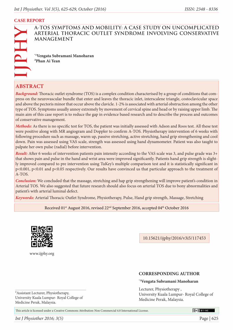
DUPLEX DOPPLERColour Doppler and colour flow imaging of left upper limb arterial system(Fig2. a,b,c,d,e and f)There is no calcified plaque or intra luminal obstruction seen in brachiocephal-ic (innominate) artery, subclavian artery, axillary and bra-chial artery. There is irregular narrowing of axillary artery with increase flow across the segment. Flow in distal axil-lary artery, proximal, mid and distal brachial artery shows reduced peak systolic velocities and loss of triphasic flow pattern. Flow in the interosseous radial and ulnar artery showed parvus tardus flow pattern. Radial artery flow at wristcould not be picked up.
a
b
Int J Physiother 2016; 3(5) Page | 627
c
d
e
fFig ure2: Duplex doppler of a,c. bracheochephalic artery, b,d. Left xillary artery e.left Ulnar artery, f. Left brachial
arteryVAS SCALE The VAS scale is a widely used subjective scale for measur-ing the intensity of the pain that experience by the patient. VAS score ranges from 0 to 10, with 0 indicates no pain and 10 being the worst pain. Patient’s VAS score was 8 before the initiation of our interventions.ADSONS TESTIt is a provacative test where bypatient is in upright posi-tion and the patient’s affected arm is passively extended, abducted and externally rotated withbreath hold and pal-pation of radial pulse. Patient was asked to perform neck extension and rotation of the head towards affected side. The test is positive if there is a marked decrease or absent of the radial pulse and it should compare to the normal side to find the patients normal pulse. This test had 79% sensititivity with no reliaibility [4].ROOS TESTIt is a common test included in the examination of the shoulder, especially in presence of Thoracic Outlet Syn-drome (TOS). It is also knows as the Elevated Arm Stress
Test (EAST) or Hands Up Test. The patient raises their arms to 90 degrees of abduction in the frontal plane of the body with the arms fully externally rotated and elbows at 90 degrees of flexion then the patient opens and closes their hands for up to 3 minutes. It is positive if the patient is unable to hold the arms up for the 3 minutes, or if the patient experiences pain, heaviness or parasthesia in the shoulder, arm or hands.this test had 100% sensitivity with relaibility of 0.42-1.0 [4].GRADING OF PULSEThe grading of pulse shown in the table. Before interven-tion the patients pulse was 0 on the left side and 3+ on the right side.
Table 1. Grading of pulse [5]
Grade Description0 No pulse
1+ A faint, but detectable pulse2+ A slightly more diminished pulse than normal3+ normal pulse4+ Bounding pulse
HAND GRIP STRENGTHPatient’s hand grip strength was measured initially using hand held Basline hydraulic hand dynamometer. Paients hand grip strength is measured initially before the inter-vention and followed by the end of 2,3,4,5 and 6thweek after the intervention. This patient asked to perform three times in each session and mean of these three values were tak-en in standing position with 90 degree of shoulder flexion. Right side grip strength was28+2 kg, but we concentrated more on the affected (left) side.
Figure 3: Hand dynamometerPHARMACOLOGICAL INTERVENTIONClopivas is started from 2012 to till date and it is used to prevent the formation of blood clots with established peripheral artery disease (poor circulation in the limbs caused by blocked arteries). Telista 40 mg (angiotensin re-ceptor blocker) also started in 2012 to till date for her high blood pressure. INTERVENTIONPhysiotherapy intervention of 6 weeks with following pro-cedure such as massage, warm up, passive stretching, ac-tive stretching, hand grip strengthening and cool down. At the beginning of the intervention outcome measures such
Int J Physiother 2016; 3(5) Page | 628
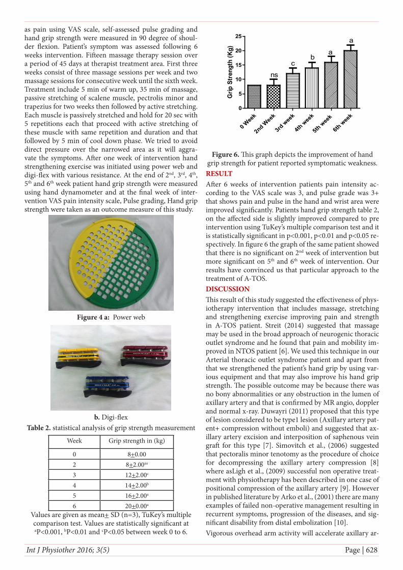
as pain using VAS scale, self-assessed pulse grading and hand grip strength were measured in 90 degree of shoul-der flexion. Patient’s symptom was assessed following 6 weeks intervention. Fifteen massage therapy session over a period of 45 days at therapist treatment area. First three weeks consist of three massage sessions per week and two massage sessions for consecutive week until the sixth week. Treatment include 5 min of warm up, 35 min of massage, passive stretching of scalene muscle, pectrolis minor and trapezius for two weeks then followed by active stretching. Each muscle is passively stretched and hold for 20 sec with 5 repetitions each that proceed with active stretching of these muscle with same repetition and duration and that followed by 5 min of cool down phase. We tried to avoid direct pressure over the narrowed area as it will aggra-vate the symptoms. After one week of intervention hand strengthening exercise was initiated using power web and digi-flex with various resistance. At the end of 2nd, 3rd, 4th, 5th and 6th week patient hand grip strength were measured using hand dynamometer and at the final week of inter-vention VAS pain intensity scale, Pulse grading, Hand grip strength were taken as an outcome measure of this study.
Figure 4 a: Power web
b. Digi-flexTable 2. statistical analysis of grip strength measurement
Week Grip strength in (kg)
0 8+0.002 8+2.00ns
3 12+2.00c
4 14+2.00b
5 16+2.00a
6 20+0.00a
Values are given as mean+ SD (n=3), TuKey’s multiple comparison test. Values are statistically significant at aP<0.001, bP<0.01 and cP<0.05 between week 0 to 6.
0 Wee
k
2nd W
eek
3rd w
eek
4th w
eek
5th w
eek
6th w
eek
0
5
10
15
20
25 a
a
ns
bc
Grip
Str
engt
h (K
g)
Figure 6. This graph depicts the improvement of hand grip strength for patient reported symptomatic weakness.RESULT After 6 weeks of intervention patients pain intensity ac-cording to the VAS scale was 3, and pulse grade was 3+ that shows pain and pulse in the hand and wrist area were improved significantly. Patients hand grip strength table 2, on the affected side is slightly improved compared to pre intervention using TuKey’s multiple comparison test and it is statistically significant in p<0.001, p<0.01 and p<0.05 re-spectively. In figure 6 the graph of the same patient showed that there is no significant on 2nd week of intervention but more significant on 5th and 6th week of intervention. Our results have convinced us that particular approach to the treatment of A-TOS. DISCUSSION This result of this study suggested the effectiveness of phys-iotherapy intervention that includes massage, stretching and strengthening exercise improving pain and strength in A-TOS patient. Streit (2014) suggested that massage may be used in the broad approach of neurogenic thoracic outlet syndrome and he found that pain and mobility im-proved in NTOS patient [6]. We used this technique in our Arterial thoracic outlet syndrome patient and apart from that we strengthened the patient’s hand grip by using var-ious equipment and that may also improve his hand grip strength. The possible outcome may be because there was no bony abnormalities or any obstruction in the lumen of axillary artery and that is confirmed by MR angio, doppler and normal x-ray. Duwayri (2011) proposed that this type of lesion considered to be type1 lesion (Axillary artery pat-ent+ compression without emboli) and suggested that ax-illary artery excision and interposition of saphenous vein graft for this type [7]. Simovitch et al., (2006) suggested that pectoralis minor tenotomy as the procedure of choice for decompressing the axillary artery compression [8] where asLigh et al., (2009) successful non operative treat-ment with physiotherapy has been described in one case of positional compression of the axillary artery [9]. However in published literature by Arko et al., (2001) there are many examples of failed non-operative management resulting in recurrent symptoms, progression of the diseases, and sig-nificant disability from distal embolization [10].Vigorous overhead arm activity will accelerate axillary ar-
Int J Physiother 2016; 3(5) Page | 629
tery restenosis, neither angioplasty or stent placement can be recommended in this patient population [7]. However this patient using clopivas, that will prevent the formation of clot or thrombus that leads to emboli. Pulse examination is also an important tool in patient assessment. Diminished or absent pulse are the sign of impaired blood flow. More valuable information can be gathered from examination of the peripheral pulses in addition to the status of the arterial system itself [5]. Shyam kumar et al., (2008) suggested that grip strength measurement for standing is stronger than sitting and there difference in grip strength in different posture due to change in the length of the muscle [11] and Su et al., (1994) stated that shoulder with 180 degree of flexion has a high-est grip strength than 0 degree flexion [12]. But sometime with 180 degree of shoulder will aggravate the symptoms of this patient, thus 90 degree of shoulder flexionwas cho-sen as a testing position in standing. The study provides quantifiable data that support the possibility that physio-therapy may reduce ATOS related symptoms. Results from this study indicate that consistent physiotherapy may be ef-fective for managing key symptoms and improve strength. CONCLUSIONWe concluded that the massage, stretching and hap grip strengthening will improve patient’s condition in Arterial TOS. We also suggested that future research should also fo-cus on arterial TOS due to bony abnormalities and patient’s with arterial luminal defect.ACKNOWLEDGEMENT: The author would like to thank to the patient who involved in this study.CONFLICT OF INTEREST: NoneREFERENCE[1] Miller TA, Sangha H, Ross DC. Neurogenic thoracic
outlet syndrome: a treatable disorder. The natural his-tory and outcome in a case series. Clin Neurophysiol. 2011; 123(7):69-70.
[2] Gustaw W, Barbara S, Jolanta P. Epidemiolo-gy and Pathogenesis of thoracic outlet syndrome. Current Issues in Pharmacy and Medical Scienc-es.2015;28(1):24-26.
[3] Lindgren KA, Oksala I. Long term outcome of surgery for thoracic outlet syndrome. American journal of sur-gery. 1995; 169(3):358-60.
[4] Hooper TL, Denton J, McGalliard MK, Brismée JM, Sizer PS Jr. Thoracic outlet syndrome: a controversial clinical condition. Part 1: anatomy, and clinical ex-amination/diagnosis. J Man Manip Ther. 2010 Jun; 18(2):74-83.
[5] Hill RD, Smith RB III. Examination of the Extremi-ties: Pulses, Bruits, and Phlebitis. In: Walker HK, Hall WD, Hurst JW, editors. Clinical Methods: The History, Physical, and Laboratory Examinations. 3rd edition. Boston: Butterworths; 1990. Chapter 30.
[6] Streit RS. NTOS symptoms and mobility: A case study on neurogenic thoracic outlet syndrome involving massage therapy. J Bodywork & Movement therapies. 2014;18(1):42-48.
[7] Duwayri Y M, Emery V B, Driskill M R, Earley J A, Wright R W, Paletta G A, Thompson R W.Positional compression of the axillary artery causing upper ex-tremity thrombosis and embolism in the elite overhead throwing athlete. J Vasc Surg. 2011 May;53(5):1329-40.
[8] Simovitch RW, Bal GK, Basamania CJ. Thoracic out-let syndrome in a competitive baseball player second-ary to the anomalous insertion of an atrophic pecto-ralis minor muscle: a case report. Am J Sports Med. 2006;34(6):1016-9.
[9] Ligh CA, Schluman BL, Safran MR. Case reports: Un-usual cause of shoulder pain in a collegiate baseball player. Clin Orthop Relat Res. 2009;467(10):2744-8.
[10] Arko FR, Harris EJ, Zarins CK, Olcott Ct. Vascular complication in high-performance athletes. J Vasc Surg. 2001;33(5):935-42.
[11] Shyam kumar AJ, Parmar V, Ahmed S, Kar S, Harper WM. A study of grip endurance and strength in dif-ferent elbow positions. J Orthop Traumatol Invest. 2008;9(4):209-11.
[12] Su C, Lin JH, Chein TH, Cheng KF, Sung YT. Grip strength in different position of elbow and shoulder. Arch Phys Med Rehabi. 1994; 75(7): 812-5.
CitationManoharan, V. S., & Yean, P. A. (2016). A-TOS SYMPTOMS AND MOBILITY: A CASE STUDY ON UNCOMPLI-CATED ARTERIAL THORACIC OUTLET SYNDROME INVOLVING CONSERVATIVE MANAGEMENT. Inter-national Journal of Physiotherapy, 3(5), 625-629.