Embed Size (px)
Citation preview
fmicb-07-01618 October 20, 2016 Time: 17:12 # 1
ORIGINAL RESEARCHpublished: 24 October 2016
doi: 10.3389/fmicb.2016.01618
Edited by:Yuji Morita,
Aichi Gakuin University, Japan
Reviewed by:Vassiliy Bavro,
University of Birmingham, UKXian-Zhi Li,
Health Canada, CanadaJanet MacInnes,
University of Guelph, Canada
*Correspondence:Xintian Wen
†These authors have contributedequally to this work.
Specialty section:This article was submitted to
Antimicrobials, Resistanceand Chemotherapy,
a section of the journalFrontiers in Microbiology
Received: 21 June 2016Accepted: 28 September 2016
Published: 24 October 2016
Citation:Li Y, Cao S, Zhang L, Lau GW,
Wen Y, Wu R, Zhao Q, Huang X,Yan Q, Huang Y and Wen X (2016) A
TolC-Like Protein of Actinobacilluspleuropneumoniae Is Involved inAntibiotic Resistance and Biofilm
Formation. Front. Microbiol. 7:1618.doi: 10.3389/fmicb.2016.01618
A TolC-Like Protein of Actinobacilluspleuropneumoniae Is Involved inAntibiotic Resistance and BiofilmFormationYing Li1†, Sanjie Cao1†, Luhua Zhang1,2†, Gee W. Lau2, Yiping Wen1, Rui Wu1, Qin Zhao1,Xiaobo Huang1, Qigui Yan1, Yong Huang1 and Xintian Wen1*
1 Research Center of Swine Diseases, College of Veterinary Medicine, Sichuan Agricultural University, Chengdu, China,2 Department of Pathobiology, University of Illinois at Urbana-Champaign, Urbana, IL, USA
Actinobacillus pleuropneumoniae is the etiologic agent of porcine contagiouspleuropneumonia, a significant disease that causes serious economic losses tothe swine industry worldwide. Persistent infections caused by bacterial biofilms arerecalcitrant to treat because of the particular drug resistance of biofilm-dwelling cells.TolC, a key component of multidrug efflux pumps, are responsible for multidrugresistance (MDR) in many Gram-negative bacteria. In this study, we identified two TolC-like proteins, TolC1 and TolC2, in A. pleuropneumoniae. Deletion of tolC1, but not tolC2,caused a significant reduction in biofilm formation, as well as increased drug sensitivityof both planktonic and biofilm cells. The genetic-complementation of the tolC1 mutationrestored the competent biofilm and drug resistance. Besides, biofilm formation wasinhibited and drug sensitivity was increased by the addition of phenylalanine-argininebeta-naphthylamide (PAβN), a well-known efflux pump inhibitor (EPI), suggesting a rolefor EPI in antibacterial strategies toward drug tolerance of A. pleuropneumoniae. Takentogether, TolC1 is required for biofilm formation and is a part of the MDR machineryof both planktonic and biofilm cells, which could supplement therapeutic strategies forresistant bacteria and biofilm-related infections of A. pleuropneumoniae clinical isolateSC1516.
Keywords: TolC, biofilm formation, multidrug resistance, PAβN, Actinobacillus pleuropneumoniae
INTRODUCTION
Actinobacillus pleuropneumoniae, a facultative anaerobic Gram-negative coccobacillus belongingto the family Pasteurellaceae, is the etiological agent of porcine pleuropneumonia characterizedby serious fibrino-hemorrhagic necrotizing pneumonia and fibrinous pleurisy (Bosse et al.,2002). This disease often manifests as severe outbreaks, causing significant economic losses inporcine industry worldwide. Even though considerable progress has been made in unmaskingthe pathogenesis of A. pleuropneumoniae, much remains to be studied (Chiers et al., 2010).In particular, little is known regarding how A. pleuropneumoniae confers resistance againstbactericidal substances for survival and spread within the host.
Frontiers in Microbiology | www.frontiersin.org 1 October 2016 | Volume 7 | Article 1618

fmicb-07-01618 October 20, 2016 Time: 17:12 # 2
Li et al. TolC-Like Proteins of Actinobacillus pleuropneumoniae
Bacteria often live in compact microbial communitiesenclosed in a self-produced matrix called biofilms that attach tobiotic or abiotic surfaces (Van Acker et al., 2014). Biofilms protectbacterial cells by decreasing their susceptibility to bactericidesand the host immune system, which makes biofilm formation thebasis of many persistent infections in humans and animals (Mah,2012). Bacteria growing in biofilms are often largely resistant toantimicrobial agents and the mechanisms of drug resistance ofbiofilm cells are not fully understood. In A. pleuropneumoniae,biofilm formation is prevalent among field isolates (Kaplan andMulks, 2005). In addition, antimicrobial treatments were stillnecessary to prevent the spread of A. pleuropneumoniae, forthat the currently available vaccination provides only partialprotection and limited impact on morbidity (Bossé et al., 2015).Various antibiotic resistance patterns have been reported inA. pleuropneumoniae, which become a problem for the controlof porcine pleuropneumonia outbreaks (Wang et al., 2010;Kucerova et al., 2011; Pridmore et al., 2011). Therefore, antibioticresistance and biofilm formation present big challenges forantimicrobial therapies in A. pleuropneumoniae.
In A. pleuropneumoniae, several resistance genes or plasmidswere identified (Wang et al., 2010; Archambault et al., 2012),while drug efflux pumps were poorly understood. In most Gram-negative bacteria, efflux pumps that actively extrude multiplestructural classes of antibacterials out of the cells, play a keyrole in multidrug resistance (MDR) phenotypes (Blair et al.,2014). Efflux pumps were also suggested to be involved inantimicrobial resistance of biofilms and in the ability to formbiofilms in several bacterial species: Escherichia coli, Klebsiellapneumoniae, and Salmonella typhimurium (Kvist et al., 2008; VanAcker and Coenye, 2016). Little is known about the contributionof efflux pumps to antibiotic resistance and biofilm formation inA. pleuropneumoniae.
Classically, efflux transporters can be divided into five families:ABC (ATP-binding cassette), RND (resistance nodulationdivision), MFS (major facilitator superfamily), SMR (smallMDR), and MATE (multidrug and toxic compound extrusion)families or superfamilies (Li et al., 2015). Usually, the first threefamilies of transporters function in the form of tripartite effluxpumps, which involve a periplasmic adaptor protein and anouter membrane efflux protein (OEP), which is usually TolC ora TolC-like homolog (Koronakis et al., 2004; Du et al., 2015).Clinically, the most significant efflux pumps in Gram-negativebacteria are members of the RND family (Morita et al., 2012; Aneset al., 2015), among which AcrAB-TolC of E. coli is best studied(Zgurskaya et al., 2011). With a channel-tunnel structure, TolCfunctions as an exit duct of AcrAB-TolC system for diverse typesof substances when recruited by the substrate-engaged innermembrane complexes, conferring intrinsic resistance to a largevariety of antimicrobial agents such as antibiotics, detergentsas well as host-derived bile salts and antimicrobial peptides(Piddock, 2006; Horiyama et al., 2010). In fact, bacteria may havea number of different transporters, but only a limited numberof OEPs, which makes TolC family proteins a key component ofefflux pumps (Koronakis et al., 2004).
In this study, we identified two TolC-like proteins, encodedby the genes APL_RS01335 (tolC1) and APL_RS04400 (tolC2),
respectively, in A. pleuropneumoniae genome. Analyses of themutants revealed that tolC1, but not tolC2, was involved inMDR of planktonic and biofilm cells. In addition, tolC1 wasalso shown to be required for biofilm formation. Chemicalinhibition of efflux pumps by Phe-Arg-β-naphthylamide (PAβN)not only increased the drug susceptibility, but also repressedbiofilm formation and eradicated mature biofilms, indicatinga potential application of efflux pump inhibitors (EPIs) asadjunctive therapies in antibacterial and antibiofilm strategies inA. pleuropneumoniae.
MATERIALS AND METHODS
Strains and Growth ConditionsBacterial strains, plasmids and primers used in this study arelisted in Tables 1 and 2. Wild-type A. pleuropneumoniae strainSC1516, isolated from an outbreak on a farm in Sichuan,China in 2014, was used in the current study. SC1516 andderivatives were grown in Tryptic Soy Broth (TSB, DifcoLaboratories, Detroit, MI, USA) or on Tryptic Soy agar (TSA,Difco Laboratories, Detroit, MI, USA) supplemented with 5%bovine serum (Invitrogen, USA) and 0.01% β-nicotinamideadenine dinucleotide (NAD). Where necessary, the mediawere supplemented with 50 µg/ml kanamycin or 5 µg/mlchloramphenicol. E. coli strains were grown on Luria-Bertani(LB, Difco Laboratories, Detroit, MI, USA) agar or in broth.When required, the media were supplemented with 25 µg/mlchloramphenicol and 1 mM diaminopimelic acid (Sigma-Aldrich, St. Louis, MO, USA). Unless otherwise stated, all strainswere grown at 37◦C.
Bioinformatic AnalysisThe NCBI BLAST package was used to perform homologysearches, and DNAMAN 4.0 was employed for similarityand identity analyses. A neighbor-joining tree containing 34sequences from members of the TolC family was constructedusing Mega 5.0 software program, and viewed through iTOL1.
Construction of tolC1 and tolC2 Mutantsand Genetically Complemented Strainsin A. pleuropneumoniaeFor creation of the tolC1 mutant, a DNA fragment of 842-bp obtained from tolC1 (from 24 to 865 bp) was joined withthe chloramphenicol acetyl transferase gene (cat) from pMC-Express (Bosse et al., 2009) by overlap PCR. The resulting PCRproduct was cloned into a pMD19-T vector (Takara, Japan)to give pLCT. pLCT was electroporated into SC1516 and thetransformants were selected on TSA supplemented with 5 µg/mlchloramphenicol, 5% bovine serum and 0.01% NAD, resulting inthe tolC1 mutant.
To delete the full length of tolC2 gene, the 1000-bpupstream and downstream regions flanking the tolC2 genewere PCR-amplified from chromosomal DNA of SC1516,
1http://itol.embl.de/
Frontiers in Microbiology | www.frontiersin.org 2 October 2016 | Volume 7 | Article 1618
fmicb-07-01618 October 20, 2016 Time: 17:12 # 3
Li et al. TolC-Like Proteins of Actinobacillus pleuropneumoniae
and fused using overlap extension PCR. The resulting PCRproduct was cloned into the suicide vector pEMOC2 togenerate plasmid pEMOC2-1tolC2. The resulting plasmidpEMOC2-1tolC2 was conjugated into SC1516 using theE. coli strain β2155 (Xie et al., 2013). After two homologousrecombination steps, the tolC2 mutant was identified byPCR.
For genetic complementation, the entire codingregions of tolC1 and tolC2 together with theirpromoters were cloned into the shuttle vector pLS88 togenerate plasmids pLStolC1 and pLStolC2, respectively.The resulting plasmids were electroporated into thecorresponding mutant strains and the transformantswere selected on TSA with 5% bovine serum and
TABLE 1 | Bacterial strains and plasmids used in this study.
Strain or plasmid Characteristicsa Source or reference
Strains
Actinobacillus pleuropneumoniae
SC1516 Serovar 7 clinical isolate Laboratory collection
tolC1::cat tolC1 insertion mutant of SC1516 This study
1tolC2 tolC2 deletion mutant of SC1516 This study
tolC1::cat tolC1+ The complemented strain of tolC1::cat containing the tolC1 gene This study
1tolC2/tolC2 The complemented strain of 1tolC2 containing the tolC2 gene This study
Escherichia coli
DH5α General cloning host strain TAKARA
BL21 (DE3) General expression host strain TAKARA
β2155 thrB1004 pro thi hsdS lacZ1M15 (F′
lacZ1M15 lacIq traD36 proA+ proB+) 1dap::erm (Ermr) Baltes et al., 2003
Plasmids
pMD19-T Ampr, E. coli cloning vector TAKARA
pET39b Kanr, E. coli expression vector Novagen
pEMOC2 Transconjugation vector based on pBluescript SK with mobRP4, a polycloning site, Cmr, andtranscriptional fusion of the omlA promoter with the sacB gene
Baltes et al., 2003
pEMOC2-1tolC2 1000 bp left and 1000 bp right homologous fragment of tolC2 cloned into pEMOC2 This study
pMC-express Cmr, expression vector of A. pleuropneumoniae Bosse et al., 2009
pLCT pMD19-T carrying the 24–865 bp of tolC1 gene fragment and an cat cassette This study
pLS88 Broad-host-range shuttle vector from Haemophilus ducreyi; Strr Kanr Willson et al., 1989
pLStolC1 Kanr, tolC1 gene with its native promoter in pLS88 This study
pLStolC2 Kanr, tolC2 gene with its native promoter in pLS88 This study
aErmr, erythromycin resistance; Ampr, ampicillin resistance; Kanr, kanamycin resistance; Cmr, chloramphenicol resistance; Strr, streptomycin resistance; cat,chloramphenicol acetyltransferase.
TABLE 2 | Primers used in this study.
Primer name Sequence (5′→3′)a Description
tolC1-pET39b-F AGTACTGACGGTTCGCTTGAACAAG (ScaI) Primers for tolC1 gene cloning and expression
tolC1-pET39b-R CTCGAGTTATCTTCTATATTTACCC (XhoI)
tolC2-pET39b-F AGTACTTTGCAGTCCGATATTGAATTACC (ScaI) Primers for tolC2 gene cloning and expression
tolC2-pET39b-R CTCGAGTTACCAACCGCCCCCAATCGAT(XhoI)
tolC2-L-F AGCGGGCCCGTCAAAAACGGTTCCGTTGC (ApaI) Cloning the 1000 bp upstream of tolC2 into pEMOC2
tolC2-L-R AGTTGTTCCTATATTCTTAT
tolC2-R-F ATAAGAATATAGGAACAACTAATCTATACATTTCGAACGG Cloning the 1000 bp downstream of tolC2 into pEMOC2
tolC2-R-R ATTTGCGGCCGCTAAATATTCCGCATAAACAC (NotI)
tolC1-F AACACTCTCCGTTGTACTTG Primers used for cloning the 24–865 bp of tolC1 gene
tolC1-R AATAAGGGTAACTATTGCCGCATCCGGGCGATTTGCGATTG
cat-F CGGCAATAGTTACCCTTATT Primers used for amplification of the chloramphenicol resistance gene
cat-R TTCAACTAACGGGGCAGGTT
tolC1-pls88-F CGGAATTCGCGTTTAAAACAAGCTTCTC (EcoRI) Primers used for amplification of tolC1 gene and its native promoter
tolC1-pls88-R CATGCCATGGAGCGCAATACTATCTTCTAT (NcoI)
tolC2-pls88-F CGGAATTCCACAAAGTCTAACCCAACTT (EcoRI) Primers used for amplification of tolC2 gene and its native promoter
tolC2-pls88-R CATGCCATGGTTATTGCTCCGTTCGAAATG (NcoI)
aRestriction sites are underlined. The 20-bp extensions required for overlap PCR are indicated in bold text.
Frontiers in Microbiology | www.frontiersin.org 3 October 2016 | Volume 7 | Article 1618
fmicb-07-01618 October 20, 2016 Time: 17:12 # 4
Li et al. TolC-Like Proteins of Actinobacillus pleuropneumoniae
0.01% NAD containing kanamycin or kanamycin andchloramphenicol.
Expression of Recombinant TolC1 andTolC2 and Generation of Rabbit AntiseraRecombinant proteins of TolC1 and TolC2 were generated usingan E. coli expression system. Briefly, PCR fragments containingtolC1 or tolC2 genes minus their signal peptide sequences wereamplified from genomic DNA of SC1516 using primers tolC1-pET39b-F/R and tolC2-pET39b-F/R, respectively. The resultingPCR products were cloned into the ScaI and XhoI sites ofpET39b to form plasmid pET39b-TolC1 and pET39b-TolC2.These recombinant plasmids were transformed into E. coli BL21(DE3) and induced to express by the addition of 0.5 mMisopropyl-beta-D-thiogalactopyranoside (IPTG). Recombinantproteins were purified by Ni affinity chromatography usingProfinity IMAC Ni-Charged Resin (Bio-Rad) as described (Zhanget al., 2016).
Rabbit immunization was carried out in strict accordancewith procedures approved by the Sichuan AgriculturalUniversity Institutional Animal Care and Use Committee.Antisera were generated by immunized each rabbitsubcutaneously with 0.5 mg of recombinant TolC1 andTolC2 containing the same volume of complete Freund’sadjuvant (Sigma) on day 0 (primary immunization). Ondays 14 and 21, the rabbits were immunized subcutaneouslywith 0.5 mg of recombinant proteins that were mixed withincomplete Freund’s adjuvant (Sigma). Serum samples werecollected 1 week after the final immunization and stored at−70◦C.
Western BlottingWestern blotting analyses were performed as describedpreviously (Zhang et al., 2015). Briefly, whole-cell extracts ofwild-type strain SC1516, mutants and genetically complementedstrains were separated on 12% Sodium dodecylsulfate(SDS) – polyacrylamide gel electrophoresis (PAGE) andelectrotransferred onto nitrocellulose membranes, respectively.After blocking in 5% skimmed milk in TBST (Tris-HCl bufferedsaline containing 0.05% Tween 20) at room temperature for30 min, the membranes were incubated with rabbit antiserumfor 3 h at room temperature. After three washes with TBST,the membranes were hybridized with horseradish peroxidase-conjugated goat anti-rabbit IgG antibody (Bioss, China) for30 min at room temperature. Membranes were washed five timeswith TBST and the signal was detected with the Immun-StarWestern C Kit (Bio-Rad, USA) according to the manufacturer’sinstructions.
In vitro Growth AssaysGrowth of A. pleuropneumoniae SC1516 and its derivatives wasdetermined by transferring 1 ml overnight culture into 100 mlof fresh TSB with 5% bovine serum and 0.01% NAD. Bacteriacultures were incubated while shaking at 37◦C. Growth wasmonitored by measuring the optical density (OD) at 600 nm(OD600) with the SmartSpec Plus spectrophotometer (Bio-Rad).
Antibiotic Susceptibility AssaysAssays on the minimal inhibitory concentration for planktoniccells (MIC-P) were carried out using 17 antibiotics, detergentsand dyes that were reported to be efflux pump substrates(Martinez et al., 2009). The MIC-P was determined by abroth micro-dilution assay using serial twofold dilution in TSBwith 5% bovine serum and 0.01% NAD. The microtiter plateswere incubated at 37◦C and visually scored for growth orno growth at 24 h. All antibiotics used in this study wereobtained from Sigma-Aldrich, Co. (St. Louis, MO, USA). Theexperiments were independently performed at least three timesin triplicate.
The minimal bactericidal concentrations for biofilms (MBC-B) were determined as described (Zhang and Mah, 2008).Overnight-grown biofilms in a 96-well microtiter plate wereexposed to serially diluted antibiotics of choice for at least 12 h.Then, fresh TSB supplemented with 5% bovine serum and 0.01%NAD was used to replace the antibiotic-containing medium andincubated for additional 24 h. The viability of bacteria in biofilmswas assessed by transferring a spot of the culture onto a TSAwith 5% bovine serum and 0.01% NAD. The experiments wereindependently performed at least three times in triplicate.
For co-treatments of antibiotics and PAβN, appropriate finalconcentration (60 µg/ml for the MIC-P assay and 160 µg/ml forthe MBC-B assay) of PAβN (Sigma, USA) was added into themedium containing antibiotics.
Crystal Violet Biofilm AssaysBiofilm formation assays were performed as described previouslywith minor changes (Kaplan et al., 2004; Zhang et al., 2016).Briefly, overnight cultures of SC1516 and its derivatives werediluted 1/100 into fresh TSB with 5% bovine serum and 0.01%NAD and incubated statically in sterile 96-well microtiter plates(Costar 3599, Corning, NY, USA). After 12 h at 37◦C, bacterialgrowth (OD600) was measured using a microplate reader (Bio-Rad iMarkTM microplate Reader). Culture supernatant was thenremoved and the wells were washed by immersing in water andstained with 100 µl of crystal violet (0.1%, w/v) per well for5 min at room temperature. After that, crystal violet was removedand washed with distilled water. The bound dye was released byadding 100 µl of acetic acid (33%, v/v) and the optical density wasmeasured at 595 nm using a microplate reader.
For the biofilm inhibition assays, overnight broth culture ofSC1516 was diluted (1/100) into fresh TSB with 5% bovine serumand 0.01% NAD in the presence of PAβN (0, 20, 40, and 60 µg/ml)alone, or with subminimal inhibitory concentrations of antibiotic(0.25 µg/ml of ceftazidime, 0.0625 µg/ml of ofloxacin, or 1 µg/mlof gentamicin) for planktonic cells (sub-MIC-P). Control wellswere without any antibiotics or PAβN. Biofilms quantified asdescribed above.
For the eradication assays of established biofilms, overnightbiofilms of SC1516 in a 96-well microtiter plate were exposedto PAβN (160 µg/ml), or subminimal-bactericidal concentrationfor biofilms (sub-MBC-B) of antibiotic (1 µg/ml of ceftazidime,1 µg/ml of ofloxacin, or 10 µg/ml of gentamicin) with or withoutPAβN. Control wells were without any antibiotics or PAβN. After
Frontiers in Microbiology | www.frontiersin.org 4 October 2016 | Volume 7 | Article 1618
fmicb-07-01618 October 20, 2016 Time: 17:12 # 5
Li et al. TolC-Like Proteins of Actinobacillus pleuropneumoniae
24 h of incubation, supernatants were removed and replaced withfresh TSB with 5% bovine serum and 0.01% NAD for additional12 h. Biofilms were stained with crystal violet and quantified asdescribed above. All crystal violet biofilm assays were performedat least three times in triplicate.
Confocal Laser Scanning MicroscopyStatic biofilms were grown on 20-mm2 coverglasses submergedin 3 ml TSB with 5% bovine serum and 0.01% NAD in 6-well microtiter plate for 12 h. The supernatants were removedand the biofilms were exposed to 10 µg/ml of gentamicin withor without 160 µg/ml of PAβN for additional 24 h. Thenbiofilms were washed with water three times and stained withthe LIVE/DEAD@ BacLightTM Bacterial Viability Kit (catalog no.L13152; Molecular Probes, Invitrogen, USA) or Wheat GermAgglutinin (WGA)–Oregon Green@ 488 (Invitrogen, MolecularProbes, Eugene, OR, USA). Plates were incubated for 15 minat room temperature in the dark and washed for three timeswith water. Stained biofilms were examined with a NikonA1R confocal scanning laser microscope (CLSM). The imageswere analyzed using the NIS-Elements AR software (Nikon,Japan).
Statistical AnalysisData were analyzed with the GraphPad Prism version 6.0(GraphPad Software, San Diego, CA, USA). One- or two-wayanalysis of variance (ANOVA) was employed to determine thedifference between more than two groups. Differences wereconsidered statistically significant at ∗P < 0.05.
RESULTS
A. pleuropneumoniae Encodes Two TolCHomologsBioinformatic analysis showed the presence of two tolC homologsin the genomes of all serotypes of A. pleuropneumoniae availablein NCBI. In strain SC1516, the two TolC homologs, namedTolC1 and TolC2, are encoded by tolC1 and tolC2, with GenBankaccession numbers KU705812 and KU705813, respectively.TolC1 showed similarity to a number of bacterial TolC proteinsdetected through a BLAST search against the GenBank, includingrepresentative members from Haemophilus influenzae (HI1462;56% identity), Klebsiella pneumoniae (CusC; 26% identity) andE. coli (TolC; 22% identity). TolC2 showed similarity to Vibriocholerae VceC (30% identity), Pseudomonas aeruginosa OprM(27% identity) and H. influenzae HI1462 (23% identity). Besides,TolC1 and TolC2 proteins are both 464 amino acids with99.78 and 98.98% sequence identity among different serotypes ofA. pleuropneumoniae, respectively.
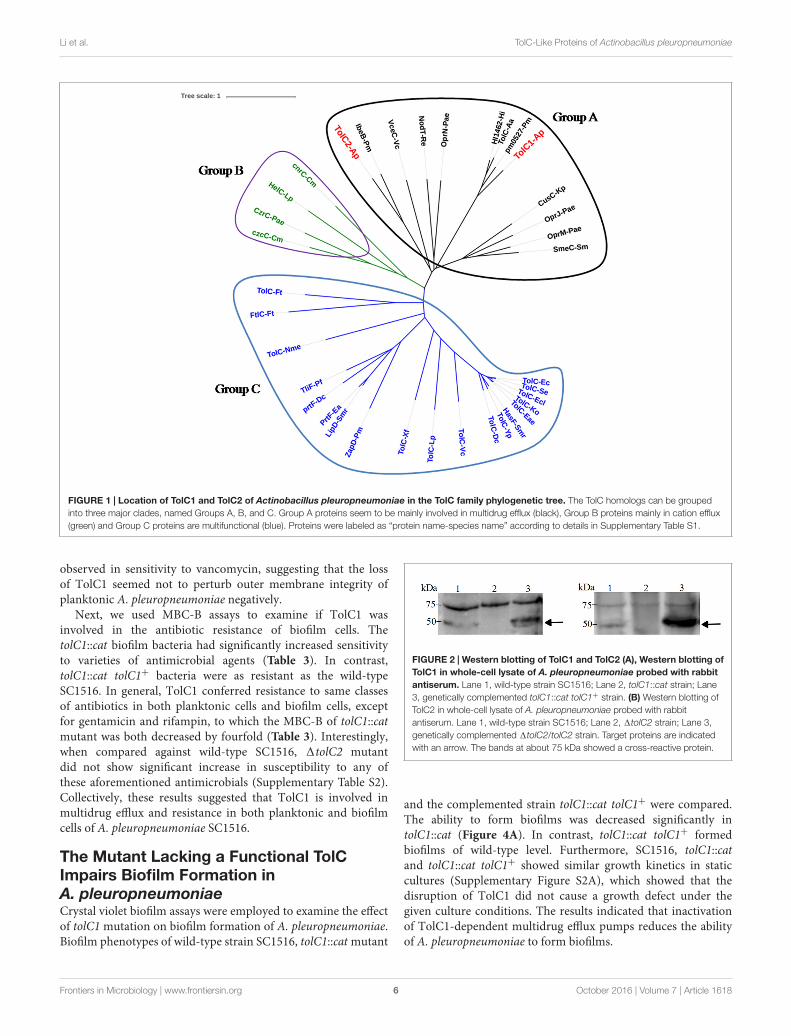
Phylogenetic analysis was performed to investigate theevolutionary relationships of A. pleuropneumoniae TolC1 andTolC2 to other members of the TolC family. Thirty six proteins(see Supplementary Table S1) were clustered into three differentgroups (groups A, B, and C; Figure 1). Both TolC1 and TolC2of A. pleuropneumoniae were phylogenetically clustered into thegroup A that seems to be mainly involved in the multidrug efflux.
Construction of tolC1 and tolC2 Mutantsand Genetically Complemented Strainsin A. pleuropneumoniaeInitially, we attempted to inactivate both tolC1 and tolC2 genes bythe suicide vector pEMOC2 carrying a counter-selection system(Xie et al., 2013). A 1tolC2 was easily obtained while multipleattempts to delete tolC1 were unsuccessful. Therefore, tolC1 wasdisrupted using plasmid pLCT as described in Section “Materialsand Methods.” Gene disruption in tolC1::cat and 1tolC2mutants and genetic complementation in tolC1::cat tolC1+ and1tolC2/tolC2 strains were confirmed by PCR (SupplementaryFigure S1).
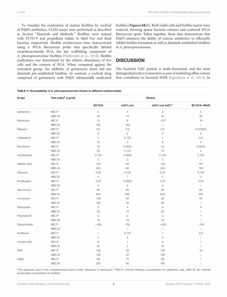
To further confirm the mutations, Western blotting wasperformed with rabbit antisera. We do not know how muchcloned proteins were incorporated to the membrane fraction, butthe mutation can be confirmed by reading the positive/negativeband. For TolC1, the antiserum identified a 50 kDa protein inthe wild-type strain SC1516 and in the genetically complementedstrain tolC1::cat tolC1+, but absent in the tolC1::cat strain.Similarly, the antiserum against TolC2 detected a protein withthe expected size in both SC1516 and genetically complemented1tolC2/tolC2, but not in 1tolC2 (Figure 2). The results wereconsistent with the PCR data, confirming that both tolC1::cat and1tolC2 have lost their ability to express corresponding proteins.In contrast, the expression of TolC1 and TolC2 were restored inboth genetically complemented mutants.
Next, we compared the growth of the wild-type SC1516 andits derivatives. The tolC1::cat strain showed a slight growth delayin the log phage, but reached similar optical density in thestationary phase (Figure 3A). Genetic complementation restoredthe growth kinetics of tolC1::cat tolC1+ strain to the wild-typelevel. There was no significant difference in growth kineticsbetween SC1516 and 1tolC2 (Figure 3B).
TolC1, but not TolC2, Is Part of theMultidrug Resistance Machinery ofA. pleuropneumoniaeTo determine the roles of TolC1 and TolC2 in MDR of planktoniccells, MIC-P assays were carried out using a broth micro-dilutionassay. As shown in Table 3, for the tolC1::cat strain, the MICsof novobiocin, ceftazidime, and lincomycin decreased by 256-, 8- and 4-fold, while the MICs of nalidixic acid, kanamycinand tetracycline decreased by twofold, respectively. Additionally,tolC1::cat also showed enhanced susceptibility to variousfluoroquinolones. Moreover, tolC1::cat was also more susceptibleto non-antibiotic antimicrobials, including acriflavine, crystalviolet and SDS by 8-, 8-, and 4-fold, respectively. In contrast,tolC1::cat showed similar levels of resistance to its parentalwild-type SC1516 for gentamicin, rifampin and polymyxin B,suggesting that these compounds might not be substrates ofTolC1-dependent efflux systems. Resistance to the majority ofthe compounds was returned back to wild-type levels in thegenetically complemented strain tolC1::cat tolC1+, confirmingthat the observed antibiotic susceptibility of tolC1::cat mutantwas not caused by polarity effects. In addition, no change was
Frontiers in Microbiology | www.frontiersin.org 5 October 2016 | Volume 7 | Article 1618
fmicb-07-01618 October 20, 2016 Time: 17:12 # 6
Li et al. TolC-Like Proteins of Actinobacillus pleuropneumoniae
FIGURE 1 | Location of TolC1 and TolC2 of Actinobacillus pleuropneumoniae in the TolC family phylogenetic tree. The TolC homologs can be groupedinto three major clades, named Groups A, B, and C. Group A proteins seem to be mainly involved in multidrug efflux (black), Group B proteins mainly in cation efflux(green) and Group C proteins are multifunctional (blue). Proteins were labeled as “protein name-species name” according to details in Supplementary Table S1.
observed in sensitivity to vancomycin, suggesting that the lossof TolC1 seemed not to perturb outer membrane integrity ofplanktonic A. pleuropneumoniae negatively.
Next, we used MBC-B assays to examine if TolC1 wasinvolved in the antibiotic resistance of biofilm cells. ThetolC1::cat biofilm bacteria had significantly increased sensitivityto varieties of antimicrobial agents (Table 3). In contrast,tolC1::cat tolC1+ bacteria were as resistant as the wild-typeSC1516. In general, TolC1 conferred resistance to same classesof antibiotics in both planktonic cells and biofilm cells, exceptfor gentamicin and rifampin, to which the MBC-B of tolC1::catmutant was both decreased by fourfold (Table 3). Interestingly,when compared against wild-type SC1516, 1tolC2 mutantdid not show significant increase in susceptibility to any ofthese aforementioned antimicrobials (Supplementary Table S2).Collectively, these results suggested that TolC1 is involved inmultidrug efflux and resistance in both planktonic and biofilmcells of A. pleuropneumoniae SC1516.
The Mutant Lacking a Functional TolCImpairs Biofilm Formation inA. pleuropneumoniaeCrystal violet biofilm assays were employed to examine the effectof tolC1 mutation on biofilm formation of A. pleuropneumoniae.Biofilm phenotypes of wild-type strain SC1516, tolC1::cat mutant
FIGURE 2 | Western blotting of TolC1 and TolC2 (A), Western blotting ofTolC1 in whole-cell lysate of A. pleuropneumoniae probed with rabbitantiserum. Lane 1, wild-type strain SC1516; Lane 2, tolC1::cat strain; Lane3, genetically complemented tolC1::cat tolC1+ strain. (B) Western blotting ofTolC2 in whole-cell lysate of A. pleuropneumoniae probed with rabbitantiserum. Lane 1, wild-type strain SC1516; Lane 2, 1tolC2 strain; Lane 3,genetically complemented 1tolC2/tolC2 strain. Target proteins are indicatedwith an arrow. The bands at about 75 kDa showed a cross-reactive protein.
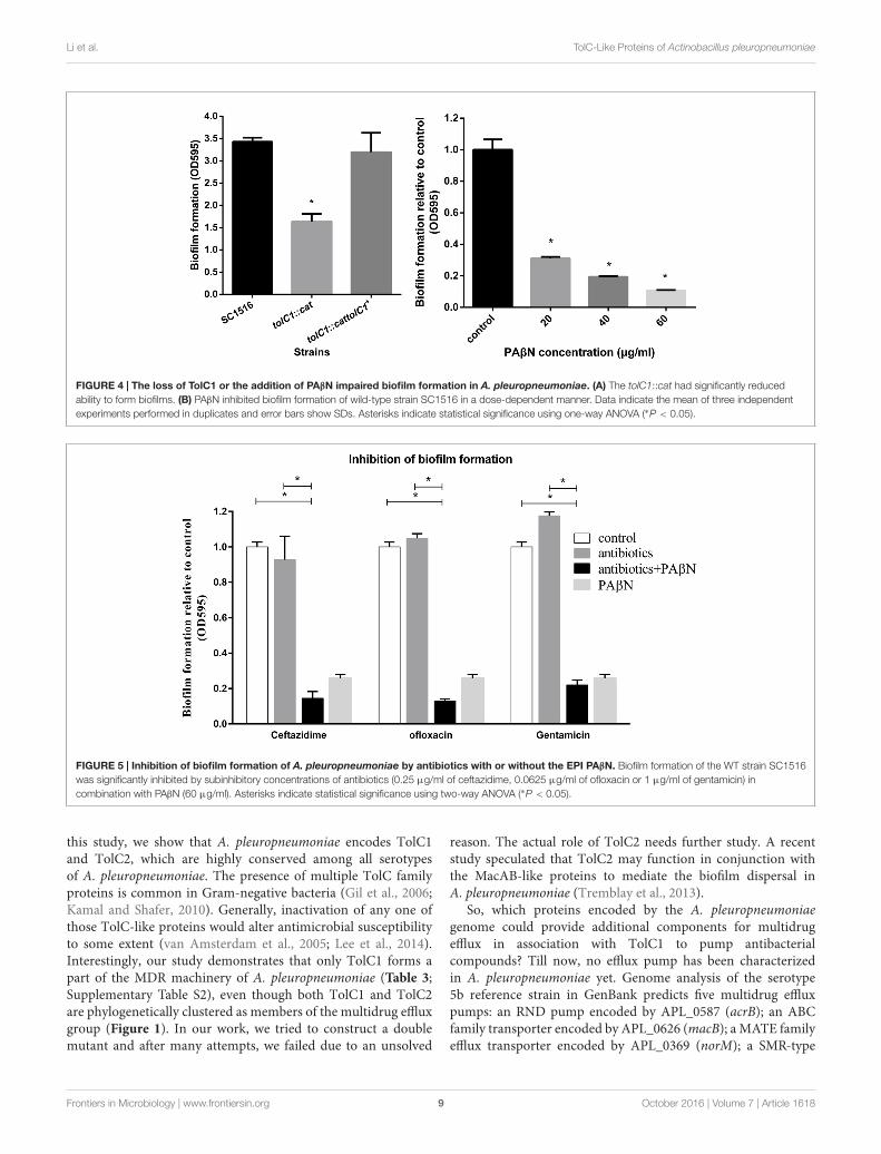
and the complemented strain tolC1::cat tolC1+ were compared.The ability to form biofilms was decreased significantly intolC1::cat (Figure 4A). In contrast, tolC1::cat tolC1+ formedbiofilms of wild-type level. Furthermore, SC1516, tolC1::catand tolC1::cat tolC1+ showed similar growth kinetics in staticcultures (Supplementary Figure S2A), which showed that thedisruption of TolC1 did not cause a growth defect under thegiven culture conditions. The results indicated that inactivationof TolC1-dependent multidrug efflux pumps reduces the abilityof A. pleuropneumoniae to form biofilms.
Frontiers in Microbiology | www.frontiersin.org 6 October 2016 | Volume 7 | Article 1618

fmicb-07-01618 October 20, 2016 Time: 17:12 # 7
Li et al. TolC-Like Proteins of Actinobacillus pleuropneumoniae
FIGURE 3 | Growth kinetics of the wild-type strain SC1516, tolC mutants and genetically complemented strains in TSB broth. (A) The tolC1 mutantshowed a reduced growth rate in the log phage, while later reached similar optical density in the stationary phase. (B) The tolC2 mutant exhibited the same growthkinetics with the wild-type strain. Data presented here are means of triplicate experiments and error bars represent standard deviations.
PAβN Restores the Drug Susceptibility ofPlanktonic and Biofilm Cells inA. pleuropneumoniaePAβN is a competitive inhibitor of RND efflux pumps (Zechiniand Versace, 2009). We examined the susceptibility of wild-type SC1516 and tolC1::cat mutant to PAβN. The MIC ofPAβN for tolC1::cat was eightfold lower than that for SC1516and the genetically complemented tolC1::cat tolC1+ (Table 3),indicating that PAβN was transported in a TolC1-dependentway in A. pleuropneumoniae. To determine the competitiveinhibitory effect of PAβN on efflux pumps, novobiocin wasemployed as an indicator. Based on the MIC (80 µg/ml)of PAβN for wild-type SC1516, sub-MICs of PAβN (20, 40,and 60 µg/ml) were added for the novobiocin susceptibilityassays. At 20 µg/ml, PAβN was insufficient to block drugefflux and the sensitivity to novobiocin was not affected. Thenovobiocin sensitivity was significantly increased 16-fold in thepresence of 40 µg/ml of PAβN and 64-fold in the presenceof 60 µg/ml of PAβN. Therefore, 60 µg/ml of PAβN wasemployed as the optimum concentration in subsequent MIC-P assays, and 160 µg/ml (1/2 MBC-B, Table 3) in the biofilmassays.
To determine the effect of PAβN on drug susceptibilityof A. pleuropneumoniae planktonic cells, MIC-P assays werecarried out in the presence of cocktail drugs composed ofPAβN and antibiotics. PAβN increased the susceptibility ofSC1516 to rifampin by 256-fold. Additionally, the susceptibilityto ceftazidime, ofloxacin, lincomycin, and polymyxin Bwas increased by 2–4 fold. Moreover, MICs of acriflavine,deoxycholate, crystal violet, and SDS were decreased by 2–8 fold,while the MIC of novobiocin decreased by 64-fold (Table 3).
To assess the efficacy of PAβN on the antibiotic sensitivityof biofilm cells, MBC-B assays were performed with PAβN in acocktail with novobiocin, ofloxacin, ceftazidime, nalidixic acid,gentamicin, and rifampin, respectively. Overnight biofilm cellsshowed 2–4 fold higher sensitivity to PAβN-antibiotic cocktailsthan individual antibiotic alone (Table 3). Taken together, theseresults indicate that PAβN efficiently inhibits multidrug effluxpumps and increases the killing activity of various classes
of antibiotics against both planktonic and biofilm cells ofA. pleuropneumoniae.
PAβN Enhances the Inhibitory Effects ofAntibiotics on Bacterial BiofilmFormationCrystal violet staining assay was also employed to assess theeffect of PAβN on biofilm formation. The addition of PAβNsignificantly reduced biofilm formation of SC1516 in a dose-dependent manner (Figure 4B). The effective concentrationsof PAβN required were below the MIC (80 µg/ml), indicatingthat PAβN performed a specific antibiofilm effect rather thana substantial inhibition on planktonic growth (SupplementaryFigure S2B).
Because PAβN increased drug susceptibility of biofilm cellsand suppressed biofilm formation, we examined if the antibiofilmefficacies of sub-MIC-P of antibiotics (ceftazidime, ofloxacin, orgentamicin) could be enhanced by PAβN. Low concentration ofantibiotic alone caused no significant inhibition, and in somecases, or even induced biofilm formation (Figure 5). Importantly,when compared against individual antibiotics alone, addition ofPAβN significantly enhanced the ability of these antimicrobialsto inhibit biofilm formation by the wild-type strain SC1516(Figure 5). Furthermore, cocktails of PAβN with ceftazidimeor ofloxacin were slightly more effective than PAβN alone ininhibiting biofilm formation.
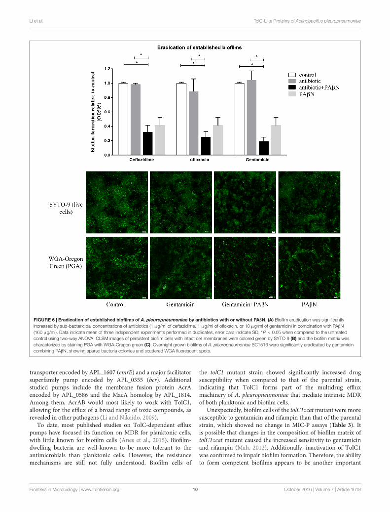
PAβN Enhances the Eradication Effectsof Antibiotics on Established BiofilmsGiven its significant inhibitory effect on biofilm formation, thecocktail PAβN-antibiotic strategy might be efficacious in theeradication on established biofilms. Biofilm eradication assayswere performed using sub-MBC-B of ceftazidime, ofloxacin,and gentamicin plus PAβN. As shown in Figure 6A, lowconcentration of each antibiotic alone had no effect on biofilmeradication, while the combination of each antibiotic withPAβN diminished the mature biofilms significantly. Theseresults indicated that the eradication activity of sub-MBC-B ofantibiotics on biofilms was substantially enhanced by PAβN.
Frontiers in Microbiology | www.frontiersin.org 7 October 2016 | Volume 7 | Article 1618
fmicb-07-01618 October 20, 2016 Time: 17:12 # 8
Li et al. TolC-Like Proteins of Actinobacillus pleuropneumoniae
To visualize the eradication of mature biofilms by cocktailof PAβN-antibiotics, CLSM assays were performed as describedin Section “Materials and Methods.” Biofilms were stainedwith SYTO-9 and propidium iodide to label live and deadbacteria, respectively. Biofilm architectures were characterizedusing a WGA fluorescent probe that specifically labeledexopolysaccharide PGA, the key scaffolding component ofA. pleuropneumoniae biofilms (Hathroubi et al., 2016). Biofilmeradication was determined by the relative abundance of livecells and the content of PGA. When compared against theuntreated group, the addition of gentamicin alone did notdiminish pre-established biofilms. In contrast, a cocktail drugcomposed of gentamicin with PAβN substantially eradicated
biofilms (Figures 6B,C). Both viable cells and biofilm matrix werereduced, showing sparse bacteria colonies and scattered WGAfluorescent spots. Taken together, these data demonstrate thatPAβN enhances the ability of various antibiotics to efficientlyinhibit biofilm formation as well as diminish established biofilmsin A. pleuropneumoniae.
DISCUSSION
The bacterial TolC protein is multi-functional, and the mostdistinguished role is to function as part of multidrug efflux systemthat contributes to bacterial MDR (Zgurskaya et al., 2011). In
TABLE 3 | Susceptibility of A. pleuropneumoniae strains to different antimicrobials.
Drugs Test indexb (µg/ml) Strains
SC1516 tolC1::cat tolC1::cat tolC1+ SC1516+PAβN
Gentamicin MIC-P 4 4 4 4
MBC-B 40 10 40 20
Kanamycin MIC-P 16 8 >32a 16
MBC-B 160 160 / /
Rifampin MIC-P 0.5 0.5 0.5 0.015625
MBC-B 8 2 8 2
Ceftazidime MIC-P 1 0.125 1 0.5
MBC-B 16 4 8 2
Novobiocin MIC-P 16 0.0625 16 0.0625
MBC-B 32 0.125 16 4
Ciprofloxacin MIC-P 0.125 0.0625 0.125 0.125
MBC-B 2 2 2 /
Naldixic acid MIC-P 128 64 128 64
MBC-B 320 80 320 160
Ofloxacin MIC-P 0.25 0.125 0.25 0.125
MBC-B 4 1 4 2
Enrofloxacin MIC-P 0.25 0.0625 0.25 0.25
MBC-B 2 2 2 /
Vancomycin MIC-P 64 64 64 64
MBC-B 640 640 640 640
Lincomycin MIC-P 128 32 32 32
MBC-B 160 40 80 /
Tetracycline MIC-P 8 4 8 4
MBC-B 32 16 32 /
Polymyxin B MIC-P 2 2 2 1
MBC-B 16 16 16 /
Deoxycholate MIC-P >400 100 >400 <100
MBC-B / / / /
Acriflavine MIC-P 1 0.125 1 0.5
MBC-B 4 2 4 /
Crystal violet MIC-P 8 1 4 1
MBC-B 32 2 16 /
SDS MIC-P 128 32 128 16
MBC-B 160 40 160 /
PAβN MIC-P 80 10 80 /
MBC-B 320 80 160 /
aThe plasmids used in the complemented strains confer resistance to kanamycin. bMIC-P, minimal inhibitory concentration for planktonic cells; MBC-B, the minimalbactericidal concentration for biofilms.
Frontiers in Microbiology | www.frontiersin.org 8 October 2016 | Volume 7 | Article 1618
fmicb-07-01618 October 20, 2016 Time: 17:12 # 9
Li et al. TolC-Like Proteins of Actinobacillus pleuropneumoniae
FIGURE 4 | The loss of TolC1 or the addition of PAβN impaired biofilm formation in A. pleuropneumoniae. (A) The tolC1::cat had significantly reducedability to form biofilms. (B) PAβN inhibited biofilm formation of wild-type strain SC1516 in a dose-dependent manner. Data indicate the mean of three independentexperiments performed in duplicates and error bars show SDs. Asterisks indicate statistical significance using one-way ANOVA (∗P < 0.05).
FIGURE 5 | Inhibition of biofilm formation of A. pleuropneumoniae by antibiotics with or without the EPI PAβN. Biofilm formation of the WT strain SC1516was significantly inhibited by subinhibitory concentrations of antibiotics (0.25 µg/ml of ceftazidime, 0.0625 µg/ml of ofloxacin or 1 µg/ml of gentamicin) incombination with PAβN (60 µg/ml). Asterisks indicate statistical significance using two-way ANOVA (∗P < 0.05).
this study, we show that A. pleuropneumoniae encodes TolC1and TolC2, which are highly conserved among all serotypesof A. pleuropneumoniae. The presence of multiple TolC familyproteins is common in Gram-negative bacteria (Gil et al., 2006;Kamal and Shafer, 2010). Generally, inactivation of any one ofthose TolC-like proteins would alter antimicrobial susceptibilityto some extent (van Amsterdam et al., 2005; Lee et al., 2014).Interestingly, our study demonstrates that only TolC1 forms apart of the MDR machinery of A. pleuropneumoniae (Table 3;Supplementary Table S2), even though both TolC1 and TolC2are phylogenetically clustered as members of the multidrug effluxgroup (Figure 1). In our work, we tried to construct a doublemutant and after many attempts, we failed due to an unsolved
reason. The actual role of TolC2 needs further study. A recentstudy speculated that TolC2 may function in conjunction withthe MacAB-like proteins to mediate the biofilm dispersal inA. pleuropneumoniae (Tremblay et al., 2013).
So, which proteins encoded by the A. pleuropneumoniaegenome could provide additional components for multidrugefflux in association with TolC1 to pump antibacterialcompounds? Till now, no efflux pump has been characterizedin A. pleuropneumoniae yet. Genome analysis of the serotype5b reference strain in GenBank predicts five multidrug effluxpumps: an RND pump encoded by APL_0587 (acrB); an ABCfamily transporter encoded by APL_0626 (macB); a MATE familyefflux transporter encoded by APL_0369 (norM); a SMR-type
Frontiers in Microbiology | www.frontiersin.org 9 October 2016 | Volume 7 | Article 1618
fmicb-07-01618 October 20, 2016 Time: 17:12 # 10
Li et al. TolC-Like Proteins of Actinobacillus pleuropneumoniae
FIGURE 6 | Eradication of established biofilms of A. pleuropneumoniae by antibiotics with or without PAβN. (A) Biofilm eradication was significantlyincreased by sub-bactericidal concentrations of antibiotics (1 µg/ml of ceftazidime, 1 µg/ml of ofloxacin, or 10 µg/ml of gentamicin) in combination with PAβN(160 µg/ml). Data indicate mean of three independent experiments performed in duplicates, error bars indicate SD, ∗P < 0.05 when compared to the untreatedcontrol using two-way ANOVA. CLSM images of persistent biofilm cells with intact cell membranes were colored green by SYTO 9 (B) and the biofilm matrix wascharacterized by staining PGA with WGA-Oregon green (C). Overnight grown biofilms of A. pleuropneumoniae SC1516 were significantly eradicated by gentamicincombining PAβN, showing sparse bacteria colonies and scattered WGA fluorescent spots.
transporter encoded by APL_1607 (emrE) and a major facilitatorsuperfamily pump encoded by APL_0355 (bcr). Additionalstudied pumps include the membrane fusion protein AcrAencoded by APL_0586 and the MacA homolog by APL_1814.Among them, AcrAB would most likely to work with TolC1,allowing for the efflux of a broad range of toxic compounds, asrevealed in other pathogens (Li and Nikaido, 2009).
To date, most published studies on TolC-dependent effluxpumps have focused its function on MDR for planktonic cells,with little known for biofilm cells (Anes et al., 2015). Biofilm-dwelling bacteria are well-known to be more tolerant to theantimicrobials than planktonic cells. However, the resistancemechanisms are still not fully understood. Biofilm cells of
the tolC1 mutant strain showed significantly increased drugsusceptibility when compared to that of the parental strain,indicating that TolC1 forms part of the multidrug effluxmachinery of A. pleuropneumoniae that mediate intrinsic MDRof both planktonic and biofilm cells.
Unexpectedly, biofilm cells of the tolC1::cat mutant were moresusceptible to gentamicin and rifampin than that of the parentalstrain, which showed no change in MIC-P assays (Table 3). Itis possible that changes in the composition of biofilm matrix oftolC1::cat mutant caused the increased sensitivity to gentamicinand rifampin (Mah, 2012). Additionally, inactivation of TolC1was confirmed to impair biofilm formation. Therefore, the abilityto form competent biofilms appears to be another important
Frontiers in Microbiology | www.frontiersin.org 10 October 2016 | Volume 7 | Article 1618
fmicb-07-01618 October 20, 2016 Time: 17:12 # 11
Li et al. TolC-Like Proteins of Actinobacillus pleuropneumoniae
function of TolC1 in contribution to MDR of biofilm cells ofA. pleuropneumoniae.
Bacterial efflux pumps, especially the clinically relevantAcrAB-TolC, are often considered as important targets fordeveloping novel antibacterial treatments (Blair et al., 2014).Consequently, EPIs, especially those against RND-type pumps,attracted a lot of attention as potential adjunctive therapiesthat would improve the potency of antibiotics and decreasethe emergence of MDR bacteria (Opperman and Nguyen,2015). The EPI PAβN is known as a competitive inhibitor ofRND efflux pumps (Kinana et al., 2016). In this study, theincreased susceptibility of tolC1::cat to PAβN suggested a rolefor TolC1 in PAβN efflux. Given the predicted efflux pumps inA. pleuropneumoniae, PAβN is most likely to target AcrB andcompetitively inhibit AcrAB-TolC system. Our results showedthat PAβN inhibited the efflux pumps of A. pleuropneumoniaein a dose-dependent manner, with 60 µg/ml the most effectiveagainst planktonic bacteria and 160 µg/ml against the biofilmcells. Significantly, this concentration contrasted with that in aprevious study, in which 20 µg/ml of PAβN was sufficient toincrease drug susceptibility from 77 to 96% for E. coli MG1655(Sáenz et al., 2004). This discrepancy suggested that the effectsof PAβN may vary to different bacterial species (Lister et al.,2012).
PAβN has been used as a classical EPI of RND pumps, butdue to the dicationic character, its membrane-permeabilizingeffect, especially when used at high concentrations, has frequentlybeen ignored (Li et al., 2015). The OM-permeabilizing effectcould also increase antibiotic susceptibility, which is likely tointerfere with the judgment of PAβN on efflux pump inhibition.In this study, when combined with 60 µg/ml PAβN in MIC-P assays, the MIC of vancomycin—an antibiotic that is unableto cross the outer membrane of Gram-negative bacteria—wasnot changed (Table 3). These data suggested that 60 µg/ml ofPAβN seemed not to cause substantial outer membrane damageof A. pleuropneumoniae. When 160 µg/ml of PAβN was usedin biofilm cells, the susceptibility to vancomycin was also notchanged with or without PAβN (Table 3). Besides, the effect ofPAβN on drug susceptibility of biofilm cells were also determinedin the presence of 1 mM MgSO4 to stabilize the outer membrane,as described previously (Lamers et al., 2013). As a result, thesupplementation with magnesium did not restore the resistanceto gentamicin, rifampin, ceftazidime, novobiocin, nalidixic acid,ofloxacin (data not shown), indicating that the ability of PAβN toincrease the drug susceptibility of biofilm cells seemed not due toits permeabilizing activity.
Bacteria are capable of establishing biofilms on almost anysurfaces. Therefore, prevention of biofilm formation is the key inthwarting biofilm-related infections. In clinics, bacteria are oftenexposed to low concentration of antibiotics at the beginning andend of a drug regimen, or continuously during low-dose therapy(Kaplan, 2011). Several studies, including ours, have shown thatlow doses of some antimicrobials induce biofilm formation ina variety of bacterial species (Figure 5; Hoffman et al., 2005;Kaplan et al., 2012). Importantly, our results indicate that PAβNnot only effectively suppresses the induction of biofilm formationby low doses of antibiotics, but also significantly enhances the
therapeutic potential of low doses of antibiotics in inhibitingbiofilm formation of A. pleuropneumoniae.
Bacterial cells within established biofilms are highly resistantto external antimicrobial agents (Mah, 2012). Because effluxpumps confer many types of bacterial drug resistance (Blair et al.,2014), not surprisingly, interference with the efflux pump activityof biofilm cells restores the antibiotic sensitivity of biofilms.Therefore, cocktail drugs composed of EPI with antibiotics areprojected to have greater potential of destroying establishedbiofilms. Indeed, PAβN significantly increases the ability of sub-bactericidal concentration of antibiotics to eradicate establishedbiofilms of A. pleuropneumoniae. The demonstration thatPAβN suppresses biofilm formation whilst also potentiates theeradication of established biofilms implies important therapeuticvalue of the EPI.
CONCLUSION
We have characterized two TolC-like proteins, TolC1 andTolC2, and only TolC1 function as a component of theMDR machinery of A. pleuropneumoniae. TolC1-dependentefflux pump(s) contributed to drug resistance of cells inplanktonic culture as well as in biofilms. Moreover, wedemonstrated a biofilm defect for the mutant lacking TolC1,which facilitating an important role of TolC1 in biofilmformation of A. pleuropneumoniae. Chemical inhibiting effluxpumps by PAβN improved the efficacy of antibiotics as well asrepressed biofilm formation of A. pleuropneumoniae. In addition,cocktail drugs composed of EPI with low concentration ofantibiotics, showed great potential to eradicate biofilms, which isimportant in treating chronic infectious diseases. These findingsmay contribute to understanding the molecular basis of MDR ofA. pleuropneumoniae, especially for the drug-tolerance biofilmcells, and representing a step in controlling MDR and biofilm-related infections in A. pleuropneumoniae.
AUTHOR CONTRIBUTIONS
SC, XW, and YW designed the experiments. XH, RW, QY,and QZ performed the experiments with assistance of YH. YLand LZ analyzed the data and wrote the paper, GL edited themanuscript. All authors read, commented on and approved thefinal manuscript.
ACKNOWLEDGMENT
We thank Dr. Janine T. Bossé (Imperial College London) forthe gift of plasmid pMC-Express and Dr. Liancheng Lei (JilinUniversity) for the gift of plasmid pEMOC2.
SUPPLEMENTARY MATERIAL
The Supplementary Material for this article can be found onlineat: http://journal.frontiersin.org/article/10.3389/fmicb.2016.01618
Frontiers in Microbiology | www.frontiersin.org 11 October 2016 | Volume 7 | Article 1618
fmicb-07-01618 October 20, 2016 Time: 17:12 # 12
Li et al. TolC-Like Proteins of Actinobacillus pleuropneumoniae
REFERENCESAnes, J., Mccusker, M. P., Fanning, S., and Martins, M. (2015). The ins and
outs of RND efflux pumps in Escherichia coli. Front. Microbiol. 6:587. doi:10.3389/fmicb.2015.00587
Archambault, M., Harel, J., Goure, J., Tremblay, Y. D., and Jacques, M. (2012).Antimicrobial susceptibilities and resistance genes of Canadian isolates ofActinobacillus pleuropneumoniae. Microb. Drug Resist. 18, 198–206. doi:10.1089/mdr.2011.0150
Baltes, N., Tonpitak, W., Hennig-Pauka, I., Gruber, A. D., and Gerlach, G.-F. (2003). Actinobacillus pleuropneumoniae serotype 7 siderophore receptorFhuA is not required for virulence. FEMS Microbiol. Lett. 220, 41–48. doi:10.1016/S0378-1097(03)00064-8
Blair, J. M., Richmond, G. E., and Piddock, L. J. (2014). Multidrug efflux pumps inGram-negative bacteria and their role in antibiotic resistance. Future Microbiol.9, 1165–1177. doi: 10.2217/fmb.14.66
Bosse, J. T., Durham, A. L., Rycroft, A. N., Kroll, J. S., and Langford, P. R. (2009).New plasmid tools for genetic analysis of Actinobacillus pleuropneumoniaeand other pasteurellaceae. Appl. Environ. Microbiol. 75, 6124–6131. doi:10.1128/AEM.00809-09
Bosse, J. T., Janson, H., Sheehan, B. J., Beddek, A. J., Rycroft, A. N., Kroll, J. S.,et al. (2002). Actinobacillus pleuropneumoniae: pathobiology and pathogenesisof infection. Microbes Infect. 4, 225–235. doi: 10.1016/S1286-4579(01)01534-9
Bossé, J. T., Li, Y., Walker, S., Atherton, T., Fernandez Crespo, R.,Williamson, S. M., et al. (2015). Identification of dfrA14 in twodistinct plasmids conferring trimethoprim resistance in Actinobacilluspleuropneumoniae. J. Antimicrob. Chemother. 70, 2217–2222. doi: 10.1093/jac/dkv121
Chiers, K., De Waele, T., Pasmans, F., Ducatelle, R., and Haesebrouck, F. (2010).Virulence factors of Actinobacillus pleuropneumoniae involved in colonization,persistence and induction of lesions in its porcine host. Vet. Res. 41:65. doi:10.1051/vetres/2010037
Du, D., Van Veen, H. W., and Luisi, B. F. (2015). Assembly and operation ofbacterial tripartite multidrug efflux pumps. Trends Microbiol. 23, 311–319. doi:10.1016/j.tim.2015.01.010
Gil, H., Platz, G. J., Forestal, C. A., Monfett, M., Bakshi, C. S., Sellati, T. J.,et al. (2006). Deletion of TolC orthologs in Francisella tularensis identifiesroles in multidrug resistance and virulence. Proc. Natl. Acad. Sci. U.S.A. 103,12897–12902. doi: 10.1073/pnas.0602582103
Hathroubi, S., Hancock, M. A., Bossé, J. T., Langford, P. R., Tremblay, Y. D. N.,Labrie, J., et al. (2016). Surface polysaccharide mutants reveal that absence of Oantigen reduces biofilm formation of Actinobacillus pleuropneumoniae. Infect.Immun. 84, 127–137. doi: 10.1128/IAI.00912-15
Hoffman, L. R., D’argenio, D. A., Maccoss, M. J., Zhang, Z., Jones, R. A., and Miller,S. I. (2005). Aminoglycoside antibiotics induce bacterial biofilm formation.Nature 436, 1171–1175. doi: 10.1038/nature03912
Horiyama, T., Yamaguchi, A., and Nishino, K. (2010). TolC dependency ofmultidrug efflux systems in Salmonella enterica serovar Typhimurium.J. Antimicrob. Chemother. 65, 1372–1376. doi: 10.1093/jac/dkq160
Kamal, N., and Shafer, W. M. (2010). Biologic activities of the TolC-like protein ofNeisseria meningitidis as assessed by functional complementation in Escherichiacoli. Antimicrob. Agents Chemother. 54, 506–508. doi: 10.1128/AAC.01168-09
Kaplan, J. B. (2011). Antibiotic-induced biofilm formation. Int. J. Artif. Organs 34,737–751. doi: 10.5301/ijao.5000027
Kaplan, J. B., Izano, E. A., Gopal, P., Karwacki, M. T., Kim, S., Bose, J. L.,et al. (2012). Low levels of beta-lactam antibiotics induce extracellular DNArelease and biofilm formation in Staphylococcus aureus. MBio 3:e00198-12. doi:10.1128/mBio.00198-12
Kaplan, J. B., and Mulks, M. H. (2005). Biofilm formation is prevalent among fieldisolates of Actinobacillus pleuropneumoniae. Vet. Microbiol. 108, 89–94. doi:10.1016/j.vetmic.2005.02.011
Kaplan, J. B., Velliyagounder, K., Ragunath, C., Rohde, H., Mack, D., Knobloch,J. K., et al. (2004). Genes involved in the synthesis and degradationof matrix polysaccharide in Actinobacillus actinomycetemcomitans andActinobacillus pleuropneumoniae biofilms. J. Bacteriol. 186, 8213–8220. doi:10.1128/JB.186.24.8213-8220.2004
Kinana, A. D., Vargiu, A. V., May, T., and Nikaido, H. (2016). Aminoacyl beta-naphthylamides as substrates and modulators of AcrB multidrug efflux pump.Proc. Natl. Acad. Sci. U.S.A. 113, 1405–1410. doi: 10.1073/pnas.1525143113
Koronakis, V., Eswaran, J., and Hughes, C. (2004). Structure and function of TolC:the bacterial exit duct for proteins and drugs. Annu. Rev. Biochem. 73, 467–489.doi: 10.1146/annurev.biochem.73.011303.074104
Kucerova, Z., Hradecka, H., Nechvatalova, K., and Nedbalcova, K. (2011).Antimicrobial susceptibility of Actinobacillus pleuropneumoniae isolates fromclinical outbreaks of porcine respiratory diseases. Vet. Microbiol. 150, 203–206.doi: 10.1016/j.vetmic.2011.01.016
Kvist, M., Hancock, V., and Klemm, P. (2008). Inactivation of efflux pumpsabolishes bacterial biofilm formation. Appl. Environ. Microbiol. 74, 7376–7382.doi: 10.1128/AEM.01310-08
Lamers, R. P., Cavallari, J. F., and Burrows, L. L. (2013). The effluxinhibitor phenylalanine-arginine beta-naphthylamide (PAbetaN) permeabilizesthe outer membrane of gram-negative bacteria. PLoS ONE 8:e60666. doi:10.1371/journal.pone.0060666
Lee, S., Song, S., and Lee, K. (2014). Functional analysis of TolC homologs in Vibriovulnificus. Curr. Microbiol. 68, 729–734. doi: 10.1007/s00284-014-0537-4
Li, X. Z., and Nikaido, H. (2009). Efflux-mediated drug resistance inbacteria: an update. Drugs 69, 1555–1623. doi: 10.2165/11317030-000000000-00000
Li, X. Z., Plesiat, P., and Nikaido, H. (2015). The challenge of efflux-mediatedantibiotic resistance in Gram-negative bacteria. Clin. Microbiol. Rev. 28, 337–418. doi: 10.1128/CMR.00117-14
Lister, I. M., Raftery, C., Mecsas, J., and Levy, S. B. (2012). Yersinia pestis AcrAB-TolC in antibiotic resistance and virulence. Antimicrob. Agents Chemother. 56,1120–1123. doi: 10.1128/AAC.05338-11
Mah, T.-F. (2012). Biofilm-specific antibiotic resistance. Future Microbiol. 7, 1061–1072. doi: 10.2217/fmb.12.76
Martinez, J. L., Sanchez, M. B., Martinez-Solano, L., Hernandez, A., Garmendia, L.,Fajardo, A., et al. (2009). Functional role of bacterial multidrug efflux pumpsin microbial natural ecosystems. FEMS Microbiol. Rev. 33, 430–449. doi:10.1111/j.1574-6976.2008.00157.x
Morita, Y., Tomida, J., and Kawamura, Y. (2012). MexXY multidrugefflux system of Pseudomonas aeruginosa. Front. Microbiol. 3:408. doi:10.3389/fmicb.2012.00408
Opperman, T. J., and Nguyen, S. T. (2015). Recent advances toward amolecular mechanism of efflux pump inhibition. Front. Microbiol. 6:421. doi:10.3389/fmicb.2015.00421
Piddock, L. J. (2006). Multidrug-resistance efflux pumps? not just for resistance.Nat. Rev. Microbiol. 4, 629–636. doi: 10.1038/nrmicro1464
Pridmore, A., Burch, D., and Lees, P. (2011). Determination of minimuminhibitory and minimum bactericidal concentrations of tiamulin against fieldisolates of Actinobacillus pleuropneumoniae. Vet. Microbiol. 151, 409–412. doi:10.1016/j.vetmic.2011.03.016
Sáenz, Y., Ruiz, J., Zarazaga, M., Teixidó, M., Torres, C., and Vila, J. (2004).Effect of the efflux pump inhibitor Phe-Arg-β-naphthylamide on the MICvalues of the quinolones, tetracycline and chloramphenicol, in Escherichiacoli isolates of different origin. J. Antimicrob. Chemother. 53, 544–545. doi:10.1093/jac/dkh117
Tremblay, Y. D., Deslandes, V., and Jacques, M. (2013). Actinobacilluspleuropneumoniae genes expression in biofilms cultured under static conditionsand in a drip-flow apparatus. BMC Genomics 14:364. doi: 10.1186/1471-2164-14-364
Van Acker, H., and Coenye, T. (2016). The role of efflux and physiologicaladaptation in biofilm tolerance and resistance. J. Biol. Chem. 291, 12565–12572.doi: 10.1074/jbc.R115.707257
Van Acker, H., Van Dijck, P., and Coenye, T. (2014). Molecular mechanisms ofantimicrobial tolerance and resistance in bacterial and fungal biofilms. TrendsMicrobiol. 22, 326–333. doi: 10.1016/j.tim.2014.02.001
van Amsterdam, K., Bart, A., and Van Der Ende, A. (2005). A Helicobacter pyloriTolC efflux pump confers resistance to metronidazole. Antimicrob. AgentsChemother. 49, 1477–1482. doi: 10.1128/AAC.49.4.1477-1482.2005
Wang, Y. C., Chan, J. P., Yeh, K. S., Chang, C. C., Hsuan, S. L., Hsieh,Y. M., et al. (2010). Molecular characterization of enrofloxacin resistantActinobacillus pleuropneumoniae isolates. Vet. Microbiol. 142, 309–312. doi:10.1016/j.vetmic.2009.09.067
Frontiers in Microbiology | www.frontiersin.org 12 October 2016 | Volume 7 | Article 1618
fmicb-07-01618 October 20, 2016 Time: 17:12 # 13
Li et al. TolC-Like Proteins of Actinobacillus pleuropneumoniae
Willson, P. J., Albritton, W. L., Slaney, L., and Setlow, J. K. (1989). Characterizationof a multiple antibiotic resistance plasmid from Haemophilus ducreyi.Antimicrob. Agents Chemother. 33, 1627–1630. doi: 10.1128/AAC.33.9.1627
Xie, F., Zhang, Y., Li, G., Zhou, L., Liu, S., and Wang, C. (2013). The ClpP proteaseis required for the stress tolerance and biofilm formation in Actinobacilluspleuropneumoniae. PLoS ONE 8:e53600. doi: 10.1371/journal.pone.0053600
Zechini, B., and Versace, I. (2009). Inhibitors of multidrug resistant effluxsystems in bacteria. Recent Pat. Antiinfect. Drug Discov. 4, 37–50. doi:10.2174/157489109787236256
Zgurskaya, H. I., Krishnamoorthy, G., Ntreh, A., and Lu, S. (2011). Mechanismand function of the outer membrane channel TolC in multidrugresistance and physiology of enterobacteria. Front. Microbiol. 2:189. doi:10.3389/fmicb.2011.00189
Zhang, L., Li, Y., Dai, K., Wen, X., Wu, R., Huang, X., et al. (2015).Establishment of a successive markerless mutation system in Haemophilusparasuis through natural transformation. PLoS ONE 10:e0127393. doi:10.1371/journal.pone.0127393
Zhang, L., Li, Y., Wen, Y., Lau, G. W., Huang, X., Wu, R., et al. (2016). HtrA isimportant for stress resistance and virulence in Haemophilus parasuis. Infect.Immun. 84, 2209–2219. doi: 10.1128/IAI.00147-16
Zhang, L., and Mah, T. F. (2008). Involvement of a novel efflux system inbiofilm-specific resistance to antibiotics. J. Bacteriol. 190, 4447–4452. doi:10.1128/JB.01655-07
Conflict of Interest Statement: The authors declare that the research wasconducted in the absence of any commercial or financial relationships that couldbe construed as a potential conflict of interest.
Copyright © 2016 Li, Cao, Zhang, Lau, Wen, Wu, Zhao, Huang, Yan, Huang andWen. This is an open-access article distributed under the terms of the CreativeCommons Attribution License (CC BY). The use, distribution or reproduction inother forums is permitted, provided the original author(s) or licensor are creditedand that the original publication in this journal is cited, in accordance with acceptedacademic practice. No use, distribution or reproduction is permitted which does notcomply with these terms.
Frontiers in Microbiology | www.frontiersin.org 13 October 2016 | Volume 7 | Article 1618