Embed Size (px)
Citation preview

lable at ScienceDirect
Water Research 132 (2018) 361e370
Contents lists avai
Water Research
journal homepage: www.elsevier .com/locate/watres
A tiered approach to assess effects of diclofenac on the brown musselPerna perna: A contribution to characterize the hazard
Mayana Karoline Fontes a, b, Paloma Kachel Gusso-Choueri b, Luciane Alves Maranho a, c,Denis Moledo de Souza Abessa b, Wesley Almeida Mazur c, d, Bruno Galv~ao de Campos b,Luciana Lopes Guimar~aes c, d, Marcos Sergio de Toledo d, Daniel Lebre e,Joyce Rodrigues Marques e, Andreia Arantes Felicio f, Augusto Cesar a, c,Eduardo Alves Almeida g, Camilo Dias Seabra Pereira a, c, *
a Departamento de Ciencias do Mar, Universidade Federal de S~ao Paulo, Rua Maria M�aximo, 168, 11030-100 Santos, Brazilb Instituto de Biociencias, Campus do Litoral Paulista, Universidade Estadual Paulista “Júlio de Mesquita Filho”, Infante Dom Henrique, s/n, 11330-900 S~aoVicente, Brazilc Laborat�orio de Ecotoxicologia, Universidade Santa Cecília, Rua Oswaldo Cruz 266, 11045-907 Santos, Brazild Departamento de Bioquímica da Universidade Federal de S~ao Paulo, Rua Botucatu, 862, 04023-901 S~ao Paulo, Brazile CEMSA e Centro de Espectrometria de Massas Aplicada, CIETEC/IPEN, Av. Prof. Lineu Prestes, 2242, Salas 112 e 113, 05508-000 S~ao Paulo, Brazilf Universidade Estadual Paulista Júlio de Mesquita Filho e Campus S~ao Jos�e do Rio Preto, Rua Crist�ov~ao Colombo 2265, 15054-000 S~ao Jos�e do Rio Preto, SP,Brazilg Fundaç~ao Universidade Regional de Blumenau, Rua Antonio da Veiga 498, Itoupava Seca, 89030-103 Blumenau, Brazil
a r t i c l e i n f o
Article history:Available online 29 December 2017
Keywords:PharmaceuticalsNonsteroidal anti-inflammatory drugMarine environmentNon-target organism
* Corresponding author. Departamento de Cienciasde S~ao Paulo, Rua Maria M�aximo, 168, 11030-100 San
E-mail addresses: [email protected], camilo
https://doi.org/10.1016/j.watres.2017.12.0770043-1354/© 2017 Elsevier Ltd. All rights reserved.
a b s t r a c t
Pharmaceutical discharges into the aquatic ecosystem are of environmental concern and sewage treat-ment plants (STPs) have been pointed out as the major source of these compounds to coastal zones,where oceanic disposal of sewage occurs through submarine outfalls. Diclofenac (DCF) is one of the mostfrequently detected pharmaceuticals in water, but little is known about the effects on marine organisms.In this study, we employed a tiered approach involving the determination of environmental concen-trations of DCF in marine water and the adverse biological effects for fertilization, embryo-larvaldevelopment and biomarker responses of the mussel Perna perna. Results indicate that effects infertilization rate and embryo-larval development were found in the order of mg$L�1. However, lowconcentrations of DCF (ng$L�1) significantly decreased the lysosomal membrane stability and COX ac-tivity, as well as triggered DNA damage, oxidative stress and changes in antioxidant defenses. Our resultspoint to an environmental hazard at coastal ecosystems and suggest the need for improvements in thetreatment of domestic wastewater aiming to reduce DCF concentrations, as well as regulation on currentenvironmental legislation and monitoring of aquatic ecosystems.
© 2017 Elsevier Ltd. All rights reserved.
1. Introduction
The marine environment is exposed to a wide range of pollut-ants associated with anthropogenic sources, such as metals, poly-cyclic aromatic hydrocarbons, plastic debris and activepharmaceuticals compounds and their metabolites (Aguirre-Martínez et al., 2013a; Diniz et al., 2015). Pharmaceutical and
do Mar, Universidade Federaltos, [email protected] (C.D.S. Pereira).
Personal Care Products (PPCPs) have been detected in surface wa-ters (Hernando et al., 2006), groundwater (Heberer and Feldmann,2005), drinking water (Rodil et al., 2012), marine water (Pereiraet al., 2016) and Sewage Treatment Plants (STP) were identified asthe major source of water contamination (McClellan and Halden,2010). Although pharmaceuticals have been detected in the envi-ronment in trace-concentrations from ng.L�1 to mg.L�1, it has beendemonstrated that they can adversely affect the health status ofaquatic organisms (Aguirre-Martínez et al., 2013b).
Urban sewage is considered a main cause of marine pollution,especially in coastal areas, due to a high population density. Inthese areas, two different alternatives have been adopted for
M.K. Fontes et al. / Water Research 132 (2018) 361e370362
disposal of urban sewage: oceanic disposal system through a sub-marine sewage outfall composed of a pre-conditioning plant; 2)primary and secondary treatment of effluent waste release ininland waters. However, some pharmaceuticals are not totallyeliminated because the conventional technology of treatment usedin STPs is insufficient to completely remove these compounds(Ferrari et al., 2003). Furthermore, the continuous input of phar-maceuticals into the water can give them a pseudo-persistencestate (Hernando et al., 2006).
One of most frequently detected pharmaceuticals in water isthe Diclofenac (DCF), an anti-inflammatory drug that is widelyused as analgesic, antirheumatic compound, antiarthritic, work-ing by cyclooxygenase inhibition and thus blocking the prosta-glandin synthesis (van den Brandhof and Montforts, 2010). DCFhas a Kow¼ 4.5, which makes it a compound with certain lip-ophilicity, facilitating its bioaccumulation in animal tissues(Cleuvers, 2004). Several studies have demonstrated that DCFexposure induces negative effects in non-target organisms suchas alterations and necrosis in trout gills (Hoeger et al., 2005;Triebskorn et al., 2004), impairment of the osmoregulatoryability of crabs (Eades and Waring, 2010), lower scope for growthin mussels (Ericson et al., 2010), bioaccumulation and significantbiomarker responses (Gonzalez-Rey and Bebianno, 2014). More-over, DCF exposure affected larval development of mussels(Fabbri et al., 2014), caused deleterious effects in zebrafish em-bryos (Feito et al., 2012) oxidative stress in crustaceans (G�omez-Oliv�an et al., 2014) and affected gill integrity and pituitary geneexpression in trout (Gr€oner et al., 2015, 2017). However, mostprevious studies have highlighted the adverse effects of DCF infreshwater organisms, and little effort has been applied to thestudy of the negative effects of this pharmaceutical in marineorganisms.
Recently, the European Union established regulatory guidanceto assess the presence of pharmaceuticals in the aquatic envi-ronment (Directive, 2013/39/EU amending Directives, 2000/60/EC and 2008/105/EC) (European Commission, 2013) and thepriority substances in Water Policy. Moreover, the EuropeanCommission updated the monitoring watch list of priority sub-stances in the field of water policy, including the sex hormones17alpha-ethinylestradiol and 17beta-estradiol, and the non-steroidal anti-inflammatory (NSAID) DCF (Loli�c et al., 2015). Inthis context, DCF and other pharmaceuticals have been includedin monitoring programs and environmental risk assessmentsaround the world, for example in Canada (Kleywegt et al., 2007)and Korea (Han et al., 2006). However, Brazilian regulation onenvironmental risk assessment and PPCPs discharges remainunchanged (Pereira et al., 2016).
Under this reasoning, the present work assumes that DCF iscapable of causing adverse effects in non-target organisms, morespecifically the marine bivalve Perna perna, at environmentallyrelevant concentrations, denoting the ecological risk of this drug.Bivalve mollusks as Perna perna play an important role at aquaticecosystems. They are sessile and filter large quantities of surfacewater for feeding and respiration, besides to accumulate organicpollutants and metals, and these are highly desirable features inecotoxicological studies (Rittschof and McClellan-Green, 2005).
Our study aimed to conduct an ecotoxicological study using atiered approach to determine the environmental concentrationin the water column and associated adverse biological effects.This approach included: a) identification and quantification ofthis pharmaceutical in marine water sampled near the disposalarea of the submarine sewage outfall; b) acute toxicity testsassessing reproduction endpoints; c) 96-h exposure assay for theevaluation of sublethal responses through biomarkers.
2. Material and methods
2.1. Chemicals
Standards of DCF (2-[(2,6-dichlorophenyl)amino]benzeneaceticacid, CAS number 15307-79-6, purity� 98%) were obtained fromSigma-Aldrich (Steinheim, Germany), as well as all other chemicalsused for fertilization and embryo-larval development assays andbiomarkers analysis.
2.2. Tier 0
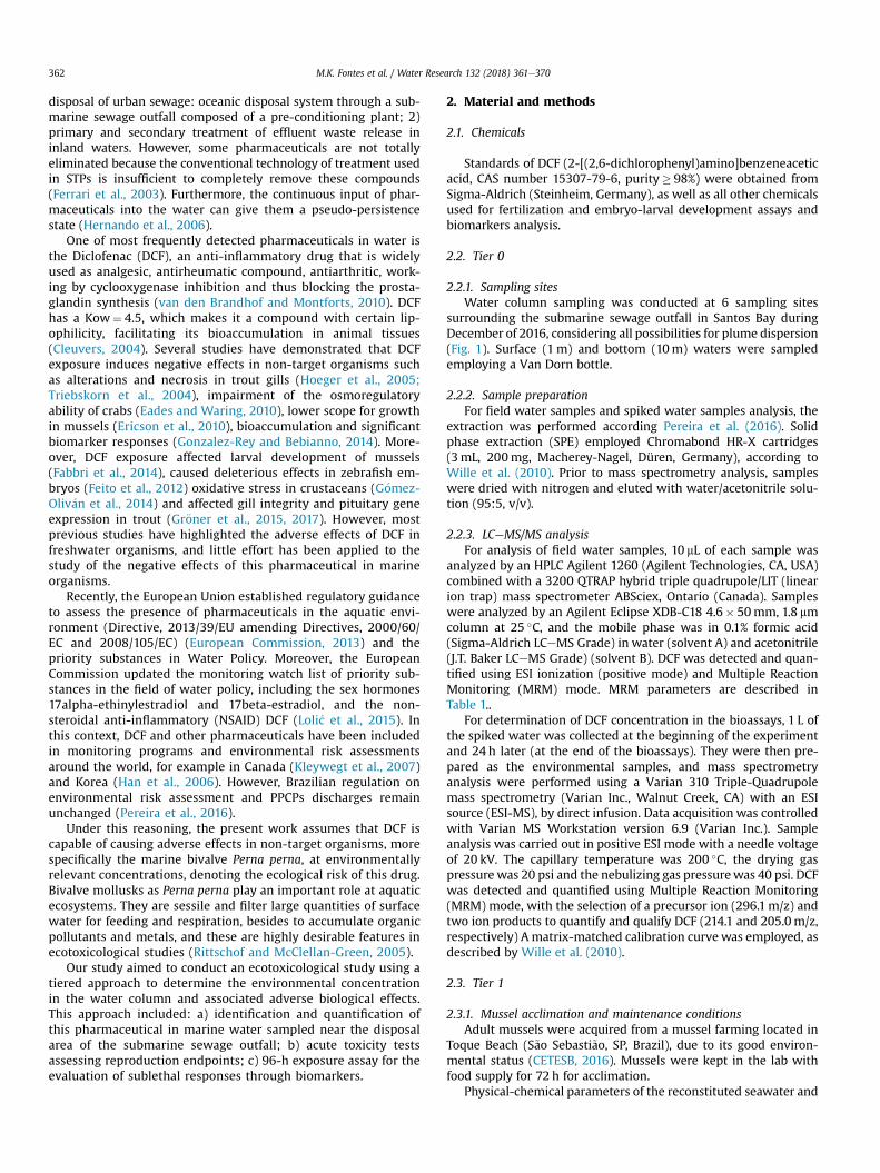
2.2.1. Sampling sitesWater column sampling was conducted at 6 sampling sites
surrounding the submarine sewage outfall in Santos Bay duringDecember of 2016, considering all possibilities for plume dispersion(Fig. 1). Surface (1m) and bottom (10m) waters were sampledemploying a Van Dorn bottle.
2.2.2. Sample preparationFor field water samples and spiked water samples analysis, the
extraction was performed according Pereira et al. (2016). Solidphase extraction (SPE) employed Chromabond HR-X cartridges(3mL, 200mg, Macherey-Nagel, Düren, Germany), according toWille et al. (2010). Prior to mass spectrometry analysis, sampleswere dried with nitrogen and eluted with water/acetonitrile solu-tion (95:5, v/v).
2.2.3. LCeMS/MS analysisFor analysis of field water samples, 10 mL of each sample was
analyzed by an HPLC Agilent 1260 (Agilent Technologies, CA, USA)combined with a 3200 QTRAP hybrid triple quadrupole/LIT (linearion trap) mass spectrometer ABSciex, Ontario (Canada). Sampleswere analyzed by an Agilent Eclipse XDB-C18 4.6� 50mm, 1.8 mmcolumn at 25 �C, and the mobile phase was in 0.1% formic acid(Sigma-Aldrich LCeMS Grade) in water (solvent A) and acetonitrile(J.T. Baker LCeMS Grade) (solvent B). DCF was detected and quan-tified using ESI ionization (positive mode) and Multiple ReactionMonitoring (MRM) mode. MRM parameters are described inTable 1..
For determination of DCF concentration in the bioassays, 1 L ofthe spiked water was collected at the beginning of the experimentand 24 h later (at the end of the bioassays). They were then pre-pared as the environmental samples, and mass spectrometryanalysis were performed using a Varian 310 Triple-Quadrupolemass spectrometry (Varian Inc., Walnut Creek, CA) with an ESIsource (ESI-MS), by direct infusion. Data acquisition was controlledwith Varian MS Workstation version 6.9 (Varian Inc.). Sampleanalysis was carried out in positive ESI mode with a needle voltageof 20 kV. The capillary temperature was 200 �C, the drying gaspressure was 20 psi and the nebulizing gas pressurewas 40 psi. DCFwas detected and quantified using Multiple Reaction Monitoring(MRM) mode, with the selection of a precursor ion (296.1m/z) andtwo ion products to quantify and qualify DCF (214.1 and 205.0m/z,respectively) A matrix-matched calibration curve was employed, asdescribed by Wille et al. (2010).
2.3. Tier 1
2.3.1. Mussel acclimation and maintenance conditionsAdult mussels were acquired from a mussel farming located in
Toque Beach (S~ao Sebasti~ao, SP, Brazil), due to its good environ-mental status (CETESB, 2016). Mussels were kept in the lab withfood supply for 72 h for acclimation.
Physical-chemical parameters of the reconstituted seawater and

Fig. 1. Sampling stations located adjacent to submarine sewage outfall in Santos Bay.
Table 1Parameters of Multiple Reactions Monitoring for the positive and negative ion mode, limit of detection, limit of quantification and retention time.
Compounds Q1 Q3 DP (V) CE (V) CXP (V) LOD (ng$L�1) LOQ (ng$L�1) RT (min.)
Diclofenac 296.1 214.1 21 39 4 0.81 3.0 5.77250.0 21 25 4207.1 21 33 4
Q1 (first quadrupole); Q3 (last quadrupole); DP (Declustering potential); CE (Collision Energy); CXP (Collision Exit Potential); LOD (Limits of detection); LOQ (Limits ofquantification); RT (Retention Time); MIM (Multiple ion monitoring). In Q3, in the upper cell is the quantifier ion and in the lower cell is the qualifier ion.
M.K. Fontes et al. / Water Research 132 (2018) 361e370 363
treatments were measured at the beginning and at the end of theassays (fertilization and embryo-larval assays), or at everyreplacement of test solutions (NRRT assays, session 2.4.1.1). Tem-perature ranged from 21 �C to 23 �C, pH ranged from 8.1 to 8.25,salinity ranged from 34 to 36 PSU, and dissolved oxygen rangedfrom 6.0 to 6.8mg.L�1.
2.3.2. Fertilization and embryo-larval development assaysFertilization assay was performed following USEPA protocol
(2002) adapted to Perna perna according to Zaroni et al. (2005). Thegametes (eggs and sperm) were obtained by thermal stimulation(from 10 �C to 30 �C) of fifty individuals during 30min. The eggssolution was filtered in a 0.75 mm membrane. From the sperm so-lution, about 2mL were diluted in 48mL of reconstituted seawater.Given the lipophilic nature of DCF and its low water solubility(23.73mg/L at 25 �C; Research Corporation, 2006), dimethyl sulf-oxide (DMSO) was used as carrier solvent (Parolini et al., 2011). Asolvent concentration of 1 ml.L�1 was used in each DCF concen-tration. So, a DCF working solution (1000mg.L�1) was prepared bydiluting 100mg of DCF in 60 mL of DMSO. From this stock solutionall DCF test solutions were prepared (31.25; 62.5; 125; 250; 500 and1000mg L�1). The sperm was exposed to two controls (seawaterand seawater plus DMSO 1 ml.L�1) for 60min, in quadruplicate.
Then, approximately 2000 ovules were added to each tube and
after 45min were added 500 mL of formaldehyde. 100 eggs fromeach replicate were assessed and the observation of a fertilizationmembrane or the beginning of cellular divisions was employed toidentify fertilization. Three assays were performed to obtain amean value of DCF concentration causing fertilization inhibition at50% of exposed ovules (IC50).
The embryo-larval development assaywas performed accordingto the protocol recommended by ASTM (1992) for mussels, withminor adaptations proposed by Zaroni et al. (2005). Fifty in-dividuals were induced to spawn (as described in section 2.3.2 forthe detailed characterization of this procedure). The gametes werecollected separately and the fertilization was induced adding 2mLof sperm solution to a 2000 ovules solution. When the cleavagesbegan, about 500 embryos were added to each test-tube containingdifferent concentrations of DCF (0.01; 0.1; 1; 10 and 100mg L�1).The test durationwas 48 h at 25 �C and salinity of 35 PSU. The assaywas conducted in quadruplicate and employed two controls(seawater and seawater plus DMSO 1 ml.L�1), since a solvent con-centration of 1 ml.L-1 was used in each DCF concentration. The first100 larvae from each test-tube were assessed, considering a “Dshape” as regular development. The assay was performed threetimes to obtain mean values of DCF concentration that causeembryo-larval development inhibition of 50% of the exposed or-ganisms (IC50; 48 h), the No Observable Effect Concentration

M.K. Fontes et al. / Water Research 132 (2018) 361e370364
(NOEC) and the Lowest Observable Effect Concentration (LOEC)means.
2.4. Tier 2. biomarkers responses
2.4.1. Biomarker assay2.4.1.1. Mussel exposure. Mussel P. perna (n¼ 80, 60± 1mm) wereacclimatized for one week in a 300 L aquarium filled with seawaterunder controlled conditions. After this period, the mussels (n¼ 10,1 mussel L�1) were exposed in aquaria to different concentrationsof DCF (20; 200 and 2000 ng L�1), and a solvent concentration of1 ml.L�1 was used in each DCF concentration. These concentrationswere chosen based on data obtained from environmental moni-toring performed by Pereira et al. (2016), which found a concen-tration of 19.4 ng L�1 of DCF in Santos Bay. A solvent control(1 ml.L�1) was set in parallel with the DCF bioassay, and for eachtreatment, two aquaria of 10 L and 10 animals per aquaria wereused. Seawater was changed daily (24 h) and DCF nominal con-centration was restored. Seawater was filtered (200 mm) providingphytoplankton as mussels’ food source, and no additional food wasadded. For each 48 and 96 h of exposure, themussels were removedfrom the aquaria for haemolymph extraction.
Before the bioassay has started, lysosomal membrane stability(LMS) was determined in some specimens from the acclimatedaquaria, to certify the health status of the organisms that wereexposed to the DCF concentrations. LMSwas determined by NeutralRed Retention Time (NRRT) method (Lowe and Pipe, 1994).
2.4.1.2. Neutral Red Retention Time assay (NRRT). This method isapplied to haemolymph withdrawn from the posterior adductormuscle of living bivalves, as described by Lowe and Pipe (1994). Theendpoint was the time when at least 50% of the examined cells byoptical microscopy (400� ) exhibited dye loss from the lysosomesto the cytosol. After the withdrawal of their haemolymph, eachmussel was dissected, and gill and digestive gland were storedat �80 �C until the biomarkers’ analyses.
2.4.1.3. Tissue preparation. Gills and digestive gland from each in-dividual were excised and homogenized (homogenized frac-tiondHF) in a TriseHCl buffer. An aliquot of the homogenate wasseparated for the analysis of DNA damage and lipid peroxidation.Another aliquot was centrifuged at 15,000� g for 20min at 4 �Cand the supernatant were employed for biomarkers de-terminations (EROD, DBF, GST, GPx, AChE and COX). Total proteinwas determined for both aliquots and tissues according to Bradford(1976).
2.4.1.4. Ethoxyresorufin O-deethylase (EROD). Ethoxyresorufin O-deethylase activity was assessed by the transformation of 7-ethoxyresorufin in resorufin at 485 nm (excitation) and 580 nm(emission) according to Gagn�e and Blaise (1993). Results wereexpressed as pmol/min/mg protein.
2.4.1.5. Dibenzylfluorescein dealkylase (DBF). The activity ofDibenzylfluorescein dealkylase was evaluated employing themethod described by Gagn�e et al. (2007). Fluorescence wasmeasured at 485 nm (excitation) and 516 nm (emission) and resultswere expressed as pmol/min/mg protein.
2.4.1.6. Glutathione S-transferase activity (GST). Glutathione S-transferase activity was determined by the method adapted fromMcFarland et al. (1999). The rate of reaction was measured byabsorbance in a spectrophotometer at 340 nm at every 5min for30min at 30 �C. Results were expressed as OD/min/mg proteins.
2.4.1.7. Glutathione peroxidase activity (GPX). Glutathione peroxi-dase activity was determined following the protocol employed byMcFarland et al. (1999). Absorbance at 340 nmwas measured every30 s for 30min and results were expressed as nmol/min/mgprotein.
2.4.1.8. DNA damage. DNA damage was assessed by the alkalineprecipitation assay (Olive, 1988) using fluorescence to measureDNA strand breaks (Gagn�e et al., 1995). DNA strands were quanti-fied using fluorescence 360 nm (excitation) and 450 nm (emission)after staining with Hoechst dye. Standard solutions of salmonsperm DNAwere used for calibration. Results were expressed as mgDNA strands/mg protein.
2.4.1.9. Lipid peroxidation. Levels of MDA in tissues were quantifiedaccording to Hong et al. (2000) by HPLC coupled to an UV/Vis de-tector set at 532 nm. The MDA quantification was established on acalibration curve using as standard MDA obtained by tetrame-thoxypropane hydrolysis. Chromatogram monitoring and peakidentification and quantification were performed using the EZChrom Elite software (Agilent Technologies). Results wereexpressed as pmol TBARS/mg tissue.
2.4.1.10. Cholinesterase (ChE). Cholinesterase activity was evalu-ated by the method described by Ellman et al. (1961). The absor-bance at 412 nmwas measured every 1min for 7min. Results wereexpressed as nmol min/mg protein.
2.4.1.11. Cyclooxygenase (COX). COX activity was measured asdescribed by Fujimoto et al. (2002). The samples were incubated in50mM Tris-HCl, 0.05% Tween-20, 50 mM arachidonate, 2 mMdichlorofluorescein and 0.1 mgmL�1 horseradish peroxidase. Fluo-rescence was measured at 485 nm and 530 nm and results wereexpressed as RFU/min/mg protein.
2.5. Statistical analysis
For the fertilization assay, an EC50 was calculated by TrimmedSpearman-Karber. The linear interpolation method was used tocalculate the IC50 (48 h) for the embryo-larval development assay.Biomarker results of T0, water and solvent controls (DMSO) wereanalyzed by ANOVA and showed no significant differences. Thus,two-way ANOVA followed by the Dunnett's test were used toidentify the concentrations which were significantly different, aswell as differences between the periods analyzed, employing sol-vent control as a reference. Statistical differences were consideredsignificant when p� .05. Prism v.7a Software was employed forANOVA and post hoc analysis.
3. Results
3.1. Environmental concentration
The environmental concentrations of DCF are shown in Table 2.DCF was quantified in Station 1 (surface and bottom) and detectedin Station 4 (bottom), but it was not detected in the referencesampling station (6).
3.2. Fertilization and embryo-larval development assays
The measured concentrations of DCF at the beginning of theexposure experiment for the in fertilization and embryo-larval as-says (T0) and after 24 h (T24h) are shown in Table 3.
Fertilization ratewas significantly inhibited at all concentrationstested (p< .05) so that a NOEC could not be provided. The LOEC of

Fig. 2. Inhibition of fertilization (A) and embryo-larval development (B) in Perna pernaafter exposure to different concentrations of DCF (ANOVA - Dunnett's test, p< .05).Error bars indicate the standard errors.
Table 4Measured concentration of DCF in spiked water of biomarkers assays.
Nominal concentration (ng.L�1) Measuredconcentration
Reduction (%)
t 0 t 24
20 22.71 <LOQ ND200 211.33 7.20 96.592000 2214.66 77.69 96.49
t0, beginning of the experiment; t24, after 24 h of exposure. ND, not determined.
Table 2Environmental concentration of DCF in surface and bottom water samples (1e6sampling stations) from Santos Bay.
DCF concentration (ng.L�1)
1 2 3 4 5 6
Surface 4.01 <LOD <LOD <LOD <LOD <LODBottom 4.78 <LOD <LOD <LOQ <LOD <LOD
<LOD, below limit of detection. < LOQ, below limit of quantification.
Table 3Measured concentrations of DCF in spiked water of fertilization and embryo-larvalassays.
Nominal concentration (mg.L�1) Measuredconcentration
Reduction (%)
t 0 t 24
0.01 0.0093 0.0003 96.771.0 1.5207 0.0454 97.0131.25 41.3400 0.8300 97.99100.0 104.5013 3.5339 96.61250.0 260.3400 8.0100 96.931000.0 1013.9600 13.0800 98.71
t0, beginning of the experiment; t24, after 24 h of exposure.
M.K. Fontes et al. / Water Research 132 (2018) 361e370 365
the DCFwas 31.25mg L�1 and the EC50 was estimated at 389mg L�1
after 24 h of exposure (95% confidence of interval: 356mg L�1 -407mg L�1). Furthermore, there was no statistically significantdifference between the control and the solvent control with DMSO(p> .05) (Fig. 2A). Only the highest DCF concentration caused asignificant inhibition of embryo-larval development compared tocontrols (p< .05) (Fig. 2B). The NOEC was at 10mg L�1, the LOEC ofthe DCF was 100mg L�1 and the IC50 was 18mg L�1 after 48 h ofexposure (95% confidence of interval: 16.3mg L�1 - 20mg L�1).
Fig. 3. NRRT in hemocytes of Perna perna exposed to DCF (ANOVA - Dunnett's test,p< .05). Asterisks indicate significance compared to control. Error bars indicate thestandard errors.
3.3. Biomarker responses
The measured concentrations of DCF at the beginning of theexposure experiment for the biomarkers assays (T0) and after 24 h(T24h) are shown in Table 4.
There was no statistically significant difference between watercontrol and the solvent control (p> .05), therefore, the concentra-tion of DMSO applied to prepare DCF stocks did not have a toxiceffect in exposed mussels. For all biomarkers, only solvent controlDMSO was considered for statistical analysis.
The LMS of Perna was significantly affected (p< .05) comparedwith control organisms, at all concentrations tested. After 48 h ofexposure, the organisms exposed to all concentrations showed asignificant decrease in the retention time of the dye. At 20 ng.L�1
the NRRT was reduced by 65% (23.57min); at 200 ng.L�1 by 52%(32.14min) at 2000 ng.L�1 by 58% (25.5min). The reduction alsooccurred after 96 h of exposure. At 20 ng.L�1 the NRRT was reducedby 46% (35.5min); at 200 ng.L-1 was reduced by 47% (28.3min) andat 2000 ng.L�1 was reduced by 47% (28.2min) (Fig. 3).
For gill tissues, EROD activity was inhibited at 200 ng. L�1, butnot at 20 and 2000 ng.L�1 during 96 h exposure (p< .05) (Fig. 4).EROD activity in the digestive gland exposed to DCF showed nosignificant differences between concentrations tested compared tocontrols (p> .05) and there were also no significant differencesbetween the times analyzed (p> .05) (Fig. 5). DBF activity in thedigestive gland tissues (Fig. 5) showed no significant differencesbetween concentrations compared to controls and no significantdifferences between exposure times (p> .05).
DCF inhibited significantly the GST activity in gill tissues (Fig. 4)at all concentrations tested after 48 h exposure (p< .05), while at
96 h exposure only the highest concentration (2000 ng. L�1)showed significantly lower GST activity compared to control andwith the activity measured after 48 h exposure (p< .05). In addi-tion, between the times, there was a significant difference betweencontrols (p< .05), being possible to observe that 96 h exposure hasa lower GST activity than after 48 h exposure (Fig. 4). With respectto digestive gland tissues, none of the concentrations tested wereable to induce any change over the control, but GST activity wassignificantly different between times in all treatments (Fig. 5).
GPx activity in the gill tissues was inhibited at 200 ng .L�1 after48h exposure, and a significantly different inhibition occurred atthe highest concentration (2000 ng .L�1) after 96h exposure(p< .05), but no significant differences were observed between thetimes analyzed (Fig. 4). Similarly, for the digestive gland tissues, nosignificant difference was observed between the concentrationsand the control or between the times analyzed (p> .05) (Fig. 5).

Fig. 4. Biochemical biomarkers in gill tissues of P. perna exposed to DCF. Letter A in-dicates significant differences from control (ANOVA - Dunnett's test, p< .05); letter Bindicates significant differences between times (ANOVA - Dunnett's test, p< .05). Errorbars indicate the standard errors.
Fig. 5. Biochemical biomarkers in digestive glands tissue of P. perna exposed to DCF.Letter A indicates significant differences from control (ANOVA - Dunnett's test,p< .05); letter B indicates significant differences between times (ANOVA, p < .05). Er-ror bars indicate the standard errors.
M.K. Fontes et al. / Water Research 132 (2018) 361e370366
Gills tissues (Fig. 4) showed a significant decrease in DNAdamage for 200 ng$L1 and 2000 ng.L�1, compared to control, after48 h exposure (p< .05). The DNA damage was also significantlydifferent between periods within control and the concentration of20 ng.L�1 showed a greater damage after 48 h exposure. However,for the digestive gland tissues (Fig. 5), the highest DNA damagevalues were measured only at the highest concentration of2000 ng.L�1 after 48 h exposure (p< .05). No significant differenceswere observed between periods analyzed (p> .05).
LPO levels were measured through MDA quantification. In thegill tissues (Fig. 4), 200 ng .L�1 and 2000 ng .L�1 concentrationsshowed LPO levels significantly higher than control after 48 hexposure (p< .05). Significant induction (p< .05) of LPO was alsoobserved in gills tissue exposed to 20 ng.L�1 concentration after96 h exposure. Furthermore, between times, LPO levels showedsignificant difference within control and within the concentrationof 20 ng. L�1 (p< .05), observable after an increase of LPO during96 h exposure (Fig. 4). Digestive gland tissues showed significantlylower LPO levels compared to control when exposed to 200 ng. L�1
and after 96 h exposure (p< .05). Furthermore, LPO levels weresignificantly different between times within the control for con-centrations of 20 ng .L�1 and 2000 ng .L�1 (Fig. 5).
ChE activity was induced in the gill tissues (Fig. 4) at the highestconcentration (2000 ng. L�1) at 48 h of exposure (p< .05). At thesame concentration, it was also possible to observe that there was asignificant difference in the enzymatic activity between times
(p< .05). However, in the digestive gland tissue, no significantdifferences were observed between different concentrations andcontrol or between times analyzed (p> .05) (Fig. 5).
COX activity was significantly inhibited in the gill tissues (Fig. 4)at concentrations of 20 ng. L�1 and 200 ng .L�1 after 48 h exposure(p< .05) and for 200 ng .L�1 and 2000 ng.L�1 after 96 h exposure.Between times, lower activity was observed at 96 h exposure forcontrol and for concentrations of 200 ng. L�1 and 2000 ng.L�1.However, DCF induced a significantly increase in the COX activityfor the digestive gland tissues (Fig. 5) at a concentration of200 ng.L�1 after 96 h exposure compared to control (p< .05). Therewas a significant difference in activity between times within con-trol and for concentrations of 200 ng.L�1 and 2000 ng.L�1, and itwas possible to observe an increased activity after 96 h exposure.No significant difference was observed after 48 h exposure(p> .05).
4. Discussion
In a previous study in Santos Bay (Pereira et al., 2016), DCF wasquantified only in the surface water sample (19.4 ng. L�1), whereasin the current study DCF was quantified in surface and bottomsamples from station 1, and detected in station 4 (bottom). Theoccurrence of DCF, especially in the bottom samples, is partiallyexplained by the lower sunlight in these samples, avoiding pho-todegradation (Baena-Nogueras et al., 2017). DCF has a short half-

M.K. Fontes et al. / Water Research 132 (2018) 361e370 367
life, and it is considered a pseudo-persistent contaminant becauseof the continuous release in the environment and the inefficiency ofconventional effluent treatment processes, which are not able tocompletely remove residual drugs (Hernando et al., 2006; Gr€oneret al., 2017).
Degradation assays with DCF under controlled conditions havedemonstrated that the sunlight is a crucial factor for DCF degra-dation (Poirier-Larabie et al., 2016). DCF concentrations found inSantos bay are within the range found in previous studies inseawater. Gros et al. (2012) found lower concentrations (4.0 ng .L-1)in the Mediterranean Sea, whereas Fang et al. (2012) found an in-termediate concentration of 53.60 ng .L-1 in coastal waters ofTaiwan. The study carried out by Loli�c et al. (2015) reported higherconcentrations of DCF (241 ng. L-1) in North Portugal.
There are few data available relating pharmaceutical exposurein the marine environment and the adverse effects in organismsfrom tropical coastal regions. Most studies have been evaluatingthe acute toxicity of DCF in freshwater environments, and differentEC50 values have been obtained such as 5.3mg .L�1 (after 72 hexposure) for Danio rerio (van den Brandhof and Montforts, 2010),27.8mg .L�1 (after 24 h exposure) for Vibrio fischeri (Schmidt et al.,2011), 68mg .L�1 (after 24 h exposure) for Daphnia magna(Cleuvers, 2003, 2004). Liu et al. (2017) showed thatDaphnia magnaexposed to DCF at 50 mg.L�1 may present a delay in egg production,suggesting that reproductive parameters are a sensitive endpointfor DCF toxicity to aquatic invertebrates.
Toxicity tests using early life stages of marine organisms (gam-etes, embryos and larval stages) have been applied to analyze thetoxicity of certain substances in a faster and more economical way(Aguirre-Martínez et al., 2015). In our study, acute toxicity wasevaluated through fertilization (EC50¼ 389mg.L�1) and embryolarval development assays (IC50¼18.04mg.L�1) and our resultsshowed that DCF might affect the reproduction of Perna pernamussels. The larval development of Ruditapes philippinarumexposed to DCF concentrations of 0.5 mg.L�1 was also negativelyaffected (Munari et al., 2016). Fabbri et al. (2014) have shown thateven lower concentrations (10 ng. L�1) may cause changes in larvaldevelopment of Mytilus galloprovincialis, suggesting that the earlylife stages are particularly affected by this compound, becausenewly hatched larvae constitute a particularly critical and sensitivelife stage. Aguirre-Martínez et al. (2015) observed that embryos ofsea urchin may lose their protective membrane at hatching, and somaymore exposed to potential toxicants. Ericson et al. (2010) notedthat concentrations of 10mg. L�1 of DCF significantly lowered scopefor growth and byssus strength in mussels Mytilus edulis trossulus.
The analysis of the toxic effects caused by certain contaminantsshould take into account not only the exposure time to thecontaminant, but also the route of exposure. Generally, the diges-tive gland is the main organ involved in xenobiotic biotransfor-mation, often generating oxy-radicals as by products (Aguirre-Martínez et al., 2013b). However, our results demonstrated thatthe gills were the most responsive organs, showing the highestactivity after 48 h exposure. This may be related to the fact that thegills are the first organs to come into contact with the environ-mental contaminant and are considered the main interface be-tween the organism, and waterborne pollutants (Trevisan et al.,2016), triggering several reactions aimed at protecting the organ-ism from the toxic effects of xenobiotics present in the aquaticenvironment (Gonzalez-Rey and Bebianno, 2014). Furthermore, theimpact of pharmaceutical exposures on gill physiology are of spe-cial interest, since adverse alterations of gills most likely affectoxygen supply and consequently biochemical oxygen-dependentreactions (Hoeger et al., 2005).
The biotransformation of foreign chemicals, including pharma-ceuticals, was examined by cytochrome P450 activities responsible
for phase 1 biotransformation of lipophilic xenobiotics. Severalauthors have reported that the induction of EROD and DBF enzymesis directly associated with these biotransformation processes, andthis may also occur in mollusks (Gagn�e et al., 2007; Lopes et al.,2012; Maranho et al., 2015; Siebert et al., 2017). DCF at 200 ng.L�1
significantly decreased the activity of the EROD, in accordance withFalfushynska et al. (2014) that observed an inhibition of EROD ac-tivity in mussels exposed to NSAIDs. Laville et al. (2004) also re-ported EROD inhibition in Onchorynchus mykiss exposed to DCF,suggesting that there is a specific interaction between this phar-maceutical or its metabolites and P450 dependent enzymes.Aguirre-Martínez et al. (2016) observed an inhibition of DBF ac-tivity in mollusks exposed to anti-inflammatory Ibuprofen (IBU),but in our study the activity of DBF was not affected by DCF. Indeed,there is a gap in knowledge regarding the role of mussels’ cyto-chrome P450 (Aguirre-Martínez et al., 2013b).
GPx and GST activities were also inhibited by DCF. GST repre-sents themajor conjugating enzyme in bivalves (Gagn�e et al., 2007),and contributes to the phase II biotransformation of xenobiotics(Aguirre-Martínez et al., 2013b). Furthermore, in bivalves, GSTrepresents the main conjugating enzyme and plays an importantrole in phase II biotransformation, especially in digestive gland,while it performs a prevalent antioxidant activity in gills. Our re-sults agree with Schmidt et al. (2014) where a significant reductionin the activity of the antioxidant system of mussels exposed to DCFoccurred at concentrations of 1 mg. L�1 and 1000 mg .L�1, suggestinga potential role of oxidative stress by DCF. Parolini et al. (2009) alsoshowed that DCF caused an inhibition of GST activity in Dreissenapolymorpha. Studies conducted by Guiloski et al. (2015) withHopilas malabaricus exposed to DCF also showed a reduction in GSTactivity at all concentrations tested (0.2 mg.kg�1, 2.0 mg. kg�1 and20 mg .kg�1), suggesting that biotransformation was inhibited.
GPx is responsible for catalyzing the reduction of hydrogenperoxide and lipid peroxide and a decrease in its activity mayindicate that the anti-oxidant capacity was suppressed, favoringthe occurrence of lipid peroxidation processes (Lu et al., 2013). Ourresults indicate a decrease in GPx activity at the concentrations of200 ng. L�1 at T 48 h and 2000 ng. L�1 at T 96 h. This ability of DCF toinhibit GPx activity has also been reported in studies with Cyprinuscarpio (Sanjuan-Reyes et al., 2013) and Hopilas malabaricus(Guiloski et al., 2015), and both authors pointed out that inhibitionof antioxidant defenses can increase the susceptibility to oxidativestress.
Pharmaceutical compounds are involved in the production ofreactive oxygen species (ROS), and are able to cause adverse effectsin both target and non-target organisms, besides altering theoxidative state of the cells during the metabolism of xenobiotics(Aguirre-Martínez et al., 2013b). ROS are normally produced byseveral kinds of chemical compounds such as NSAIDs after themetabolic processes, generating reactive products capable ofinducing oxidative stress (G�omez-Oliv�an et al., 2014), and causinglipid peroxidation, changes in gene expression, DNA damage andinactivation of enzymes can occur especially when the antioxidantdefenses are overwhelmed (Diniz et al., 2015). Nevertheless, ourresults showed a reduction in DNA damage levels in gills exposed toa concentration of 200 ng.L�1 after 48 h exposure, suggesting thatDCF exposure may stimulate another defense or repair mechanismto counteract or prevent DNA damage (Schmidt et al., 2014). Chinget al. (2001) observed that a DNA repair system might be activatedafter the mussel accumulates sufficient concentrations of toxicantsabove a specific threshold that enables the repair mechanism.
Genotoxicity was observed only in the digestive gland exposedto 2000 ng. L�1 after 48 h exposure, corroborating with Paroliniet al. (2009) who also noted that DCF could induce DNA damage.Ribas et al. (2016) also recorded damage in Hoplias malabaricus

M.K. Fontes et al. / Water Research 132 (2018) 361e370368
exposed to a concentration of 0.18 ng .mL�1. DNA damage maygenerate several negative effects in organism such as mutations,chromosomal changes or carcinogenesis, compromising thereproduction and survival of the organism (Morachis-Valdez et al.,2015).
Malondialdehyde (MDA) content was used as an indicator oflipid peroxidation and our results also showed an MDA increase ingill tissues exposed to concentrations of 200 ng .L�1 after 48 hexposure. This ability of DCF to induce lipoperoxidation corrobo-rates with Kummerov�a et al. (2016), which observed damage to cellmembranes after an increase of MDA in Lemma minor exposed toDCF. Saucedo-Vence et al. (2015) and Guiloski et al. (2015) alsonoticed an increase in MDA levels in fishes exposed to this phar-maceutical. Occurrence of LPO in mussels exposed to DCF has beendemonstrated in several studies (Quinn et al., 2011; Schmidt et al.,2011; Gonzalez-Rey and Bebianno, 2014). The occurrence of LPO isassociated with oxidative stress and may lead to cell injury, proteinand membrane damage. Therefore, we believe that this lip-operoxidation process is responsible for the decrease in the lyso-somal membrane stability (LMS) observed in the cytotoxicity assay.
DCF possibly contributed to a significant lysosomal damage,specifically lysosomal membrane destabilization in the cytotoxicityassays, since the lysosomes may accumulate several xenobiotics,dyes and drugs (Moore et al., 2008). Therefore, the NRRT assay isbased on the fact that healthy cells retain the dye longer comparedto lysosomes affected by contaminants, and has been included inenvironmental quality monitoring studies (Viarengo et al., 2007).When the lysosomal membrane is destabilized, the neutral red willleak into the cytosol of the cell quickly (Lowe and Pipe, 1994),therefore NRR assay is commonly used as a biomarker to monitormarine environments (Francioni et al., 2005; Aguirre-Martínezet al., 2013a).
Our results demonstrated a lysosomal membrane destabiliza-tion in all concentrations tested, starting from environmentalconcentrations of 20 ng .L�1. Parolini et al. (2009, 2012) showed alysosomal membrane destabilization in D. polymorpha musselsexposed to concentration of 250 mg. L�1 of DCF, and 1.5 mg .L�1 of aNSAID mixture. Studies carried out by Aguirre-Martínez et al.(2013a) also observed lysosomal membrane destabilization inmussels exposed to NSAID, with reductions of up to 60% in theNRRT, corroborating with the data obtained in our study. Thelysosomal membrane destabilization is of concern since it mayaffect the cellular nutrition, immunological defense, besides pro-ducing negative effects during embryogenesis, leading to distur-bances on larval development (Pereira et al., 2014).
NSAIDs promote the non-selective inhibition of cyclooxygenaseisoforms (COX-1 and COX-2), reducing the biosynthesis of pro-inflammatory prostaglandins (PGs) from phospholipid arach-idonic acid (AA) (Fent et al., 2006). Thus, important physiologicalfunctions such as water transport, osmoregulation, reproductionand immune defense may be affected (Gonzalez-Rey and Bebianno,2014). Our results showed an inhibition of COX activity in gill tis-sues and agree with Gagn�e et al. (2005), which observed an inhi-bition of enzymatic activity in Ellipio complanata exposed to theNSAID ibuprofen. Mehinto and Hill (2010) described a significantreduction in the activity of COX-1 and COX-2 in the gills of the fishOncorhynchus mykiss, leading to a reduction in the production ofeicosanoids that may affect blood coagulation, homeostasis andimmune response in these organisms. Ardaillou et al. (1987)observed that an increase of ROS is associated to an AA conver-sion via LOX pathway, while the inverse effect can be found whentransformed into prostaglandins via COX pathway, events that areassociated with processes of phagocytosis and concentration ofhemocytes in invertebrates (Delaporte et al., 2006). Therefore,Gonzalez-Rey and Bebianno (2011) hypothesized that a possible
accumulation of AA in cells due to blockage or decrease of COXactivity may induce an increase in H2O2 production, altering theoxidative state of the cells. COX expression has been investigated inbivalve mussels (Gagn�e et al., 2008). Quinn et al. (2011) observedthat mussels exposed to DCF showed higher LPO levels, directlyassociated with oxidative stress, resulting in the oxidation ofpolyunsaturated lipids and may be a negative effect of COX-2 in-hibition or peroxisome proliferators where oxidation occurs. Inaquatic invertebrates, PGs are involved in several important pro-cesses as, oogenesis, spermatogenesis, immune defense and iontransport (Rowley et al., 2005; Schmidt et al., 2011).
ChE is one of the most effective biomarkers of xenobiotic-drivenneurologic alterations in aquatic species, and its activity is criticalfor the neuromuscular system. AChE induction is associated withcell apoptosis because AChE is released after cell membranedisruption (Zhang et al., 2002). Measurement of AChE inhibitionhas been used as a biomarker of effect on nervous system followingexposure to emerging pollutants (Lionetto et al., 2013), but ourresults showed an increased enzymatic activity in gill tissues. Theseresults agree with Gonzalez-Rey and Bebianno (2014) that alsoobserved an induction of AChE activity in Mytilus galloprovincialisexposed to concentration of 250 ng .L�1 DCF. Zhang et al. (2002)noted that AChE induction is associated with cell apoptosis invarious mammalian cells, probably because a cell membranedisruption promotes a release of ChE. The cytotoxicity representedby lysosomal membrane disruption, as well as high levels of MDAfound in gill tissues, may be related to increased ChE in these tis-sues. Similarly, Falfushynska et al. (2014) reported an intenseapoptotic process inmussels’ gills due to high levels of caspase geneexpression in these organs, aiming at eliminating damaged cells.
5. Conclusions
This study has shown that acute exposure of DCF in order tomg.L�1 was able to impair reproductive parameters of the brownmussel Perna perna. Nevertheless, environmental relevant con-centrations triggered cellular and physiological damages throughlipid peroxidation, lysosomal membrane destabilization and COXinhibition. These biomarker responses were linked to DCF mode ofaction. Hazard characterization employing sublethal effects withecological relevance (e.g. cytotoxicity) provided valuable informa-tion on health status disturbances caused by this NSAID to a non-target marine organism.
Acknowledgments
This study was funded by CNPq (Processes n� 481358/2012e9and n�481553/2012-6). The authors thank FAPESP (Fundaç~ao deAmparo �a Pesquisa do Estado de S~ao Paulo) for financial support(Grant #2014/11742-0 and Grant 2006/07005-4). Camilo Dias Sea-bra Pereira, Augusto Cesar, Denis Abessa and Eduardo AlvesAlmeida thank CNPq for fellowships.
References
Aguirre-Martínez, G.V., Buratti, S., Fabbri, E., DelValls, A.T., Martín-Díaz, M.L., 2013a.Using lysosomal membrane stability of haemocytes in Ruditapes philippinarumas a biomarker of cellular stress to assess contamination by caffeine, ibuprofen,carbamazepine and novobiocin. J. Environ. Sci. 25, 1408e1418. https://doi.org/10.1016/S1001-0742(12)60207-1.
Aguirre-Martínez, G.V., Del Valls, T.A., Martín-Díaz, M.L., 2013b. Identification ofbiomarkers responsive to chronic exposure to pharmaceuticals in target tissuesof Carcinus maenas. Mar. Environ. Res. 87e88, 1e11. https://doi.org/10.1016/j.marenvres.2013.02.011.
Aguirre-Martínez, G.V., Owuor, M.A., Garrido-P�erez, C., Salamanca, M.J., DelValls, T.A., Martín-Díaz, M.L., 2015. Are standard tests sensitive enough toevaluate effects of human pharmaceuticals in aquatic biota? Facing changes inresearch approaches when performing risk assessment of drugs. Chemosphere

M.K. Fontes et al. / Water Research 132 (2018) 361e370 369
120, 75e85. https://doi.org/10.1016/j.chemosphere.2014.05.087.Aguirre-Martínez, G.V., DelValls, T.A., Martín-Díaz, M.L., 2016. General stress,
detoxification pathways, neurotoxicity and genotoxicity evaluated in Ruditapesphilippinarum exposed to human pharmaceuticals. Ecotoxicol. Environ. Saf. 124,18e31. https://doi.org/10.1016/j.ecoenv.2015.09.031.
Ardaillou, R., Baud, L., Sraer, J., 1987. Role of arachidonic acid metabolites andreactive oxygen species in glomerular immune-inflammatory process. SpringerSemin. Immunopathol. 9, 371e385. https://doi.org/10.1007/BF00197215.
ASTM, American Society of Testing and Materials, 1992. ASTM E 724-89 Standartguide for conduction static toxicity tests starting with embryos of four speciesof saltwater mollusks. In: ASTM e American Society for Testing and Materials.Annual Book of ASTM Standards: water and environmental technology, Phila-delphia, 11:4:377e394.
Baena-Nogueras, R.M., Gonz�alez-Mazo, E., Lara-Martín, P.A., 2017. Degradation ki-netics of pharmaceuticals and personal care products in surface waters:photolysis vs biodegradation. Sci. Total Environ. 590e591, 643e654. https://doi.org/10.1016/j.scitotenv.2017.03.015.
Bradford, M., 1976. A rapid and sensitive method for the quantification of micro-gram quantities of protein utilizing the principle of protein-dye binding. Anal.Biochem. 72, 248e254. https://doi.org/10.1016/0003-2697(76)90527-3.
CETESB. Companhia de Saneamento B�asico do Estado de S~ao Paulo, 2016. Qualidadedas praias litoraneas no Estado de S~ao Paulo. Available. http://praias.cetesb.sp.gov.br/wp-content/uploads/sites/26/2013/11/relatorio-praias-2016.pdf.
Ching, E.W.K., Siu, W.H.L., Lam, P.K.S., Xu, L., Zhang, Y., Richardson, B.J., Wu, R.S.S.,2001. DNA adduct formation and DNA strand breaks in green-lipped mussels(Perna viridis) exposed to benzo[a]pyrene: dose- and time-dependent re-lationships. Mar. Pollut. Bull. 42, 603e610. https://doi.org/S0025-326X(00)00209-5.
Cleuvers, M., 2003. Aquatic ecotoxicity of pharmaceuticals including the assessmentof combination effects. Toxicol. Lett. 142, 185e194. https://doi.org/10.1016/S0378-4274(03)00068-7.
Cleuvers, M., 2004. Mixture toxicity of the anti-inflammatory drugs diclofenac,ibuprofen, naproxen, and acetylsalicylic acid. Ecotoxicol. Environ. Saf. 59,309e315. https://doi.org/10.1016/S0147-6513(03)00141-6.
Delaporte, M., Soudant, P., Moal, J., Giudicelli, E., Lambert, C., 2006. Impact of 20: 4n-6 supplementation on the fatty acid composition and hemocyte parameters ofthe Pacific Oyster Crassostrea gigas. Lipids 41, 567e576. https://doi.org/10.1007/s11745-006-5006-9.
Diniz, M.S., Salgado, R., Pereira, V.J., Carvalho, G., Oehmen, A., Reis, M.A.,Noronha, J.P., 2015. Ecotoxicity of ketoprofen, diclofenac, atenolol and theirphotolysis byproducts in zebrafish (Danio rerio). Sci. Total Environ. 505,282e289. https://doi.org/10.1016/j.scitotenv.2014.09.103.
Eades, C., Waring, C.P., 2010. The effects of diclofenac on the physiology of the greenshore crab Carcinus maenas. Mar. Environ. Res. 69, S46eS48. https://doi.org/10.1016/j.marenvres.2009.11.001.
Ellman, G.L., Courtney, K.D., Andres Jr., V., Featherstone, R.M., 1961. A new and rapidcolorimetric determination of acetylcholinesterase activity. Biochem. Pharma-col. 7, 88e95. https://doi.org/10.1016/0006-2952(61)90145-9.
Ericson, H., Thors�en, G., Kumblad, L., 2010. Physiological effects of diclofenac,ibuprofen and propranolol on Baltic Sea blue mussels. Aquat. Toxicol. 99,223e231. https://doi.org/10.1016/j.aquatox.2010.04.017.
European Commission, 2013. Directive 2013/39/EU of 12 August 2013 AmendingDirectives 2000/60/EC and 2008/105/EC as Regards Priority Substances in theField of Water Policy. Directive.
Fabbri, R., Montagna, M., Balbi, T., Raffo, E., Palumbo, F., Canesi, L., 2014. Adaptationof the bivalve embryotoxicity assay for the high throughput screening ofemerging contaminants in Mytilus galloprovincialis. Mar. Environ. Res. 99, 1e8.https://doi.org/10.1016/j.marenvres.2014.05.007.
Falfushynska, H.I., Gnatyshyna, L.L., Osadchuk, O.Y., Farkas, A., Vehovszky, A.,Carpenter, D.O., Gyori, J., Stoliar, O.B., 2014. Diversity of the molecular responsesto separate wastewater effluents in freshwater mussels. Comp. Biochem.Physiol. C Toxicol. Pharmacol. 164, 51e58. https://doi.org/10.1016/j.cbpc.2014.04.007.
Fang, T.H., Nan, F.H., Chin, T.S., Feng, H.M., 2012. The occurrence and distribution ofpharmaceutical compounds in the effluents of a major sewage treatment plantin Northern Taiwan and the receiving coastal waters. Mar. Pollut. Bull. 64 (7),1435e1444. https://doi.org/10.1016/j.marpolbul.2012.04.008.
Feito, R., Valc�arcel, Y., Catal�a, M., 2012. Biomarker assessment of toxicity withminiaturised bioassays: diclofenac as a case study. Ecotoxicology 21, 289e296.https://doi.org/10.1007/s10646-011-0790-2.
Fent, K., Weston, A.A., Caminada, D., 2006. Ecotoxicology of human pharmaceuti-cals. Aquat. Toxicol. 76, 122e159. https://doi.org/10.1016/j.aquatox.2005.09.009.
Ferrari, B., Pax�eus, N., Lo Giudice, R., Pollio, A., Garric, J., 2003. Ecotoxicologicalimpact of pharmaceuticals found in treated wastewaters: study of carbamaz-epine, clofibric acid, and diclofenac. Ecotoxicol. Environ. Saf. 55, 359e370.https://doi.org/10.1016/S0147-6513(02)00082-9.
Francioni, E., Wagener, A., Scofield, A.L., Cavalier, B., 2005. Biomonitoring of poly-cyclic aromatic hydrocarbon in Perna perna from Guanabara Bay. Brazil. Envi-ron. Forensics 6, 361e370. https://doi.org/10.1080/15275920500351759.
Fujimoto, Y., Sakuma, S., Inoue, T., Uno, E., Fujita, T., 2002. The endocrine disruptornonylphenol preferentially blocks cyclooxygenase-1. Life Sci. 70, 2209e2214.https://doi.org/10.1016/S0024-3205(01)01538-7.
Gagn�e, F., Blaise, C., 1993. Hepatic metallothionein level and mixed function oxidaseactivity in fingerling rainbow trout (Oncorhynchus mykiss) after acute exposureto pulp and paper mill effluents. Water Res. 27, 1669e1682. https://doi.org/
10.1016/0043-1354(93)90131-Z.Gagn�e, F., Trottier, S., Blaise, C., Sproull, J., Ernst, B., 1995. Genotoxicity of sediment
extracts obtained in the vicinity of a creosote-treated wharf to rainbow trouthepatocytes. Toxicol. Lett. 78, 175e182. https://doi.org/10.1016/0378-4274(95)03259-N.
Gagn�e, F., B�erub�e, E., Fournier, M., Blaise, C., 2005. Inflammatory properties ofmunicipal effluents to Elliptio complanata musselselack of effects from anti-inflammatory drugs. Comp. Biochem. Physiol. C Toxicol. Pharmacol. 141,332e337. https://doi.org/10.1016/j.cca.2005.06.006.
Gagn�e, F., Andr�e, C., Cejka, P., Gagnon, C., Blaise, C., 2007. Toxicological effects ofprimary-treated urban wastewaters, before and after ozone treatment onfreshwater mussels (Elliptio complanata). Comp. Biochem. Physiol. C Toxicol.Pharmacol. 145, 542e552. https://doi.org/10.1016/j.cbpc.2007.01.019.
Gagn�e, F., Andre, C., Cejka, P., Hausler, R., Fournier, M., Blaise, C., 2008. Immunotoxiceffects on freshwater mussels of a primary-treated wastewater before and afterozonation: a pilot plant study. Ecotoxicol. Environ. Saf. 69, 366e373. https://doi.org/10.1016/j.ecoenv.2007.10.027.
G�omez-Oliv�an, L.M., Galar-Martínez, M., García-Medina, S., Vald�es-Alanís, A., Islas-Flores, H., Neri-Cruz, N., 2014. Genotoxic response and oxidative stress inducedby diclofenac, ibuprofen and naproxen in Daphnia magna. Drug Chem. Toxicol.37, 391e399. https://doi.org/10.3109/01480545.2013.870191.
Gonzalez-Rey, M., Bebianno, M.J., 2011. Non-steroidal anti-inflammatory drug(NSAID) ibuprofen distresses antioxidant defense system in mussel Mytilusgalloprovincialis gills. Aquat. Toxicol. 105, 264e269. https://doi.org/10.1016/j.aquatox.2011.06.015.
Gonzalez-Rey, M., Bebianno, M.J., 2014. Effects of non-steroidal anti-inflammatorydrug (NSAID) diclofenac exposure in mussel Mytilus galloprovincialis. Aquat.Toxicol. 148, 221e230. https://doi.org/10.1016/j.aquatox.2014.01.011.
Gr€oner, F., Zikov�a, A., Kloas, W., 2015. Effects of the pharmaceuticals diclofenac andmetoprolol on gene expression levels of enzymes of biotransformation, excre-tion pathways and estrogenicity in primary hepatocytes of Nile tilapia (Oreo-chromis niloticus). Comp. Biochem. Physiol. C Toxicol. Pharmacol. 167, 51e57.https://doi.org/10.1016/j.cbpc.2014.09.003.
Gr€oner, F., H€ohne, C., Kleiner, W., Kloas, W., 2017. Chronic diclofenac exposure af-fects gill integrity and pituitary gene expression and displays estrogenic activityin nile tilapia (Oreochromis niloticus). Chemosphere 166, 473e481. https://doi.org/10.1016/j.chemosphere.2016.09.116.
Gros, M., Rodríguez-Mozaz, S., Barcel�o, D., 2012. Fast and comprehensive multi-residue analysis of a broad range of human and veterinary pharmaceuticalsand some of their metabolites in surface and treated waters by ultra-high-performance liquid chromatography coupled to quadrupole-linear ion traptandem mass spectrometry. J. Chromatogr. A 1248, 104e121. https://doi.org/10.1016/j.chroma.2012.05.084.
Guiloski, I.C., Luiz, J., Ribas, C., Pereira, S., Paula, A., Neves, P., Cristina, H., Assis, S.,2015. Ecotoxicology and Environmental Safety Effects of trophic exposure todexamethasone and diclofenac in freshwater fish. Ecotoxicol. Environ. Saf. 114,204e211. https://doi.org/10.1016/j.ecoenv.2014.11.020.
Han, G., Hur, H., Kim, S., 2006. Ecotoxicological risk of pharmaceuticals fromwastewater treatment plants in Korea, occurence and toxicity to Daphniamagna. Environ. Toxicol. Chem. 25, 265e271. https://doi.org/10.1897/05-193R.1.
Heberer, T., Feldmann, D., 2005. Contribution of effluents from hospitals and privatehouseholds to the total loads of diclofenac and carbamazepine in municipalsewage effluentsemodeling versus measurements. J. Hazard Mater. 122,211e218. https://doi.org/10.1016/j.jhazmat.2005.03.007.
Hernando, M.D., Mezcua, M., Fern�andez-Alba, a R., Barcel�o, D., 2006. Environmentalrisk assessment of pharmaceutical residues in wastewater effluents, surfacewaters and sediments. Talanta 69, 334e342. https://doi.org/10.1016/j.talanta.2005.09.037.
Hoeger, B., K€ollner, B., Dietrich, D.R., Hitzfeld, B., 2005. Water-borne diclofenac af-fects kidney and gill integrity and selected immune parameters in brown trout(Salmo trutta f. fario). Aquat. Toxicol. 75, 53e64. https://doi.org/10.1016/j.aquatox.2005.07.006.
Hong, Y.L., Yeh, S.L., Chang, C.Y., Hu, M.L., 2000. Total plasma malondialdehydelevels in 16 Taiwanese college students determined by various thiobarbituricacid tests and an improved high-performance liquid chromatography-basedmethod. Clin. Biochem. 33, 619e625. https://doi.org/10.1016/S0009-9120(00)00177-6.
NWRI Scientific Assessment Report Series No.8. In: Kleywegt, S., Smyth, S.A.,Parrott, J., Schaefer, K., Lagac�e, E., Payne, M., Topp, E., Beck, A., McLaughlin, A.,Ostapyk, K. (Eds.), 2007. Pharmaceuticals and Personal Care Products in theCanadian Environment: Research and Policy Directions, p. 53.
Kummerov�a, M., Zezulka, �S., Babula, P., T�ríska, J., 2016. Possible ecological risk of twopharmaceuticals diclofenac and paracetamol demonstrated on a model plantLemna minor. J. Hazard Mater. 302, 351e361. https://doi.org/10.1016/j.jhazmat.2015.09.057.
Laville, N., Aït-Aïssa, S., Gomez, E., Casellas, C., Porcher, J.M., 2004. Effects of humanpharmaceuticals on cytotoxicity, EROD activity and ROS production in fish he-patocytes. Toxicology 196, 41e55. https://doi.org/10.1016/j.tox.2003.11.002.
Lionetto, M.G., Caricato, R., Calisi, A., Giordano, M.E., Schettino, T., 2013. Acetyl-cholinesterase as a biomarker in environmental and occupational medicine:new insights and future perspectives. Bio Med Res. Intern 2013 (8). https://doi.org/10.1155/2013/3.
Liu, Y., Wang, L., Pan, B., Whang, C., Bao, S., Nie, X., 2017. Toxic effects of diclofenacon life history parameters and the expression of detoxification-related genes inDaphnia magna. Aquatic Toxic. 183, 104e113. https://doi.org/10.1016/

M.K. Fontes et al. / Water Research 132 (2018) 361e370370
j.aquatox.2016.12.020, 0166e445.Loli�c, A., Paíga, P., Santos, L.H.M.L.M., Ramos, S., Correia, M., Delerue-Matos, C., 2015.
Assessment of non-steroidal anti-inflammatory and analgesic pharmaceuticalsin seawaters of North of Portugal: occurrence and environmental risk. Sci. TotalEnviron. 508, 240e250. https://doi.org/10.1016/j.scitotenv.2014.11.097.
Lopes, B., Ferreira, a M., Bebianno, M.J., 2012. Responses of CYP450 dependentsystem to aliphatic and aromatic hydrocarbons body burden in transplantedmussels from South coast of Portugal. Ecotoxicology 21, 730e749. https://doi.org/10.1007/s10646-011-0834-7.
Lowe, D.M., Pipe, R.K., 1994. Contaminant induced lysosomal membrane damage inmarine mussel digestive cells: an in vitro study. Aquat. Toxicol. 30, 357e365.https://doi.org/10.1016/0166-445X(94)00045-X.
Lu, G., Yang, X., Li, Z., Zhao, H., Wang, C., 2013. Contamination by metals andpharmaceuticals in northern Taihu Lake (China) and its relation to integratedbiomarker response in fish. Ecotoxicology 22, 50e59. https://doi.org/10.1007/s10646-012-1002-4.
Maranho, L.A., DelValls, T.A., Martín-Diaz, M.L., 2015. Assessing potential risks ofwastewater discharges to benthic biota: an integrated approach to biomarkerresponses in clams (Ruditapes philippinarum) exposed under controlled condi-tions. Mar. Pollut. Bull. 92, 11e24. https://doi.org/10.1016/j.marpolbul.2015.01.009.
McClellan, K., Halden, R.U., 2010. Pharmaceuticals and personal care products inarchived U.S. biosolids from the 2001 EPA National Sewage Sludge Survey.Water Res. 44, 658e668. https://doi.org/10.1016/j.watres.2009.12.032.
McFarland, V.A., Inouye, L.S., Lutz, C.H., Jarvis, A.S., Clarke, J.U., Mccant, D.D., 1999.Biomarkers of oxidative stress and genotoxicity in livers of field-collectedbrown bullhead, Ameiurus nebulosus. Arch. Environ. Contam. Toxicol. 241,236e241. https://doi.org/10.1007/s002449900510.
Mehinto, A.C., Hill, E.M., 2010. Uptake and biological effects of environmentallyrelevant concentrations of the nonsteroidal anti-inflammatory pharmaceuticaldiclofenac in rainbow trout (Oncorhynchus mykiss ). Environ. Sci. Technol. 44,2176e2182. https://doi.org/10.1021/es903702m.
Moore, M.N., Kohler, A., Lowe, D., Viarengo, A., 2008. Lysosomes and autophagy inaquatic animals. Meth. Enzymol. 451, 581e620. https://doi.org/10.1016/S0076-6879(08)03233-3.
Morachis-Valdez, G., Dubl�an-García, O., L�opez-Martínez, L.X., Galar-Martínez, M.,Saucedo-Vence, K., G�omez-Oliv�an, L.M., 2015. Chronic exposure to pollutants inMadín Reservoir (Mexico) alters oxidative stress status and flesh quality in thecommon carp Cyprinus carpio. Environ. Sci. Pollut. Res. 22, 9159e9172. https://doi.org/10.1007/s11356-014-4061-7.
Munari, M., Chemello, C., Finos, L., Ingrosso, G., Giani, M., Marin, M.G., 2016. Copingwith seawater acidification and the emerging contaminant diclofenac at thelarval stage: a tale from the clam Ruditapes philippinarum. Chemosphere 160,293e302. https://doi.org/10.1016/j.chemosphere.2016.06.095, 0045e6535.
Olive, P.L., 1988. DNA precipitation assay: a rapid and simple method for detectingDNA damage in mammalian cells. Environ. Mol. Mutagen. 11, 487e495. https://doi.org/10.1002/em.2850110409.
Parolini, M., Binelli, A., Cogni, D., Riva, C., Provini, A., 2009. An in vitro biomarkerapproach for the evaluation of the ecotoxicity of non-steroidal anti-inflam-matory drugs (NSAIDs). Toxicol. In Vitro 23, 935e942. https://doi.org/10.1016/j.tiv.2009.04.014.
Parolini, M., Binelli, A., Quinn, B., Provini, A., 2011. Cytotoxicity assessment of fourpharmaceutical compounds on the zebra mussel (Dreissena polymorpha) hae-mocytes, gill and digestive gland primary cell cultures. Chemosphere 84,91e100. https://doi.org/10.1016/j.chemosphere.2011.02.049.
Parolini, M., Binelli, A., 2012. Sub-lethal effects induced by a mixture of three non-steroidal anti-inflammatory drugs (NSAIDs) on the freshwater bivalve Dreissenapolymorpha. Ecotoxicology 21, 379e392. https://doi.org/10.1007/s10646-0110799-6.
Pereira, C.D.S., Abessa, D.M.S., Choueri, R.B., Almagro-Pastor, V., Cesar, A.,Maranho, L.A., Martín-Díaz, M.L., Torres, R.J., Gusso-Choueri, P.K., Almeida, J.E.,Cortez, F.S., Mozeto, A.A., Silbiger, H.L.N., Sousa, E.C.P.M., Del Valls, T.A.,Bainy, A.C.D., 2014. Ecological relevance of Sentinels' biomarker responses: amulti-level approach. Mar. Environ. Res. 96, 118e126. https://doi.org/10.1016/j.marenvres.2013.11.002.
Pereira, C.D.S., Maranho, L.A., Cortez, F.S., Pusceddu, F.H., Santos, A.R., Ribeiro, D.A.,Cesar, A., Guimar~aes, L.L., 2016. Occurrence of pharmaceuticals and cocaine in aBrazilian coastal zone. Sci. Total Environ. 548e549, 148e154. https://doi.org/10.1016/j.scitotenv.2016.01.051.
Poirier-Larabie, S., Segura, P.A., Gagnon, C., 2016. Degradation of the pharmaceuti-cals diclofenac and sulfamethoxazole and their transformation products undercontrolled environmental conditions. Sci. Total Environ. 557e558, 257e267.https://doi.org/10.1016/j.scitotenv.2016.03.057.
Quinn, B., Schmidt, W., O'Rourke, K., Hernan, R., 2011. Effects of the pharmaceuticals
gemfibrozil and diclofenac on biomarker expression in the zebra mussel(Dreissena polymorpha) and their comparison with standardised toxicity tests.Chemosphere 84, 657e663. https://doi.org/10.1016/j.chemosphere.2011.03.033.
Ribas, J.L.C., Zampronio, A.R., Silva de Assis, H.C., 2016. Effects of trophic exposure todiclofenac and dexamethasone on hematological parameters and immuneresponse in freshwater fish. Environ. Toxicol. Chem. 35, 975e982. https://doi.org/10.1002/etc.3240.
Rittschof, D., McClellan-Green, P., 2005. Molluscs as multidisciplinary models inenvironment toxicology. Mar. Pollut. Bull. 50, 369e373. https://doi.org/10.1016/j.marpolbul.2005.02.008.
Rodil, R., Quintana, J.B., Concha-Gra~na, E., L�opez-Mahía, P., Muniategui-Lorenzo, S.,Prada-Rodríguez, D., 2012. Emerging pollutants in sewage, surface and drinkingwater in Galicia (NW Spain). Chemosphere 86, 1040e1049. https://doi.org/10.1016/j.chemosphere.2011.11.053.
Rowley, A.F., Vogan, C.L., Taylor, G.W., Clare, A.S., 2005. Prostaglandins in non-insectan invertebrates: recent insights and unsolved problems. J. Exp. Biol. 208,3e14. https://doi.org/10.1242/jeb.01275.
Sanjuan-Reyes, N., G�omez-Oliv�an, L.M., Galar-Martínez, M., Vieyra-Reyes, P., García-Medina, S., Islas-Flores, H., Neri-Cruz, N., 2013. Effluent from an NSAID-manufacturing plant in Mexico induces oxidative stress on Cyprinus Carpio.Water Air Soil Pollut. 224 (1689) https://doi.org/10.1007/s11270-013-1689-8.
Saucedo-Vence, K., Dubl�an-García, O., L�opez-Martínez, L.X., Morachis-Valdes, G.,Galar-Martínez, M., Islas-Flores, H., G�omez-Oliv�an, L.M., 2015. Short and long-term exposure to diclofenac alter oxidative stress status in common carpCyprinus carpio. Ecotoxicology 24, 527e539. https://doi.org/10.1007/s10646-014-1401-9.
Schmidt, W., O'Rourke, K., Hernan, R., Quinn, B., 2011. Effects of the pharmaceuticalsgemfibrozil and diclofenac on the marine mussel (Mytilus spp.) and theircomparison with standardized toxicity tests. Mar. Pollut. Bull. 62, 1389e1395.https://doi.org/10.1016/j.marpolbul.2011.04.043.
Schmidt, W., Rainville, L.-C., McEneff, G., Sheehan, D., Quinn, B., 2014. A proteomicevaluation of the effects of the pharmaceuticals diclofenac and gemfibrozil onmarine mussels (Mytilus spp.): evidence for chronic sublethal effects on stress-response proteins. Drug Test. Anal. 6, 210e219. https://doi.org/10.1002/dta.1463.
Siebert, M.N., Mattos, J.J., Piazza, C.E., De Lima, D., Gomes, C.H.A.M., De Melo, C.M.R.,Bainy, A.C.D., 2017. Characterization of Ethoxyresorufin O-Deethylase activity(EROD) in oyster Crassostrea brasiliana. Comp. Biochem. Physiol. B Biochem.Mol. Biol. 203, 115e121. https://doi.org/10.1016/j.cbpb.2016.10.002.
Trevisan, R., Mello, D.F., Delapedra, G., Silva, D.G.H., Arl, M., Danielli, N.M.,Metian, M., Almeida, E. a, Dafre, A.L., 2016. Gills as a glutathione-dependentmetabolic barrier in Pacific oysters Crassostrea gigas: absorption, metabolismand excretion of a model electrophile. Aquat. Toxicol. 173, 105e119. https://doi.org/10.1016/j.aquatox.2016.01.008.
Triebskorn, R., Casper, H., Heyd, A., Eikemper, R., K€ohler, H.-R., Schwaiger, J., 2004.Toxic effects of the non-steroidal anti-inflammatory drug diclofenac. Part II:cytological effects in liver, kidney, gills and intestine of rainbow trout (Onco-rhynchus mykiss). Aquat. Toxicol. 68, 151e166. https://doi.org/10.1016/j.aquatox.2004.03.015.
USEPA United States Environmental Protection Agency, 2002. EPA/600/4-91/003-Short-term Methods for Estimating the Chronic Toxicity of Effluents andReceiving Waters to Marine and Estuarine Organisms. U.S. EnvironmentalProtection Agency, Cincinati, p. 579.
van den Brandhof, E.-J., Montforts, M., 2010. Fish embryo toxicity of carbamazepine,diclofenac and metoprolol. Ecotoxicol. Environ. Saf. 73, 1862e1866. https://doi.org/10.1016/j.ecoenv.2010.08.031.
Viarengo, A., Lowe, D., Bolognesi, C., Fabbri, E., Koehler, A., 2007. The use of bio-markers in biomonitoring: a 2-tier approach assessing the level of pollutant-induced stress syndrome in sentinel organisms. Comp. Biochem. Physiol. CToxicol. Pharmacol. 146, 281e300. https://doi.org/10.1016/j.cbpc.2007.04.011.
Wille, K., Noppe, H., Verheyden, K., Vanden Bussche, J., De Wulf, E., Van Caeter, P.,Janssen, C.R., De Brabander, H.F., Vanhaecke, L., 2010. Validation and applicationof an LC-MS/MS method for the simultaneous quantification of 13 pharma-ceuticals in seawater. Anal. Bioanal. Chem. 397, 1797e1808. https://doi.org/10.1007/s00216-010-3702-z.
Zaroni, L.P., Abessa, D.M.S., Lotufo, G.R., Sousa, E.C.P.M., Pinto, Y.A., 2005. Toxicitytesting with embryos of marine mussels: protocol standardization for Pernaperna (linnaeus, 1758). Bull. Environ. Contam. Toxicol. 74, 793e800. https://doi.org/10.1007/s00128-005-0651-x.
Zhang, X.J., Yang, L., Zhao, Q., Caen, J.P., He, H.Y., Guo, L.H., Alemany, M., Zhang, L.Y.,Shi, Y.F., 2002. Induction of acetylcholinesterase expression during apoptosis invarious cell types. Cell Death Differ. 9, 790e800. https://doi.org/10.1038/sj.cdd.4401034.