Embed Size (px)
Citation preview
S 1
A Thermostable Transketolase Evolved for Aliphatic Aldehyde Acceptors Dong Yi,a Thangavelu Saravanan,a Titu Devamani,a Franck Charmantray,b Laurence Hecquetb and Wolf-Dieter Fessner*a a Institut für Organische Chemie und Biochemie, Technische Universität Darmstadt, Alarich-Weiss-Str. 4, 64287 Darmstadt, Germany. b Clermont Université, Université Blaise Pascal, Institut de Chimie de Clermont-Ferrand, CNRS UMR 6296, ICCF, BP10448, 63177 Aubière, France. E-mail: [email protected]; Fax: +49 6151 166636; Tel: +49 6151 166666; Contents
Materials S2
Modeling of TKgst structure S2
Building of Leu382/Asp470 single-site and double-site mutagenesis libraries of TKgst S2
Screening method for mutagenesis library S3
Expression and purification of positive mutants S3
Determination of protein thermostability S3
Activity measurement of positive TKgst variants towards propanal, butanal, methoxyethanal and glycolaldehyde S4
TKgst(L382F) catalyzed preparative syntheses S4
Table S1 Screening results for mutant libraries of TKgst S5
Fig. S4 Correlation of rates for positive variants listed in Table S1 by screening against propanal and butanal, respectively S6
Fig. S5 Correlation of rates for positive variants listed in Table S1 by screening against propanal and methoxyethanal, respectively S6
Stereoselectivity determination of positive mutants S7
GC assay method for ee determination of 1,3-dihydroxypentan-2-one by peracetylation S7
GC assay method for ee determination by trifluoroacetylation S7
Exemplary GC profiles S9–S13
References S13
Electronic Supplementary Material (ESI) for ChemComm.This journal is © The Royal Society of Chemistry 2014

S 2
Materials Solvents were distilled before use. Propanal, butanal, glycolaldehyde, phenol red, triethanolamine (TEA) and thiamine diphosphate (ThDP) were purchased from Sigma–Aldrich. Methoxyethanal[1] and LiHPA[2] were synthesized according to literature procedures. NMR spectra were recorded on Bruker ARX300 spectrometer. Column chromatography was performed on Merck 60 silica gel (0.063–0.200 mesh; Millipore); analytical thin layer chromatography was performed on Merck silica gel plates 60 GF254 with anisaldehyde stain for detection. GC analysis was performed on Shimadzu GC-17A instrument using a RT-bDEXsm chiral column. Oligonucleotides were synthesized by Biomers (Ulm, Germany). QuikChange Lightning Site-Directed Mutagenesis Kit (Agilent, USA) was used for building the mutagenesis library. Gene sequencing was performed by Eurofins MWG (Ebersberg, Germany). Modeling of TKgst structure The computational simulation was created by using the SWISS-MODEL web tool,[3,4] which recommended TKban (PDB entry 3M49) as an optimum template from alignment of protein sequence data and a PDB database search. PyMOL software was used as visual tool to display and align the simulated structure. The variant structures were optimized for energy minimization by Discovery Studio software 3.5 (using default parameters), followed by superimposed with the structure of TKban (PDB entry 3M49) and TKyst (PDB entry 1NGS) to obtain a complex of variant with ThDP and substrate. This complex was optimized for energy minimization (default parameters were used). The structure with the lowest energy was chosen for interaction analysis of enzyme and substrate. Building of Leu382/Asp470 single-site and double-site mutagenesis libraries of TKgst For the construction of mutagenesis libraries of TKgst, the QuikChange kit from Agilent (USA) was used. For the single site libraries, PCR primers TKGSL382f 5'- TTT GGC GGT TCG GCG GAC NNS GCA AGC TCG AAT AAA ACG C-3' (forward primer) and TKGSL382b 5'-G CGT TTT ATT CGA GCT TGC SNN GTC CGC CGA ACC GCC AAA-3’ (backward primer) and TKGSD470f 5'-C AGC ATC GCC GTC GGC GAA NNS GGG CCG ACG CAC-3' (forward primer) and TKGSD470b 5'-GTG CGT CGG CCC SNN TTC GCC GAC GGC GAT GCT G-3’ (backward primer) were designed for Leu382 and Asp470 sites, respectively. For the double site library, PCR primers TKGSL382f 5'- TTT GGC GGT TCG GCG GAC NNS GCA AGC TCG AAT AAA ACG C-3' (forward primer) and TKGSL382b 5'-G CGT TTT ATT CGA GCT TGC SNN GTC CGC CGA ACC GCC AAA-3’ (backward primer), and TKGSD470f 5'-C AGC ATC GCC GTC GGC GAA NNS GGG CCG ACG CAC-3' (forward primer) and TKGSD470b 5'-GTG CGT CGG CCC SNN TTC GCC GAC GGC GAT GCT G-3’ (backward primer) were designed for Leu382 and Asp470 sites, respectively. The PCR used the artificial TKgst gene in vector pET47b (pET47b-TKgst) as template. The PCR products were digested by 2 µl DpnI for 1 h at 37°C. Then 2 µl PCR products was transformed into JM109 competent cells and cultured in LB medium with 30 µg/mL kanamycin. The plasmids were extracted for sequencing to evaluate the quality of mutagenesis PCR. After that, the plasmids were transformed into BL21(DE3) competent cells and cultured on LB-kanamycin agar plates overnight. 96 colonies for each single site library and 3,456 colonies for double site library were picked into 96-well plates containing 150 µl/well LB-kanamycin medium. After overnight
S 3
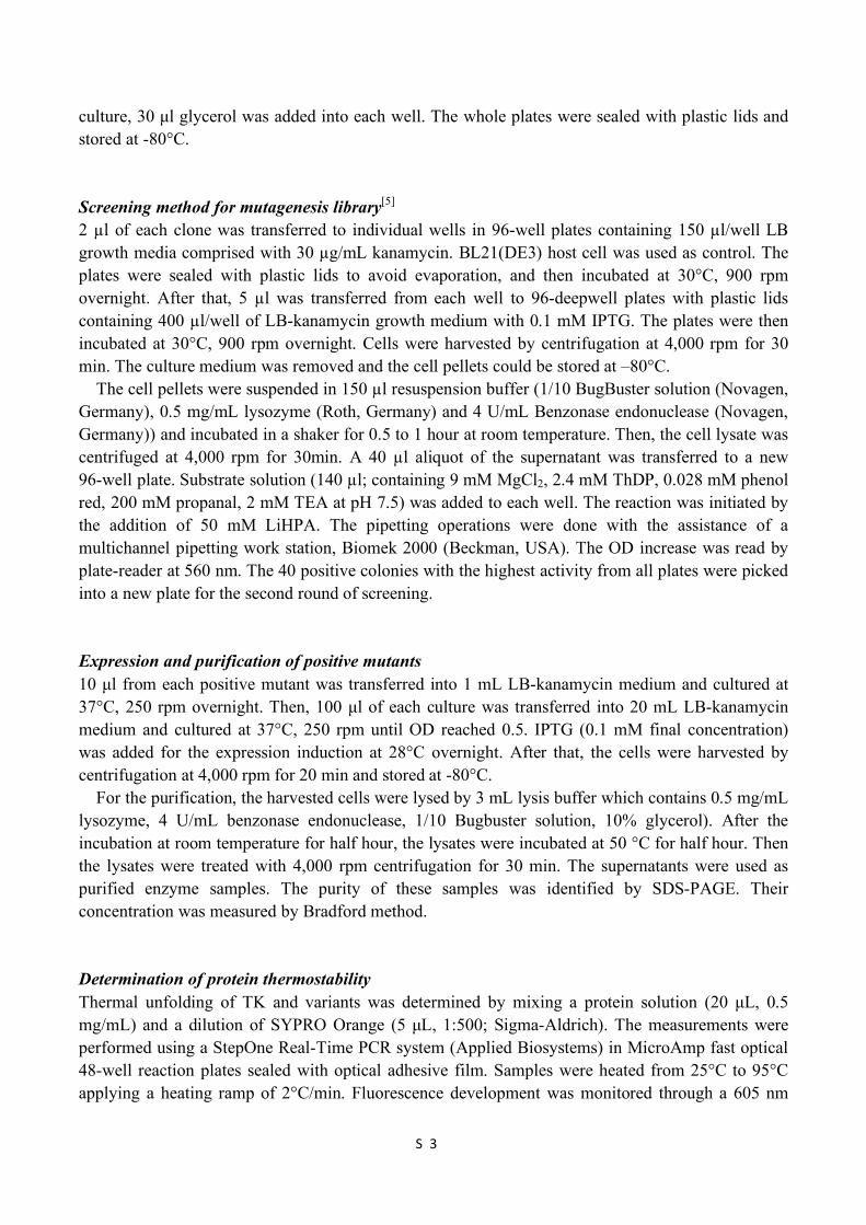
culture, 30 µl glycerol was added into each well. The whole plates were sealed with plastic lids and stored at -80°C. Screening method for mutagenesis library[5] 2 µl of each clone was transferred to individual wells in 96-well plates containing 150 µl/well LB growth media comprised with 30 µg/mL kanamycin. BL21(DE3) host cell was used as control. The plates were sealed with plastic lids to avoid evaporation, and then incubated at 30°C, 900 rpm overnight. After that, 5 µl was transferred from each well to 96-deepwell plates with plastic lids containing 400 µl/well of LB-kanamycin growth medium with 0.1 mM IPTG. The plates were then incubated at 30°C, 900 rpm overnight. Cells were harvested by centrifugation at 4,000 rpm for 30 min. The culture medium was removed and the cell pellets could be stored at –80°C.
The cell pellets were suspended in 150 µl resuspension buffer (1/10 BugBuster solution (Novagen, Germany), 0.5 mg/mL lysozyme (Roth, Germany) and 4 U/mL Benzonase endonuclease (Novagen, Germany)) and incubated in a shaker for 0.5 to 1 hour at room temperature. Then, the cell lysate was centrifuged at 4,000 rpm for 30min. A 40 µl aliquot of the supernatant was transferred to a new 96-well plate. Substrate solution (140 µl; containing 9 mM MgCl2, 2.4 mM ThDP, 0.028 mM phenol red, 200 mM propanal, 2 mM TEA at pH 7.5) was added to each well. The reaction was initiated by the addition of 50 mM LiHPA. The pipetting operations were done with the assistance of a multichannel pipetting work station, Biomek 2000 (Beckman, USA). The OD increase was read by plate-reader at 560 nm. The 40 positive colonies with the highest activity from all plates were picked into a new plate for the second round of screening. Expression and purification of positive mutants 10 µl from each positive mutant was transferred into 1 mL LB-kanamycin medium and cultured at 37°C, 250 rpm overnight. Then, 100 µl of each culture was transferred into 20 mL LB-kanamycin medium and cultured at 37°C, 250 rpm until OD reached 0.5. IPTG (0.1 mM final concentration) was added for the expression induction at 28°C overnight. After that, the cells were harvested by centrifugation at 4,000 rpm for 20 min and stored at -80°C.
For the purification, the harvested cells were lysed by 3 mL lysis buffer which contains 0.5 mg/mL lysozyme, 4 U/mL benzonase endonuclease, 1/10 Bugbuster solution, 10% glycerol). After the incubation at room temperature for half hour, the lysates were incubated at 50 °C for half hour. Then the lysates were treated with 4,000 rpm centrifugation for 30 min. The supernatants were used as purified enzyme samples. The purity of these samples was identified by SDS-PAGE. Their concentration was measured by Bradford method. Determination of protein thermostability Thermal unfolding of TK and variants was determined by mixing a protein solution (20 µL, 0.5 mg/mL) and a dilution of SYPRO Orange (5 µL, 1:500; Sigma-Aldrich). The measurements were performed using a StepOne Real-Time PCR system (Applied Biosystems) in MicroAmp fast optical 48-well reaction plates sealed with optical adhesive film. Samples were heated from 25°C to 95°C applying a heating ramp of 2°C/min. Fluorescence development was monitored through a 605 nm
S 4
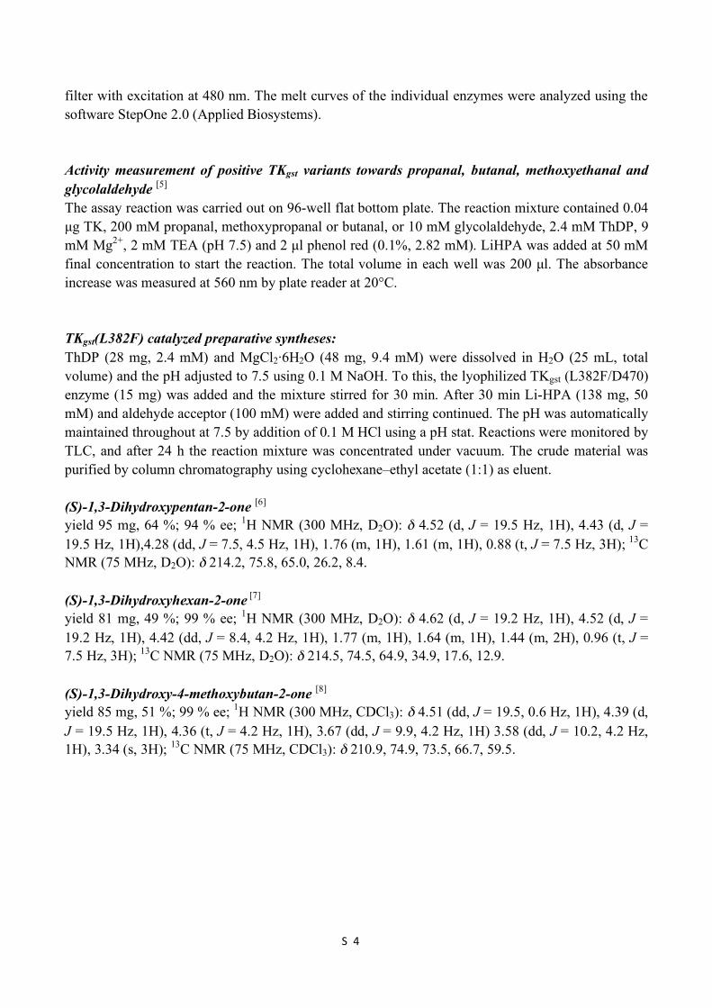
filter with excitation at 480 nm. The melt curves of the individual enzymes were analyzed using the software StepOne 2.0 (Applied Biosystems). Activity measurement of positive TKgst variants towards propanal, butanal, methoxyethanal and glycolaldehyde [5] The assay reaction was carried out on 96-well flat bottom plate. The reaction mixture contained 0.04 µg TK, 200 mM propanal, methoxypropanal or butanal, or 10 mM glycolaldehyde, 2.4 mM ThDP, 9 mM Mg2+, 2 mM TEA (pH 7.5) and 2 µl phenol red (0.1%, 2.82 mM). LiHPA was added at 50 mM final concentration to start the reaction. The total volume in each well was 200 µl. The absorbance increase was measured at 560 nm by plate reader at 20°C. TKgst(L382F) catalyzed preparative syntheses: ThDP (28 mg, 2.4 mM) and MgCl2·6H2O (48 mg, 9.4 mM) were dissolved in H2O (25 mL, total volume) and the pH adjusted to 7.5 using 0.1 M NaOH. To this, the lyophilized TKgst (L382F/D470) enzyme (15 mg) was added and the mixture stirred for 30 min. After 30 min Li-HPA (138 mg, 50 mM) and aldehyde acceptor (100 mM) were added and stirring continued. The pH was automatically maintained throughout at 7.5 by addition of 0.1 M HCl using a pH stat. Reactions were monitored by TLC, and after 24 h the reaction mixture was concentrated under vacuum. The crude material was purified by column chromatography using cyclohexane–ethyl acetate (1:1) as eluent. (S)-1,3-Dihydroxypentan-2-one [6] yield 95 mg, 64 %; 94 % ee; 1H NMR (300 MHz, D2O): δ 4.52 (d, J = 19.5 Hz, 1H), 4.43 (d, J = 19.5 Hz, 1H),4.28 (dd, J = 7.5, 4.5 Hz, 1H), 1.76 (m, 1H), 1.61 (m, 1H), 0.88 (t, J = 7.5 Hz, 3H); 13C NMR (75 MHz, D2O): δ 214.2, 75.8, 65.0, 26.2, 8.4. (S)-1,3-Dihydroxyhexan-2-one [7] yield 81 mg, 49 %; 99 % ee; 1H NMR (300 MHz, D2O): δ 4.62 (d, J = 19.2 Hz, 1H), 4.52 (d, J = 19.2 Hz, 1H), 4.42 (dd, J = 8.4, 4.2 Hz, 1H), 1.77 (m, 1H), 1.64 (m, 1H), 1.44 (m, 2H), 0.96 (t, J = 7.5 Hz, 3H); 13C NMR (75 MHz, D2O): δ 214.5, 74.5, 64.9, 34.9, 17.6, 12.9. (S)-1,3-Dihydroxy-4-methoxybutan-2-one [8] yield 85 mg, 51 %; 99 % ee; 1H NMR (300 MHz, CDCl3): δ 4.51 (dd, J = 19.5, 0.6 Hz, 1H), 4.39 (d, J = 19.5 Hz, 1H), 4.36 (t, J = 4.2 Hz, 1H), 3.67 (dd, J = 9.9, 4.2 Hz, 1H) 3.58 (dd, J = 10.2, 4.2 Hz, 1H), 3.34 (s, 3H); 13C NMR (75 MHz, CDCl3): δ 210.9, 74.9, 73.5, 66.7, 59.5.
S 5
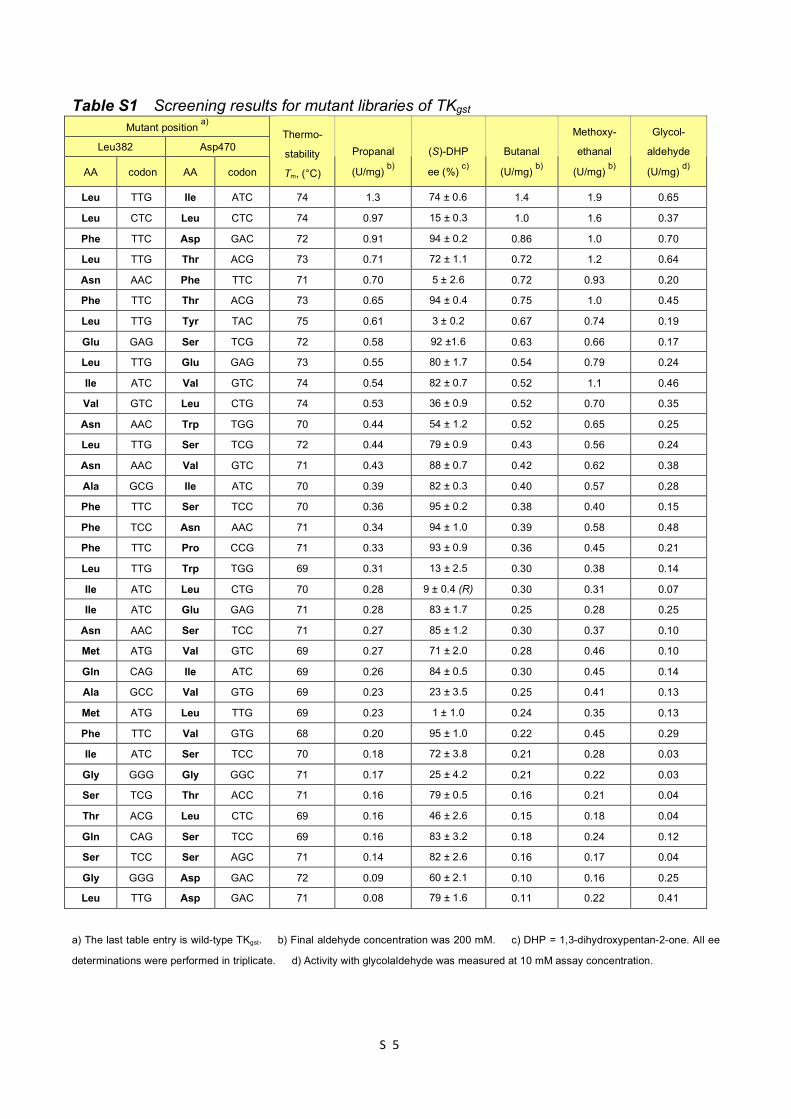
Table S1 Screening results for mutant libraries of TKgst Mutant position a)
Leu382 Asp470
AA codon AA codon
Thermo-
stability
Tm, (°C)
Propanal
(U/mg) b)
(S)-DHP
ee (%) c)
Butanal
(U/mg) b)
Methoxy-
ethanal
(U/mg) b)
Glycol-
aldehyde
(U/mg) d)
Leu TTG Ile ATC 74 1.3 74 ± 0.6 1.4 1.9 0.65
Leu CTC Leu CTC 74 0.97 15 ± 0.3 1.0 1.6 0.37
Phe TTC Asp GAC 72 0.91 94 ± 0.2 0.86 1.0 0.70
Leu TTG Thr ACG 73 0.71 72 ± 1.1 0.72 1.2 0.64
Asn AAC Phe TTC 71 0.70 5 ± 2.6 0.72 0.93 0.20
Phe TTC Thr ACG 73 0.65 94 ± 0.4 0.75 1.0 0.45
Leu TTG Tyr TAC 75 0.61 3 ± 0.2 0.67 0.74 0.19
Glu GAG Ser TCG 72 0.58 92 ±1.6 0.63 0.66 0.17
Leu TTG Glu GAG 73 0.55 80 ± 1.7 0.54 0.79 0.24
Ile ATC Val GTC 74 0.54 82 ± 0.7 0.52 1.1 0.46
Val GTC Leu CTG 74 0.53 36 ± 0.9 0.52 0.70 0.35
Asn AAC Trp TGG 70 0.44 54 ± 1.2 0.52 0.65 0.25
Leu TTG Ser TCG 72 0.44 79 ± 0.9 0.43 0.56 0.24
Asn AAC Val GTC 71 0.43 88 ± 0.7 0.42 0.62 0.38
Ala GCG Ile ATC 70 0.39 82 ± 0.3 0.40 0.57 0.28
Phe TTC Ser TCC 70 0.36 95 ± 0.2 0.38 0.40 0.15
Phe TCC Asn AAC 71 0.34 94 ± 1.0 0.39 0.58 0.48
Phe TTC Pro CCG 71 0.33 93 ± 0.9 0.36 0.45 0.21
Leu TTG Trp TGG 69 0.31 13 ± 2.5 0.30 0.38 0.14
Ile ATC Leu CTG 70 0.28 9 ± 0.4 (R) 0.30 0.31 0.07
Ile ATC Glu GAG 71 0.28 83 ± 1.7 0.25 0.28 0.25
Asn AAC Ser TCC 71 0.27 85 ± 1.2 0.30 0.37 0.10
Met ATG Val GTC 69 0.27 71 ± 2.0 0.28 0.46 0.10
Gln CAG Ile ATC 69 0.26 84 ± 0.5 0.30 0.45 0.14
Ala GCC Val GTG 69 0.23 23 ± 3.5 0.25 0.41 0.13
Met ATG Leu TTG 69 0.23 1 ± 1.0 0.24 0.35 0.13
Phe TTC Val GTG 68 0.20 95 ± 1.0 0.22 0.45 0.29
Ile ATC Ser TCC 70 0.18 72 ± 3.8 0.21 0.28 0.03
Gly GGG Gly GGC 71 0.17 25 ± 4.2 0.21 0.22 0.03
Ser TCG Thr ACC 71 0.16 79 ± 0.5 0.16 0.21 0.04
Thr ACG Leu CTC 69 0.16 46 ± 2.6 0.15 0.18 0.04
Gln CAG Ser TCC 69 0.16 83 ± 3.2 0.18 0.24 0.12
Ser TCC Ser AGC 71 0.14 82 ± 2.6 0.16 0.17 0.04
Gly GGG Asp GAC 72 0.09 60 ± 2.1 0.10 0.16 0.25
Leu TTG Asp GAC 71 0.08 79 ± 1.6 0.11 0.22 0.41
a) The last table entry is wild-type TKgst. b) Final aldehyde concentration was 200 mM. c) DHP = 1,3-dihydroxypentan-2-one. All ee
determinations were performed in triplicate. d) Activity with glycolaldehyde was measured at 10 mM assay concentration.
S 6
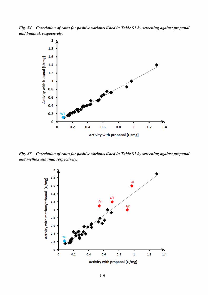
Fig. S4 Correlation of rates for positive variants listed in Table S1 by screening against propanal and butanal, respectively.
Fig. S5 Correlation of rates for positive variants listed in Table S1 by screening against propanal and methoxyethanal, respectively.
S 7
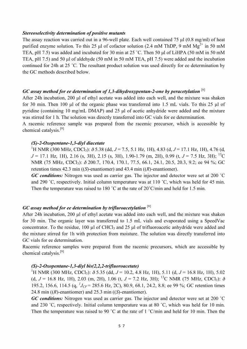
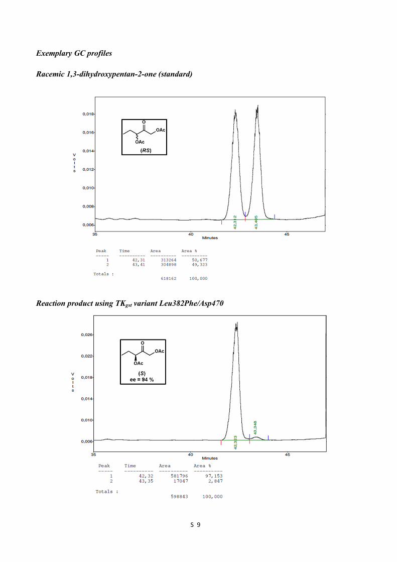
Stereoselectivity determination of positive mutants The assay reaction was carried out in a 96-well plate. Each well contained 75 µl (0.8 mg/ml) of heat purified enzyme solution. To this 25 µl of cofactor solution (2.4 mM ThDP, 9 mM Mg2+ in 50 mM TEA, pH 7.5) was added and incubated for 30 min at 25 ˚C. Then 50 µl of LiHPA (50 mM in 50 mM TEA, pH 7.5) and 50 µl of aldehyde (50 mM in 50 mM TEA, pH 7.5) were added and the incubation continued for 24h at 25 ˚C. The resultant product solution was used directly for ee determination by the GC methods described below. GC assay method for ee determination of 1,3-dihydroxypentan-2-one by peracetylation [6] After 24h incubation, 200 µl of ethyl acetate was added into each well, and the mixture was shaken for 30 min. Then 100 µl of the organic phase was transferred into 1.5 mL vials. To this 25 µl of pyridine (containing 10 mg/mL DMAP) and 25 µl of acetic anhydride were added and the mixture was stirred for 1 h. The solution was directly transferred into GC vials for ee determination. A racemic reference sample was prepared from the racemic precursor, which is accessible by chemical catalysis.[9]
(S)-2-Oxopentane-1,3-diyl diacetate 1H NMR (300 MHz, CDCl3): δ 5.38 (dd, J = 7.5, 5.1 Hz, 1H), 4.83 (d, J = 17.1 Hz, 1H), 4.76 (d, J = 17.1 Hz, 1H), 2.16 (s, 3H), 2.15 (s, 3H), 1.90-1.79 (m, 2H), 0.99 (t, J = 7.5 Hz, 3H); 13C NMR (75 MHz, CDCl3): δ 200.7, 170.4, 170.1, 77.5, 66.1, 24.1, 20.5, 20.3, 9.2; ee 94 %; GC retention times 42.3 min ((S)-enantiomer) and 43.4 min ((R)-enantiomer). GC conditions: Nitrogen was used as carrier gas. The injector and detector were set at 200 ˚C and 290 ˚C, respectively. Initial column temperature was at 110 ˚C, which was held for 45 min. Then the temperature was raised to 180 ˚C at the rate of 20˚C/min and held for 1.5 min.
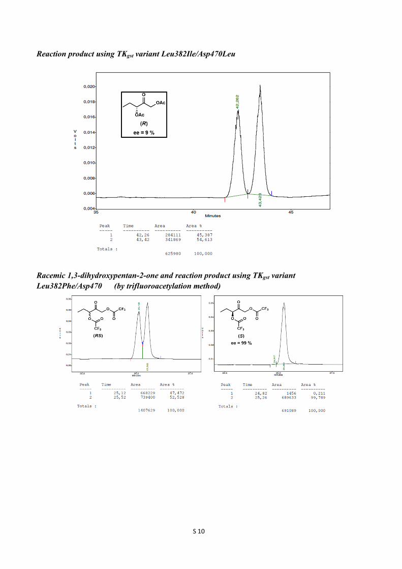
GC assay method for ee determination by trifluroacetylation [8] After 24h incubation, 200 µl of ethyl acetate was added into each well, and the mixture was shaken for 30 min. The organic layer was transferred to 1.5 mL vials and evaporated using a SpeedVac concentrator. To the residue, 100 µl of CHCl3 and 25 µl of trifluoroacetic anhydride were added and the mixture stirred for 1h with protection from moisture. The solution was directly transferred into GC vials for ee determination. Racemic reference samples were prepared from the racemic precursors, which are accessible by chemical catalysis.[9]
(S)-2-Oxopentane-1,3-diyl bis(2,2,2-trifluoroacetate) 1H NMR (300 MHz, CDCl3): δ 5.35 (dd, J = 10.2, 4.8 Hz, 1H), 5.11 (d, J = 16.8 Hz, 1H), 5.02 (d, J = 16.8 Hz, 1H), 2.03 (m, 2H), 1.06 (t, J = 7.2 Hz, 3H); 13C NMR (75 MHz, CDCl3): δ 195.2, 156.6, 114.5 (q, 1JCF = 285.6 Hz, 2C), 80.9, 68.1, 24.2, 8.8; ee 99 %; GC retention times 24.8 min ((R)-enantiomer) and 25.3 min ((S)-enantiomer). GC conditions: Nitrogen was used as carrier gas. The injector and detector were set at 200 ˚C and 230 ˚C, respectively. Initial column temperature was at 80 ˚C, which was held for 10 min. Then the temperature was raised to 90 ˚C at the rate of 1 ˚C/min and held for 10 min. Then the

S 8
temperature was raised to 170 ˚C at the rate of 20 ˚C/min and held for 1 min.
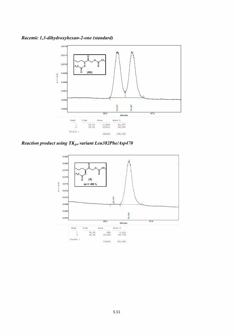
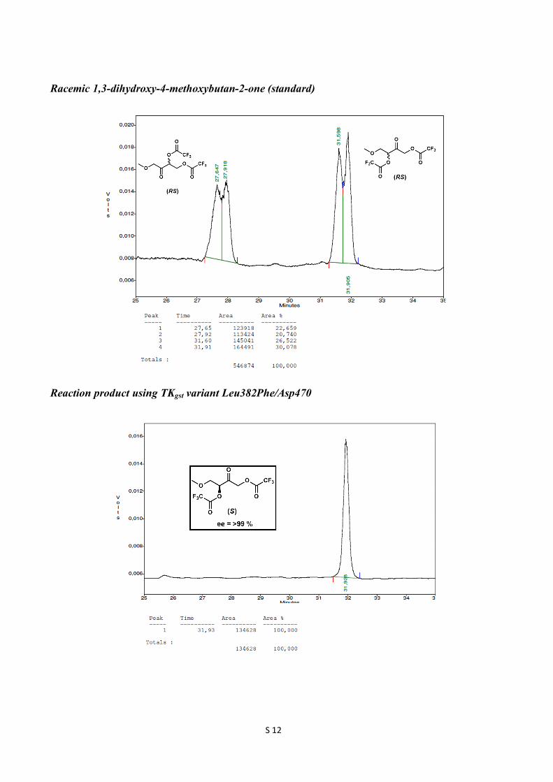
(S)-2-Oxohexane-1,3-diyl bis(2,2,2-trifluoroacetate) 1H NMR (300 MHz, CDCl3): δ 5.38 (t, J = 6 Hz, 1H), 5.10 (d, J = 17.1 Hz, 1H), 5.02 (d, J = 17.1 Hz, 1H), 1.97-1.90 (m, 2H), 1.47 (m, 2H), 0.98 (t, J = 7.5 Hz, 3H); 13C NMR (75 MHz, CDCl3): δ 195.4, 156.8 (q, 2JCF = 43.7 Hz, 2C), 114.5 (q, 1JCF = 283 Hz, 2C), 79.8, 67.9, 32.6, 17.8, 13.5; ee 99 %; GC retention times 35.5 min ((R)-enantiomer) and 36.3 min ((S)-enantiomer). GC conditions: Nitrogen was used as carrier gas. The injector and detector were set at 200 ˚C and 230 ˚C, respectively. Initial column temperature was at 80 ˚C, which was held for 10 min. Then the temperature was raised to 90 ˚C at the rate of 1 ˚C/min and held for 20 min. Then the temperature was raised to 170 ˚C at the rate of 20 ˚C/min and held for 1 min. (S)-4-Methoxy-2-oxobutane-1,3-diyl bis(2,2,2-trifluoroacetate) 1H NMR (300 MHz, CDCl3): δ 5.47 (t, J = 4.2 Hz, 1H), 5.13 (s, 2H), 3.90 (dd, J = =11.1, 4.2 Hz, 1H), 3.80 (dd, J = 11.1, 4.2 Hz, 1H), 3.39 (s, 3H); 13C NMR (75 MHz, CDCl3): δ 194.3, 156.6 (q, 2JCF = 24.7 Hz, 2C), 114.4 (q, 1JCF = 283.5 Hz, 2C), 79.1, 70.5, 69.0, 59.8; ee 99 %; GC retention times 31.6 min ((R)-enantiomer) and 31.9 min ((S)-enantiomer). GC conditions: Nitrogen was used as carrier gas. The injector and detector were set at 200 ˚C and 230 ˚C, respectively. Initial column temperature was at 80 ˚C and the temperature raised to 90 ˚C at the rate of 2 ˚C/min and held for 20 min. Then the temperature was raised to 120 ˚C at the rate of 2 ˚C/min and further the temperature was raised to 180 ˚C at the rate of 20 ˚C/min and held for 1 min.
S 9
Exemplary GC profiles Racemic 1,3-dihydroxypentan-2-one (standard)
Reaction product using TKgst variant Leu382Phe/Asp470
S 10
Reaction product using TKgst variant Leu382Ile/Asp470Leu
Racemic 1,3-dihydroxypentan-2-one and reaction product using TKgst variant Leu382Phe/Asp470 (by trifluoroacetylation method)
S 11
Racemic 1,3-dihydroxyhexan-2-one (standard)
Reaction product using TKgst variant Leu382Phe/Asp470
S 12
Racemic 1,3-dihydroxy-4-methoxybutan-2-one (standard)
Reaction product using TKgst variant Leu382Phe/Asp470
S 13
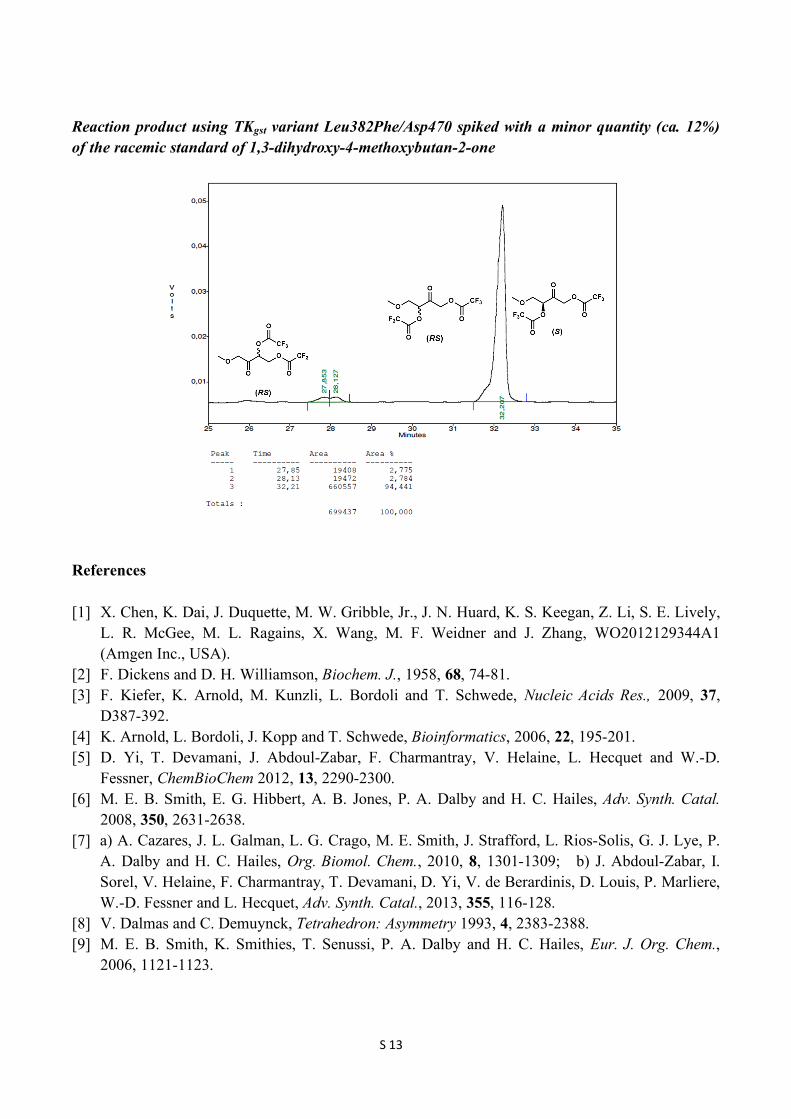
Reaction product using TKgst variant Leu382Phe/Asp470 spiked with a minor quantity (ca. 12%) of the racemic standard of 1,3-dihydroxy-4-methoxybutan-2-one
References [1] X. Chen, K. Dai, J. Duquette, M. W. Gribble, Jr., J. N. Huard, K. S. Keegan, Z. Li, S. E. Lively,
L. R. McGee, M. L. Ragains, X. Wang, M. F. Weidner and J. Zhang, WO2012129344A1 (Amgen Inc., USA).
[2] F. Dickens and D. H. Williamson, Biochem. J., 1958, 68, 74-81. [3] F. Kiefer, K. Arnold, M. Kunzli, L. Bordoli and T. Schwede, Nucleic Acids Res., 2009, 37,
D387-392. [4] K. Arnold, L. Bordoli, J. Kopp and T. Schwede, Bioinformatics, 2006, 22, 195-201. [5] D. Yi, T. Devamani, J. Abdoul-Zabar, F. Charmantray, V. Helaine, L. Hecquet and W.-D.
Fessner, ChemBioChem 2012, 13, 2290-2300. [6] M. E. B. Smith, E. G. Hibbert, A. B. Jones, P. A. Dalby and H. C. Hailes, Adv. Synth. Catal.
2008, 350, 2631-2638. [7] a) A. Cazares, J. L. Galman, L. G. Crago, M. E. Smith, J. Strafford, L. Rios-Solis, G. J. Lye, P.
A. Dalby and H. C. Hailes, Org. Biomol. Chem., 2010, 8, 1301-1309; b) J. Abdoul-Zabar, I. Sorel, V. Helaine, F. Charmantray, T. Devamani, D. Yi, V. de Berardinis, D. Louis, P. Marliere, W.-D. Fessner and L. Hecquet, Adv. Synth. Catal., 2013, 355, 116-128.
[8] V. Dalmas and C. Demuynck, Tetrahedron: Asymmetry 1993, 4, 2383-2388. [9] M. E. B. Smith, K. Smithies, T. Senussi, P. A. Dalby and H. C. Hailes, Eur. J. Org. Chem.,
2006, 1121-1123.