Embed Size (px)
Citation preview
137
Robert SchleipRobert Schleip, Ph.D.
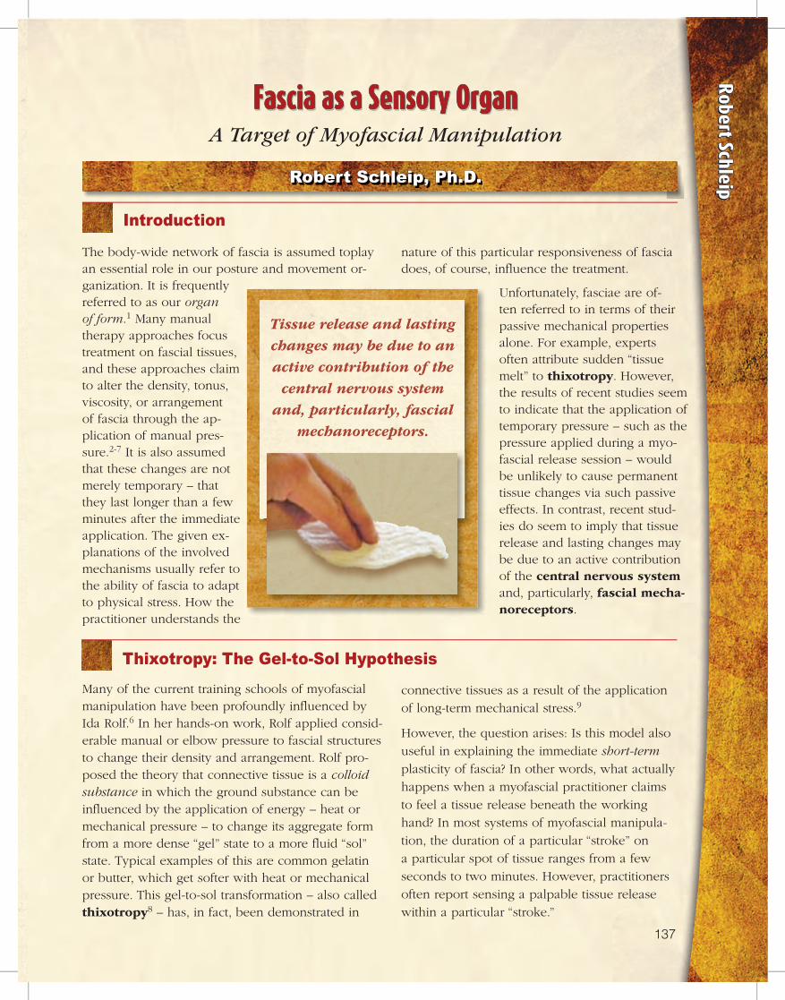
The body-wide network of fascia is assumed toplay an essential role in our posture and movement or-ganization. It is frequently referred to as our organ of form.1 Many manual therapy approaches focus treatment on fascial tissues, and these approaches claim to alter the density, tonus, viscosity, or arrangement of fascia through the ap-plication of manual pres-sure.2-7 It is also assumed that these changes are not merely temporary – that they last longer than a few minutes after the immediate application. The given ex-planations of the involved mechanisms usually refer to the ability of fascia to adapt to physical stress. How the practitioner understands the
nature of this particular responsiveness of fascia does, of course, influence the treatment.
Unfortunately, fasciae are of-ten referred to in terms of their passive mechanical properties alone. For example, experts often attribute sudden “tissue melt” to thixotropy. However, the results of recent studies seem to indicate that the application of temporary pressure – such as the pressure applied during a myo-fascial release session – would be unlikely to cause permanent tissue changes via such passive effects. In contrast, recent stud-ies do seem to imply that tissue release and lasting changes may be due to an active contribution of the central nervous system and, particularly, fascial mecha-noreceptors.
Introduction
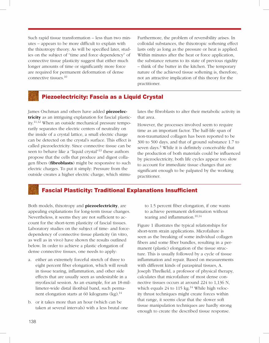
Thixotropy: The Gel-to-Sol HypothesisMany of the current training schools of myofascial manipulation have been profoundly influenced by Ida Rolf.6 In her hands-on work, Rolf applied consid-erable manual or elbow pressure to fascial structures to change their density and arrangement. Rolf pro-posed the theory that connective tissue is a colloid substance in which the ground substance can be influenced by the application of energy – heat or mechanical pressure – to change its aggregate form from a more dense “gel” state to a more fluid “sol” state. Typical examples of this are common gelatin or butter, which get softer with heat or mechanical pressure. This gel-to-sol transformation – also called thixotropy8 – has, in fact, been demonstrated in
connective tissues as a result of the application of long-term mechanical stress.9
However, the question arises: Is this model also useful in explaining the immediate short-term plasticity of fascia? In other words, what actually happens when a myofascial practitioner claims to feel a tissue release beneath the working hand? In most systems of myofascial manipula-tion, the duration of a particular “stroke” on a particular spot of tissue ranges from a few seconds to two minutes. However, practitioners often report sensing a palpable tissue release within a particular “stroke.”
A Target of Myofascial Manipulation
137136
Fascia as a Sensory Organ
Tissue release and lasting changes may be due to an active contribution of the
central nervous system and, particularly, fascial
mechanoreceptors.
Robert Schleip
138 139
James Oschman and others have added piezoelec-tricity as an intriguing explanation for fascial plastic-ity.11,12 When an outside mechanical pressure tempo-rarily separates the electric centers of neutrality on the inside of a crystal lattice, a small electric charge can be detected on the crystal’s surface. This effect is called piezoelectricity. Since connective tissue can be seen to behave like a “liquid crystal”13 these authors propose that the cells that produce and digest colla-gen fibers (fibroblasts) might be responsive to such electric charges. To put it simply: Pressure from the outside creates a higher electric charge, which stimu-
Fascial Plasticity: Traditional Explanations Insufficient
Piezoelectricity: Fascia as a Liquid Crystal
Both models, thixotropy and piezoelectricity, are appealing explanations for long-term tissue changes. Nevertheless, it seems they are not sufficient to ac-count for the short-term plasticity of fascial tissues. Laboratory studies on the subject of time- and force-dependency of connective tissue plasticity (in vitro, as well as in vivo) have shown the results outlined below. In order to achieve a plastic elongation of dense connective tissues, one needs to apply:
a. either an extremely forceful stretch of three to eight percent fiber elongation, which will result in tissue tearing, inflammation, and other side effects that are usually seen as undesirable in a myofascial session. As an example, for an 18-mil-limeter-wide distal iliotibial band, such perma-nent elongation starts at 60 kilograms (kg).14
b. or it takes more than an hour (which can be taken at several intervals) with a less brutal one
to 1.5 percent fiber elongation, if one wants to achieve permanent deformation without tearing and inflammation.10,14
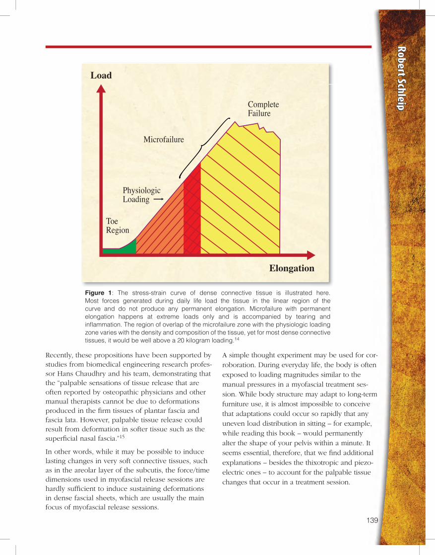
Figure 1 illustrates the typical relationships for short-term strain applications. Microfailure is seen as the breaking of some individual collagen fibers and some fiber bundles, resulting in a per-manent (plastic) elongation of the tissue struc-ture. This is usually followed by a cycle of tissue inflammation and repair. Based on measurements with different kinds of paraspinal tissues, A. Joseph Threlkeld, a professor of physical therapy, calculates that microfailure of most dense con-nective tissues occurs at around 224 to 1,136 N, which equals 24 to 115 kg.14 While high veloc-ity thrust techniques might create forces within that range, it seems clear that the slower soft tissue manipulation techniques are hardly strong enough to create the described tissue response.
lates the fibroblasts to alter their metabolic activity in that area.
However, the processes involved seem to require time as an important factor. The half-life span of non-traumatized collagen has been reported to be 300 to 500 days, and that of ground substance 1.7 to seven days.3 While it is definitely conceivable that the production of both materials could be influenced by piezoelectricity, both life cycles appear too slow to account for immediate tissue changes that are significant enough to be palpated by the working practitioner.
Such rapid tissue transformation – less than two min-utes – appears to be more difficult to explain with the thixotropy theory. As will be specified later, stud-ies on the subject of “time and force dependency” of connective tissue plasticity suggest that either much longer amounts of time or significantly more force are required for permanent deformation of dense connective tissues.10
Furthermore, the problem of reversibility arises. In colloidal substances, the thixotropic softening effect lasts only as long as the pressure or heat is applied. Within minutes after the heat or force application, the substance returns to its state of previous rigidity – think of the butter in the kitchen. The temporary nature of the achieved tissue softening is, therefore, not an attractive implication of this theory for the practitioner.
138 139
Robert Schleip
Load
Elongation
Microfailure
PhysiologicLoading
ToeRegion
CompleteFailure
Figure 1: The stress-strain curve of dense connective tissue is illustrated here. Most forces generated during daily life load the tissue in the linear region of the curve and do not produce any permanent elongation. Microfailure with permanent elongation happens at extreme loads only and is accompanied by tearing and inflammation. The region of overlap of the microfailure zone with the physiologic loading zone varies with the density and composition of the tissue, yet for most dense connective tissues, it would be well above a 20 kilogram loading.14
Recently, these propositions have been supported by studies from biomedical engineering research profes-sor Hans Chaudhry and his team, demonstrating that the “palpable sensations of tissue release that are often reported by osteopathic physicians and other manual therapists cannot be due to deformations produced in the firm tissues of plantar fascia and fascia lata. However, palpable tissue release could result from deformation in softer tissue such as the superficial nasal fascia.”15
In other words, while it may be possible to induce lasting changes in very soft connective tissues, such as in the areolar layer of the subcutis, the force/time dimensions used in myofascial release sessions are hardly sufficient to induce sustaining deformations in dense fascial sheets, which are usually the main focus of myofascial release sessions.
A simple thought experiment may be used for cor-roboration. During everyday life, the body is often exposed to loading magnitudes similar to the manual pressures in a myofascial treatment ses-sion. While body structure may adapt to long-term furniture use, it is almost impossible to conceive that adaptations could occur so rapidly that any uneven load distribution in sitting – for example, while reading this book – would permanently alter the shape of your pelvis within a minute. It seems essential, therefore, that we find additional explanations – besides the thixotropic and piezo-electric ones – to account for the palpable tissue changes that occur in a treatment session.
140 141
The Nervous System as a Rapid Self-Regulatory System
The inclusion of the nervous system in
attempting to understand fascial responsiveness is
hardly a new concept altogether.
From an evolutionary perspective, it makes sense that animals have a slowly adapting plasticity system in order to adjust to patterns of long-term use. In addi-tion to this capacity, they have also developed a more rapid system of adapting their form and local tissue density to temporary demands. Such a rapidly adapt-ing system is likely to play a key role in the lasting tissue changes reported by manual therapists. The question then becomes: Which systems allow for this rapid adaptation?
From observation, we know that an animal’s rapid regulation system is capable of adapting to how the animal perceives its interaction with the environment. The au-thor’s own experiments in treating anaesthetized people – which can give very similar results as manu-ally treating fresh pieces of raw meat – have shown that myofascial release work does not affect tis-sues in the same way as it would if the person pos-sessed intact neural regulation.16
Therefore, it seems plausible that this ability to quickly adapt to low-force manual manipulation is mediated by a finely tuned coordination system that is involved in the perception of our environment, as well as in sensing our own internal needs at any time. Traditionally, this body system has been called the nervous system.
The inclusion of the nervous system in attempting to understand fascial responsiveness is hardly a new concept altogether. Andrew Taylor Still, the founder of osteopathy, wrote more than a century ago:
The soul of man with all the streams of pure liv-ing water seems to dwell in the fascia of his body. When you deal with the fascia, you deal and do business with the branch offices of the brain, and under the general corporation law, the same as the brain itself, and why not treat it with the same degree of respect.17
Even so, many people discount the nervous system in the discussion of fascial responsiveness, because they consider the human nervous system to be orga-
nized like an old-fashioned telephone switchboard from the industrial age. Such a system would then, of course, be incapable of representing finer and more complex processes such as “life energy,” intui-tive insights, rapid movement refinements, or human empathy.
The reader is cordially invited to consider this to be an outdated model. Current concepts in neurobiol-
ogy primarily view the brain as a liquid system, in which fluid dynamics of a multitude of liquid and even gaseous neurotransmit-ters have come to the forefront.
It is a common opinion in the field that fascia must contain a different and much faster commu-nication system than the nervous system. Those who hold this view often believe that the ner-vous system cannot adapt quickly enough for the rapid responses in
human behavior. Thus, the fascia must have a sepa-rate, faster adaptation system. However, transmission of impulses in our nervous system often happens via messenger substances that travel along neural path-ways, as well as through the blood, lymph, cerebro-spinal fluid, or ground substance.18
This global system for rapid body regulation is in-separably connected to the endocrinal and immune systems, and it also works with complex feedforward system dynamics. Rather than picturing the nervous system as a hard-wired electric cable system – which in the view of many bodyworkers is then, of course, incapable of being involved in more subtle energetic phenomena – picture it in your mind’s eye as a wet tropical jungle.19
This jungle is a self-regulatory field – involving com-plex feedforward system dynamics – with an amaz-ing amount of complexity, plasticity, and continuous reorganization. Such a complex neural field could easily be involved in the rapid fascial changes expe-rienced during manual therapy.
140 141
Robert Schleip
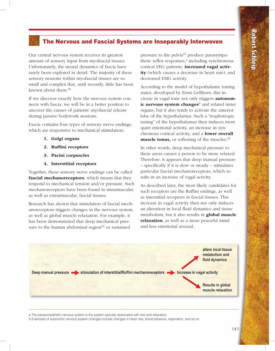
pressure to the pelvis22 produce parasympa-thetic reflex responses,a including synchronous cortical EEG patterns, increased vagal activ-ity (which causes a decrease in heart rate), and decreased EMG activity.
According to the model of hypothalamic tuning states, developed by Ernst Gellhorn, this in-crease in vagal tone not only triggers autonom-ic nervous system changesb and related inner organs, but it also tends to activate the anterior lobe of the hypothalamus. Such a “trophotropic tuning” of the hypothalamus then induces more quiet emotional activity, an increase in syn-chronous cortical activity, and a lower overall muscle tonus, or softening of the muscles.23
In other words, deep mechanical pressure to these areas causes a person to be more relaxed. Therefore, it appears that deep manual pressure – specifically if it is slow or steady – stimulates particular fascial mechanoreceptors, which re-sults in an increase of vagal activity.
As described later, the most likely candidates for such receptors are the Ruffini endings, as well as interstitial receptors in fascial tissues. This increase in vagal activity then not only induces an alteration in local fluid dynamics and tissue metabolism, but it also results in global muscle relaxation, as well as a more peaceful mind and less emotional arousal.
a The parasympathetic nervous system is the system typically associated with rest and relaxation.b Examples of autonomic nervous system changes include changes in heart rate, blood pressure, respiration, and so on.
The Nervous and Fascial Systems are Inseparably Interwoven
Our central nervous system receives its greatest amount of sensory input from myofascial tissues. Unfortunately, the neural dynamics of fascia have rarely been explored in detail. The majority of these sensory neurons within myofascial tissues are so small and complex that, until recently, little has been known about them.20
If we discover exactly how the nervous system con-nects with fascia, we will be in a better position to uncover the causes of patients’ myofascial release during passive bodywork sessions.
Fascia contains four types of sensory nerve endings, which are responsive to mechanical stimulation:
1. Golgi organs
2. Ruffini receptors
3. Pacini corpuscles
4. Interstitial receptors
Together, these sensory nerve endings can be called fascial mechanoreceptors, which means that they respond to mechanical tension and/or pressure. Such mechanoreceptors have been found in intramuscular, as well as extramuscular, fascial tissues.
Research has shown that stimulation of fascial mech-anoreceptors triggers changes in the nervous system, as well as global muscle relaxation. For example, it has been demonstrated that deep mechanical pres-sure to the human abdominal region21 or sustained
142 143
Obviously, fascia are not
the passive players we
often envision them to
be – responding to our
manual manipulations
as simply as a lump of
molding clay.
Fascial Mechanoreceptors Linked with Endocrine System
Fascial mechanoreceptors are not only intricately interwoven with the autonomic nervous system, but they are also vital to communication within the endocrine system. For example, many of the sensory neurons of the enteric brainc are mechanoreceptors, which – if activated – trigger, among other respons-es, important neuroendocrine changes. These include a change in the production of serotonin – an important cortical neurotransmitter – as well as other neuropeptides, such as the substance hista-mine, which increases inflam-matory processes.
Obviously, fascia are not the passive players we often envi-sion them to be – responding
to our manual manipulations as simply as a lump of molding clay. Their close-knit involvement with both the nervous and endocrine systems means a body-worker should approach fascial manipulation with a deeper understanding of the dynamic properties of fascial innervation.
The more we understand about the communication between fascial mechanoreceptors and the body’s systems, the more likely we are to create optimal manual therapy sessions for our clients. The following discussion delves more deeply into the relation-ship between the nervous system, the endocrine system, and fascial mechanoreceptors.
c Recent discoveries concerning the richness of the enteric nervous system25 have taught us that our “belly brain” contains more than 100 million neurons and works largely independently of the cortical brain. It is interesting to note that the limited connection between these two brains – only a few thousand neurons – consists of nine times as many neurons involved in processes in which the lower brain tells the upper one what to do, compared with the number of neurons involved in the top-down direction.
In addition, mechanoreceptors are also found abun-dantly in visceral ligaments, as well as in the dura ma-ter of the spinal cord and cranium. It, therefore, seems quite plausible that many of the benefits of osteopa-thy on these areas could be sufficiently explained by
a stimulation of mechanoreceptors. Such effects can explain profound autonomic changes without the need to rely on more esoteric assumptions, such as a breathing cerebrospinal liquid system.24
142 143
Robert Schleip
This is, of course, different in active client con-tractions, in which contractile muscle fibers acquire an increased stiffness, and the Golgi tendon organs function to provide feedback information about dynamic force changes during the contraction.28
Does this mean deep tissue work – in which the client is often passive – will not involve the Golgi reflex loop? Perhaps, but not necessarily. The in vitro studies discussed above were conducted using preparations in which muscles were surgi-cally isolated – freed from their lateral myofas-
cial adherences to surrounding structures. However, as shown by the extensive work of Peter Huijing and others, intact mus-cle tissues exhibit very different force transmission dynamics than isolated muscles.29
Furthermore, it is important to note that fewer than 10 percent of the Golgi receptors are found wholly within tendon – and the studies described above27,28 were performed only on Golgi tendon
receptors. The remaining 90 percent of the Golgi receptors are located in the muscular portions of myotendinous junctions, in the attachment tran-sitions of aponeuroses, in capsules, and in liga-ments of peripheral joints.30
Taking these considerations into account, it can-not be excluded that passive tissue stretch may be able to stimulate some Golgi receptors, particu-larly if the tissue is stretched in directions other than along the main muscle-tendon axis. In addition, if one applies strong local pressure to the fascial connection, the relatively intense, direct pressure may cause Golgi receptors to be activat-ed. However, the chance of eliciting such respons-es appears to be higher if the related muscle fibers are not in a state of complete relaxation.
Fascial Mechanoreceptor 1: The Golgi Receptors
d For clarification, the following analogy may be helpful. When a tight rope and a soft noodle are serially connected (one after the other in one straight continuous line), a pull on both of them will hardly result in any extension of the rope.
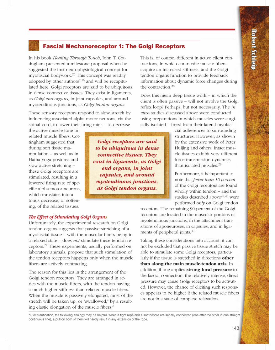
In his book Healing Through Touch, John T. Cot-tingham presented a milestone proposal when he suggested the first neurophysiological concept for myofascial bodywork.26 This concept was readily adopted by other authors7,16 and will be recapitu-lated here. Golgi receptors are said to be ubiquitous in dense connective tissues. They exist in ligaments, as Golgi end organs, in joint capsules, and around myotendinous junctions, as Golgi tendon organs.
These sensory receptors respond to slow stretch by influencing associated alpha motor neurons, via the spinal cord, to lower their firing rates – to decrease the active muscle tone in related muscle fibers. Cot-tingham suggested that during soft tissue ma-nipulation – as well as in Hatha yoga postures and slow active stretching – these Golgi receptors are stimulated, resulting in a lowered firing rate of spe-cific alpha motor neurons, which translates into a tonus decrease, or soften-ing, of the related tissues.
The Effect of Stimulating Golgi OrgansUnfortunately, the experimental research on Golgi tendon organs suggests that passive stretching of a myofascial tissue – with the muscular fibers being in a relaxed state – does not stimulate these tendon re-ceptors.27 These experiments, usually performed on laboratory animals, propose that such stimulation of the tendon receptors happens only when the muscle fibers are actively contracting.
The reason for this lies in the arrangement of the Golgi tendon receptors. They are arranged in se-ries with the muscle fibers, with the tendon having a much higher stiffness than relaxed muscle fibers. When the muscle is passively elongated, most of the stretch will be taken up, or “swallowed,” by a result-ing elastic elongation of the muscle fibers.d
Golgi receptors are said to be ubiquitous in dense connective tissues. They
exist in ligaments, as Golgi end organs, in joint
capsules, and around myotendinous junctions, as Golgi tendon organs.
144 145
Fascial Mechanoreceptors 2 and 3: Ruffini and Pacini Corpuscles
e The sympathetic nervous system is often associated with the fight or flight response. Increase in sympathetic nervous system activity results in stress. An inhibition of the sympathetic nervous system has a relaxing effect.
Both the Pacinian/Paciniform and the Ruffini bodies are found in all types of dense connective tissue – muscle fasciae, tendons, ligaments, aponeuroses, and joint capsules. In myotendinous junctions, the Pacin-ian corpuscles are more concentrated in the tendi-nous portion, as opposed to the Golgi tendon organs, which are more concentrated in the muscular portion.
They have also been shown to be more concentrated in the deeper portions of joint capsules; in deeper spi-nal ligaments; in plantar, as well as palmar tissues; in the peritoneum and enveloping muscular fasciae, such as the antebrachial, crural, and abdominal fasciae; and in the fascia of the masseter and the lateral thigh.31
The Ruffini endings are particularly densely distributed in tissues associated with regular stretching, such as the outer layer of joint capsules, the dura mater, the liga-ments of peripheral joints, and the deep dorsal fascia of the hand. At the knee joint, the Ruffini endings are especially found at anterior and posterior ligamentous and capsular structures, whereas Pacinian bodies are more accumulated medially and laterally of the joint.32
In addition, an immunohistochemical examination of the human thoracolumbar fascia revealed that it is richly populated by such mechanoreceptors.33 In this study, researchers found a high presence of Pacini and Ruffini endings, but no Golgi receptors.
The Effect of Stimulating Pacini ReceptorsThe group of Pacini receptors consists of the large Pacini corpuscles and the slightly smaller Paciniform corpuscles. The egg-shaped Pacini bodies respond to rapid changes in pressure – but not to constant unchanging pressure – and to vibration. A bit smaller are the Paciniform corpuscles, which have a similar function and sensitivity. Therefore, it seems likely that the Pacinian receptors are being stimulated only by high-velocity thrust manipulations and vibra-tory or oscillatory techniques.
Stimulation of Pacini receptors does not yield any clearly predictable muscle tonus increase or de-crease, but it does trigger heightened local pro-prioceptive attention of the central nervous system to the stimulated fascial region.
In case of a local sensomotor amnesia, as de-scribed by Thomas Hanna,34 such stimulation may, thus, have beneficial effects and may result in a refined cortical body representation and im-proved local neuromuscular coordination.
The Effect of Stimulating Ruffini EndingsThe smaller and more longitudinal Ruffini organs do not adapt as quickly as Pacini receptors, and, therefore, respond to constant pressure. In con-trast to the Pacini, the Ruffini endings are acti-vated by slow and deep “melting quality” soft tissue techniques, as well as faster strokes.
Two aspects of Ruffini endings seem important:
1. They are especially responsive to tangential forces and lateral stretch.35
2. It has been shown that stimulation of Ruffini receptors tends to induce a lowering of sym-pathetic nervous system activity.32,e This seems to fit to the common clinical finding that slow deep tissue techniques tend to have a relaxing effect on local tissues, and, indeed, on the whole organism.
In order to examine the potential neural dynam-ics of Ruffini receptors in myofascial manipu-lation, let us use the following scenario as a reference point. Imagine a practitioner working slowly with the connective tissue around the lateral ankle, in an area with no striated muscle fibers. Choosing this reference scenario allows us to focus on intrafascial dynamics only and – for the purpose of this chapter – to ignore the stimu-lation of intramuscular mechanoreceptors and other effects that would be involved in the analy-sis of many other myofascial working situations.
If the practitioner in this scenario reports a “tis-sue release,” what has happened? Possibly, the manual touch stimulated some Ruffini endings, which, in turn, triggered the central nervous system to change the tonus of some motor units in the muscle tissue that was mechanically con-nected to the tissue beneath the practitioner’s hand (Fig. 4).

144 145
Robert Schleip
In the past, it was assumed that these nerve end-ings were mostly pain receptors. Some have also been shown to be involved in thermo- or che-moception. Although many of these receptors are multimodal, research has shown that the majority
of interstitial receptors do, in fact, func-tion as mechanoreceptors.36
The Effect of Stimulating Interstitial ReceptorsWhile the type III and type IV fibers exhibit some important differences in certain physiological aspects, they do express common features regarding their mechanoreceptor functions. The large group of interstitial mechanoreceptors can be subdivided into two groups of equal size: low-threshold pressure units (LTP units) and high-threshold pressure units (HTP).
A study of cat Achilles tendon re-vealed that about half of the type III and IV endings encountered were LTP units and responded to light touch, even touch as light as “with a painter’s brush.”36 Based on this finding, doesn’t it
seem possible – if not probable – that soft tissue manipulation might involve stimulation of these type III and type IV receptors?
This raises the question of the natural functional role of interstitial mechanoreceptors in the body. What regular consequences or reactions have been associated with an excitation of this hid-den and rich sensory network? Of course, some of them function as pain receptors. However, research conducted as early as 1974 revealed that the type III and type IV receptors in the fasciae of the temporalis, masseter, and infrahy-oid muscles show “responses to the mandibular movement and the stretching of the fascia and the skin.”37
Thus, the study suggested that these nerve
Figure 2: Within a typical muscle nerve there are almost three times as many sensory neurons than motor neurons. Note that only a small portion of the sensory information comes from the type I and II receptors originating in muscle spindles – Golgi receptors, Pacinian corpuscles, and Ruffini endings. The majority of the sensory input comes from the group of type III and IV receptors, or interstitial receptors, which are intimately linked with the autonomic nervous system.
Fascial Mechanoreceptor 4: Interstitial Receptors
Interstitial neural fibers are often ignored, yet they account for almost 80 percent of the sensory fibers within a typical motor nerve.f Only a small fraction of a motor nerve’s sensory fibers belong to the well-known types I and II sensory fibers, which originate
in muscle spindles – Golgi organs, Pacini corpuscles, and Ruffini endings (Fig. 2). The majority, or four times as many, belong to an interesting group of types III and IV sensory interstitial fibers, which are hardly mentioned in most textbooks.36
These hidden neurons are much smaller in diameter than the other three fascial mechanoreceptors and are now commonly called interstitial muscle receptors. A better name would be interstitial myofascial tissue receptors because they also exist abundantly in fascia. A minority of these fibers are covered by a very thin myelin sheath (type III, also called A-delta fibers), but 90 percent of them are unmyelinated (type IV or C fibers). These interstitial receptors are slower than the type I and II sensory fibers, and most of them originate in free nerve endings.
f Although many of the nerve fibers in a typical motor nerve have a vasomotor function, regulating blood flow, the largest group of fibers is sensory. If one studies a typical muscle nerve, such as the tibial nerve, it consists of almost three times more sensory fibers than motor fibers. This points to a fascinating principle – that sensory refinement seems to be much more important than motor organization.
146 147
endings are concerned “with the sensation of posi-tion and movement of the mandible. It is, therefore, quite likely that some of these receptors may also be responsive to the tissue deformations achieved in therapeutic manual fascia manipulations.
Furthermore, the majority of these type III and type IV mechanoreceptors have been shown to have au-tonomic functions. In other words, stimulation of their sensory endings leads to a change in heart rate, blood pressure, respiration, and so on.
Stimulation of type IV receptors tends to increase arterial blood pressure,38 whereas stimulation of type III receptors can both increase and decrease blood pressure. Several studies have shown that an increase of static pressure on muscles tends to lower arterial blood pressure.36 It seems that a major function of this intricate network of interstitial tissue receptors is to fine-tune the nervous system’s regulation of blood flow according to local demands, and that this is done via very close connections with the auto-nomic nervous system.
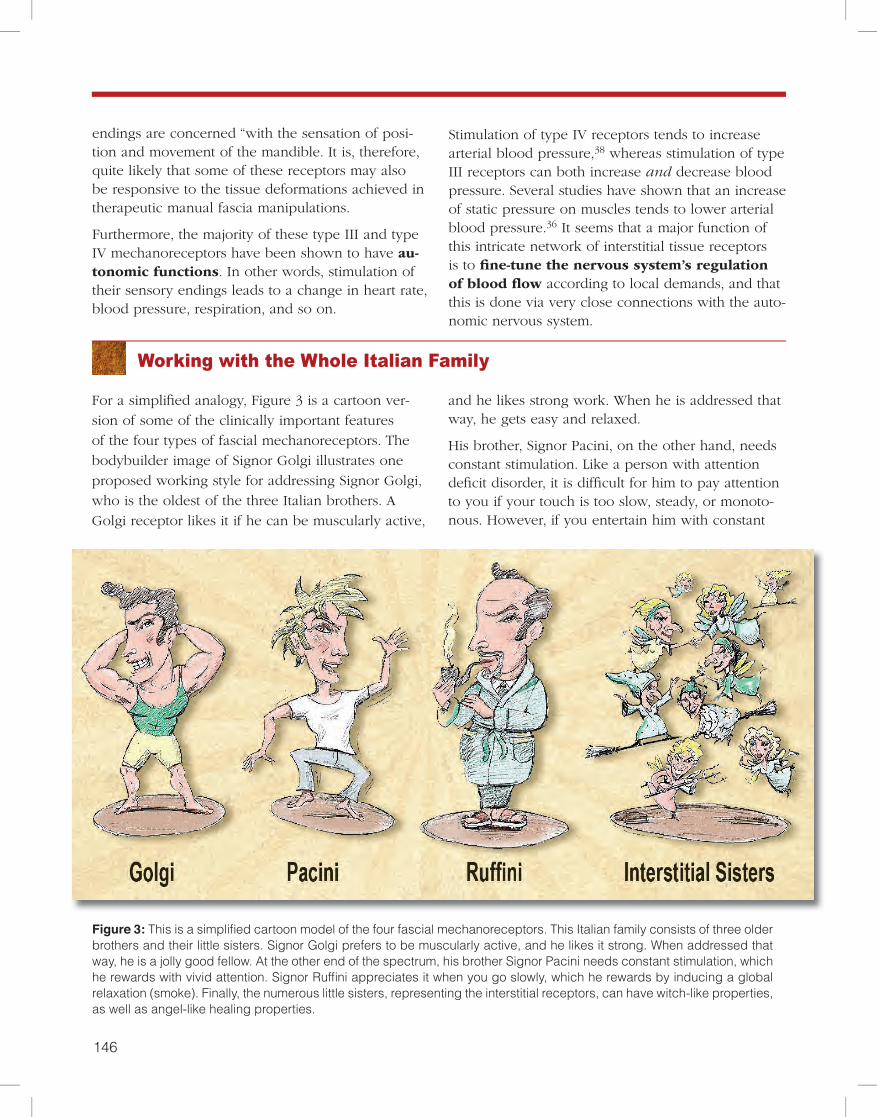
Working with the Whole Italian Family
For a simplified analogy, Figure 3 is a cartoon ver-sion of some of the clinically important features of the four types of fascial mechanoreceptors. The bodybuilder image of Signor Golgi illustrates one proposed working style for addressing Signor Golgi, who is the oldest of the three Italian brothers. A Golgi receptor likes it if he can be muscularly active,
and he likes strong work. When he is addressed that way, he gets easy and relaxed.
His brother, Signor Pacini, on the other hand, needs constant stimulation. Like a person with attention deficit disorder, it is difficult for him to pay attention to you if your touch is too slow, steady, or monoto-nous. However, if you entertain him with constant
Figure 3: This is a simplified cartoon model of the four fascial mechanoreceptors. This Italian family consists of three older brothers and their little sisters. Signor Golgi prefers to be muscularly active, and he likes it strong. When addressed that way, he is a jolly good fellow. At the other end of the spectrum, his brother Signor Pacini needs constant stimulation, which he rewards with vivid attention. Signor Ruffini appreciates it when you go slowly, which he rewards by inducing a global relaxation (smoke). Finally, the numerous little sisters, representing the interstitial receptors, can have witch-like properties, as well as angel-like healing properties.
146 147
Robert Schleip
changes and stimulation, he rewards you with vivid attention.
Finally, let’s look at the third of the three Italian brothers, Signor Ruffini. He is not the fast cappuc-cino guy, but an old-fashioned pipe smoker with a beard. Signor Ruffini likes it slow, and he prefers to address an issue at an angle, not in straightforward attacks. If you approach him in a slow manner and at the right tangential angle, he likes it a lot, and he will disperse some nice relaxing smoke that will have global effects throughout the whole body.
However, the power of the three brothers is well-matched – or even surpassed – by that of their nu-merous little sisters – the inter-stitial receptors. Some of these tiny nerve endings can have witch-like properties, evoking both temporary and long-term pain sensitization processes in their neighborhood. Others can have angel-like healing proper-ties, if addressed in the right manner.
While many practitioners may have a favorite working style, possibly primarily affect-ing only one of the four described mechanoreceptors, it is advisable to address the remaining receptor types as well, at least once a while. As in family systems therapy, it is suggested that ignoring some members may be less efficient than giving at least some atten-tion to them all.
Specific instructions on how to optimally address each of the depicted mechanoreceptors are best taught in the setting of a hands-on class. As an example, working approaches for addressing the Ruffini receptors are usually attractive to practitioners who already love a melting touch quality, in which the hand carefully listens to the tissue to understand the exact tangential angle the tissue responds to most. This myofascial working approach correlates well with important elements in traditional Rolfing® and also with slow-melting approaches in fascia-directed osteopathy.
Working with the interstitial receptors in the periosteum might recall Chua Ka, the traditional bone massage with which Mongolian warriors reportedly freed their body from fear before going into battle. Here, the pressure or shear of the practitioner on the periosteum is slowly increased until a slight sympathetic activation, along with a minimal orienting motor response, can be noticed.
These reactions may include a slight widening of the pupils, an increase or extension of a respira-tory inhalation movement, a tiny watering of the eye, a slight blushing of the face, or a minimal
turn of the head toward the practitioner. However, any motor expression of a withdrawal response, no matter how small, should alert the practitioner to back off.
Examples of a withdrawal response include nar-rowing of the eyes, neck preparation to turn away
from the practitioner, tightening of the lips, a more angular breathing movement of the lower belly wall, and so on. Ideally, the client supports the periosteal work with active movement par-ticipations from the inside, thereby increasing the pressure or shear at the working site in a moder-ated manner.
While such periosteum work should not be done on inflamed myofascial tissues or other areas with heightened pain sensitivity, it usually works quite well in those periosteal areas that are located close to pain areas, but have normal pressure sensitivity.
If preceded by respective training to extend one’s tactile sensitivity into a well-crafted hand tool, some practitioners profit from using a wooden or metal tool in the hand to direct the periosteal pressure with more precision.
Working approaches for addressing the Ruffini receptors are usually
attractive to practitioners who already love a melting
touch quality.
148 149
motor units associated with this tissue. In the case of slow deep pressure, the related mechanoreceptors are most likely the slowly adapting Ruffini endings and some of the interstitial receptors, though ad-ditional receptors might also be involved, such as spindle receptors in nearby affected muscle fibers, or possibly some intrafascial Golgi receptors.
g This is particularly shown in “genetic flexor muscles,”41 which are innervated via a ventral primary ramus from the spinal cord.
Fascial Mechanoreceptors: An Entry for Changing Skeletal Muscle Tone
Mechanoreceptor Stimulation and Gamma Motor Tone Regulation
By now, there is little doubt that myofascial manipu-lation involves stimulation of intrafascial mechano-receptors. Studies support the fact that this manipu-lation is also an entry for changing skeletal muscle tone. For example, slow deep pressure on the soft tissue of cats has been shown to lead to a reduction in muscle tonus as measured by electromyogram (EMG) activity.39 On the other hand, sudden deep tactile pressure, or pinching, and other types of strong and rapid manipulations have been shown to induce a general contraction of skeletal muscles.40,g
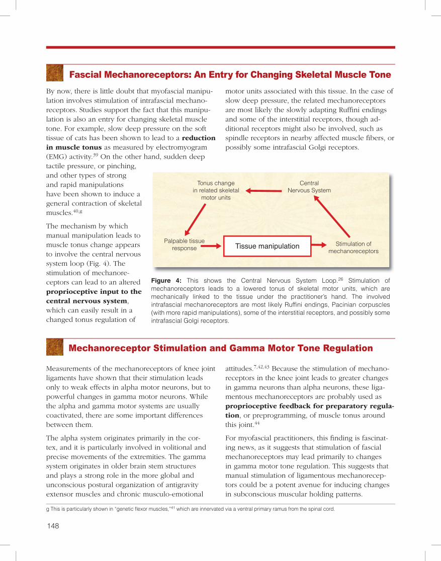
The mechanism by which manual manipulation leads to muscle tonus change appears to involve the central nervous system loop (Fig. 4). The stimulation of mechanore-ceptors can lead to an altered proprioceptive input to the central nervous system, which can easily result in a changed tonus regulation of
CentralNervous System
Tonus changein related skeletal
motor units
Palpable tissueresponse
Proprioceptivefunction
Stimulation ofmechanoreceptors
Hypothalamictuning
Interstitial receptors
and Ruffini endings
Local fluiddynamics
Fascialmyofibroblasts Autonomic
Nervous System
Tissue manipulation
Global muscletonus
Figure 12
Tissue manipulationPalpable tissueresponse
Stimulation ofmechanoreceptors
Local fluiddynamics
AutonomicNervous System
Interstitial and Ruffini
Figure 6
Manipulation of tissuePalpable tissue
response Stimulation ofmechanoreceptors
Global muscle tonus
Hypothalmic tuning
AutonomicNervous System
Interstitial and Ruffini
Figure 7
CentralNervous System
Tonus changein related skeletal
motor units
Palpable tissueresponse
Figure 4
Stimulation ofmechanoreceptors
Tissue manipulation
Figure 4: This shows the Central Nervous System Loop.26 Stimulation of mechanoreceptors leads to a lowered tonus of skeletal motor units, which are mechanically linked to the tissue under the practitioner’s hand. The involved intrafascial mechanoreceptors are most likely Ruffini endings, Pacinian corpuscles (with more rapid manipulations), some of the interstitial receptors, and possibly some intrafascial Golgi receptors.
Measurements of the mechanoreceptors of knee joint ligaments have shown that their stimulation leads only to weak effects in alpha motor neurons, but to powerful changes in gamma motor neurons. While the alpha and gamma motor systems are usually coactivated, there are some important differences between them.
The alpha system originates primarily in the cor-tex, and it is particularly involved in volitional and precise movements of the extremities. The gamma system originates in older brain stem structures and plays a strong role in the more global and unconscious postural organization of antigravity extensor muscles and chronic musculo-emotional
attitudes.7,42,43 Because the stimulation of mechano-receptors in the knee joint leads to greater changes in gamma neurons than alpha neurons, these liga-mentous mechanoreceptors are probably used as proprioceptive feedback for preparatory regula-tion, or preprogramming, of muscle tonus around this joint.44
For myofascial practitioners, this finding is fascinat-ing news, as it suggests that stimulation of fascial mechanoreceptors may lead primarily to changes in gamma motor tone regulation. This suggests that manual stimulation of ligamentous mechanorecep-tors could be a potent avenue for inducing changes in subconscious muscular holding patterns.
148 149
Robert Schleip
Muscles are Not Functional Units
Influencing Local Fluid Dynamics
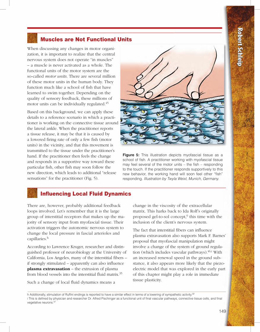
Figure 5: This illustration depicts myofascial tissue as a school of fish. A practitioner working with myofascial tissue may feel several of the motor units – the fish – responding to the touch. If the practitioner responds supportively to this new behavior, the working hand will soon feel other “fish” responding. Illustration by Twyla Weixl, Munich, Germany.
When discussing any changes in motor organi-zation, it is important to realize that the central nervous system does not operate “in muscles” – a muscle is never activated as a whole. The functional units of the motor system are the so-called motor units. There are several million of these motor units in the human body. They function much like a school of fish that have learned to swim together. Depending on the quality of sensory feedback, these millions of motor units can be individually regulated.45
Based on this background, we can apply these details to a reference scenario in which a practi-tioner is working on the connective tissue around the lateral ankle. When the practitioner reports a tissue release, it may be that it is caused by a lowered firing rate of only a few fish (motor units) in the vicinity, and that this movement is transmitted to the tissue under the practitioner’s hand. If the practitioner then feels the change and responds in a supportive way toward these particular fish, other fish may soon follow the new direction, which leads to additional “release sensations” for the practitioner (Fig. 5).
There are, however, probably additional feedback loops involved. Let’s remember that it is the large group of interstitial receptors that makes up the ma-jority of sensory input from myofascial tissue. Their activation triggers the autonomic nervous system to change the local pressure in fascial arterioles and capillaries.h
According to Lawrence Kruger, researcher and distin-guished professor of neurobiology at the University of California, Los Angeles, many of the interstitial fibers – if strongly stimulated – apparently can also influence plasma extravasation – the extrusion of plasma from blood vessels into the interstitial fluid matrix.35
Such a change of local fluid dynamics means a
change in the viscosity of the extracellular matrix. This harks back to Ida Rolf’s originally proposed gel-to-sol concept,6 this time with the inclusion of the client’s nervous system.
The fact that interstitial fibers can influence plasma extravasation also supports Mark F. Barnes’ proposal that myofascial manipulation might involve a change of the system of ground regula-tion (which includes vascular pathways).46,i With an increased renewal speed in the ground sub-stance, it also appears more likely that the piezo-electric model that was explored in the early part of this chapter might play a role in immediate tissue plasticity.
h Additionally, stimulation of Ruffini endings is reported to have a similar effect in terms of a lowering of sympathetic activity.32
i This is defined by physician and researcher Dr. Alfred Pischinger as a functional unit of final vascular pathways, connective tissue cells, and final vegetative neurons.47
150 151
Given that a myofascial manipulation then affects both the local blood supply and the local tis-sue viscosity, it is quite conceivable that these tis-sue changes could be rapid and significant enough to be felt by the listening hand of a sensitive practi-
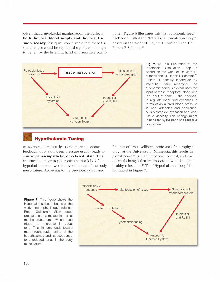
tioner. Figure 6 illustrates this first autonomic feed-back loop, called the “Intrafascial Circulation Loop,” based on the work of Dr. Jere H. Mitchell and Dr. Robert F. Schmidt.36
Figure 6: This illustration of the Intrafascial Circulation Loop is based on the work of Dr. Jere H. Mitchell and Dr. Robert F. Schmidt.36
Fascia is densely innervated by interstitial tissue receptors. The autonomic nervous system uses the input of these receptors, along with the input of some Ruffini endings, to regulate local fluid dynamics in terms of an altered blood pressure in local arterioles and capillaries, plus plasma extravasation and local tissue viscosity. This change might then be felt by the hand of a sensitive practitioner.
Hypothalamic TuningIn addition, there is at least one more autonomic feedback loop. Slow deep pressure usually leads to a more parasympathetic, or relaxed, state. This activates the more trophotropic anterior lobe of the hypothalamus to lower the overall tonus of the body musculature. According to the previously discussed
findings of Ernst Gellhorn, professor of neurophysi-ology at the University of Minnesota, this results in global neuromuscular, emotional, cortical, and en-docrinal changes that are associated with deep and healthy relaxation.23 This “Hypothalamus Loop” is illustrated in Figure 7.
Figure 7: This figure shows the Hypothalamus Loop, based on the work of neurophysiology professor Ernst Gellhorn.23 Slow deep pressure can stimulate interstitial mechanoreceptors, which can trigger an increase in vagal tone. This, in turn, leads toward more trophotropic tuning of the hypothalamus and, subsequently, to a reduced tonus in the body musculature.
CentralNervous System
Tonus changein related skeletal
motor units
Palpable tissueresponse
Proprioceptivefunction
Stimulation ofmechanoreceptors
Hypothalamictuning
Interstitial receptors
and Ruffini endings
Local fluiddynamics
Fascialmyofibroblasts Autonomic
Nervous System
Tissue manipulation
Global muscletonus
Figure 12
Tissue manipulationPalpable tissueresponse
Stimulation ofmechanoreceptors
Local fluiddynamics
AutonomicNervous System
Interstitial and Ruffini
Figure 6
Manipulation of tissuePalpable tissue
response Stimulation ofmechanoreceptors
Global muscle tonus
Hypothalmic tuning
AutonomicNervous System
Interstitial and Ruffini
Figure 7
CentralNervous System
Tonus changein related skeletal
motor units
Palpable tissueresponse
Figure 4
Stimulation ofmechanoreceptors
Tissue manipulation
CentralNervous System
Tonus changein related skeletal
motor units
Palpable tissueresponse
Proprioceptivefunction
Stimulation ofmechanoreceptors
Hypothalamictuning
Interstitial receptors
and Ruffini endings
Local fluiddynamics
Fascialmyofibroblasts Autonomic
Nervous System
Tissue manipulation
Global muscletonus
Figure 12
Tissue manipulationPalpable tissueresponse
Stimulation ofmechanoreceptors
Local fluiddynamics
AutonomicNervous System
Interstitial and Ruffini
Figure 6
Manipulation of tissuePalpable tissue
response Stimulation ofmechanoreceptors
Global muscle tonus
Hypothalmic tuning
AutonomicNervous System
Interstitial and Ruffini
Figure 7
CentralNervous System
Tonus changein related skeletal
motor units
Palpable tissueresponse
Figure 4
Stimulation ofmechanoreceptors
Tissue manipulation

150 151
Robert Schleip
A Brief Review
In summary of what we have learned so far, consider Table 1. We have discovered that fasciae are rich in four types of mechanoreceptors. Each type has a pre-ferred location and is responsive to a different type of manual manipulation. When stimulated, each type
Mechanoreceptors in FasciaKnown results of
stimulationResponsive toPreferred locationReceptor type
Golgi
Type Ib
• Myotendinous junctions
• Attachment areas of aponeuroses
• Ligaments of peripheral joints
• Joint capsules
Muscular contractionin golgi tendon organs
Probably to strong stretch only in other golgi receptors
Tonus decrease in related striated motor fibers
Pacini
and
Paciniform
Type II
• Myotendinous junctions
• Deep capsular layers
• Spinal ligaments
• Investing muscular tissues
Rapid pressure changes and vibrations
Proprioceptive feedback for movement control
(sense of kinesthesia)
Ruffini
Type II
• Ligaments of peripheral joints
• Dura mater
• Outer capsular layers and other tissues associated with regular stretching
Like Pacini, but also to sustained pressure
Especially responsive to tangential forces
(lateral stretch)
Inhibition of sympathetic activity
Interstitial
Types III and IV
• Most abundant receptor type, found almost everywhere, even inside bones
• Highest density in periosteum
Rapid as well as sustained pressure changes
(50 percent are high-threshold units, and 50 percent are
low-threshold units)
Changes in vasodilation
Plus, apparently in plasma extravasation
Table 1
of receptor has a different effect on the body – many of these effects are made possible by the complex feedback loops previously discussed.
152 153
A possible explanation for the contraction of fascia held under isometric conditions could be the exis-tence of cells with muscle-like contractile properties in the lumbodorsal fascia. Indeed, many visceral muscles possess the ability to contract spontane-ously. Price et al (Price 1981) demonstrated that strained and isometrically held intestinal muscles undergo relaxation followed by contraction.50 In order to test these specimens in a relaxed state
(without spontaneous contrac-tion), they used diverse tech-niques to suppress spontaneous activity, amongst them the use of epinephrine. A histological study of lumbodorsal fascia would, therefore, be desirable to evaluate whether muscles play a role in the contraction observed.48
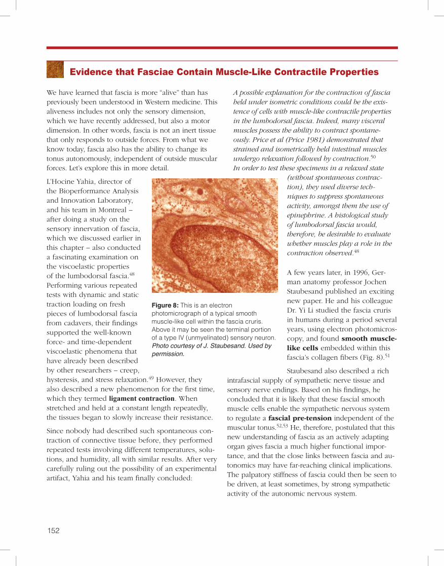
A few years later, in 1996, Ger-man anatomy professor Jochen Staubesand published an exciting new paper. He and his colleague Dr. Yi Li studied the fascia cruris in humans during a period several years, using electron photomicros-copy, and found smooth muscle-like cells embedded within this fascia’s collagen fibers (Fig. 8).51
Staubesand also described a rich intrafascial supply of sympathetic nerve tissue and sensory nerve endings. Based on his findings, he concluded that it is likely that these fascial smooth muscle cells enable the sympathetic nervous system to regulate a fascial pre-tension independent of the muscular tonus.52,53 He, therefore, postulated that this new understanding of fascia as an actively adapting organ gives fascia a much higher functional impor-tance, and that the close links between fascia and au-tonomics may have far-reaching clinical implications. The palpatory stiffness of fascia could then be seen to be driven, at least sometimes, by strong sympathetic activity of the autonomic nervous system.
Evidence that Fasciae Contain Muscle-Like Contractile Properties
We have learned that fascia is more “alive” than has previously been understood in Western medicine. This aliveness includes not only the sensory dimension, which we have recently addressed, but also a motor dimension. In other words, fascia is not an inert tissue that only responds to outside forces. From what we know today, fascia also has the ability to change its tonus autonomously, independent of outside muscular forces. Let’s explore this in more detail.
L’Hocine Yahia, director of the Bioperformance Analysis and Innovation Laboratory, and his team in Montreal – after doing a study on the sensory innervation of fascia, which we discussed earlier in this chapter – also conducted a fascinating examination on the viscoelastic properties of the lumbodorsal fascia.48 Performing various repeated tests with dynamic and static traction loading on fresh pieces of lumbodorsal fascia from cadavers, their findings supported the well-known force- and time-dependent viscoelastic phenomena that have already been described by other researchers – creep, hysteresis, and stress relaxation.49 However, they also described a new phenomenon for the first time, which they termed ligament contraction. When stretched and held at a constant length repeatedly, the tissues began to slowly increase their resistance.
Since nobody had described such spontaneous con-traction of connective tissue before, they performed repeated tests involving different temperatures, solu-tions, and humidity, all with similar results. After very carefully ruling out the possibility of an experimental artifact, Yahia and his team finally concluded:
Figure 8: This is an electron photomicrograph of a typical smooth muscle-like cell within the fascia cruris. Above it may be seen the terminal portion of a type IV (unmyelinated) sensory neuron. Photo courtesy of J. Staubesand. Used by permission.
152 153
Robert Schleip
Fascial Contractions Caused by Multiple Factors
How Stress Can Lead to Fascial Stiffness
j Fascial myofibroblasts can be seen as a phenotype of fibroblast with very high contractile properties. It has long been known that fibroblasts often transform into myofibroblasts, which stress fiber bundles containing smooth muscle actin and can, therefore, actively contract in a smooth muscle-like manner. This happens in pathologies such as Dupuytren’s contracture, liver cirrhosis, rheumatic arthritis, and frozen shoulder. However, it is also a productive element of early wound healing, and myofibroblasts are regularly found in healthy skin, as well as in the spleen, uterus, ovaries, circula-tory vessels, periodontal ligaments, and pulmonary septa.32
k Interestingly, our original hypothesis – that autonomic nervous system neurotransmitters, such as adrenaline, noradrenaline, and acetylcholine, might be able to elicit contractile responses large enough to translate to a palpable tissue response – was thwarted.
Inspired by the combined findings of Yahia and Staubesand, my research group at Ulm University conducted a histological examination of fascial tis-sues from different anatomical locations in 32 hu-man donors, ages 17 to 91. It was demonstrated that myofibroblasts exist in all of these fascial tissues.54 Myofibroblasts are known to have high contractile properties and can actively contract in a smooth muscle-like manner.j
With in vitro examinations, we also explored the contractile capacity of rat lumbar fasciae in response to chemical stimulation. We discovered that substanc-es associated with wound healing and inflammation – such as thromboxane and mepyramine – were able to trigger long-lasting tissue contractions.k These contractile responses were large enough to predict a palpable tissue response when applied to the ana-tomical properties of the human body.
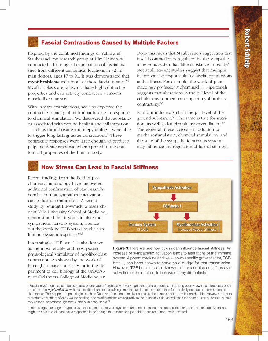
Recent findings from the field of psy-choneuroimmunology have uncovered additional confirmation of Staubesand’s conclusion that sympathetic activation causes fascial contractions. A recent study by Sourojit Bhowmick, a research-er at Yale University School of Medicine, demonstrated that if you stimulate the sympathetic nervous system, it sends out the cytokine TGF-beta-1 to elicit an immune system response.58,l
Interestingly, TGF-beta-1 is also known as the most reliable and most potent physiological stimulator of myofibroblast contraction. As shown by the work of James J. Tomasek, a professor in the de-partment of cell biology at the Universi-ty of Oklahoma College of Medicine, an
Sympathetic Activation
TGF-beta-1
Immune SystemT3 Cells
Myofibroblast ActivationIncreased Fascial Stiffness
Figure 9: Here we see how stress can influence fascial stiffness. An increase of sympathetic activation leads to alterations of the immune system. A potent cytokine and well-known specific growth factor, TGF-beta-1, has been shown to serve as a bridge for that transmission. However, TGF-beta-1 is also known to increase tissue stiffness via activation of the contractile behavior of myofibroblasts.
Does this mean that Staubesand’s suggestion that fascial contraction is regulated by the sympathet-ic nervous system has little substance in reality? Not at all. Recent studies suggest that multiple factors can be responsible for fascial contractions and stiffness. For example, the work of phar-macology professor Mohammad H. Pipelzadeh suggests that alterations in the pH level of the cellular environment can impact myofibroblast contractility.55
Pain can induce a shift in the pH level of the ground substance.56 The same is true for nutri-tion, as well as for chronic hyperventilation.57 Therefore, all these factors – in addition to mechanostimulation, chemical stimulation, and the state of the sympathetic nervous system – may influence the regulation of fascial stiffness.
154 155
Figure 9 and Figure 10, therefore, illustrate our pro-posed connections between the sympathetic nervous system and fascial contractions.
increase in TGF-beta-1 can induce tissue contracture mediated by myofibroblasts.59 These studies indicate that an increase in sympathetic activation, as caused by stress, can lead to increased fascial stiffness.
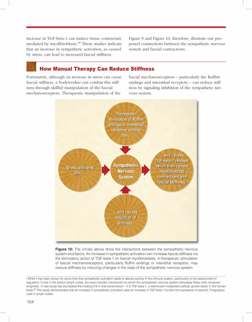
How Manual Therapy Can Reduce StiffnessFortunately, although an increase in stress can cause fascial stiffness, a bodyworker can combat this stiff-ness through skillful manipulation of the fascial mechanoreceptors. Therapeutic manipulation of the
fascial mechanoreceptors – particularly the Ruffini endings and interstitial receptors – can reduce stiff-ness by signaling inhibition of the sympathetic ner-vous system.
Figure 10: The circles above show the interactions between the sympathetic nervous system and fascia. An increase in sympathetic activation can increase fascial stiffness via the stimulatory action of TGF-beta-1 on fascial myofibroblasts. A therapeutic stimulation of fascial mechanoreceptors, particularly Ruffini endings or interstitial receptors, may reduce stiffness by inducing changes in the state of the sympathetic nervous system.
l While it has been known for some time that sympathetic activation leads to altered activity in the immune system, particularly to the deployment of regulatory T-cells in the body’s lymph nodes, the exact transfer mechanism by which the sympathetic nervous system stimulates these cells remained enigmatic. A new study has elucidated the missing link in that transmission – it is TGF-beta-1, a well-known multipotent cellular growth factor in the human body.58 This study demonstrates that an increase in sympathetic activation uses an increase in TGF-beta-1 to elicit the expression of specific T-regulatory cells in lymph nodes.
154 155
Robert Schleip
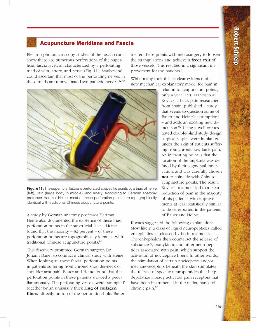
Figure 11: The superficial fascia is perforated at specific points by a triad of nerve (left), vein (large body in middle), and artery. According to German anatomy professor Hartmut Heine, most of these perforation points are topographically identical with traditional Chinese acupuncture points.
Acupuncture Meridians and Fascia
Electron photomicroscopy studies of the fascia cruris show there are numerous perforations of the super-ficial fascia layer, all characterized by a perforating triad of vein, artery, and nerve (Fig. 11). Staubesand could ascertain that most of the perforating nerves in these triads are unmyelinated sympathetic nerves.52,53
A study by German anatomy professor Hartmut Heine also documented the existence of these triad perforation points in the superficial fascia. Heine found that the majority – 82 percent – of these perforation points are topographically identical with traditional Chinese acupuncture points.60
This discovery prompted German surgeon Dr. Johann Bauer to conduct a clinical study with Heine. When looking at these fascial perforation points in patients suffering from chronic shoulder-neck or shoulder-arm pain, Bauer and Heine found that the perforation points in these patients showed a pecu-liar anomaly. The perforating vessels were “strangled” together by an unusually thick ring of collagen fibers, directly on top of the perforation hole. Bauer
treated these points with microsurgery to loosen the strangulations and achieve a freer exit of those vessels. This resulted in a significant im-provement for the patients.61
While many took this as clear evidence of a new mechanical explanatory model for pain in
relation to acupuncture points, only a year later, Francisco M. Kovacs, a back pain researcher from Spain, published a study that seems to question some of Bauer and Heine’s assumptions – and adds an exciting new di-mension.62 Using a well-orches-trated double-blind study design, surgical staples were implanted under the skin of patients suffer-ing from chronic low back pain. An interesting point is that the location of the implants was de-fined by their segmental inner-vation, and was carefully chosen not to coincide with Chinese acupuncture points. The result: Kovacs’ treatment led to a clear reduction of pain in the majority of his patients, with improve-ments at least statistically similar to those reported in the patients of Bauer and Heine.
Kovacs suggested the following explanation: Most likely, a class of liquid neuropeptides called enkephalins is released by both treatments. The enkephalins then counteract the release of substance P, bradykinin, and other neuropep-tides associated with pain, which support the activation of nociceptive fibers. In other words, the stimulation of certain nociceptors and/or mechanoreceptors beneath the skin stimulates the release of specific neuropeptides that help depolarize already activated pain receptors that have been instrumental in the maintenance of chronic pain.62
156 157
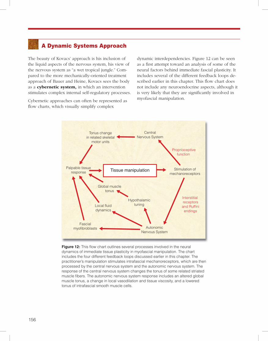
Figure 12: This flow chart outlines several processes involved in the neural dynamics of immediate tissue plasticity in myofascial manipulation. The chart includes the four different feedback loops discussed earlier in this chapter. The practitioner’s manipulation stimulates intrafascial mechanoreceptors, which are then processed by the central nervous system and the autonomic nervous system. The response of the central nervous system changes the tonus of some related striated muscle fibers. The autonomic nervous system response includes an altered global muscle tonus, a change in local vasodilation and tissue viscosity, and a lowered tonus of intrafascial smooth muscle cells.
A Dynamic Systems Approach
The beauty of Kovacs’ approach is his inclusion of the liquid aspects of the nervous system, his view of the nervous system as “a wet tropical jungle.” Com-pared to the more mechanically-oriented treatment approach of Bauer and Heine, Kovacs sees the body as a cybernetic system, in which an intervention stimulates complex internal self-regulatory processes.
Cybernetic approaches can often be represented as flow charts, which visually simplify complex
dynamic interdependencies. Figure 12 can be seen as a first attempt toward an analysis of some of the neural factors behind immediate fascial plasticity. It includes several of the different feedback loops de-scribed earlier in this chapter. This flow chart does not include any neuroendocrine aspects, although it is very likely that they are significantly involved in myofascial manipulation.
CentralNervous System
Tonus changein related skeletal
motor units
Palpable tissueresponse
Proprioceptivefunction
Stimulation ofmechanoreceptors
Hypothalamictuning
Interstitial receptors
and Ruffini endings
Local fluiddynamics
Fascialmyofibroblasts Autonomic
Nervous System
Tissue manipulation
Global muscletonus
Figure 12
Tissue manipulationPalpable tissueresponse
Stimulation ofmechanoreceptors
Local fluiddynamics
AutonomicNervous System
Interstitial and Ruffini
Figure 6
Manipulation of tissuePalpable tissue
response Stimulation ofmechanoreceptors
Global muscle tonus
Hypothalmic tuning
AutonomicNervous System
Interstitial and Ruffini
Figure 7
CentralNervous System
Tonus changein related skeletal
motor units
Palpable tissueresponse
Figure 4
Stimulation ofmechanoreceptors
Tissue manipulation
156 157
Robert Schleip
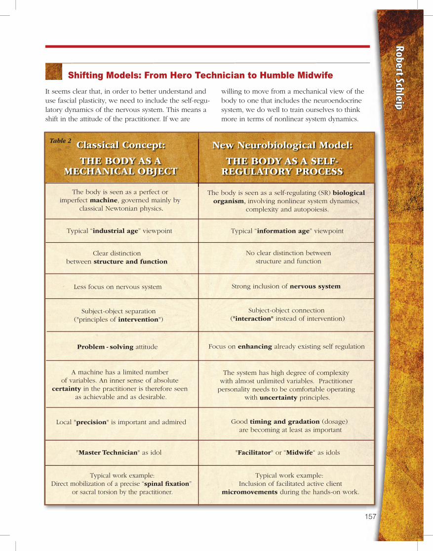
Shifting Models: From Hero Technician to Humble MidwifeIt seems clear that, in order to better understand and use fascial plasticity, we need to include the self-regu-latory dynamics of the nervous system. This means a shift in the attitude of the practitioner. If we are
willing to move from a mechanical view of the body to one that includes the neuroendocrine system, we do well to train ourselves to think more in terms of nonlinear system dynamics.
Classical Concept:
THE BODY AS A MECHANICAL OBJECT
The body is seen as a perfect or imperfect machine, governed mainly by
classical Newtonian physics.
The body is seen as a self-regulating (SR) biological organism, involving nonlinear system dynamics,
complexity and autopoiesis.
Typical “industrial age” viewpoint Typical “information age” viewpoint
Clear distinctionbetween structure and function
No clear distinction betweenstructure and function
Less focus on nervous system Strong inclusion of nervous system
Subject-object separation("principles of intervention")
Subject-object connection ("interaction" instead of intervention)
Problem - solving attitude Focus on enhancing already existing self regulation
A machine has a limited number of variables. An inner sense of absolute
certainty in the practitioner is therefore seen as achievable and as desirable.
The system has high degree of complexity with almost unlimited variables. Practitioner
personality needs to be comfortable operating with uncertainty principles.
Local "precision" is important and admired Good timing and gradation (dosage) are becoming at least as important
"Master Technician" as idol "Facilitator" or “Midwife“ as idols
Typical work example:Direct mobilization of a precise “spinal �xation”
or sacral torsion by the practitioner.
Typical work example: Inclusion of facilitated active client
micromovements during the hands-on work.
New Neurobiological Model:
THE BODY AS A SELF-REGULATORY PROCESS
Table 2
158 159
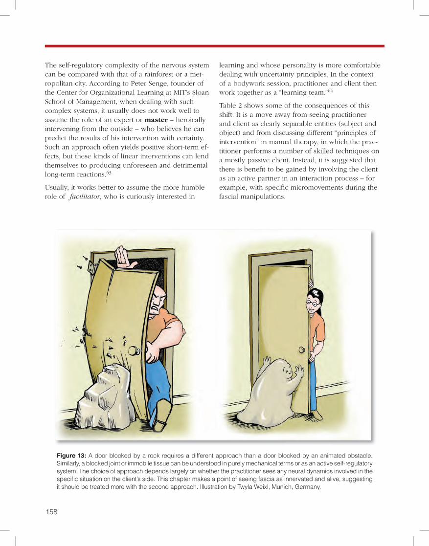
The self-regulatory complexity of the nervous system can be compared with that of a rainforest or a met-ropolitan city. According to Peter Senge, founder of the Center for Organizational Learning at MIT’s Sloan School of Management, when dealing with such complex systems, it usually does not work well to assume the role of an expert or master – heroically intervening from the outside – who believes he can predict the results of his intervention with certainty. Such an approach often yields positive short-term ef-fects, but these kinds of linear interventions can lend themselves to producing unforeseen and detrimental long-term reactions.63
Usually, it works better to assume the more humble role of facilitator, who is curiously interested in
Figure 13: A door blocked by a rock requires a different approach than a door blocked by an animated obstacle. Similarly, a blocked joint or immobile tissue can be understood in purely mechanical terms or as an active self-regulatory system. The choice of approach depends largely on whether the practitioner sees any neural dynamics involved in the specific situation on the client’s side. This chapter makes a point of seeing fascia as innervated and alive, suggesting it should be treated more with the second approach. Illustration by Twyla Weixl, Munich, Germany.
learning and whose personality is more comfortable dealing with uncertainty principles. In the context of a bodywork session, practitioner and client then work together as a “learning team.”64
Table 2 shows some of the consequences of this shift. It is a move away from seeing practitioner and client as clearly separable entities (subject and object) and from discussing different “principles of intervention” in manual therapy, in which the prac-titioner performs a number of skilled techniques on a mostly passive client. Instead, it is suggested that there is benefit to be gained by involving the client as an active partner in an interaction process – for example, with specific micromovements during the fascial manipulations.
158 159
Robert Schleip
Note that the common distinction between struc-ture – bones, dense fibrous connective tissue – and function – neuromuscular organization – is no longer useful within this new picture. Nobel laureate Lud-wig von Bertalanffy puts it this way:
The antithesis of structure and function, morphology and physi-
ology, is based upon a static conception of the organism. In a
machine, there is a fixed arrangement that can be set in motion
but can also be at rest. In a similar way, the pre-established struc-
ture of, say, the heart is distinguished from its function, namely,
rhythmical contraction. Actually, this separation between a
pre-established structure and processes occurring in that struc-
ture does not apply to the living organism. For the organism is
the expression of an everlasting, orderly process, though, on the
other hand, this process is sustained by underlying structures and
organized forms. What is described in morphology as organic
forms and structures is in reality a momentary cross section
through a spatio-temporal pattern.
What are called structures are slow processes of long duration,
functions are quick processes of short duration. If we say that a
function such as the contraction of a muscle is performed by a
structure, it means that a quick and short process is superimposed
on a long-lasting and slowly running wave.65
The role of a “Master Technician” in Table 2 can best be described by the following story. The heat-ing system of a big steamboat was broken and for
several days, and nobody could fix it. Finally, a master technician was called in. He just walked around and looked at everything, then he took out a little hammer from his pocket and hit a little valve, which immediately fixed the problem, and the machine started working again. When his bill of $1,000 arrived, the captain didn’t want to believe there could be such a high sum for so little work. He asked for an itemized bill, and the next day, the new bill arrived. It said:
For adjusting a little valve: $ 0.01
For knowing where: $ 999.99
Many bodywork practitioners still worship this story as an ideal of mastery in their work, al-though it clearly belongs in the realm of dealing with a mechanical universe. If one is willing to deal with fascia in a dynamic systems perspec-tive, it is more appropriate to assume the role of a midwife or facilitator who is skillfully assist-ing a self-regulatory process of the organism. This ideal is expressed in the Chinese saying:
Give a man a fish, and you feed him for a day.
Teach him how to fish, and you feed him for a lifetime.
Active Client Participation
A groundbreaking study by Dr. Lorimer Moseley, professor of clinical neurosciences and director of the Body in Mind research group, demonstrated that skillful mechanostimulation on the hand can be a very effective treatment for complex regional pain syndrome. However, the effectiveness of the treat-ment depends on the cortical attention of the pa-tient. If the patients were asked to accompany each touch with detailed attention, via a perceptual dis-crimination task, the treatment was highly effective. The exact same type of mechanostimulation showed zero effectiveness when the patients were allowed to read the newspaper and were not required to give detailed attention to each stimulation.66
The proposed mechanism for the effectiveness of the treatment included a remapping of the respec-
tive body part representation in the somatomotor cortex. While such cortical remapping may not have the same importance in all other musculo-skeletal dysfunctions, most body therapists would probably agree to the notion that the ultimate sustainability question – How long do the session changes last? – depends to a large degree on how much the client “owns” or “embodies” the recently gained relationships within his or her internal body schema and psychological body image.
How can we engage the client to pay curious attention to each detail of our fascial manipula-tion? Working at the edge of pain works well with most clients. The same goes for the per-ception and projection of charisma and status
160 161
on the practitioner, for an atmosphere of magic, or the setting of appropriately timed pauses after each intervention.
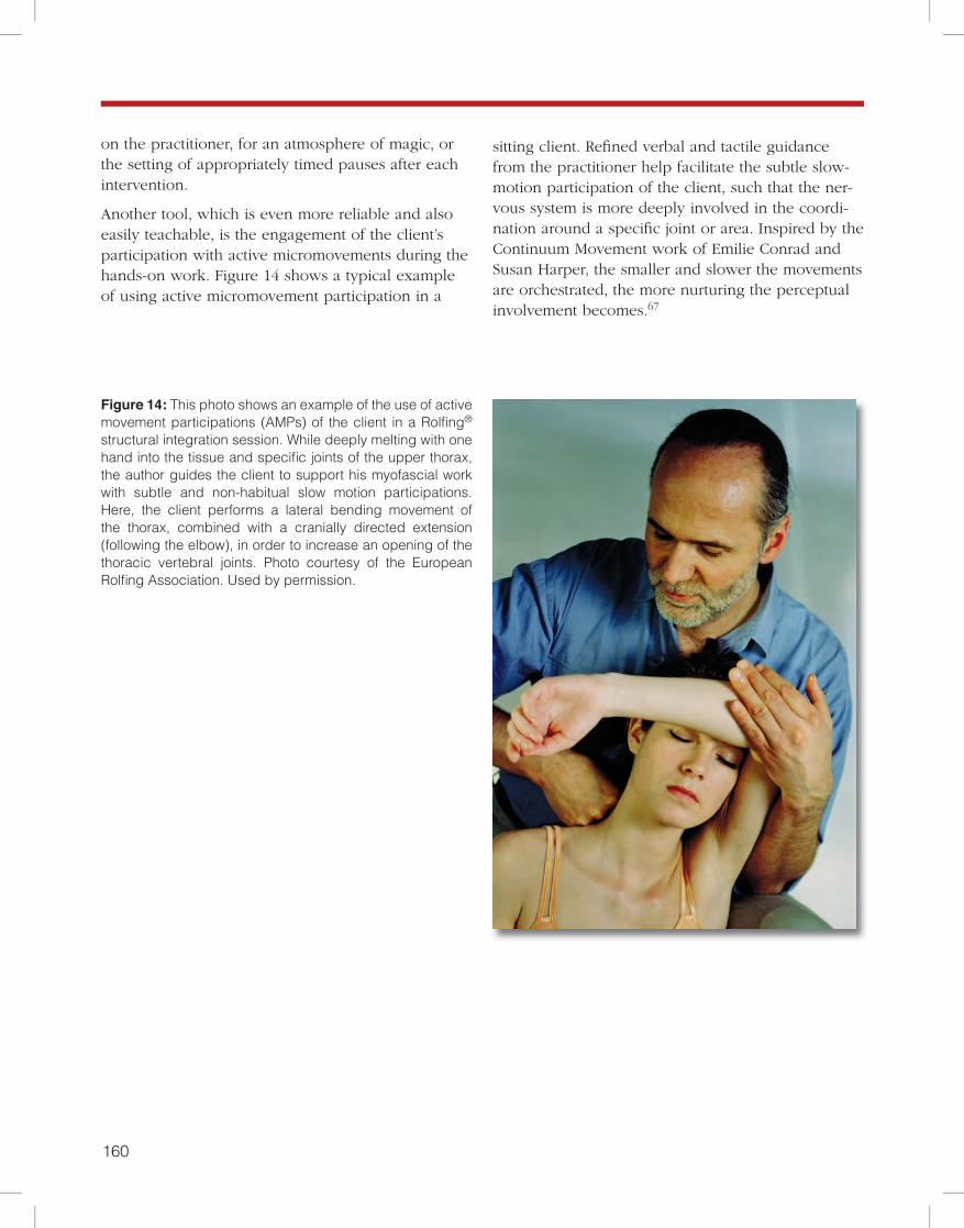
Another tool, which is even more reliable and also easily teachable, is the engagement of the client’s participation with active micromovements during the hands-on work. Figure 14 shows a typical example of using active micromovement participation in a
Figure 14: This photo shows an example of the use of active movement participations (AMPs) of the client in a Rolfing® structural integration session. While deeply melting with one hand into the tissue and specific joints of the upper thorax, the author guides the client to support his myofascial work with subtle and non-habitual slow motion participations. Here, the client performs a lateral bending movement of the thorax, combined with a cranially directed extension (following the elbow), in order to increase an opening of the thoracic vertebral joints. Photo courtesy of the European Rolfing Association. Used by permission.
sitting client. Refined verbal and tactile guidance from the practitioner help facilitate the subtle slow-motion participation of the client, such that the ner-vous system is more deeply involved in the coordi-nation around a specific joint or area. Inspired by the Continuum Movement work of Emilie Conrad and Susan Harper, the smaller and slower the movements are orchestrated, the more nurturing the perceptual involvement becomes.67
160 161
Robert Schleip
Conclusion
References
Fascia is alive. The practitioner working with fascial tissue should understand that it is innervated by four different kinds of mechanoreceptors. Without an in-clusion of the responsiveness of these mechanorecep-tors to various kinds of touch, the immediate effects of tissue release in myofascial manipulation cannot be adequately explained.
Manual stimulation of these sensory endings prob-ably leads to tonus changes in the motor units that are mechanically linked to the tissue beneath the practi-tioner’s hand. At least some of these responses are pri-marily regulated by a change in gamma motor tone.
Of particular interest are the Ruffini organs, with their high responsiveness to tangential pressure, and the very rich network of interstitial receptors. Stimulation of both these receptors can trigger pro-found changes in the autonomic nervous system.
There are strong links between fascia and the autonomic nervous system that affect fascial tonus and local tissue viscosity. Therefore, a shift from a mechanically-oriented “technician” point of view toward an inclusion of the self-regulation dynamics of the client’s nervous system is advocated.
1. Varela, F.J., & Frenk, S. (1987). The organ of form: towards a theory of biological shape. J Social Biol Struct, 10, 73-83.
2. Barnes J.F. (1990). Myofascial Release: The Search for Excellence. Paoli, PA: Rehabilitation Services Inc.
3. Cantu, R.I., & Grodin, A.J. (1992). Myofascial Manipu-lation: Theory and Clinical Application. Gaithersburg, MD: Aspen Publishers.
4. Chaitow, L. (1980). Soft-Tissue Manipulation. Roches-ter, VT: Healing Arts Press.
5. Paoletti, S. (1998). Les fascias – Role des tissues dans la mecanique humaine. Vannes cedex, France: Le Prisme.
6. Rolf, I.P. (1977). Rolfing: The Integration of Human Structures. Santa Monica, CA: Dennis-Landman.
7. Ward, R.C. (1993). Myofascial Release Concepts. In J.V. Basmajian and R.E. Nyberg (Eds.), Rational Manual Therapies. Baltimore, MD: Williams & Wilkins.
8. Juhan, D. (1987). Job’s Body: A Handbook for Body-work. Barrytown, NY: Station Hill Press.
9. Twomey, L., & Taylor, J. (1982). Flexion, creep, dys-function and hysteresis in the lumbar vertebral column. Spine, 7(2), 116-122.
10. Currier, D.P., & Nelson, R.M. (1992). Dynamics of Hu-man Biologic Tissues. Philadelphia, PA: F.A. Davis Com-pany.
11. Oschman, J.L. (2000). Energy Medicine. Edinburgh, United Kingdom: Churchill Livingstone.
12. Athenstaedt, H. (1974). Pyroelectric and piezoelectric properties of vertebrates. Ann NY Acad Sci, 238, 68-110.
13. Juhan, D. (1998). Job’s Body: A Handbook for Body-
work (3rd ed.). Barrytown, NY: Station Hill Press.
14. Threlkeld, A.J. (1992). The Effects of Manual Therapy on Connective Tissue. Phys Ther, 72(12), 893-901.
15. Chaudhry, H., Schleip, R., Zhiming, J., Bukiet, B., Maney, M., & Findley, T. (2008). Three-Dimensional Mathematical Model for Deformation of Human Fasciae in Manual Therapy. J Am Osteopath Assoc, 108(8), 379-390.
16. Schleip, R. (1989). A new explanation of the effect of Rolfing. Rolf Lines, 15(1), 18-20.
17. Still, A.T. (1899). Philosophy of Osteopathy. Kirks-ville, MO: Academy of Osteopathy.
18. Kandel, E.R. (1995). Essentials of neural science and behavior. New York, NY: Appleton & Lange.
19. Schleip, R. (2000). Lichtblicke im Dschungel der Gehirnforschung. FeldenkraisZEIT, 1, 47-56.
20. Engeln, H. (1994). Konzert der Muskeln und Sinne. GEO Wissen, 1(Mai), 90-97.
21. Folkow, B., Gelin, L.E., Lindell, S.E., Stenberg, K., & Thoren, O. (1962). Cardiovascular Reactions Dur-ing Abdominal Surgery. Ann Surg, 156(6), 905-913.
22. Koizumi, K., & Brooks, C.M. (1972). The integra-tion of autonomic system reactions: a discussion of autonomic reflexes, their control and their associa-tion with somatic reactions. Ergeb Physiol, 67, 1-68.
23. Gellhorn, E. (1967). Principles of Autonomic-Somatic Integrations: Physiological Basis and Psycho-logical and Clinical Implications. Minneapolis, MN: University of Minnesota Press.
162 163
24. Arbuckle, B.E. (1994). Selected Writings. Indianapolis, IN: American Academy of Osteopathy.
25. Gershon, M.D. (1999). The Second Brain. New York, NY: Harper-Collins.
26. Cottingham, J.T. (1985). Healing Through Touch: A History and a Review of the Physiological Evidence. Boul-der, CO: Rolf Institute.
27. Jami, L. (1992). Golgi tendon organs in mammalian skeletal muscle: functional properties and central actions. Physiol Rev, 73(3), 623-666.
28. Lederman, E. (1997). Fundamentals of Manual Thera-py. Edinburgh, United Kingdom: Churchill Livingstone.
29. Huijing, P.A. (2009). Epimuscular myofascial force transmission: a historical review and implications for new research. International Society of Biomechanics Muybridge Award Lecture, Taipei, 2007. J Biomech, 42(1), 9-21.
30. Burke, D., & Gandeva, S.C. (1990). Peripheral Motor System. In G. Paxinos (Ed.), The Human Nervous System (p. 133). San Diego, CA: Academic Press.
31. Stilwell, D. (1957). Regional variations in the inner-vation of deep fasciae and aponeuroses. Anat Record, 127(4), 635-653.
32. van den Berg, F., & Cabri, J. (1999). Angewandte Physiologie – Das Bindegewebe des Bewegungsapparates verstehen und beeinflussen. Stuttgart, Germany: Georg Thieme Verlag.
33. Yahia, L., Rhalmi, S., Newman, N., & Isler, M. (1992). Sensory innervation of human thoracolumbar fascia: An immunohistochemical study. Acta Orthop Scand, 63(2), 195-197.
34. Hanna, T. (1998). Somatics: Reawakening the Mind’s Control of Movement, Flexibility, and Health. Cambridge, MA: Da Capo Press.
35. Kruger, L. (1987). Cutaneous Sensory System. In G. Adelman (Ed.), Encyclopedia of Neuroscience (p. 293). Boston, MA: Birkhäuser.
36. Mitchell, J.H., & Schmidt, R.F. (1983). Cardiovascular Reflex Control by Afferent Fibers from Skeletal Muscle Receptors. In J.T. Shepherd et al (Eds.), Handbook of Physiology, The Cardiovascular System, Peripheral Cir-culation and Organ Blood Flow (pp. 623-658). Bethesda, MD: American Physiological Society.
37. Sakada, S. (1974). Mechanoreceptors in fascia, peri-osteum and periodontal ligament. Bull Tokyo Med Dent Univ, 21, S11-13.
38. Coote, J.H., Pérez-Gonzáles, J.F. (1970). The response of some sympathetic neurons to volleys in various affer-ent nerves. J Physiol-London, 208, 261-278.
39. Johansson, B. (1962). Circulatory response to stimulation of somatic afferents. Acta Physiol Scand, 62(S198), 1-92.
40. Eble, J.N. (1960). Patterns of response of the paraver-tebral musculature to visceral stimuli. Am J Physiol, 198, 429-433.
41. Schleip, R. (1993). Primary reflexes and structural typology. Rolf Lines, 21(3), 37-47.
42. Glaser, V. (1980). Eutonie. Heidelberg, Germany: Karl F. Haug Verlag.
43. Henatsch, H-D. (1976). Bauplan der peripheren und zentralen sensomotorischen Kontrollen. In Physiologie des Menschen (vol. 14). München, Germany: Urban and Schwarzenberg.
44. Johansson, H., Sjolander, P., & Sojka, P. (1991). Recep-tors in the knee joint ligaments and their role in the bio-mechanics of the joint. Critical Rev Biomed Eng, 18(5), 341-368.
45. Basmajian, J.V., & De Luca, C.J. (1985). Muscles Alive: Their Functions Revealed by Electromyography. Balti-more, MD: Williams & Wilkins.
46. Barnes, M.F. (1997). The Basic Science of Myofascial Release. J Bodywork Move Ther, 1(4), 231-238.
47. Pischinger, A. (1991). Matrix and Matrix Regulation: Basis for a Holistic Theory in Medicine. Brussels, Bel-gium: Haug International.
48. Yahia, L.H., Pigeon, P., & DesRosiers, E.A. (1993). Viscoelastic properties of the human lumbodorsal fascia. J Biomed Eng, 15(5), 425-429.
49. Chaitow, L., & DeLany, J.W. (2000). Clinical Applica-tion of Neuromuscular Techniques, Volume 1. Edinburgh, United Kingdom: Churchill Livingstone.
50. Price, J.M., Patitucci, P., & Fung, Y.C. (1981). Biome-chanics: Mechanical Properties of Living Tissues (pp. 371-391). New York, NY: Springer Verlag.
51. Staubesand, J., & Li, Y. (1996). Zum Feinbau der Fascia cruris mit besonderer Berücksichtigung epi- und intrafaszialer Nerven. Manuelle Medizin, 34, 196-200.
52. Staubesand, J., & Li, Y. (1997). Begriff und Substrat der Faziensklerose bei chronisch-venöser Insuffizienz. Phlébologie, 26, 72-79.
53. Staubesand, J., et al. (1997). La structure fine de l´aponévrose jambière. Phlébologie, 50(1), 105-113.
54. Schleip, R., Lehmann-Horn, F., & Klingler, W. (2006). Fascia is able to contract in a smooth muscle-like manner and thereby influence musculoskeletal mechanics. In D. Liepsch (Ed.), Proceedings of the 5th World Congress of Biomechanics, July 29 – August 4, 2006, Munich, Ger-many (pp. 51-54). Pianoro, Italy: Medimond International Proceedings.
55. Pipelzadeh, M.H., & Naylor, I.L. (1998). The in vitro enhancement of rat myofibroblast contractility by altera-tions to the pH of the physiological solution. Eur J Phar-macol, 357(2-3), 257-259.
162 163
Robert Schleip
56. Steen, K.H., Steen, A.E., Kreysel, H.W., & Reeh, P.W. (1996). Inflammatory mediators potentiate pain induced by experimental tissue acidosis. Pain, 66(2-3): 163-70.
57. Chaitow, L., Bradley, D., & Gilbert, C. (2002) Multi-disciplinary Approaches to Breathing Pattern Disorders. Edinburgh, United Kingdom: Churchill Livingstone.
58. Bhowmick, S., Singh, A., Flavell, R.A., Clark, R.B., O’Rourke, J., & Cone, R.E. (2009). The sympathetic nervous system modulates CD4(+)FoxP3(+) regulatory T cells via a TGF-beta-dependent mechanism. J Leukoc Biol, 86(6), 1275–1283.
59. Tomasek, J.J, Gabbiani, G., Hinz, B., Chaponnier, C., & Brown, R.A. (2002). Myofibroblasts and mechano-reg-ulation of connective tissue remodeling. Nat Rev Mol Cell Biol, 3, 349-363.
60. Heine, H. (1995). Functional anatomy of traditional Chinese acupuncture points. Acta Anat, 152, 293.
61. Bauer, J., & Heine, H. (1998). Akupunkturpunkte und Fibromyalgie – Möglichkeiten chirurgischer Intervention. Biologische Medizin, 6(12), 257-261.
62. Kovacs, F.M., Abraira, V., Pozo, F., Kleinbaum, D.G., Beltrán, J., Mateo, I., … Morillas, L. (1997). Lo-cal and Remote Sustained Trigger Point Therapy for Exacerbations of Chronic Low Back Pain: A Random-ized, Double-Blind, Controlled, Multicenter Trial. Spine, 22(7), 786-797.
63. Senge, P.M. (1990). The Fifth Discipline: The Art and Practice of The Learning Organization. New York, NY: Doubleday.
64. Petersen, S. (2000) Mana Integrative Therapies. Manaia, New Zealand: Mana Publications.
65. von Bertalanffy, L. (1952). Problems of Life. New York, NY: Harper and Row.
66. Moseley, G.L., Zalucki, N.M., & Wiech, K. (2008). Tactile discrimination, but not tactile stimulation alone, reduces chronic limb pain. Pain, 137(3), 600-608.
67. Conrad, E. (2007). Life on Land: The Story of Con-tinuum. Berkeley, CA: North Atlantic Books.
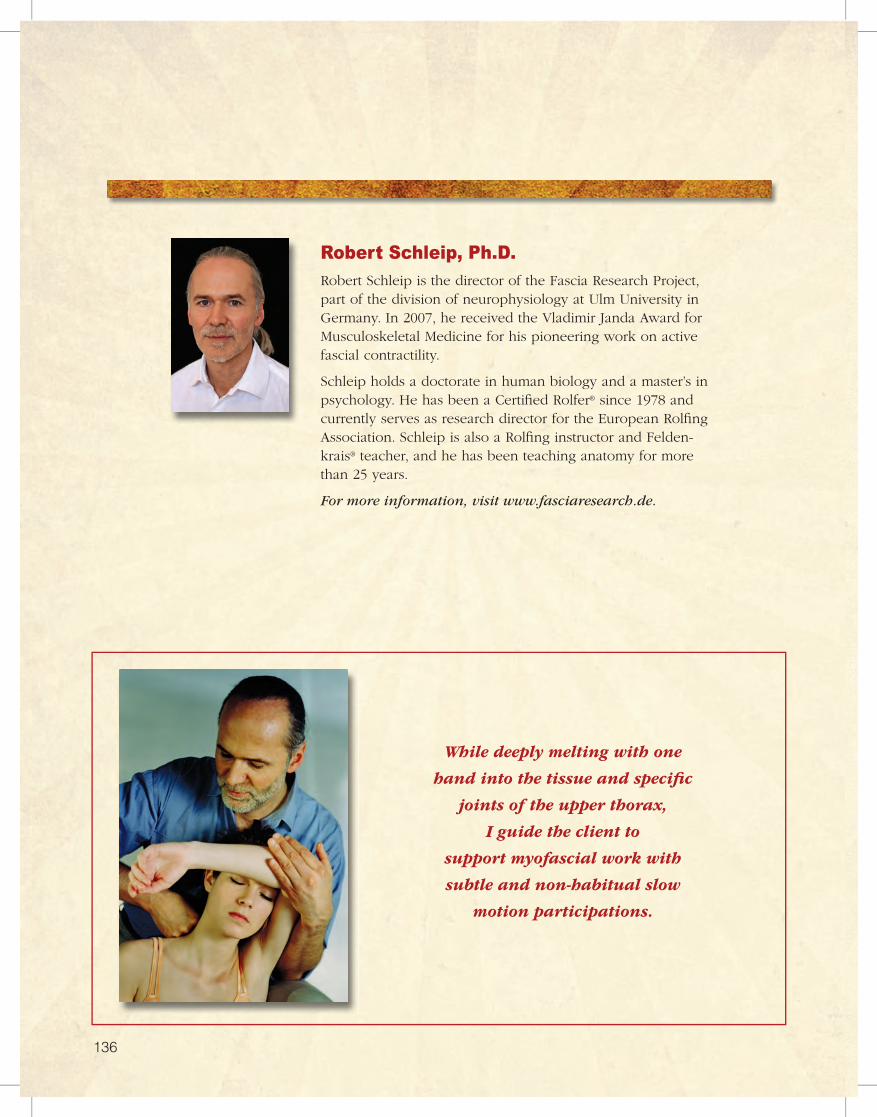
137
Robert Schleip, Ph.D.Robert Schleip is the director of the Fascia Research Project, part of the division of neurophysiology at Ulm University in Germany. In 2007, he received the Vladimir Janda Award for Musculoskeletal Medicine for his pioneering work on active fascial contractility.
Schleip holds a doctorate in human biology and a master’s in psychology. He has been a Certified Rolfer® since 1978 and currently serves as research director for the European Rolfing Association. Schleip is also a Rolfing instructor and Felden-krais® teacher, and he has been teaching anatomy for more than 25 years.
For more information, visit www.fasciaresearch.de.
While deeply melting with one
hand into the tissue and specific
joints of the upper thorax,
I guide the client to
support myofascial work with
subtle and non-habitual slow
motion participations.
137136


![Current Trends in Medicine - Somato Publications · myofascial pain [28,29]. Myofascial pain is associated with myofascial trigger points (MTPs), muscles in sustained contraction](https://img.pdfslide.us/doc/110x75/5e43a3f992ffb312756e8245/current-trends-in-medicine-somato-publications-myofascial-pain-2829-myofascial.jpg)


![Libya [Libyan Tagline Here] By, Logan Threlkeld Pd. 1](https://img.pdfslide.us/doc/110x75/56649e4e5503460f94b44d55/libya-libyan-tagline-here-by-logan-threlkeld-pd-1.jpg)