Embed Size (px)
Citation preview

Review/Model Systems Series
A Systems Approach to the Cellular Analysisof Associative Learning in the PondSnail LymnaeaPaul R. Benjamin,1 Kevin Staras, and Gyorgy KemenesSussex Centre for Neuroscience, School of Biological Sciences, University of Sussex, Falmer, Brighton, East Sussex, BN1 9QG, UK
We show that appetitive and aversive conditioning can be analyzed at the cellular level in the well-describedneural circuitries underlying rhythmic feeding and respiration in the pond snail, Lymnaea stagnalis. To relateelectrical changes directly to behavior, the snails were first trained and the neural changes recorded atmultiple sites in reduced preparations made from the same animals. Changes in neural activity followingconditioning could be recorded at the level of motoneurons, central pattern generator interneurons andmodulatory neurons. Of significant interest was recent work showing that neural correlates of long-termmemory could be recorded in the feeding network following single-trial appetitive chemical conditioning.Available information on the synaptic connectivity and transmitter content of identified neurons within theLymnaea circuits will allow further work on the synaptic and molecular mechanisms of learning and memory.
A large body of work using the very successful cellular andmolecular approach to learning and memory has providedimportant models of plasticity in both invertebrates andvertebrates (Byrne 1987; Milner et al. 1998). These studiesemphasize changes occurring at a single locus. However,there is increasing evidence to suggest that most forms oflearning involve changes at several different sites in thebrain (Wolpaw 1997; Lisberger 1998), suggesting that morecomplex models will be required. The identification of mul-tiple sites of plasticity requires a systems approach to theanalysis of learning and memory, which makes it importantto first identify the electrical changes that result from con-ditioning throughout a neural network and then attempt torelate these to behavioral plasticity. Given the complexityof the brain, systems-level analyses of learning and memoryhave often been inevitable in vertebrate studies striving tolink learning-related behavioral and cellular changes. In ouropinion there is a clear requirement for a similar approachin numerically more simple invertebrate systems, where100–1000s of neurons form circuits, especially because ofthe evidence that the invertebrate central nervous system(CNS) can also show changes resulting from conditioning inseveral different structures (e.g., antennal lobes, mushroombodies, and lateral protocerebral lobes of the honeybee;Hammer 1997). A systems approach to invertebrate learn-ing is particularly useful if we have no prior informationabout the numbers and locations of sites of plasticity in thevarious elements of the nervous system. Despite the recentdevelopments in in vivo single-cell recording and imagingtechniques in vertebrates (Fetcho and O’Malley 1997; Svo-boda et al. 1997), studies using invertebrates still have theadded advantage that more specific learning-related
changes in electrical activity at the single identified cell orsingle identified synapse level can be examined throughouta specific behavioral neural network.
One of the most promising candidates for a systems-level analysis of learning in invertebrates is the pond snailLymnaea stagnalis where a number of behavioral associa-tive conditioning paradigms have been used successfullyand, importantly, cellular traces of behavioral conditioninghave been found in both isolated and semi-intact prepara-tions. The discovery that the feeding response of Lymnaeacan be subjected to both appetitive and aversive classicalconditioning (Audesirk et al. 1982; Kemenes and Benjamin1989a; Kojima et al. 1996), together with the fact that thecircuit-generating feeding ingestion movements is wellknown (Benjamin and Elliott 1989; Yeoman et al. 1995;Brierley et al. 1997; Staras et al. 1998a), has made feasible acellular analysis of feeding-related associative memory for-mation. Aversive operant conditioning of the respiratorybehavior also has been demonstrated in Lymnaea (Lukow-iak et al. 1996, 1998) and because the circuit underlyingrespiration is another network that has been described indetail (Syed et al. 1990; Syed and Winlow 1991b), it was alsopossible to subject this form of learning to a cellular analysis(Spencer et al. 1999).
To understand the learning-induced electrical changesin the neural networks underlying feeding and respiration itis necessary first to consider their organization at the cellu-lar level (Fig. 1).
The Feeding NetworkFeeding in Lymnaea consists of a sequence of repetitivemovements called rasps. During each rasp, the mouthopens and a toothed radula is scraped forward over the foodsubstrate (protraction phase). Food is then lifted into the
1Corresponding author.E-MAIL [email protected]; FAX 44 1273 678535.
LEARNING & MEMORY 7:124–131 © 2000 by Cold Spring Harbor Laboratory Press ISSN1072-0502/00 $5.00
&L E A R N I N G M E M O R Y
www.learnmem.org
124
Cold Spring Harbor Laboratory Press on April 7, 2022 - Published by learnmem.cshlp.orgDownloaded from
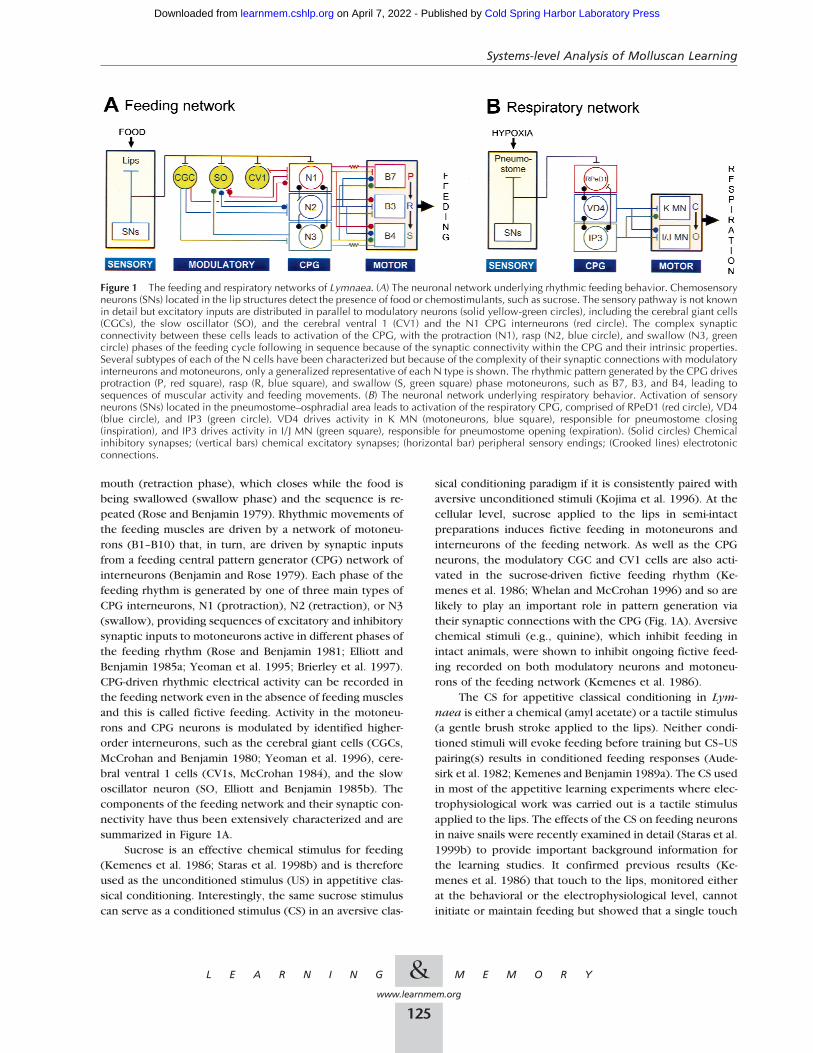
mouth (retraction phase), which closes while the food isbeing swallowed (swallow phase) and the sequence is re-peated (Rose and Benjamin 1979). Rhythmic movements ofthe feeding muscles are driven by a network of motoneu-rons (B1–B10) that, in turn, are driven by synaptic inputsfrom a feeding central pattern generator (CPG) network ofinterneurons (Benjamin and Rose 1979). Each phase of thefeeding rhythm is generated by one of three main types ofCPG interneurons, N1 (protraction), N2 (retraction), or N3(swallow), providing sequences of excitatory and inhibitorysynaptic inputs to motoneurons active in different phases ofthe feeding rhythm (Rose and Benjamin 1981; Elliott andBenjamin 1985a; Yeoman et al. 1995; Brierley et al. 1997).CPG-driven rhythmic electrical activity can be recorded inthe feeding network even in the absence of feeding musclesand this is called fictive feeding. Activity in the motoneu-rons and CPG neurons is modulated by identified higher-order interneurons, such as the cerebral giant cells (CGCs,McCrohan and Benjamin 1980; Yeoman et al. 1996), cere-bral ventral 1 cells (CV1s, McCrohan 1984), and the slowoscillator neuron (SO, Elliott and Benjamin 1985b). Thecomponents of the feeding network and their synaptic con-nectivity have thus been extensively characterized and aresummarized in Figure 1A.
Sucrose is an effective chemical stimulus for feeding(Kemenes et al. 1986; Staras et al. 1998b) and is thereforeused as the unconditioned stimulus (US) in appetitive clas-sical conditioning. Interestingly, the same sucrose stimuluscan serve as a conditioned stimulus (CS) in an aversive clas-
sical conditioning paradigm if it is consistently paired withaversive unconditioned stimuli (Kojima et al. 1996). At thecellular level, sucrose applied to the lips in semi-intactpreparations induces fictive feeding in motoneurons andinterneurons of the feeding network. As well as the CPGneurons, the modulatory CGC and CV1 cells are also acti-vated in the sucrose-driven fictive feeding rhythm (Ke-menes et al. 1986; Whelan and McCrohan 1996) and so arelikely to play an important role in pattern generation viatheir synaptic connections with the CPG (Fig. 1A). Aversivechemical stimuli (e.g., quinine), which inhibit feeding inintact animals, were shown to inhibit ongoing fictive feed-ing recorded on both modulatory neurons and motoneu-rons of the feeding network (Kemenes et al. 1986).
The CS for appetitive classical conditioning in Lym-naea is either a chemical (amyl acetate) or a tactile stimulus(a gentle brush stroke applied to the lips). Neither condi-tioned stimuli will evoke feeding before training but CS–USpairing(s) results in conditioned feeding responses (Aude-sirk et al. 1982; Kemenes and Benjamin 1989a). The CS usedin most of the appetitive learning experiments where elec-trophysiological work was carried out is a tactile stimulusapplied to the lips. The effects of the CS on feeding neuronsin naive snails were recently examined in detail (Staras et al.1999b) to provide important background information forthe learning studies. It confirmed previous results (Ke-menes et al. 1986) that touch to the lips, monitored eitherat the behavioral or the electrophysiological level, cannotinitiate or maintain feeding but showed that a single touch
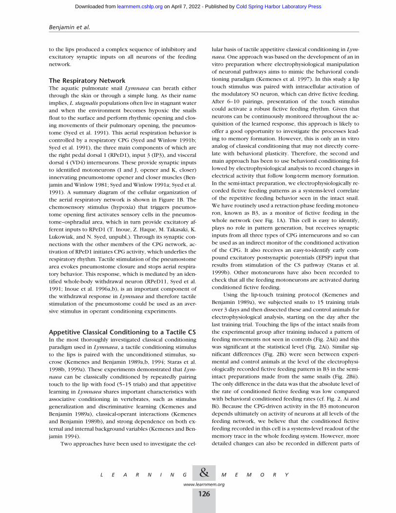
Figure 1 The feeding and respiratory networks of Lymnaea. (A) The neuronal network underlying rhythmic feeding behavior. Chemosensoryneurons (SNs) located in the lip structures detect the presence of food or chemostimulants, such as sucrose. The sensory pathway is not knownin detail but excitatory inputs are distributed in parallel to modulatory neurons (solid yellow-green circles), including the cerebral giant cells(CGCs), the slow oscillator (SO), and the cerebral ventral 1 (CV1) and the N1 CPG interneurons (red circle). The complex synapticconnectivity between these cells leads to activation of the CPG, with the protraction (N1), rasp (N2, blue circle), and swallow (N3, greencircle) phases of the feeding cycle following in sequence because of the synaptic connectivity within the CPG and their intrinsic properties.Several subtypes of each of the N cells have been characterized but because of the complexity of their synaptic connections with modulatoryinterneurons and motoneurons, only a generalized representative of each N type is shown. The rhythmic pattern generated by the CPG drivesprotraction (P, red square), rasp (R, blue square), and swallow (S, green square) phase motoneurons, such as B7, B3, and B4, leading tosequences of muscular activity and feeding movements. (B) The neuronal network underlying respiratory behavior. Activation of sensoryneurons (SNs) located in the pneumostome–osphradial area leads to activation of the respiratory CPG, comprised of RPeD1 (red circle), VD4(blue circle), and IP3 (green circle). VD4 drives activity in K MN (motoneurons, blue square), responsible for pneumostome closing(inspiration), and IP3 drives activity in I/J MN (green square), responsible for pneumostome opening (expiration). (Solid circles) Chemicalinhibitory synapses; (vertical bars) chemical excitatory synapses; (horizontal bar) peripheral sensory endings; (Crooked lines) electrotonicconnections.
Systems-level Analysis of Molluscan Learning
&L E A R N I N G M E M O R Y
www.learnmem.org
125
Cold Spring Harbor Laboratory Press on April 7, 2022 - Published by learnmem.cshlp.orgDownloaded from
to the lips produced a complex sequence of inhibitory andexcitatory synaptic inputs on all neurons of the feedingnetwork.
The Respiratory NetworkThe aquatic pulmonate snail Lymnaea can breath eitherthrough the skin or through a simple lung. As their nameimplies, L. stagnalis populations often live in stagnant waterand when the environment becomes hypoxic the snailsfloat to the surface and perform rhythmic opening and clos-ing movements of their pulmonary opening, the pneumos-tome (Syed et al. 1991). This aerial respiration behavior iscontrolled by a respiratory CPG (Syed and Winlow 1991b;Syed et al. 1991), the three main components of which arethe right pedal dorsal 1 (RPeD1), input 3 (IP3), and visceraldorsal 4 (VD4) interneurons. These provide synaptic inputsto identified motoneurons (I and J, opener and K, closer)innervating pneumostome opener and closer muscles (Ben-jamin and Winlow 1981; Syed and Winlow 1991a; Syed et al.1991). A summary diagram of the cellular organization ofthe aerial respiratory network is shown in Figure 1B. Thechemosensory stimulus (hypoxia) that triggers pneumos-tome opening first activates sensory cells in the pneumos-tome–osphradial area, which in turn provide excitatory af-ferent inputs to RPeD1 (T. Inoue, Z. Haque, M. Takasaki, K.Lukowiak, and N. Syed, unpubl.). Through its synaptic con-nections with the other members of the CPG network, ac-tivation of RPeD1 initiates CPG activity, which underlies therespiratory rhythm. Tactile stimulation of the pneumostomearea evokes pneumostome closure and stops aerial respira-tory behavior. This response, which is mediated by an iden-tified whole-body withdrawal neuron (RPeD11, Syed et al.1991; Inoue et al. 1996a,b), is an important component ofthe withdrawal response in Lymnaea and therefore tactilestimulation of the pneumostome could be used as an aver-sive stimulus in operant conditioning experiments.
Appetitive Classical Conditioning to a Tactile CSIn the most thoroughly investigated classical conditioningparadigm used in Lymnaea, a tactile conditioning stimulusto the lips is paired with the unconditioned stimulus, su-crose (Kemenes and Benjamin 1989a,b, 1994; Staras et al.1998b, 1999a). These experiments demonstrated that Lym-naea can be classically conditioned by repeatedly pairingtouch to the lip with food (5–15 trials) and that appetitivelearning in Lymnaea shares important characteristics withassociative conditioning in vertebrates, such as stimulusgeneralization and discriminative learning (Kemenes andBenjamin 1989a), classical-operant interactions (Kemenesand Benjamin 1989b), and strong dependence on both ex-ternal and internal background variables (Kemenes and Ben-jamin 1994).
Two approaches have been used to investigate the cel-
lular basis of tactile appetitive classical conditioning in Lym-naea. One approach was based on the development of an invitro preparation where electrophysiological manipulationof neuronal pathways aims to mimic the behavioral condi-tioning paradigm (Kemenes et al. 1997). In this study a liptouch stimulus was paired with intracellular activation ofthe modulatory SO neuron, which can drive fictive feeding.After 6–10 pairings, presentation of the touch stimuluscould activate a robust fictive feeding rhythm. Given thatneurons can be continuously monitored throughout the ac-quisition of the learned response, this approach is likely tooffer a good opportunity to investigate the processes lead-ing to memory formation. However, this is only an in vitroanalog of classical conditioning that may not directly corre-late with behavioral plasticity. Therefore, the second andmain approach has been to use behavioral conditioning fol-lowed by electrophysiological analysis to record changes inelectrical activity that follow long-term memory formation.In the semi-intact preparation, we electrophysiologically re-corded fictive feeding patterns as a systems-level correlateof the repetitive feeding behavior seen in the intact snail.We have routinely used a retraction-phase feeding motoneu-ron, known as B3, as a monitor of fictive feeding in thewhole network (see Fig. 1A). This cell is easy to identify,plays no role in pattern generation, but receives synapticinputs from all three types of CPG interneurons and so canbe used as an indirect monitor of the conditioned activationof the CPG. It also receives an easy-to-identify early com-pound excitatory postsynaptic potentials (EPSP) input thatresults from stimulation of the CS pathway (Staras et al.1999b). Other motoneurons have also been recorded tocheck that all the feeding motoneurons are activated duringconditioned fictive feeding.
Using the lip-touch training protocol (Kemenes andBenjamin 1989a), we subjected snails to 15 training trialsover 3 days and then dissected these and control animals forelectrophysiological analysis, starting on the day after thelast training trial. Touching the lips of the intact snails fromthe experimental group after training induced a pattern offeeding movements not seen in controls (Fig. 2Aii) and thiswas significant at the statistical level (Fig. 2Ai). Similar sig-nificant differences (Fig. 2Bi) were seen between experi-mental and control animals at the level of the electrophysi-ologically recorded fictive feeding pattern in B3 in the semi-intact preparations made from the same snails (Fig. 2Bii).The only difference in the data was that the absolute level ofthe rate of conditioned fictive feeding was low comparedwith behavioral conditioned feeding rates (cf. Fig. 2, Ai andBi). Because the CPG-driven activity in the B3 motoneurondepends ultimately on activity of neurons at all levels of thefeeding network, we believe that the conditioned fictivefeeding recorded in this cell is a systems-level readout of thememory trace in the whole feeding system. However, moredetailed changes can also be recorded in different parts of
Benjamin et al.
&L E A R N I N G M E M O R Y
www.learnmem.org
126
Cold Spring Harbor Laboratory Press on April 7, 2022 - Published by learnmem.cshlp.orgDownloaded from

the network. One of these is the early EPSP that occurs inthe B3 before the onset of the fictive feeding pattern. Theamplitude (Fig. 2C), but not the latency and duration, of theEPSP was enhanced significantly after conditioning. In satedsnails the conditioned fictive feeding response to touch waslost but the increase in the EPSP amplitude persisted (Staraset al. 1999b). This suggests that there is unlikely to be a
causal link between increases in amplitude inB3 and generation of the fictive feeding pattern.
One candidate for initiating CPG activityfollowing conditioning is the modulatory inter-neuron cell type known as CV1. This neuron iscapable of driving a fictive feeding neuron viaits connections with the N1M cells of the CPGnetwork and activity in these cells normally ac-companies unconditioned feeding patternsstimulated by sucrose (Whelan and McCrohan1996). Significantly, CV1 is the only protractionphase interneuron type that is depolarized bytouch applied to the lips in naive snails (Staraset al. 1999b). Despite their small size, recentlyin ongoing studies we have been able to recorda sufficient number of CV1 cells in semi-intactpreparations for quantitative analysis. We findthat they are significantly more active followingtouch in conditioned snails compared with con-trols and that they show the typical patternedactivity seen with sugar (P.R. Benjamin et al.,unpubl.)
Electrical correlates of classical condition-ing were recorded at several levels within thefeeding circuit and these could all be potentialsites of plasticity. That some sites could be quiteearly in the CS pathway was revealed by extra-cellularly recording mechanosensory fibers lo-cated in the connective between the cerebraland buccal ganglia (Staras et al. 1999a). Tactileresponses could be recorded in these fibers and,following conditioning, the number of spikesoccurring early in this response increased, com-pared with controls.
Appetitive Classical Conditioning to aChemical CSIn the original successful formulation of appeti-tive classical conditioning in Lymnaea, snailswere subjected to a chemical conditioning pro-tocol using amyl acetate as the CS and sucroseas the US (Audesirk et al. 1982). Following train-ing, the explicitly paired (CS–US) experimentalgroup showed significantly greater feeding re-sponses to amyl acetate over their own naiveresponses and all the standard control groups(Random, Explicitly Unpaired, CS alone, US
alone). As might be predicted for appetitive classical con-ditioning, both age and motivational state (hunger vs. sati-ety) influenced learning (Audesirk et al. 1982). Both hungryand sated young snails could acquire the conditioned re-sponse but in the latter group its expression was only ap-parent when the animals were starved before testing. Onthe other hand, old snails could only acquire the condi-
Figure 2 Cellular effects of appetitive conditioning with a tactile stimulus (touch tothe lip). (A) Behaviorally-recorded feeding responses. (Ai), Change in feeding rateafter tactile conditioning is significantly greater in the experimental group (15 pair-ings of lip tactile CS and sucrose US) vs. the control group (random CS and USpresentation). (Aii) Example of feeding response to lip tactile stimulation in anexperimental vs. a control animal. Solid vertical bars represent single rasp events.(B) Electrophysiologically recorded fictive feeding responses in reduced prepara-tions derived from the behaviorally-trained animals in A. (Bi) Change in fictivefeeding rate after tactile conditioning is significantly greater in the experimentalgroup versus the control group. (Bii) Example of fictive feeding response to lip tactilestimulation in an experimental vs. a control semi-intact preparation monitored usingthe motoneuron, B3. Fictive feeding cycles are seen as characteristic inputs on theB3. (C) A cellular correlate of tactile conditioning. (Ci) Amplitude of the B3 EPSPevoked by the tactile stimulus is significantly larger in conditioned vs. controlpreparations. (Cii) Example of the touch-evoked EPSP in an experimental and con-trol preparation. Statistical data are presented as medians and interquartile ranges.After Staras et al. (1999a).
Systems-level Analysis of Molluscan Learning
&L E A R N I N G M E M O R Y
www.learnmem.org
127
Cold Spring Harbor Laboratory Press on April 7, 2022 - Published by learnmem.cshlp.orgDownloaded from

tioned response if they were maintained in ahungry state during training. The significance ofmotivational state became even more apparentwhen it was realized that if snails were starvedlong enough (for 5 days) before and throughoutthe experiment, even a single pairing of amylacetate and sucrose resulted in long-termmemory that lasted for at least 19 days (Alex-ander et al. 1984). This is a remarkable exampleof single trial learning that we have replicated inour laboratory and use now as a starting pointfor an analysis of the time course of the mecha-nisms underlying long term memory formation.
An in vitro correlate of an appetitive con-ditioned response was also found after chemicalappetitive classical conditioning of intact ani-mals with 15 paired applications of amyl acetateas the CS and sucrose as the US (Whelan andMcCrohan 1996). The electrophysiological testsin this experiment concentrated on the CV1cells, a pair of modulatory interneurones in thecerebral ganglia (McCrohan 1984). In mostsemi-intact preparations (∼80%) taken from ex-perimental snails, application of amyl acetate tothe lips led to depolarization of the membrane and in ∼30%of them to strong rhythmical bursting activity. Interestingly,in some preparations where the buccal feeding central pat-tern generator system was removed, CV1 continued toshow an excitatory response to amyl acetate. This indicatesthat, at least in the case of chemical appetitive classicalconditioning, one site of plasticity may be located in thecerebral ganglia.
We have recently obtained similar results followingsingle trial chemical classical conditioning to the same CS.The example shown in Figure 3 shows that amyl acetate canactivate a fictive feeding pattern in conditioned (Fig. 3A)but not control (Fig. 3B) snails and further statistical analysis(not shown) confirmed that this difference between experi-mentals and controls was significant. Fictive feeding wasalso monitored on the B3 motoneuron, as in the tactileconditioning experiments, and patterned fictive feeding ac-tivity could be recorded on both CV1 and the B3. This dataon chemical conditioning also indicates that training canlead to equivalent systems level responses that were previ-ously reported for tactile conditioning except that it occursafter only 1 training trial rather than 15. This success ingetting a robust electrical correlate of chemical condition-ing makes further cellular analysis possible.
Aversive Classical Conditioning to aChemical CSAversive classical conditioning in Lymnaea was based onpairing sucrose with an aversive stimulus such as KCl,which inhibits feeding and evokes a withdrawal response
(Kojima et al. 1996, 1997). After eight trials, trained animalsshowed a significantly weaker feeding response to sucrosethan did controls and this memory lasted for more than onemonth (Kojima et al. 1997).
A cellular analysis of this learned response was carriedout on isolated brains dissected from conditioned and con-trol animals. In particular, the synaptic connection betweenthe modulatory CGCs and the CPG interneuron, N1M, wasexamined. In conditioned animals compared to controls, asignificant increase in the size of inhibitory postsynapticpotentials (IPSPs) recorded in the N1M was observed fol-lowing an artificial depolarization of the CGCs. Because theCGCs are known to play a critical gating role in feedingbehavior (Yeoman et al. 1994) and the N1M is a pivotalmember of the feeding CPG (Kemenes and Elliott 1994),this enhanced IPSP may be an important cellular correlateof the conditioned taste-aversion learning (Kojima et al.1997).
Operant Conditioning of theRespiratory NetworkThe aerial respiratory behavior of L. stagnalis has been usedin a series of experiments investigating the behavioral andcellular mechanisms of operant conditioning (Lukowiak etal. 1996, 1998; Spencer et al. 1999). Hypoxia triggers pneu-mostome opening and this was used as the operantly con-ditioned behavior. Tactile stimulation of the pneumostomearea evokes pneumostome closure and stops aerial respira-tory behavior. Animals were tested and trained in an artifi-cially-created hypoxic nitrogen-rich environment to in-
Figure 3 One trial chemical conditioning. (A) Response to amyl acetate in thefeeding motoneuron B3 and modulatory interneuron CV1 in a semi-intact lip brainpreparation derived from an animal that was behaviorally conditioned with a singlepairing of amyl acetate and sucrose. Amyl acetate evokes maintained rhythmicfictive feeding activity in both cell-types. (B) B3 and CV1 neurons are silent follow-ing amyl acetate presentation recorded in a preparation derived from an animalreceiving a single unpaired presentation of sucrose. (P.R. Benjamin et al. unpubl.)
Benjamin et al.
&L E A R N I N G M E M O R Y
www.learnmem.org
128
Cold Spring Harbor Laboratory Press on April 7, 2022 - Published by learnmem.cshlp.orgDownloaded from

crease the level of respiratory behavior (Lukowiak et al.1996). In the operantly trained group a tactile stimulus wasapplied to the pneumostome area each time aerial respira-tion was attempted by the animal. Suitable yoked and hy-poxic control groups were also used. The number of open-ings, latency to first opening, and total breath durationswere recorded in pre- and post-training periods. Only theoperantly conditioned group showed significant changesbetween the pre- and post-training behaviors, with signifi-cant reductions in openings and total breathing time andsignificant increases in the latency to first breath (Lukowiaket al. 1996). It has since been demonstrated that a memoryfor this conditioned response could persist for at least 4weeks when a spaced training procedure was used (Lukow-iak et al. 1998).
More recently, neural changes associated with thislearned behavior have been identified in the isolated CNSderived from operantly conditioned animals (Spencer et al.1999). Specifically, spontaneous patterned activity in theIP3 interneuron, which is involved in pneumostome open-ing (see Fig. 1B) showed a significant reduction comparedwith activity in the IP3 neurons of brains derived fromyoked controls. Furthermore, a higher percentage of RPeD1CPG interneurons, which are important in the onset of therespiratory cycle, were silent in conditioned versus controlpreparations (Spencer et al. 1999). They also observed areduction in the ability of the RPeD1 cells to induce IP3activity. One speculation arising from these findings is thathigher-order neurons involved in withdrawal responses,such as RPeD11, may be providing maintained inhibitoryinputs to the respiratory CPG as a result of the operantconditioning (Spencer et al. 1999).
Conclusions and DiscussionThe picture emerging from the work we have reviewedsuggests that different forms of associative conditioning inLymnaea result in electrical changes that can be recordedat several different locations in the nervous system. Thisrepresents an intermediate or systems level of analysis be-tween behavior and the cellular and molecular studies ofsynaptic plasticity. It provides an opportunity to carry out adirect quantitative comparison of behavior and its electro-physiological correlate. The expression of the memory orits read-out (Byrne 1985) was mainly recorded as a globalpattern of activity at the level of motoneurons and interneu-rons of the feeding and respiratory networks.
Our detailed knowledge of the Lymnaea feeding cir-cuitry allowed us to infer that the rhythmic activity of thefeeding CPG in response to the CS was increased followingappetitive classical conditioning (Whelan and McCrohan1996; Staras et al. 1998b, 1999a). Other changes associatedwith appetitive classical conditioning were recorded at thelevel of the motoneurons (enhancement of an early EPSPresponse) and the modulatory interneurons (activation of
the CV1s). The activation of the synaptic connection be-tween the CV1 modulatory interneuron and the CPG neu-rons is considered to be particularly important because CV1cells can drive activity in the CPG network and this is likelyto be essential to obtain a full readout of the feeding motorprogram. These results are consistent with a multistage ormultisite model of appetitive classical conditioning but donot rule out a single common site of plasticity, particularlyas a very short latency change in activity could be recordedin what was interpreted as part of the mechanosensory CSpathway. Recently, a pairing-specific strengthening of themechanoafferent pathway also has been found after tactileappetitive classical conditioning of feeding in Aplysia (Lech-ner et al. 2000). Changes at the level of sensory neuronshave also been shown to be major sites of plasticity in aver-sive classical conditioning of both the gill withdrawal reflexof Aplysia (Hawkins et al. 1983) and phototactic behaviorin Hermissenda (Crow and Alkon 1980).
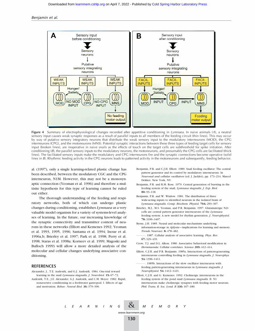
The causal link between the various sites of electricalchange after appetitive classical conditioning have not beenestablished, but we can make certain inferences based onthe temporal sequences of events recorded after the appli-cation of the CS following conditioning, and relating thisinformation to our knowledge of the synaptic connectivityof the circuit (see Fig. 1A). These inferences led to a gen-eralized working model for appetitive learning summarizedin Figure 4. We know in naive animals (Fig. 4A) that sensoryinputs, such as touch and amyl actetate, produce weak sub-threshold synaptic responses in neurons of the feeding cir-cuit (Staras et al. 1999b; G. Kemenes, unpubl.). Recent evi-dence indicates that distribution of the weak tactile inputsto the modulatory interneurons, the CPG interneurons andthe motoneurons may occur via sensory integratory neu-rons (Fig. 4A) and we make the assumption here that thesame is true for chemical inputs. Potential synaptic interac-tions between the three types of feeding target cells forsensory input are shown by the dashed lines in Figure 4A.After classical conditioning (Fig. 4B), the sensory inputs tothe modulatory neurons (CV1), motoneurons (B3), and pre-sumably the CPG cells, are facilitated. The facilitated sen-sory input makes the modulatory and CPG interneurons fireand the synaptic connections become operative (solid linesin Fig. 4B). The CPG neurons then drive rhythmic feedingactivity in the motoneurons leading to sequences of mus-cular activity and feeding movements. Whether the specificplastic changes after tactile versus chemical conditioningare the same remains to be investigated.
Multiple changes were also recorded in the respiratorynetwork after aversive operant conditioning of the aerialrespiratory behavior (Spencer et al. 1999). This indicatesthat a multisite hypothesis of plastic changes after associa-tive conditioning in Lymnaea is now strongly supported bywork on both classical and operant conditioning. In theaversive classical conditioning paradigm used by Kojima et
Systems-level Analysis of Molluscan Learning
&L E A R N I N G M E M O R Y
www.learnmem.org
129
Cold Spring Harbor Laboratory Press on April 7, 2022 - Published by learnmem.cshlp.orgDownloaded from
al. (1997), only a single learning-related plastic change hasbeen described, between the modulatory CGC and the CPGinterneuron, N1M. However, this may not be a monosyn-aptic connection (Yeoman et al. 1996) and therefore a mul-tisite hypothesis for this type of learning cannot be ruledout either.
The thorough understanding of the feeding and respi-ratory networks, both of which can undergo plasticchanges during conditioning, establishes Lymnaea as a veryvaluable model organism for a variety of systems-level analy-ses of learning. In the future, our increasing knowledge ofthe synaptic connectivity and transmitter content of neu-rons in these networks (Elliott and Kemenes 1992; Yeomanet al. 1993, 1995, 1996; Santama et al. 1994; Inoue et al.1996a,b; Brierley et al. 1997; Park et al. 1998; Perry et al.1998; Staras et al. 1998a; Korneev et al. 1999; Magoski andBulloch 1999) will allow a more detailed analysis of themolecular and cellular changes underlying associative con-ditioning.
REFERENCESAlexander, J., T.E. Audesirk, and G.J. Audesirk. 1984. One-trial reward
learning in the snail Lymnaea stagnalis. J. Neurobiol. 15: 67–72.Audesirk, T.E., J.E. Alexander, G.J. Audesirk, and C.M. Moyer. 1982. Rapid,
nonaversive conditioning in a freshwater gastropod. I. Effects of ageand motivation. Behav. Neural Biol. 36: 379–390.
Benjamin, P.R. and C.J.H. Elliott. 1989. Snail feeding oscillator: The centralpattern generator and its control by modulatory interneurons. InNeuronal and cellular oscillators (ed. J. Jacklet), pp. 173–214. MarcelDekker, New York, NY.
Benjamin, P.R. and R.M. Rose. 1979. Central generation of bursting in thefeeding system of the snail, Lymnaea stagnalis. J. Exp. Biol.80: 93–118.
Benjamin, P.R. and W. Winlow. 1981. The distribution of threewide-acting inputs to identified neurons in the isolated brain ofLymnaea stagnalis. Comp. Biochem. Physiol. 70A: 293–307.
Brierley, M.J., M.S. Yeoman, and P.R. Benjamin. 1997. Glutamatergic N2vcells are central pattern generator interneurons of the Lymnaeafeeding system: A new model for rhythm generation. J. Neurophysiol.78: 3396–3407.
Byrne, J.H. 1985. Neural and molecular mechanisms underlyinginformation-storage in Aplysia—implications for learning and memory.Trends Neurosci. 8: 478–482.
. 1987. Cellular analysis of associative learning. Phys. Rev.67: 329–439.
Crow, T.J. and D.L. Alkon. 1980. Associative behavioral modification inHermissenda: Cellular correlates. Science 209: 412–414.
Elliott, C.J.H. and P.R. Benjamin. 1985a. Interactions of pattern-generatinginterneurons controlling feeding in Lymnaea stagnalis. J. Neurophys.54: 1396–1411.
. 1985b. Interactions of the slow oscillator interneuron withfeeding pattern-generating interneurons in Lymnaea stagnalis. J.Neurophysiol. 54: 1412–1421.
Elliott, C.J.H. and G. Kemenes. 1992. Cholinergic interneurons in thefeeding system of the pond snail Lymnaea stagnalis. II. N1interneurons make cholinergic synapses with feeding motor neurons.Phil. Trans. R. Soc. Lond. B 336: 167–180.
Figure 4 Summary of electrophysiological changes recorded after appetitive conditioning in Lymnaea. In naive animals (A), a neutralsensory input causes weak synaptic responses as a result of parallel inputs to all members of the feeding circuit (thin lines). This may occurby way of putative sensory integratory neurons that distribute the weak sensory input to the modulatory interneurons (MOD), the CPGinterneurons (CPG), and the motoneurons (MNS). Potential synaptic interactions between these three types of feeding target cells for sensoryinput (broken lines), are inoperative in naive snails as the effects of touch on the target cells are subthreshold for spike initiation. Afterconditioning (B), the parallel sensory inputs to the modulatory neurons, the motoneurons, and presumably the CPG cells are facilitated (thicklines). The facilitated sensory inputs make the modulatory and CPG interneurons fire and the synaptic connections become operative (solidlines in B). Rhythmic feeding activity in the CPG neurons leads to patterned activity in the motoneurons and subsequently, feeding behavior.
Benjamin et al.
&L E A R N I N G M E M O R Y
www.learnmem.org
130
Cold Spring Harbor Laboratory Press on April 7, 2022 - Published by learnmem.cshlp.orgDownloaded from

Fetcho, J.F. and D.M. O’Malley. 1997. Imaging neuronal networks inbehaving animals. Curr. Opin. Neurobiol. 7: 832–838.
Hammer, M. 1997. The neural basis of associative reward learning inhoneybees. Trends Neurosci. 20: 245–252.
Hawkins, R.D., T.W. Abrams, T.J. Carew, and E.R. Kandel. 1983. A cellularmechanism of classical conditioning in Aplysia: Activity-dependentamplification of presynaptic facilitation. Science 219: 400–405.
Inoue, T., M. Takasaki, K. Lukowiak, and N. Syed. 1996a. Identification ofa putative mechanosensory neuron in Lymnaea: Characterization of itssynaptic and functional connections with the whole-body withdrawalinterneuron. J. Neurophysiol. 76: 3230–3238.
. 1996b. Inhibition of the respiratory pattern-generating neurons byan identified whole-body withdrawal interneuron of Lymnaeastagnalis. J. Exp. Biol. 199: 1887–1898.
Kemenes, G. and P.R. Benjamin. 1989a. Appetitive learning in snails showscharacteristics of conditioning in vertebrates. Brain Res.489: 163–166.
. 1989b. Goal-tracking behaviour in the pond snail Lymnaeastagnalis. Behav. Neural Biol. 52: 260–270.
. 1994. Training in a novel environment improves the appetitivelearning performance of the snail, Lymnaea stagnalis. Behav. NeuralBiol. 61: 139–149.
Kemenes, G., C.J.H. Elliott, and P.R. Benjamin. 1986. Chemical and tactileinputs to the Lymnaea feeding system: Effects on behaviour andneural circuitry. J. Exp. Biol. 122: 113–137.
Kemenes, G., K. Staras, and P.R. Benjamin. 1997. In vitro appetitiveclassical conditioning of the feeding response in the pond snailLymnaea stagnalis. J. Neurophysiol. 78: 2351–2362.
Kojima, S., M. Yamanaka, Y. Fujito, and E. Ito. 1996. Differentialneuroethological effects of aversive and appetitive reinforcing stimulion associative learning in Lymnaea stagnalis. Zool. Sci. 13: 803–812.
Kojima, S., H. Nanakamura, S. Nagayama, Y. Fujito, and E. Ito. 1997.Enhancement of an inhibitory input to the feeding central patterngenerator in Lymnaea stagnalis during conditioned taste-aversionlearning. Neurosci. Lett. 230: 179–182.
Korneev, S.A., J.H. Park, and M. O’Shea. 1999. Neuronal expression ofneural nitric oxide synthase (nNOS) protein is suppressed by anantisense RNA transcribed from an NOS pseudogene. J. Neurosci.19: 7711–7720.
Lechner, H., D.A. Baxter,, and J.H. Byrne. 2000. Classical conditioning offeeding in Aplysia: II. Neurophysiological correlates. J. Neurosci. 20,in press.
Lisberger, S.G. 1998. Cerebellar LTD: A molecular mechanism ofbehavioral learning? Cell 92: 701–704.
Lukowiak, K., E. Ringseis, G. Spencer, W. Wildering, and N. Syed. 1996.Operant conditioning of aerial respiratory behaviour in Lymnaeastagnalis. J. Exp. Biol. 199: 683–691.
Lukowiak, K., R. Cotter, J. Westly, E. Ringseis, and G. Spencer. 1998.Long-term memory of an operantly conditioned respiratory behaviourpattern in Lymnaea stagnalis. J. Exp. Biol. 201: 877–882.
Magoski, N.S. and A.G. Bulloch. 1999. Dopamine activates two differentreceptors to produce variability in sign at an identified synapse. J.Neurophysiol. 81: 1330–1340.
McCrohan, C.R. 1984. Initiation of feeding motor output by an identifiedinterneurone in the snail Lymnaea stagnalis. J. Exp. Biol.113: 351–366.
McCrohan, C.R. and P.R. Benjamin. 1980. Synaptic relationships of thecerebral giant cells with motoneurones in the feeding system ofLymnaea stagnalis. J. Exp. Biol. 85: 169–186.
Milner, B., L.R. Squire, and E.R. Kandel. 1998. Cognitive neuroscience andthe study of memory. Neuron 20: 445–468.
Park, J.H., V.A. Straub, and M. O’Shea. 1998. Anterograde signaling bynitric oxide: Characterization and in vitro reconstitution of anidentified nitrergic synapse. J. Neurosci. 18: 5463–5476.
Perry, S.J., V.A. Straub, G. Kemenes, N. Santama, B.M. Worster, J.F. Burke,and P.R. Benjamin. 1998. Neural modulation of gut motility bymyomodulin peptides and acetylcholine in the snail Lymnaea. J.Neurophysiol. 79: 2460–2474.
Rose, R.M. and P.R. Benjamin. 1979. The relationship of the central motorpattern to the feeding cycle of Lymnaea stagnalis. J. Exp. Biol.80: 137–163.
. 1981. Interneuronal control of feeding in Lymnaea stagnalis. II.The interneuronal mechanisms generating feeding cycles. J. Exp. Biol.92: 203–228.
Santama, N., M. Brierley, J.F. Burke, and P.R. Benjamin. 1994. Neuralnetwork controlling feeding in Lymnaea stagnalis:Immunocytochemical localization of myomodulin, small cardioactivepeptide, buccalin, and FMRFamide-related peptides. J. Comp. Neurol.342: 352–365.
Spencer, G.E., N.I. Syed, and K. Lukowiak. 1999. Neural changes afteroperant conditioning of the aerial respiratory behavior in Lymnaeastagnalis. J. Neurosci. 19: 1836–1843.
Staras, K., G. Kemenes, and P.R. Benjamin. 1998a. Pattern-generating rolefor motoneurons in a rhythmically active neuronal network. J.Neurosci. 18: 3669–3688.
. 1998b. Neurophysiological correlates of unconditioned andconditioned feeding behavior in the pond snail Lymnaea stagnalis. J.Neurophysiol. 79: 3030–3040.
. 1999a. Cellular traces of behavioral classical conditioning can berecorded at several specific sites in a simple nervous system. J.Neurosci. 19: 347–357.
. 1999b. Electrophysiological and behavioral analysis of lip touch asa component of the food stimulus in the snail Lymnaea. J.Neurophysiol. 81: 1261–1273.
Svoboda, K., W. Denk, D. Kleinfeld, and D.W. Tank. 1997. In vivodendritic calcium dynamics in neocortical pyramidal neurons. Nature385: 161–165.
Syed, N.I., A.G. Bulloch, and K. Lukowiak. 1990. In vitro reconstruction ofthe respiratory central pattern generator of the mollusk Lymnaea.Science 250: 282–285.
Syed, N.I, D. Harrison, and W. Winlow. 1991. Respiratory behavior in thepond snail Lymnaea stagnalis. I. Behavioral analysis and theidentification of motor neurons. J. Comp. Physiol. A 169: 541–555.
Syed, N.I. and W. Winlow. 1991a. Coordination of locomotor andcardiorespiratory networks of Lymnaea stagnalis by a pair ofidentified interneurones. J. Exp. Biol. 158: 37–62.
. 1991b. Respiratory behavior in the pond snail Lymnaea stagnalis.II. Neural elements of the central pattern generator (CPG). J. Comp.Physiol. A 169: 557–568.
Whelan, H.A. and C.R. McCrohan. 1996. Food-related conditioning andneuronal correlates in the freshwater snail Lymnaea stagnalis. J. Moll.Stud. 62: 483–494.
Wolpaw, J.R. 1997. The complex structure of a simple memory. TrendsNeurosci. 20: 588–594.
Yeoman, M.S., D.C. Parish, and P.R. Benjamin. 1993. A cholinergicmodulatory interneuron in the feeding system of the snail, Lymnaea.J. Neurophysiol. 70: 37–50.
Yeoman, M.S., A.W. Pieneman, G.P. Ferguson, A. Ter Maat, and P.R.Benjamin. 1994. Modulatory role for the serotonergic cerebral giantcells in the feeding system of the snail, Lymnaea. I. Fine wirerecording in the intact animal and pharmacology. J. Neurophysiolol.72: 1357–1371.
Yeoman, M.S., A. Vehovszky, G. Kemenes, C.J.H. Elliott, and P.R.Benjamin. 1995. Novel interneuron having hybrid modulatory-centralpattern generator-properties in the feeding system of the snail,Lymnaea stagnalis. J. Neurophysiol. 73: 112–124.
Yeoman, M.S., M.J. Brierley, and P.R. Benjamin. 1996. Central patterngenerator interneurons are targets for the modulatory serotonergiccerebral giant cells in the feeding system of Lymnaea. J.Neurophysiol. 75: 11–25.
Received January 19, 2000; accepted in revised form March 14, 2000.
Systems-level Analysis of Molluscan Learning
&L E A R N I N G M E M O R Y
www.learnmem.org
131
Cold Spring Harbor Laboratory Press on April 7, 2022 - Published by learnmem.cshlp.orgDownloaded from

10.1101/lm.7.3.124Access the most recent version at doi: 7:2000, Learn. Mem.
Paul R. Benjamin, Kevin Staras and György Kemenes
LymnaeaLearning in the Pond Snail A Systems Approach to the Cellular Analysis of Associative
References
http://learnmem.cshlp.org/content/7/3/124.full.html#ref-list-1
This article cites 51 articles, 18 of which can be accessed free at:
License
ServiceEmail Alerting
click here.top right corner of the article or
Receive free email alerts when new articles cite this article - sign up in the box at the
Cold Spring Harbor Laboratory Press
Cold Spring Harbor Laboratory Press on April 7, 2022 - Published by learnmem.cshlp.orgDownloaded from